9qj1
;Crystal structure of S-adenosyl-L-homocysteine hydrolase from P. aeruginosa, Q65N mutant soaked with adenosine and probed with rubidium to confirm disruption of a potassium binding site. ;
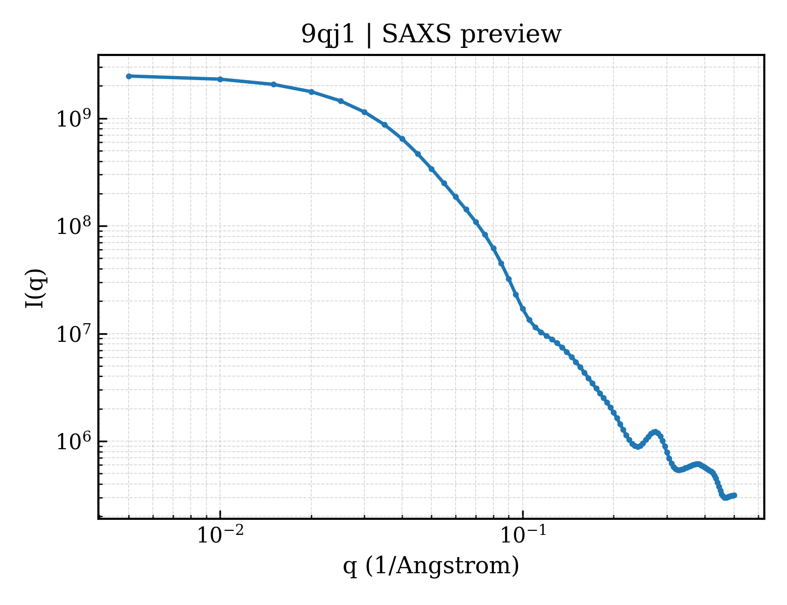
SAXS Profile
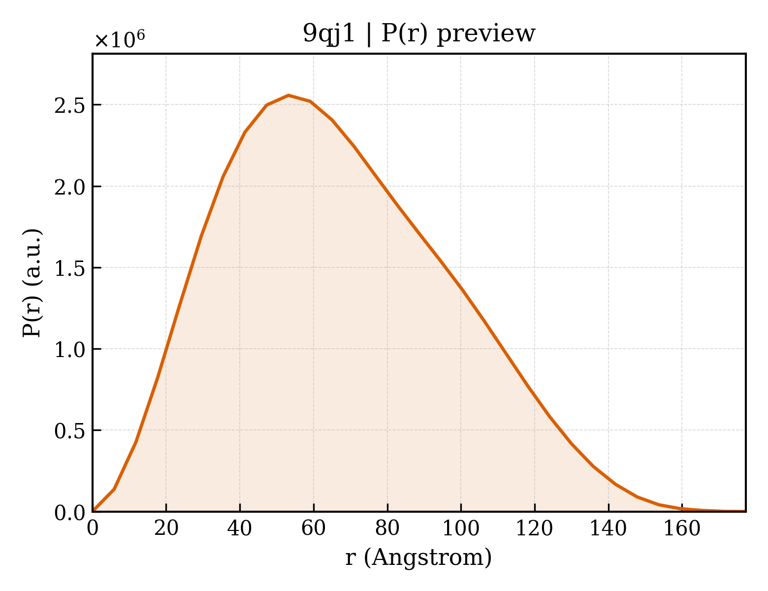
P(r) Distribution
1. Structure Basics
| entry_id | 9qj1 |
| deposition_date | 2025-03-18 |
| last_revision | 2026-04-08 |
| title | ;Crystal structure of S-adenosyl-L-homocysteine hydrolase from P. aeruginosa, Q65N mutant soaked with adenosine and probed with rubidium to confirm disruption of a potassium binding site. ; |
| keywords | Protein dynamics, metal binding site disruption, anomalous data, ion coordination, SAHase, AHCY, HYDROLASE; HYDROLASE |
| method | X-RAY DIFFRACTION |
Download Data
2. SAXS Parameters (CRYSOL)
| rg_guinier | 51.72 Å |
| rg_electron | 51.82 Å |
| i0 | 2526050000.00 |
| molecular_weight | 413420.0 kDa |
| excluded_volume | 514330 ų |
| envelope_volume | 645340 ų |
| shell_volume | 101130 ų |
| envelope_diameter | 180.2 Å |
| shell_rg | 56.83 Å |
| envelope_rg | 51.38 Å |
| shape_rg | 51.85 Å |
| total_rg | 51.86 Å |
| total_atoms | 28979 |
| n_residues | 3688 |
| n_harmonics | 20 |
| q_range | — – 0.5000 Å−1 |
| n_points | 101 |
| shell_type | directional |
| solvent_density | 0.3340 e/ų |
| contrast_shell | 0.0300 e/ų |
| crysol_version | 4.1.3 |
3. P(r) Analysis (GNOM)
| dmax | 177.3 Å |
| rg_real | 51.78 Å |
| rg_real_error | 1.48 Å |
| i0_real | 2.5260e+09 |
| i0_real_error | 4.7960e+07 |
| rg_reciprocal | 51.66 Å |
| i0_reciprocal | 2526000000.0000 |
| total_estimate | 0.8707 |
| solution_quality | GOOD a GOOD solution |
| n_peaks | 1 |
| r_peak_primary | 53.2 Å |
| skewness | 0.368 |
| kurtosis | -0.472 |
| angular_range | — – 0.1500 Å−1 |
| current_alpha | 0.0001 |
| highest_alpha | 349100000.0000 |
| n_real_points | 31 |
| gnom_version | 4.1.3 |
| quality_criteria | AN1: 0.000; Oscil: 0.824; Stabil: 1.000; Sysdev: 1.000; Positv: 1.000; Valcen: 1.000; Smooth: 0.842 |