9u3i
Crystal structure of a phenylalanine-modifi ed thrombin-binding DNA aptamer
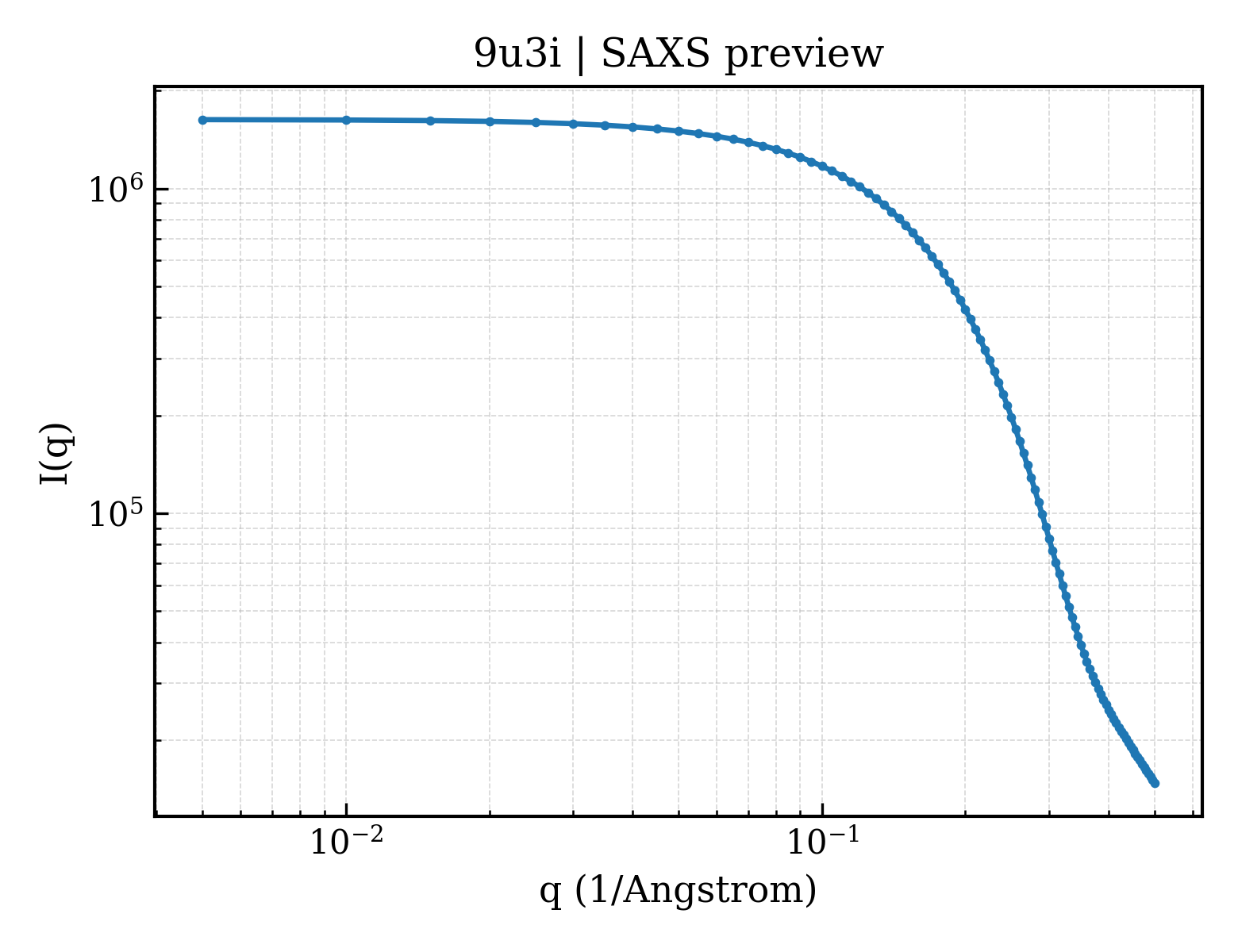
SAXS Profile
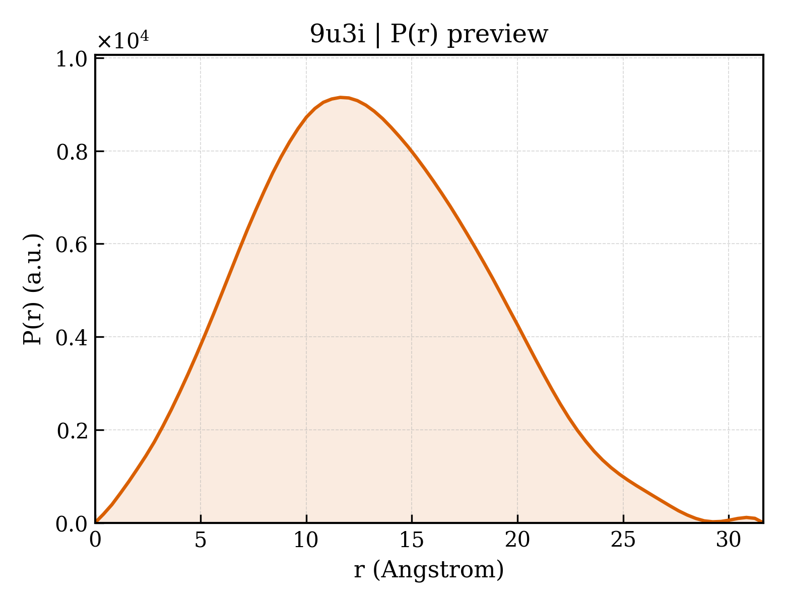
P(r) Distribution
1. Structure Basics
| entry_id | 9u3i |
| deposition_date | 2025-03-18 |
| last_revision | 2026-03-25 |
| title | Crystal structure of a phenylalanine-modifi ed thrombin-binding DNA aptamer |
| keywords | DNA, aptamer; DNA |
| method | X-RAY DIFFRACTION |
Download Data
2. SAXS Parameters (CRYSOL)
| rg_guinier | 9.96 Å |
| rg_electron | 9.10 Å |
| i0 | 1630540.00 |
| molecular_weight | 4663.0 kDa |
| excluded_volume | 3668 ų |
| envelope_volume | 5689 ų |
| shell_volume | 5670 ų |
| envelope_diameter | 29.7 Å |
| shell_rg | 13.83 Å |
| envelope_rg | 9.58 Å |
| shape_rg | 9.02 Å |
| total_rg | 10.02 Å |
| total_atoms | 316 |
| n_residues | 14 |
| n_harmonics | 20 |
| q_range | — – 0.5000 Å−1 |
| n_points | 101 |
| shell_type | directional |
| solvent_density | 0.3340 e/ų |
| contrast_shell | 0.0300 e/ų |
| crysol_version | 4.1.3 |
3. P(r) Analysis (GNOM)
| dmax | 31.6 Å |
| rg_real | 9.92 Å |
| rg_real_error | 0.25 Å |
| i0_real | 1.6310e+06 |
| i0_real_error | 1.6380e+04 |
| rg_reciprocal | 9.92 Å |
| i0_reciprocal | 1631000.0000 |
| total_estimate | 0.8967 |
| solution_quality | GOOD a GOOD solution |
| n_peaks | 2 |
| r_peak_primary | 11.6 Å |
| skewness | 0.233 |
| kurtosis | -0.365 |
| angular_range | — – 0.5000 Å−1 |
| current_alpha | 0.0000 |
| highest_alpha | 95960.0000 |
| n_real_points | 80 |
| gnom_version | 4.1.3 |
| quality_criteria | AN1: 0.000; Oscil: 0.909; Stabil: 1.000; Sysdev: 1.000; Positv: 1.000; Valcen: 1.000; Smooth: 0.927 |