1bjn

STRUCTURE OF PHOSPHOSERINE AMINOTRANSFERASE FROM ESCHERICHIA COLI

Method: X-RAY DIFFRACTION Dmax: 90.1 Å Quality: REASONABLE

1. Protein Identity and Related Structures Protein Identity & Related Structures

PHOSPHOSERINE AMINOTRANSFERASE

Escherichia coli

UniProt P23721

State in the Current Structure

Assembly Oligomeric State Construct Mutations and Modifications Ligands, Ions and Associated Components Method and Experimental Conditions Structure Quality
1 Protein homooligomer Homooligomer Protein × 2 PDB declaration: dimeric(2) Consistent with protein copy count Chain A; UniProt 3–362 Chain B; UniProt 3–362 Non-standard monomer:Yes (specific site not provided by mmCIF) No other associated polymer X-RAY DIFFRACTION X-ray crystallization conditions:VAPOR DIFFUSION, HANGING DROP;277 K;PROTEIN WAS CRYSTALLIZED AT 4-7 DEGREES CELSIUS BY HANGING DROP METHOD, USING PEG4000 AS A PRECIPITATING AGENT, BUFFERED WITH SODIUM ACETATE TO PH 7.2 IN THE DROP AND PH 5.6 IN THE RESERVOIR. THE PH-GRADIENT WAS ESSENTIAL FOR THE CRYSTALLIZATION., vapor diffusion - hanging drop, temperature 277K Resolution 2.30 Å R-free 0.201

Other States of the Same Protein in the Database

Each row is a biological assembly of the same UniProt protein in another PDB entry. The “Difference from current entry” column identifies evidence-level differences; no tag means the currently parsed fields agree.

No other PDB entry for the same UniProt protein was found.

View Construct and Data Evidence
UniProt name SERC_ECOLI
Isoform
PDB entities 1
Chains and sequence ranges Author chain A; PDBConstruct 1–360; UniProt 3–362 Author chain B; PDBConstruct 1–360; UniProt 3–362

The page prioritizes protein identity, the current assembly, associated components, oligomeric state and cross-PDB links. Chain mapping and sequence ranges are retained as data evidence. Internal IDs, import timestamps and assembly operation expressions are maintenance fields and are not shown here.

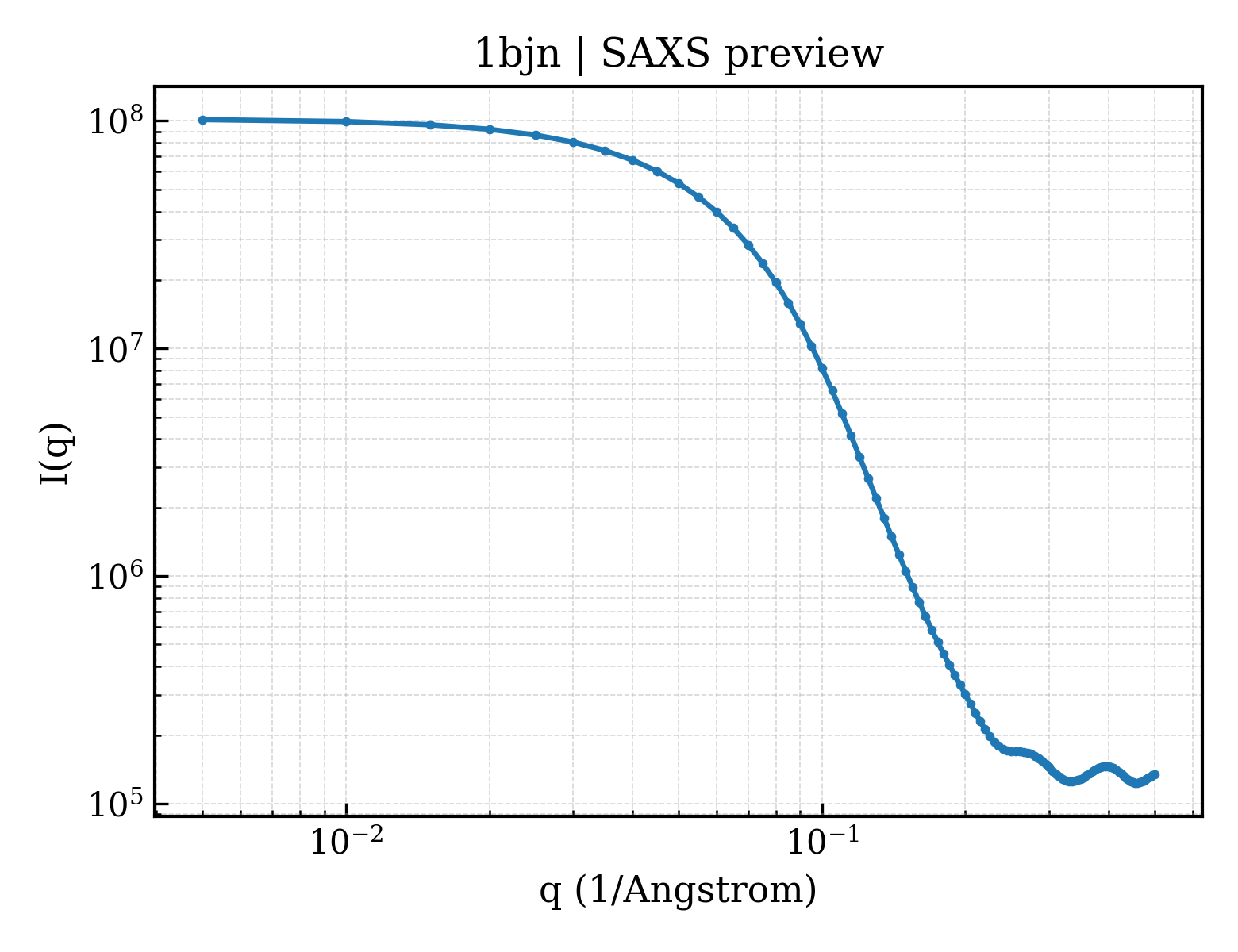
SAXS scattering curve SAXS Profile

SAXS profile for 1bjn

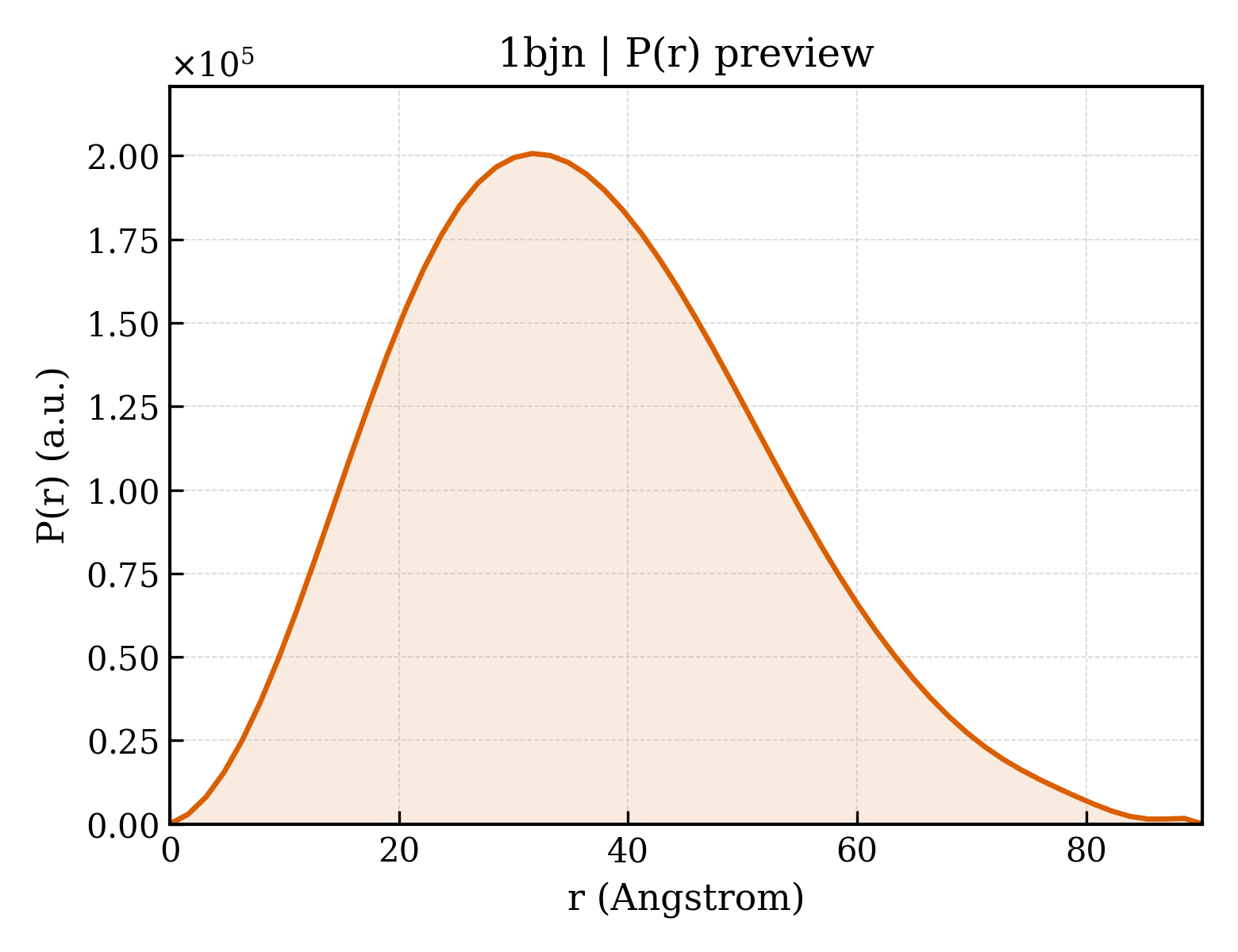
P(r) Distance Distribution P(r) Distribution

P(r) distribution for 1bjn
Download Download

2. Structure Basics 2. Structure Basics

Entry ID entry_id1bjn
Deposition date deposition_date1998-06-25
Structure title titleSTRUCTURE OF PHOSPHOSERINE AMINOTRANSFERASE FROM ESCHERICHIA COLI
Keywords keywordsAMINOTRANSFERASE, L-SERINE BIOSYNTHESIS; AMINOTRANSFERASE
Experimental Method methodX-RAY DIFFRACTION

3. SAXS Parameters (CRYSOL theoretical calculation) 3. SAXS Parameters (CRYSOL)

Radius of gyration Rg (Guinier) rg_guinier27.94
Radius of gyration Rg (electron density) rg_electron26.86
Forward intensity I(0) i0101935000.00
Molecular weight molecular_weight79471.0 kDa
Excluded volume excluded_volume99356 ų
Envelope volume envelope_volume119390 ų
Hydration-shell volume shell_volume36487 ų
Envelope diameter envelope_diameter95.0
Shell Rg shell_rg34.96
Envelope Rg envelope_rg27.16
Shape Rg shape_rg26.86
Total Rg total_rg27.64
Total atoms total_atoms5597
Residues n_residues718
Spherical-harmonic order n_harmonics20
q range q_range— – 0.5000 −1
Data points n_points101
Shell type shell_typedirectional
Solvent electron density solvent_density0.3340 e/ų
Shell contrast contrast_shell0.0300 e/ų
CRYSOL version crysol_version4.1.3

4. P(r) Distance Distribution (GNOM inversion) 4. P(r) Analysis (GNOM)

Maximum dimension Dmax dmax90.1
Rg (real space) rg_real27.91
Rg uncertainty (real space) rg_real_error0.53
I(0) (real space) i0_real1.0190e+08
I(0) uncertainty (real space) i0_real_error1.5350e+06
Rg (reciprocal space) rg_reciprocal27.92
I(0) (reciprocal space) i0_reciprocal101900000.0000
Solution quality estimate total_estimate0.6782
Solution quality rating solution_quality REASONABLE a REASONABLE solution
P(r) peaks n_peaks2
Primary peak position r_peak_primary31.6
Skewness Skewness skewness0.364
Kurtosis Kurtosis kurtosis-0.278
Angular range angular_range— – 0.2850 −1
Current regularization parameter α current_alpha0.0000
Highest regularization parameter α highest_alpha49030000.0000
Real-space data points n_real_points58
GNOM version gnom_version4.1.3
Quality Criteria quality_criteria AN1: 0.000; Oscil: 0.882; Stabil: 1.000; Sysdev: 0.075; Positv: 1.000; Valcen: 1.000; Smooth: 0.940

5. Crystallography and Experiment 5. Crystallography & Experiment

6. Entities and Polymers Entities & Polymers (2)

7. Fold Classification (SCOP + CATH) 6 domains

SCOP 2.08 (2 domains)

Domain ID domain_idd1bjna_
Class classc — Alpha and beta proteins (a/b)
Fold Fold foldc.67 — PLP-dependent transferase-like
Superfamily Superfamily superfamilyc.67.1 — PLP-dependent transferases
Family Family familyc.67.1.4 — GABA-aminotransferase-like
Domain ID domain_idd1bjnb_
Class classc — Alpha and beta proteins (a/b)
Fold Fold foldc.67 — PLP-dependent transferase-like
Superfamily Superfamily superfamilyc.67.1 — PLP-dependent transferases
Family Family familyc.67.1.4 — GABA-aminotransferase-like

CATH v4.4 (4 domains)

Domain ID domain_id1bjnA01
Class class3 — Alpha Beta
Architecture architecture40 — 3-Layer(aba) Sandwich
Topology topology640 — Aspartate Aminotransferase; domain 2
Homologous superfamily homologous superfamily10 — Type I PLP-dependent aspartate aminotransferase-like (Major domain)
Domain ID domain_id1bjnA02
Class class3 — Alpha Beta
Architecture architecture90 — Alpha-Beta Complex
Topology topology1150 — Aspartate Aminotransferase, domain 1
Homologous superfamily homologous superfamily10 — Aspartate Aminotransferase, domain 1
Domain ID domain_id1bjnB01
Class class3 — Alpha Beta
Architecture architecture40 — 3-Layer(aba) Sandwich
Topology topology640 — Aspartate Aminotransferase; domain 2
Homologous superfamily homologous superfamily10 — Type I PLP-dependent aspartate aminotransferase-like (Major domain)
Domain ID domain_id1bjnB02
Class class3 — Alpha Beta
Architecture architecture90 — Alpha-Beta Complex
Topology topology1150 — Aspartate Aminotransferase, domain 1
Homologous superfamily homologous superfamily10 — Aspartate Aminotransferase, domain 1

8. Citations (3)

9. Files and Curves (10)