1cto

NMR STRUCTURE OF THE C-TERMINAL DOMAIN OF THE LIGAND-BINDING REGION OF MURINE GRANULOCYTE COLONY-STIMULATING FACTOR RECEPTOR, MINIMIZED AVERAGE STRUCTURE

Method: SOLUTION NMR Dmax: 52.9 Å Quality: GOOD

1. Protein Identity and Related Structures Protein Identity & Related Structures

GRANULOCYTE COLONY-STIMULATING FACTOR RECEPTOR

Mus musculus

UniProt P40223

State in the Current Structure

Assembly Oligomeric State Construct Mutations and Modifications Ligands, Ions and Associated Components Method and Experimental Conditions Structure Quality
1 Protein monomer Monomer Protein × 1 PDB declaration: monomeric(1) Consistent with protein copy count Chain A; UniProt 211–333 Fragment:C-TERMINAL DOMAIN OF THE LIGAND-BINDING REGION Mutation:INS(G1,S2,S3) No other associated polymer SOLUTION NMR NMR measurement conditions:pH 5.5;303 K Resolution not provided

Other States of the Same Protein in the Database

Each row is a biological assembly of the same UniProt protein in another PDB entry. The “Difference from current entry” column identifies evidence-level differences; no tag means the currently parsed fields agree.

3 other PDB entries and 4 assemblies. Open the comparison page and filter oligomeric states

View Construct and Data Evidence
UniProt name CSF3R_MOUSE
Isoform
PDB entities 1
Chains and sequence ranges Author chain A; PDBConstruct 1–109; UniProt 211–333

The page prioritizes protein identity, the current assembly, associated components, oligomeric state and cross-PDB links. Chain mapping and sequence ranges are retained as data evidence. Internal IDs, import timestamps and assembly operation expressions are maintenance fields and are not shown here.

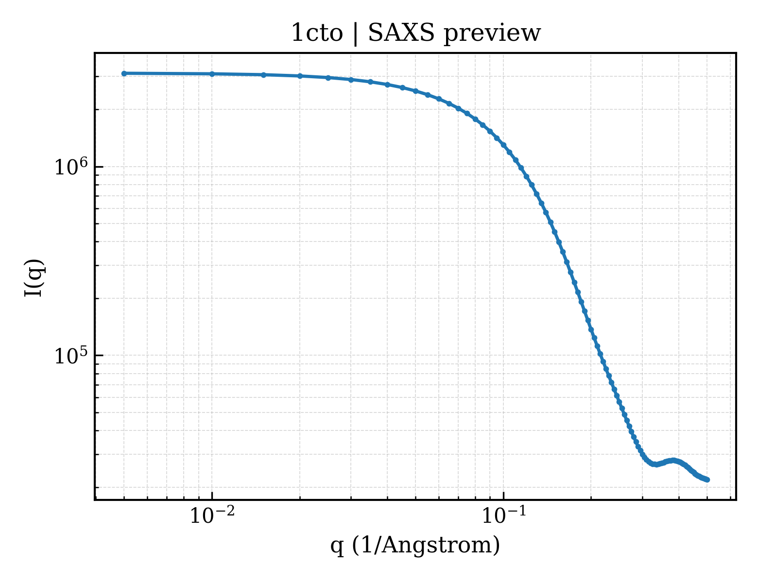
SAXS scattering curve SAXS Profile

SAXS profile for 1cto

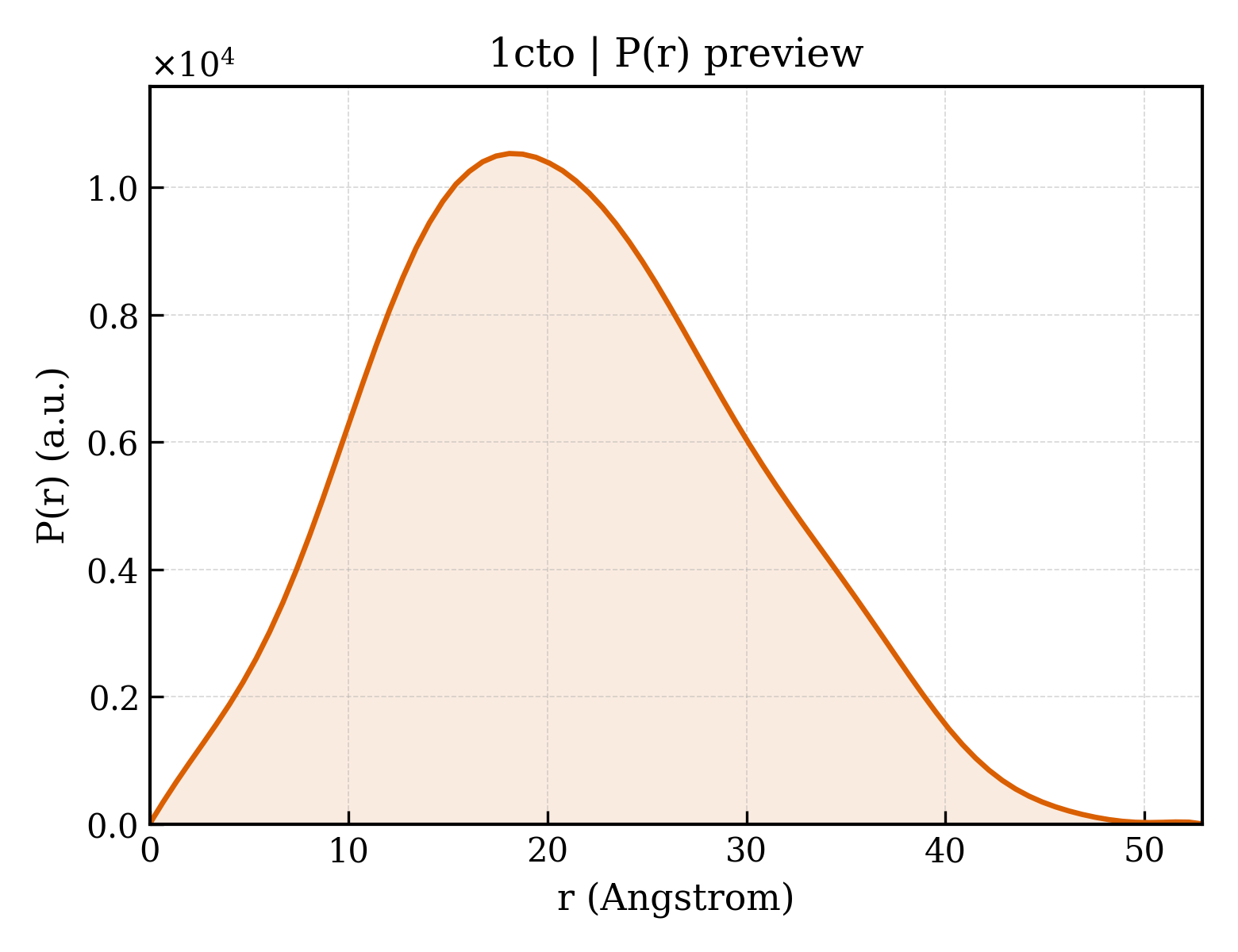
P(r) Distance Distribution P(r) Distribution

P(r) distribution for 1cto
Download Download

2. Structure Basics 2. Structure Basics

Entry ID entry_id1cto
Deposition date deposition_date1996-09-25
Structure title titleNMR STRUCTURE OF THE C-TERMINAL DOMAIN OF THE LIGAND-BINDING REGION OF MURINE GRANULOCYTE COLONY-STIMULATING FACTOR RECEPTOR, MINIMIZED AVERAGE STRUCTURE
Keywords keywordsBINDING PROTEIN, CYTOKINE RECEPTOR; BINDING PROTEIN
Experimental Method methodSOLUTION NMR

3. SAXS Parameters (CRYSOL theoretical calculation) 3. SAXS Parameters (CRYSOL)

Radius of gyration Rg (Guinier) rg_guinier16.16
Radius of gyration Rg (electron density) rg_electron15.04
Forward intensity I(0) i03117500.00
Molecular weight molecular_weight12525.0 kDa
Excluded volume excluded_volume15757 ų
Envelope volume envelope_volume18956 ų
Hydration-shell volume shell_volume11270 ų
Envelope diameter envelope_diameter51.8
Shell Rg shell_rg19.92
Envelope Rg envelope_rg15.41
Shape Rg shape_rg15.02
Total Rg total_rg16.15
Total atoms total_atoms1739
Residues n_residues109
Spherical-harmonic order n_harmonics20
q range q_range— – 0.5000 −1
Data points n_points101
Shell type shell_typedirectional
Solvent electron density solvent_density0.3340 e/ų
Shell contrast contrast_shell0.0300 e/ų
CRYSOL version crysol_version4.1.3

4. P(r) Distance Distribution (GNOM inversion) 4. P(r) Analysis (GNOM)

Maximum dimension Dmax dmax52.9
Rg (real space) rg_real16.13
Rg uncertainty (real space) rg_real_error0.24
I(0) (real space) i0_real3.1180e+06
I(0) uncertainty (real space) i0_real_error3.8430e+04
Rg (reciprocal space) rg_reciprocal16.13
I(0) (reciprocal space) i0_reciprocal3118000.0000
Solution quality estimate total_estimate0.8949
Solution quality rating solution_quality GOOD a GOOD solution
P(r) peaks n_peaks2
Primary peak position r_peak_primary18.1
Skewness Skewness skewness0.263
Kurtosis Kurtosis kurtosis-0.419
Angular range angular_range— – 0.4950 −1
Current regularization parameter α current_alpha0.0000
Highest regularization parameter α highest_alpha599500.0000
Real-space data points n_real_points80
GNOM version gnom_version4.1.3
Quality Criteria quality_criteria AN1: 0.000; Oscil: 0.881; Stabil: 1.000; Sysdev: 1.000; Positv: 1.000; Valcen: 0.986; Smooth: 0.999

5. Crystallography and Experiment 5. Crystallography & Experiment

6. Entities and Polymers Entities & Polymers (1)

7. Fold Classification (SCOP + CATH) 2 domains

SCOP 2.08 (1 domains)

Domain ID domain_idd1ctoa_
Class classb — All beta proteins
Fold Fold foldb.1 — Immunoglobulin-like beta-sandwich
Superfamily Superfamily superfamilyb.1.2 — Fibronectin type III
Family Family familyb.1.2.1 — Fibronectin type III

CATH v4.4 (1 domains)

Domain ID domain_id1ctoA00
Class class2 — Mainly Beta
Architecture architecture60 — Sandwich
Topology topology40 — Immunoglobulin-like
Homologous superfamily homologous superfamily10 — Immunoglobulins

8. Citations (2)

9. Files and Curves (10)