2lbp
;STRUCTURE OF THE L-LEUCINE-BINDING PROTEIN REFINED AT 2.4 ANGSTROMS RESOLUTION AND COMPARISON WITH THE LEU(SLASH)ILE(SLASH)VAL-BINDING PROTEIN STRUCTURE ;
SAXS 散射曲线 SAXS Profile

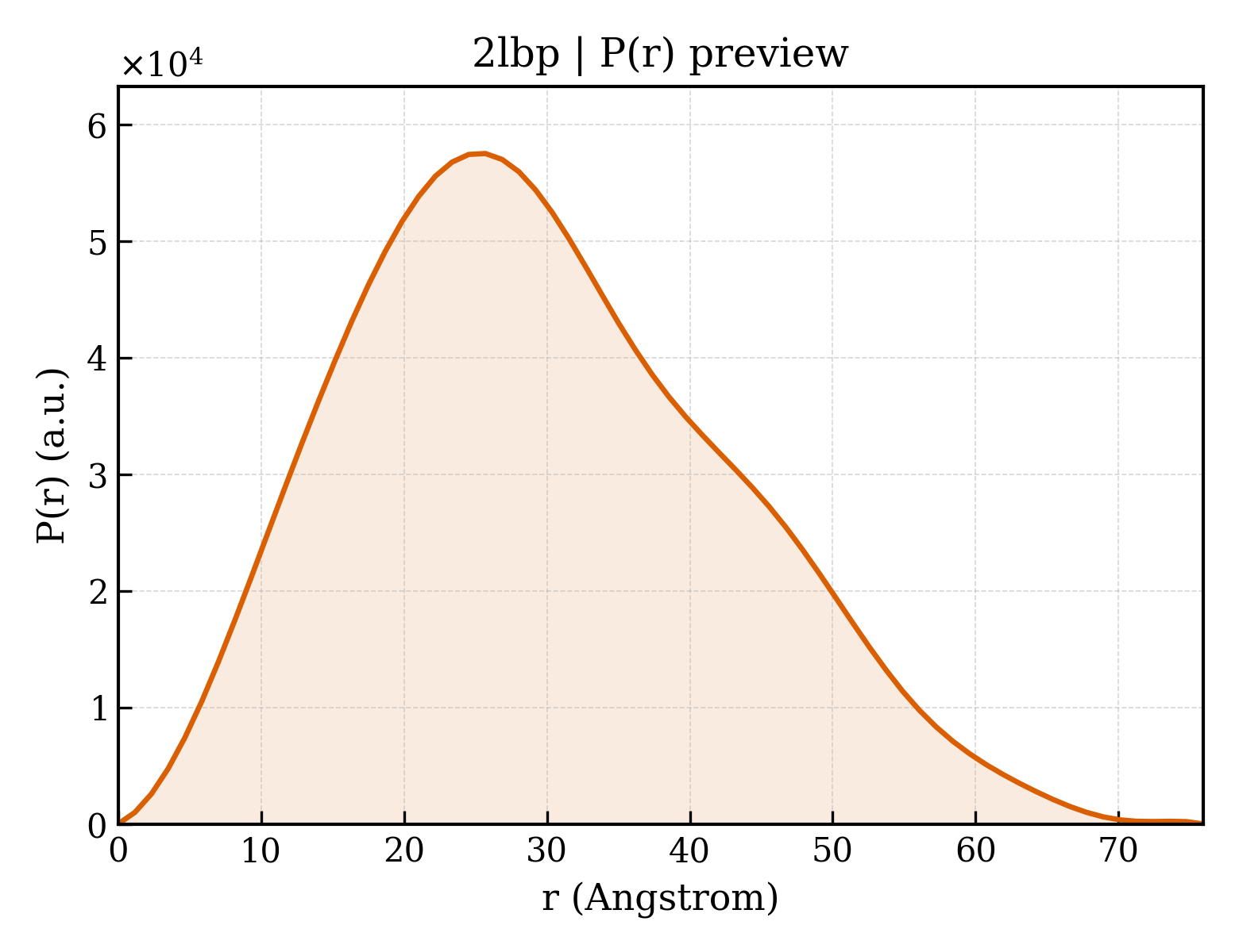
P(r) 距离分布 P(r) Distribution
1. 结构基本信息 1. Structure Basics
| 条目编号 entry_id | 2lbp |
| 沉积日期 deposition_date | 1989-04-10 |
| 结构标题 title | ;STRUCTURE OF THE L-LEUCINE-BINDING PROTEIN REFINED AT 2.4 ANGSTROMS RESOLUTION AND COMPARISON WITH THE LEU(SLASH)ILE(SLASH)VAL-BINDING PROTEIN STRUCTURE ; |
| 关键词 keywords | PERIPLASMIC BINDING PROTEIN; PERIPLASMIC BINDING PROTEIN |
| 实验方法 method | X-RAY DIFFRACTION |
2. SAXS 参数 (CRYSOL 理论计算) 2. SAXS Parameters (CRYSOL)
| 回转半径 Rg (Guinier) rg_guinier | 22.86 Å |
| 回转半径 Rg (电子) rg_electron | 22.08 Å |
| 零角强度 I(0) i0 | 23792500.00 |
| 分子量 molecular_weight | 36959.0 kDa |
| 排除体积 excluded_volume | 46143 ų |
| 包络体积 envelope_volume | 54426 ų |
| 水化壳体积 shell_volume | 21068 ų |
| 包络直径 envelope_diameter | 79.2 Å |
| 壳层 Rg shell_rg | 28.26 Å |
| 包络 Rg envelope_rg | 22.09 Å |
| 形状 Rg shape_rg | 22.05 Å |
| 总 Rg total_rg | 22.94 Å |
| 总原子数 total_atoms | 2600 |
| 残基数 n_residues | 346 |
| 球谐函数阶数 n_harmonics | 20 |
| q 范围 q_range | — – 0.5000 Å−1 |
| 数据点数 n_points | 101 |
| 壳层类型 shell_type | directional |
| 溶剂电子密度 solvent_density | 0.3340 e/ų |
| 壳层衬度 contrast_shell | 0.0300 e/ų |
| CRYSOL 版本 crysol_version | 4.1.3 |
3. P(r) 距离分布 (GNOM 反演) 3. P(r) Analysis (GNOM)
| 最大尺寸 Dmax dmax | 75.9 Å |
| Rg (实空间) rg_real | 22.89 Å |
| Rg 误差 (实空间) rg_real_error | 0.45 Å |
| I(0) (实空间) i0_real | 2.3790e+07 |
| I(0) 误差 (实空间) i0_real_error | 3.3710e+05 |
| Rg (倒空间) rg_reciprocal | 22.88 Å |
| I(0) (倒空间) i0_reciprocal | 23790000.0000 |
| 解质量估计 total_estimate | 0.8854 |
| 解质量评级 solution_quality | GOOD a GOOD solution |
| P(r) 峰数 n_peaks | 2 |
| 主峰位置 r_peak_primary | 25.7 Å |
| 偏度 Skewness skewness | 0.375 |
| 峰度 Kurtosis kurtosis | -0.375 |
| 角度范围 angular_range | — – 0.3450 Å−1 |
| 当前正则化参数 α current_alpha | 0.0001 |
| 最高正则化参数 α highest_alpha | 3952000.0000 |
| 实空间数据点数 n_real_points | 66 |
| GNOM 版本 gnom_version | 4.1.3 |
| 质量判据 quality_criteria | AN1: 0.000; Oscil: 0.844; Stabil: 1.000; Sysdev: 1.000; Positv: 1.000; Valcen: 0.975; Smooth: 0.997 |
4. 晶体学与实验 4. Crystallography & Experiment
5. 实体与聚合体信息 (2)
6. 折叠分类 (SCOP + CATH) 3 domains
SCOP 2.08 (1 domains)
| 结构域编号 domain_id | d2lbpa_ |
| 类 Class class | c — Alpha and beta proteins (a/b) |
| 折叠类型 Fold fold | c.93 — Periplasmic binding protein-like I |
| 超家族 Superfamily superfamily | c.93.1 — Periplasmic binding protein-like I |
| 家族 Family family | c.93.1.1 — L-arabinose binding protein-like |
CATH v4.4 (2 domains)
| 结构域编号 domain_id | 2lbpA01 |
| 类 Class class | 3 — Alpha Beta |
| 架构 Architecture architecture | 40 — 3-Layer(aba) Sandwich |
| 拓扑 Topology topology | 50 — Rossmann fold |
| 同源超家族 H-superfamily homologous superfamily | 2300 — Response regulator |
| 结构域编号 domain_id | 2lbpA02 |
| 类 Class class | 3 — Alpha Beta |
| 架构 Architecture architecture | 40 — 3-Layer(aba) Sandwich |
| 拓扑 Topology topology | 50 — Rossmann fold |
| 同源超家族 H-superfamily homologous superfamily | 2300 — Response regulator |