Polymeric-immunoglobulin receptor
Homo sapiens
State in the Current Structure
| Assembly | Oligomeric State | Construct | Mutations and Modifications | Ligands, Ions and Associated Components | Method and Experimental Conditions | Structure Quality |
|---|---|---|---|---|---|---|
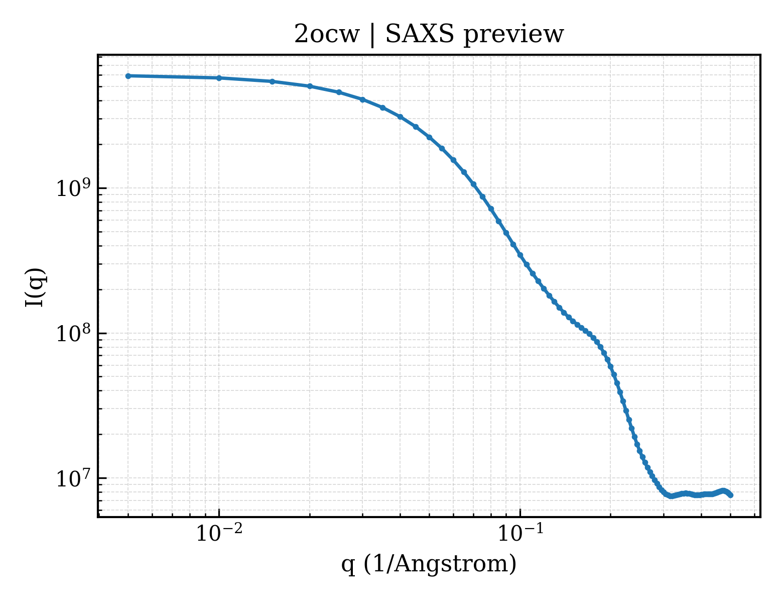
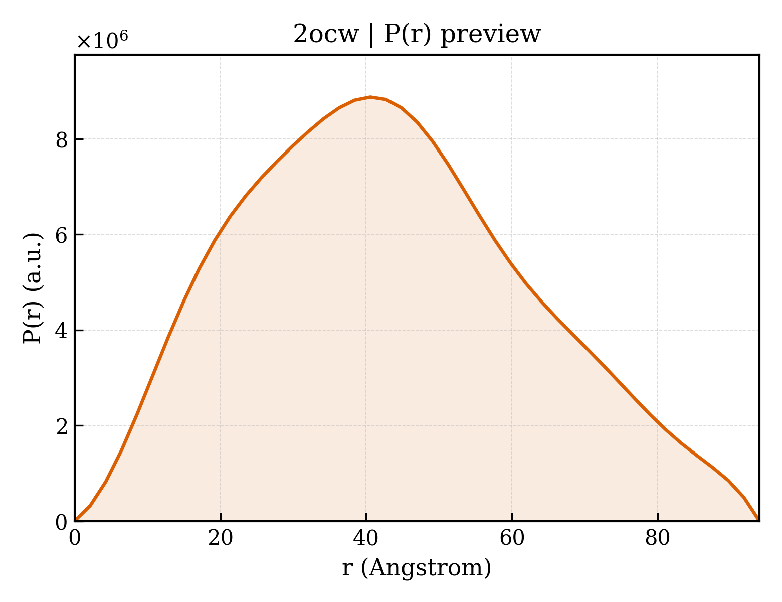
| 1 | Protein monomer Monomer Protein × 1 PDB declaration: monomeric(1) Consistent with protein copy count | Chain A; UniProt 19–603 | Not recorded | No other associated polymer | SOLUTION SCATTERING mmCIF provides none of the parsed experimental conditions | Resolution not provided |
Other States of the Same Protein in the Database
Each row is a biological assembly of the same UniProt protein in another PDB entry. The “Difference from current entry” column identifies evidence-level differences; no tag means the currently parsed fields agree.
| Other PDB | Difference from Current Entry 2OCW | Assembly / Oligomeric State | Construct | Mutations and Modifications | Ligands, Ions and Non-polymers | Method and Experimental Conditions | Structure Quality |
|---|---|---|---|---|---|---|---|
| 1XED Crystal Structure of a Ligand-Binding Domain of the Human Polymeric Ig Receptor, pIgR Deposited 2004-09-10 | Different construct Different ligand/ion Different experimental method Different experimental conditions Different structure-quality metrics | Assembly 1 Protein monomer Monomer;Protein × 1 PDB declaration: monomeric |
Chain A
19–127(109 aa)
Fragment:N-Terminal Domain I
|
Not recorded | MG MAGNESIUM ION × 1 |
X-RAY DIFFRACTION
X-ray crystallization conditions
pH 6.5;293 K;PEG 8000, Magnesium acetate, cacodylate, pH 6.5, VAPOR DIFFUSION, HANGING DROP, temperature 293K, pH 6.50
|
Resolution 1.90 Å R-free 0.244 |
| 1XED Crystal Structure of a Ligand-Binding Domain of the Human Polymeric Ig Receptor, pIgR Deposited 2004-09-10 | Different construct Different ligand/ion Different experimental method Different experimental conditions Different structure-quality metrics | Assembly 2 Protein monomer Monomer;Protein × 1 PDB declaration: monomeric |
Chain B
19–127(109 aa)
Fragment:N-Terminal Domain I
|
Not recorded | MG MAGNESIUM ION × 1 |
X-RAY DIFFRACTION
X-ray crystallization conditions
pH 6.5;293 K;PEG 8000, Magnesium acetate, cacodylate, pH 6.5, VAPOR DIFFUSION, HANGING DROP, temperature 293K, pH 6.50
|
Resolution 1.90 Å R-free 0.244 |
| 1XED Crystal Structure of a Ligand-Binding Domain of the Human Polymeric Ig Receptor, pIgR Deposited 2004-09-10 | Different construct Different experimental method Different experimental conditions Different structure-quality metrics | Assembly 3 Protein monomer Monomer;Protein × 1 PDB declaration: monomeric |
Chain C
19–127(109 aa)
Fragment:N-Terminal Domain I
|
Not recorded | No recorded non-water small molecule |
X-RAY DIFFRACTION
X-ray crystallization conditions
pH 6.5;293 K;PEG 8000, Magnesium acetate, cacodylate, pH 6.5, VAPOR DIFFUSION, HANGING DROP, temperature 293K, pH 6.50
|
Resolution 1.90 Å R-free 0.244 |
| 1XED Crystal Structure of a Ligand-Binding Domain of the Human Polymeric Ig Receptor, pIgR Deposited 2004-09-10 | Different construct Different experimental method Different experimental conditions Different structure-quality metrics | Assembly 4 Protein monomer Monomer;Protein × 1 PDB declaration: monomeric |
Chain D
19–127(109 aa)
Fragment:N-Terminal Domain I
|
Not recorded | No recorded non-water small molecule |
X-RAY DIFFRACTION
X-ray crystallization conditions
pH 6.5;293 K;PEG 8000, Magnesium acetate, cacodylate, pH 6.5, VAPOR DIFFUSION, HANGING DROP, temperature 293K, pH 6.50
|
Resolution 1.90 Å R-free 0.244 |
| 1XED Crystal Structure of a Ligand-Binding Domain of the Human Polymeric Ig Receptor, pIgR Deposited 2004-09-10 | Different construct Different experimental method Different experimental conditions Different structure-quality metrics | Assembly 5 Protein monomer Monomer;Protein × 1 PDB declaration: monomeric |
Chain E
19–127(109 aa)
Fragment:N-Terminal Domain I
|
Not recorded | No recorded non-water small molecule |
X-RAY DIFFRACTION
X-ray crystallization conditions
pH 6.5;293 K;PEG 8000, Magnesium acetate, cacodylate, pH 6.5, VAPOR DIFFUSION, HANGING DROP, temperature 293K, pH 6.50
|
Resolution 1.90 Å R-free 0.244 |
| 1XED Crystal Structure of a Ligand-Binding Domain of the Human Polymeric Ig Receptor, pIgR Deposited 2004-09-10 | Different construct Different experimental method Different experimental conditions Different structure-quality metrics | Assembly 6 Protein monomer Monomer;Protein × 1 PDB declaration: monomeric |
Chain F
19–127(109 aa)
Fragment:N-Terminal Domain I
|
Not recorded | No recorded non-water small molecule |
X-RAY DIFFRACTION
X-ray crystallization conditions
pH 6.5;293 K;PEG 8000, Magnesium acetate, cacodylate, pH 6.5, VAPOR DIFFUSION, HANGING DROP, temperature 293K, pH 6.50
|
Resolution 1.90 Å R-free 0.244 |
| 1XED Crystal Structure of a Ligand-Binding Domain of the Human Polymeric Ig Receptor, pIgR Deposited 2004-09-10 | Different construct Different mutation/modification Different oligomeric state Different ligand/ion Different experimental method Different experimental conditions Different structure-quality metrics | Assembly 7 Protein homooligomer Homooligomer;Protein × 4 PDB declaration: tetrameric |
Chain A
19–127(109 aa)
Fragment:N-Terminal Domain I
Chain B
19–127(109 aa)
Fragment:N-Terminal Domain I
Chain C
19–127(109 aa)
Fragment:N-Terminal Domain I
Chain D
19–127(109 aa)
Fragment:N-Terminal Domain I
|
Not recorded | MG MAGNESIUM ION × 2 |
X-RAY DIFFRACTION
X-ray crystallization conditions
pH 6.5;293 K;PEG 8000, Magnesium acetate, cacodylate, pH 6.5, VAPOR DIFFUSION, HANGING DROP, temperature 293K, pH 6.50
|
Resolution 1.90 Å R-free 0.244 |
| 1XED Crystal Structure of a Ligand-Binding Domain of the Human Polymeric Ig Receptor, pIgR Deposited 2004-09-10 | Different construct Different mutation/modification Different oligomeric state Different ligand/ion Different experimental method Different experimental conditions Different structure-quality metrics | Assembly 8 Protein homooligomer Homooligomer;Protein × 2 PDB declaration: dimeric |
Chain B
19–127(109 aa)
Fragment:N-Terminal Domain I
Chain D
19–127(109 aa)
Fragment:N-Terminal Domain I
|
Not recorded | MG MAGNESIUM ION × 1 |
X-RAY DIFFRACTION
X-ray crystallization conditions
pH 6.5;293 K;PEG 8000, Magnesium acetate, cacodylate, pH 6.5, VAPOR DIFFUSION, HANGING DROP, temperature 293K, pH 6.50
|
Resolution 1.90 Å R-free 0.244 |
| 1XED Crystal Structure of a Ligand-Binding Domain of the Human Polymeric Ig Receptor, pIgR Deposited 2004-09-10 | Different construct Different mutation/modification Different oligomeric state Different ligand/ion Different experimental method Different experimental conditions Different structure-quality metrics | Assembly 9 Protein homooligomer Homooligomer;Protein × 2 PDB declaration: dimeric |
Chain A
19–127(109 aa)
Fragment:N-Terminal Domain I
Chain C
19–127(109 aa)
Fragment:N-Terminal Domain I
|
Not recorded | MG MAGNESIUM ION × 1 |
X-RAY DIFFRACTION
X-ray crystallization conditions
pH 6.5;293 K;PEG 8000, Magnesium acetate, cacodylate, pH 6.5, VAPOR DIFFUSION, HANGING DROP, temperature 293K, pH 6.50
|
Resolution 1.90 Å R-free 0.244 |
| 3CHN Solution structure of human secretory IgA1 Deposited 2008-03-10 | Different construct Different mutation/modification Different oligomeric state | Assembly 1 Protein heterocomplex Heteromer;Protein × 10 PDB declaration: decameric |
Chain J
353–458(106 aa)
Fragment:Ig-like V-type domain 4
Chain S
19–603(585 aa)
|
Not recorded | No recorded non-water small molecule | SOLUTION SCATTERING mmCIF provides none of the parsed conditions | Resolution not provided |
| 3CM9 Solution Structure of Human SIgA2 Deposited 2008-03-21 | Different construct Different mutation/modification Different oligomeric state | Assembly 1 Protein heterocomplex Heteromer;Protein × 10 PDB declaration: decameric |
Chain J
353–458(106 aa)
Fragment:Ig-like V-type domain 4
Chain S
19–603(585 aa)
|
Not recorded | No recorded non-water small molecule | SOLUTION SCATTERING mmCIF provides none of the parsed conditions | Resolution not provided |
| 5D4K Crystal structure of the human polymeric Ig receptor (pIgR) ectodomain Deposited 2015-08-07 | Different construct Different oligomeric state Different ligand/ion Different experimental method Different experimental conditions Different structure-quality metrics | Assembly 1 Other combination Monomer;Protein × 1 PDB declaration: monomeric |
Chain A
19–565(547 aa)
Fragment:ectodomain (UNP residues 19-565)
|
Not recorded | NAG 2-acetamido-2-deoxy-beta-D-glucopyranose × 3 SO4 SULFATE ION × 2 |
X-RAY DIFFRACTION
X-ray crystallization conditions
VAPOR DIFFUSION, SITTING DROP;pH 5.5;295 K;0.2 M ammonium sulfate, 0.1M BisTris pH 5.5, 31% Polyethylene glycol 3350
|
Resolution 2.60 Å R-free 0.254 |
| 5D4K Crystal structure of the human polymeric Ig receptor (pIgR) ectodomain Deposited 2015-08-07 | Different construct Different oligomeric state Different ligand/ion Different experimental method Different experimental conditions Different structure-quality metrics | Assembly 2 Other combination Monomer;Protein × 1 PDB declaration: monomeric |
Chain B
19–565(547 aa)
Fragment:ectodomain (UNP residues 19-565)
|
Not recorded | NAG 2-acetamido-2-deoxy-beta-D-glucopyranose × 3 SO4 SULFATE ION × 2 |
X-RAY DIFFRACTION
X-ray crystallization conditions
VAPOR DIFFUSION, SITTING DROP;pH 5.5;295 K;0.2 M ammonium sulfate, 0.1M BisTris pH 5.5, 31% Polyethylene glycol 3350
|
Resolution 2.60 Å R-free 0.254 |
| 6KXS Cryo-EM structure of human IgM-Fc in complex with the J chain and the ectodomain of pIgR Deposited 2019-09-12 | Different construct Different oligomeric state Different ligand/ion Different experimental method Different experimental conditions Different structure-quality metrics | Assembly 1 Other combination Heteromer;Protein × 12 PDB declaration: dodecameric |
Chain P
19–565(547 aa)
|
Not recorded | NAG 2-acetamido-2-deoxy-beta-D-glucopyranose × 10 |
ELECTRON MICROSCOPY
cryo-EM buffer
pH 7.4
cryo-EM vitrification conditions
Cryogen ETHANE
|
Resolution 3.40 Å |
| 6LX3 Cryo-EM structure of human secretory immunoglobulin A Deposited 2020-02-10 | Different construct Different oligomeric state Different experimental method Different experimental conditions Different structure-quality metrics | Assembly 1 Insufficient information Heteromer;Protein × 6 PDB declaration: hexameric |
Chain P
1–565(565 aa)
|
Not recorded | No recorded non-water small molecule |
ELECTRON MICROSCOPY
cryo-EM buffer
pH 7.4
cryo-EM vitrification conditions
Cryogen ETHANE
|
Resolution 3.15 Å |
| 6LXW Cryo-EM structure of human secretory immunoglobulin A in complex with the N-terminal domain of SpsA Deposited 2020-02-12 | Different construct Different oligomeric state Different experimental method Different experimental conditions Different structure-quality metrics | Assembly 1 Insufficient information Heteromer;Protein × 7 PDB declaration: heptameric |
Chain P
1–565(565 aa)
|
Not recorded | No recorded non-water small molecule |
ELECTRON MICROSCOPY
cryo-EM buffer
pH 7.4
cryo-EM vitrification conditions
Cryogen ETHANE
|
Resolution 3.27 Å |
| 6UE7 Structure of dimeric sIgA complex Deposited 2019-09-20 | Different construct Different oligomeric state Different ligand/ion Different experimental method Different experimental conditions Different structure-quality metrics | Assembly 1 Other combination Heteromer;Protein × 6 PDB declaration: hexameric |
Chain C
19–603(585 aa)
Fragment:UNP residues 19-603
|
Not recorded | NAG 2-acetamido-2-deoxy-beta-D-glucopyranose × 7 |
ELECTRON MICROSCOPY
cryo-EM buffer
pH 7.2
cryo-EM vitrification conditions
Cryogen ETHANE
|
Resolution 2.90 Å |
| 6UE8 Structure of tetrameric sIgA complex (Class 1) Deposited 2019-09-20 | Different construct Different oligomeric state Different ligand/ion Different experimental method Different experimental conditions Different structure-quality metrics | Assembly 1 Other combination Heteromer;Protein × 10 PDB declaration: decameric |
Chain C
19–603(585 aa)
Fragment:UNP residues 19-603
|
Not recorded | NAG 2-acetamido-2-deoxy-beta-D-glucopyranose × 5 |
ELECTRON MICROSCOPY
cryo-EM buffer
pH 7.2
cryo-EM vitrification conditions
Cryogen ETHANE
|
Resolution 3.00 Å |
| 6UE9 Structure of tetrameric sIgA complex (Class 2) Deposited 2019-09-20 | Different construct Different oligomeric state Different ligand/ion Different experimental method Different experimental conditions Different structure-quality metrics | Assembly 1 Other combination Heteromer;Protein × 10 PDB declaration: decameric |
Chain C
19–603(585 aa)
Fragment:UNP residues 19-603
|
Not recorded | NAG 2-acetamido-2-deoxy-beta-D-glucopyranose × 7 |
ELECTRON MICROSCOPY
cryo-EM buffer
pH 7.2
cryo-EM vitrification conditions
Cryogen ETHANE
|
Resolution 2.90 Å |
| 6UEA Structure of pentameric sIgA complex Deposited 2019-09-20 | Different construct Different oligomeric state Different ligand/ion Different experimental method Different experimental conditions Different structure-quality metrics | Assembly 1 Other combination Heteromer;Protein × 12 PDB declaration: dodecameric |
Chain C
19–603(585 aa)
Fragment:UNP residues 19-603
|
Not recorded | NAG 2-acetamido-2-deoxy-beta-D-glucopyranose × 2 |
ELECTRON MICROSCOPY
cryo-EM buffer
pH 7.2
cryo-EM vitrification conditions
Cryogen ETHANE
|
Resolution 3.00 Å |
| 7K0C Structure of Secretory IgM Core Deposited 2020-09-04 | Different oligomeric state Different experimental method Different experimental conditions Different structure-quality metrics | Assembly 1 Other combination Heteromer;Protein × 12 PDB declaration: dodecameric |
Chain C
19–603(585 aa)
|
Not recorded | No recorded non-water small molecule |
ELECTRON MICROSCOPY
cryo-EM buffer
pH 7.2
cryo-EM vitrification conditions
Cryogen ETHANE;Blot for 3.5 seconds before plunging with blot force 7.
|
Resolution 3.30 Å |
| 7YSG Cryo-EM structure of human FcmR bound to sIgM Deposited 2022-08-12 | Different construct Different oligomeric state Different ligand/ion Different experimental method Different experimental conditions Different structure-quality metrics | Assembly 1 Other combination Heteromer;Protein × 16 PDB declaration: hexadecameric |
Chain P
19–559(541 aa)
|
Not recorded | NAG 2-acetamido-2-deoxy-beta-D-glucopyranose × 10 |
ELECTRON MICROSCOPY
cryo-EM buffer
pH 7.4
cryo-EM vitrification conditions
Cryogen ETHANE
|
Resolution 3.18 Å |
| 8SKU Structure of human SIgA1 in complex with human CD89 (FcaR1) Deposited 2023-04-20 | Different construct Different oligomeric state Different ligand/ion Different experimental method Different experimental conditions Different structure-quality metrics | Assembly 1 Other combination Heteromer;Protein × 8 PDB declaration: octameric |
Chain E
19–565(547 aa)
|
Not recorded | NAG 2-acetamido-2-deoxy-beta-D-glucopyranose × 4 |
ELECTRON MICROSCOPY
cryo-EM buffer
pH 7
cryo-EM vitrification conditions
Cryogen ETHANE
|
Resolution 3.20 Å |
| 8SKV Structure of human SIgA1 in complex with Streptococcus pyogenes protein M4 (Arp4) Deposited 2023-04-20 | Different construct Different oligomeric state Different ligand/ion Different experimental method Different experimental conditions Different structure-quality metrics | Assembly 1 Other combination Heteromer;Protein × 8 PDB declaration: octameric |
Chain E
19–565(547 aa)
|
Not recorded | NAG 2-acetamido-2-deoxy-beta-D-glucopyranose × 7 |
ELECTRON MICROSCOPY
cryo-EM buffer
pH 7
cryo-EM vitrification conditions
Cryogen ETHANE
|
Resolution 3.10 Å |
15 other PDB entries and 24 assemblies. Open the comparison page and filter oligomeric states
View Construct and Data Evidence
| UniProt name | PIGR_HUMAN |
| Isoform | — |
| PDB entities | 1 |
| Chains and sequence ranges | Author chain A; PDBConstruct 1–585; UniProt 19–603 |