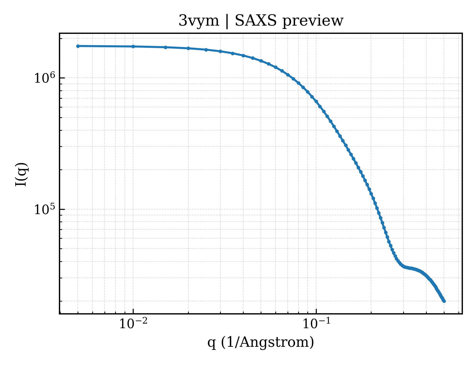
Cytochrome c-552
Hydrogenobacter thermophilus
State in the Current Structure
| Assembly | Oligomeric State | Construct | Mutations and Modifications | Ligands, Ions and Associated Components | Method and Experimental Conditions | Structure Quality |
|---|---|---|---|---|---|---|
| 1 | Protein homooligomer Homooligomer Protein × 2 PDB declaration: dimeric(2) Consistent with protein copy count | Chain A; UniProt 19–98 | Not recorded | HEC HEME C × 2 | X-RAY DIFFRACTION X-ray crystallization conditions:VAPOR DIFFUSION, SITTING DROP;pH 7;295 K;100mM HEPES buffer, 800mM ammonium sulfate, 45% (v/v) 2-methyl-2,4-pentanediol, pH 7.0, VAPOR DIFFUSION, SITTING DROP, temperature 295K | Resolution 2.00 Å R-free 0.264 |
Other States of the Same Protein in the Database
Each row is a biological assembly of the same UniProt protein in another PDB entry. The “Difference from current entry” column identifies evidence-level differences; no tag means the currently parsed fields agree.
| Other PDB | Difference from Current Entry 3VYM | Assembly / Oligomeric State | Construct | Mutations and Modifications | Ligands, Ions and Non-polymers | Method and Experimental Conditions | Structure Quality |
|---|---|---|---|---|---|---|---|
| 1AYG SOLUTION STRUCTURE OF CYTOCHROME C-552, NMR, 20 STRUCTURES Deposited 1997-11-04 | Different oligomeric state Different ligand/ion Different experimental method Different experimental conditions Different structure-quality metrics | Assembly 1 Protein monomer Monomer;Protein × 1 PDB declaration: monomeric |
Chain A
19–98(80 aa)
|
Not recorded | HEC HEME C × 1 |
SOLUTION NMR
NMR measurement conditions
pH 4.8;298 K;Ionic strength (raw mmCIF value) 120mM ACETATE;Pressure 1
NMR sample composition
90% H2O/10% D2O, OR 99.98% D2O CONTAINING 120MM DEUTERATED ACETATE BUFFER
|
Resolution not provided |
| 1YNR Crystal structure of the cytochrome c-552 from Hydrogenobacter thermophilus at 2.0 resolution Deposited 2005-01-25 | Different construct Different mutation/modification Different oligomeric state Different ligand/ion Different experimental conditions Different structure-quality metrics | Assembly 1 Protein homooligomer Homooligomer;Protein × 4 PDB declaration: tetrameric |
Chain A
19–98(80 aa)
Chain B
19–98(80 aa)
Chain C
19–98(80 aa)
Chain D
19–98(80 aa)
|
Not recorded | SO4 SULFATE ION × 3 HEC HEME C × 4 MPD (4S)-2-METHYL-2,4-PENTANEDIOL × 8 |
X-RAY DIFFRACTION
X-ray crystallization conditions
VAPOR DIFFUSION, HANGING DROP;pH 7;294 K;MPD, ammonium sulphate, pH 7.0, VAPOR DIFFUSION, HANGING DROP, temperature 294K
|
Resolution 2.00 Å R-free 0.218 |
| 2AI5 Solution Structure of Cytochrome C552, determined by Distributed Computing Implementation for NMR data Deposited 2005-07-29 | Different oligomeric state Different ligand/ion Different experimental method Different experimental conditions Different structure-quality metrics | Assembly 1 Protein monomer Monomer;Protein × 1 PDB declaration: monomeric |
Chain A
19–98(80 aa)
|
Not recorded | HEC HEME C × 1 |
SOLUTION NMR
NMR measurement conditions
pH 4.8;298 K;Ionic strength (raw mmCIF value) 120mM ACETATE BUFFER;Pressure 1
NMR sample composition
2mM CYTOCHROME C-552 | 90% H2O/10% D2O
|
Resolution not provided |
| 4ZID Dimeric Hydrogenobacter thermophilus cytochrome c552 obtained from Escherichia coli Deposited 2015-04-28 | Different construct Different experimental conditions Different structure-quality metrics | Assembly 1 Protein homooligomer Homooligomer;Protein × 2 PDB declaration: dimeric |
Chain A
19–98(80 aa)
Fragment:UNP residues 19-98
|
Not recorded | HEC HEME C × 2 |
X-RAY DIFFRACTION
X-ray crystallization conditions
VAPOR DIFFUSION, SITTING DROP;pH 6.5;293 K;1.6 M sodium citrate buffer
|
Resolution 1.80 Å R-free 0.203 |
| 5AUR Hydrogenobacter thermophilus cytochrome c552 dimer formed by domain swapping at N-terminal region Deposited 2015-06-08 | Different construct Different mutation/modification Different ligand/ion Different experimental conditions Different structure-quality metrics | Assembly 1 Protein homooligomer Homooligomer;Protein × 2 PDB declaration: dimeric |
Chain A
19–98(80 aa)
Chain C
19–98(80 aa)
|
Not recorded | HEC HEME C × 2 IOD IODIDE ION × 10 |
X-RAY DIFFRACTION
X-ray crystallization conditions
VAPOR DIFFUSION, SITTING DROP;277 K;200 mM potassium iodide, 15% (w/v) PEG 3350
|
Resolution 1.26 Å R-free 0.229 |
| 5AUR Hydrogenobacter thermophilus cytochrome c552 dimer formed by domain swapping at N-terminal region Deposited 2015-06-08 | Different construct Different mutation/modification Different ligand/ion Different experimental conditions Different structure-quality metrics | Assembly 2 Protein homooligomer Homooligomer;Protein × 2 PDB declaration: dimeric |
Chain E
19–98(80 aa)
Chain G
19–98(80 aa)
|
Not recorded | HEC HEME C × 2 IOD IODIDE ION × 1 |
X-RAY DIFFRACTION
X-ray crystallization conditions
VAPOR DIFFUSION, SITTING DROP;277 K;200 mM potassium iodide, 15% (w/v) PEG 3350
|
Resolution 1.26 Å R-free 0.229 |
| 5AUS Hydrogenobacter thermophilus cytochrome c552 dimer formed by domain swapping at C-terminal region Deposited 2015-06-08 | Different construct Different mutation/modification Different experimental conditions Different structure-quality metrics | Assembly 1 Protein homooligomer Homooligomer;Protein × 2 PDB declaration: dimeric |
Chain A
19–98(80 aa)
Chain C
19–98(80 aa)
|
Not recorded | HEC HEME C × 2 |
X-RAY DIFFRACTION
X-ray crystallization conditions
VAPOR DIFFUSION, SITTING DROP;pH 8.5;277 K;100 mM Tris-HCl, 200 mM sodium acetate, 30% w/v PEG 4000
|
Resolution 1.30 Å R-free 0.202 |
| 5XEC Heterodimer constructed from PA cyt c551-HT cyt c552 and HT cyt c552-PA cyt c551 chimeric proteins Deposited 2017-04-04 | Different construct Different mutation/modification Different oligomeric state Different experimental conditions Different structure-quality metrics | Assembly 1 Insufficient information Homooligomer;Protein × 2 PDB declaration: dimeric |
Chain A
37–98(62 aa)
Fragment:UNP RESIDUES 23-42,UNP RESIDUES 37-98
Chain C
19–36(18 aa)
Fragment:UNP RESIDUES 19-36,UNP RESIDUES 43-104
|
Not recorded | HEC HEME C × 2 |
X-RAY DIFFRACTION
X-ray crystallization conditions
VAPOR DIFFUSION, SITTING DROP;pH 8.5;277 K;100 mM Tris-HCl containing 200 mM sodium acetate, 30% w/v PEG 4000
|
Resolution 1.10 Å R-free 0.197 |
| 5XED Heterodimer constructed from M61A PA cyt c551-HT cyt c552 and HT cyt c552-PA cyt c551 chimeric proteins Deposited 2017-04-04 | Different construct Different mutation/modification Different oligomeric state Different experimental conditions Different structure-quality metrics | Assembly 1 Insufficient information Homooligomer;Protein × 2 PDB declaration: dimeric |
Chain A
37–98(62 aa)
Fragment:UNP RESIDUES 23-42,UNP RESIDUES 37-98
Chain C
19–36(18 aa)
Fragment:UNP RESIDUES 19-36,UNP RESIDUES 43-104
|
Mutation:M61A | HEC HEME C × 2 |
X-RAY DIFFRACTION
X-ray crystallization conditions
VAPOR DIFFUSION, SITTING DROP;pH 6.5;277 K;100 mM MES containing 25% w/v PEG 6000
|
Resolution 1.55 Å R-free 0.238 |
8 other PDB entries and 9 assemblies. Open the comparison page and filter oligomeric states
View Construct and Data Evidence
| UniProt name | CY552_HYDTT |
| Isoform | — |
| PDB entities | 1 |
| Chains and sequence ranges | Author chain A; PDBConstruct 1–80; UniProt 19–98 |