查看完整 summary.json 原文
{
  "code": "SASDBG6",
  "status": "Published",
  "type_of_curve": "Single concentration",
  "angular_unit": "1/A",
  "project": {
    "title": "Structural analysis reveals the flexible C-terminus of Nop15 undergoes rearrangement to recognize a pre-ribosomal RNA folding intermediate.",
    "publication": {
      "title": "Structural analysis reveals the flexible C-terminus of Nop15 undergoes rearrangement to recognize a pre-ribosomal RNA folding intermediate.",
      "author_list": "Zhang J, Gonzalez LE, Hall TMT",
      "journal": "Nucleic Acids Res",
      "doi": "10.1093/nar/gkw961",
      "pmid": "27789691",
      "published_date": "2017 Mar 17"
    },
    "status": "released",
    "submitted_date": "2016-09-18",
    "released_date": "2017-04-05"
  },
  "pddf_data": "https://www.sasbdb.org/media/p_of_R_files/SASDBG6.out",
  "intensities_data": "https://www.sasbdb.org/media/intensities_files/SASDBG6.dat",
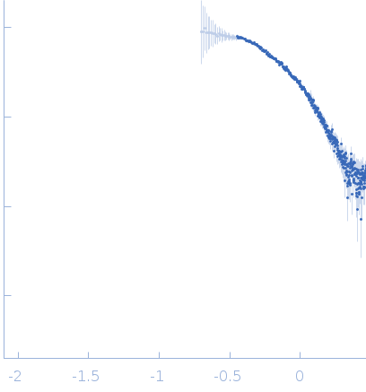
  "intensities_log_plot": "https://www.sasbdb.org/media/intensities_files/scattering_plots/SASDBG6_dat_img.png",
  "intensities_kratky_plot": "https://www.sasbdb.org/media/intensities_files/scattering_plots/SASDBG6_kratky_img.png",
  "pddf_plot": "https://www.sasbdb.org/media/p_of_R_files/pofr_images/SASDBG6_pofr_img.png",
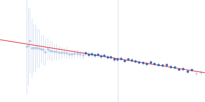
  "intensities_guinier_plot": "https://www.sasbdb.org/media/intensities_files/scattering_plots/SASDBG6_guinier_img.png",
  "sascif_data": "https://www.sasbdb.org/media/sascif/sascif_files/SASDBG6.sascif",
  "experiment": {
    "instrument": {
      "detector": {
        "type": null,
        "name": "MAR 165 CCD",
        "resolution": null
      },
      "name": "Advanced Light Source (ALS)",
      "city": "Berkeley, CA",
      "country": "USA",
      "beamline_name": "12.3.1 (SIBYLS)",
      "beam_geometry": "",
      "type_of_source": "X-ray synchrotron",
      "point_source": null,
      "line_collimation": null,
      "sample_path_length": null,
      "line_collimation_slitlength": null,
      "line_collimation_integrationwidth": null,
      "xray_energy": null,
      "beam_profile_ah": null,
      "beam_profile_al": null
    },
    "sample": {
      "molecule": [
        {
          "long_name": "Ribosome biogenesis protein 15",
          "short_name": "Nop15",
          "sequence": "KDKKTLEEYS GIIYVSRLPH GFHEKELSKY FAQFGDLKEV RLARNKKTGN SRHYGFLEFV NKEDAMIAQE \r\nSMNNYLLMGH LLQVRVLPKG AKIEKLYKYK KRVLVEKGIT KPVKQLKDNMKQKHEERIKK LAKSGIEFKW",
          "organism": "Saccharomyces cerevisiae",
          "uniprot_code": "P53927",
          "uniprot_range_first": 81,
          "uniprot_range_last": 220,
          "oligomerization": "monomer",
          "molecular_type": "protein",
          "uniprot_sequence": "MVKSTSKTSTKETVTKQPTEEKPIQEKEELALETSSSSSDEEDEKDEDEIEGLAASDDEQ\nSGTHKIKRLNPKKQANEKKSKDKKTLEEYSGIIYVSRLPHGFHEKELSKYFAQFGDLKEV\nRLARNKKTGNSRHYGFLEFVNKEDAMIAQESMNNYLLMGHLLQVRVLPKGAKIEKLYKYK\nKRVLVEKGITKPVKQLKDNMKQKHEERIKKLAKSGIEFKW",
          "mw": 16.541,
          "total_mw": 16.541,
          "number_molecules": 1,
          "complex_state": false,
          "deuteration": null,
          "molecule_source": "biological",
          "molecule_description": null
        }
      ],
      "buffer": {
        "name": "25 mM HEPES, 500 mM NaCl, 2 mM DTT",
        "concentration_unit": null,
        "comment": null,
        "additive": null,
        "concentration": null,
        "pka": null,
        "ph": 7.5,
        "deuteration": null
      },
      "purity_method": null,
      "name": "Ribosome biogenesis protein 15 (Nop15)",
      "ext_coefficient": null,
      "contrast": null,
      "specific_vol": null,
      "dry_vol": null,
      "absorbption": null,
      "deuteration": null,
      "mixture": null
    },
    "contributor": [
      {
        "affiliation": [
          {
            "short_name": "NIH",
            "address": "Bethesda, Maryland, USA",
            "full_name": "National Institutes of Health, Bethesda, MD",
            "webpage": "https://www.nih.gov/"
          }
        ],
        "contributor_name": "Jun",
        "contributor_surname": "Zhang",
        "orcid": null
      }
    ],
    "concentration_method": null,
    "concentration_unit": null,
    "date": "2013-04-25",
    "storage_temperature": 4.0,
    "cell_temperature": 4.0,
    "exposure_time": 0.5,
    "number_of_frames": 1,
    "wavelength": 0.1,
    "sample_detector_distance": null,
    "concentration_min": 0.7,
    "concentration_max": 3.0,
    "sample_volume": null,
    "flow_rate": null,
    "s_min": 0.198,
    "s_max": 2.934,
    "total_exposure_time": null,
    "seccolumn": null
  },
  "fits": [
    {
      "models": [
        {
          "model_plot": "https://www.sasbdb.org/media/pdb_file/images/SASDBG6_fit1_model1_img.png",
          "software": "EOM/RANCH",
          "pdb_link": [
            {
              "pdb_code": "5t9p",
              "ext_code": "5T9P",
              "ext_type": "pdb",
              "difference_with_model": "other"
            }
          ],
          "model_title": null,
          "type_of_model": "mix",
          "software_version": "",
          "model_data": "https://www.sasbdb.org/media/pdb_file/SASDBG6_fit1_model1.pdb",
          "model_mw": 15.99,
          "bead_radius": 1.9,
          "log": "https://www.sasbdb.org/media/log_files/logFile_001_1.log",
          "symmetry": "",
          "comment": "",
          "user": 157
        },
        {
          "model_plot": "https://www.sasbdb.org/media/pdb_file/images/SASDBG6_fit1_model2_img.png",
          "software": "EOM/RANCH",
          "pdb_link": [],
          "model_title": null,
          "type_of_model": "mix",
          "software_version": "",
          "model_data": "https://www.sasbdb.org/media/pdb_file/SASDBG6_fit1_model2.pdb",
          "model_mw": 15.99,
          "bead_radius": 1.9,
          "log": "https://www.sasbdb.org/media/log_files/logFile_001_1_D8AznaA.log",
          "symmetry": "",
          "comment": "",
          "user": 157
        },
        {
          "model_plot": "https://www.sasbdb.org/media/pdb_file/images/SASDBG6_fit1_model3_img.png",
          "software": "EOM/RANCH",
          "pdb_link": [],
          "model_title": null,
          "type_of_model": "mix",
          "software_version": "",
          "model_data": "https://www.sasbdb.org/media/pdb_file/SASDBG6_fit1_model3.pdb",
          "model_mw": 15.99,
          "bead_radius": 1.9,
          "log": "https://www.sasbdb.org/media/log_files/logFile_001_1_6M5amPg.log",
          "symmetry": "",
          "comment": "",
          "user": 157
        }
      ],
      "fit_unit": "1/A",
      "fit_plot": "https://www.sasbdb.org/media/fitting_files/scattering_plots/SASDBG6_fit1_fit_img.png",
      "software": null,
      "chi_square_value": 0.649,
      "p_value": 0.0,
      "fit_residual_plot": "fit4columns497_fitresiduals_img.png",
      "fit_data": "https://www.sasbdb.org/media/fitting_files/SASDBG6_fit1.fit",
      "fit_log": null,
      "software_version": null,
      "description": null
    }
  ],
  "estimated_volume_method": null,
  "pddf_software": "ATSAS GNOM",
  "pddf_software_version": "GNOM 5",
  "i0_calibration_standard": null,
  "description": null,
  "experiment_description": "Synchrotron SAXS data from solutions of ribosome biogenesis protein 15 (Nop15) in 25 mM HEPES, 500 mM NaCl, 2 mM DTT, pH 7.5 were collected on the BL 12.3.1 (SIBYLS) beam line at the Advanced Light Source (ALS, Berkeley, CA, USA) using a MAR Image Plate detector at a wavelength of λ = 0.1 nm (I(s) vs s, where s = 4π sin θ/λ, and 2θ is the scattering angle). Solute concentrations ranging between 0.7 and 3 mg/ml were measured at 4 °C (the data displayed in this entry: 0.7 mg/ml). One 0.5 second frame was collected per sample. The data were normalized to the intensity of the transmitted beam, radially averaged and the scattering of the solvent-blank was subtracted.",
  "tags": [],
  "intensity_unit": null,
  "experimental_mw": 16.4,
  "experimental_mw_error": null,
  "guinier_i0_mw": null,
  "guinier_i0_mw_error": null,
  "porod_mw": 23.0,
  "porod_mw_error": null,
  "pddf_i0": 125.6,
  "pddf_i0_error": 3.5,
  "guinier_i0": 113.76,
  "guinier_i0_error": 3.6,
  "pddf_rg": 2.86,
  "pddf_rg_error": 0.09,
  "guinier_rg": 2.4,
  "guinier_rg_error": 0.09,
  "pddf_dmax": 10.3,
  "pddf_dmax_error": null,
  "porod_volume": 38.0,
  "porod_volume_error": null,
  "estimated_volume": null,
  "estimated_volume_error": null,
  "guinier_point_first": 27,
  "guinier_point_last": 56,
  "pddf_point_first": null,
  "pddf_point_last": null,
  "i0_calibration_standard_data": null,
  "intensities_log_log_plot": "SASDBG6_datloglog_img.png",
  "symmetry": null,
  "last_modified": "2019-12-06T13:07:15.624532+01:00",
  "bragg_peak": []
}