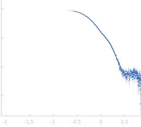
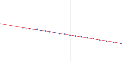
2. Measurements Reported by SASBDB Reported Results
Metric
Method
Value
Uncertainty
Unit
dmax
P(r)
11.2
—
Å
i0
Guinier
0.023
0.0002
—
i0
P(r)
0.0237
0.0002
—
mw
Experimental
29.9
—
kDa
mw
Guinier I(0)
29.9
—
kDa
mw
Porod
29.8
—
kDa
porod_volume
Porod
38.0
—
ų
rg
Guinier
2.73
0.03
Å
rg
P(r)
2.92
0.04
Å
These values are reported by SASBDB; SAXSdb did not recalculate them from the experimental curve.
▼
3. Source Fits and Models Source Fits & Models
This entry has no source fit record.
▶
4. Source File Index Source Files
Category
Path
Download status
Size
curve
sasbdb/entries/f3/sasdcf3/source/SASDCF3.dat
downloaded
23850
full_entry_zip
sasbdb/entries/f3/sasdcf3/source/SASDCF3.zip
downloaded
15878
pddf
sasbdb/entries/f3/sasdcf3/source/SASDCF3.out
downloaded
21686
sascif
sasbdb/entries/f3/sasdcf3/source/SASDCF3.sascif
downloaded
47064
summary
sasbdb/entries/f3/sasdcf3/source/summary.json
downloaded
6721
▶
5. Experiment Description and Publication Experiment & Publication
Original Experiment Description
SAXS data from solutions of Nucleolysin TIA-1 isoform p40 in 10 mM potassium phosphate, 50 mM NaCl, 10 mM DTT, pH 6 were were collected using a Rigaku BioSAXS-1000 instrument (Munich, Germany) equipped with a Pilatus 100K detector at a sample-detector distance of 0.5 m and at a wavelength of λ = 0.1542 nm (I(s) vs s, where s = 4πsinθ/λ, and 2θ is the scattering angle). Solute concentrations ranging between 2.5 and 5 mg/ml were measured at 20°C. Eight successive 7200 second frames were collected. The data were normalized to the intensity of the transmitted beam and radially averaged; the scattering of the solvent-blank was subtracted and the different curves were scaled for protein concentration.
Related Publication
Title
Segmental, Domain-Selective Perdeuteration and Small-Angle Neutron Scattering for Structural Analysis of Multi-Domain Proteins.
Authors
Sonntag M, Jagtap PKA, Simon B, Appavou MS, Geerlof A, Stehle R, Gabel F, Hennig J, Sattler M
DOI / PMID
10.1002/anie.201702904 / 28636238
▼
6. Complete Source Record Complete Source Record
The following content comes directly from the SASBDB entry. Missing values are shown as “—”; SAXSdb does not infer undeclared units.
SAXS data from solutions of Nucleolysin TIA-1 isoform p40 in 10 mM potassium phosphate, 50 mM NaCl, 10 mM DTT, pH 6 were were collected using a Rigaku BioSAXS-1000 instrument (Munich, Germany) equipped with a Pilatus 100K detector at a sample-detector distance of 0.5 m and at a wavelength of λ = 0.1542 nm (I(s) vs s, where s = 4πsinθ/λ, and 2θ is the scattering angle). Solute concentrations ranging between 2.5 and 5 mg/ml were measured at 20°C. Eight successive 7200 second frames were collected. The data were normalized to the intensity of the transmitted beam and radially averaged; the scattering of the solvent-blank was subtracted and the different curves were scaled for protein concentration.