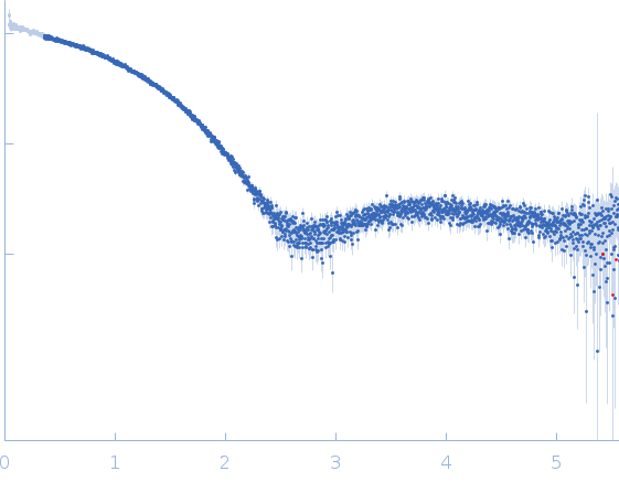
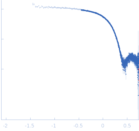
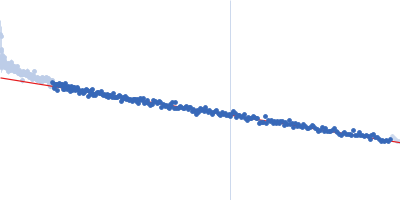
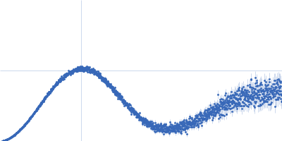
Synchrotron SAXS data from solutions of cytochrome C in 50 mM Tris, pH 7.4 were collected at the EMBL-P12 beam line at the PETRA III storage ring (Hamburg, Germany) using an Eiger 4M detector at a sample-detector distance of 1.6 m and at a wavelength of λ = 0.124 nm (I(s) vs s, where s = 4πsinθ/λ and 2θ is the scattering angle). One solute concentration of 2.00 mg/ml was measured at 20°C. A single 1.3 millisecond frame was collected. The data were normalized to the intensity of the transmitted beam and radially averaged; the scattering of the solvent-blank was subtracted and the data were scaled to the protein concentration.
European Molecular Biology Laboratory (EMBL) - Hamburg outstation
experiment.contributor[0].affiliation[0].webpage
http://www.embl-hamburg.de/index.php
experiment.contributor[0].contributor_name
Clement
experiment.contributor[0].contributor_surname
Blanchet
experiment.contributor[0].orcid
null
experiment.concentration_method
null
experiment.concentration_unit
null
experiment.date
2016-12-21
experiment.storage_temperature
20.0
experiment.cell_temperature
20.0
experiment.exposure_time
0.001
experiment.number_of_frames
1
experiment.wavelength
0.124
experiment.sample_detector_distance
1.6
experiment.concentration_min
null
experiment.concentration_max
2.0
experiment.sample_volume
null
experiment.flow_rate
null
experiment.s_min
0.035
experiment.s_max
5.57
experiment.total_exposure_time
null
experiment.seccolumn
null
fits
[]
estimated_volume_method
null
pddf_software
null
pddf_software_version
null
i0_calibration_standard
null
description
null
experiment_description
Synchrotron SAXS data from solutions of cytochrome C in 50 mM Tris, pH 7.4 were collected at the EMBL-P12 beam line at the PETRA III storage ring (Hamburg, Germany) using an Eiger 4M detector at a sample-detector distance of 1.6 m and at a wavelength of λ = 0.124 nm (I(s) vs s, where s = 4πsinθ/λ and 2θ is the scattering angle). One solute concentration of 2.00 mg/ml was measured at 20°C. A single 1.3 millisecond frame was collected. The data were normalized to the intensity of the transmitted beam and radially averaged; the scattering of the solvent-blank was subtracted and the data were scaled to the protein concentration.