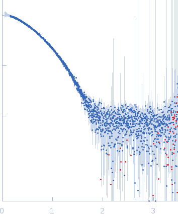
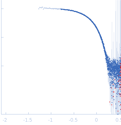
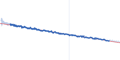
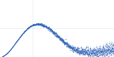
Candida antarctica lipase B - with guanidine-HCl unfolding series, in the presence of dithiothreitol
Data type: SASBDB experimental dataStatus: PublishedCurve type: Single concentrationLast updated: 2023-06-21T07:56:39.492710+02:00
In addition to the SAXS profile displayed above, two lipase B unfolding series in the presence of DTT spanning 0 M to 6 M guanidine hydrochloride are included in the full entry zip archive.
▼
1. Samples, Components and Experimental Conditions Sample & Experiment
Sample 1 · Candida antarctica lipase B - with guanidine-HCl unfolding series, in the presence of dithiothreitol
2. Measurements Reported by SASBDB Reported Results
Metric
Method
Value
Uncertainty
Unit
i0
Guinier
1269.62
2.25
arbitrary
mw
Experimental
35.1
—
kDa
mw
Guinier I(0)
35.1
—
kDa
rg
Guinier
2.36
0.1
nm
These values are reported by SASBDB; SAXSdb did not recalculate them from the experimental curve.
▼
3. Source Fits and Models Source Fits & Models
This entry has no source fit record.
▶
4. Source File Index Source Files
Category
Path
Download status
Size
curve
sasbdb/entries/k3/sasddk3/source/SASDDK3.dat
downloaded
79629
full_entry_zip
sasbdb/entries/k3/sasddk3/source/SASDDK3.zip
downloaded
504204
pddf
sasbdb/entries/k3/sasddk3/source/SASDDK3.out
not_listed
—
sascif
sasbdb/entries/k3/sasddk3/source/SASDDK3.sascif
downloaded
99077
summary
sasbdb/entries/k3/sasddk3/source/summary.json
downloaded
6769
▶
5. Experiment Description and Publication Experiment & Publication
Original Experiment Description
Synchrotron SAXS data from solutions of Candida antarctica lipase B - with a guanidine-HCl unfolding series under reducing conditions - in 100 mM NaCl, 20 mM Na2HPO4, 10 mM DTT, pH 6, were collected on the EMBL-P12 beam line at the PETRA III storage ring (DESY, Hamburg, Germany) using a Pilatus 2M detector at a sample-detector distance of 3.1 m and at a wavelength of λ = 0.124 nm (l(s) vs s, where s = 4πsinθ/λ, and 2θ is the scattering angle). One solute concentration of 4.65 mg/ml was measured at 10°C. 29 successive 0.030 second frames were collected. The data were normalized to the intensity of the transmitted beam and radially averaged; the scattering of the solvent-blank was subtracted.
Related Publication
Title
Machine Learning Methods for X-Ray Scattering Data Analysis from Biomacromolecular Solutions.
Authors
Franke D, Jeffries CM, Svergun DI
DOI / PMID
10.1016/j.bpj.2018.04.018 / 29874600
▼
6. Complete Source Record Complete Source Record
The following content comes directly from the SASBDB entry. Missing values are shown as “—”; SAXSdb does not infer undeclared units.
European Molecular Biology Laboratory (EMBL) - Hamburg outstation
experiment.contributor[0].affiliation[0].webpage
http://www.embl-hamburg.de/index.php
experiment.contributor[0].contributor_name
Cy M
experiment.contributor[0].contributor_surname
Jeffries
experiment.contributor[0].orcid
null
experiment.concentration_method
null
experiment.concentration_unit
null
experiment.date
2013-07-29
experiment.storage_temperature
10.0
experiment.cell_temperature
10.0
experiment.exposure_time
0.03
experiment.number_of_frames
29
experiment.wavelength
0.124
experiment.sample_detector_distance
3.1
experiment.concentration_min
null
experiment.concentration_max
4.65
experiment.sample_volume
null
experiment.flow_rate
null
experiment.s_min
0.054
experiment.s_max
3.491
experiment.total_exposure_time
null
experiment.seccolumn
null
fits
[]
estimated_volume_method
null
pddf_software
null
pddf_software_version
null
i0_calibration_standard
null
description
In addition to the SAXS profile displayed above, two lipase B unfolding series in the presence of DTT spanning 0 M to 6 M guanidine hydrochloride are included in the full entry zip archive.
experiment_description
Synchrotron SAXS data from solutions of Candida antarctica lipase B - with a guanidine-HCl unfolding series under reducing conditions - in 100 mM NaCl, 20 mM Na2HPO4, 10 mM DTT, pH 6, were collected on the EMBL-P12 beam line at the PETRA III storage ring (DESY, Hamburg, Germany) using a Pilatus 2M detector at a sample-detector distance of 3.1 m and at a wavelength of λ = 0.124 nm (l(s) vs s, where s = 4πsinθ/λ, and 2θ is the scattering angle). One solute concentration of 4.65 mg/ml was measured at 10°C. 29 successive 0.030 second frames were collected. The data were normalized to the intensity of the transmitted beam and radially averaged; the scattering of the solvent-blank was subtracted.