2. Measurements Reported by SASBDB Reported Results
Metric
Method
Value
Uncertainty
Unit
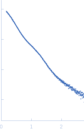
dmax
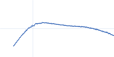
P(r)
15.9
—
Å
i0
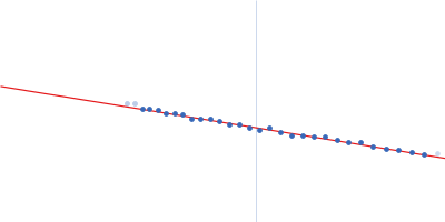
Guinier
9.11
0.055
arbitrary
i0
P(r)
8.819
—
arbitrary
mw
Experimental
109.0
—
kDa
porod_volume
Porod
132.0
—
ų
rg
Guinier
4.4
0.07
Å
rg
P(r)
4.37
—
Å
These values are reported by SASBDB; SAXSdb did not recalculate them from the experimental curve.
▼
3. Source Fits and Models Source Fits & Models
This entry has no source fit record.
▶
4. Source File Index Source Files
Category
Path
Download status
Size
curve
sasbdb/entries/w9/sasddw9/source/SASDDW9.dat
downloaded
17439
full_entry_zip
sasbdb/entries/w9/sasddw9/source/SASDDW9.zip
downloaded
17782
pddf
sasbdb/entries/w9/sasddw9/source/SASDDW9.out
downloaded
45862
sascif
sasbdb/entries/w9/sasddw9/source/SASDDW9.sascif
downloaded
64236
summary
sasbdb/entries/w9/sasddw9/source/summary.json
downloaded
7387
▶
5. Experiment Description and Publication Experiment & Publication
Original Experiment Description
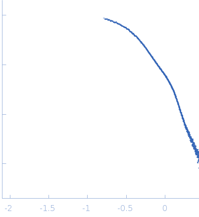
Synchrotron SAXS measurements (l(s) vs s, where s = 4πsinθ/λ, and 2θ is the scattering angle; λ = 0.11 nm) were carried out at the beam line X12SA of the Swiss Light Source (Paul Scherrer Institut, Villigen, Switzerland). Quartz capillaries (inner diameter 1 mm) were mounted in a sample holder and filled with buffer (20 mM HEPES, 150 mM NaCl, 5 mM MgCl2, 5 % w/v glycerol, pH 7.5). The sample holder was cooled to 10 °C for all measurements. Scattering of the buffer was determined at 20 positions along the capillary (spacing 0.5 mm), exposing each position with 11.2 keV photons for 0.5 s using 200 successive frames. Each position was measured 10 times and scattering intensities, after radial averaging to produce 1D-scattering patterns, were averaged. Scattered X-rays were detected by a Pilatus 2M detector (Dectris, Baden-Dättwil, Switzerland) at the end of an evacuated flight tube, 2.13m from the sample position. Buffer was replaced by the protein sample without moving the sample holder or the capillaries. Lights in the experimental hutch were switched off and after 5 minutes, scattering of the protein-containing solutions was measured at the same positions using the procedure described above. Due to poor behaviour of this sample, molecular weight and Rg estimates from the Guinier region need to be treated with caution. This sample is mostly provided to illustrate the differences that occur upon nucleotide binding of mPAC. Solute concentrations ranging between 4.7 and 7.5 mg/ml were measured and the concentration series data extrapolated to infinite dilution generating the final SAXS profile displayed in this entry.
▼
6. Complete Source Record Complete Source Record
The following content comes directly from the SASBDB entry. Missing values are shown as “—”; SAXSdb does not infer undeclared units.
Synchrotron SAXS measurements (l(s) vs s, where s = 4πsinθ/λ, and 2θ is the scattering angle; λ = 0.11 nm) were carried out at the beam line X12SA of the Swiss Light Source (Paul Scherrer Institut, Villigen, Switzerland). Quartz capillaries (inner diameter 1 mm) were mounted in a sample holder and filled with buffer (20 mM HEPES, 150 mM NaCl, 5 mM MgCl2, 5 % w/v glycerol, pH 7.5). The sample holder was cooled to 10 °C for all measurements. Scattering of the buffer was determined at 20 positions along the capillary (spacing 0.5 mm), exposing each position with 11.2 keV photons for 0.5 s using 200 successive frames. Each position was measured 10 times and scattering intensities, after radial averaging to produce 1D-scattering patterns, were averaged. Scattered X-rays were detected by a Pilatus 2M detector (Dectris, Baden-Dättwil, Switzerland) at the end of an evacuated flight tube, 2.13m from the sample position. Buffer was replaced by the protein sample without moving the sample holder or the capillaries. Lights in the experimental hutch were switched off and after 5 minutes, scattering of the protein-containing solutions was measured at the same positions using the procedure described above. Due to poor behaviour of this sample, molecular weight and Rg estimates from the Guinier region need to be treated with caution. This sample is mostly provided to illustrate the differences that occur upon nucleotide binding of mPAC. Solute concentrations ranging between 4.7 and 7.5 mg/ml were measured and the concentration series data extrapolated to infinite dilution generating the final SAXS profile displayed in this entry.
{
  "code": "SASDDW9",
  "status": "Published",
  "type_of_curve": "Merged",
  "angular_unit": "1/A",
  "project": {
    "title": "mPAC Delta132",
    "publication": null,
    "status": "released",
    "submitted_date": "2018-07-25",
    "released_date": "2021-09-20"
  },
  "pddf_data": "https://www.sasbdb.org/media/p_of_R_files/SASDDW9.out",
  "intensities_data": "https://www.sasbdb.org/media/intensities_files/SASDDW9.dat",
  "intensities_log_plot": "https://www.sasbdb.org/media/intensities_files/scattering_plots/SASDDW9_dat_img.png",
  "intensities_kratky_plot": "https://www.sasbdb.org/media/intensities_files/scattering_plots/SASDDW9_kratky_img.png",
  "pddf_plot": "https://www.sasbdb.org/media/p_of_R_files/pofr_images/SASDDW9_pofr_img.png",
  "intensities_guinier_plot": "https://www.sasbdb.org/media/intensities_files/scattering_plots/SASDDW9_guinier_img.png",
  "sascif_data": "https://www.sasbdb.org/media/sascif/sascif_files/SASDDW9.sascif",
  "experiment": {
    "instrument": {
      "detector": {
        "type": null,
        "name": "Pilatus 2M",
        "resolution": null
      },
      "name": "Swiss Light Source",
      "city": "Villigen",
      "country": "Switzerland",
      "beamline_name": "cSAXS",
      "beam_geometry": null,
      "type_of_source": "X-ray synchrotron",
      "point_source": null,
      "line_collimation": null,
      "sample_path_length": null,
      "line_collimation_slitlength": null,
      "line_collimation_integrationwidth": null,
      "xray_energy": null,
      "beam_profile_ah": null,
      "beam_profile_al": null
    },
    "sample": {
      "molecule": [
        {
          "long_name": "PAS fold family",
          "short_name": "mPAC",
          "sequence": "GAMGSQAYRKGVQETVKLRDQAIAASSVGIVIADARLPDMPLIYVNPAFEEITGYSDAEVLGYNCRFLQGKDTSQPAVDQLRAAIKAGENCTVTLLNYRKDGTPFWNELTISPIYDDHNNLTHFVGIQSDISDRIKAEQALRLEQEKSERLLLNILPKPIVDQLKQFEGSLAQQFTEATILFADIVGFTQLSAQMSPLELLNLLNNIFSVFDKLAEKHGIEKIKTIGDAYMAVAGLPVANDHHAEAIANMALDMQQAIQQFKTPQGEPFQIRIGINTGLVVAGVIGIKKFSYDLWGDAVNVASRMESSGLPGKIQVTAATKERLQDKYVFEKRGAIVVKGKGEMINYWLVGKQE",
          "organism": "Coleofasciculus chthonoplastes PCC 7420",
          "uniprot_code": "B4VKN6",
          "uniprot_range_first": 133,
          "uniprot_range_last": 483,
          "oligomerization": "dimer",
          "molecular_type": "protein",
          "uniprot_sequence": "MNPSCEENEPNLFHGEASLQAFFDLSAELLCIRDSNGYFREINSVWEKTLGWTLDELRSR\nPWLEFVHPDDVAFTFDMENQCHTLQDNKTPICLKNRFRCRDGSYRWLSWRLGAYQNSVSH\nGIAHDVTESNWRGSQAYRKGVQETVKLRDQAIAASSVGIVIADARLPDMPLIYVNPAFEE\nITGYSDAEVLGYNCRFLQGKDTSQPAVDQLRAAIKAGENCTVTLLNYRKDGTPFWNELTI\nSPIYDDHNNLTHFVGIQSDISDRIKAEQALRLEQEKSERLLLNILPKPIVDQLKQFEGSL\nAQQFTEATILFADIVGFTQLSAQMSPLELLNLLNNIFSVFDKLAEKHGIEKIKTIGDAYM\nAVAGLPVANDHHAEAIANMALDMQQAIQQFKTPQGEPFQIRIGINTGLVVAGVIGIKKFS\nYDLWGDAVNVASRMESSGLPGKIQVTAATKERLQDKYVFEKRGAIVVKGKGEMINYWLVG\nKQE",
          "mw": 39.015,
          "total_mw": 78.03,
          "number_molecules": 2,
          "complex_state": false,
          "deuteration": null,
          "molecule_source": "biological",
          "molecule_description": null
        }
      ],
      "buffer": {
        "name": "20 mM HEPES, 150 mM NaCl, 5 mM MgCl2, 5 % w/v Glycerol",
        "concentration_unit": null,
        "comment": null,
        "additive": null,
        "concentration": null,
        "pka": null,
        "ph": 7.5,
        "deuteration": null
      },
      "purity_method": null,
      "name": "PAS fold family protein mPAC-Δ132 in the dark state (mPAC-Δ132, dark, without adenosine-5'-[(α,β)-methyleno]triphosphate, ApCpp)",
      "ext_coefficient": null,
      "contrast": null,
      "specific_vol": null,
      "dry_vol": null,
      "absorbption": null,
      "deuteration": null,
      "mixture": null
    },
    "contributor": [
      {
        "affiliation": [
          {
            "short_name": null,
            "address": "Germany",
            "full_name": "Max Planck Institute for Medical Research",
            "webpage": "http://www.mpimf-heidelberg.mpg.de/en"
          }
        ],
        "contributor_name": "Robert",
        "contributor_surname": "Lindner",
        "orcid": ""
      }
    ],
    "concentration_method": null,
    "concentration_unit": null,
    "date": "2015-03-11",
    "storage_temperature": 0.0,
    "cell_temperature": 10.0,
    "exposure_time": 0.5,
    "number_of_frames": 200,
    "wavelength": 0.11,
    "sample_detector_distance": 2.13,
    "concentration_min": 4.7,
    "concentration_max": 7.5,
    "sample_volume": null,
    "flow_rate": null,
    "s_min": 0.16,
    "s_max": 2.744,
    "total_exposure_time": null,
    "seccolumn": null
  },
  "fits": [],
  "estimated_volume_method": null,
  "pddf_software": "ATSAS GNOM",
  "pddf_software_version": "5.0",
  "i0_calibration_standard": null,
  "description": null,
  "experiment_description": "Synchrotron SAXS measurements (l(s) vs s, where s = 4πsinθ/λ, and 2θ is the scattering angle;  λ = 0.11 nm) were carried out at the beam line X12SA of the Swiss Light Source (Paul Scherrer Institut, Villigen, Switzerland). Quartz capillaries (inner diameter 1 mm) were mounted in a sample holder and filled with buffer (20 mM HEPES, 150 mM NaCl, 5 mM MgCl2, 5 % w/v glycerol, pH 7.5). The sample holder was cooled to 10 °C for all measurements. Scattering of the buffer was determined at 20 positions along the capillary (spacing 0.5 mm), exposing each position with 11.2 keV photons for 0.5 s using 200 successive frames. Each position was measured 10 times and scattering intensities, after radial averaging to produce 1D-scattering patterns, were averaged. Scattered X-rays were detected by a Pilatus 2M detector (Dectris, Baden-Dättwil, Switzerland) at the end of an evacuated flight tube, 2.13m from the sample position. Buffer was replaced by the protein sample without moving the sample holder or the capillaries. Lights in the experimental hutch were switched off and after 5 minutes, scattering of the protein-containing solutions was measured at the same positions using the procedure described above. Due to poor behaviour of this sample, molecular weight and Rg estimates from the Guinier region need to be treated with caution. This sample is mostly provided to illustrate the differences that occur upon nucleotide binding of mPAC. Solute concentrations ranging between 4.7 and 7.5 mg/ml were measured and the concentration series data extrapolated to infinite dilution generating the final SAXS profile displayed in this entry.",
  "tags": [],
  "intensity_unit": "arbitrary",
  "experimental_mw": 109.0,
  "experimental_mw_error": null,
  "guinier_i0_mw": null,
  "guinier_i0_mw_error": null,
  "porod_mw": null,
  "porod_mw_error": null,
  "pddf_i0": 8.819,
  "pddf_i0_error": null,
  "guinier_i0": 9.11,
  "guinier_i0_error": 0.055,
  "pddf_rg": 4.37,
  "pddf_rg_error": null,
  "guinier_rg": 4.4,
  "guinier_rg_error": 0.07,
  "pddf_dmax": 15.9,
  "pddf_dmax_error": null,
  "porod_volume": 132.0,
  "porod_volume_error": null,
  "estimated_volume": null,
  "estimated_volume_error": null,
  "guinier_point_first": 3,
  "guinier_point_last": 30,
  "pddf_point_first": null,
  "pddf_point_last": null,
  "i0_calibration_standard_data": null,
  "intensities_log_log_plot": "SASDDW9_datloglog_img.png",
  "symmetry": null,
  "last_modified": "2021-09-20T13:38:19.015615+02:00",
  "bragg_peak": []
}