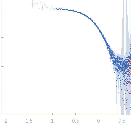
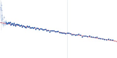
2. Measurements Reported by SASBDB Reported Results
Metric
Method
Value
Uncertainty
Unit
dmax
P(r)
9.5
—
nm
i0
Guinier
19.9306
—
arbitrary
i0
P(r)
20.05
—
arbitrary
mw
Experimental
23.6
—
kDa
mw
Porod
26.8
—
kDa
porod_volume
Porod
42.9
—
nm³
rg
Guinier
2.5
0.02
nm
rg
P(r)
2.592
—
nm
These values are reported by SASBDB; SAXSdb did not recalculate them from the experimental curve.
▼
3. Source Fits and Models Source Fits & Models
This entry has no source fit record.
▶
4. Source File Index Source Files
Category
Path
Download status
Size
curve
sasbdb/entries/x2/sasdux2/source/SASDUX2.dat
downloaded
57970
full_entry_zip
sasbdb/entries/x2/sasdux2/source/SASDUX2.zip
downloaded
33633
pddf
sasbdb/entries/x2/sasdux2/source/SASDUX2.out
downloaded
62564
sascif
sasbdb/entries/x2/sasdux2/source/SASDUX2.sascif
not_available
—
summary
sasbdb/entries/x2/sasdux2/source/summary.json
downloaded
6044
▶
5. Experiment Description and Publication Experiment & Publication
Original Experiment Description
Synchrotron SAXS data from solutions of cell division protein SepF (C-terminal domain) in 25 mM Tris, 100 mM NaCl, 10% glycerol, 1 mM EDTA, pH 8.5 were collected on the BM29 beam line at the ESRF (Grenoble, France) using a Pilatus3 2M detector at a sample-detector distance of 2.9 m and at a wavelength of λ = 0.1 nm (I(s) vs s, where s = 4πsinθ/λ, and 2θ is the scattering angle). One solute concentration of 3.00 mg/ml was measured at 20°C. 10 successive 0.500 second frames were collected. The data were normalized to the intensity of the transmitted beam and radially averaged; the scattering of the solvent-blank was subtracted.
▼
6. Complete Source Record Complete Source Record
The following content comes directly from the SASBDB entry. Missing values are shown as “—”; SAXSdb does not infer undeclared units.
Synchrotron SAXS data from solutions of cell division protein SepF (C-terminal domain) in 25 mM Tris, 100 mM NaCl, 10% glycerol, 1 mM EDTA, pH 8.5 were collected on the BM29 beam line at the ESRF (Grenoble, France) using a Pilatus3 2M detector at a sample-detector distance of 2.9 m and at a wavelength of λ = 0.1 nm (I(s) vs s, where s = 4πsinθ/λ, and 2θ is the scattering angle). One solute concentration of 3.00 mg/ml was measured at 20°C. 10 successive 0.500 second frames were collected. The data were normalized to the intensity of the transmitted beam and radially averaged; the scattering of the solvent-blank was subtracted.