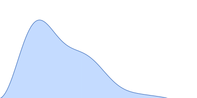
The following content comes directly from the SASBDB entry. Missing values are shown as “—”; SAXSdb does not infer undeclared units.
Every source field is expanded here, including empty values, list members and fields already shown above.
View the complete summary.json source
{
  "code": "SASDWP5",
  "status": "Published",
  "type_of_curve": "Single concentration",
  "angular_unit": "1/A",
  "project": {
    "title": "Dissecting the molecular basis underlying mycobacterial cell-wall hydrolysis by the catalytic domains of D29LysA and DS6ALysA phage endolysins.",
    "publication": {
      "title": "Dissecting the molecular basis underlying mycobacterial cell-wall hydrolysis by the catalytic domains of D29LysA and DS6ALysA phage endolysins.",
      "author_list": "Ceballos-Zúñiga F, Galvez-Larrosa L, Muñoz IG, Infantes L, Fernández-Carrillo J, Pérez-Dorado I",
      "journal": "Int J Biol Macromol",
      "doi": "10.1016/j.ijbiomac.2025.148896",
      "pmid": "41207583",
      "published_date": "2025 Nov 7"
    },
    "status": "released",
    "submitted_date": "2025-01-21",
    "released_date": "2025-11-10"
  },
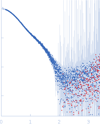
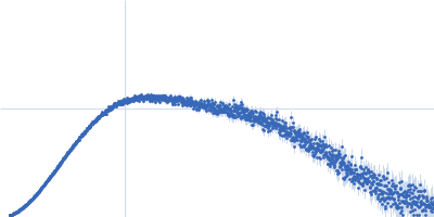
  "pddf_data": "https://www.sasbdb.org/media/p_of_R_files/SASDWP5.out",
  "intensities_data": "https://www.sasbdb.org/media/intensities_files/SASDWP5.dat",
  "intensities_log_plot": "https://www.sasbdb.org/media/intensities_files/scattering_plots/SASDWP5_dat_img.png",
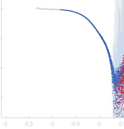
  "intensities_kratky_plot": "https://www.sasbdb.org/media/intensities_files/scattering_plots/SASDWP5_kratky_img.png",
  "pddf_plot": "https://www.sasbdb.org/media/p_of_R_files/pofr_images/SASDWP5_pofr_img.png",
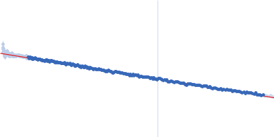
  "intensities_guinier_plot": "https://www.sasbdb.org/media/intensities_files/scattering_plots/SASDWP5_guinier_img.png",
  "sascif_data": "https://www.sasbdb.org/media/sascif/sascif_files/SASDWP5.sascif",
  "experiment": {
    "instrument": {
      "detector": {
        "type": null,
        "name": "Eiger 4M",
        "resolution": 75.0
      },
      "name": "Diamond Light Source",
      "city": "Didcot",
      "country": "UK",
      "beamline_name": "B21",
      "beam_geometry": null,
      "type_of_source": "X-ray synchrotron",
      "point_source": null,
      "line_collimation": null,
      "sample_path_length": null,
      "line_collimation_slitlength": null,
      "line_collimation_integrationwidth": null,
      "xray_energy": null,
      "beam_profile_ah": null,
      "beam_profile_al": null
    },
    "sample": {
      "molecule": [
        {
          "long_name": "N-acetylmuramoyl-L-alanine amidase (Mycobacteriophage DS6A)",
          "short_name": "DS6A with His-Tag",
          "sequence": ">DS6A-HisTag\r\n\r\nMHHHHHHSSGLEVLFQGP MMLTVESFAAAMGNSLSVDRYRQLFPAAVESMVACGCTTVNRAAMWLAQVGHESGGLRWM\r\nEELASGAAYEWRSDLGNTQAGDGVRFKGRGPIQITGRYNYRKVSEWAHAQGIVPTPTYFV\r\nDNPTQLASDQYGFIGVSWYWQHGGPRPGQINGFADAGDILSGSRCVNGWVTTPNGMPDRT\r\nERWNRCRAMGDQILPAGAGTDNGGDGMAWTGDPVWLEDVLRPVLGDRLRVLPSWQMYGHG\r\nDFKDIRGVMVHHTGNARETAESIRKGRPDLRGPLSNIHIAPDGTVTLVAAGVCWHAGAGS\r\nYPWLPTNNANWHMIGIECAWPTIRPNGTYDEREPWPDAQIIAMRDTCAALTKRLGWDASR\r\nVIGHKEYAGASQGKWDPGNLDMGWFRGEVAKAMRG",
          "organism": null,
          "uniprot_code": "G8I4E0",
          "uniprot_range_first": 1,
          "uniprot_range_last": 395,
          "oligomerization": "monomer",
          "molecular_type": "protein",
          "uniprot_sequence": "MMLTVESFAAAMGNSLSVDRYRQLFPAAVESMVACGCTTVNRAAMWLAQVGHESGGLRWM\nEELASGAAYEWRSDLGNTQAGDGVRFKGRGPIQITGRYNYRKVSEWAHAQGIVPTPTYFV\nDNPTQLASDQYGFIGVSWYWQHGGPRPGQINGFADAGDILSGSRCVNGWVTTPNGMPDRT\nERWNRCRAMGDQILPAGAGTDNGGDGMAWTGDPVWLEDVLRPVLGDRLRVLPSWQMYGHG\nDFKDIRGVMVHHTGNARETAESIRKGRPDLRGPLSNIHIAPDGTVTLVAAGVCWHAGAGS\nYPWLPTNNANWHMIGIECAWPTIRPNGTYDEREPWPDAQIIAMRDTCAALTKRLGWDASR\nVIGHKEYAGASQGKWDPGNLDMGWFRGEVAKAMRGEFDPKAPAPAPNPPADNDNGGFLMA\nLNDKQQHDLYNAVMTIAAVAADIQTQLRGPGLRGWPQLGRNANGEALTLVDAVGVIKAIA\nDREAGR",
          "mw": 45.374,
          "total_mw": 45.374,
          "number_molecules": 1,
          "complex_state": false,
          "deuteration": null,
          "molecule_source": "biological",
          "molecule_description": "Monomer state of a N-terminal and central domain of DS6ALysA with 6xHis tag"
        }
      ],
      "buffer": {
        "name": "20 mM Tris, 150 mM NaCl and 1 mM DTT",
        "concentration_unit": null,
        "comment": null,
        "additive": null,
        "concentration": null,
        "pka": null,
        "ph": 8.0,
        "deuteration": null
      },
      "purity_method": null,
      "name": "N-acetylmuramoyl-L-alanine amidase (with His-Tag)",
      "ext_coefficient": null,
      "contrast": null,
      "specific_vol": null,
      "dry_vol": null,
      "absorbption": null,
      "deuteration": null,
      "mixture": null
    },
    "contributor": [
      {
        "affiliation": [
          {
            "short_name": null,
            "address": null,
            "full_name": "Spanish National Cancer Research Centre (CNIO)",
            "webpage": null
          }
        ],
        "contributor_name": "INÉS",
        "contributor_surname": "MUÑOZ",
        "orcid": "https://orcid.org/0000-0001-6732-4059"
      }
    ],
    "concentration_method": null,
    "concentration_unit": null,
    "date": "2023-03-30",
    "storage_temperature": 4.0,
    "cell_temperature": 18.0,
    "exposure_time": 3.0,
    "number_of_frames": 600,
    "wavelength": 0.1,
    "sample_detector_distance": 3.7,
    "concentration_min": null,
    "concentration_max": 5.6,
    "sample_volume": null,
    "flow_rate": null,
    "s_min": 0.045,
    "s_max": 3.399,
    "total_exposure_time": null,
    "seccolumn": null
  },
  "fits": [
    {
      "models": [
        {
          "model_plot": "https://www.sasbdb.org/media/pdb_file/images/SASDWP5_fit1_model1_img.png",
          "software": "Coot",
          "pdb_link": [],
          "model_title": null,
          "type_of_model": "atomic",
          "software_version": null,
          "model_data": "https://www.sasbdb.org/media/pdb_file/SASDWP5_fit1_model1.pdb",
          "model_mw": 43.28,
          "bead_radius": null,
          "log": null,
          "symmetry": "P1",
          "comment": null,
          "user": 973
        }
      ],
      "fit_unit": "1/A",
      "fit_plot": "https://www.sasbdb.org/media/fitting_files/scattering_plots/SASDWP5_fit1_fixed_fit_img.png",
      "software": "FoXS",
      "chi_square_value": 1.242133,
      "p_value": 0.0,
      "fit_residual_plot": "SASDWP5_fit1_fitresiduals_img.png",
      "fit_data": "https://www.sasbdb.org/media/fitting_files/SASDWP5_fit1.fit",
      "fit_log": null,
      "software_version": null,
      "description": ""
    },
    {
      "models": [
        {
          "model_plot": "https://www.sasbdb.org/media/pdb_file/images/SASDWP5_fit2_model1_img.png",
          "software": "DAMMIN",
          "pdb_link": [],
          "model_title": null,
          "type_of_model": "dummy",
          "software_version": null,
          "model_data": "https://www.sasbdb.org/media/pdb_file/SASDWP5_fit2_model1.pdb",
          "model_mw": 39.668,
          "bead_radius": 2.5,
          "log": "https://www.sasbdb.org/media/log_files/1_QBz7Fdg.log",
          "symmetry": "P1",
          "comment": null,
          "user": 973
        }
      ],
      "fit_unit": "1/A",
      "fit_plot": "https://www.sasbdb.org/media/fitting_files/scattering_plots/SASDWP5_fit2_fixed_fit_img.png",
      "software": "DAMMIN",
      "chi_square_value": 0.663,
      "p_value": 0.318,
      "fit_residual_plot": "SASDWP5_fit2_fitresiduals_img.png",
      "fit_data": "https://www.sasbdb.org/media/fitting_files/SASDWP5_fit2.fir",
      "fit_log": "https://www.sasbdb.org/media/fitting_files/log_files/1_cGpEMdJ.log",
      "software_version": null,
      "description": ""
    }
  ],
  "estimated_volume_method": null,
  "pddf_software": "ATSAS GNOM",
  "pddf_software_version": "5.0",
  "i0_calibration_standard": null,
  "description": "SAXS experiments were performed at the beamline B21 of the Diamond Light Source (Harwell Campus, UK) (Cowieson et al, 2020). Samples of 45 µl of 6His-tagged and untagged DS6AGH19-Ami2B protein at 5.6 and 5.9 mg/ml were loaded into a Sodex KW-403 column and a SRT-C SEC-300 (Sepax) column, respectively, equilibrated in buffer (20 mM Tris pH 8.0, 150 mM NaCl and 1 mM DTT) and connected to an Agilent 1200 HPLC system at 18 °C. Continuously eluting samples were exposed for 3 s in 10 s acquisition blocks using an X-ray wavelength of 1 Å, and a sample to detector (Eiger 4M) distance of 3.7 m. The data covered a momentum transfer range of 0.0032 < q < 0.34 Å−1. The frames recorded immediately before elution of the sample were subtracted from the protein scattering profiles. The Scåtter software package (www.bioisis.net) was used to analyse data, buffer-subtraction, scaling, merging and checking possible radiation damage of the samples. The Rg value was calculated with the Guinier approximation assuming that at very small angles q < 1.3/Rg. The particle distance distribution, Dmax, was calculated from the scattering pattern with GNOM, and shape estimation was carried out with DAMMIF/DAMMIN, all these programs included in the ATSAS package. The proteins molecular mass was estimated with GNOM. Interactively generated PDB-based homology models were made using the program COOT by manually adjusting the X-ray structures obtained in this work, into the envelope given by SAXS until a good correlation between the real-space scattering profile calculated for the homology model matched the experimental scattering data. This was computed with the program FoXS.",
  "experiment_description": null,
  "tags": [],
  "intensity_unit": "1/cm",
  "experimental_mw": 44.07,
  "experimental_mw_error": null,
  "guinier_i0_mw": 47.1,
  "guinier_i0_mw_error": null,
  "porod_mw": 49.2,
  "porod_mw_error": null,
  "pddf_i0": 0.03397,
  "pddf_i0_error": null,
  "guinier_i0": 0.0337834,
  "guinier_i0_error": null,
  "pddf_rg": 2.92,
  "pddf_rg_error": 0.03,
  "guinier_rg": 2.843,
  "guinier_rg_error": 0.056,
  "pddf_dmax": 10.3,
  "pddf_dmax_error": null,
  "porod_volume": 62.0,
  "porod_volume_error": null,
  "estimated_volume": null,
  "estimated_volume_error": null,
  "guinier_point_first": 80,
  "guinier_point_last": 315,
  "pddf_point_first": null,
  "pddf_point_last": null,
  "i0_calibration_standard_data": null,
  "intensities_log_log_plot": "SASDWP5_datloglog_img.png",
  "symmetry": null,
  "last_modified": "2025-11-10T15:23:44.421893+01:00",
  "bragg_peak": []
}