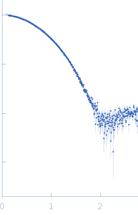
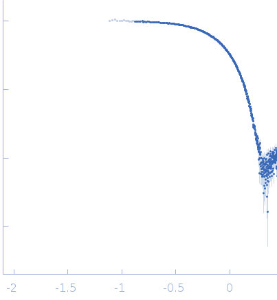
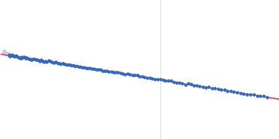
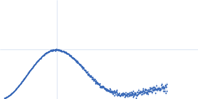
Synchrotron Size-exclusion coupled to Small angle X-ray scattering (SEC-SAXS) data from non-methylated Virulence factor family protein (VirJD1) from Brucella abortus in 50 mM Tris pH 8.0, 200 mM NaCl, 5% v/v glycerol were collected on the SWING beamline at the SOLEIL storage ring (Saint-Aubin, France) using an Eiger 4M detector at a sample-detector distance of 2 m and at a wavelength of λ = 0.1033 nm (I(s) vs s, where s = 4πsinθ/λ, and 2θ is the scattering angle). The SEC parameters were as follows: Fifty μl of protein at 15 mg/mL were loaded onto a size-exclusion column (S200 increase 5/150) at a 0.20 ml/min flow rate at 20°C. Frames were recorded every second for the duration of the size-exclusion run. Buffer subtraction was performed by averaging 100 buffer frames and 16 successive sample frames. The data were normalized to the intensity of the transmitted beam and radially averaged; the scattering of the solvent-blank was subtracted.
相关论文
标题
Crystal structure of the virulence protein J (VirJ) domain 1 from Brucella abortus.
VirJ domain 1 is the fist domain of the VirJ protein. VirJ is a periplasmic protein required for Virulence. It was found to interact with components of the VirB type IV secretion system. Its exact function is unknown. The protein construct contains an additional six non-native amino acids at the N-terminus (GIDPFT).
experiment.sample.buffer.name
50 mM Tris pH 8.0, 200 mM NaCl, 5% v/v glycerol
experiment.sample.buffer.concentration_unit
null
experiment.sample.buffer.comment
null
experiment.sample.buffer.additive
null
experiment.sample.buffer.concentration
null
experiment.sample.buffer.pka
null
experiment.sample.buffer.ph
null
experiment.sample.buffer.deuteration
null
experiment.sample.purity_method
null
experiment.sample.name
Virulence factor family protein (VirJD1) from Brucella abortus
Synchrotron Size-exclusion coupled to Small angle X-ray scattering (SEC-SAXS) data from non-methylated Virulence factor family protein (VirJD1) from Brucella abortus in 50 mM Tris pH 8.0, 200 mM NaCl, 5% v/v glycerol were collected on the SWING beamline at the SOLEIL storage ring (Saint-Aubin, France) using an Eiger 4M detector at a sample-detector distance of 2 m and at a wavelength of λ = 0.1033 nm (I(s) vs s, where s = 4πsinθ/λ, and 2θ is the scattering angle). The SEC parameters were as follows: Fifty μl of protein at 15 mg/mL were loaded onto a size-exclusion column (S200 increase 5/150) at a 0.20 ml/min flow rate at 20°C. Frames were recorded every second for the duration of the size-exclusion run. Buffer subtraction was performed by averaging 100 buffer frames and 16 successive sample frames. The data were normalized to the intensity of the transmitted beam and radially averaged; the scattering of the solvent-blank was subtracted.