查看完整 summary.json 原文
{
  "code": "SASDXV3",
  "status": "Published",
  "type_of_curve": "Single concentration",
  "angular_unit": "1/A",
  "project": {
    "title": "DNA induces non-canonical dimerization of the farnesoid X receptor.",
    "publication": {
      "title": "DNA induces non-canonical dimerization of the farnesoid X receptor.",
      "author_list": "Khan SH, Yennawar N, Okafor CD",
      "journal": "Nucleic Acids Res",
      "doi": "10.1093/nar/gkag087",
      "pmid": "41728955",
      "published_date": "2026 Feb 5"
    },
    "status": "released",
    "submitted_date": "2025-04-16",
    "released_date": "2026-02-26"
  },
  "pddf_data": "https://www.sasbdb.org/media/p_of_R_files/SASDXV3.out",
  "intensities_data": "https://www.sasbdb.org/media/intensities_files/SASDXV3.dat",
  "intensities_log_plot": "https://www.sasbdb.org/media/intensities_files/scattering_plots/SASDXV3_dat_img.png",
  "intensities_kratky_plot": "https://www.sasbdb.org/media/intensities_files/scattering_plots/SASDXV3_kratky_img.png",
  "pddf_plot": "https://www.sasbdb.org/media/p_of_R_files/pofr_images/SASDXV3_pofr_img.png",
  "intensities_guinier_plot": "https://www.sasbdb.org/media/intensities_files/scattering_plots/SASDXV3_guinier_img.png",
  "sascif_data": "https://www.sasbdb.org/media/sascif/sascif_files/SASDXV3.sascif",
  "experiment": {
    "instrument": {
      "detector": {
        "type": "Hybrid Photon Counting (HPC) X-ray detector",
        "name": "Rigaku HyPix-3000",
        "resolution": 100.0
      },
      "name": "Pennsylvania State University",
      "city": "University Park, PA",
      "country": "USA",
      "beamline_name": "Rigaku BioSAXS-2000",
      "beam_geometry": null,
      "type_of_source": "X-ray in house",
      "point_source": null,
      "line_collimation": null,
      "sample_path_length": null,
      "line_collimation_slitlength": null,
      "line_collimation_integrationwidth": null,
      "xray_energy": null,
      "beam_profile_ah": null,
      "beam_profile_al": null
    },
    "sample": {
      "molecule": [
        {
          "long_name": "inverted repeat DNA",
          "short_name": "IR1",
          "sequence": "CCGAGGTCAATGACCTCG\r\nCCGAGGTCATTGACCTCG",
          "organism": null,
          "uniprot_code": null,
          "uniprot_range_first": null,
          "uniprot_range_last": null,
          "oligomerization": "monomer",
          "molecular_type": "DNA",
          "uniprot_sequence": "",
          "mw": 11.102,
          "total_mw": 11.102,
          "number_molecules": 1,
          "complex_state": false,
          "deuteration": null,
          "molecule_source": "biological",
          "molecule_description": ""
        },
        {
          "long_name": "Isoform 3 of Bile acid receptor",
          "short_name": "FXR",
          "sequence": "KKPRMGASAGRIKGDELCVVCGDRASGYHYNALTCEGCKGFFRRSITKNAVYKCKNGGNCVMDMYMRRKCQECRLRKCKEMGMLAECMYTGLLTEIQCKSKRLRKNVKQHADQTVNEDSEGRDLRQVTSTTKSCREKTELTPDQQTLLHFIMDSYNKQRMPQEITNKILKEEFSAEENFLILTEMATNHVQVLVEFTKKLPGFQTLDHEDQIALLKGSAVEAMFLRSAEIFNKKLPSGHSDLLEERIRNSGISDEYITPMFSFYKSIGELKMTQEEYALLTAIVILSPDRQYIKDREAVEKLQEPLLDVLQKLCKIHQPENPQHFACLLGRLTELRTFNHHHAEMLMSWRVNDHKFT",
          "organism": "Homo sapiens",
          "uniprot_code": "Q96RI1",
          "uniprot_range_first": 110,
          "uniprot_range_last": 466,
          "oligomerization": "dimer",
          "molecular_type": "protein",
          "uniprot_sequence": "MGSKMNLIEHSHLPTTDEFSFSENLFGVLTEQVAGPLGQNLEVEPYSQYSNVQFPQVQPQ\nISSSSYYSNLGFYPQQPEEWYSPGIYELRRMPAETLYQGETEVAEMPVTKKPRMGASAGR\nIKGDELCVVCGDRASGYHYNALTCEGCKGFFRRSITKNAVYKCKNGGNCVMDMYMRRKCQ\nECRLRKCKEMGMLAECMYTGLLTEIQCKSKRLRKNVKQHADQTVNEDSEGRDLRQVTSTT\nKSCREKTELTPDQQTLLHFIMDSYNKQRMPQEITNKILKEEFSAEENFLILTEMATNHVQ\nVLVEFTKKLPGFQTLDHEDQIALLKGSAVEAMFLRSAEIFNKKLPSGHSDLLEERIRNSG\nISDEYITPMFSFYKSIGELKMTQEEYALLTAIVILSPDRQYIKDREAVEKLQEPLLDVLQ\nKLCKIHQPENPQHFACLLGRLTELRTFNHHHAEMLMSWRVNDHKFTPLLCEIWDVQ",
          "mw": 41.264,
          "total_mw": 82.528,
          "number_molecules": 2,
          "complex_state": false,
          "deuteration": null,
          "molecule_source": "biological",
          "molecule_description": ""
        }
      ],
      "buffer": {
        "name": "50 mM HEPES, 150 mM NaCl, 5 % glycerol, 1 mM DTT, 20 µM CDCA",
        "concentration_unit": null,
        "comment": null,
        "additive": null,
        "concentration": null,
        "pka": null,
        "ph": 7.4,
        "deuteration": null
      },
      "purity_method": null,
      "name": "Farnesoid X receptor homo-dimer bound to inverted repeat DNA",
      "ext_coefficient": null,
      "contrast": null,
      "specific_vol": null,
      "dry_vol": null,
      "absorbption": null,
      "deuteration": null,
      "mixture": null
    },
    "contributor": [
      {
        "affiliation": [
          {
            "short_name": "PSU",
            "address": "Pennsylvania, USA",
            "full_name": "Pennsylvania State University (PSU)",
            "webpage": "https://www.psu.edu/"
          }
        ],
        "contributor_name": "Neela",
        "contributor_surname": "Yennawar",
        "orcid": ""
      }
    ],
    "concentration_method": null,
    "concentration_unit": null,
    "date": "2024-05-09",
    "storage_temperature": 4.0,
    "cell_temperature": 4.0,
    "exposure_time": 600.0,
    "number_of_frames": 6,
    "wavelength": 0.154,
    "sample_detector_distance": 4.955,
    "concentration_min": null,
    "concentration_max": 0.6,
    "sample_volume": null,
    "flow_rate": null,
    "s_min": 0.034,
    "s_max": 6.106,
    "total_exposure_time": null,
    "seccolumn": null
  },
  "fits": [
    {
      "models": [
        {
          "model_plot": "https://www.sasbdb.org/media/pdb_file/images/SASDXV3_fit1_model1_img.png",
          "software": "AlphaFold",
          "pdb_link": [],
          "model_title": null,
          "type_of_model": "atomic",
          "software_version": null,
          "model_data": "https://www.sasbdb.org/media/pdb_file/SASDXV3_fit1_model1.pdb",
          "model_mw": 93.05,
          "bead_radius": null,
          "log": null,
          "symmetry": null,
          "comment": null,
          "user": 1001
        }
      ],
      "fit_unit": "1/A",
      "fit_plot": "https://www.sasbdb.org/media/fitting_files/scattering_plots/SASDXV3_fit1_fixed_fit_img.png",
      "software": "FoXS",
      "chi_square_value": 1.939,
      "p_value": 0.0001,
      "fit_residual_plot": "SASDXV3_fit1_fitresiduals_img.png",
      "fit_data": "https://www.sasbdb.org/media/fitting_files/SASDXV3_fit1.fit",
      "fit_log": null,
      "software_version": null,
      "description": ""
    }
  ],
  "estimated_volume_method": null,
  "pddf_software": "ATSAS GNOM",
  "pddf_software_version": null,
  "i0_calibration_standard": null,
  "description": "Chenodeoxycholic acid (CDCA): https://www.ebi.ac.uk/chembl/explore/compound/CHEMBL240597",
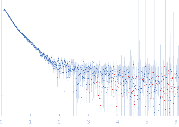
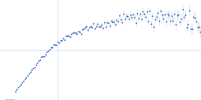
  "experiment_description": "SAXS data from solutions of farnesoid X receptor homo-dimer bound to inverted repeat DNA in 50 mM HEPES, 150 mM NaCl, 5 % glycerol, 1 mM DTT, 20 µM CDCA, pH 7.4 were collected on a Rigaku BioSAXS-2000 instrument (Pennsylvania State University, University Park, PA, USA) using a Rigaku HyPix-3000 detector at a sample-detector distance of 5.0 m and at a wavelength of λ = 0.154 nm (I(s) vs s, where s = 4πsinθ/λ, and 2θ is the scattering angle). One solute concentration of 0.60 mg/ml was measured at 4°C. Six successive 600 second frames were collected. The data were normalized to the intensity of the transmitted beam and radially averaged; the scattering of the solvent-blank was subtracted.",
  "tags": [],
  "intensity_unit": "arbitrary",
  "experimental_mw": 94.7,
  "experimental_mw_error": null,
  "guinier_i0_mw": null,
  "guinier_i0_mw_error": null,
  "porod_mw": null,
  "porod_mw_error": null,
  "pddf_i0": 6.051,
  "pddf_i0_error": null,
  "guinier_i0": 5.74754,
  "guinier_i0_error": null,
  "pddf_rg": 5.83,
  "pddf_rg_error": null,
  "guinier_rg": 5.05,
  "guinier_rg_error": 0.078,
  "pddf_dmax": 23.2,
  "pddf_dmax_error": null,
  "porod_volume": 120.0,
  "porod_volume_error": null,
  "estimated_volume": null,
  "estimated_volume_error": null,
  "guinier_point_first": 9,
  "guinier_point_last": 28,
  "pddf_point_first": null,
  "pddf_point_last": null,
  "i0_calibration_standard_data": null,
  "intensities_log_log_plot": "SASDXV3_datloglog_img.png",
  "symmetry": null,
  "last_modified": "2026-02-26T13:30:46.344061+01:00",
  "bragg_peak": []
}