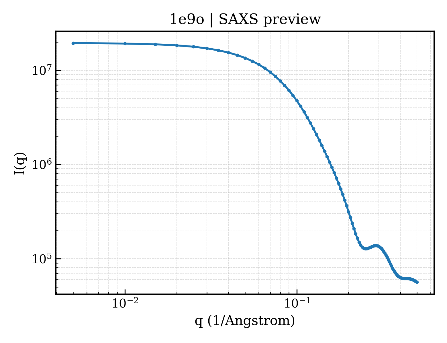
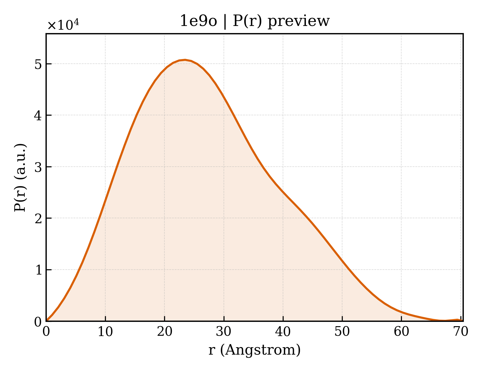
SUPEROXIDE DISMUTASE
OrganismNot specified
State in the Current Structure
| Assembly | Oligomeric State | Construct | Mutations and Modifications | Ligands, Ions and Associated Components | Method and Experimental Conditions | Structure Quality |
|---|---|---|---|---|---|---|
| 1 | Protein homooligomer Homooligomer Protein × 2 PDB declaration: dimeric(2) Consistent with protein copy count | Chain A; UniProt 1–151 Chain B; UniProt 1–151 | Not recorded | CU COPPER (II) ION × 4 | X-RAY DIFFRACTION X-ray crystallization conditions:pH 4;PEG-4000, NACL, pH 4.00 | Resolution 1.85 Å R-free 0.230 |
Other States of the Same Protein in the Database
Each row is a biological assembly of the same UniProt protein in another PDB entry. The “Difference from current entry” column identifies evidence-level differences; no tag means the currently parsed fields agree.
| Other PDB | Difference from Current Entry 1E9O | Assembly / Oligomeric State | Construct | Mutations and Modifications | Ligands, Ions and Non-polymers | Method and Experimental Conditions | Structure Quality |
|---|---|---|---|---|---|---|---|
| 1CB4 CRYSTAL STRUCTURE OF COPPER, ZINC SUPEROXIDE DISMUTASE Deposited 1999-02-26 | Different construct Different ligand/ion Different experimental conditions Different structure-quality metrics | Assembly 1 Protein homooligomer Homooligomer;Protein × 2 PDB declaration: dimeric |
Chain A
2–152(151 aa)
Chain B
2–152(151 aa)
|
Not recorded | CU COPPER (II) ION × 3 ZN ZINC ION × 1 |
X-RAY DIFFRACTION
X-ray crystallization conditions
pH 6;277 K;pH 6.00, temperature 277.00K
|
Resolution 2.30 Å R-free 0.228 |
| 1CBJ CRYSTAL STRUCTURE OF BOVINE SUPEROXIDE DISMUTASE CRYSTAL. Deposited 1999-02-26 | Different construct Different ligand/ion Different experimental conditions Different structure-quality metrics | Assembly 1 Protein homooligomer Homooligomer;Protein × 2 PDB declaration: dimeric |
Chain A
2–152(151 aa)
Chain B
2–152(151 aa)
|
Not recorded | CU COPPER (II) ION × 2 ZN ZINC ION × 2 |
X-RAY DIFFRACTION
X-ray crystallization conditions
pH 6.5;pH 6.50
|
Resolution 1.65 Å R-free 0.228 |
| 1COB CRYSTAL STRUCTURE SOLUTION AND REFINEMENT OF THE SEMISYNTHETIC COBALT SUBSTITUTED BOVINE ERYTHROCYTE ENZYME SUPEROXIDE DISMUTASE AT 2.0 ANGSTROMS RESOLUTION Deposited 1992-02-19 | Different ligand/ion Different experimental conditions Different structure-quality metrics | Assembly 1 Protein homooligomer Homooligomer;Protein × 2 PDB declaration: dimeric |
Chain A
1–151(151 aa)
Chain B
1–151(151 aa)
|
Not recorded | CU COPPER (II) ION × 2 CO COBALT (II) ION × 2 | X-RAY DIFFRACTION mmCIF provides none of the parsed conditions | Resolution 2.00 Å |
| 1E9P Crystal structure of bovine Cu, Zn SOD to 1.7 Angstrom (3 of 3) Deposited 2000-10-25 | Different ligand/ion Different experimental conditions Different structure-quality metrics | Assembly 1 Protein homooligomer Homooligomer;Protein × 2 PDB declaration: dimeric |
Chain A
1–151(151 aa)
Chain B
1–151(151 aa)
|
Not recorded | CU COPPER (II) ION × 3 ZN ZINC ION × 1 |
X-RAY DIFFRACTION
X-ray crystallization conditions
pH 6.5;pH 6.50
|
Resolution 1.70 Å R-free 0.202 |
| 1E9Q Crystal structure of bovine Cu Zn SOD - (1 of 3) Deposited 2000-10-26 | Different ligand/ion Different experimental conditions Different structure-quality metrics | Assembly 1 Protein homooligomer Homooligomer;Protein × 2 PDB declaration: dimeric |
Chain A
1–151(151 aa)
Chain B
1–151(151 aa)
|
Not recorded | CU COPPER (II) ION × 3 ZN ZINC ION × 1 |
X-RAY DIFFRACTION
X-ray crystallization conditions
pH 6.5;pH 6.50
|
Resolution 1.75 Å R-free 0.205 |
| 1Q0E Atomic resolution (1.15 ) crystal structure of bovine copper, zinc superoxide dismutase Deposited 2003-07-16 | Different construct Different mutation/modification Different oligomeric state Different ligand/ion Different experimental conditions Different structure-quality metrics | Assembly 1 Protein monomer Monomer;Protein × 1 PDB declaration: monomeric |
Chain A
1–151(151 aa)
Fragment:Copper, Zinc Superoxide Dismutase
|
Non-standard monomer:Yes (specific site not provided by mmCIF) | CU COPPER (II) ION × 1 ZN ZINC ION × 1 |
X-RAY DIFFRACTION
X-ray crystallization conditions
VAPOR DIFFUSION, SITTING DROP;pH 6.5;277 K;15 % PEG-4000, 50 mM glycyl-glycine , 100 mM NaCl, pH 6.5, VAPOR DIFFUSION, SITTING DROP, temperature 277K
|
Resolution 1.15 Å R-free 0.166 |
| 1Q0E Atomic resolution (1.15 ) crystal structure of bovine copper, zinc superoxide dismutase Deposited 2003-07-16 | Different construct Different mutation/modification Different oligomeric state Different ligand/ion Different experimental conditions Different structure-quality metrics | Assembly 2 Protein monomer Monomer;Protein × 1 PDB declaration: monomeric |
Chain B
1–151(151 aa)
Fragment:Copper, Zinc Superoxide Dismutase
|
Non-standard monomer:Yes (specific site not provided by mmCIF) | CU COPPER (II) ION × 1 ZN ZINC ION × 1 |
X-RAY DIFFRACTION
X-ray crystallization conditions
VAPOR DIFFUSION, SITTING DROP;pH 6.5;277 K;15 % PEG-4000, 50 mM glycyl-glycine , 100 mM NaCl, pH 6.5, VAPOR DIFFUSION, SITTING DROP, temperature 277K
|
Resolution 1.15 Å R-free 0.166 |
| 1SDA CRYSTAL STRUCTURE OF PEROXYNITRITE-MODIFIED BOVINE CU,ZN SUPEROXIDE DISMUTASE Deposited 1993-01-13 | Different mutation/modification Different ligand/ion Different experimental conditions Different structure-quality metrics | Assembly 1 Protein homooligomer Homooligomer;Protein × 2 PDB declaration: dimeric |
Chain O
1–151(151 aa)
Chain Y
1–151(151 aa)
|
Non-standard monomer:Yes (specific site not provided by mmCIF) Non-standard monomer:Yes (specific site not provided by mmCIF) | CU COPPER (II) ION × 2 ZN ZINC ION × 2 | X-RAY DIFFRACTION mmCIF provides none of the parsed conditions | Resolution 2.50 Å |
| 1SDA CRYSTAL STRUCTURE OF PEROXYNITRITE-MODIFIED BOVINE CU,ZN SUPEROXIDE DISMUTASE Deposited 1993-01-13 | Different mutation/modification Different ligand/ion Different experimental conditions Different structure-quality metrics | Assembly 2 Protein homooligomer Homooligomer;Protein × 2 PDB declaration: dimeric |
Chain B
1–151(151 aa)
Chain G
1–151(151 aa)
|
Non-standard monomer:Yes (specific site not provided by mmCIF) Non-standard monomer:Yes (specific site not provided by mmCIF) | CU COPPER (II) ION × 2 ZN ZINC ION × 2 | X-RAY DIFFRACTION mmCIF provides none of the parsed conditions | Resolution 2.50 Å |
| 1SXA CRYSTAL STRUCTURE OF REDUCED BOVINE ERYTHROCYTE SUPEROXIDE DISMUTASE AT 1.9 ANGSTROMS RESOLUTION Deposited 1995-03-17 | Different ligand/ion Different experimental conditions Different structure-quality metrics | Assembly 1 Protein homooligomer Homooligomer;Protein × 2 PDB declaration: dimeric |
Chain A
1–151(151 aa)
Chain B
1–151(151 aa)
|
Not recorded | CU COPPER (II) ION × 2 ZN ZINC ION × 2 | X-RAY DIFFRACTION mmCIF provides none of the parsed conditions | Resolution 1.90 Å |
| 1SXB CRYSTAL STRUCTURE OF REDUCED BOVINE ERYTHROCYTE SUPEROXIDE DISMUTASE AT 1.9 ANGSTROMS RESOLUTION Deposited 1995-03-17 | Different ligand/ion Different experimental conditions Different structure-quality metrics | Assembly 1 Protein homooligomer Homooligomer;Protein × 2 PDB declaration: dimeric |
Chain A
1–151(151 aa)
Chain B
1–151(151 aa)
|
Not recorded | CU COPPER (II) ION × 2 ZN ZINC ION × 2 | X-RAY DIFFRACTION mmCIF provides none of the parsed conditions | Resolution 2.00 Å |
| 1SXC CRYSTAL STRUCTURE OF REDUCED BOVINE ERYTHROCYTE SUPEROXIDE DISMUTASE AT 1.9 ANGSTROMS RESOLUTION Deposited 1995-03-17 | Different ligand/ion Different experimental conditions Different structure-quality metrics | Assembly 1 Protein homooligomer Homooligomer;Protein × 2 PDB declaration: dimeric |
Chain A
1–151(151 aa)
Chain B
1–151(151 aa)
|
Not recorded | CU COPPER (II) ION × 2 ZN ZINC ION × 2 | X-RAY DIFFRACTION mmCIF provides none of the parsed conditions | Resolution 1.90 Å |
| 1SXN REDUCED BOVINE SUPEROXIDE DISMUTASE AT PH 5.0 Deposited 1997-09-17 | Different ligand/ion Different experimental conditions Different structure-quality metrics | Assembly 1 Protein homooligomer Homooligomer;Protein × 2 PDB declaration: dimeric |
Chain A
1–151(151 aa)
Chain B
1–151(151 aa)
|
Not recorded | CU COPPER (II) ION × 2 ZN ZINC ION × 2 CA CALCIUM ION × 2 |
X-RAY DIFFRACTION
X-ray crystallization conditions
excess nitrogen atmosphere;pH 5;20 % PEG6K, 20MM HEPES PH 5.0, SODIUM DITHIONITE (EXCESS), NITROGEN ATMOSPHERE, excess nitrogen atmosphere
|
Resolution 1.90 Å |
| 1SXS Reduced bovine superoxide dismutase at pH 5.0 complexed with thiocyanate Deposited 1998-09-24 | Different ligand/ion Different experimental conditions Different structure-quality metrics | Assembly 1 Protein homooligomer Homooligomer;Protein × 2 PDB declaration: dimeric |
Chain A
1–151(151 aa)
Chain B
1–151(151 aa)
|
Not recorded | SCN THIOCYANATE ION × 2 CU COPPER (II) ION × 2 ZN ZINC ION × 2 CA CALCIUM ION × 2 |
X-RAY DIFFRACTION
X-ray crystallization conditions
excess nitrogen atmosphere;pH 5;20 % PEG6K, 20MM HEPES, 100MM NASCN, PH 5.0, SODIUM DITHIONITE (EXCESS), NITROGEN ATMOSPHERE, excess nitrogen atmosphere
|
Resolution 2.00 Å |
| 1SXZ Reduced bovine superoxide dismutase at pH 5.0 complexed with azide Deposited 1998-09-22 | Different ligand/ion Different experimental conditions Different structure-quality metrics | Assembly 1 Protein homooligomer Homooligomer;Protein × 2 PDB declaration: dimeric |
Chain A
1–151(151 aa)
Chain B
1–151(151 aa)
|
Not recorded | CU COPPER (II) ION × 2 ZN ZINC ION × 2 CA CALCIUM ION × 2 AZI AZIDE ION × 2 |
X-RAY DIFFRACTION
X-ray crystallization conditions
excess nitrogen atmosphere;pH 5;20 % PEG6K, 20MM HEPES PH 5.0, SODIUM DITHIONITE (EXCESS), NITROGEN ATMOSPHERE, excess nitrogen atmosphere
|
Resolution 2.05 Å |
| 2AEO Crystal structure of cisplatinated bovine Cu,Zn superoxide dismutase Deposited 2005-07-23 | Different oligomeric state Different ligand/ion Different experimental conditions Different structure-quality metrics | Assembly 1 Protein homooligomer Homooligomer;Protein × 4 PDB declaration: tetrameric |
Chain A
1–151(151 aa)
Chain B
1–151(151 aa)
|
Not recorded | CU COPPER (II) ION × 4 ZN ZINC ION × 4 CPT Cisplatin × 4 |
X-RAY DIFFRACTION
X-ray crystallization conditions
VAPOR DIFFUSION, HANGING DROP;pH 7.5;298 K;50mM Tris HCl, 18% PEG 4000, 20% 2-propanol, pH 7.5, VAPOR DIFFUSION, HANGING DROP, temperature 298K
|
Resolution 1.80 Å R-free 0.236 |
| 2SOD DETERMINATION AND ANALYSIS OF THE 2 ANGSTROM STRUCTURE OF COPPER, ZINC SUPEROXIDE DISMUTASE Deposited 1980-03-25 | Different mutation/modification Different ligand/ion Different experimental conditions Different structure-quality metrics | Assembly 1 Protein homooligomer Homooligomer;Protein × 2 PDB declaration: dimeric |
Chain O
1–151(151 aa)
Chain Y
1–151(151 aa)
|
Non-standard monomer:Yes (specific site not provided by mmCIF) Non-standard monomer:Yes (specific site not provided by mmCIF) | CU COPPER (II) ION × 2 ZN ZINC ION × 2 | X-RAY DIFFRACTION mmCIF provides none of the parsed conditions | Resolution 2.00 Å |
| 2SOD DETERMINATION AND ANALYSIS OF THE 2 ANGSTROM STRUCTURE OF COPPER, ZINC SUPEROXIDE DISMUTASE Deposited 1980-03-25 | Different mutation/modification Different ligand/ion Different experimental conditions Different structure-quality metrics | Assembly 2 Protein homooligomer Homooligomer;Protein × 2 PDB declaration: dimeric |
Chain B
1–151(151 aa)
Chain G
1–151(151 aa)
|
Non-standard monomer:Yes (specific site not provided by mmCIF) Non-standard monomer:Yes (specific site not provided by mmCIF) | CU COPPER (II) ION × 2 ZN ZINC ION × 2 | X-RAY DIFFRACTION mmCIF provides none of the parsed conditions | Resolution 2.00 Å |
| 2Z7U Crystal Structure of H2O2 treated Cu,Zn-SOD Deposited 2007-08-28 | Different construct Different ligand/ion Different experimental conditions Different structure-quality metrics | Assembly 1 Protein homooligomer Homooligomer;Protein × 2 PDB declaration: dimeric |
Chain A
2–152(151 aa)
Chain B
2–152(151 aa)
|
Not recorded | ZN ZINC ION × 2 CU COPPER (II) ION × 2 |
X-RAY DIFFRACTION
X-ray crystallization conditions
VAPOR DIFFUSION, SITTING DROP;pH 6.4;295 K;20% (w/v) PEG 4000, 20% (w/v) iso-propanol, 0.09M MES, 1mM EDTA, pH 6.4-6.7, VAPOR DIFFUSION, SITTING DROP, temperature 295K
|
Resolution 2.10 Å R-free 0.238 |
| 2Z7W Crystal Structure of H2O2 treated Cu,Zn-SOD Deposited 2007-08-29 | Different construct Different ligand/ion Different experimental conditions Different structure-quality metrics | Assembly 1 Protein homooligomer Homooligomer;Protein × 2 PDB declaration: dimeric |
Chain A
2–152(151 aa)
Chain B
2–152(151 aa)
|
Not recorded | ZN ZINC ION × 2 CU COPPER (II) ION × 2 |
X-RAY DIFFRACTION
X-ray crystallization conditions
VAPOR DIFFUSION, SITTING DROP;pH 6.4;295 K;20% (w/v) PEG 4000, 20% (w/v) iso-propanol, 0.09M MES, 1mM EDTA, pH 6.4-6.7, VAPOR DIFFUSION, SITTING DROP, temperature 295K
|
Resolution 1.80 Å R-free 0.234 |
| 2Z7Y Crystal Structure of H2O2 treated Cu,Zn-SOD Deposited 2007-08-30 | Different construct Different ligand/ion Different experimental conditions Different structure-quality metrics | Assembly 1 Protein homooligomer Homooligomer;Protein × 2 PDB declaration: dimeric |
Chain A
2–152(151 aa)
Chain B
2–152(151 aa)
|
Not recorded | ZN ZINC ION × 2 CU COPPER (II) ION × 2 |
X-RAY DIFFRACTION
X-ray crystallization conditions
VAPOR DIFFUSION, SITTING DROP;pH 6.4;295 K;20% (w/v) PEG 4000, 20% (w/v) iso-propanol, 0.09M MES, 1mM EDTA, pH 6.4-6.7, VAPOR DIFFUSION, SITTING DROP, temperature 295K
|
Resolution 1.55 Å R-free 0.210 |
| 2Z7Z Crystal Structure of H2O2 treated Cu,Zn-SOD Deposited 2007-08-30 | Different construct Different ligand/ion Different experimental conditions Different structure-quality metrics | Assembly 1 Protein homooligomer Homooligomer;Protein × 2 PDB declaration: dimeric |
Chain A
2–152(151 aa)
Chain B
2–152(151 aa)
|
Not recorded | ZN ZINC ION × 2 CU COPPER (II) ION × 2 |
X-RAY DIFFRACTION
X-ray crystallization conditions
VAPOR DIFFUSION, SITTING DROP;pH 6.4;295 K;20% (w/v) PEG 4000, 20% (w/v) iso-propanol, 0.09M MES, 1mM EDTA, pH 6.4-6.7, VAPOR DIFFUSION, SITTING DROP, temperature 295K
|
Resolution 1.85 Å R-free 0.228 |
| 2ZOW Crystal Structure of H2O2 treated Cu,Zn-SOD Deposited 2008-06-11 | Different construct Different ligand/ion Different experimental conditions Different structure-quality metrics | Assembly 1 Protein homooligomer Homooligomer;Protein × 2 PDB declaration: dimeric |
Chain A
2–152(151 aa)
Chain B
2–152(151 aa)
|
Not recorded | ZN ZINC ION × 2 CU1 COPPER (I) ION × 2 |
X-RAY DIFFRACTION
X-ray crystallization conditions
VAPOR DIFFUSION, SITTING DROP;pH 6.4;295 K;20% (w/v) PEG 4000, 20% (w/v) iso-propanol, 0.09M MES, 1mM EDTA, pH 6.4, VAPOR DIFFUSION, SITTING DROP, temperature 295K
|
Resolution 1.45 Å R-free 0.221 |
| 3HW7 High pressure (0.57 GPa) crystal structure of bovine copper, zinc superoxide dismutase at 2.0 angstroms Deposited 2009-06-17 | Different construct Different ligand/ion Different experimental conditions Different structure-quality metrics | Assembly 1 Protein homooligomer Homooligomer;Protein × 2 PDB declaration: dimeric |
Chain A
2–152(151 aa)
Chain B
2–152(151 aa)
|
Not recorded | CU1 COPPER (I) ION × 2 ZN ZINC ION × 2 CU COPPER (II) ION × 1 |
X-RAY DIFFRACTION
X-ray crystallization conditions
VAPOR DIFFUSION, HANGING DROP;pH 6;291 K;pH 6, VAPOR DIFFUSION, HANGING DROP, temperature 291K
|
Resolution 2.00 Å R-free 0.218 |
| 3SOD CHANGES IN CRYSTALLOGRAPHIC STRUCTURE AND THERMOSTABILITY OF A CU,ZN SUPEROXIDE DISMUTASE MUTANT RESULTING FROM THE REMOVAL OF BURIED CYSTEINE Deposited 1990-06-26 | Different mutation/modification Different ligand/ion Different experimental conditions Different structure-quality metrics | Assembly 1 Protein homooligomer Homooligomer;Protein × 2 PDB declaration: dimeric |
Chain O
1–151(151 aa)
Chain Y
1–151(151 aa)
|
Non-standard monomer:Yes (specific site not provided by mmCIF) Non-standard monomer:Yes (specific site not provided by mmCIF) | CU COPPER (II) ION × 2 ZN ZINC ION × 2 | X-RAY DIFFRACTION mmCIF provides none of the parsed conditions | Resolution 2.10 Å |
| 3SOD CHANGES IN CRYSTALLOGRAPHIC STRUCTURE AND THERMOSTABILITY OF A CU,ZN SUPEROXIDE DISMUTASE MUTANT RESULTING FROM THE REMOVAL OF BURIED CYSTEINE Deposited 1990-06-26 | Different mutation/modification Different ligand/ion Different experimental conditions Different structure-quality metrics | Assembly 2 Protein homooligomer Homooligomer;Protein × 2 PDB declaration: dimeric |
Chain B
1–151(151 aa)
Chain G
1–151(151 aa)
|
Non-standard monomer:Yes (specific site not provided by mmCIF) Non-standard monomer:Yes (specific site not provided by mmCIF) | CU COPPER (II) ION × 2 ZN ZINC ION × 2 | X-RAY DIFFRACTION mmCIF provides none of the parsed conditions | Resolution 2.10 Å |
| 8IQ0 Crystal structure of hydrogen sulfide-bound superoxide dismutase in oxidized state Deposited 2023-03-15 | Different construct Different ligand/ion Different experimental conditions Different structure-quality metrics | Assembly 1 Protein homooligomer Homooligomer;Protein × 2 PDB declaration: dimeric |
Chain A
2–152(151 aa)
Chain B
2–152(151 aa)
|
Not recorded | CU COPPER (II) ION × 2 ZN ZINC ION × 2 |
X-RAY DIFFRACTION
X-ray crystallization conditions
VAPOR DIFFUSION, SITTING DROP;pH 5.5;291 K;25%PEG4000, 150mM ammonium sulfate and 100 mM MES
|
Resolution 1.88 Å R-free 0.244 |
| 8IQ0 Crystal structure of hydrogen sulfide-bound superoxide dismutase in oxidized state Deposited 2023-03-15 | Different construct Different ligand/ion Different experimental conditions Different structure-quality metrics | Assembly 2 Protein homooligomer Homooligomer;Protein × 2 PDB declaration: dimeric |
Chain C
2–152(151 aa)
Chain D
2–152(151 aa)
|
Not recorded | CU COPPER (II) ION × 2 ZN ZINC ION × 2 EDO 1,2-ETHANEDIOL × 1 |
X-RAY DIFFRACTION
X-ray crystallization conditions
VAPOR DIFFUSION, SITTING DROP;pH 5.5;291 K;25%PEG4000, 150mM ammonium sulfate and 100 mM MES
|
Resolution 1.88 Å R-free 0.244 |
| 8IQ0 Crystal structure of hydrogen sulfide-bound superoxide dismutase in oxidized state Deposited 2023-03-15 | Different construct Different ligand/ion Different experimental conditions Different structure-quality metrics | Assembly 3 Protein homooligomer Homooligomer;Protein × 2 PDB declaration: dimeric |
Chain E
2–152(151 aa)
Chain F
2–152(151 aa)
|
Not recorded | CU COPPER (II) ION × 2 ZN ZINC ION × 2 GOL GLYCEROL × 1 SO4 SULFATE ION × 1 |
X-RAY DIFFRACTION
X-ray crystallization conditions
VAPOR DIFFUSION, SITTING DROP;pH 5.5;291 K;25%PEG4000, 150mM ammonium sulfate and 100 mM MES
|
Resolution 1.88 Å R-free 0.244 |
| 8IQ0 Crystal structure of hydrogen sulfide-bound superoxide dismutase in oxidized state Deposited 2023-03-15 | Different construct Different ligand/ion Different experimental conditions Different structure-quality metrics | Assembly 4 Protein homooligomer Homooligomer;Protein × 2 PDB declaration: dimeric |
Chain G
2–152(151 aa)
Chain H
2–152(151 aa)
|
Not recorded | CU COPPER (II) ION × 2 ZN ZINC ION × 2 |
X-RAY DIFFRACTION
X-ray crystallization conditions
VAPOR DIFFUSION, SITTING DROP;pH 5.5;291 K;25%PEG4000, 150mM ammonium sulfate and 100 mM MES
|
Resolution 1.88 Å R-free 0.244 |
| 8IQ0 Crystal structure of hydrogen sulfide-bound superoxide dismutase in oxidized state Deposited 2023-03-15 | Different construct Different ligand/ion Different experimental conditions Different structure-quality metrics | Assembly 5 Protein homooligomer Homooligomer;Protein × 2 PDB declaration: dimeric |
Chain I
2–152(151 aa)
Chain J
2–152(151 aa)
|
Not recorded | CU COPPER (II) ION × 2 ZN ZINC ION × 2 SO4 SULFATE ION × 1 |
X-RAY DIFFRACTION
X-ray crystallization conditions
VAPOR DIFFUSION, SITTING DROP;pH 5.5;291 K;25%PEG4000, 150mM ammonium sulfate and 100 mM MES
|
Resolution 1.88 Å R-free 0.244 |
| 8IQ0 Crystal structure of hydrogen sulfide-bound superoxide dismutase in oxidized state Deposited 2023-03-15 | Different construct Different ligand/ion Different experimental conditions Different structure-quality metrics | Assembly 6 Protein homooligomer Homooligomer;Protein × 2 PDB declaration: dimeric |
Chain K
2–152(151 aa)
Chain L
2–152(151 aa)
|
Not recorded | CU COPPER (II) ION × 2 ZN ZINC ION × 2 SO4 SULFATE ION × 1 CL CHLORIDE ION × 1 |
X-RAY DIFFRACTION
X-ray crystallization conditions
VAPOR DIFFUSION, SITTING DROP;pH 5.5;291 K;25%PEG4000, 150mM ammonium sulfate and 100 mM MES
|
Resolution 1.88 Å R-free 0.244 |
| 8IQ0 Crystal structure of hydrogen sulfide-bound superoxide dismutase in oxidized state Deposited 2023-03-15 | Different construct Different ligand/ion Different experimental conditions Different structure-quality metrics | Assembly 7 Protein homooligomer Homooligomer;Protein × 2 PDB declaration: dimeric |
Chain M
2–152(151 aa)
Chain N
2–152(151 aa)
|
Not recorded | CU COPPER (II) ION × 2 ZN ZINC ION × 2 EDO 1,2-ETHANEDIOL × 1 H2S HYDROSULFURIC ACID × 1 |
X-RAY DIFFRACTION
X-ray crystallization conditions
VAPOR DIFFUSION, SITTING DROP;pH 5.5;291 K;25%PEG4000, 150mM ammonium sulfate and 100 mM MES
|
Resolution 1.88 Å R-free 0.244 |
| 8IQ0 Crystal structure of hydrogen sulfide-bound superoxide dismutase in oxidized state Deposited 2023-03-15 | Different construct Different ligand/ion Different experimental conditions Different structure-quality metrics | Assembly 8 Protein homooligomer Homooligomer;Protein × 2 PDB declaration: dimeric |
Chain O
2–152(151 aa)
Chain P
2–152(151 aa)
|
Not recorded | CU COPPER (II) ION × 2 ZN ZINC ION × 2 CL CHLORIDE ION × 1 |
X-RAY DIFFRACTION
X-ray crystallization conditions
VAPOR DIFFUSION, SITTING DROP;pH 5.5;291 K;25%PEG4000, 150mM ammonium sulfate and 100 mM MES
|
Resolution 1.88 Å R-free 0.244 |
| 8IQ1 Crystal structure of hydrogen sulfide-bound superoxide dismutase in reduced state Deposited 2023-03-15 | Different construct Different ligand/ion Different experimental conditions Different structure-quality metrics | Assembly 1 Protein homooligomer Homooligomer;Protein × 2 PDB declaration: dimeric |
Chain A
2–152(151 aa)
Chain B
2–152(151 aa)
|
Not recorded | CU COPPER (II) ION × 2 ZN ZINC ION × 2 EDO 1,2-ETHANEDIOL × 5 H2S HYDROSULFURIC ACID × 1 SO4 SULFATE ION × 1 CL CHLORIDE ION × 1 |
X-RAY DIFFRACTION
X-ray crystallization conditions
VAPOR DIFFUSION, SITTING DROP;291 K;25% PEG 4000, 150 mM amimonium sulfate and 100 mM MES, pH 5.5
|
Resolution 1.80 Å R-free 0.217 |
| 8IQ1 Crystal structure of hydrogen sulfide-bound superoxide dismutase in reduced state Deposited 2023-03-15 | Different construct Different ligand/ion Different experimental conditions Different structure-quality metrics | Assembly 2 Protein homooligomer Homooligomer;Protein × 2 PDB declaration: dimeric |
Chain C
2–152(151 aa)
Chain D
2–152(151 aa)
|
Not recorded | CU COPPER (II) ION × 2 ZN ZINC ION × 2 EDO 1,2-ETHANEDIOL × 4 SO4 SULFATE ION × 1 CL CHLORIDE ION × 2 |
X-RAY DIFFRACTION
X-ray crystallization conditions
VAPOR DIFFUSION, SITTING DROP;291 K;25% PEG 4000, 150 mM amimonium sulfate and 100 mM MES, pH 5.5
|
Resolution 1.80 Å R-free 0.217 |
| 8IQ1 Crystal structure of hydrogen sulfide-bound superoxide dismutase in reduced state Deposited 2023-03-15 | Different construct Different ligand/ion Different experimental conditions Different structure-quality metrics | Assembly 3 Protein homooligomer Homooligomer;Protein × 2 PDB declaration: dimeric |
Chain E
2–152(151 aa)
Chain F
2–152(151 aa)
|
Not recorded | CU COPPER (II) ION × 2 ZN ZINC ION × 2 EDO 1,2-ETHANEDIOL × 4 H2S HYDROSULFURIC ACID × 1 CL CHLORIDE ION × 2 GOL GLYCEROL × 1 |
X-RAY DIFFRACTION
X-ray crystallization conditions
VAPOR DIFFUSION, SITTING DROP;291 K;25% PEG 4000, 150 mM amimonium sulfate and 100 mM MES, pH 5.5
|
Resolution 1.80 Å R-free 0.217 |
| 8IQ1 Crystal structure of hydrogen sulfide-bound superoxide dismutase in reduced state Deposited 2023-03-15 | Different construct Different ligand/ion Different experimental conditions Different structure-quality metrics | Assembly 4 Protein homooligomer Homooligomer;Protein × 2 PDB declaration: dimeric |
Chain G
2–152(151 aa)
Chain H
2–152(151 aa)
|
Not recorded | CU COPPER (II) ION × 2 ZN ZINC ION × 2 EDO 1,2-ETHANEDIOL × 5 H2S HYDROSULFURIC ACID × 1 CL CHLORIDE ION × 2 |
X-RAY DIFFRACTION
X-ray crystallization conditions
VAPOR DIFFUSION, SITTING DROP;291 K;25% PEG 4000, 150 mM amimonium sulfate and 100 mM MES, pH 5.5
|
Resolution 1.80 Å R-free 0.217 |
24 other PDB entries and 38 assemblies. Open the comparison page and filter oligomeric states
View Construct and Data Evidence
| UniProt name | SODC_BOVIN |
| Isoform | — |
| PDB entities | 1, 2 |
| Chains and sequence ranges | Author chain A; PDBConstruct 2–152; UniProt 1–151 Author chain B; PDBConstruct 2–152; UniProt 1–151 |