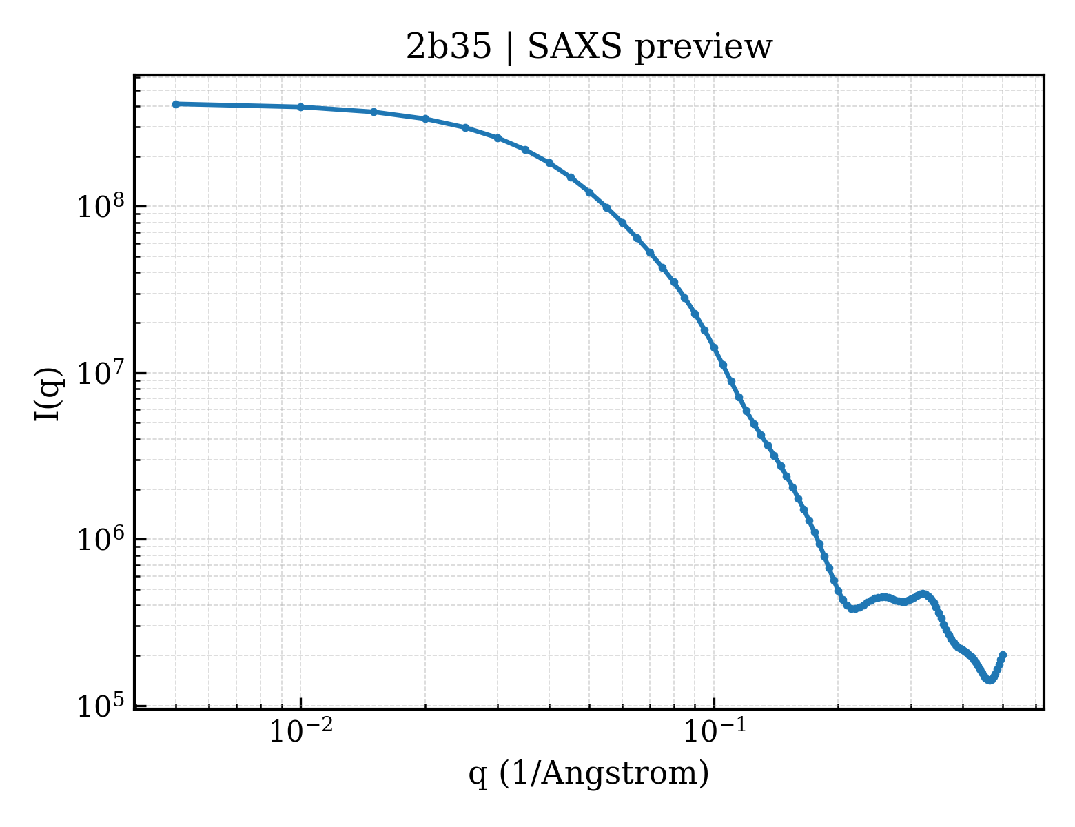
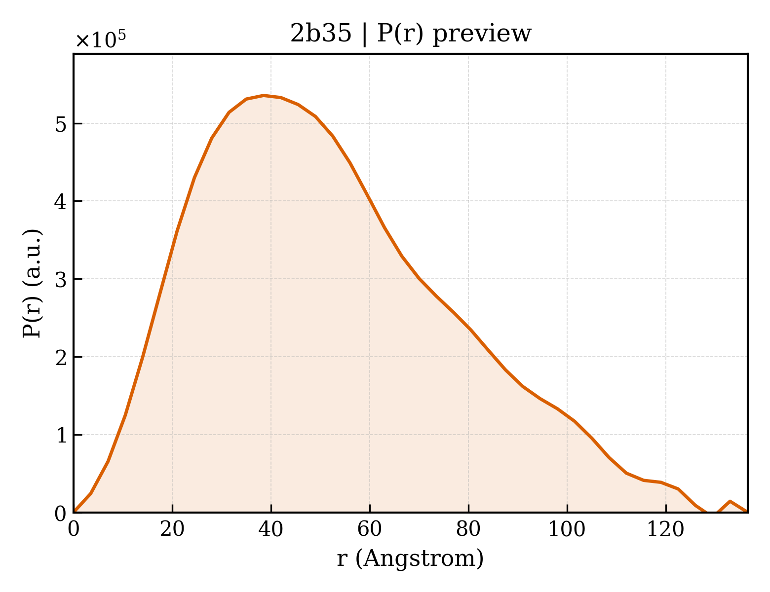
Enoyl-[acyl-carrier-protein] reductase [NADH]
Mycobacterium tuberculosis
State in the Current Structure
| Assembly | Oligomeric State | Construct | Mutations and Modifications | Ligands, Ions and Associated Components | Method and Experimental Conditions | Structure Quality |
|---|---|---|---|---|---|---|
| 1 | Protein homooligomer Homooligomer Protein × 4 PDB declaration: tetrameric(4) Consistent with protein copy count | Chain A; UniProt 1–269 Chain B; UniProt 1–269 | Not recorded | TCL TRICLOSAN × 4 NAD NICOTINAMIDE-ADENINE-DINUCLEOTIDE × 4 | X-RAY DIFFRACTION X-ray crystallization conditions:VAPOR DIFFUSION, HANGING DROP;pH 6.8;295 K;PEG 4000, DMSO, ammonium acetate, NAD+, ADA, pH 6.8, VAPOR DIFFUSION, HANGING DROP, temperature 295K | Resolution 2.30 Å R-free 0.334 |
| 2 | Protein homooligomer Homooligomer Protein × 4 PDB declaration: tetrameric(4) Consistent with protein copy count | Chain C; UniProt 1–269 Chain D; UniProt 1–269 Chain E; UniProt 1–269 Chain F; UniProt 1–269 | Not recorded | TCL TRICLOSAN × 4 NAD NICOTINAMIDE-ADENINE-DINUCLEOTIDE × 4 | X-RAY DIFFRACTION X-ray crystallization conditions:VAPOR DIFFUSION, HANGING DROP;pH 6.8;295 K;PEG 4000, DMSO, ammonium acetate, NAD+, ADA, pH 6.8, VAPOR DIFFUSION, HANGING DROP, temperature 295K | Resolution 2.30 Å R-free 0.334 |
Other States of the Same Protein in the Database
Each row is a biological assembly of the same UniProt protein in another PDB entry. The “Difference from current entry” column identifies evidence-level differences; no tag means the currently parsed fields agree.
| Other PDB | Difference from Current Entry 2B35 | Assembly / Oligomeric State | Construct | Mutations and Modifications | Ligands, Ions and Non-polymers | Method and Experimental Conditions | Structure Quality |
|---|---|---|---|---|---|---|---|
| 1BVR M.TB. ENOYL-ACP REDUCTASE (INHA) IN COMPLEX WITH NAD+ AND C16-FATTY-ACYL-SUBSTRATE Deposited 1998-09-17 | Different construct Different ligand/ion Different experimental conditions Different structure-quality metrics | Assembly 1 Protein homooligomer Homooligomer;Protein × 4 PDB declaration: tetrameric |
Chain C
2–269(268 aa)
Chain D
2–269(268 aa)
Chain E
2–269(268 aa)
Chain F
2–269(268 aa)
|
Not recorded | NAD NICOTINAMIDE-ADENINE-DINUCLEOTIDE × 4 THT TRANS-2-HEXADECENOYL-(N-ACETYL-CYSTEAMINE)-THIOESTER × 2 |
X-RAY DIFFRACTION
X-ray crystallization conditions
pH 6.8;10% PEG-4000, 6% DMSO, 100 MM AMMONIUM ACETATE, AND 100 MM ADA, PH 6.8
|
Resolution 2.80 Å R-free 0.344 |
| 1BVR M.TB. ENOYL-ACP REDUCTASE (INHA) IN COMPLEX WITH NAD+ AND C16-FATTY-ACYL-SUBSTRATE Deposited 1998-09-17 | Different construct Different oligomeric state Different ligand/ion Different experimental conditions Different structure-quality metrics | Assembly 2 Protein homooligomer Homooligomer;Protein × 2 PDB declaration: dimeric |
Chain A
2–269(268 aa)
Chain B
2–269(268 aa)
|
Not recorded | NAD NICOTINAMIDE-ADENINE-DINUCLEOTIDE × 2 THT TRANS-2-HEXADECENOYL-(N-ACETYL-CYSTEAMINE)-THIOESTER × 2 |
X-RAY DIFFRACTION
X-ray crystallization conditions
pH 6.8;10% PEG-4000, 6% DMSO, 100 MM AMMONIUM ACETATE, AND 100 MM ADA, PH 6.8
|
Resolution 2.80 Å R-free 0.344 |
| 1BVR M.TB. ENOYL-ACP REDUCTASE (INHA) IN COMPLEX WITH NAD+ AND C16-FATTY-ACYL-SUBSTRATE Deposited 1998-09-17 | Different construct Different ligand/ion Different experimental conditions Different structure-quality metrics | Assembly 3 Protein homooligomer Homooligomer;Protein × 4 PDB declaration: tetrameric |
Chain A
2–269(268 aa)
Chain B
2–269(268 aa)
|
Not recorded | NAD NICOTINAMIDE-ADENINE-DINUCLEOTIDE × 4 THT TRANS-2-HEXADECENOYL-(N-ACETYL-CYSTEAMINE)-THIOESTER × 4 |
X-RAY DIFFRACTION
X-ray crystallization conditions
pH 6.8;10% PEG-4000, 6% DMSO, 100 MM AMMONIUM ACETATE, AND 100 MM ADA, PH 6.8
|
Resolution 2.80 Å R-free 0.344 |
| 1ENY CRYSTAL STRUCTURE AND FUNCTION OF THE ISONIAZID TARGET OF MYCOBACTERIUM TUBERCULOSIS Deposited 1995-01-27 | Different construct Different mutation/modification Different oligomeric state Different ligand/ion Different experimental conditions Different structure-quality metrics | Assembly 1 Protein homooligomer Homooligomer;Protein × 2 PDB declaration: dimeric |
Chain A
3–269(267 aa)
|
Not recorded | NAD NICOTINAMIDE-ADENINE-DINUCLEOTIDE × 2 | X-RAY DIFFRACTION mmCIF provides none of the parsed conditions | Resolution 2.20 Å |
| 1ENY CRYSTAL STRUCTURE AND FUNCTION OF THE ISONIAZID TARGET OF MYCOBACTERIUM TUBERCULOSIS Deposited 1995-01-27 | Different construct Different mutation/modification Different ligand/ion Different experimental conditions Different structure-quality metrics | Assembly 2 Protein homooligomer Homooligomer;Protein × 4 PDB declaration: tetrameric |
Chain A
3–269(267 aa)
|
Not recorded | NAD NICOTINAMIDE-ADENINE-DINUCLEOTIDE × 4 | X-RAY DIFFRACTION mmCIF provides none of the parsed conditions | Resolution 2.20 Å |
| 1ENZ CRYSTAL STRUCTURE AND FUNCTION OF THE ISONIAZID TARGET OF MYCOBACTERIUM TUBERCULOSIS Deposited 1995-01-27 | Different construct Different mutation/modification Different oligomeric state Different ligand/ion Different experimental conditions Different structure-quality metrics | Assembly 1 Protein homooligomer Homooligomer;Protein × 2 PDB declaration: dimeric |
Chain A
3–269(267 aa)
|
Mutation:S94A | NAD NICOTINAMIDE-ADENINE-DINUCLEOTIDE × 2 | X-RAY DIFFRACTION mmCIF provides none of the parsed conditions | Resolution 2.70 Å |
| 1ENZ CRYSTAL STRUCTURE AND FUNCTION OF THE ISONIAZID TARGET OF MYCOBACTERIUM TUBERCULOSIS Deposited 1995-01-27 | Different construct Different mutation/modification Different ligand/ion Different experimental conditions Different structure-quality metrics | Assembly 2 Protein homooligomer Homooligomer;Protein × 4 PDB declaration: tetrameric |
Chain A
3–269(267 aa)
|
Mutation:S94A | NAD NICOTINAMIDE-ADENINE-DINUCLEOTIDE × 4 | X-RAY DIFFRACTION mmCIF provides none of the parsed conditions | Resolution 2.70 Å |
| 1P44 Targeting tuberculosis and malaria through inhibition of enoyl reductase: compound activity and structural data Deposited 2003-04-21 | Different ligand/ion Different experimental conditions Different structure-quality metrics | Assembly 1 Protein homooligomer Homooligomer;Protein × 4 PDB declaration: tetrameric |
Chain A
1–269(269 aa)
Chain B
1–269(269 aa)
|
Not recorded | NAD NICOTINAMIDE-ADENINE-DINUCLEOTIDE × 4 GEQ 5-{[4-(9H-FLUOREN-9-YL)PIPERAZIN-1-YL]CARBONYL}-1H-INDOLE × 4 |
X-RAY DIFFRACTION
X-ray crystallization conditions
VAPOR DIFFUSION, HANGING DROP;pH 6.8;291 K;100 mM ADA, 150 mM ammonium acetate, 12% PEG 3350, pH 6.8, VAPOR DIFFUSION, HANGING DROP, temperature 291K
|
Resolution 2.70 Å R-free 0.288 |
| 1P44 Targeting tuberculosis and malaria through inhibition of enoyl reductase: compound activity and structural data Deposited 2003-04-21 | Different ligand/ion Different experimental conditions Different structure-quality metrics | Assembly 2 Protein homooligomer Homooligomer;Protein × 4 PDB declaration: tetrameric |
Chain C
1–269(269 aa)
Chain D
1–269(269 aa)
Chain E
1–269(269 aa)
Chain F
1–269(269 aa)
|
Not recorded | NAD NICOTINAMIDE-ADENINE-DINUCLEOTIDE × 4 GEQ 5-{[4-(9H-FLUOREN-9-YL)PIPERAZIN-1-YL]CARBONYL}-1H-INDOLE × 2 |
X-RAY DIFFRACTION
X-ray crystallization conditions
VAPOR DIFFUSION, HANGING DROP;pH 6.8;291 K;100 mM ADA, 150 mM ammonium acetate, 12% PEG 3350, pH 6.8, VAPOR DIFFUSION, HANGING DROP, temperature 291K
|
Resolution 2.70 Å R-free 0.288 |
| 1P45 Targeting tuberculosis and malaria through inhibition of enoyl reductase: compound activity and structural data Deposited 2003-04-21 | Different ligand/ion Different experimental conditions Different structure-quality metrics | Assembly 1 Protein homooligomer Homooligomer;Protein × 4 PDB declaration: tetrameric |
Chain A
1–269(269 aa)
Chain B
1–269(269 aa)
|
Not recorded | NAD NICOTINAMIDE-ADENINE-DINUCLEOTIDE × 4 TCL TRICLOSAN × 6 |
X-RAY DIFFRACTION
X-ray crystallization conditions
VAPOR DIFFUSION, HANGING DROP;pH 8;291 K;100 mM Tris, pH 8, VAPOR DIFFUSION, HANGING DROP, temperature 291K
|
Resolution 2.60 Å R-free 0.290 |
| 1ZID LONG FATTY ACID CHAIN ENOYL-ACP REDUCTASE (INHA) IN COMPLEX WITH AN ISONICOTINIC-ACYL-NADH INHIBITOR Deposited 1997-03-25 | Different construct Different mutation/modification Different ligand/ion Different experimental conditions Different structure-quality metrics | Assembly 1 Protein homooligomer Homooligomer;Protein × 4 PDB declaration: tetrameric |
Chain A
3–269(267 aa)
|
Mutation:T2A | ZID ISONICOTINIC-ACETYL-NICOTINAMIDE-ADENINE DINUCLEOTIDE × 4 |
X-RAY DIFFRACTION
X-ray crystallization conditions
pH 7.5;12% MPD, 4% DMSO, 50MM NACITRATE, 100 MM HEPES, PH 7.5
|
Resolution 2.70 Å R-free 0.297 |
| 2AQ8 Crystal structure of wild-type of Enoyl-ACP(CoA) reductase from Mycobacterium tuberculosis in complex with NADH. Deposited 2005-08-17 | Different construct Different mutation/modification Different ligand/ion Different experimental conditions Different structure-quality metrics | Assembly 1 Protein homooligomer Homooligomer;Protein × 4 PDB declaration: tetrameric |
Chain A
1–269(269 aa)
|
Not recorded | NAI 1,4-DIHYDRONICOTINAMIDE ADENINE DINUCLEOTIDE × 4 LYS LYSINE × 4 |
X-RAY DIFFRACTION
X-ray crystallization conditions
VAPOR DIFFUSION, HANGING DROP;pH 7.2;293 K;0.05 M Sodium citrate, 0.05 M Hepes, 8-15% 2-Methyl-2-4-pentanediol (MPD), pH 7.2, VAPOR DIFFUSION, HANGING DROP, temperature 293K
|
Resolution 1.92 Å R-free 0.213 |
| 2AQH Crystal structure of Isoniazid-resistant I21V Enoyl-ACP(CoA) reductase mutant enzyme from Mycobacterium tuberculosis in complex with NADH Deposited 2005-08-18 | Different construct Different mutation/modification Different ligand/ion Different experimental conditions Different structure-quality metrics | Assembly 1 Protein homooligomer Homooligomer;Protein × 4 PDB declaration: tetrameric |
Chain A
1–269(269 aa)
|
Mutation:I21V | NAI 1,4-DIHYDRONICOTINAMIDE ADENINE DINUCLEOTIDE × 4 |
X-RAY DIFFRACTION
X-ray crystallization conditions
VAPOR DIFFUSION, HANGING DROP;pH 7.2;293 K;0.05M Sodium citrate, 0.05M Hepes, 8-15% 2-Methyl-2-4-pentanediol (MPD), pH 7.2, VAPOR DIFFUSION, HANGING DROP, temperature 293K
|
Resolution 2.01 Å R-free 0.227 |
| 2AQI Crystal structure of Isoniazid-resistant I47T Enoyl-ACP(CoA) reductase mutant enzyme from Mycobacterium tuberculosis in complex with NADH Deposited 2005-08-18 | Different construct Different mutation/modification Different ligand/ion Different experimental conditions Different structure-quality metrics | Assembly 1 Protein homooligomer Homooligomer;Protein × 4 PDB declaration: tetrameric |
Chain A
1–269(269 aa)
|
Mutation:I47T | NAI 1,4-DIHYDRONICOTINAMIDE ADENINE DINUCLEOTIDE × 4 |
X-RAY DIFFRACTION
X-ray crystallization conditions
VAPOR DIFFUSION, HANGING DROP;pH 7.2;293 K;0.05M Sodium citrate, 0.05M Hepes, 8-15% 2-Methyl-2-4-pentanediol (MPD), pH 7.2, VAPOR DIFFUSION, HANGING DROP, temperature 293K
|
Resolution 2.20 Å R-free 0.213 |
| 2AQK Crystal structure of Isoniazid-resistant S94A Enoyl-ACP(CoA) reductase mutant enzyme from Mycobacterium tuberculosis in complex with NADH Deposited 2005-08-18 | Different construct Different mutation/modification Different ligand/ion Different experimental conditions Different structure-quality metrics | Assembly 1 Protein homooligomer Homooligomer;Protein × 4 PDB declaration: tetrameric |
Chain A
1–269(269 aa)
|
Mutation:S94A | NAI 1,4-DIHYDRONICOTINAMIDE ADENINE DINUCLEOTIDE × 4 |
X-RAY DIFFRACTION
X-ray crystallization conditions
VAPOR DIFFUSION, HANGING DROP;pH 7.2;293 K;0.05M Sodium citrate, 0.05M Hepes, 8-15% 2-Methyl-2-4-pentanediol (MPD), pH 7.2, VAPOR DIFFUSION, HANGING DROP, temperature 293K
|
Resolution 2.30 Å R-free 0.242 |
| 2B36 Crystal structure of Mycobacterium tuberculosis enoyl reductase (InhA) inhibited by 5-pentyl-2-phenoxyphenol Deposited 2005-09-19 | Different ligand/ion Different experimental conditions Different structure-quality metrics | Assembly 1 Protein homooligomer Homooligomer;Protein × 4 PDB declaration: tetrameric |
Chain A
1–269(269 aa)
Chain B
1–269(269 aa)
|
Not recorded | 5PP 5-PENTYL-2-PHENOXYPHENOL × 4 NAD NICOTINAMIDE-ADENINE-DINUCLEOTIDE × 4 |
X-RAY DIFFRACTION
X-ray crystallization conditions
VAPOR DIFFUSION, HANGING DROP;pH 6.8;295 K;PEG 4000, DMSO, ammonium acetate, NAD+, ADA , pH 6.8, VAPOR DIFFUSION, HANGING DROP, temperature 295K
|
Resolution 2.80 Å R-free 0.295 |
| 2B36 Crystal structure of Mycobacterium tuberculosis enoyl reductase (InhA) inhibited by 5-pentyl-2-phenoxyphenol Deposited 2005-09-19 | Different ligand/ion Different experimental conditions Different structure-quality metrics | Assembly 2 Protein homooligomer Homooligomer;Protein × 4 PDB declaration: tetrameric |
Chain C
1–269(269 aa)
Chain D
1–269(269 aa)
Chain E
1–269(269 aa)
Chain F
1–269(269 aa)
|
Not recorded | 5PP 5-PENTYL-2-PHENOXYPHENOL × 4 NAD NICOTINAMIDE-ADENINE-DINUCLEOTIDE × 4 |
X-RAY DIFFRACTION
X-ray crystallization conditions
VAPOR DIFFUSION, HANGING DROP;pH 6.8;295 K;PEG 4000, DMSO, ammonium acetate, NAD+, ADA , pH 6.8, VAPOR DIFFUSION, HANGING DROP, temperature 295K
|
Resolution 2.80 Å R-free 0.295 |
| 2B37 Crystal structure of Mycobacterium tuberculosis enoyl reductase (InhA) inhibited by 5-octyl-2-phenoxyphenol Deposited 2005-09-19 | Different ligand/ion Different experimental conditions Different structure-quality metrics | Assembly 1 Protein homooligomer Homooligomer;Protein × 4 PDB declaration: tetrameric |
Chain A
1–269(269 aa)
Chain B
1–269(269 aa)
Chain E
1–269(269 aa)
Chain F
1–269(269 aa)
|
Not recorded | NAD NICOTINAMIDE-ADENINE-DINUCLEOTIDE × 4 |
X-RAY DIFFRACTION
X-ray crystallization conditions
VAPOR DIFFUSION, HANGING DROP;pH 6.8;295 K;PEG 4000, DMSO, ammonium acetate, NAD+, ADA , pH 6.8, VAPOR DIFFUSION, HANGING DROP, temperature 295K
|
Resolution 2.60 Å R-free 0.294 |
| 2B37 Crystal structure of Mycobacterium tuberculosis enoyl reductase (InhA) inhibited by 5-octyl-2-phenoxyphenol Deposited 2005-09-19 | Different ligand/ion Different experimental conditions Different structure-quality metrics | Assembly 2 Protein homooligomer Homooligomer;Protein × 4 PDB declaration: tetrameric |
Chain C
1–269(269 aa)
Chain D
1–269(269 aa)
|
Not recorded | NAD NICOTINAMIDE-ADENINE-DINUCLEOTIDE × 4 8PS 5-OCTYL-2-PHENOXYPHENOL × 4 |
X-RAY DIFFRACTION
X-ray crystallization conditions
VAPOR DIFFUSION, HANGING DROP;pH 6.8;295 K;PEG 4000, DMSO, ammonium acetate, NAD+, ADA , pH 6.8, VAPOR DIFFUSION, HANGING DROP, temperature 295K
|
Resolution 2.60 Å R-free 0.294 |
| 2H9I Mycobacterium tuberculosis InhA bound with ETH-NAD adduct Deposited 2006-06-09 | Different construct Different mutation/modification Different ligand/ion Different experimental conditions Different structure-quality metrics | Assembly 1 Protein homooligomer Homooligomer;Protein × 4 PDB declaration: tetrameric |
Chain A
3–269(267 aa)
|
Not recorded | EAD {(2R,3S,4R,5R)-5-[(4S)-3-(AMINOCARBONYL)-4-(2-ETHYLISONICOTINOYL)PYRIDIN-1(4H)-YL]-3,4-DIHYDROXYTETRAHYDROFURAN-2-YL}METHYL [(2R,3S,4R,5R)-5-(6-AMINO-9H-PURIN-9-YL)-3,4-DIHYDROXYTETRAHYDROFURAN-2-YL]METHYL DIHYDROGEN DIPHOSPHATE × 4 |
X-RAY DIFFRACTION
X-ray crystallization conditions
VAPOR DIFFUSION, HANGING DROP;pH 7.5;289 K;12% MDP, 4% DMSO, 0.1M Hepes, 0.025M Sodium Citrate, pH 7.5, VAPOR DIFFUSION, HANGING DROP, temperature 289K
|
Resolution 2.20 Å R-free 0.261 |
| 2IDZ Crystal structure of wild type Enoyl-ACP(CoA) reductase from Mycobacterium tuberculosis in complex with NADH-INH Deposited 2006-09-15 | Different construct Different mutation/modification Different ligand/ion Different experimental conditions Different structure-quality metrics | Assembly 1 Protein homooligomer Homooligomer;Protein × 4 PDB declaration: tetrameric |
Chain A
2–269(268 aa)
|
Not recorded | ZID ISONICOTINIC-ACETYL-NICOTINAMIDE-ADENINE DINUCLEOTIDE × 4 |
X-RAY DIFFRACTION
X-ray crystallization conditions
VAPOR DIFFUSION, HANGING DROP;pH 7.2;293 K;0.05M SODIUM CITRATE, 0.05M HEPES, 8-15% 2-METHYL-2-4-PENTANEDIOL (MPD), PH 7.2, TEMPERATURE 293K , VAPOR DIFFUSION, HANGING DROP
|
Resolution 2.00 Å R-free 0.226 |
| 2IE0 Crystal Structure of Isoniazid-resistant I21V Enoyl-ACP(COA) Reductase Mutant Enzyme From MYCOBACTERIUM TUBERCULOSIS in Complex with NADH-INH Deposited 2006-09-15 | Different construct Different mutation/modification Different ligand/ion Different experimental conditions Different structure-quality metrics | Assembly 1 Protein homooligomer Homooligomer;Protein × 4 PDB declaration: tetrameric |
Chain A
2–269(268 aa)
|
Mutation:i21v | ZID ISONICOTINIC-ACETYL-NICOTINAMIDE-ADENINE DINUCLEOTIDE × 4 |
X-RAY DIFFRACTION
X-ray crystallization conditions
VAPOR DIFFUSION, HANGING DROP;pH 7.2;293 K;0.05M SODIUM CITRATE, 0.05M HEPES, 8-15% 2-METHYL-2-4-PENTANEDIOL (MPD), PH 7.2, VAPOR DIFFUSION, HANGING DROP, temperature 293K
|
Resolution 2.20 Å R-free 0.237 |
| 2IEB Crystal Structure of Isoniazid-resistant S94A ENOYL-ACP(COA) Reductase Mutant Enzyme from MYCOBACTERIUM TUBERCULOSIS in Complex with NADH-INH Deposited 2006-09-18 | Different construct Different mutation/modification Different ligand/ion Different experimental conditions Different structure-quality metrics | Assembly 1 Protein homooligomer Homooligomer;Protein × 4 PDB declaration: tetrameric |
Chain A
2–269(268 aa)
|
Mutation:S94A | ZID ISONICOTINIC-ACETYL-NICOTINAMIDE-ADENINE DINUCLEOTIDE × 4 |
X-RAY DIFFRACTION
X-ray crystallization conditions
VAPOR DIFFUSION, HANGING DROP;pH 7.2;293 K;0.05 M SODIUM CITRATE, 0.05 M
HEPES, 8-15% 2-METHYL-2-4-PENTANEDIOL (MPD), PH 7.2., VAPOR DIFFUSION, HANGING DROP, temperature 293K
|
Resolution 2.20 Å R-free 0.240 |
| 2IED CRYSTAL STRUCTURE of ISONIAZID-RESISTANT S94A ENOYL-ACP(COA) REDUCTASE MUTANT ENZYME FROM MYCOBACTERIUM TUBERCULOSIS UNCOMPLEXED Deposited 2006-09-18 | Different construct Different mutation/modification Different ligand/ion Different experimental conditions Different structure-quality metrics | Assembly 1 Protein homooligomer Homooligomer;Protein × 4 PDB declaration: tetrameric |
Chain A
2–269(268 aa)
Chain B
2–269(268 aa)
Chain C
2–269(268 aa)
Chain D
2–269(268 aa)
|
Mutation:S94A Mutation:S94A Mutation:S94A Mutation:S94A | No recorded non-water small molecule |
X-RAY DIFFRACTION
X-ray crystallization conditions
VAPOR DIFFUSION, HANGING DROP;pH 5.6;293 K;100 mM sodium citrate, pH 5.6, 200 mM ammonium acetate and 20-30% of the PEG 4000, VAPOR DIFFUSION, HANGING DROP, temperature 293K
|
Resolution 2.14 Å R-free 0.255 |
| 2NSD Enoyl acyl carrier protein reductase InhA in complex with N-(4-methylbenzoyl)-4-benzylpiperidine Deposited 2006-11-03 | Different construct Different mutation/modification Different oligomeric state Different ligand/ion Different experimental conditions Different structure-quality metrics | Assembly 1 Protein monomer Monomer;Protein × 1 PDB declaration: monomeric |
Chain A
1–269(269 aa)
|
Not recorded | NAD NICOTINAMIDE-ADENINE-DINUCLEOTIDE × 1 4PI N-(4-METHYLBENZOYL)-4-BENZYLPIPERIDINE × 1 |
X-RAY DIFFRACTION
X-ray crystallization conditions
VAPOR DIFFUSION, HANGING DROP;pH 7.5;295 K;8% MPD, 50mM Sodium citrate pH 6.5, 100mM HEPES pH 7.5, VAPOR DIFFUSION, HANGING DROP, temperature 295K
|
Resolution 1.90 Å R-free 0.259 |
| 2NSD Enoyl acyl carrier protein reductase InhA in complex with N-(4-methylbenzoyl)-4-benzylpiperidine Deposited 2006-11-03 | Different construct Different mutation/modification Different oligomeric state Different ligand/ion Different experimental conditions Different structure-quality metrics | Assembly 2 Protein monomer Monomer;Protein × 1 PDB declaration: monomeric |
Chain B
1–269(269 aa)
|
Not recorded | NAD NICOTINAMIDE-ADENINE-DINUCLEOTIDE × 1 4PI N-(4-METHYLBENZOYL)-4-BENZYLPIPERIDINE × 1 |
X-RAY DIFFRACTION
X-ray crystallization conditions
VAPOR DIFFUSION, HANGING DROP;pH 7.5;295 K;8% MPD, 50mM Sodium citrate pH 6.5, 100mM HEPES pH 7.5, VAPOR DIFFUSION, HANGING DROP, temperature 295K
|
Resolution 1.90 Å R-free 0.259 |
| 2NTJ Mycobacterium tuberculosis InhA bound with PTH-NAD adduct Deposited 2006-11-07 | Different construct Different mutation/modification Different ligand/ion Different experimental conditions Different structure-quality metrics | Assembly 1 Protein homooligomer Homooligomer;Protein × 4 PDB declaration: tetrameric |
Chain A
3–269(267 aa)
Chain B
3–269(267 aa)
|
Mutation:D2A Mutation:D2A | P1H {(2R,3S,4R,5R)-5-[(4S)-3-(AMINOCARBONYL)-4-(2-PROPYLISONICOTINOYL)PYRIDIN-1(4H)-YL]-3,4-DIHYDROXYTETRAHYDROFURAN-2-YL}M ETHYL [(2R,3S,4R,5R)-5-(6-AMINO-9H-PURIN-9-YL)-3,4-DIHYDROXYTETRAHYDROFURAN-2-YL]METHYL DIHYDROGEN DIPHOSPHATE × 4 |
X-RAY DIFFRACTION
X-ray crystallization conditions
VAPOR DIFFUSION, HANGING DROP;pH 7.5;289 K;12% MDP, 4% DMSO, 0.1M Hepes, 0.025M Sodium Citrate, pH 7.5, VAPOR DIFFUSION, HANGING DROP, temperature 289K
|
Resolution 2.50 Å R-free 0.268 |
| 2NV6 Mycobacterium tuberculosis InhA (S94A) bound with INH-NAD adduct Deposited 2006-11-10 | Different construct Different mutation/modification Different ligand/ion Different experimental conditions Different structure-quality metrics | Assembly 1 Protein homooligomer Homooligomer;Protein × 4 PDB declaration: tetrameric |
Chain A
2–269(268 aa)
|
Mutation:D2A, S94A | ZID ISONICOTINIC-ACETYL-NICOTINAMIDE-ADENINE DINUCLEOTIDE × 4 |
X-RAY DIFFRACTION
X-ray crystallization conditions
VAPOR DIFFUSION, HANGING DROP;pH 7.5;289 K;12% MPD, 4% DMSO, 0.1 M Hepes, pH 7.5, VAPOR DIFFUSION, HANGING DROP, temperature 289K
|
Resolution 1.90 Å R-free 0.228 |
| 2PR2 Structure of Mycobacterium tuberculosis enoyl-ACP reductase with bound INH-NADP. Deposited 2007-05-03 | Different construct Different mutation/modification Different ligand/ion Different experimental conditions Different structure-quality metrics | Assembly 1 Protein homooligomer Homooligomer;Protein × 4 PDB declaration: tetrameric |
Chain A
1–269(269 aa)
|
Not recorded | DG1 (4S)-ISONICOTINIC-ACETYL-NICOTINAMIDE-ADENINE DINUCLEOTIDE × 4 |
X-RAY DIFFRACTION
X-ray crystallization conditions
VAPOR DIFFUSION;pH 9;291 K;Cocrystallization with Inh-NADP at 4.8 mM 20 25% of 2-methyl-2,4-pentanediol, 100 mM Bicine pH 9.0., Vapour diffusion under oil, temperature 291K, VAPOR DIFFUSION
|
Resolution 2.50 Å R-free 0.217 |
| 2X22 crystal structure of M. tuberculosis InhA inhibited by PT70 Deposited 2010-01-10 | Different ligand/ion Different experimental conditions Different structure-quality metrics | Assembly 1 Protein homooligomer Homooligomer;Protein × 4 PDB declaration: tetrameric |
Chain A
1–269(269 aa)
Chain B
1–269(269 aa)
|
Not recorded | NAD NICOTINAMIDE-ADENINE-DINUCLEOTIDE × 4 TCU 5-HEXYL-2-(2-METHYLPHENOXY)PHENOL × 4 DMS DIMETHYL SULFOXIDE × 4 |
X-RAY DIFFRACTION
X-ray crystallization conditions
12-16% (W/V) PEG4000, 1% DMSO, 100MM ADA PH 6.8, 250MM AMMONIUM ACETATE
|
Resolution 2.10 Å R-free 0.216 |
| 2X23 crystal structure of M. tuberculosis InhA inhibited by PT70 Deposited 2010-01-10 | Different ligand/ion Different experimental conditions Different structure-quality metrics | Assembly 1 Protein homooligomer Homooligomer;Protein × 4 PDB declaration: tetrameric |
Chain A
1–269(269 aa)
Chain B
1–269(269 aa)
Chain E
1–269(269 aa)
Chain G
1–269(269 aa)
|
Not recorded | NAD NICOTINAMIDE-ADENINE-DINUCLEOTIDE × 4 TCU 5-HEXYL-2-(2-METHYLPHENOXY)PHENOL × 4 DMS DIMETHYL SULFOXIDE × 1 |
X-RAY DIFFRACTION
X-ray crystallization conditions
12-16% (W/V) PEG 4000, 1% DMSO, 250MM AMMONIUM ACETATE, 100MM ADA PH 6.8
|
Resolution 1.81 Å R-free 0.203 |
| 3FNE Crystal structure of InhA bound to triclosan derivative 17 Deposited 2008-12-24 | Different ligand/ion Different experimental conditions Different structure-quality metrics | Assembly 1 Protein homooligomer Homooligomer;Protein × 4 PDB declaration: tetrameric |
Chain A
1–269(269 aa)
Chain B
1–269(269 aa)
Chain C
1–269(269 aa)
Chain D
1–269(269 aa)
|
Not recorded | NAD NICOTINAMIDE-ADENINE-DINUCLEOTIDE × 4 8PC 2-(2,4-DICHLOROPHENOXY)-5-(PYRIDIN-2-YLMETHYL)PHENOL × 4 |
X-RAY DIFFRACTION
X-ray crystallization conditions
VAPOR DIFFUSION, HANGING DROP;pH 7.2;293 K;20% PEG 3350, 6% DMSO, 0.1 m N-(2-acetamido)iminodiacetic
acid pH 6.8, 0.08 m NH4OAc, VAPOR DIFFUSION, HANGING DROP, temperature 293K
|
Resolution 1.98 Å R-free 0.253 |
| 3FNF Crystal structure of InhA bound to triclosan derivative Deposited 2008-12-24 | Different ligand/ion Different experimental conditions Different structure-quality metrics | Assembly 1 Protein homooligomer Homooligomer;Protein × 4 PDB declaration: tetrameric |
Chain A
1–269(269 aa)
Chain B
1–269(269 aa)
Chain C
1–269(269 aa)
Chain D
1–269(269 aa)
|
Not recorded | NAD NICOTINAMIDE-ADENINE-DINUCLEOTIDE × 4 JPM 5-benzyl-2-(2,4-dichlorophenoxy)phenol × 4 |
X-RAY DIFFRACTION
X-ray crystallization conditions
VAPOR DIFFUSION, HANGING DROP;pH 6.8;293 K;20% PEG 3350, 6% DMSO, 0.1M ADA pH 6.8, and 0.08 M ammonium acetate, VAPOR DIFFUSION, HANGING DROP, temperature 293K
|
Resolution 2.30 Å R-free 0.260 |
| 3FNG Crystal structure of InhA bound to triclosan derivative Deposited 2008-12-24 | Different construct Different mutation/modification Different ligand/ion Different experimental conditions Different structure-quality metrics | Assembly 1 Protein homooligomer Homooligomer;Protein × 4 PDB declaration: tetrameric |
Chain A
1–269(269 aa)
|
Not recorded | NAD NICOTINAMIDE-ADENINE-DINUCLEOTIDE × 4 JPL 5-(cyclohexylmethyl)-2-(2,4-dichlorophenoxy)phenol × 4 |
X-RAY DIFFRACTION
X-ray crystallization conditions
VAPOR DIFFUSION, HANGING DROP;pH 7.5;293 K;0.1 M Hepes pH 7.5, 12% MPD, and 4% DMSO, VAPOR DIFFUSION, HANGING DROP, temperature 293K
|
Resolution 1.97 Å R-free 0.253 |
| 3FNH Crystal structure of InhA bound to triclosan derivative Deposited 2008-12-24 | Different construct Different mutation/modification Different ligand/ion Different experimental conditions Different structure-quality metrics | Assembly 1 Protein homooligomer Homooligomer;Protein × 4 PDB declaration: tetrameric |
Chain A
1–269(269 aa)
|
Not recorded | NAD NICOTINAMIDE-ADENINE-DINUCLEOTIDE × 4 JPJ 2-(2,4-DICHLOROPHENOXY)-5-(2-PHENYLETHYL)PHENOL × 4 |
X-RAY DIFFRACTION
X-ray crystallization conditions
VAPOR DIFFUSION, HANGING DROP;pH 7.5;293 K;0.1 M Hepes pH 7.5, 12% MPD, and 4% DMSO, VAPOR DIFFUSION, HANGING DROP, temperature 293K
|
Resolution 2.80 Å R-free 0.268 |
| 3OEW Crystal structure of wild-type InhA:NADH complex Deposited 2010-08-13 | Different construct Different mutation/modification Different ligand/ion Different experimental conditions Different structure-quality metrics | Assembly 1 Protein homooligomer Homooligomer;Protein × 4 PDB declaration: tetrameric |
Chain A
1–269(269 aa)
|
Not recorded | NAD NICOTINAMIDE-ADENINE-DINUCLEOTIDE × 4 MPD (4S)-2-METHYL-2,4-PENTANEDIOL × 4 |
X-RAY DIFFRACTION
X-ray crystallization conditions
VAPOR DIFFUSION, HANGING DROP;pH 7.2;291 K;100 mM Hepes 7.2, 50 mM sodium citrate 6.5, 8-12% MPD, 4% DMSO, VAPOR DIFFUSION, HANGING DROP, temperature 291K
|
Resolution 2.20 Å R-free 0.209 |
| 3OEY Crystal structure of InhA_T266E:NADH complex Deposited 2010-08-13 | Different construct Different mutation/modification Different ligand/ion Different experimental conditions Different structure-quality metrics | Assembly 1 Protein homooligomer Homooligomer;Protein × 4 PDB declaration: tetrameric |
Chain A
1–269(269 aa)
|
Mutation:T266E | NAD NICOTINAMIDE-ADENINE-DINUCLEOTIDE × 4 MPD (4S)-2-METHYL-2,4-PENTANEDIOL × 4 |
X-RAY DIFFRACTION
X-ray crystallization conditions
VAPOR DIFFUSION, HANGING DROP;pH 7.2;291 K;100 mM Hepes 7.2, 50 mM sodium citrate 6.5, 8-12% MPD, 4% DMSO, VAPOR DIFFUSION, HANGING DROP, temperature 291K
|
Resolution 2.00 Å R-free 0.214 |
| 3OF2 Crystal structure of InhA_T266D:NADH complex Deposited 2010-08-13 | Different construct Different mutation/modification Different ligand/ion Different experimental conditions Different structure-quality metrics | Assembly 1 Protein homooligomer Homooligomer;Protein × 4 PDB declaration: tetrameric |
Chain A
1–269(269 aa)
|
Mutation:T266D | NAD NICOTINAMIDE-ADENINE-DINUCLEOTIDE × 4 MPD (4S)-2-METHYL-2,4-PENTANEDIOL × 4 |
X-RAY DIFFRACTION
X-ray crystallization conditions
VAPOR DIFFUSION, HANGING DROP;pH 7.2;291 K;100 mM Hepes 7.2, 50 mM sodium citrate 6.5, 8-12% MPD, 4% DMSO
, VAPOR DIFFUSION, HANGING DROP, temperature 291K
|
Resolution 2.00 Å R-free 0.203 |
| 4BII How nature bridges the gap: Crystallographic elucidation of pyridomycin binding to InhA Deposited 2013-04-10 | Different ligand/ion Different experimental conditions Different structure-quality metrics | Assembly 1 Protein homooligomer Homooligomer;Protein × 4 PDB declaration: tetrameric |
Chain A
1–269(269 aa)
Chain B
1–269(269 aa)
Chain C
1–269(269 aa)
Chain D
1–269(269 aa)
|
Not recorded | NAD NICOTINAMIDE-ADENINE-DINUCLEOTIDE × 3 PYW Pyridomycin × 3 | X-RAY DIFFRACTION mmCIF provides none of the parsed conditions | Resolution 1.95 Å R-free 0.205 |
| 4BQP Mtb InhA complex with Methyl-thiazole compound 7 Deposited 2013-05-31 | Different ligand/ion Different experimental conditions Different structure-quality metrics | Assembly 1 Protein homooligomer Homooligomer;Protein × 4 PDB declaration: tetrameric |
Chain A
1–269(269 aa)
Chain B
1–269(269 aa)
|
Not recorded | NAD NICOTINAMIDE-ADENINE-DINUCLEOTIDE × 4 NA SODIUM ION × 2 VMY (1S)-1-(5-{[1-(2,6-DIFLUOROBENZYL)-1H-PYRAZOL-3-YL]AMINO}-1,3,4-THIADIAZOL-2-YL)-1-(4-METHYL-1,3-THIAZOL-2-YL)ETHANOL × 4 | X-RAY DIFFRACTION mmCIF provides none of the parsed conditions | Resolution 1.89 Å R-free 0.233 |
| 4BQP Mtb InhA complex with Methyl-thiazole compound 7 Deposited 2013-05-31 | Different ligand/ion Different experimental conditions Different structure-quality metrics | Assembly 2 Protein homooligomer Homooligomer;Protein × 4 PDB declaration: tetrameric |
Chain C
1–269(269 aa)
Chain D
1–269(269 aa)
Chain E
1–269(269 aa)
Chain F
1–269(269 aa)
|
Not recorded | NAD NICOTINAMIDE-ADENINE-DINUCLEOTIDE × 4 VMY (1S)-1-(5-{[1-(2,6-DIFLUOROBENZYL)-1H-PYRAZOL-3-YL]AMINO}-1,3,4-THIADIAZOL-2-YL)-1-(4-METHYL-1,3-THIAZOL-2-YL)ETHANOL × 4 | X-RAY DIFFRACTION mmCIF provides none of the parsed conditions | Resolution 1.89 Å R-free 0.233 |
| 4BQP Mtb InhA complex with Methyl-thiazole compound 7 Deposited 2013-05-31 | Different ligand/ion Different experimental conditions Different structure-quality metrics | Assembly 3 Protein homooligomer Homooligomer;Protein × 4 PDB declaration: tetrameric |
Chain C
1–269(269 aa)
Chain D
1–269(269 aa)
Chain E
1–269(269 aa)
Chain F
1–269(269 aa)
|
Not recorded | NAD NICOTINAMIDE-ADENINE-DINUCLEOTIDE × 4 VMY (1S)-1-(5-{[1-(2,6-DIFLUOROBENZYL)-1H-PYRAZOL-3-YL]AMINO}-1,3,4-THIADIAZOL-2-YL)-1-(4-METHYL-1,3-THIAZOL-2-YL)ETHANOL × 4 | X-RAY DIFFRACTION mmCIF provides none of the parsed conditions | Resolution 1.89 Å R-free 0.233 |
| 4BQR Mtb InhA complex with Methyl-thiazole compound 11 Deposited 2013-05-31 | Different ligand/ion Different experimental conditions Different structure-quality metrics | Assembly 1 Protein homooligomer Homooligomer;Protein × 4 PDB declaration: tetrameric |
Chain A
1–269(269 aa)
Chain B
1–269(269 aa)
Chain C
1–269(269 aa)
Chain D
1–269(269 aa)
|
Not recorded | NAD NICOTINAMIDE-ADENINE-DINUCLEOTIDE × 4 IBH (NZ)-2-[2,6-bis(fluoranyl)phenyl]-N-[5-[(1S)-1-(4-methyl-1,3-thiazol-2-yl)-1-oxidanyl-ethyl]-3H-1,3,4-thiadiazol-2-ylidene]ethanamide × 4 | X-RAY DIFFRACTION mmCIF provides none of the parsed conditions | Resolution 2.05 Å R-free 0.232 |
| 4COD Encoded library technology as a source of hits for the discovery and lead optimization of a potent and selective class of bactericidal direct inhibitors of Mycobacterium tuberculosis InhA Deposited 2014-01-28 | Different ligand/ion Different experimental conditions Different structure-quality metrics | Assembly 1 Protein homooligomer Homooligomer;Protein × 4 PDB declaration: tetrameric |
Chain B
1–269(269 aa)
Chain D
1–269(269 aa)
Chain F
1–269(269 aa)
Chain H
1–269(269 aa)
|
Not recorded | KV1 N-((3R,5S)-1-(benzofuran-3-carbonyl)-5-(ethylcarbamoyl)pyrrolidin-3-yl)-3-ethyl-1-methyl-1H-pyrazole-5-carboxamide × 4 NAD NICOTINAMIDE-ADENINE-DINUCLEOTIDE × 4 |
X-RAY DIFFRACTION
X-ray crystallization conditions
pH 8.5;10% PEG 8K, 0.1M TRIS PH8.5 25% ETHYLENE GLYCOL USED AS CRYO.
|
Resolution 2.40 Å R-free 0.206 |
| 4DQU Mycobacterium tuberculosis InhA-D148G mutant in complex with NADH Deposited 2012-02-16 | Different construct Different mutation/modification Different ligand/ion Different experimental conditions Different structure-quality metrics | Assembly 1 Protein homooligomer Homooligomer;Protein × 4 PDB declaration: tetrameric |
Chain A
1–269(269 aa)
|
Mutation:D148G | NAI 1,4-DIHYDRONICOTINAMIDE ADENINE DINUCLEOTIDE × 4 |
X-RAY DIFFRACTION
X-ray crystallization conditions
VAPOR DIFFUSION;pH 9;291 K;8% (v/v) MPD and 0.1 M Bicine, pH 9, VAPOR DIFFUSION, temperature 291K
|
Resolution 2.45 Å R-free 0.254 |
| 4DRE Mycobacterium tuberculosis InhA in complex with NADH Deposited 2012-02-17 | Different construct Different mutation/modification Different ligand/ion Different experimental conditions Different structure-quality metrics | Assembly 1 Protein homooligomer Homooligomer;Protein × 4 PDB declaration: tetrameric |
Chain A
1–269(269 aa)
|
Not recorded | NAI 1,4-DIHYDRONICOTINAMIDE ADENINE DINUCLEOTIDE × 4 |
X-RAY DIFFRACTION
X-ray crystallization conditions
VAPOR DIFFUSION;pH 8;291 K;10% (v/v) MPD and 0.1 M TRIS, pH 8, VAPOR DIFFUSION, temperature 291K
|
Resolution 2.40 Å R-free 0.249 |
| 4DRE Mycobacterium tuberculosis InhA in complex with NADH Deposited 2012-02-17 | Different construct Different mutation/modification Different oligomeric state Different ligand/ion Different experimental conditions Different structure-quality metrics | Assembly 2 Protein homooligomer Homooligomer;Protein × 2 PDB declaration: dimeric |
Chain A
1–269(269 aa)
|
Not recorded | NAI 1,4-DIHYDRONICOTINAMIDE ADENINE DINUCLEOTIDE × 2 |
X-RAY DIFFRACTION
X-ray crystallization conditions
VAPOR DIFFUSION;pH 8;291 K;10% (v/v) MPD and 0.1 M TRIS, pH 8, VAPOR DIFFUSION, temperature 291K
|
Resolution 2.40 Å R-free 0.249 |
| 4DRE Mycobacterium tuberculosis InhA in complex with NADH Deposited 2012-02-17 | Different construct Different mutation/modification Different oligomeric state Different ligand/ion Different experimental conditions Different structure-quality metrics | Assembly 3 Protein homooligomer Homooligomer;Protein × 2 PDB declaration: dimeric |
Chain A
1–269(269 aa)
|
Not recorded | NAI 1,4-DIHYDRONICOTINAMIDE ADENINE DINUCLEOTIDE × 2 |
X-RAY DIFFRACTION
X-ray crystallization conditions
VAPOR DIFFUSION;pH 8;291 K;10% (v/v) MPD and 0.1 M TRIS, pH 8, VAPOR DIFFUSION, temperature 291K
|
Resolution 2.40 Å R-free 0.249 |
| 4DTI Mycobacterium tuberculosis InhA-S94A mutant in complex with NADH Deposited 2012-02-21 | Different construct Different mutation/modification Different ligand/ion Different experimental conditions Different structure-quality metrics | Assembly 1 Protein homooligomer Homooligomer;Protein × 4 PDB declaration: tetrameric |
Chain A
1–269(269 aa)
|
Mutation:S94A | NAI 1,4-DIHYDRONICOTINAMIDE ADENINE DINUCLEOTIDE × 4 |
X-RAY DIFFRACTION
X-ray crystallization conditions
VAPOR DIFFUSION;pH 7.5;291 K;8% (v/v) MPD, 50mM Sodium Citrate, pH 6.5 and 0.1 M Hepes, VAPOR DIFFUSION, temperature 291K
|
Resolution 1.90 Å R-free 0.212 |
| 4OHU Crystal structure of Mycobacterium tuberculosis InhA in complex with inhibitor PT92 Deposited 2014-01-18 | Different ligand/ion Different experimental conditions Different structure-quality metrics | Assembly 1 Protein homooligomer Homooligomer;Protein × 4 PDB declaration: tetrameric |
Chain A
1–269(269 aa)
Chain B
1–269(269 aa)
Chain C
1–269(269 aa)
Chain D
1–269(269 aa)
|
Not recorded | NAD NICOTINAMIDE-ADENINE-DINUCLEOTIDE × 4 2TK 2-(2-bromophenoxy)-5-hexylphenol × 4 |
X-RAY DIFFRACTION
X-ray crystallization conditions
VAPOR DIFFUSION, HANGING DROP;pH 6.4;298 K;100 mM Bis-tris, 200 mM NaCl, 14% PEG 3350, 4% DMSO, pH 6.4, VAPOR DIFFUSION, HANGING DROP, temperature 298K
|
Resolution 1.60 Å R-free 0.188 |
| 4OIM Crystal structure of Mycobacterium tuberculosis InhA in complex with inhibitor PT119 in 2.4 M acetate Deposited 2014-01-20 | Different construct Different mutation/modification Different ligand/ion Different experimental conditions Different structure-quality metrics | Assembly 1 Protein homooligomer Homooligomer;Protein × 4 PDB declaration: tetrameric |
Chain A
1–269(269 aa)
|
Not recorded | NAD NICOTINAMIDE-ADENINE-DINUCLEOTIDE × 4 JUS 2-(2-CYANOPHENOXY)-5-HEXYLPHENOL × 4 ACT ACETATE ION × 44 |
X-RAY DIFFRACTION
X-ray crystallization conditions
VAPOR DIFFUSION, HANGING DROP;pH 5;298 K;2.4 M sodium acetate, pH 5.0, VAPOR DIFFUSION, HANGING DROP, temperature 298K
|
Resolution 1.85 Å R-free 0.198 |
| 4OXK Multiple binding modes of inhibitor PT155 to the Mycobacterium tuberculosis enoyl-ACP reductase InhA within a tetramer Deposited 2014-02-05 | Different ligand/ion Different experimental conditions Different structure-quality metrics | Assembly 1 Protein homooligomer Homooligomer;Protein × 4 PDB declaration: tetrameric |
Chain A
1–269(269 aa)
Chain B
1–269(269 aa)
Chain C
1–269(269 aa)
Chain D
1–269(269 aa)
|
Not recorded | NAD NICOTINAMIDE-ADENINE-DINUCLEOTIDE × 4 1S5 5-(4-amino-2-methylphenoxy)-2-hexyl-4-hydroxy-1-methylpyridinium × 4 2NV 3,6,9,12,15-pentaoxaoctadecan-17-amine × 2 |
X-RAY DIFFRACTION
X-ray crystallization conditions
VAPOR DIFFUSION, HANGING DROP;pH 8;298 K;100 mM HEPES pH 8.0, 32% Jeffamine ED-2001 pH 7.0
|
Resolution 1.84 Å R-free 0.177 |
| 4OXN Substrate-like binding mode of inhibitor PT155 to the Mycobacterium tuberculosis enoyl-ACP reductase InhA Deposited 2014-02-05 | Different ligand/ion Different experimental conditions Different structure-quality metrics | Assembly 1 Protein homooligomer Homooligomer;Protein × 4 PDB declaration: tetrameric |
Chain A
1–269(269 aa)
Chain B
1–269(269 aa)
|
Not recorded | NAD NICOTINAMIDE-ADENINE-DINUCLEOTIDE × 4 1S5 5-(4-amino-2-methylphenoxy)-2-hexyl-4-hydroxy-1-methylpyridinium × 4 CL CHLORIDE ION × 4 2NV 3,6,9,12,15-pentaoxaoctadecan-17-amine × 16 EPE 4-(2-HYDROXYETHYL)-1-PIPERAZINE ETHANESULFONIC ACID × 2 |
X-RAY DIFFRACTION
X-ray crystallization conditions
VAPOR DIFFUSION, HANGING DROP;pH 8;298 K;100 mM HEPES pH 8.0, 32% Jeffamine ED-2001 pH 7.0
|
Resolution 2.29 Å R-free 0.205 |
| 4OXY Substrate-binding loop movement with inhibitor PT10 in the tetrameric Mycobacterium tuberculosis enoyl-ACP reductase InhA Deposited 2014-02-09 | Different ligand/ion Different experimental conditions Different structure-quality metrics | Assembly 1 Protein homooligomer Homooligomer;Protein × 4 PDB declaration: tetrameric |
Chain A
1–269(269 aa)
Chain B
1–269(269 aa)
Chain C
1–269(269 aa)
Chain D
1–269(269 aa)
|
Not recorded | NAD NICOTINAMIDE-ADENINE-DINUCLEOTIDE × 4 1TN 5-hexyl-2-(2-nitrophenoxy)phenol × 4 |
X-RAY DIFFRACTION
X-ray crystallization conditions
VAPOR DIFFUSION, HANGING DROP;pH 6.8;298 K;250 mM ammonium acetate, 14% PEG 4000, 6% DMSO
|
Resolution 2.35 Å R-free 0.220 |
| 4OYR Competition of the small inhibitor PT91 with large fatty acyl substrate of the Mycobacterium tuberculosis enoyl-ACP reductase InhA by induced substrate-binding loop refolding Deposited 2014-02-13 | Different ligand/ion Different experimental conditions Different structure-quality metrics | Assembly 1 Protein homooligomer Homooligomer;Protein × 4 PDB declaration: tetrameric |
Chain A
1–269(269 aa)
Chain B
1–269(269 aa)
Chain C
1–269(269 aa)
Chain D
1–269(269 aa)
|
Not recorded | NAD NICOTINAMIDE-ADENINE-DINUCLEOTIDE × 4 1US 2-(2-chloranylphenoxy)-5-hexyl-phenol × 4 |
X-RAY DIFFRACTION
X-ray crystallization conditions
VAPOR DIFFUSION, HANGING DROP;pH 6.8;298 K;100 mM ADA, 200 mM ammonium acetate, 16% PEG 4000, 6% DMSO
|
Resolution 2.30 Å R-free 0.244 |
43 other PDB entries and 55 assemblies. Open the comparison page and filter oligomeric states
View Construct and Data Evidence
| UniProt name | INHA_MYCTU |
| Isoform | — |
| PDB entities | 1 |
| Chains and sequence ranges | Author chain A; PDBConstruct 1–269; UniProt 1–269 Author chain B; PDBConstruct 1–269; UniProt 1–269 Author chain C; PDBConstruct 1–269; UniProt 1–269 Author chain D; PDBConstruct 1–269; UniProt 1–269 Author chain E; PDBConstruct 1–269; UniProt 1–269 Author chain F; PDBConstruct 1–269; UniProt 1–269 |