|
1AV6
VACCINIA METHYLTRANSFERASE VP39 COMPLEXED WITH M7G CAPPED RNA HEXAMER AND S-ADENOSYLHOMOCYSTEINE
Deposited 1997-09-26
|
Different construct
Different mutation/modification
Different oligomeric state
Different ligand/ion
Different experimental conditions
Different structure-quality metrics
|
Assembly 1
Protein–RNA
Monomer;Protein × 1
PDB declaration: dimeric
|
Chain A
3–297(295 aa)
|
Mutation:C-TERMINAL DELETION OF 26 RESIDUES
|
MGT 7N-METHYL-8-HYDROGUANOSINE-5'-TRIPHOSPHATE × 1
SAH S-ADENOSYL-L-HOMOCYSTEINE × 1
|
X-RAY DIFFRACTION
X-ray crystallization conditions
pH 8.5;pH 8.50
|
Resolution 2.80 Å
R-free 0.285
|
|
1B42
VACCINIA METHYLTRANSFERASE VP39 COMPLEXED WITH M1ADE AND S-ADENOSYLHOMOCYSTEINE
Deposited 1999-01-05
|
Different mutation/modification
Different oligomeric state
Different ligand/ion
Different experimental conditions
Different structure-quality metrics
|
Assembly 1
Protein homooligomer
Homooligomer;Protein × 2
PDB declaration: dimeric
|
Chain A
1–297(297 aa)
|
Mutation:C-TERMINAL DELETION OF 26 RESIDUES
|
SAH S-ADENOSYL-L-HOMOCYSTEINE × 2
M1A 6-AMINO-1-METHYLPURINE × 2
|
X-RAY DIFFRACTION
X-ray crystallization conditions
pH 6.5;pH 6.5
|
Resolution 2.20 Å
R-free 0.256
|
|
1BKY
VACCINIA METHYLTRANSFERASE VP39 COMPLEXED WITH M1CYT AND S-ADENOSYLHOMOCYSTEINE
Deposited 1998-07-13
|
Different construct
Different mutation/modification
Different oligomeric state
Different ligand/ion
Different experimental conditions
Different structure-quality metrics
|
Assembly 1
Protein homooligomer
Homooligomer;Protein × 2
PDB declaration: dimeric
|
Chain A
1–307(307 aa)
|
Mutation:C-TERMINAL DELETION OF 26 RESIDUES
|
SAH S-ADENOSYL-L-HOMOCYSTEINE × 2
1MC 1-METHYLCYTOSINE × 2
|
X-RAY DIFFRACTION
X-ray crystallization conditions
pH 6.5;pH 6.5
|
Resolution 2.00 Å
R-free 0.261
|
|
1EAM
VACCINIA METHYLTRANSFERASE VP39 MUTANT (EC: 2.7.7.19)
Deposited 1999-01-26
|
Different construct
Different mutation/modification
Different oligomeric state
Different ligand/ion
Different experimental conditions
Different structure-quality metrics
|
Assembly 1
Protein monomer
Monomer;Protein × 1
PDB declaration: monomeric
|
Chain A
1–307(307 aa)
|
Mutation:C-TERMINAL DELETION OF 26 RESIDUES & E233A
|
SAH S-ADENOSYL-L-HOMOCYSTEINE × 1
|
X-RAY DIFFRACTION
X-ray crystallization conditions
pH 6.5;pH 6.5
|
Resolution 2.00 Å
R-free 0.268
|
|
1EQA
VACCINIA METHYLTRANSFERASE VP39 MUTANT E233Q COMPLEXED WITH M7G AND S-ADENOSYLHOMOCYSTEINE
Deposited 1999-01-05
|
Different mutation/modification
Different oligomeric state
Different ligand/ion
Different experimental conditions
Different structure-quality metrics
|
Assembly 1
Protein homooligomer
Homooligomer;Protein × 2
PDB declaration: dimeric
|
Chain A
1–297(297 aa)
|
Mutation:C-TERMINAL DELETION OF 26 RESIDUES
|
SAH S-ADENOSYL-L-HOMOCYSTEINE × 2
MG7 7-METHYLGUANOSINE × 2
|
X-RAY DIFFRACTION
X-ray crystallization conditions
pH 6.5;pH 6.5
|
Resolution 2.20 Å
R-free 0.255
|
|
1JSZ
Crystal Structure Analysis of N7,9-dimethylguanine-VP39 complex
Deposited 2001-08-19
|
Different construct
Different mutation/modification
Different oligomeric state
Different ligand/ion
Different experimental conditions
Different structure-quality metrics
|
Assembly 1
Protein monomer
Monomer;Protein × 1
PDB declaration: monomeric
|
Chain A
1–307(307 aa)
Fragment:residues 1-307
|
Not recorded
|
SAH S-ADENOSYL-L-HOMOCYSTEINE × 1
NDM 7,9-DIMETHYLGUANINE × 1
|
X-RAY DIFFRACTION
X-ray crystallization conditions
VAPOR DIFFUSION, HANGING DROP;pH 4.5;298 K;PEG 8000, sodium citrate, ammonium sulphate, pH 4.5, VAPOR DIFFUSION, HANGING DROP, temperature 298.0K
|
Resolution 1.93 Å
R-free 0.252
|
|
1JTE
Crystal Structure Analysis of VP39 F180W mutant
Deposited 2001-08-20
|
Different construct
Different mutation/modification
Different oligomeric state
Different ligand/ion
Different experimental conditions
Different structure-quality metrics
|
Assembly 1
Protein monomer
Monomer;Protein × 1
PDB declaration: monomeric
|
Chain A
1–307(307 aa)
Fragment:residues 1-307
|
Mutation:F180W
|
SAH S-ADENOSYL-L-HOMOCYSTEINE × 1
|
X-RAY DIFFRACTION
X-ray crystallization conditions
VAPOR DIFFUSION, HANGING DROP;pH 4.5;298 K;PEG 8000, sodium citrate, ammonium sulphate, pH 4.5, VAPOR DIFFUSION, HANGING DROP, temperature 298.0K
|
Resolution 2.00 Å
R-free 0.285
|
|
1JTF
Crystal Structure Analysis of VP39-F180W mutant and m7GpppG complex
Deposited 2001-08-20
|
Different construct
Different mutation/modification
Different oligomeric state
Different ligand/ion
Different experimental conditions
Different structure-quality metrics
|
Assembly 1
Protein monomer
Monomer;Protein × 1
PDB declaration: monomeric
|
Chain A
1–307(307 aa)
Fragment:residues 1-307
|
Mutation:F180W
|
SAH S-ADENOSYL-L-HOMOCYSTEINE × 1
M7G 7N-METHYL-8-HYDROGUANOSINE-5'-DIPHOSPHATE × 1
|
X-RAY DIFFRACTION
X-ray crystallization conditions
VAPOR DIFFUSION, HANGING DROP;pH 4.5;298 K;PEG 8000, sodium citrate, ammonium sulphate, pH 4.5, VAPOR DIFFUSION, HANGING DROP, temperature 298K
|
Resolution 2.60 Å
R-free 0.279
|
|
1P39
DC26 MUTANT OF VACCINIA VIRUS PROTEIN VP39 IN COMPLEX WITH S-ADENOSYLHOMOCYSTEINE AND M7G(5')PPPG
Deposited 1996-12-16
|
Different construct
Different mutation/modification
Different oligomeric state
Different ligand/ion
Different experimental conditions
Different structure-quality metrics
|
Assembly 1
Protein homooligomer
Homooligomer;Protein × 2
PDB declaration: dimeric
|
Chain A
1–307(307 aa)
|
Mutation:26 C-TERMINAL RESIDUES DELETED
|
SAH S-ADENOSYL-L-HOMOCYSTEINE × 2
MGT 7N-METHYL-8-HYDROGUANOSINE-5'-TRIPHOSPHATE × 2
|
X-RAY DIFFRACTION
X-ray crystallization conditions
pH 4.5;DC26 CRYSTALLIZED IN 2MM ADOHCY AND 2MM M7GPPPG., pH 4.5
|
Resolution 2.00 Å
|
|
1V39
DC26 MUTANT OF VACCINIA VIRUS PROTEIN VP39 IN COMPLEX WITH S-ADENOSYLHOMOCYSTEINE AND M7G(5')PPPG
Deposited 1996-12-16
|
Different construct
Different mutation/modification
Different oligomeric state
Different ligand/ion
Different experimental conditions
Different structure-quality metrics
|
Assembly 1
Protein homooligomer
Homooligomer;Protein × 2
PDB declaration: dimeric
|
Chain A
1–307(307 aa)
|
Mutation:26 C-TERMINAL RESIDUES DELETED
|
SAH S-ADENOSYL-L-HOMOCYSTEINE × 2
M7G 7N-METHYL-8-HYDROGUANOSINE-5'-DIPHOSPHATE × 2
|
X-RAY DIFFRACTION
X-ray crystallization conditions
pH 6.5;DC26/ADOHCY CRYSTAL SOAKED IN 10MM M7GPPPG., pH 6.5
|
Resolution 1.80 Å
|
|
1VP3
VACCINIA VIRUS PROTEIN VP39 IN COMPLEX WITH S-ADENOSYLHOMOCYSTEINE
Deposited 1996-11-21
|
Different construct
Different mutation/modification
Different oligomeric state
Different ligand/ion
Different experimental conditions
Different structure-quality metrics
|
Assembly 1
Protein homooligomer
Homooligomer;Protein × 2
PDB declaration: dimeric
|
Chain A
1–333(333 aa)
|
Not recorded
|
SAH S-ADENOSYL-L-HOMOCYSTEINE × 2
|
X-RAY DIFFRACTION
X-ray crystallization conditions
pH 4.5;pH 4.5
|
Resolution 1.90 Å
|
|
1VP9
DC26 MUTANT OF VACCINIA VIRUS PROTEIN VP39 IN COMPLEX WITH S-ADENOSYLHOMOCYSTEINE
Deposited 1996-11-23
|
Different construct
Different mutation/modification
Different oligomeric state
Different ligand/ion
Different experimental conditions
Different structure-quality metrics
|
Assembly 1
Protein homooligomer
Homooligomer;Protein × 2
PDB declaration: dimeric
|
Chain A
1–307(307 aa)
|
Mutation:26 C-TERMINAL RESIDUES DELETED
|
SAH S-ADENOSYL-L-HOMOCYSTEINE × 2
|
X-RAY DIFFRACTION
X-ray crystallization conditions
pH 6.5;pH 6.5
|
Resolution 1.95 Å
|
|
1VPT
AS11 VARIANT OF VACCINIA VIRUS PROTEIN VP39 IN COMPLEX WITH S-ADENOSYL-L-METHIONINE
Deposited 1996-03-20
|
Different construct
Different mutation/modification
Different oligomeric state
Different ligand/ion
Different experimental conditions
Different structure-quality metrics
|
Assembly 1
Protein homooligomer
Homooligomer;Protein × 2
PDB declaration: dimeric
|
Chain A
1–333(333 aa)
|
Mutation:AS11 VARIANT (R140A, K142A, R143A)
|
SAM S-ADENOSYLMETHIONINE × 2
|
X-RAY DIFFRACTION
X-ray crystallization conditions
pH 4.5;pH 4.5
|
Resolution 1.80 Å
R-free 0.250
|
|
2GA9
Crystal Structure of the Heterodimeric Vaccinia Virus Polyadenylate Polymerase with Bound ATP-gamma-S
Deposited 2006-03-08
|
Different mutation/modification
Different oligomeric state
Different ligand/ion
Different experimental conditions
Different structure-quality metrics
|
Assembly 1
Protein heterocomplex
Heteromer;Protein × 2
PDB declaration: dimeric
|
Chain A
1–297(297 aa)
|
Mutation:R140A K142A R143A
|
CA CALCIUM ION × 2
AGS PHOSPHOTHIOPHOSPHORIC ACID-ADENYLATE ESTER × 2
|
X-RAY DIFFRACTION
X-ray crystallization conditions
VAPOR DIFFUSION, HANGING DROP;pH 8.7;293 K;20% PEG 4000, 0.240 M CaCl2, 5% glycerol, pH 8.7, VAPOR DIFFUSION, HANGING DROP, temperature 293K
|
Resolution 2.30 Å
R-free 0.260
|
|
2GAF
Crystal Structure of the Vaccinia Polyadenylate Polymerase Heterodimer (apo form)
Deposited 2006-03-08
|
Different mutation/modification
Different oligomeric state
Different ligand/ion
Different experimental conditions
Different structure-quality metrics
|
Assembly 1
Protein heterocomplex
Heteromer;Protein × 2
PDB declaration: dimeric
|
Chain A
1–297(297 aa)
|
Mutation:R140A, K142A, R143A
Non-standard monomer:Yes (specific site not provided by mmCIF)
|
No recorded non-water small molecule
|
X-RAY DIFFRACTION
X-ray crystallization conditions
VAPOR DIFFUSION, HANGING DROP;pH 8.7;293 K;20% PEG 4000, 0.24 M CaCl2, 5% glycerol, pH 8.7, VAPOR DIFFUSION, HANGING DROP, temperature 293K
|
Resolution 2.40 Å
R-free 0.288
|
|
2VP3
DC26 MUTANT OF VACCINIA VIRUS PROTEIN VP39 IN COMPLEX WITH S-ADENOSYLHOMOCYSTEINE AND M7G(5')PPPG
Deposited 1996-12-16
|
Different construct
Different mutation/modification
Different oligomeric state
Different ligand/ion
Different experimental conditions
Different structure-quality metrics
|
Assembly 1
Protein homooligomer
Homooligomer;Protein × 2
PDB declaration: dimeric
|
Chain A
1–307(307 aa)
|
Mutation:26 C-TERMINAL RESIDUES DELETED
|
SAH S-ADENOSYL-L-HOMOCYSTEINE × 2
M7G 7N-METHYL-8-HYDROGUANOSINE-5'-DIPHOSPHATE × 2
|
X-RAY DIFFRACTION
X-ray crystallization conditions
pH 6.5;DC26/ADOHCY CRYSTAL SOAKED IN 10MM M7GDP., pH 6.5
|
Resolution 1.95 Å
|
|
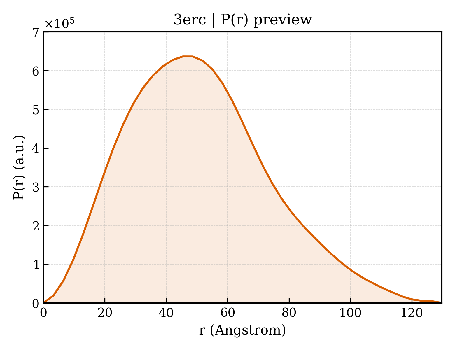
3ER8
Crystal structure of the heterodimeric vaccinia virus mRNA polyadenylate polymerase complex with two fragments of RNA
Deposited 2008-10-01
|
Different oligomeric state
Different ligand/ion
Different experimental conditions
Different structure-quality metrics
|
Assembly 1
Protein–DNA
Heteromer;Protein × 2
PDB declaration: tetrameric
|
Chain A
1–297(297 aa)
|
Mutation:R140A,K142A,R143A
|
No recorded non-water small molecule
|
X-RAY DIFFRACTION
X-ray crystallization conditions
VAPOR DIFFUSION, SITTING DROP;pH 8.7;295 K;proteins(4-5mg.ml) in 10mM Tris-HCl, pH8.7, 75 mM NaCl, 0.5 mM DTT, mixed with equal volume buffer which composed of 10mM Tris-HCl, pH 8.7, 15-20% PEG 4000, 5% glycerol, 0.5 mM DTT. room temperature for several days., VAPOR DIFFUSION, SITTING DROP, temperature 295K
|
Resolution 3.18 Å
R-free 0.285
|
|
3ER8
Crystal structure of the heterodimeric vaccinia virus mRNA polyadenylate polymerase complex with two fragments of RNA
Deposited 2008-10-01
|
Different oligomeric state
Different ligand/ion
Different experimental conditions
Different structure-quality metrics
|
Assembly 2
Protein–DNA
Heteromer;Protein × 2
PDB declaration: tetrameric
|
Chain B
1–297(297 aa)
|
Mutation:R140A,K142A,R143A
|
No recorded non-water small molecule
|
X-RAY DIFFRACTION
X-ray crystallization conditions
VAPOR DIFFUSION, SITTING DROP;pH 8.7;295 K;proteins(4-5mg.ml) in 10mM Tris-HCl, pH8.7, 75 mM NaCl, 0.5 mM DTT, mixed with equal volume buffer which composed of 10mM Tris-HCl, pH 8.7, 15-20% PEG 4000, 5% glycerol, 0.5 mM DTT. room temperature for several days., VAPOR DIFFUSION, SITTING DROP, temperature 295K
|
Resolution 3.18 Å
R-free 0.285
|
|
3ER9
Crystal structure of the heterodimeric vaccinia virus mRNA polyadenylate polymerase complex with UU and 3'-deoxy ATP
Deposited 2008-10-01
|
Different oligomeric state
Different ligand/ion
Different experimental conditions
Different structure-quality metrics
|
Assembly 1
Protein–RNA
Heteromer;Protein × 2
PDB declaration: trimeric
|
Chain A
1–297(297 aa)
|
Mutation:R140A,K142A,R143A
|
GOL GLYCEROL × 3
3AT 3'-DEOXYADENOSINE-5'-TRIPHOSPHATE × 1
CA CALCIUM ION × 2
|
X-RAY DIFFRACTION
X-ray crystallization conditions
VAPOR DIFFUSION, SITTING DROP;pH 8.7;295 K;proteins(4-5mg.ml) in 10mM tris-HCl, pH 8.7, 75 mM NaCl, 0.5 mM DTT, mixed with equal volume buffer which composed of 10mM Tris-HCl, pH 8.7, 15-20% PEG 4000, 5% glycerol, 0.5 mM DTT. Room temperature for several days., VAPOR DIFFUSION, SITTING DROP, temperature 295K
|
Resolution 2.06 Å
R-free 0.250
|
|
3MAG
VACCINIA METHYLTRANSFERASE VP39 COMPLEXED WITH M3ADE AND S-ADENOSYLHOMOCYSTEINE
Deposited 1998-07-12
|
Different construct
Different mutation/modification
Different oligomeric state
Different ligand/ion
Different experimental conditions
Different structure-quality metrics
|
Assembly 1
Protein homooligomer
Homooligomer;Protein × 2
PDB declaration: dimeric
|
Chain A
1–307(307 aa)
|
Mutation:C-TERMINAL DELETION OF 26 RESIDUES
|
SAH S-ADENOSYL-L-HOMOCYSTEINE × 2
3MA 6-AMINO-3-METHYLPURINE × 2
|
X-RAY DIFFRACTION
X-ray crystallization conditions
pH 6.5;pH 6.5
|
Resolution 1.80 Å
R-free 0.251
|
|
3MCT
VACCINIA METHYLTRANSFERASE VP39 COMPLEXED WITH M3CYT AND S-ADENOSYLHOMOCYSTEINE
Deposited 1999-01-05
|
Different mutation/modification
Different oligomeric state
Different ligand/ion
Different experimental conditions
Different structure-quality metrics
|
Assembly 1
Protein homooligomer
Homooligomer;Protein × 2
PDB declaration: dimeric
|
Chain A
1–297(297 aa)
|
Mutation:C-TERMINAL DELETION OF 26 RESIDUES
|
SAH S-ADENOSYL-L-HOMOCYSTEINE × 2
3MC 3-METHYLCYTOSINE × 2
|
X-RAY DIFFRACTION
X-ray crystallization conditions
pH 6.5;pH 6.5
|
Resolution 2.00 Å
R-free 0.285
|
|
4DCG
VACCINIA METHYLTRANSFERASE VP39 MUTANT D182A COMPLEXED WITH M7G AND S-ADENOSYLHOMOCYSTEINE
Deposited 1998-07-13
|
Different construct
Different mutation/modification
Different oligomeric state
Different ligand/ion
Different experimental conditions
Different structure-quality metrics
|
Assembly 1
Protein monomer
Monomer;Protein × 1
PDB declaration: monomeric
|
Chain A
1–307(307 aa)
|
Mutation:D182A, C-TERMINAL DELETION OF 26 RESIDUES
|
SAH S-ADENOSYL-L-HOMOCYSTEINE × 1
MG7 7-METHYLGUANOSINE × 1
|
X-RAY DIFFRACTION
X-ray crystallization conditions
pH 6.5;pH 6.5
|
Resolution 1.80 Å
R-free 0.258
|