|
1CYN
CYCLOPHILIN B COMPLEXED WITH [D-(CHOLINYLESTER)SER8]-CYCLOSPORIN
Deposited 1995-05-22
|
Different construct
Different ligand/ion
Different experimental conditions
Different structure-quality metrics
|
Assembly 1
Protein heterocomplex
Heteromer;Protein × 2
PDB declaration: dimeric
|
Chain A
31–208(178 aa)
|
Not recorded
|
No recorded non-water small molecule
|
X-RAY DIFFRACTION
mmCIF provides none of the parsed conditions
|
Resolution 1.85 Å
|
|
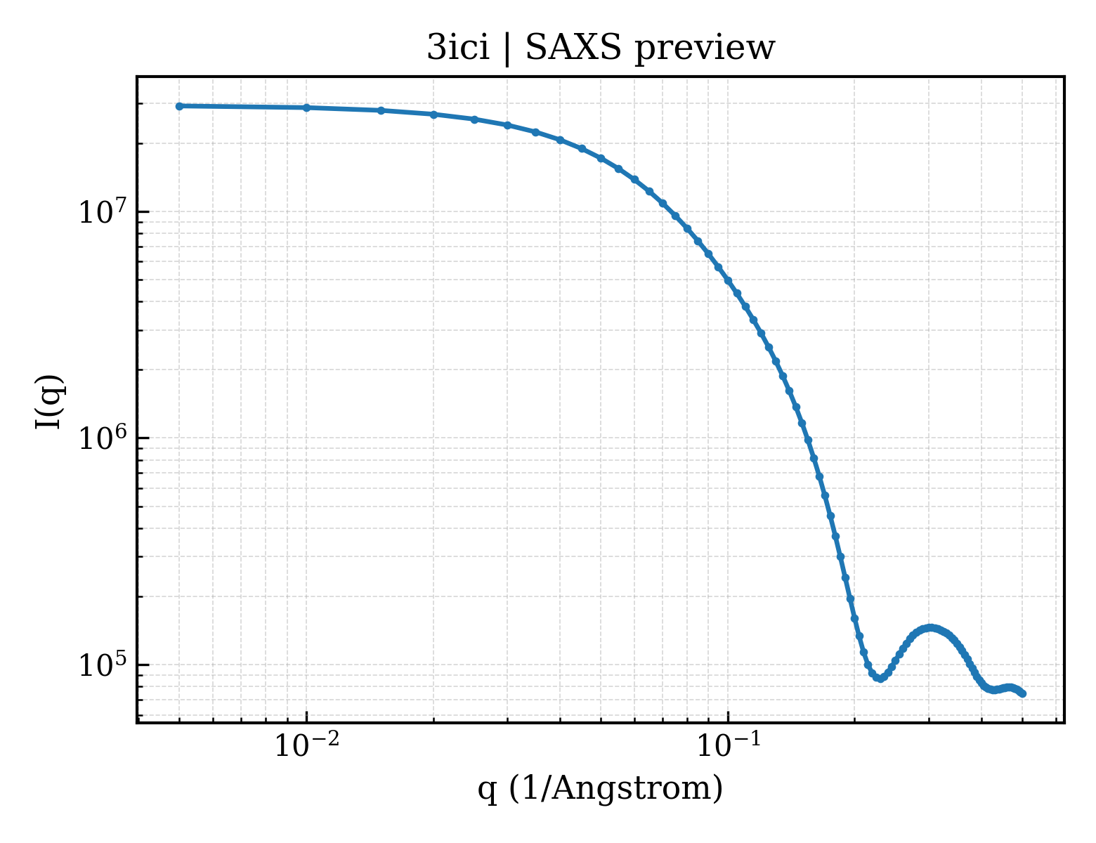
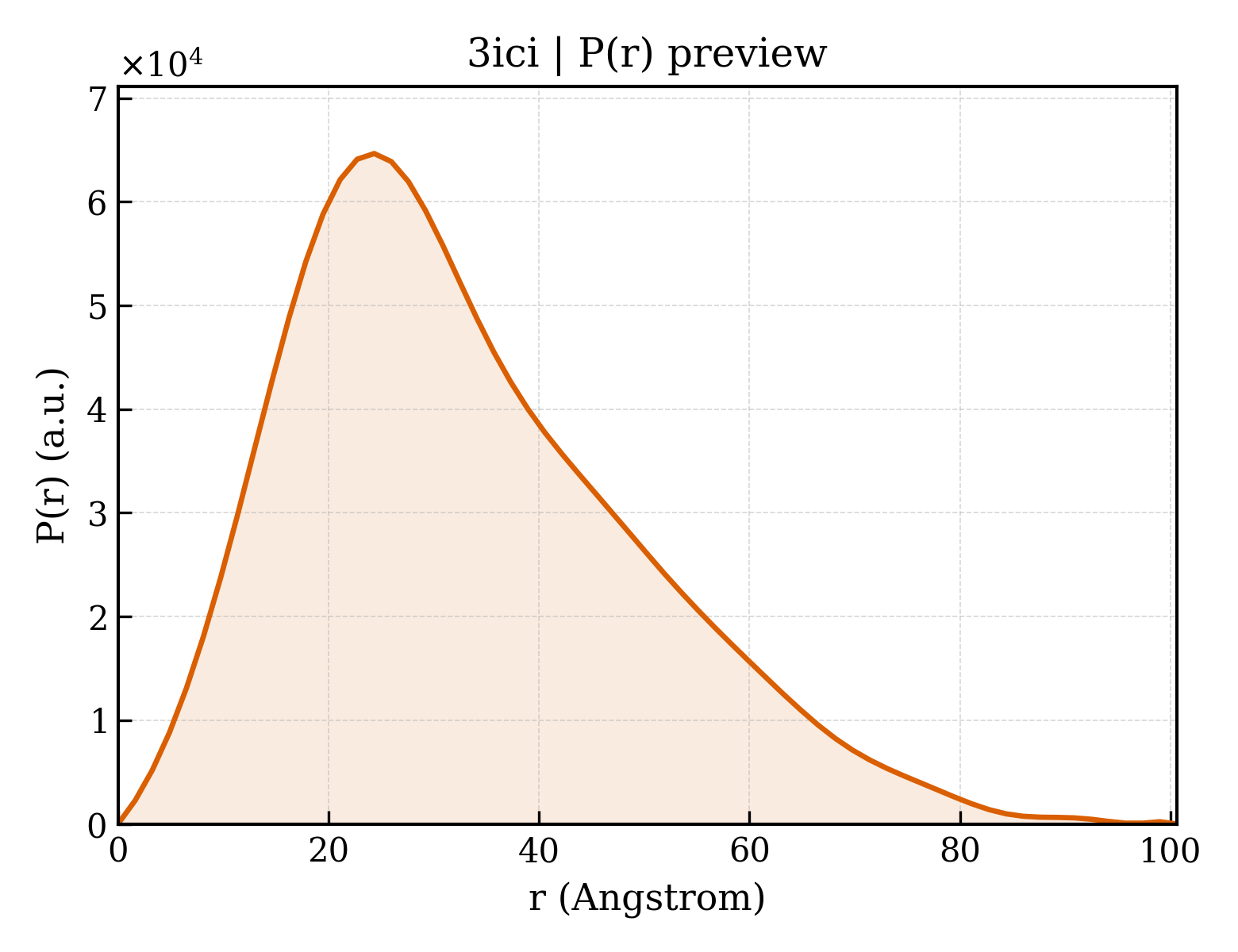
3ICH
Crystal structure of cyclophilin B at 1.2 A resolution
Deposited 2009-07-17
|
Different ligand/ion
Different experimental conditions
Different structure-quality metrics
|
Assembly 1
Protein monomer
Monomer;Protein × 1
PDB declaration: monomeric
|
Chain A
34–216(183 aa)
Fragment:UNP residues 34-216
|
Not recorded
|
No recorded non-water small molecule
|
X-RAY DIFFRACTION
X-ray crystallization conditions
VAPOR DIFFUSION, HANGING DROP;pH 6;295 K;25% w/v PEG 1500, 0.1M MMT buffer, pH 6.0, VAPOR DIFFUSION, HANGING DROP, temperature 295K
|
Resolution 1.20 Å
R-free 0.162
|
|
8K0F
Human collagen prolyl processing enzyme complex, P3H1/CRTAP/PPIB heterotrimer, in its apo state
Deposited 2023-07-08
|
Different construct
Different oligomeric state
Different ligand/ion
Different experimental method
Different experimental conditions
Different structure-quality metrics
|
Assembly 1
Protein heterocomplex
Heteromer;Protein × 3
PDB declaration: trimeric
|
Chain C
1–216(216 aa)
|
Not recorded
|
NAG 2-acetamido-2-deoxy-beta-D-glucopyranose × 4
FE FE (III) ION × 1
|
ELECTRON MICROSCOPY
cryo-EM buffer
pH 7.5
cryo-EM vitrification conditions
Cryogen ETHANE
|
Resolution 3.37 Å
|
|
8K0I
Human collagen prolyl processing enzyme complex, P3H1/CRTAP/PPIB heterotrimer, in its dual-ternary state
Deposited 2023-07-09
|
Different construct
Different mutation/modification
Different oligomeric state
Different ligand/ion
Different experimental method
Different experimental conditions
Different structure-quality metrics
|
Assembly 1
Protein heterocomplex
Heteromer;Protein × 6
PDB declaration: hexameric
|
Chain C
1–216(216 aa)
Chain c
1–216(216 aa)
|
Not recorded
|
NAG 2-acetamido-2-deoxy-beta-D-glucopyranose × 6
|
ELECTRON MICROSCOPY
cryo-EM buffer
pH 7.5
cryo-EM vitrification conditions
Cryogen ETHANE
|
Resolution 3.62 Å
|
|
8K0M
Human collagen prolyl processing enzyme complex, P3H1/CRTAP/PPIB heterotrimer, bound to 2-oxoglutarate
Deposited 2023-07-09
|
Different construct
Different oligomeric state
Different ligand/ion
Different experimental method
Different experimental conditions
Different structure-quality metrics
|
Assembly 1
Protein heterocomplex
Heteromer;Protein × 3
PDB declaration: trimeric
|
Chain C
1–216(216 aa)
|
Not recorded
|
NAG 2-acetamido-2-deoxy-beta-D-glucopyranose × 4
FE FE (III) ION × 1
AKG 2-OXOGLUTARIC ACID × 1
|
ELECTRON MICROSCOPY
cryo-EM buffer
pH 7.5
cryo-EM vitrification conditions
Cryogen ETHANE
|
Resolution 3.17 Å
|
|
8K17
Human collagen prolyl processing enzyme complex, P3H1/CRTAP/PPIB heterotrimer, bound to collagen alpha-1(I) chain
Deposited 2023-07-10
|
Different construct
Different oligomeric state
Different ligand/ion
Different experimental method
Different experimental conditions
Different structure-quality metrics
|
Assembly 1
Protein heterocomplex
Heteromer;Protein × 4
PDB declaration: tetrameric
|
Chain C
1–216(216 aa)
|
Not recorded
|
NAG 2-acetamido-2-deoxy-beta-D-glucopyranose × 4
FE FE (III) ION × 1
|
ELECTRON MICROSCOPY
cryo-EM buffer
pH 7.5
cryo-EM vitrification conditions
Cryogen ETHANE
|
Resolution 3.18 Å
|
|
8KC9
Human collagen prolyl processing enzyme complex, P3H1/CRTAP/PPIB heterotrimer, bound to cyclosporin A
Deposited 2023-08-06
|
Different construct
Different oligomeric state
Different ligand/ion
Different experimental method
Different experimental conditions
Different structure-quality metrics
|
Assembly 1
Protein heterocomplex
Heteromer;Protein × 4
PDB declaration: tetrameric
|
Chain C
1–216(216 aa)
|
Not recorded
|
NAG 2-acetamido-2-deoxy-beta-D-glucopyranose × 4
FE FE (III) ION × 1
|
ELECTRON MICROSCOPY
cryo-EM buffer
pH 7.5
cryo-EM vitrification conditions
Cryogen ETHANE
|
Resolution 3.75 Å
|