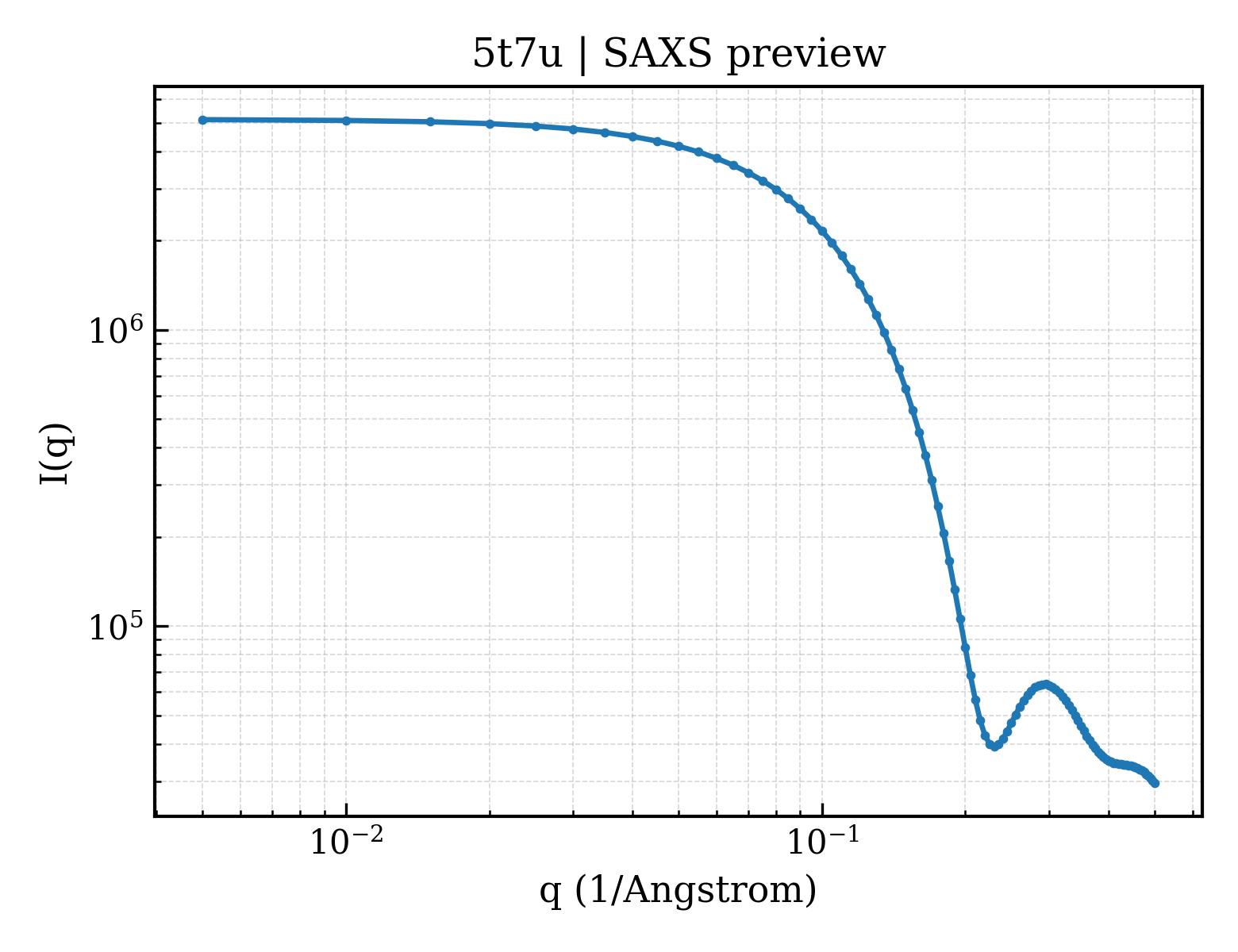
Galectin-8
Homo sapiens
State in the Current Structure
| Assembly | Oligomeric State | Construct | Mutations and Modifications | Ligands, Ions and Associated Components | Method and Experimental Conditions | Structure Quality |
|---|---|---|---|---|---|---|
| 1 | Protein monomer Monomer Protein × 1 PDB declaration: monomeric(1) Consistent with protein copy count | Chain A; UniProt 1–155 | Fragment:N-terminal Domaine (UNP residues 1-155) Non-standard monomer:Yes (specific site not provided by mmCIF) | GOL GLYCEROL × 2 NA SODIUM ION × 1 CL CHLORIDE ION × 1 | X-RAY DIFFRACTION X-ray crystallization conditions:VAPOR DIFFUSION, HANGING DROP;293 K;10 mM sodium phosphate, 137 mM sodium chloride, 2.7 mM potassium chloride, 1.8 mM potassium phosphate | Resolution 1.58 Å R-free 0.147 |
Other States of the Same Protein in the Database
Each row is a biological assembly of the same UniProt protein in another PDB entry. The “Difference from current entry” column identifies evidence-level differences; no tag means the currently parsed fields agree.
| Other PDB | Difference from Current Entry 5T7U | Assembly / Oligomeric State | Construct | Mutations and Modifications | Ligands, Ions and Non-polymers | Method and Experimental Conditions | Structure Quality |
|---|---|---|---|---|---|---|---|
| 2YRO Solution structure of the C-terminal Gal-bind lectin protein from Human Galectin-8 Deposited 2007-04-02 | Different construct Different mutation/modification Different ligand/ion Different experimental method Different experimental conditions Different structure-quality metrics | Assembly 1 Protein monomer Monomer;Protein × 1 PDB declaration: monomeric |
Chain A
175–316(142 aa)
Fragment:Gal-bind_lectin domain
|
Not recorded | No recorded non-water small molecule |
SOLUTION NMR
NMR measurement conditions
pH 7;296 K;Ionic strength (raw mmCIF value) 120mM;Pressure ambient
NMR sample composition
1.28mM Gal-binding lectin U-15N, 13C; 20mM d-Tris-HCl(pH 7.0); 100mM NaCl; 1mM d-DTT; 0.02% NaN3; 90% H2O, 10% D2O | 90% H2O/10% D2O
|
Resolution not provided |
| 3AP4 Crystal structure of the galectin-8 N-terminal carbohydrate recognition domain in complex with lactose Deposited 2010-10-11 | Different construct Different mutation/modification Different oligomeric state Different ligand/ion Different experimental conditions Different structure-quality metrics | Assembly 1 Other combination Monomer;Protein × 1 PDB declaration: monomeric |
Chain A
1–154(154 aa)
Fragment:N-terminal carbohydrate recognition domain
|
Not recorded | No recorded non-water small molecule |
X-RAY DIFFRACTION
X-ray crystallization conditions
VAPOR DIFFUSION, HANGING DROP;pH 7;293.15 K;0.275mM protein, 10mM Hepes-NaOH, 2.75mM lactose, 50mM sodium chloride, 0.5mM DTT, 50mM ammonium fluoride, 12% PEG 3350, pH 7.0, VAPOR DIFFUSION, HANGING DROP, temperature 293.15K
|
Resolution 2.33 Å R-free 0.251 |
| 3AP4 Crystal structure of the galectin-8 N-terminal carbohydrate recognition domain in complex with lactose Deposited 2010-10-11 | Different construct Different mutation/modification Different oligomeric state Different ligand/ion Different experimental conditions Different structure-quality metrics | Assembly 2 Other combination Monomer;Protein × 1 PDB declaration: monomeric |
Chain B
1–154(154 aa)
Fragment:N-terminal carbohydrate recognition domain
|
Not recorded | No recorded non-water small molecule |
X-RAY DIFFRACTION
X-ray crystallization conditions
VAPOR DIFFUSION, HANGING DROP;pH 7;293.15 K;0.275mM protein, 10mM Hepes-NaOH, 2.75mM lactose, 50mM sodium chloride, 0.5mM DTT, 50mM ammonium fluoride, 12% PEG 3350, pH 7.0, VAPOR DIFFUSION, HANGING DROP, temperature 293.15K
|
Resolution 2.33 Å R-free 0.251 |
| 3AP4 Crystal structure of the galectin-8 N-terminal carbohydrate recognition domain in complex with lactose Deposited 2010-10-11 | Different construct Different mutation/modification Different oligomeric state Different ligand/ion Different experimental conditions Different structure-quality metrics | Assembly 3 Other combination Monomer;Protein × 1 PDB declaration: monomeric |
Chain C
1–154(154 aa)
Fragment:N-terminal carbohydrate recognition domain
|
Not recorded | No recorded non-water small molecule |
X-RAY DIFFRACTION
X-ray crystallization conditions
VAPOR DIFFUSION, HANGING DROP;pH 7;293.15 K;0.275mM protein, 10mM Hepes-NaOH, 2.75mM lactose, 50mM sodium chloride, 0.5mM DTT, 50mM ammonium fluoride, 12% PEG 3350, pH 7.0, VAPOR DIFFUSION, HANGING DROP, temperature 293.15K
|
Resolution 2.33 Å R-free 0.251 |
| 3AP4 Crystal structure of the galectin-8 N-terminal carbohydrate recognition domain in complex with lactose Deposited 2010-10-11 | Different construct Different mutation/modification Different oligomeric state Different ligand/ion Different experimental conditions Different structure-quality metrics | Assembly 4 Other combination Monomer;Protein × 1 PDB declaration: monomeric |
Chain D
1–154(154 aa)
Fragment:N-terminal carbohydrate recognition domain
|
Not recorded | No recorded non-water small molecule |
X-RAY DIFFRACTION
X-ray crystallization conditions
VAPOR DIFFUSION, HANGING DROP;pH 7;293.15 K;0.275mM protein, 10mM Hepes-NaOH, 2.75mM lactose, 50mM sodium chloride, 0.5mM DTT, 50mM ammonium fluoride, 12% PEG 3350, pH 7.0, VAPOR DIFFUSION, HANGING DROP, temperature 293.15K
|
Resolution 2.33 Å R-free 0.251 |
| 3AP5 Crystal structure of the galectin-8 N-terminal carbohydrate recognition domain Deposited 2010-10-11 | Different construct Different mutation/modification Different ligand/ion Different experimental conditions Different structure-quality metrics | Assembly 1 Protein monomer Monomer;Protein × 1 PDB declaration: monomeric |
Chain A
1–154(154 aa)
Fragment:N-terminal carbohydrate recognition domain
|
Not recorded | No recorded non-water small molecule |
X-RAY DIFFRACTION
X-ray crystallization conditions
VAPOR DIFFUSION, HANGING DROP;pH 7;293.15 K;0.275mM protein, 10mM Hepes-NaOH, 50mM sodium chloride, 0.5mM DTT, 125mM ammonium fluoride, 8% PEG 3350 , pH 7.0, VAPOR DIFFUSION, HANGING DROP, temperature 293.15K
|
Resolution 1.92 Å R-free 0.237 |
| 3AP6 Crystal structure of the galectin-8 N-terminal carbohydrate recognition domain in complex with lactose 3'-sulfate Deposited 2010-10-12 | Different construct Different mutation/modification Different oligomeric state Different ligand/ion Different experimental conditions Different structure-quality metrics | Assembly 1 Other combination Monomer;Protein × 1 PDB declaration: monomeric |
Chain A
1–154(154 aa)
Fragment:N-terminal carbohydrate recognition domain
|
Not recorded | SO4 SULFATE ION × 1 |
X-RAY DIFFRACTION
X-ray crystallization conditions
VAPOR DIFFUSION, HANGING DROP;pH 7;293.15 K;0.275mM protein, 10mM Hepes-NaOH, 2.75mM lactose 3'-sulfate, 50mM sodium chloride, 0.5mM DTT, 50mM ammonium fluoride, 9% PEG 3350, pH 7.0, VAPOR DIFFUSION, HANGING DROP, temperature 293.15K
|
Resolution 1.58 Å R-free 0.242 |
| 3AP6 Crystal structure of the galectin-8 N-terminal carbohydrate recognition domain in complex with lactose 3'-sulfate Deposited 2010-10-12 | Different construct Different mutation/modification Different oligomeric state Different ligand/ion Different experimental conditions Different structure-quality metrics | Assembly 2 Other combination Monomer;Protein × 1 PDB declaration: monomeric |
Chain B
1–154(154 aa)
Fragment:N-terminal carbohydrate recognition domain
|
Not recorded | SO4 SULFATE ION × 1 |
X-RAY DIFFRACTION
X-ray crystallization conditions
VAPOR DIFFUSION, HANGING DROP;pH 7;293.15 K;0.275mM protein, 10mM Hepes-NaOH, 2.75mM lactose 3'-sulfate, 50mM sodium chloride, 0.5mM DTT, 50mM ammonium fluoride, 9% PEG 3350, pH 7.0, VAPOR DIFFUSION, HANGING DROP, temperature 293.15K
|
Resolution 1.58 Å R-free 0.242 |
| 3AP6 Crystal structure of the galectin-8 N-terminal carbohydrate recognition domain in complex with lactose 3'-sulfate Deposited 2010-10-12 | Different construct Different mutation/modification Different oligomeric state Different ligand/ion Different experimental conditions Different structure-quality metrics | Assembly 3 Other combination Monomer;Protein × 1 PDB declaration: monomeric |
Chain C
1–154(154 aa)
Fragment:N-terminal carbohydrate recognition domain
|
Not recorded | SO4 SULFATE ION × 1 |
X-RAY DIFFRACTION
X-ray crystallization conditions
VAPOR DIFFUSION, HANGING DROP;pH 7;293.15 K;0.275mM protein, 10mM Hepes-NaOH, 2.75mM lactose 3'-sulfate, 50mM sodium chloride, 0.5mM DTT, 50mM ammonium fluoride, 9% PEG 3350, pH 7.0, VAPOR DIFFUSION, HANGING DROP, temperature 293.15K
|
Resolution 1.58 Å R-free 0.242 |
| 3AP6 Crystal structure of the galectin-8 N-terminal carbohydrate recognition domain in complex with lactose 3'-sulfate Deposited 2010-10-12 | Different construct Different mutation/modification Different oligomeric state Different ligand/ion Different experimental conditions Different structure-quality metrics | Assembly 4 Other combination Monomer;Protein × 1 PDB declaration: monomeric |
Chain D
1–154(154 aa)
Fragment:N-terminal carbohydrate recognition domain
|
Not recorded | SO4 SULFATE ION × 1 |
X-RAY DIFFRACTION
X-ray crystallization conditions
VAPOR DIFFUSION, HANGING DROP;pH 7;293.15 K;0.275mM protein, 10mM Hepes-NaOH, 2.75mM lactose 3'-sulfate, 50mM sodium chloride, 0.5mM DTT, 50mM ammonium fluoride, 9% PEG 3350, pH 7.0, VAPOR DIFFUSION, HANGING DROP, temperature 293.15K
|
Resolution 1.58 Å R-free 0.242 |
| 3AP7 Crystal structure of the galectin-8 N-terminal carbohydrate recognition domain in complex with lactose sialic acid Deposited 2010-10-12 | Different construct Different mutation/modification Different oligomeric state Different ligand/ion Different experimental conditions Different structure-quality metrics | Assembly 1 Other combination Monomer;Protein × 1 PDB declaration: monomeric |
Chain A
1–154(154 aa)
Fragment:N-terminal carbohydrate recognition domain
|
Not recorded | CL CHLORIDE ION × 1 |
X-RAY DIFFRACTION
X-ray crystallization conditions
VAPOR DIFFUSION, HANGING DROP;pH 7;293.15 K;0.275mM protein, 10mM Hepes-NaOH, 2.75mM lactose sialic acid, 50mM sodium chloride, 0.5mM DTT, 80mM ammonium
fluoride, 7.5% PEG 3350, pH 7.0, VAPOR DIFFUSION, HANGING DROP, temperature 293.15K
|
Resolution 1.53 Å R-free 0.188 |
| 3AP9 Crystal structure of the galectin-8 N-terminal carbohydrate recognition domain in complex with Lacto-N-fucopentaose III Deposited 2010-10-12 | Different construct Different mutation/modification Different oligomeric state Different ligand/ion Different experimental conditions Different structure-quality metrics | Assembly 1 Other combination Monomer;Protein × 1 PDB declaration: monomeric |
Chain A
1–154(154 aa)
Fragment:N-terminal carbohydrate recognition domain
|
Not recorded | MPD (4S)-2-METHYL-2,4-PENTANEDIOL × 1 CL CHLORIDE ION × 3 |
X-RAY DIFFRACTION
X-ray crystallization conditions
VAPOR DIFFUSION, HANGING DROP;pH 7;293.15 K;0.275mM protein, 10mM Hepes-NaOH, 2.75mM Lacto-N-fucopentaose III, 50mM sodium chloride, 0.5mM DTT, 50mM ammonium fluoride, 12% PEG 3350, pH 7.0, VAPOR DIFFUSION, HANGING DROP, temperature 293.15K
|
Resolution 1.33 Å R-free 0.178 |
| 3APB Crystal structure of the galectin-8 N-terminal carbohydrate recognition domain in complex with iodide Deposited 2010-10-14 | Different construct Different mutation/modification Different ligand/ion Different experimental conditions Different structure-quality metrics | Assembly 1 Protein monomer Monomer;Protein × 1 PDB declaration: monomeric |
Chain A
1–154(154 aa)
Fragment:N-terminal carbohydrate recognition domain
|
Not recorded | IOD IODIDE ION × 13 |
X-RAY DIFFRACTION
X-ray crystallization conditions
VAPOR DIFFUSION, HANGING DROP;pH 7;293.15 K;0.55mM protein, 10mM Hepes-NaOH, 50mM sodium chloride, 0.5mM DTT, 125mM ammonium fluoride, 8% PEG 3350, pH 7.0, VAPOR DIFFUSION, HANGING DROP, temperature 293.15K
|
Resolution 1.95 Å R-free 0.242 |
| 3APB Crystal structure of the galectin-8 N-terminal carbohydrate recognition domain in complex with iodide Deposited 2010-10-14 | Different construct Different mutation/modification Different ligand/ion Different experimental conditions Different structure-quality metrics | Assembly 2 Protein monomer Monomer;Protein × 1 PDB declaration: monomeric |
Chain B
1–154(154 aa)
Fragment:N-terminal carbohydrate recognition domain
|
Not recorded | IOD IODIDE ION × 16 |
X-RAY DIFFRACTION
X-ray crystallization conditions
VAPOR DIFFUSION, HANGING DROP;pH 7;293.15 K;0.55mM protein, 10mM Hepes-NaOH, 50mM sodium chloride, 0.5mM DTT, 125mM ammonium fluoride, 8% PEG 3350, pH 7.0, VAPOR DIFFUSION, HANGING DROP, temperature 293.15K
|
Resolution 1.95 Å R-free 0.242 |
| 3OJB Crystal structure of C-terminal domain of human galectin-8 Deposited 2010-08-21 | Different construct Different mutation/modification Different ligand/ion Different experimental conditions Different structure-quality metrics | Assembly 1 Protein monomer Monomer;Protein × 1 PDB declaration: monomeric |
Chain A
186–315(130 aa)
Fragment:C-terminal domain
|
Not recorded | No recorded non-water small molecule |
X-RAY DIFFRACTION
X-ray crystallization conditions
VAPOR DIFFUSION, HANGING DROP;pH 6.5;281 K;27.5% PEG 8000, 0.2M ammonium phosphate, 6.5% Ethylene Glycol, 0.1M Na Cacodylate, VAPOR DIFFUSION, HANGING DROP, temperature 281K
|
Resolution 3.01 Å R-free 0.292 |
| 3OJB Crystal structure of C-terminal domain of human galectin-8 Deposited 2010-08-21 | Different construct Different mutation/modification Different ligand/ion Different experimental conditions Different structure-quality metrics | Assembly 2 Protein monomer Monomer;Protein × 1 PDB declaration: monomeric |
Chain B
186–315(130 aa)
Fragment:C-terminal domain
|
Not recorded | No recorded non-water small molecule |
X-RAY DIFFRACTION
X-ray crystallization conditions
VAPOR DIFFUSION, HANGING DROP;pH 6.5;281 K;27.5% PEG 8000, 0.2M ammonium phosphate, 6.5% Ethylene Glycol, 0.1M Na Cacodylate, VAPOR DIFFUSION, HANGING DROP, temperature 281K
|
Resolution 3.01 Å R-free 0.292 |
| 3OJB Crystal structure of C-terminal domain of human galectin-8 Deposited 2010-08-21 | Different construct Different mutation/modification Different ligand/ion Different experimental conditions Different structure-quality metrics | Assembly 3 Protein monomer Monomer;Protein × 1 PDB declaration: monomeric |
Chain C
186–315(130 aa)
Fragment:C-terminal domain
|
Not recorded | No recorded non-water small molecule |
X-RAY DIFFRACTION
X-ray crystallization conditions
VAPOR DIFFUSION, HANGING DROP;pH 6.5;281 K;27.5% PEG 8000, 0.2M ammonium phosphate, 6.5% Ethylene Glycol, 0.1M Na Cacodylate, VAPOR DIFFUSION, HANGING DROP, temperature 281K
|
Resolution 3.01 Å R-free 0.292 |
| 3OJB Crystal structure of C-terminal domain of human galectin-8 Deposited 2010-08-21 | Different construct Different mutation/modification Different ligand/ion Different experimental conditions Different structure-quality metrics | Assembly 4 Protein monomer Monomer;Protein × 1 PDB declaration: monomeric |
Chain D
186–315(130 aa)
Fragment:C-terminal domain
|
Not recorded | No recorded non-water small molecule |
X-RAY DIFFRACTION
X-ray crystallization conditions
VAPOR DIFFUSION, HANGING DROP;pH 6.5;281 K;27.5% PEG 8000, 0.2M ammonium phosphate, 6.5% Ethylene Glycol, 0.1M Na Cacodylate, VAPOR DIFFUSION, HANGING DROP, temperature 281K
|
Resolution 3.01 Å R-free 0.292 |
| 3VKL Protease-resistant mutant form of human Galectin-8 in complex with two lactose molecules Deposited 2011-11-18 | Different construct Different mutation/modification Different oligomeric state Different ligand/ion Different experimental conditions Different structure-quality metrics | Assembly 1 Other combination Monomer;Protein × 1 PDB declaration: monomeric |
Chain A
1–155(155 aa)
Fragment:N-terminal carbohydrate recognition domain, C-terminal carbohydrate recognition domain
Chain A
184–317(134 aa)
Fragment:N-terminal carbohydrate recognition domain, C-terminal carbohydrate recognition domain
|
Not recorded | EDO 1,2-ETHANEDIOL × 12 |
X-RAY DIFFRACTION
X-ray crystallization conditions
VAPOR DIFFUSION, HANGING DROP;pH 9;293 K;2-3%(v/v) 1,4-dioxane, 9-10%(w/v) PEG20000, 0.1M bicine, pH 9.0, VAPOR DIFFUSION, HANGING DROP, temperature 293K
|
Resolution 2.55 Å R-free 0.264 |
| 3VKL Protease-resistant mutant form of human Galectin-8 in complex with two lactose molecules Deposited 2011-11-18 | Different construct Different mutation/modification Different oligomeric state Different ligand/ion Different experimental conditions Different structure-quality metrics | Assembly 2 Other combination Monomer;Protein × 1 PDB declaration: monomeric |
Chain B
1–155(155 aa)
Fragment:N-terminal carbohydrate recognition domain, C-terminal carbohydrate recognition domain
Chain B
184–317(134 aa)
Fragment:N-terminal carbohydrate recognition domain, C-terminal carbohydrate recognition domain
|
Not recorded | EDO 1,2-ETHANEDIOL × 8 |
X-RAY DIFFRACTION
X-ray crystallization conditions
VAPOR DIFFUSION, HANGING DROP;pH 9;293 K;2-3%(v/v) 1,4-dioxane, 9-10%(w/v) PEG20000, 0.1M bicine, pH 9.0, VAPOR DIFFUSION, HANGING DROP, temperature 293K
|
Resolution 2.55 Å R-free 0.264 |
| 3VKM Protease-resistant mutant form of Human Galectin-8 in complex with sialyllactose and lactose Deposited 2011-11-18 | Different construct Different mutation/modification Different oligomeric state Different ligand/ion Different experimental conditions Different structure-quality metrics | Assembly 1 Other combination Monomer;Protein × 1 PDB declaration: monomeric |
Chain A
1–155(155 aa)
Fragment:N-terminal carbohydrate recognition domain, C-terminal carbohydrate recognition domain
Chain A
184–317(134 aa)
Fragment:N-terminal carbohydrate recognition domain, C-terminal carbohydrate recognition domain
|
Not recorded | EDO 1,2-ETHANEDIOL × 11 |
X-RAY DIFFRACTION
X-ray crystallization conditions
VAPOR DIFFUSION, HANGING DROP;pH 9;293 K;2-3%(v/v) 1,4-dioxane, 9-10%(w/v) PEG20000, 0.1M bicine, pH 9.0, VAPOR DIFFUSION, HANGING DROP, temperature 293K
|
Resolution 2.98 Å R-free 0.261 |
| 3VKM Protease-resistant mutant form of Human Galectin-8 in complex with sialyllactose and lactose Deposited 2011-11-18 | Different construct Different mutation/modification Different oligomeric state Different ligand/ion Different experimental conditions Different structure-quality metrics | Assembly 2 Other combination Monomer;Protein × 1 PDB declaration: monomeric |
Chain B
1–155(155 aa)
Fragment:N-terminal carbohydrate recognition domain, C-terminal carbohydrate recognition domain
Chain B
184–317(134 aa)
Fragment:N-terminal carbohydrate recognition domain, C-terminal carbohydrate recognition domain
|
Not recorded | EDO 1,2-ETHANEDIOL × 13 |
X-RAY DIFFRACTION
X-ray crystallization conditions
VAPOR DIFFUSION, HANGING DROP;pH 9;293 K;2-3%(v/v) 1,4-dioxane, 9-10%(w/v) PEG20000, 0.1M bicine, pH 9.0, VAPOR DIFFUSION, HANGING DROP, temperature 293K
|
Resolution 2.98 Å R-free 0.261 |
| 3VKN Galectin-8 N-terminal domain in free form Deposited 2011-11-18 | Different construct Different mutation/modification Different ligand/ion Different experimental conditions Different structure-quality metrics | Assembly 1 Protein monomer Monomer;Protein × 1 PDB declaration: monomeric |
Chain A
1–153(153 aa)
Fragment:N-TERMINAL CARBOHYDRATE RECOGNITION DOMAIN
|
Not recorded | CL CHLORIDE ION × 1 |
X-RAY DIFFRACTION
X-ray crystallization conditions
VAPOR DIFFUSION, SITTING DROP;pH 9.5;293 K;30%(v/v) PEG 400, 0.1M CHES, pH 9.5, VAPOR DIFFUSION, SITTING DROP, temperature 293K
|
Resolution 1.98 Å R-free 0.251 |
| 3VKN Galectin-8 N-terminal domain in free form Deposited 2011-11-18 | Different construct Different mutation/modification Different ligand/ion Different experimental conditions Different structure-quality metrics | Assembly 2 Protein monomer Monomer;Protein × 1 PDB declaration: monomeric |
Chain B
1–153(153 aa)
Fragment:N-TERMINAL CARBOHYDRATE RECOGNITION DOMAIN
|
Not recorded | No recorded non-water small molecule |
X-RAY DIFFRACTION
X-ray crystallization conditions
VAPOR DIFFUSION, SITTING DROP;pH 9.5;293 K;30%(v/v) PEG 400, 0.1M CHES, pH 9.5, VAPOR DIFFUSION, SITTING DROP, temperature 293K
|
Resolution 1.98 Å R-free 0.251 |
| 3VKO Galectin-8 N-terminal domain in complex with sialyllactosamine Deposited 2011-11-18 | Different construct Different mutation/modification Different oligomeric state Different ligand/ion Different experimental conditions Different structure-quality metrics | Assembly 1 Other combination Monomer;Protein × 1 PDB declaration: monomeric |
Chain A
1–153(153 aa)
Fragment:N-TERMINAL CARBOHYDRATE RECOGNITION DOMAIN
|
Not recorded | CL CHLORIDE ION × 1 |
X-RAY DIFFRACTION
X-ray crystallization conditions
VAPOR DIFFUSION, SITTING DROP;pH 9.5;293 K;30%(v/v) PEG400, 0.1M CHES, pH 9.5, VAPOR DIFFUSION, SITTING DROP, temperature 293K
|
Resolution 2.08 Å R-free 0.249 |
| 3VKO Galectin-8 N-terminal domain in complex with sialyllactosamine Deposited 2011-11-18 | Different construct Different mutation/modification Different oligomeric state Different ligand/ion Different experimental conditions Different structure-quality metrics | Assembly 2 Other combination Monomer;Protein × 1 PDB declaration: monomeric |
Chain B
1–153(153 aa)
Fragment:N-TERMINAL CARBOHYDRATE RECOGNITION DOMAIN
|
Not recorded | No recorded non-water small molecule |
X-RAY DIFFRACTION
X-ray crystallization conditions
VAPOR DIFFUSION, SITTING DROP;pH 9.5;293 K;30%(v/v) PEG400, 0.1M CHES, pH 9.5, VAPOR DIFFUSION, SITTING DROP, temperature 293K
|
Resolution 2.08 Å R-free 0.249 |
| 4BMB Crystal structure of the N terminal domain of human Galectin 8 Deposited 2013-05-07 | Different construct Different mutation/modification Different oligomeric state Different ligand/ion Different experimental conditions Different structure-quality metrics | Assembly 1 Other combination Monomer;Protein × 1 PDB declaration: monomeric |
Chain A
4–153(150 aa)
Fragment:N TERMINAL DOMAIN, RESIDUES 4-153
|
Not recorded | ZN ZINC ION × 1 NA SODIUM ION × 1 GOL GLYCEROL × 1 |
X-RAY DIFFRACTION
X-ray crystallization conditions
20% W/V PEG 4000, 100 MM HEPES SODIUM SALT (PH 7.2) AND 10 MM ZINC CHLORIDE
|
Resolution 1.35 Å R-free 0.177 |
| 4BME Crystal structure of the N terminal domain of human Galectin 8, F19Y mutant Deposited 2013-05-07 | Different construct Different mutation/modification Different oligomeric state Different ligand/ion Different experimental conditions Different structure-quality metrics | Assembly 1 Other combination Monomer;Protein × 1 PDB declaration: monomeric |
Chain A
4–155(152 aa)
Fragment:N TERMINAL DOMAIN, RESIDUES 4-155
|
Mutation:YES | No recorded non-water small molecule |
X-RAY DIFFRACTION
X-ray crystallization conditions
30% W/V PEG 4000, 100 MM MES SODIUM SALT (PH 6.5)
|
Resolution 2.00 Å R-free 0.241 |
| 4BME Crystal structure of the N terminal domain of human Galectin 8, F19Y mutant Deposited 2013-05-07 | Different construct Different mutation/modification Different oligomeric state Different ligand/ion Different experimental conditions Different structure-quality metrics | Assembly 2 Other combination Monomer;Protein × 1 PDB declaration: monomeric |
Chain B
4–155(152 aa)
Fragment:N TERMINAL DOMAIN, RESIDUES 4-155
|
Mutation:YES | No recorded non-water small molecule |
X-RAY DIFFRACTION
X-ray crystallization conditions
30% W/V PEG 4000, 100 MM MES SODIUM SALT (PH 6.5)
|
Resolution 2.00 Å R-free 0.241 |
| 4FQZ Crystal structure of a protease-resistant mutant form of human galectin-8 Deposited 2012-06-26 | Different construct Different mutation/modification Different oligomeric state Different ligand/ion Different experimental conditions Different structure-quality metrics | Assembly 1 Other combination Monomer;Protein × 1 PDB declaration: monomeric |
Chain A
1–155(155 aa)
Fragment:N-terminal carbohydrate recognition domain, C-terminal carbohydrate recognition domain
Chain A
184–317(134 aa)
Fragment:N-terminal carbohydrate recognition domain, C-terminal carbohydrate recognition domain
|
Not recorded | EDO 1,2-ETHANEDIOL × 10 |
X-RAY DIFFRACTION
X-ray crystallization conditions
VAPOR DIFFUSION, HANGING DROP;pH 9;293 K;10% PEG 20000, 0.1M Bicine, 2% Dioxane, pH 9.0, VAPOR DIFFUSION, HANGING DROP, temperature 293K
|
Resolution 2.80 Å R-free 0.253 |
| 4GXL The crystal structure of Galectin-8 C-CRD in complex with NDP52 Deposited 2012-09-04 | Different construct Different mutation/modification Different oligomeric state Different ligand/ion Different experimental conditions Different structure-quality metrics | Assembly 1 Protein heterocomplex Heteromer;Protein × 2 PDB declaration: dimeric |
Chain A
186–317(132 aa)
Fragment:UNP residues 186-317
|
Not recorded | GOL GLYCEROL × 2 |
X-RAY DIFFRACTION
X-ray crystallization conditions
VAPOR DIFFUSION, SITTING DROP;pH 7.5;283 K;2% PEG 400, 0.1M HEPES sodium pH 7.5, 2.0M ammonium sulfate, VAPOR DIFFUSION, SITTING DROP, temperature 283K
|
Resolution 2.02 Å R-free 0.216 |
| 4HAN Crystal structure of Galectin 8 with NDP52 peptide Deposited 2012-09-27 | Different construct Different mutation/modification Different oligomeric state Different ligand/ion Different experimental conditions Different structure-quality metrics | Assembly 1 Protein heterocomplex Heteromer;Protein × 4 PDB declaration: tetrameric |
Chain A
1–155(155 aa)
Fragment:UNP residues 1-155. 184-317
Chain A
184–317(134 aa)
Fragment:UNP residues 1-155. 184-317
Chain B
1–155(155 aa)
Fragment:UNP residues 1-155. 184-317
Chain B
184–317(134 aa)
Fragment:UNP residues 1-155. 184-317
|
Not recorded | NAD NICOTINAMIDE-ADENINE-DINUCLEOTIDE × 2 PEG DI(HYDROXYETHYL)ETHER × 3 |
X-RAY DIFFRACTION
X-ray crystallization conditions
VAPOR DIFFUSION, HANGING DROP;pH 8.5;295 K;0.1M Tris-Hcl pH8.5, 30~34% (v/w) PEG400, 200mM LiSO4, and 10mM Nicotinamide adenine dinucleotide, VAPOR DIFFUSION, HANGING DROP, temperature 295K
|
Resolution 2.55 Å R-free 0.214 |
| 5GZC Crystal structure of Galectin-8 N-CRD with part of linker Deposited 2016-09-28 | Different construct Different mutation/modification Different ligand/ion Different experimental conditions Different structure-quality metrics | Assembly 1 Protein monomer Monomer;Protein × 1 PDB declaration: monomeric |
Chain A
7–158(152 aa)
Fragment:carbohydrate recognition domain (UNP RESIDUES 7-158)
|
Not recorded | GOL GLYCEROL × 1 NA SODIUM ION × 1 |
X-RAY DIFFRACTION
X-ray crystallization conditions
VAPOR DIFFUSION, HANGING DROP;298 K;PEG 2000MME
|
Resolution 1.08 Å R-free 0.163 |
| 5GZD Galectin-8 N-terminal domain carbohydrate recognition domain Deposited 2016-09-28 | Different construct Different mutation/modification Different oligomeric state Different ligand/ion Different experimental conditions Different structure-quality metrics | Assembly 1 Other combination Monomer;Protein × 1 PDB declaration: monomeric |
Chain A
7–154(148 aa)
Fragment:carbohydrate recognition domain (UNP RESIDUES 7-154)
|
Not recorded | No recorded non-water small molecule |
X-RAY DIFFRACTION
X-ray crystallization conditions
VAPOR DIFFUSION, HANGING DROP;298 K;PEG 2000MME
|
Resolution 1.19 Å R-free 0.212 |
| 5GZE Galectin-8 N-terminal domain carbohydrate recognition domain Deposited 2016-09-28 | Different construct Different mutation/modification Different oligomeric state Different ligand/ion Different experimental conditions Different structure-quality metrics | Assembly 1 Other combination Monomer;Protein × 1 PDB declaration: monomeric |
Chain A
7–154(148 aa)
Fragment:carbohydrate recognition domain (UNP RESIDUES 7-154)
|
Not recorded | No recorded non-water small molecule |
X-RAY DIFFRACTION
X-ray crystallization conditions
VAPOR DIFFUSION, HANGING DROP;298 K;PEG 2000MME
|
Resolution 1.32 Å R-free 0.278 |
| 5GZF Galectin-8 N-terminal domain carbohydrate recognition domain Deposited 2016-09-28 | Different construct Different mutation/modification Different oligomeric state Different ligand/ion Different experimental conditions Different structure-quality metrics | Assembly 1 Other combination Monomer;Protein × 1 PDB declaration: monomeric |
Chain A
1–186(186 aa)
Fragment:carbohydrate recognition domain (UNP RESIDUES 1-186)
|
Not recorded | NA SODIUM ION × 1 NI NICKEL (II) ION × 1 |
X-RAY DIFFRACTION
X-ray crystallization conditions
VAPOR DIFFUSION, SITTING DROP;pH 8.5;298 K;0.1M Tris pH 8.5, 0.01M NiCl2, 20% (W/V) PEG 2000 MME
|
Resolution 2.00 Å R-free 0.218 |
| 5GZG Galectin-8 N-terminal domain carbohydrate recognition domain Deposited 2016-09-28 | Different construct Different mutation/modification Different oligomeric state Different ligand/ion Different experimental conditions Different structure-quality metrics | Assembly 1 Other combination Monomer;Protein × 1 PDB declaration: monomeric |
Chain A
1–186(186 aa)
Fragment:carbohydrate recognition domain (UNP RESIDUES 1-186)
|
Not recorded | NA SODIUM ION × 1 NI NICKEL (II) ION × 1 |
X-RAY DIFFRACTION
X-ray crystallization conditions
VAPOR DIFFUSION, SITTING DROP;298 K;0.1M Tris pH 8.5, 0.01M NiCl2,20% (W/V) PEG2000 MME
|
Resolution 2.00 Å R-free 0.258 |
| 5T7I Crystal structure of galectin-8N in complex with LNnT Deposited 2016-09-05 | Different oligomeric state Different ligand/ion Different structure-quality metrics | Assembly 1 Other combination Monomer;Protein × 1 PDB declaration: monomeric |
Chain A
1–155(155 aa)
Fragment:N-terminal Domaine (UNP residues 1-155)
|
Non-standard monomer:Yes (specific site not provided by mmCIF) | No recorded non-water small molecule |
X-RAY DIFFRACTION
X-ray crystallization conditions
VAPOR DIFFUSION, HANGING DROP;293 K;10 mM sodium phosphate, 137 mM sodium chloride, 2.7 mM potassium chloride, 1.8 mM potassium phosphate
|
Resolution 2.00 Å R-free 0.217 |
| 5T7S Crystal structure of galectin-8N in complex with Lactose Deposited 2016-09-05 | Different mutation/modification Different oligomeric state Different ligand/ion Different structure-quality metrics | Assembly 1 Other combination Monomer;Protein × 1 PDB declaration: monomeric |
Chain A
1–155(155 aa)
Fragment:N-terminal Domaine (UNP residues 1-155)
|
Mutation:M56V Non-standard monomer:Yes (specific site not provided by mmCIF) | GOL GLYCEROL × 1 |
X-RAY DIFFRACTION
X-ray crystallization conditions
VAPOR DIFFUSION, HANGING DROP;293 K;10 mM sodium phosphate, 137 mM sodium chloride, 2.7 mM potassium chloride, 1.8 mM potassium phosphate
|
Resolution 1.90 Å R-free 0.180 |
| 5T7T Galectin-8 N terminal domain in complex with LNT Deposited 2016-09-05 | Different mutation/modification Different oligomeric state Different ligand/ion Different structure-quality metrics | Assembly 1 Other combination Monomer;Protein × 1 PDB declaration: monomeric |
Chain A
1–155(155 aa)
Fragment:N-terminal Domaine (UNP residues 1-155)
|
Mutation:M56V Non-standard monomer:Yes (specific site not provided by mmCIF) | CL CHLORIDE ION × 1 |
X-RAY DIFFRACTION
X-ray crystallization conditions
VAPOR DIFFUSION, HANGING DROP;293 K;10 mM sodium phosphate, 137 mM sodium chloride, 2.7 mM potassium chloride, 1.8 mM potassium phosphate
|
Resolution 1.96 Å R-free 0.215 |
| 5VWG Galectin-8 N terminal domain in complex with Methyl 3-O-[1-carboxyethyl]-beta-D-galactopyranoside Deposited 2017-05-21 | Different construct Different mutation/modification Different ligand/ion Different experimental conditions Different structure-quality metrics | Assembly 1 Protein monomer Monomer;Protein × 1 PDB declaration: monomeric |
Chain A
1–155(155 aa)
Fragment:N-terminal domain (UNP residues 1-155)
|
Mutation:M56V Non-standard monomer:Yes (specific site not provided by mmCIF) | 9QG methyl 3-O-[(1R)-1-carboxyethyl]-beta-D-galactopyranoside × 1 |
X-RAY DIFFRACTION
X-ray crystallization conditions
VAPOR DIFFUSION;277 K;10 mM sodium phosphate, 137 mM sodium chloride, 2.7 mM potassium chloride, 1.8 mM potassium phosphate
|
Resolution 2.10 Å R-free 0.251 |
| 6W4Z Galectin-8N terminal domain in complex with Methyl 3-O-[3-O-benzyloxy]-malonyl-beta-D-galactopyranoside Deposited 2020-03-12 | Different construct Different mutation/modification Different ligand/ion Different experimental conditions Different structure-quality metrics | Assembly 1 Protein monomer Monomer;Protein × 1 PDB declaration: monomeric |
Chain A
2–154(153 aa)
|
Mutation:M54V | SZS methyl 3-O-[3-(benzyloxy)-3-oxopropanoyl]-beta-D-galactopyranoside × 1 CL CHLORIDE ION × 1 EDO 1,2-ETHANEDIOL × 1 |
X-RAY DIFFRACTION
X-ray crystallization conditions
VAPOR DIFFUSION, HANGING DROP;298.15 K;0.1M sodium citrate dihydrate pH 5.6, 20% PEG 4000, 10% IPA
|
Resolution 1.59 Å R-free 0.226 |
| 6W4Z Galectin-8N terminal domain in complex with Methyl 3-O-[3-O-benzyloxy]-malonyl-beta-D-galactopyranoside Deposited 2020-03-12 | Different construct Different mutation/modification Different ligand/ion Different experimental conditions Different structure-quality metrics | Assembly 2 Protein monomer Monomer;Protein × 1 PDB declaration: monomeric |
Chain B
2–154(153 aa)
|
Mutation:M54V | SZS methyl 3-O-[3-(benzyloxy)-3-oxopropanoyl]-beta-D-galactopyranoside × 1 EDO 1,2-ETHANEDIOL × 1 PG4 TETRAETHYLENE GLYCOL × 1 |
X-RAY DIFFRACTION
X-ray crystallization conditions
VAPOR DIFFUSION, HANGING DROP;298.15 K;0.1M sodium citrate dihydrate pH 5.6, 20% PEG 4000, 10% IPA
|
Resolution 1.59 Å R-free 0.226 |
| 6Z6Y Galectin-8 N-terminal domain in complex with a sulfatide mimicking a sphingolipid Deposited 2020-05-29 | Different construct Different mutation/modification Different ligand/ion Different experimental conditions Different structure-quality metrics | Assembly 1 Protein monomer Monomer;Protein × 1 PDB declaration: monomeric |
Chain A
5–160(156 aa)
|
Not recorded | L4L [(2~{R},3~{R},4~{S},5~{S},6~{R})-2-[(2~{S})-2-acetamido-3-oxidanylidene-pent-4-enoxy]-6-(hydroxymethyl)-3,5-bis(oxidanyl)oxan-4-yl] hydrogen sulfate × 1 ACT ACETATE ION × 6 PGE TRIETHYLENE GLYCOL × 1 |
X-RAY DIFFRACTION
X-ray crystallization conditions
VAPOR DIFFUSION, SITTING DROP;pH 7.6;298 K;0.1 M Na-HEPES, pH 7.6, 10% PEG 4000 and 20% Isopropanol
|
Resolution 1.34 Å R-free 0.194 |
| 7AEN Galectin-8 N-terminal carbohydrate recognition domain in complex with methyl 3-O-((7-carboxy)quinolin-2-yl)-methoxy)-beta-D-galactopyranoside Deposited 2020-09-17 | Different construct Different mutation/modification Different ligand/ion Different experimental conditions Different structure-quality metrics | Assembly 1 Protein monomer Monomer;Protein × 1 PDB declaration: monomeric |
Chain A
7–156(150 aa)
|
Not recorded | R8B methyl 3-O-((7-carboxy) quinolin-2-yl)-methyl)-beta-D-galactopyranoside × 1 GOL GLYCEROL × 1 CL CHLORIDE ION × 1 |
X-RAY DIFFRACTION
X-ray crystallization conditions
LIQUID DIFFUSION;pH 8;293.5 K;buffer (10mM Tris/HCl pH 8.0, 150 mM NaCl, 10 mm lactose and 1 mM TCEP) mixed with reservoir 25% (w/v) PEG 2000 monomethylethyer (MME), a cryo solution (10 mM Tris/HCl pH 8.0, 50 mM NaCl, 10 mM lactose, 25% (w/v) PEG 2000 MME, 20% ethylene glycol (EG) and 1 mM TCEP) and flash-frozen in liquid nitrogen.
A co-crystal with lactose, grown from a seeded drop with 24% (w/v) PEG 2000 MME in the reservoir, was used to soak in compound 1 by transferring crystals in three steps to different soaking drops. First to a 2 ul drop with glycerol ( 20% (v/v) glycerol, 25% (w/v PEG 2000 MME, 10 mM Tris/HCl pH 8.0, 50 mM NaCl and 1 mM TCEP) and secondly to a 2 ul drop with 10 mM compound 1 (10 mM Tris/HCl pH 8.0, 50 mM NaCl, 5 mM compound 1, 25% (w/v) PEG 2000 MME and 1 mM TCEP) and thirdly to a second drop with 5 mM compound 1 (same composition as before).
|
Resolution 1.60 Å R-free 0.212 |
| 7AEN Galectin-8 N-terminal carbohydrate recognition domain in complex with methyl 3-O-((7-carboxy)quinolin-2-yl)-methoxy)-beta-D-galactopyranoside Deposited 2020-09-17 | Different construct Different mutation/modification Different ligand/ion Different experimental conditions Different structure-quality metrics | Assembly 2 Protein monomer Monomer;Protein × 1 PDB declaration: monomeric |
Chain B
7–156(150 aa)
|
Not recorded | R8B methyl 3-O-((7-carboxy) quinolin-2-yl)-methyl)-beta-D-galactopyranoside × 1 |
X-RAY DIFFRACTION
X-ray crystallization conditions
LIQUID DIFFUSION;pH 8;293.5 K;buffer (10mM Tris/HCl pH 8.0, 150 mM NaCl, 10 mm lactose and 1 mM TCEP) mixed with reservoir 25% (w/v) PEG 2000 monomethylethyer (MME), a cryo solution (10 mM Tris/HCl pH 8.0, 50 mM NaCl, 10 mM lactose, 25% (w/v) PEG 2000 MME, 20% ethylene glycol (EG) and 1 mM TCEP) and flash-frozen in liquid nitrogen.
A co-crystal with lactose, grown from a seeded drop with 24% (w/v) PEG 2000 MME in the reservoir, was used to soak in compound 1 by transferring crystals in three steps to different soaking drops. First to a 2 ul drop with glycerol ( 20% (v/v) glycerol, 25% (w/v PEG 2000 MME, 10 mM Tris/HCl pH 8.0, 50 mM NaCl and 1 mM TCEP) and secondly to a 2 ul drop with 10 mM compound 1 (10 mM Tris/HCl pH 8.0, 50 mM NaCl, 5 mM compound 1, 25% (w/v) PEG 2000 MME and 1 mM TCEP) and thirdly to a second drop with 5 mM compound 1 (same composition as before).
|
Resolution 1.60 Å R-free 0.212 |
| 7ALS Alpha and beta Lactose isomer structure of Galectin 8, N-terminal domain Deposited 2020-10-07 | Different construct Different mutation/modification Different oligomeric state Different ligand/ion Different experimental conditions Different structure-quality metrics | Assembly 1 Other combination Homooligomer;Protein × 2 PDB declaration: dimeric |
Chain A
4–158(155 aa)
Chain B
4–158(155 aa)
|
Not recorded | CL CHLORIDE ION × 3 |
X-RAY DIFFRACTION
X-ray crystallization conditions
VAPOR DIFFUSION, SITTING DROP;pH 8;293.5 K;24% (w/v) PEG 2000 MME
|
Resolution 1.35 Å R-free 0.199 |
| 7P11 Galectin-8 N-terminal carbohydrate recognition domain in complex with quinoline D-galactal ligand Deposited 2021-07-01 | Different construct Different mutation/modification Different ligand/ion Different experimental conditions Different structure-quality metrics | Assembly 1 Protein monomer Monomer;Protein × 1 PDB declaration: monomeric |
Chain A
7–156(150 aa)
|
Not recorded | 5KM 2-[[(2~{R},3~{R},4~{R})-2-(hydroxymethyl)-3-oxidanyl-3,4-dihydro-2~{H}-pyran-4-yl]oxymethyl]quinoline-7-carboxylic acid × 1 CL CHLORIDE ION × 1 |
X-RAY DIFFRACTION
X-ray crystallization conditions
LIQUID DIFFUSION;pH 8;293.5 K;Buffer (10mM Tris/HCl pH 8.0, 150 mM NaCl, 10 mm lactose and 1 mM TCEP) mixed with reservoir 25% (w/v) PEG 2000 monomethylethyer (MME), a cryo solution (10 mM Tris/HCl pH 8.0, 50 mM NaCl, 10 mM lactose, 25% (w/v) PEG 2000 MME, 20% ethylene glycol (EG) and 1 mM TCEP) and flash-frozen in liquid nitrogen.
A co-crystal with lactose, grown from a seeded drop with 24% (w/v) PEG 2000 MME in the reservoir, was used to soak in compound 1 by transferring crystals in three steps to different soaking drops. First to a 2 ul drop with glycerol ( 20% (v/v) glycerol, 25% (w/v PEG 2000 MME, 10 mM Tris/HCl pH 8.0, 50 mM NaCl and 1 mM TCEP) and secondly to a 2 ul drop with 10 mM compound 1 (10 mM Tris/HCl pH 8.0, 50 mM NaCl, 5 mM compound 1, 25% (w/v) PEG 2000 MME and 1 mM TCEP) and thirdly to a second drop with 5 mM compound 1 (same composition as before).
|
Resolution 2.10 Å R-free 0.263 |
| 7P11 Galectin-8 N-terminal carbohydrate recognition domain in complex with quinoline D-galactal ligand Deposited 2021-07-01 | Different construct Different mutation/modification Different ligand/ion Different experimental conditions Different structure-quality metrics | Assembly 2 Protein monomer Monomer;Protein × 1 PDB declaration: monomeric |
Chain B
6–156(151 aa)
|
Not recorded | 5KM 2-[[(2~{R},3~{R},4~{R})-2-(hydroxymethyl)-3-oxidanyl-3,4-dihydro-2~{H}-pyran-4-yl]oxymethyl]quinoline-7-carboxylic acid × 1 |
X-RAY DIFFRACTION
X-ray crystallization conditions
LIQUID DIFFUSION;pH 8;293.5 K;Buffer (10mM Tris/HCl pH 8.0, 150 mM NaCl, 10 mm lactose and 1 mM TCEP) mixed with reservoir 25% (w/v) PEG 2000 monomethylethyer (MME), a cryo solution (10 mM Tris/HCl pH 8.0, 50 mM NaCl, 10 mM lactose, 25% (w/v) PEG 2000 MME, 20% ethylene glycol (EG) and 1 mM TCEP) and flash-frozen in liquid nitrogen.
A co-crystal with lactose, grown from a seeded drop with 24% (w/v) PEG 2000 MME in the reservoir, was used to soak in compound 1 by transferring crystals in three steps to different soaking drops. First to a 2 ul drop with glycerol ( 20% (v/v) glycerol, 25% (w/v PEG 2000 MME, 10 mM Tris/HCl pH 8.0, 50 mM NaCl and 1 mM TCEP) and secondly to a 2 ul drop with 10 mM compound 1 (10 mM Tris/HCl pH 8.0, 50 mM NaCl, 5 mM compound 1, 25% (w/v) PEG 2000 MME and 1 mM TCEP) and thirdly to a second drop with 5 mM compound 1 (same composition as before).
|
Resolution 2.10 Å R-free 0.263 |
| 7P1M Galectin-8 N-terminal carbohydrate recognition domain in complex with benzimidazole D-galactal ligand Deposited 2021-07-02 | Different construct Different mutation/modification Different oligomeric state Different ligand/ion Different experimental conditions Different structure-quality metrics | Assembly 1 Protein homooligomer Homooligomer;Protein × 2 PDB declaration: dimeric |
Chain A
6–156(151 aa)
Chain B
6–156(151 aa)
|
Not recorded | 4IU 2-[[(2R,3R,4R)-2-(hydroxymethyl)-3-oxidanyl-3,4-dihydro-2H-pyran-4-yl]oxymethyl]-3-methyl-benzimidazole-5-carboxylic acid × 2 CL CHLORIDE ION × 1 |
X-RAY DIFFRACTION
X-ray crystallization conditions
LIQUID DIFFUSION;pH 8;293.5 K;Buffer (10mM Tris/HCl pH 8.0, 150 mM NaCl, 10 mm lactose and 1 mM TCEP) mixed with reservoir 25% (w/v) PEG 2000 monomethylethyer (MME), a cryo solution (10 mM Tris/HCl pH 8.0, 50 mM NaCl, 10 mM lactose, 25% (w/v) PEG 2000 MME, 20% ethylene glycol (EG) and 1 mM TCEP) and flash-frozen in liquid nitrogen.
A co-crystal with lactose, grown from a seeded drop with 24% (w/v) PEG 2000 MME in the reservoir, was used to soak in compound 1 by transferring crystals in three steps to different soaking drops. First to a 2 ul drop with glycerol ( 20% (v/v) glycerol, 25% (w/v PEG 2000 MME, 10 mM Tris/HCl pH 8.0, 50 mM NaCl and 1 mM TCEP) and secondly to a 2 ul drop with 10 mM compound 1 (10 mM Tris/HCl pH 8.0, 50 mM NaCl, 5 mM compound 1, 25% (w/v) PEG 2000 MME and 1 mM TCEP) and thirdly to a second drop with 5 mM compound 1 (same composition as before).
|
Resolution 1.52 Å R-free 0.207 |
| 8HL9 Crystal structure of Galectin-8 C-CRD with part of linker Deposited 2022-11-29 | Different construct Different mutation/modification Different ligand/ion Different experimental conditions Different structure-quality metrics | Assembly 1 Protein monomer Monomer;Protein × 1 PDB declaration: monomeric |
Chain A
171–317(147 aa)
|
Not recorded | No recorded non-water small molecule |
X-RAY DIFFRACTION
X-ray crystallization conditions
VAPOR DIFFUSION, HANGING DROP;293 K;0.1M Hepes pH 7.0, 0.2M MgCl2, 30% (W/V) PEG 3350
|
Resolution 1.60 Å R-free 0.251 |
| 9FXZ Galectin-8 N-terminal carbohydrate recognition domain in complex with 4-(bromophenyl)phthalazinone D-galactal ligand Deposited 2024-07-02 | Different construct Different mutation/modification Different ligand/ion Different experimental conditions Different structure-quality metrics | Assembly 1 Protein monomer Monomer;Protein × 1 PDB declaration: monomeric |
Chain A
1–317(317 aa)
|
Not recorded | V19 4-(4-bromophenyl)-2-[[(2~{R},3~{R},4~{R})-2-(hydroxymethyl)-3-oxidanyl-3,4-dihydro-2~{H}-pyran-4-yl]oxymethyl]phthalazin-1-one × 1 CL CHLORIDE ION × 1 |
X-RAY DIFFRACTION
X-ray crystallization conditions
VAPOR DIFFUSION, HANGING DROP;pH 8;293.5 K;A co-crystal with lactose, grown from 12 mg/ml galectin-8N in 10 mM lactose, 10 mM Tris/HCl pH 8.0, 1 mM TCEP and 150 mM sodium chloride mixed with 25% (w/v) PEG 2000 MME) in a hanging drop, was used to soak the compound by transferring crystals in three steps to soaking drops. These were 2 uL drops with a soaking solution of 20% ethylene glycol, 25% PEG 2000 MME, 10 mM Tris pH 8, 1 mM TCEP, 50 mM NaCl, and 2 mM compound 10. The incubation times in the first two drops were about 10 min and in the third drop about 24 h. Then the crystals were transferred to a cryo solution with the same constituents and flash-frozen in liquid nitrogen.
|
Resolution 1.30 Å R-free 0.200 |
| 9FXZ Galectin-8 N-terminal carbohydrate recognition domain in complex with 4-(bromophenyl)phthalazinone D-galactal ligand Deposited 2024-07-02 | Different construct Different mutation/modification Different ligand/ion Different experimental conditions Different structure-quality metrics | Assembly 2 Protein monomer Monomer;Protein × 1 PDB declaration: monomeric |
Chain B
1–317(317 aa)
|
Not recorded | V19 4-(4-bromophenyl)-2-[[(2~{R},3~{R},4~{R})-2-(hydroxymethyl)-3-oxidanyl-3,4-dihydro-2~{H}-pyran-4-yl]oxymethyl]phthalazin-1-one × 1 |
X-RAY DIFFRACTION
X-ray crystallization conditions
VAPOR DIFFUSION, HANGING DROP;pH 8;293.5 K;A co-crystal with lactose, grown from 12 mg/ml galectin-8N in 10 mM lactose, 10 mM Tris/HCl pH 8.0, 1 mM TCEP and 150 mM sodium chloride mixed with 25% (w/v) PEG 2000 MME) in a hanging drop, was used to soak the compound by transferring crystals in three steps to soaking drops. These were 2 uL drops with a soaking solution of 20% ethylene glycol, 25% PEG 2000 MME, 10 mM Tris pH 8, 1 mM TCEP, 50 mM NaCl, and 2 mM compound 10. The incubation times in the first two drops were about 10 min and in the third drop about 24 h. Then the crystals were transferred to a cryo solution with the same constituents and flash-frozen in liquid nitrogen.
|
Resolution 1.30 Å R-free 0.200 |
| 9FYJ N-terminal domain of human galectin-8 in complex with an alpha-galactoside ligand Deposited 2024-07-03 | Different construct Different mutation/modification Different oligomeric state Different ligand/ion Different experimental conditions Different structure-quality metrics | Assembly 1 Protein homooligomer Homooligomer;Protein × 2 PDB declaration: dimeric |
Chain A
4–156(153 aa)
Chain B
4–156(153 aa)
|
Not recorded | A1IHE 2-[[(2~{R},3~{R},4~{S},5~{S},6~{R})-2-(3,4-dichlorophenyl)sulfanyl-6-(hydroxymethyl)-5-oxidanyl-3-prop-2-ynoxy-oxan-4-yl]oxymethyl]-3-methyl-benzimidazole-5-carboxylic acid × 2 CL CHLORIDE ION × 1 |
X-RAY DIFFRACTION
X-ray crystallization conditions
VAPOR DIFFUSION, HANGING DROP;pH 7.5;293 K;Drops consisted of 2 ul galectin-8N at 13.1 mg/ml in PBS buffer, 1 mM ligand, 5% DMSO mixed with 2 ul of reservoir solution consisting of 35% (w/v) Morpheus precipitant mix 4, 0.16 M Morpheus alcohol mix, 61 mM MES, 39 mM imidazole, pH 7.5. Morpheus precipitant mix 4 is made up of 25% (w/v) each of methane pentane diol (MPD), polyethylene glycol (PEG) 1000 and PEG 3350. Morpheus alcohol mix contains 0.2 M each of 1,6-hexanediol, 1-butanol, 1,2-propanediol, 2-propanol, 1,4-butanediol and 1,3-propanediol (1.2 M total).
|
Resolution 1.08 Å R-free 0.150 |
| 9KH4 Crystal structure of Galectin-8 N-CRD with N-acetyllactosamine Deposited 2024-11-09 | Different construct Different mutation/modification Different ligand/ion Different experimental conditions Different structure-quality metrics | Assembly 1 Protein monomer Monomer;Protein × 1 PDB declaration: monomeric |
Chain A
4–153(150 aa)
|
Not recorded | NA SODIUM ION × 1 RNQ ~{N}-[(2~{R},3~{R},4~{R},5~{S},6~{R})-6-(hydroxymethyl)-5-[(2~{S},3~{R},4~{S},5~{R},6~{R})-6-(hydroxymethyl)-3,4,5-tris(oxidanyl)oxan-2-yl]oxy-2,4-bis(oxidanyl)oxan-3-yl]ethanamide × 1 NI NICKEL (II) ION × 1 |
X-RAY DIFFRACTION
X-ray crystallization conditions
VAPOR DIFFUSION, SITTING DROP;298 K;PEG
|
Resolution 1.75 Å R-free 0.212 |
36 other PDB entries and 55 assemblies. Open the comparison page and filter oligomeric states
View Construct and Data Evidence
| UniProt name | LEG8_HUMAN |
| Isoform | — |
| PDB entities | 1 |
| Chains and sequence ranges | Author chain A; PDBConstruct 1–155; UniProt 1–155 |