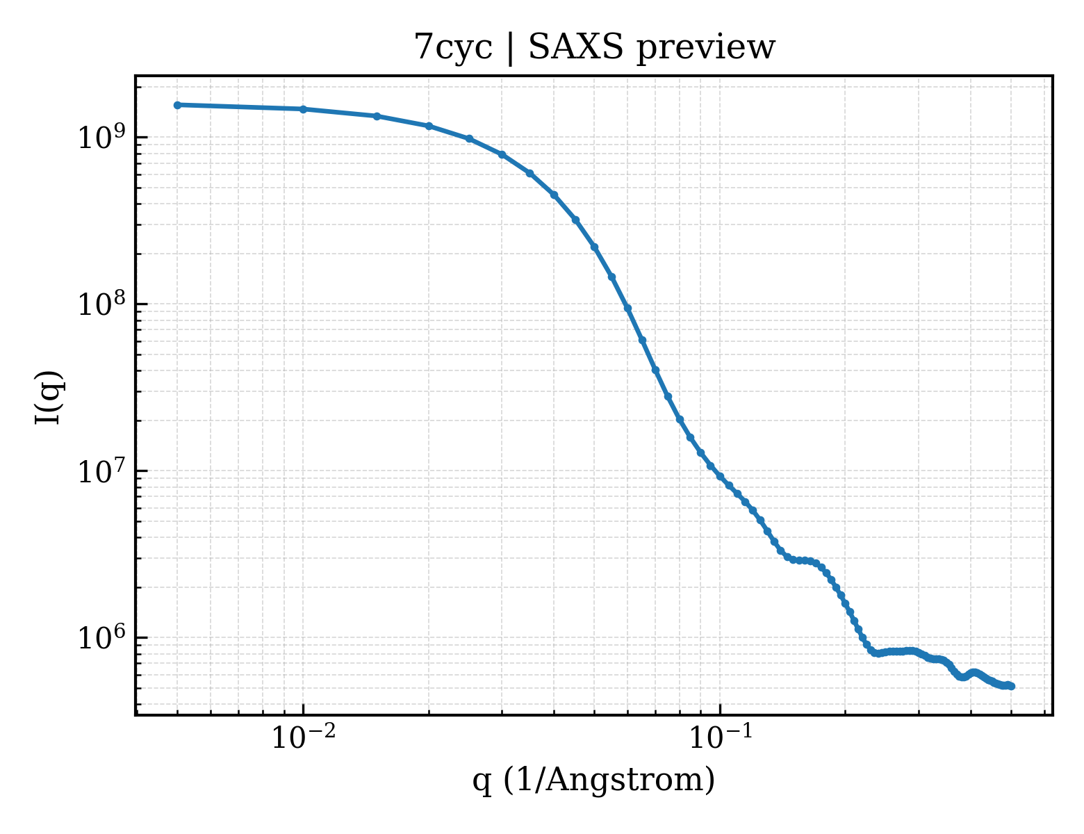
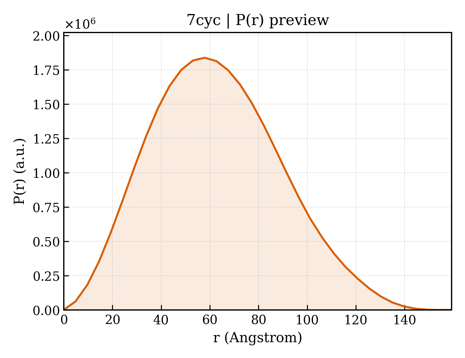
Spike glycoprotein
Human coronavirus 229E
State in the Current Structure
| Assembly | Oligomeric State | Construct | Mutations and Modifications | Ligands, Ions and Associated Components | Method and Experimental Conditions | Structure Quality |
|---|---|---|---|---|---|---|
| 1 | Other combination Homooligomer Protein × 3 其他Polymer 12 PDB declaration: trimeric(3) Consistent with protein copy count | Chain A; UniProt 1–1116 Chain B; UniProt 1–1116 Chain C; UniProt 1–1116 | Not recorded | ;alpha-D-mannopyranose-(1-2)-alpha-D-mannopyranose-(1-3)-[alpha-D-mannopyranose-(1-2)-alpha-D-mannopyranose-(1-6)]beta-D-mannopyranose-(1-4)-2-acetamido-2-deoxy-beta-D-glucopyranose-(1-4)-2-acetamido-2-deoxy-beta-D-glucopyranose ; × 3 beta-D-mannopyranose-(1-4)-2-acetamido-2-deoxy-beta-D-glucopyranose-(1-4)-2-acetamido-2-deoxy-beta-D-glucopyranose × 6 2-acetamido-2-deoxy-beta-D-glucopyranose-(1-4)-2-acetamido-2-deoxy-beta-D-glucopyranose × 3 NAG 2-acetamido-2-deoxy-beta-D-glucopyranose × 48 | ELECTRON MICROSCOPY cryo-EM buffer:pH 7.2 cryo-EM vitrification conditions:Cryogen ETHANE | Resolution 3.21 Å |
Other States of the Same Protein in the Database
Each row is a biological assembly of the same UniProt protein in another PDB entry. The “Difference from current entry” column identifies evidence-level differences; no tag means the currently parsed fields agree.
| Other PDB | Difference from Current Entry 7CYC | Assembly / Oligomeric State | Construct | Mutations and Modifications | Ligands, Ions and Non-polymers | Method and Experimental Conditions | Structure Quality |
|---|---|---|---|---|---|---|---|
| 5YL9 1.86 Angstrom crystal structure of human Coronavirus 229E fusion core Deposited 2017-10-17 | Different construct Different mutation/modification Different oligomeric state Different ligand/ion Different experimental method Different experimental conditions Different structure-quality metrics | Assembly 1 Protein homooligomer Homooligomer;Protein × 6 PDB declaration: hexameric |
Chain A
785–872(88 aa)
Fragment:UNP residues 785-872
Chain B
1052–1104(53 aa)
Fragment:UNP residues 1052-1104
|
Not recorded | No recorded non-water small molecule |
X-RAY DIFFRACTION
X-ray crystallization conditions
EVAPORATION;293 K;citric acid, BIS-TRIS propane, PEG3350
|
Resolution 1.86 Å R-free 0.206 |
| 5ZHY Structural characterization of the HCoV-229E fusion core Deposited 2018-03-13 | Different construct Different mutation/modification Different oligomeric state Different ligand/ion Different experimental method Different experimental conditions Different structure-quality metrics | Assembly 1 Protein homooligomer Homooligomer;Protein × 3 PDB declaration: trimeric |
Chain A
789–856(68 aa)
Fragment:UNP residues 789-856. UNP residues 1053-1105
Chain A
1053–1105(53 aa)
Fragment:UNP residues 789-856. UNP residues 1053-1105
Chain B
789–856(68 aa)
Fragment:UNP residues 789-856. UNP residues 1053-1105
Chain B
1053–1105(53 aa)
Fragment:UNP residues 789-856. UNP residues 1053-1105
Chain C
789–856(68 aa)
Fragment:UNP residues 789-856. UNP residues 1053-1105
Chain C
1053–1105(53 aa)
Fragment:UNP residues 789-856. UNP residues 1053-1105
|
Not recorded | No recorded non-water small molecule |
X-RAY DIFFRACTION
X-ray crystallization conditions
VAPOR DIFFUSION, SITTING DROP;293 K;1.5 M Ammonium sulfate, 12% (v/v) Glycerol, 100 mM Tris/HCl, PH 8.5
|
Resolution 2.44 Å R-free 0.263 |
| 5ZHY Structural characterization of the HCoV-229E fusion core Deposited 2018-03-13 | Different construct Different mutation/modification Different oligomeric state Different ligand/ion Different experimental method Different experimental conditions Different structure-quality metrics | Assembly 2 Protein homooligomer Homooligomer;Protein × 3 PDB declaration: trimeric |
Chain D
789–856(68 aa)
Fragment:UNP residues 789-856. UNP residues 1053-1105
Chain D
1053–1105(53 aa)
Fragment:UNP residues 789-856. UNP residues 1053-1105
Chain E
789–856(68 aa)
Fragment:UNP residues 789-856. UNP residues 1053-1105
Chain E
1053–1105(53 aa)
Fragment:UNP residues 789-856. UNP residues 1053-1105
Chain F
789–856(68 aa)
Fragment:UNP residues 789-856. UNP residues 1053-1105
Chain F
1053–1105(53 aa)
Fragment:UNP residues 789-856. UNP residues 1053-1105
|
Not recorded | No recorded non-water small molecule |
X-RAY DIFFRACTION
X-ray crystallization conditions
VAPOR DIFFUSION, SITTING DROP;293 K;1.5 M Ammonium sulfate, 12% (v/v) Glycerol, 100 mM Tris/HCl, PH 8.5
|
Resolution 2.44 Å R-free 0.263 |
| 5ZUV Crystal Structure of the Human Coronavirus 229E HR1 motif in complex with pan-CoVs inhibitor EK1 Deposited 2018-05-08 | Different construct Different oligomeric state Different ligand/ion Different experimental method Different experimental conditions Different structure-quality metrics | Assembly 1 Protein homooligomer Homooligomer;Protein × 3 PDB declaration: trimeric |
Chain A
785–873(89 aa)
Fragment:UNP residues 785-873
Chain B
785–873(89 aa)
Fragment:UNP residues 785-873
Chain C
785–873(89 aa)
Fragment:UNP residues 785-873
|
Not recorded | CL CHLORIDE ION × 2 |
X-RAY DIFFRACTION
X-ray crystallization conditions
EVAPORATION;pH 7.5;293 K;0.05M MgCl2, 0.1M HEPES, pH 7.5, 30% PEG550MME
|
Resolution 2.21 Å R-free 0.248 |
| 6ATK Crystal structure of the human coronavirus 229E spike protein receptor binding domain in complex with human aminopeptidase N Deposited 2017-08-29 | Different construct Different mutation/modification Different oligomeric state Different ligand/ion Different experimental method Different experimental conditions Different structure-quality metrics | Assembly 1 Other combination Heteromer;Protein × 2 PDB declaration: dimeric |
Chain D
293–435(143 aa)
Fragment:UNP residues 294-432
|
Not recorded | NAG 2-acetamido-2-deoxy-beta-D-glucopyranose × 6 ZN ZINC ION × 1 |
X-RAY DIFFRACTION
X-ray crystallization conditions
VAPOR DIFFUSION, HANGING DROP;298 K;8% PEG 8000, 1mM GSSG, 1mM GSH, 5% Glycerol, 100mM MES, 1ug/mL endo-beta-N-acetylglucosaminidase A
|
Resolution 3.50 Å R-free 0.267 |
| 6ATK Crystal structure of the human coronavirus 229E spike protein receptor binding domain in complex with human aminopeptidase N Deposited 2017-08-29 | Different construct Different mutation/modification Different oligomeric state Different ligand/ion Different experimental method Different experimental conditions Different structure-quality metrics | Assembly 2 Other combination Heteromer;Protein × 2 PDB declaration: dimeric |
Chain E
293–435(143 aa)
Fragment:UNP residues 294-432
|
Not recorded | NAG 2-acetamido-2-deoxy-beta-D-glucopyranose × 5 ZN ZINC ION × 1 |
X-RAY DIFFRACTION
X-ray crystallization conditions
VAPOR DIFFUSION, HANGING DROP;298 K;8% PEG 8000, 1mM GSSG, 1mM GSH, 5% Glycerol, 100mM MES, 1ug/mL endo-beta-N-acetylglucosaminidase A
|
Resolution 3.50 Å R-free 0.267 |
| 6ATK Crystal structure of the human coronavirus 229E spike protein receptor binding domain in complex with human aminopeptidase N Deposited 2017-08-29 | Different construct Different mutation/modification Different oligomeric state Different ligand/ion Different experimental method Different experimental conditions Different structure-quality metrics | Assembly 3 Other combination Heteromer;Protein × 2 PDB declaration: dimeric |
Chain F
293–435(143 aa)
Fragment:UNP residues 294-432
|
Not recorded | NAG 2-acetamido-2-deoxy-beta-D-glucopyranose × 4 ZN ZINC ION × 1 |
X-RAY DIFFRACTION
X-ray crystallization conditions
VAPOR DIFFUSION, HANGING DROP;298 K;8% PEG 8000, 1mM GSSG, 1mM GSH, 5% Glycerol, 100mM MES, 1ug/mL endo-beta-N-acetylglucosaminidase A
|
Resolution 3.50 Å R-free 0.267 |
| 7CYD Cryo-EM structures of Alphacoronavirus spike glycoprotein Deposited 2020-09-03 | Different oligomeric state Different ligand/ion Different structure-quality metrics | Assembly 1 Other combination Homooligomer;Protein × 3 PDB declaration: trimeric |
Chain A
1–1116(1116 aa)
Chain B
1–1116(1116 aa)
Chain C
1–1116(1116 aa)
|
Not recorded | NAG 2-acetamido-2-deoxy-beta-D-glucopyranose × 42 |
ELECTRON MICROSCOPY
cryo-EM buffer
pH 7.2
cryo-EM vitrification conditions
Cryogen ETHANE
|
Resolution 3.55 Å |
| 7VN9 Crystal structure of human coronavirus 229E spike protein receptor-binding domain in complex with C04 Fab Deposited 2021-10-10 | Different construct Different mutation/modification Different oligomeric state Different ligand/ion Different experimental method Different experimental conditions Different structure-quality metrics | Assembly 1 Other combination Heteromer;Protein × 3 PDB declaration: trimeric |
Chain E
294–435(142 aa)
Fragment:receptor-binding domain
|
Not recorded | No recorded non-water small molecule |
X-RAY DIFFRACTION
X-ray crystallization conditions
VAPOR DIFFUSION, HANGING DROP;pH 5;293 K;1.6M AmSO4, 0.1M Critic Acid pH 5.0
|
Resolution 4.49 Å R-free 0.288 |
| 7VN9 Crystal structure of human coronavirus 229E spike protein receptor-binding domain in complex with C04 Fab Deposited 2021-10-10 | Different construct Different mutation/modification Different oligomeric state Different ligand/ion Different experimental method Different experimental conditions Different structure-quality metrics | Assembly 2 Other combination Heteromer;Protein × 3 PDB declaration: trimeric |
Chain C
294–435(142 aa)
Fragment:receptor-binding domain
|
Not recorded | NAG 2-acetamido-2-deoxy-beta-D-glucopyranose × 1 |
X-RAY DIFFRACTION
X-ray crystallization conditions
VAPOR DIFFUSION, HANGING DROP;pH 5;293 K;1.6M AmSO4, 0.1M Critic Acid pH 5.0
|
Resolution 4.49 Å R-free 0.288 |
| 7VNG Crystal structure of human coronavirus 229E spike protein receptor-binding domain in complex with S11 Fab Deposited 2021-10-11 | Different construct Different mutation/modification Different oligomeric state Different ligand/ion Different experimental method Different experimental conditions Different structure-quality metrics | Assembly 1 Protein heterocomplex Heteromer;Protein × 3 PDB declaration: trimeric |
Chain D
294–435(142 aa)
Fragment:receptor binding domain
|
Not recorded | NAG 2-acetamido-2-deoxy-beta-D-glucopyranose × 2 |
X-RAY DIFFRACTION
X-ray crystallization conditions
VAPOR DIFFUSION, HANGING DROP;pH 7;293 K;3.6M Sodium Formate, 10% Glycerol
|
Resolution 3.80 Å R-free 0.318 |
| 7YI6 bnAb 3D1 in complex with 6-mer HR1 peptide from HCoV-229E S protein Deposited 2022-07-15 | Different construct Different mutation/modification Different oligomeric state Different ligand/ion Different experimental method Different experimental conditions Different structure-quality metrics | Assembly 1 Protein heterocomplex Heteromer;Protein × 3 PDB declaration: trimeric |
Chain A
835–841(7 aa)
|
Not recorded | EDO 1,2-ETHANEDIOL × 4 CL CHLORIDE ION × 1 |
X-RAY DIFFRACTION
X-ray crystallization conditions
VAPOR DIFFUSION, HANGING DROP;298 K;0.2 M Ammonium sulfate, 0.1 M BIS-TRIS buffer at pH 5.5 and 25% (w/v) polyethylene glycol 3350
|
Resolution 2.28 Å R-free 0.220 |
| 7YI6 bnAb 3D1 in complex with 6-mer HR1 peptide from HCoV-229E S protein Deposited 2022-07-15 | Different construct Different mutation/modification Different oligomeric state Different ligand/ion Different experimental method Different experimental conditions Different structure-quality metrics | Assembly 2 Protein heterocomplex Heteromer;Protein × 3 PDB declaration: trimeric |
Chain C
835–841(7 aa)
|
Not recorded | EDO 1,2-ETHANEDIOL × 1 PEG DI(HYDROXYETHYL)ETHER × 1 |
X-RAY DIFFRACTION
X-ray crystallization conditions
VAPOR DIFFUSION, HANGING DROP;298 K;0.2 M Ammonium sulfate, 0.1 M BIS-TRIS buffer at pH 5.5 and 25% (w/v) polyethylene glycol 3350
|
Resolution 2.28 Å R-free 0.220 |
8 other PDB entries and 13 assemblies. Open the comparison page and filter oligomeric states
View Construct and Data Evidence
| UniProt name | SPIKE_CVH22 |
| Isoform | — |
| PDB entities | 1 |
| Chains and sequence ranges | Author chain A; PDBConstruct 1–1116; UniProt 1–1116 Author chain B; PDBConstruct 1–1116; UniProt 1–1116 Author chain C; PDBConstruct 1–1116; UniProt 1–1116 |