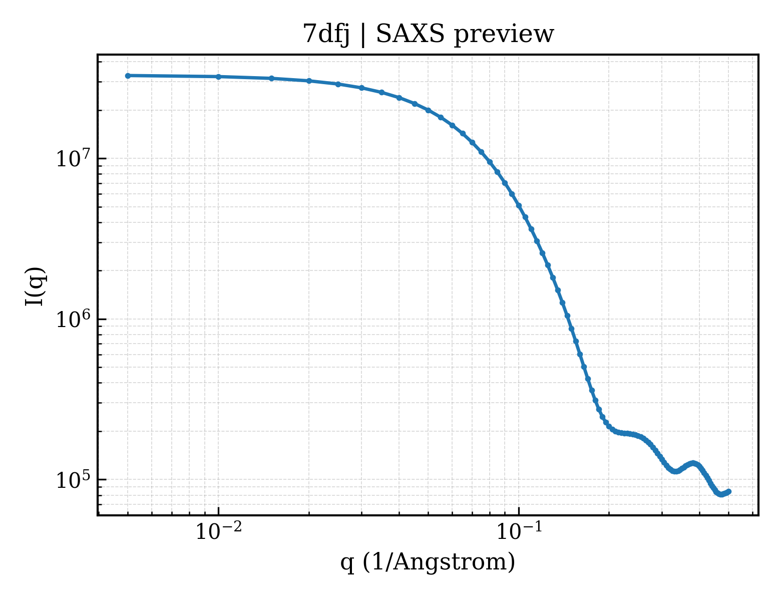
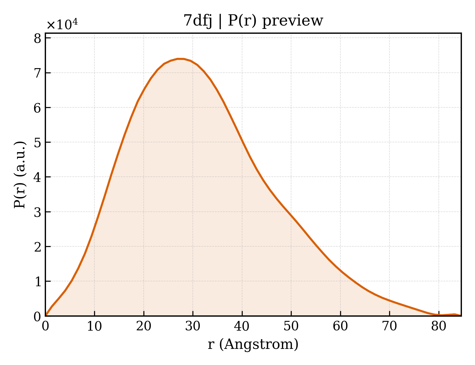
Xylose isomerase
OrganismNot specified
State in the Current Structure
| Assembly | Oligomeric State | Construct | Mutations and Modifications | Ligands, Ions and Associated Components | Method and Experimental Conditions | Structure Quality |
|---|---|---|---|---|---|---|
| 1 | Protein homooligomer Homooligomer Protein × 4 PDB declaration: tetrameric(4) Consistent with protein copy count | Chain A; UniProt 1–388 | Not recorded | MG MAGNESIUM ION × 8 | X-RAY DIFFRACTION X-ray crystallization conditions:BATCH MODE;pH 7;293.5 K;Tris-HCl, Ammonium sulfate, Magnesium sulfate | Resolution 1.50 Å R-free 0.248 |
Other States of the Same Protein in the Database
Each row is a biological assembly of the same UniProt protein in another PDB entry. The “Difference from current entry” column identifies evidence-level differences; no tag means the currently parsed fields agree.
| Other PDB | Difference from Current Entry 7DFJ | Assembly / Oligomeric State | Construct | Mutations and Modifications | Ligands, Ions and Non-polymers | Method and Experimental Conditions | Structure Quality |
|---|---|---|---|---|---|---|---|
| 1GW9 Tri-iodide derivative of Xylose Isomerase from Streptomyces Rubiginosus Deposited 2002-03-13 | Different construct Different ligand/ion Different experimental conditions Different structure-quality metrics | Assembly 1 Protein homooligomer Homooligomer;Protein × 4 PDB declaration: tetrameric |
Chain A
1–387(387 aa)
|
Not recorded | CA CALCIUM ION × 8 LXC beta-L-xylopyranose × 8 IOD IODIDE ION × 116 |
X-RAY DIFFRACTION
X-ray crystallization conditions
pH 7.5;1.8M AMMONIUM SULPHATE, 10MM TRIS-HCL AT PH7.5 AND THEN SOAKED IN AN EQUIVALENT SOLUTION WITH 50MM KI/50MM I2 AND 1.9M L-XYLOSE ADDED AS CRYO-PROTECTANT., pH 7.50
|
Resolution 1.55 Å R-free 0.188 |
| 1MNZ Atomic structure of Glucose isomerase Deposited 2002-09-06 | Different construct Different ligand/ion Different experimental conditions Different structure-quality metrics | Assembly 1 Protein homooligomer Homooligomer;Protein × 4 PDB declaration: tetrameric |
Chain A
0–387(388 aa)
|
Not recorded | MG MAGNESIUM ION × 4 CA CALCIUM ION × 4 TRS 2-AMINO-2-HYDROXYMETHYL-PROPANE-1,3-DIOL × 4 MRD (4R)-2-METHYLPENTANE-2,4-DIOL × 4 |
X-RAY DIFFRACTION
X-ray crystallization conditions
EVAPORATION;pH 7.5;295 K;MPD, Magnesium chloride, Tris base, pH 7.5, EVAPORATION, temperature 295K
|
Resolution 0.99 Å R-free 0.134 |
| 1O1H STRUCTURE OF GLUCOSE ISOMERASE DERIVATIZED WITH KR. Deposited 2002-11-07 | Different construct Different mutation/modification Different oligomeric state Different ligand/ion Different experimental conditions Different structure-quality metrics | Assembly 1 Protein homooligomer Homooligomer;Protein × 2 PDB declaration: dimeric |
Chain A
1–387(387 aa)
Chain B
1–387(387 aa)
|
Not recorded | CA CALCIUM ION × 2 MG MAGNESIUM ION × 2 CL CHLORIDE ION × 4 KR KRYPTON × 4 TRS 2-AMINO-2-HYDROXYMETHYL-PROPANE-1,3-DIOL × 2 MPD (4S)-2-METHYL-2,4-PENTANEDIOL × 2 |
X-RAY DIFFRACTION
X-ray crystallization conditions
pH 7;MPD, MgCl2, TRIS, pH 7.00
|
Resolution 1.40 Å R-free 0.161 |
| 1O1H STRUCTURE OF GLUCOSE ISOMERASE DERIVATIZED WITH KR. Deposited 2002-11-07 | Different construct Different mutation/modification Different ligand/ion Different experimental conditions Different structure-quality metrics | Assembly 2 Protein homooligomer Homooligomer;Protein × 4 PDB declaration: tetrameric |
Chain A
1–387(387 aa)
Chain B
1–387(387 aa)
|
Not recorded | CA CALCIUM ION × 4 MG MAGNESIUM ION × 4 CL CHLORIDE ION × 8 KR KRYPTON × 8 TRS 2-AMINO-2-HYDROXYMETHYL-PROPANE-1,3-DIOL × 4 MPD (4S)-2-METHYL-2,4-PENTANEDIOL × 4 |
X-RAY DIFFRACTION
X-ray crystallization conditions
pH 7;MPD, MgCl2, TRIS, pH 7.00
|
Resolution 1.40 Å R-free 0.161 |
| 1OAD Glucose isomerase from Streptomyces rubiginosus in P21212 crystal form Deposited 2003-01-08 | Different construct Different mutation/modification Different ligand/ion Different experimental conditions Different structure-quality metrics | Assembly 1 Protein homooligomer Homooligomer;Protein × 4 PDB declaration: tetrameric |
Chain A
2–388(387 aa)
Chain B
2–388(387 aa)
|
Not recorded | MN MANGANESE (II) ION × 4 MG MAGNESIUM ION × 4 MRD (4R)-2-METHYLPENTANE-2,4-DIOL × 4 MQD 2-METHYLPENTANE-1,2,4-TRIOL × 4 TRS 2-AMINO-2-HYDROXYMETHYL-PROPANE-1,3-DIOL × 2 MPD (4S)-2-METHYL-2,4-PENTANEDIOL × 2 |
X-RAY DIFFRACTION
X-ray crystallization conditions
pH 7;17 MG/ML PROTEIN, 12 % MPD, 0.1 M MGCL2, 50 MM TRIS BUFFER PH 7.0
|
Resolution 1.50 Å R-free 0.186 |
| 1XIB MODES OF BINDING SUBSTRATES AND THEIR ANALOGUES TO THE ENZYME D-XYLOSE ISOMERASE Deposited 1994-03-07 | Different construct Different ligand/ion Different experimental conditions Different structure-quality metrics | Assembly 1 Protein homooligomer Homooligomer;Protein × 4 PDB declaration: tetrameric |
Chain A
1–387(387 aa)
|
Not recorded | MN MANGANESE (II) ION × 8 | X-RAY DIFFRACTION mmCIF provides none of the parsed conditions | Resolution 1.60 Å |
| 1XIC MODES OF BINDING SUBSTRATES AND THEIR ANALOGUES TO THE ENZYME D-XYLOSE ISOMERASE Deposited 1994-03-07 | Different construct Different ligand/ion Different experimental conditions Different structure-quality metrics | Assembly 1 Protein homooligomer Homooligomer;Protein × 4 PDB declaration: tetrameric |
Chain A
1–387(387 aa)
|
Not recorded | XLS D-xylose × 4 MN MANGANESE (II) ION × 8 | X-RAY DIFFRACTION mmCIF provides none of the parsed conditions | Resolution 1.60 Å |
| 1XID MODES OF BINDING SUBSTRATES AND THEIR ANALOGUES TO THE ENZYME D-XYLOSE ISOMERASE Deposited 1994-03-07 | Different construct Different ligand/ion Different experimental conditions Different structure-quality metrics | Assembly 1 Protein homooligomer Homooligomer;Protein × 4 PDB declaration: tetrameric |
Chain A
1–387(387 aa)
|
Not recorded | MN MANGANESE (II) ION × 8 ASC ASCORBIC ACID × 4 | X-RAY DIFFRACTION mmCIF provides none of the parsed conditions | Resolution 1.70 Å |
| 1XIE MODES OF BINDING SUBSTRATES AND THEIR ANALOGUES TO THE ENZYME D-XYLOSE ISOMERASE Deposited 1994-03-07 | Different construct Different ligand/ion Different experimental conditions Different structure-quality metrics | Assembly 1 Protein homooligomer Homooligomer;Protein × 4 PDB declaration: tetrameric |
Chain A
1–387(387 aa)
|
Not recorded | ASO 1,5-anhydro-D-glucitol × 4 MN MANGANESE (II) ION × 8 | X-RAY DIFFRACTION mmCIF provides none of the parsed conditions | Resolution 1.70 Å |
| 1XIF MODES OF BINDING SUBSTRATES AND THEIR ANALOGUES TO THE ENZYME D-XYLOSE ISOMERASE Deposited 1994-03-07 | Different construct Different ligand/ion Different experimental conditions Different structure-quality metrics | Assembly 1 Protein homooligomer Homooligomer;Protein × 4 PDB declaration: tetrameric |
Chain A
1–387(387 aa)
|
Not recorded | GLC alpha-D-glucopyranose × 4 MN MANGANESE (II) ION × 8 | X-RAY DIFFRACTION mmCIF provides none of the parsed conditions | Resolution 1.60 Å |
| 1XIG MODES OF BINDING SUBSTRATES AND THEIR ANALOGUES TO THE ENZYME D-XYLOSE ISOMERASE Deposited 1994-03-07 | Different construct Different ligand/ion Different experimental conditions Different structure-quality metrics | Assembly 1 Protein homooligomer Homooligomer;Protein × 4 PDB declaration: tetrameric |
Chain A
1–387(387 aa)
|
Not recorded | XYL Xylitol × 4 MN MANGANESE (II) ION × 8 | X-RAY DIFFRACTION mmCIF provides none of the parsed conditions | Resolution 1.70 Å |
| 1XIH MODES OF BINDING SUBSTRATES AND THEIR ANALOGUES TO THE ENZYME D-XYLOSE ISOMERASE Deposited 1994-03-07 | Different construct Different ligand/ion Different experimental conditions Different structure-quality metrics | Assembly 1 Protein homooligomer Homooligomer;Protein × 4 PDB declaration: tetrameric |
Chain A
1–387(387 aa)
|
Not recorded | SOR sorbitol × 4 MN MANGANESE (II) ION × 4 | X-RAY DIFFRACTION mmCIF provides none of the parsed conditions | Resolution 1.70 Å |
| 1XII MODES OF BINDING SUBSTRATES AND THEIR ANALOGUES TO THE ENZYME D-XYLOSE ISOMERASE Deposited 1994-03-07 | Different construct Different ligand/ion Different experimental conditions Different structure-quality metrics | Assembly 1 Protein homooligomer Homooligomer;Protein × 4 PDB declaration: tetrameric |
Chain A
1–387(387 aa)
|
Not recorded | XUL D-XYLULOSE × 4 MN MANGANESE (II) ION × 8 | X-RAY DIFFRACTION mmCIF provides none of the parsed conditions | Resolution 1.70 Å |
| 1XIJ MODES OF BINDING SUBSTRATES AND THEIR ANALOGUES TO THE ENZYME D-XYLOSE ISOMERASE Deposited 1994-03-07 | Different construct Different ligand/ion Different experimental conditions Different structure-quality metrics | Assembly 1 Protein homooligomer Homooligomer;Protein × 4 PDB declaration: tetrameric |
Chain A
1–387(387 aa)
|
Not recorded | THE THREONATE ION × 4 MN MANGANESE (II) ION × 4 | X-RAY DIFFRACTION mmCIF provides none of the parsed conditions | Resolution 1.70 Å |
| 1XIS A METAL-MEDIATED HYDRIDE SHIFT MECHANISM FOR XYLOSE ISOMERASE BASED ON THE 1.6 ANGSTROMS STREPTOMYCES RUBIGINOSUS STRUCTURES WITH XYLITOL AND D-XYLOSE Deposited 1991-03-25 | Different construct Different ligand/ion Different experimental conditions Different structure-quality metrics | Assembly 1 Protein homooligomer Homooligomer;Protein × 4 PDB declaration: tetrameric |
Chain A
1–387(387 aa)
|
Not recorded | MN MANGANESE (II) ION × 8 | X-RAY DIFFRACTION mmCIF provides none of the parsed conditions | Resolution 1.60 Å |
| 2G4J Anomalous substructure of Glucose isomerase Deposited 2006-02-22 | Different construct Different ligand/ion Different experimental conditions Different structure-quality metrics | Assembly 1 Protein homooligomer Homooligomer;Protein × 4 PDB declaration: tetrameric |
Chain A
1–387(387 aa)
|
Not recorded | CA CALCIUM ION × 4 MG MAGNESIUM ION × 4 CL CHLORIDE ION × 4 | X-RAY DIFFRACTION mmCIF provides none of the parsed conditions | Resolution 1.85 Å R-free 0.177 |
| 2GLK High-resolution study of D-Xylose isomerase, 0.94A resolution. Deposited 2006-04-05 | Different construct Different ligand/ion Different experimental conditions Different structure-quality metrics | Assembly 1 Protein homooligomer Homooligomer;Protein × 4 PDB declaration: tetrameric |
Chain A
1–387(387 aa)
|
Not recorded | MN MANGANESE (II) ION × 8 GOL GLYCEROL × 16 |
X-RAY DIFFRACTION
X-ray crystallization conditions
VAPOR DIFFUSION;pH 8;293 K;50mM tris-HCl,25%AMSO4,2mM Mn2+ and 2mM Co2+ , XI @ 25mg/ml., pH 8, VAPOR DIFFUSION, temperature 293K
|
Resolution 0.94 Å R-free 0.128 |
| 2GUB Crystal Structure of Metal Free D-Xylose Isomerase. Deposited 2006-04-29 | Different construct Different ligand/ion Different experimental conditions Different structure-quality metrics | Assembly 1 Protein homooligomer Homooligomer;Protein × 4 PDB declaration: tetrameric |
Chain A
1–387(387 aa)
|
Not recorded | No recorded non-water small molecule |
X-RAY DIFFRACTION
X-ray crystallization conditions
SMALL TUBES;pH 8;279 K;Crystallization from solution containing metal free enzyme at 20 mg/ml, 0.8M AmSO4, 2mM Pipes.
Crystallization occurred over 2 day period in sealed tubes., pH 8.0, SMALL TUBES, temperature 279K
|
Resolution 1.80 Å R-free 0.193 |
| 2GVE Time-of-Flight Neutron Diffraction Structure of D-Xylose Isomerase Deposited 2006-05-02 | Different construct Different ligand/ion Different experimental method Different experimental conditions Different structure-quality metrics | Assembly 1 Protein homooligomer Homooligomer;Protein × 4 PDB declaration: tetrameric |
Chain A
1–387(387 aa)
|
Not recorded | CO COBALT (II) ION × 8 |
NEUTRON DIFFRACTION
X-ray crystallization conditions
LIQUID DIFFUSION;pH 8;293 K;50mM tris-HCl,38%AMSO4,2mM Mn2+ and 2mM Co2+,XI @ 125mg/ml, pH 8.0, LIQUID DIFFUSION, temperature 293K
|
Resolution 2.20 Å R-free 0.319 |
| 2XIS A METAL-MEDIATED HYDRIDE SHIFT MECHANISM FOR XYLOSE ISOMERASE BASED ON THE 1.6 ANGSTROMS STREPTOMYCES RUBIGINOSUS STRUCTURES WITH XYLITOL AND D-XYLOSE Deposited 1991-03-25 | Different construct Different ligand/ion Different experimental conditions Different structure-quality metrics | Assembly 1 Protein homooligomer Homooligomer;Protein × 4 PDB declaration: tetrameric |
Chain A
1–387(387 aa)
|
Not recorded | XYL Xylitol × 4 MG MAGNESIUM ION × 8 | X-RAY DIFFRACTION mmCIF provides none of the parsed conditions | Resolution 1.71 Å |
| 3CWH D-xylose Isomerase in complex with linear product, per-deuterated xylulose Deposited 2008-04-21 | Different ligand/ion Different experimental method Different experimental conditions Different structure-quality metrics | Assembly 1 Protein homooligomer Homooligomer;Protein × 4 PDB declaration: tetrameric |
Chain A
1–388(388 aa)
|
Not recorded | XUL D-XYLULOSE × 4 MG MAGNESIUM ION × 8 OH HYDROXIDE ION × 4 |
NEUTRON DIFFRACTION
X-ray crystallization conditions
pH 8;293 K;50MM TRIS-HCL, 38%(NH4)2SO4, 2MM MG2+ XI @ 125 MG/ML, PH=8.0, LIQUID DIFFUSION, T=293K; NATIVE CRYSTAL WERE SOAKED WITH 0.2M PER-DEUTERATED XYLOSE IN D2O BUFFERED IN 50MM TRIS-DCL, PH=7.6 (PD=8.0), 2MM MGCL2
|
Resolution 2.20 Å R-free 0.288 |
| 3GNX Structure of dehydrated D-xylose isomerase from streptomyces rubiginosus Deposited 2009-03-18 | Different construct Different mutation/modification Different ligand/ion Different experimental conditions Different structure-quality metrics | Assembly 1 Protein homooligomer Homooligomer;Protein × 4 PDB declaration: tetrameric |
Chain A
2–388(387 aa)
Chain E
2–388(387 aa)
|
Not recorded | XYL Xylitol × 4 MN MANGANESE (II) ION × 8 |
X-RAY DIFFRACTION
X-ray crystallization conditions
pH 7;278 K;6mM Tris-HCl, 33mg/ml crystalline suspension, 0.91M ammonium sulfate, 1mM magnesium sulfate (Hampton Research Ltd.); Soaked in xylitol solution, pH 7.0, temperature 278K
|
Resolution 2.00 Å R-free 0.206 |
| 3KBJ Room temperature X-ray structure of apo-D-Xylose Isomerase Deposited 2009-10-20 | Different ligand/ion Different experimental conditions Different structure-quality metrics | Assembly 1 Protein homooligomer Homooligomer;Protein × 4 PDB declaration: tetrameric |
Chain A
1–388(388 aa)
|
Not recorded | No recorded non-water small molecule |
X-RAY DIFFRACTION
X-ray crystallization conditions
pH 7.7;290 K;40mg/ml protein, 30%(v/v) ammonium sulfate (sat.), pH 7.7, temperature 290K
|
Resolution 2.00 Å R-free 0.226 |
| 3KBM Room Temperature X-ray structure of D-Xylose Isomerase complexed with 2Cd(2+) co-factors and d12-D-alpha-glucose in the cyclic form Deposited 2009-10-20 | Different ligand/ion Different experimental conditions Different structure-quality metrics | Assembly 1 Protein homooligomer Homooligomer;Protein × 4 PDB declaration: tetrameric |
Chain A
1–388(388 aa)
|
Not recorded | CD CADMIUM ION × 8 GLC alpha-D-glucopyranose × 4 |
X-RAY DIFFRACTION
X-ray crystallization conditions
pH 7.7;290 K;40mg/ml protein, 5mM CdCl2; 500mM d12-D-glucose, 30% (v/v) ammonium sulfate (sat.), batch, pH 7.7, temperature 290K
|
Resolution 2.00 Å R-free 0.211 |
| 3KBN Room temperature structure of D-Xylose Isomerase in complex with 2Ni(2+) co-factors and d12-D-glucose in the linear form Deposited 2009-10-20 | Different ligand/ion Different experimental conditions Different structure-quality metrics | Assembly 1 Protein homooligomer Homooligomer;Protein × 4 PDB declaration: tetrameric |
Chain A
1–388(388 aa)
|
Not recorded | NI NICKEL (II) ION × 12 GLO D-glucose × 4 |
X-RAY DIFFRACTION
X-ray crystallization conditions
pH 7.7;290 K;40mg/ml protein, 5mM NiCl2, 500mM d12-D-glucose 30% (v/v) ammonium sulfate (sat.), pH 7.7, temperature 290K
|
Resolution 1.53 Å R-free 0.181 |
| 3KBS Room Temperature X-ray structure of D-Xylose Isomerase in complex with 2Cd(2+) co-factors Deposited 2009-10-20 | Different ligand/ion Different experimental conditions Different structure-quality metrics | Assembly 1 Protein homooligomer Homooligomer;Protein × 4 PDB declaration: tetrameric |
Chain A
1–388(388 aa)
|
Not recorded | CD CADMIUM ION × 8 |
X-RAY DIFFRACTION
X-ray crystallization conditions
batch;pH 7.7;293 K;40mg/ml protein, 5mM CdCl2, 30% (v/v) ammonium sulfate (sat.), pH 7.7, batch, temperature 293K
|
Resolution 1.80 Å R-free 0.284 |
| 3KBV Room temperature structure of D-Xylose Isomerase in complex with 2Ni(2+) co-factors Deposited 2009-10-20 | Different ligand/ion Different experimental conditions Different structure-quality metrics | Assembly 1 Protein homooligomer Homooligomer;Protein × 4 PDB declaration: tetrameric |
Chain A
1–388(388 aa)
|
Not recorded | NI NICKEL (II) ION × 8 |
X-RAY DIFFRACTION
X-ray crystallization conditions
batch;pH 7.7;290 K;40mg/ml protein, 5mM NiCl2, 30% (v/v) ammonium sulfate (sat.), pH 7.7, batch, temperature 290K
|
Resolution 1.80 Å R-free 0.223 |
| 3KBW Room temperature X-ray mixed-metal structure of D-Xylose Isomerase in complex with Ni(2+) and Mg(2+) co-factors Deposited 2009-10-20 | Different ligand/ion Different experimental conditions Different structure-quality metrics | Assembly 1 Protein homooligomer Homooligomer;Protein × 4 PDB declaration: tetrameric |
Chain A
1–388(388 aa)
|
Not recorded | NI NICKEL (II) ION × 4 MG MAGNESIUM ION × 4 |
X-RAY DIFFRACTION
X-ray crystallization conditions
batch;pH 7.7;290 K;40mg/ml protein, 2mM NiCl2, 2mM MgCl2, 30% (v/v) ammonium sulfate (sat.), pH 7.7, batch, temperature 290K
|
Resolution 1.60 Å R-free 0.167 |
| 3KCJ Room temperature neutron structure of apo-D-Xylose Isomerase (refined jointly with X-ray structure 3KBJ) Deposited 2009-10-21 | Different ligand/ion Different experimental method Different experimental conditions Different structure-quality metrics | Assembly 1 Protein homooligomer Homooligomer;Protein × 4 PDB declaration: tetrameric |
Chain A
1–388(388 aa)
|
Not recorded | No recorded non-water small molecule |
Experimental method not declared
X-ray crystallization conditions
pH 7.7;293 K;50MM HEPES, 40% v/v (NH4)2SO4 (sat.), protein 40 MG/ML, PH 7.7, BATCH METHOD, temperature 293K
|
Resolution 1.80 Å |
| 3KCL Room temperature neutron structure of D-Xylose Isomerase in complex with two Cd2+ cations and d12-D-alpha-glucose in the ring form (refined jointly with X-ray structure 3KBM) Deposited 2009-10-21 | Different ligand/ion Different experimental method Different experimental conditions Different structure-quality metrics | Assembly 1 Protein homooligomer Homooligomer;Protein × 4 PDB declaration: tetrameric |
Chain A
1–388(388 aa)
|
Not recorded | CD CADMIUM ION × 8 GLC alpha-D-glucopyranose × 4 |
Experimental method not declared
X-ray crystallization conditions
pH 7.7;293 K;50MM HEPES, 40% v/v (NH4)2SO4 (sat.), protein 40 MG/ML, PH=7.7, BATCH METHOD, APO-XI CRYSTALS WERE WITH 5mM CDCL2 SALT, 0.5M PER-DEUTERATED D-GLUCOSE IN D2O, temperature 293K
|
Resolution 2.00 Å |
| 3KCO Room temperature neutron structure of D-Xylose Isomerase in complex with two Ni2+ cations and d12-D-glucose in the linear form (refined jointly with X-ray structure 3KBN) Deposited 2009-10-21 | Different ligand/ion Different experimental method Different experimental conditions Different structure-quality metrics | Assembly 1 Protein homooligomer Homooligomer;Protein × 4 PDB declaration: tetrameric |
Chain A
1–388(388 aa)
|
Not recorded | NI NICKEL (II) ION × 12 GLO D-glucose × 4 |
Experimental method not declared
X-ray crystallization conditions
pH 7.7;293 K;50MM HEPES, 40% v/v (NH4)2SO4 (sat.), protein 40 MG/ML, PH=7.7, BATCH METHOD, APO-XI CRYSTALS WERE SOAKED WITH 5mM NiCl2 SALT, 0.5M PER-DEUTERATED D-GLUCOSE IN D2O, temperature 293K
|
Resolution 1.80 Å |
| 3N4A Crystal structure of D-Xylose Isomerase in complex with S-1,2-Propandiol Deposited 2010-05-21 | Different construct Different ligand/ion Different experimental conditions Different structure-quality metrics | Assembly 1 Protein homooligomer Homooligomer;Protein × 4 PDB declaration: tetrameric |
Chain A
2–388(387 aa)
|
Not recorded | MN MANGANESE (II) ION × 8 CL CHLORIDE ION × 4 PGO S-1,2-PROPANEDIOL × 4 |
X-RAY DIFFRACTION
X-ray crystallization conditions
VAPOR DIFFUSION, SITTING DROP;pH 6;277 K;20-28% Propandiol, 0.2M MgCl, 50mM MES, pH 6.0, VAPOR DIFFUSION, SITTING DROP, temperature 277K
|
Resolution 1.94 Å R-free 0.182 |
| 3QYS Room Temperature X-ray Structure of D-Xylose Isomerase in complex with 0.6Ni2+ cation bound in M2 metal binding site at pH=5.8 Deposited 2011-03-03 | Different ligand/ion Different experimental conditions Different structure-quality metrics | Assembly 1 Protein homooligomer Homooligomer;Protein × 4 PDB declaration: tetrameric |
Chain A
1–388(388 aa)
|
Not recorded | NI NICKEL (II) ION × 4 |
X-RAY DIFFRACTION
X-ray crystallization conditions
batch crystallization;pH 5.8;280 K;apo-XI was crystallized with ammonium sulfate as precipitant; 0.2mM Ni2+ salt was added to make the complex, pH 5.8, batch crystallization, temperature 280K
|
Resolution 1.85 Å R-free 0.217 |
| 3QZA Joint neutron and X-ray structure of apo-D-Xylose Isomerase at pH=5.9 Deposited 2011-03-04 | Different ligand/ion Different experimental method Different experimental conditions Different structure-quality metrics | Assembly 1 Protein homooligomer Homooligomer;Protein × 4 PDB declaration: tetrameric |
Chain A
1–388(388 aa)
|
Not recorded | D8U deuterium(1+) × 4 |
Experimental method not declared
X-ray crystallization conditions
BATCH MODE;pH 5.9;280 K;crystals grew with ammonium sulfate as a precipitant at pH of 5.9, batch crystallization, temperature 280K
|
Resolution not provided |
| 3U3H X-Ray Crystallographic Analysis of D-Xylose Isomerase-Catalyzed Isomerization of (R)-Glyceraldehyde Deposited 2011-10-05 | Different ligand/ion Different experimental conditions Different structure-quality metrics | Assembly 1 Protein homooligomer Homooligomer;Protein × 4 PDB declaration: tetrameric |
Chain A
1–388(388 aa)
|
Not recorded | 03W (2R)-propane-1,1,2,3-tetrol × 4 FMT FORMIC ACID × 4 MG MAGNESIUM ION × 8 MRD (4R)-2-METHYLPENTANE-2,4-DIOL × 4 |
X-RAY DIFFRACTION
X-ray crystallization conditions
pH 7;298 K;20mg/ml XI in water was mixed with an equal volume of well solution consisting of 0.2-0.3M Mg formate, pH 7.0, VAPOR DIFFUSION, HANGING DROP, temperature 298K
|
Resolution 0.97 Å R-free 0.125 |
| 3XIS A METAL-MEDIATED HYDRIDE SHIFT MECHANISM FOR XYLOSE ISOMERASE BASED ON THE 1.6 ANGSTROMS STREPTOMYCES RUBIGINOSUS STRUCTURES WITH XYLITOL AND D-XYLOSE Deposited 1991-03-25 | Different construct Different ligand/ion Different experimental conditions Different structure-quality metrics | Assembly 1 Protein homooligomer Homooligomer;Protein × 4 PDB declaration: tetrameric |
Chain A
1–387(387 aa)
|
Not recorded | XYS alpha-D-xylopyranose × 4 XLS D-xylose × 4 MG MAGNESIUM ION × 8 | X-RAY DIFFRACTION mmCIF provides none of the parsed conditions | Resolution 1.60 Å |
| 4A8I Protein crystallization and microgravity: glucose isomerase crystals grown during the PCDF-PROTEIN mission Deposited 2011-11-21 | Different ligand/ion Different experimental conditions Different structure-quality metrics | Assembly 1 Protein homooligomer Homooligomer;Protein × 4 PDB declaration: tetrameric |
Chain A
1–388(388 aa)
|
Not recorded | EDO 1,2-ETHANEDIOL × 4 CO COBALT (II) ION × 8 |
X-RAY DIFFRACTION
X-ray crystallization conditions
pH 7;32,7 MG/ML PROTEIN, 0.6 M AMMONIUM SULPHATE, 100 MM HEPES PH 7.0
|
Resolution 0.95 Å R-free 0.132 |
| 4A8L Protein crystallization and microgravity: glucose isomerase crystals grown during the PCDF-PROTEIN mission Deposited 2011-11-21 | Different ligand/ion Different experimental conditions Different structure-quality metrics | Assembly 1 Protein homooligomer Homooligomer;Protein × 4 PDB declaration: tetrameric |
Chain A
1–388(388 aa)
|
Not recorded | EDO 1,2-ETHANEDIOL × 4 CO COBALT (II) ION × 8 |
X-RAY DIFFRACTION
X-ray crystallization conditions
pH 7;32.7 MG/ML PROTEIN, 0.6 M AMMONIUM SULPHATE, 100 MM HEPES PH 7.0
|
Resolution 1.35 Å R-free 0.137 |
| 4A8N Protein crystallization and microgravity: glucose isomerase crystals grown during the PCDF-PROTEIN mission Deposited 2011-11-21 | Different ligand/ion Different experimental conditions Different structure-quality metrics | Assembly 1 Protein homooligomer Homooligomer;Protein × 4 PDB declaration: tetrameric |
Chain A
1–388(388 aa)
|
Not recorded | GOL GLYCEROL × 8 CO COBALT (II) ION × 8 |
X-RAY DIFFRACTION
X-ray crystallization conditions
pH 7;28.7 MG/ML PROTEIN, 1.4 MG/ML PROTEIN COVALENTLY LABELED WITH BIS(2,2'-BIPYRIDINE)-4,4'-DICARBOXY BIPYRIDINE-RUTHENIUM DI(N-SUCCINIMIDYL ESTER) BIS(HEXAFLUOROPHOSPHATE), 0.9M AMMONIUM SULPHATE, 100MM HEPES PH 7.0
|
Resolution 1.20 Å R-free 0.128 |
| 4A8R Protein crystallization and microgravity: glucose isomerase crystals grown during the PCDF-PROTEIN mission Deposited 2011-11-21 | Different ligand/ion Different experimental conditions Different structure-quality metrics | Assembly 1 Protein homooligomer Homooligomer;Protein × 4 PDB declaration: tetrameric |
Chain A
1–388(388 aa)
|
Not recorded | GOL GLYCEROL × 8 CO COBALT (II) ION × 8 |
X-RAY DIFFRACTION
X-ray crystallization conditions
pH 7;28.7 MG/ML PROTEIN, 1.4 MG/ML PROTEIN COVALENTLY LABELED WITH A RUTHENIUM-CONTAINING LABEL, 0.9 M AMMONIUM SULPHATE, 100 MM HEPES PH 7.0
|
Resolution 1.42 Å R-free 0.130 |
| 4DUO Room-temperature X-ray structure of D-Xylose Isomerase in complex with 2Mg2+ ions and xylitol at pH 7.7 Deposited 2012-02-22 | Different ligand/ion Different experimental conditions Different structure-quality metrics | Assembly 1 Protein homooligomer Homooligomer;Protein × 4 PDB declaration: tetrameric |
Chain A
1–388(388 aa)
|
Not recorded | MG MAGNESIUM ION × 8 XYL Xylitol × 4 |
X-RAY DIFFRACTION
X-ray crystallization conditions
batch;pH 7.7;290 K;apo-XI crystals were grown with AmSO4 by batch; to obtain the complex Mg2+ was injected into drops containing crystals up to concentration 5 mM; subsequently, xylitol was injected up to concentration of 100 mM, pH 7.7, temperature 290K
|
Resolution 2.00 Å R-free 0.200 |
| 4DVO Room-temperature joint X-ray/neutron structure of D-xylose isomerase in complex with 2Ni2+ and per-deuterated D-sorbitol at pH 5.9 Deposited 2012-02-23 | Different ligand/ion Different experimental method Different experimental conditions Different structure-quality metrics | Assembly 1 Protein homooligomer Homooligomer;Protein × 4 PDB declaration: tetrameric |
Chain A
1–388(388 aa)
|
Not recorded | NI NICKEL (II) ION × 12 SOR sorbitol × 4 |
Experimental method not declared
X-ray crystallization conditions
batch;pH 5.9;290 K;apo-Xylose isomerase crystals were grown with AmSO4 as precipitant and 0.1 M MES at pH 5.9; they then were injected with Ni2+ salt up to the concentration of 5mM and D-sorbitol up to the concentration of 300mM, batch, temperature 290K
|
Resolution 2.00 Å R-free 0.214 |
| 4E3V Crystal Structure of XYLOSE ISOMERASE FROM STREPTOMYCES RUBIGINOSUS Cryoprotected in Proline Deposited 2012-03-10 | Different ligand/ion Different experimental conditions Different structure-quality metrics | Assembly 1 Protein homooligomer Homooligomer;Protein × 4 PDB declaration: tetrameric |
Chain A
1–388(388 aa)
|
Not recorded | PRO PROLINE × 4 SO4 SULFATE ION × 8 MG MAGNESIUM ION × 4 MN MANGANESE (II) ION × 4 |
X-RAY DIFFRACTION
X-ray crystallization conditions
VAPOR DIFFUSION, SITTING DROP;pH 8.5;294 K;2.0M ammonium sulfate, 0.1M Tris-HCl, pH 8.5, VAPOR DIFFUSION, SITTING DROP, temperature 294K
|
Resolution 1.50 Å R-free 0.179 |
| 4J4K Crystal structure of glucose isomerase Deposited 2013-02-07 | Different oligomeric state Different ligand/ion Different experimental conditions Different structure-quality metrics | Assembly 1 Protein monomer Monomer;Protein × 1 PDB declaration: monomeric |
Chain A
1–388(388 aa)
|
Not recorded | ZN ZINC ION × 4 MPD (4S)-2-METHYL-2,4-PENTANEDIOL × 2 ACT ACETATE ION × 2 |
X-RAY DIFFRACTION
X-ray crystallization conditions
VAPOR DIFFUSION, HANGING DROP;pH 6.5;295 K;10% MPD, 0.1M MES pH 6.5, 0.2M Mg Acetate, VAPOR DIFFUSION, HANGING DROP, temperature 295K
|
Resolution 1.90 Å R-free 0.163 |
| 4J4K Crystal structure of glucose isomerase Deposited 2013-02-07 | Different oligomeric state Different ligand/ion Different experimental conditions Different structure-quality metrics | Assembly 2 Protein homooligomer Homooligomer;Protein × 2 PDB declaration: dimeric |
Chain A
1–388(388 aa)
|
Not recorded | ZN ZINC ION × 8 MPD (4S)-2-METHYL-2,4-PENTANEDIOL × 4 ACT ACETATE ION × 4 |
X-RAY DIFFRACTION
X-ray crystallization conditions
VAPOR DIFFUSION, HANGING DROP;pH 6.5;295 K;10% MPD, 0.1M MES pH 6.5, 0.2M Mg Acetate, VAPOR DIFFUSION, HANGING DROP, temperature 295K
|
Resolution 1.90 Å R-free 0.163 |
| 4LNC Neutron structure of the cyclic glucose bound Xylose Isomerase E186Q mutant Deposited 2013-07-11 | Different ligand/ion Different experimental method Different experimental conditions Different structure-quality metrics | Assembly 1 Protein homooligomer Homooligomer;Protein × 4 PDB declaration: tetrameric |
Chain A
1–388(388 aa)
|
Not recorded | GLC alpha-D-glucopyranose × 4 MN MANGANESE (II) ION × 8 MG MAGNESIUM ION × 4 |
Experimental method not declared
X-ray crystallization conditions
pH 7.7;295 K;Large crystals were grown in 1.5 ml Eppendorf tubes with 15% ammonium sulfate, 72 mg/ml D-xylose isomerase at ph 7.7, Batch method, temperature 295K
|
Resolution not provided |
| 4QDP Joint X-ray and neutron structure of Streptomyces rubiginosus D-xylose isomerase in complex with two Cd2+ ions and cyclic beta-L-arabinose Deposited 2014-05-14 | Different ligand/ion Different experimental method Different experimental conditions Different structure-quality metrics | Assembly 1 Protein homooligomer Homooligomer;Protein × 4 PDB declaration: tetrameric |
Chain A
1–388(388 aa)
|
Not recorded | CD CADMIUM ION × 8 ARB beta-L-arabinopyranose × 4 |
Experimental method not declared
X-ray crystallization conditions
batch;pH 7.7;291 K;0.1M HEPES, pH 7.7, 30% ammonium sulfate, batch, temperature 291K
|
Resolution 2.00 Å R-free 0.247 |
| 4QDW Joint X-ray and neutron structure of Streptomyces rubiginosus D-xylose isomerase in complex with two Ni2+ ions and linear L-arabinose Deposited 2014-05-14 | Different ligand/ion Different experimental method Different experimental conditions Different structure-quality metrics | Assembly 1 Protein homooligomer Homooligomer;Protein × 4 PDB declaration: tetrameric |
Chain A
1–388(388 aa)
|
Not recorded | NI NICKEL (II) ION × 12 LAI L-arabinose × 4 |
Experimental method not declared
X-ray crystallization conditions
batch;pH 7.7;291 K;30% ammonium sulfate, 0.1 M HEPES pH 7.7, batch, temperature 291K
|
Resolution 1.80 Å R-free 0.179 |
| 4QE1 Room temperature X-ray structure of D-xylose isomerase in complex with two Cd2+ ions and L-ribulose Deposited 2014-05-15 | Different ligand/ion Different experimental conditions Different structure-quality metrics | Assembly 1 Protein homooligomer Homooligomer;Protein × 4 PDB declaration: tetrameric |
Chain A
1–388(388 aa)
|
Not recorded | CD CADMIUM ION × 8 RUU alpha-L-ribulofuranose × 4 |
X-RAY DIFFRACTION
X-ray crystallization conditions
batch;pH 7.7;291 K;30% ammonium sulfate, 0.1 M HEPES pH 7.7, batch, temperature 291K
|
Resolution 1.55 Å R-free 0.190 |
| 4QE4 Room temperature X-ray structure of D-xylose isomerase in complex with two Ni2+ ions and L-ribulose Deposited 2014-05-15 | Different ligand/ion Different experimental conditions Different structure-quality metrics | Assembly 1 Protein homooligomer Homooligomer;Protein × 4 PDB declaration: tetrameric |
Chain A
1–388(388 aa)
|
Not recorded | NI NICKEL (II) ION × 8 34V beta-L-ribulofuranose × 4 |
X-RAY DIFFRACTION
X-ray crystallization conditions
batch;pH 7.7;291 K;30% ammonium sulfate, 0.1M HEPES ph 7.7, batch, temperature 291K
|
Resolution 1.70 Å R-free 0.180 |
| 4QE5 Room temperature X-ray structure of D-xylose isomerase in complex with two Mg2+ ions and L-ribulose Deposited 2014-05-15 | Different ligand/ion Different experimental conditions Different structure-quality metrics | Assembly 1 Protein homooligomer Homooligomer;Protein × 4 PDB declaration: tetrameric |
Chain A
1–388(388 aa)
|
Not recorded | MG MAGNESIUM ION × 8 RUU alpha-L-ribulofuranose × 4 |
X-RAY DIFFRACTION
X-ray crystallization conditions
batch;pH 7.7;291 K;30% ammonium sulfate, 0.1M HEPES pH 7.7, batch, temperature 291K
|
Resolution 1.56 Å R-free 0.188 |
| 4QEE Room temperature X-ray structure of D-xylose isomerase in complex with two Ni2+ ions and L-ribose Deposited 2014-05-15 | Different ligand/ion Different experimental conditions Different structure-quality metrics | Assembly 1 Protein homooligomer Homooligomer;Protein × 4 PDB declaration: tetrameric |
Chain A
1–388(388 aa)
|
Not recorded | NI NICKEL (II) ION × 8 Z6J alpha-L-ribofuranose × 4 |
X-RAY DIFFRACTION
X-ray crystallization conditions
batch;pH 7.7;291 K;30% ammonium sulfate, 0.1M HEPES pH 7.7, batch, temperature 291K
|
Resolution 1.60 Å R-free 0.191 |
| 4QEH Room temperature X-ray structure of D-xylose isomerase in complex with two Mg2+ ions and L-ribose Deposited 2014-05-16 | Different ligand/ion Different experimental conditions Different structure-quality metrics | Assembly 1 Protein homooligomer Homooligomer;Protein × 4 PDB declaration: tetrameric |
Chain A
1–388(388 aa)
|
Not recorded | MG MAGNESIUM ION × 8 32O beta-L-ribofuranose × 4 |
X-RAY DIFFRACTION
X-ray crystallization conditions
batch;pH 7.7;291 K;30% ammonium sulfate, 0.1M HEPES pH 7.7, batch, temperature 291K
|
Resolution 1.55 Å R-free 0.184 |
| 4US6 New Crystal Form of Glucose Isomerase Grown in Short Peptide Supramolecular Hydrogels Deposited 2014-07-03 | Different construct Different mutation/modification Different ligand/ion Different experimental conditions Different structure-quality metrics | Assembly 1 Protein homooligomer Homooligomer;Protein × 4 PDB declaration: tetrameric |
Chain A
1–388(388 aa)
Chain B
1–388(388 aa)
|
Not recorded | CA CALCIUM ION × 4 MG MAGNESIUM ION × 6 GOL GLYCEROL × 12 NA SODIUM ION × 4 |
X-RAY DIFFRACTION
X-ray crystallization conditions
pH 8.5;GEL COUNTERDIFFUSION: 1 M AMMONIUM SULPHATE, 10 MM TRIS PH 8.5
|
Resolution 1.20 Å R-free 0.132 |
| 4W4Q Glucose isomerase structure determined by serial femtosecond crystallography at SACLA Deposited 2014-08-15 | Different ligand/ion Different experimental conditions Different structure-quality metrics | Assembly 1 Protein homooligomer Homooligomer;Protein × 4 PDB declaration: tetrameric |
Chain A
1–388(388 aa)
|
Not recorded | CA CALCIUM ION × 4 |
X-RAY DIFFRACTION
X-ray crystallization conditions
BATCH MODE;pH 8.5;278 K;40%(w/v) PEG4000, 0.2 M lithium sulfate, 0.1 M Tris-HCl
|
Resolution 2.00 Å R-free 0.196 |
| 4XIS A METAL-MEDIATED HYDRIDE SHIFT MECHANISM FOR XYLOSE ISOMERASE BASED ON THE 1.6 ANGSTROMS STREPTOMYCES RUBIGINOSUS STRUCTURES WITH XYLITOL AND D-XYLOSE Deposited 1991-03-25 | Different construct Different ligand/ion Different experimental conditions Different structure-quality metrics | Assembly 1 Protein homooligomer Homooligomer;Protein × 4 PDB declaration: tetrameric |
Chain A
1–387(387 aa)
|
Not recorded | XLS D-xylose × 4 XYS alpha-D-xylopyranose × 4 MN MANGANESE (II) ION × 8 | X-RAY DIFFRACTION mmCIF provides none of the parsed conditions | Resolution 1.60 Å |
| 4ZB0 A dehydrated form of glucose isomerase collected at room temperature. Deposited 2015-04-14 | Different construct Different mutation/modification Different ligand/ion Different experimental conditions Different structure-quality metrics | Assembly 1 Protein homooligomer Homooligomer;Protein × 4 PDB declaration: tetrameric |
Chain A
2–388(387 aa)
Fragment:UNP residues 2-388
Chain B
2–388(387 aa)
Fragment:UNP residues 2-388
|
Not recorded | MN MANGANESE (II) ION × 20 GLC alpha-D-glucopyranose × 4 FRU beta-D-fructofuranose × 4 |
X-RAY DIFFRACTION
X-ray crystallization conditions
VAPOR DIFFUSION, SITTING DROP;pH 7;293 K;0.1 M Hepes, 10% PEG 400, 0.05 M Manganese Chloride, 20% Glucose
|
Resolution 2.00 Å R-free 0.180 |
| 4ZB2 A native form of glucose isomerase collected at room temperature. Deposited 2015-04-14 | Different ligand/ion Different experimental conditions Different structure-quality metrics | Assembly 1 Protein homooligomer Homooligomer;Protein × 4 PDB declaration: tetrameric |
Chain A
1–388(388 aa)
|
Not recorded | MN MANGANESE (II) ION × 8 GLC alpha-D-glucopyranose × 4 FRU beta-D-fructofuranose × 4 |
X-RAY DIFFRACTION
X-ray crystallization conditions
VAPOR DIFFUSION, SITTING DROP;pH 7;293 K;0.1M Hepes, 10% PEG 400, 0.05 M Manganese Chloride, 20% Glucose
|
Resolution 2.00 Å R-free 0.140 |
| 4ZB5 A form of glucose isomerase collected at 100K. Deposited 2015-04-14 | Different construct Different ligand/ion Different experimental conditions Different structure-quality metrics | Assembly 1 Protein homooligomer Homooligomer;Protein × 4 PDB declaration: tetrameric |
Chain A
1–387(387 aa)
|
Not recorded | MN MANGANESE (II) ION × 8 GLC alpha-D-glucopyranose × 4 |
X-RAY DIFFRACTION
X-ray crystallization conditions
VAPOR DIFFUSION, SITTING DROP;pH 7;293 K;0.1M Hepes, 10% PEG 400, 0.05 M Manganese Chloride, 20% Glucose
|
Resolution 2.00 Å R-free 0.154 |
| 4ZBC A dehydrated form of glucose isomerase collected at 100K. Deposited 2015-04-14 | Different construct Different mutation/modification Different ligand/ion Different experimental conditions Different structure-quality metrics | Assembly 1 Protein homooligomer Homooligomer;Protein × 4 PDB declaration: tetrameric |
Chain A
1–387(387 aa)
Chain B
1–387(387 aa)
|
Not recorded | MN MANGANESE (II) ION × 22 GLC alpha-D-glucopyranose × 8 FRU beta-D-fructofuranose × 8 |
X-RAY DIFFRACTION
X-ray crystallization conditions
VAPOR DIFFUSION, SITTING DROP;pH 7;293 K;0.1 M Hepes, 10% PEG 400, 0.05 M Manganese Chloride, 20% Glucose
|
Resolution 2.00 Å R-free 0.182 |
| 5AVH The 0.90 angstrom structure (I222) of glucose isomerase crystallized in high-strength agarose hydrogel Deposited 2015-06-16 | Different construct Different oligomeric state Different ligand/ion Different experimental conditions Different structure-quality metrics | Assembly 1 Protein monomer Monomer;Protein × 1 PDB declaration: monomeric |
Chain A
2–387(386 aa)
|
Not recorded | No recorded non-water small molecule |
X-RAY DIFFRACTION
X-ray crystallization conditions
MICROBATCH;pH 7;293 K;0.1M Tris-HCl (pH 7.0), 10% (v/v) MPD, 0.1M Ca acetate
|
Resolution 0.90 Å R-free 0.127 |
| 5AVN The 1.03 angstrom structure (P212121) of glucose isomerase crystallized in high-strength agarose hydrogel Deposited 2015-06-23 | Different construct Different oligomeric state Different ligand/ion Different experimental conditions Different structure-quality metrics | Assembly 1 Protein monomer Monomer;Protein × 1 PDB declaration: monomeric |
Chain A
2–388(387 aa)
|
Not recorded | MN MANGANESE (II) ION × 1 CA CALCIUM ION × 1 SO4 SULFATE ION × 2 |
X-RAY DIFFRACTION
X-ray crystallization conditions
MICROBATCH;pH 7.5;293 K;0.1 M Hepes-NaOH (pH 7.5), 1.75 M (NH4)2SO4
|
Resolution 1.03 Å R-free 0.150 |
| 5AVN The 1.03 angstrom structure (P212121) of glucose isomerase crystallized in high-strength agarose hydrogel Deposited 2015-06-23 | Different construct Different oligomeric state Different ligand/ion Different experimental conditions Different structure-quality metrics | Assembly 2 Protein monomer Monomer;Protein × 1 PDB declaration: monomeric |
Chain B
2–388(387 aa)
|
Not recorded | MN MANGANESE (II) ION × 1 CA CALCIUM ION × 1 SO4 SULFATE ION × 2 |
X-RAY DIFFRACTION
X-ray crystallization conditions
MICROBATCH;pH 7.5;293 K;0.1 M Hepes-NaOH (pH 7.5), 1.75 M (NH4)2SO4
|
Resolution 1.03 Å R-free 0.150 |
| 5AVN The 1.03 angstrom structure (P212121) of glucose isomerase crystallized in high-strength agarose hydrogel Deposited 2015-06-23 | Different construct Different mutation/modification Different oligomeric state Different ligand/ion Different experimental conditions Different structure-quality metrics | Assembly 3 Protein homooligomer Homooligomer;Protein × 2 PDB declaration: dimeric |
Chain A
2–388(387 aa)
Chain B
2–388(387 aa)
|
Not recorded | MN MANGANESE (II) ION × 2 CA CALCIUM ION × 2 SO4 SULFATE ION × 4 |
X-RAY DIFFRACTION
X-ray crystallization conditions
MICROBATCH;pH 7.5;293 K;0.1 M Hepes-NaOH (pH 7.5), 1.75 M (NH4)2SO4
|
Resolution 1.03 Å R-free 0.150 |
| 5I7G Metal free Glucose Isomerase collected at room temperature using the HC1b humidity controller Deposited 2016-02-17 | Different ligand/ion Different experimental conditions Different structure-quality metrics | Assembly 1 Protein homooligomer Homooligomer;Protein × 4 PDB declaration: tetrameric |
Chain A
1–388(388 aa)
|
Not recorded | No recorded non-water small molecule |
X-RAY DIFFRACTION
X-ray crystallization conditions
VAPOR DIFFUSION, SITTING DROP;pH 7.5;293 K;10 mM Hepes pH 7.5, 1.6 M Ammonium Sulphate
|
Resolution 1.21 Å R-free 0.162 |
| 5VR0 Crystal structure of glucose isomerase from Streptomyces rubiginosus Deposited 2017-05-09 | Different ligand/ion Different experimental conditions Different structure-quality metrics | Assembly 1 Protein homooligomer Homooligomer;Protein × 4 PDB declaration: tetrameric |
Chain A
1–388(388 aa)
|
Not recorded | MRD (4R)-2-METHYLPENTANE-2,4-DIOL × 4 MN MANGANESE (II) ION × 8 CL CHLORIDE ION × 48 CA CALCIUM ION × 12 |
X-RAY DIFFRACTION
X-ray crystallization conditions
VAPOR DIFFUSION, HANGING DROP;pH 7;293 K;0.1 M CaCl2, 16-22% MPD (v/v) and 0.1 M Tris-HCl pH 7.0
|
Resolution 1.70 Å R-free 0.142 |
| 5Y4I Crystal structure of glucose isomerase in complex with glycerol in one metal binding mode Deposited 2017-08-03 | Different ligand/ion Different experimental conditions Different structure-quality metrics | Assembly 1 Protein homooligomer Homooligomer;Protein × 4 PDB declaration: tetrameric |
Chain A
1–388(388 aa)
|
Not recorded | ACT ACETATE ION × 4 MG MAGNESIUM ION × 4 GOL GLYCEROL × 4 |
X-RAY DIFFRACTION
X-ray crystallization conditions
VAPOR DIFFUSION, HANGING DROP;295.5 K;16%(w/v) PEG400 and 100mM MgCl2
|
Resolution 1.91 Å R-free 0.170 |
| 5Y4J Crystal structure of glucose isomerase in complex with xylitol inhibitor in one metal binding mode Deposited 2017-08-03 | Different construct Different mutation/modification Different ligand/ion Different experimental conditions Different structure-quality metrics | Assembly 1 Protein homooligomer Homooligomer;Protein × 4 PDB declaration: tetrameric |
Chain A
3–386(384 aa)
Fragment:UNP residues 3-386
|
Mutation:Q21E | MG MAGNESIUM ION × 4 XYL Xylitol × 4 |
X-RAY DIFFRACTION
X-ray crystallization conditions
VAPOR DIFFUSION, HANGING DROP;295.5 K;16% (w/v) PEG400 and 100 mM MgCl2
|
Resolution 1.40 Å R-free 0.180 |
| 5ZYC Crystal Structure of Glucose Isomerase Soaked with Mn2+ Deposited 2018-05-24 | Different ligand/ion Different experimental conditions Different structure-quality metrics | Assembly 1 Protein homooligomer Homooligomer;Protein × 4 PDB declaration: tetrameric |
Chain A
1–388(388 aa)
|
Not recorded | ACT ACETATE ION × 4 MN MANGANESE (II) ION × 8 EDO 1,2-ETHANEDIOL × 4 |
X-RAY DIFFRACTION
X-ray crystallization conditions
VAPOR DIFFUSION, HANGING DROP;293.5 K;PEG 400, MgCl2
|
Resolution 1.75 Å R-free 0.184 |
| 5ZYD Crystal Structure of Glucose Isomerase Soaked with Glucose Deposited 2018-05-24 | Different ligand/ion Different experimental conditions Different structure-quality metrics | Assembly 1 Protein homooligomer Homooligomer;Protein × 4 PDB declaration: tetrameric |
Chain A
1–388(388 aa)
|
Not recorded | MG MAGNESIUM ION × 4 ACT ACETATE ION × 4 |
X-RAY DIFFRACTION
X-ray crystallization conditions
VAPOR DIFFUSION, HANGING DROP;293.5 K;PEG 400, MgCl2
|
Resolution 1.40 Å R-free 0.165 |
| 5ZYE Crystal Structure of Glucose Isomerase Soaked with Mn2+ and Glucose Deposited 2018-05-24 | Different ligand/ion Different experimental conditions Different structure-quality metrics | Assembly 1 Protein homooligomer Homooligomer;Protein × 4 PDB declaration: tetrameric |
Chain A
1–388(388 aa)
|
Not recorded | MN MANGANESE (II) ION × 8 GLC alpha-D-glucopyranose × 4 |
X-RAY DIFFRACTION
X-ray crystallization conditions
VAPOR DIFFUSION, HANGING DROP;293.5 K;PEG 400, MgCl2
|
Resolution 1.40 Å R-free 0.186 |
| 6IRK Crystal structure of glucose isomerase by fixed-target serial femtosecond crystallography Deposited 2018-11-13 | Different construct Different structure-quality metrics | Assembly 1 Protein homooligomer Homooligomer;Protein × 4 PDB declaration: tetrameric |
Chain A
1–388(388 aa)
Fragment:UNP residues 3-388
|
Not recorded | MG MAGNESIUM ION × 8 |
X-RAY DIFFRACTION
X-ray crystallization conditions
BATCH MODE;pH 7;293.5 K;Tris-HCl, Ammonium sulfate, Magnesium sulfate
|
Resolution 1.75 Å R-free 0.203 |
| 6KCA Room temperature structure of glucose isomerase delivered in shortening A by serial millisecond crystallography Deposited 2019-06-27 | Different structure-quality metrics | Assembly 1 Protein homooligomer Homooligomer;Protein × 4 PDB declaration: tetrameric |
Chain A
1–388(388 aa)
|
Not recorded | MG MAGNESIUM ION × 8 |
X-RAY DIFFRACTION
X-ray crystallization conditions
BATCH MODE;pH 7;293.5 K;Tris-HCl, Ammonium sulfate, Magnesium sulfate
|
Resolution 1.90 Å R-free 0.214 |
| 6KCC Room temperature structure of glucose isomerase delivered in shortening B by serial millisecond crystallography Deposited 2019-06-27 | Different structure-quality metrics | Assembly 1 Protein homooligomer Homooligomer;Protein × 4 PDB declaration: tetrameric |
Chain A
1–388(388 aa)
|
Not recorded | MG MAGNESIUM ION × 8 |
X-RAY DIFFRACTION
X-ray crystallization conditions
BATCH MODE;pH 7;293.5 K;Tris-HCl, Ammonium sulfate, Magnesium sulfate
|
Resolution 2.00 Å R-free 0.201 |
| 6KD2 Room temperature structure of glucose isomerase delivered in gelatin by serial millisecond crystallography Deposited 2019-06-30 | Different structure-quality metrics | Assembly 1 Protein homooligomer Homooligomer;Protein × 4 PDB declaration: tetrameric |
Chain A
1–388(388 aa)
|
Not recorded | MG MAGNESIUM ION × 8 |
X-RAY DIFFRACTION
X-ray crystallization conditions
BATCH MODE;pH 7;293.5 K;Tris-HCl, Ammonium sulfate, Magnesium sulfate
|
Resolution 1.70 Å R-free 0.224 |
| 6LL2 Crystal structure of glucose isomerase by fixed-target serial femtosecond crystallography Deposited 2019-12-21 | Different structure-quality metrics | Assembly 1 Protein homooligomer Homooligomer;Protein × 4 PDB declaration: tetrameric |
Chain A
1–388(388 aa)
|
Not recorded | MG MAGNESIUM ION × 8 |
X-RAY DIFFRACTION
X-ray crystallization conditions
BATCH MODE;pH 7;293.5 K;Tris-HCl, Ammonium sulfate, Magnesium sulfate
|
Resolution 1.75 Å R-free 0.197 |
| 6OQZ Crystal structure of Glucose Isomerase from Non-merohedrally twinned crystals Deposited 2019-04-29 | Different construct Different ligand/ion Different experimental conditions Different structure-quality metrics | Assembly 1 Protein homooligomer Homooligomer;Protein × 4 PDB declaration: tetrameric |
Chain A
2–387(386 aa)
|
Not recorded | MN MANGANESE (II) ION × 8 MG MAGNESIUM ION × 4 MPD (4S)-2-METHYL-2,4-PENTANEDIOL × 4 |
X-RAY DIFFRACTION
X-ray crystallization conditions
VAPOR DIFFUSION, HANGING DROP;pH 7.5;294 K;0.05 mM TrisHCl buffer pH 7.5, 0.1 M MnCl2 and 14% MPD
|
Resolution 1.60 Å R-free 0.220 |
| 6QNC Liquid Application Method for time-resolved Analyses (LAMA) by serial synchrotron crystallography, Xylose Isomerase 0.1 s timepoint Deposited 2019-02-10 | Different ligand/ion Different experimental conditions Different structure-quality metrics | Assembly 1 Protein homooligomer Homooligomer;Protein × 4 PDB declaration: tetrameric |
Chain A
1–388(388 aa)
|
Not recorded | MG MAGNESIUM ION × 4 GLC alpha-D-glucopyranose × 4 CO COBALT (II) ION × 4 |
X-RAY DIFFRACTION
X-ray crystallization conditions
BATCH MODE;293 K;35% (w/v) PEG3350, 200 mM lithium sulfate and 10 mM Hepes/NaOH pH 7.5
|
Resolution 1.90 Å R-free 0.190 |
| 6QND Liquid Application Method for time-resolved Analyses (LAMA) by serial synchrotron crystallography, Xylose Isomerase 60 s timepoint Deposited 2019-02-11 | Different oligomeric state Different ligand/ion Different experimental conditions Different structure-quality metrics | Assembly 1 Protein monomer Monomer;Protein × 1 PDB declaration: monomeric |
Chain A
1–388(388 aa)
|
Not recorded | GLO D-glucose × 1 MG MAGNESIUM ION × 1 CO COBALT (II) ION × 1 |
X-RAY DIFFRACTION
X-ray crystallization conditions
BATCH MODE;293 K;35% (w/v) PEG3350, 200 mM lithium sulfate and 10 mM Hepes/NaOH pH 7.5
|
Resolution 2.00 Å R-free 0.219 |
| 6QNH Liquid Application Method for time-resolved Analyses (LAMA) by serial synchrotron crystallography, Xylose Isomerase 0ms timepoint Deposited 2019-02-11 | Different ligand/ion Different experimental conditions Different structure-quality metrics | Assembly 1 Protein homooligomer Homooligomer;Protein × 4 PDB declaration: tetrameric |
Chain A
1–388(388 aa)
|
Not recorded | MG MAGNESIUM ION × 4 CO COBALT (II) ION × 4 |
X-RAY DIFFRACTION
X-ray crystallization conditions
BATCH MODE;293 K;35% (w/v) PEG3350, 200 mM lithium sulfate and 10 mM Hepes/NaOH pH 7.5
|
Resolution 1.85 Å R-free 0.196 |
| 6QNI Liquid Application Method for time-resolved Analyses (LAMA) by serial synchrotron crystallography, Xylose Isomerase 1.0 s timepoint Deposited 2019-02-11 | Different oligomeric state Different ligand/ion Different experimental conditions Different structure-quality metrics | Assembly 1 Protein monomer Monomer;Protein × 1 PDB declaration: monomeric |
Chain A
1–388(388 aa)
|
Not recorded | MG MAGNESIUM ION × 1 GLC alpha-D-glucopyranose × 1 CO COBALT (II) ION × 1 |
X-RAY DIFFRACTION
X-ray crystallization conditions
BATCH MODE;293 K;35% (w/v) PEG3350, 200 mM lithium sulfate and 10 mM Hepes/NaOH pH 7.5
|
Resolution 1.85 Å R-free 0.196 |
| 6QNJ Liquid Application Method for time-resolved Analyses (LAMA) by serial synchrotron crystallography, Xylose Isomerase 4.5 s timepoint Deposited 2019-02-11 | Different oligomeric state Different ligand/ion Different experimental conditions Different structure-quality metrics | Assembly 1 Protein monomer Monomer;Protein × 1 PDB declaration: monomeric |
Chain A
1–388(388 aa)
|
Not recorded | MG MAGNESIUM ION × 1 GLC alpha-D-glucopyranose × 1 CO COBALT (II) ION × 1 |
X-RAY DIFFRACTION
X-ray crystallization conditions
BATCH MODE;293 K;35% (w/v) PEG3350, 200 mM lithium sulfate and 10 mM Hepes/NaOH pH 7.5
|
Resolution 1.85 Å R-free 0.209 |
| 6QRR X-ray radiation dose series on xylose isomerase - 0.13 MGy Deposited 2019-02-19 | Different mutation/modification Different ligand/ion Different experimental conditions Different structure-quality metrics | Assembly 1 Protein homooligomer Homooligomer;Protein × 4 PDB declaration: tetrameric |
Chain A
1–388(388 aa)
|
Mutation:E186Q | EDO 1,2-ETHANEDIOL × 48 IPA ISOPROPYL ALCOHOL × 4 MN MANGANESE (II) ION × 8 MG MAGNESIUM ION × 8 NA SODIUM ION × 8 |
X-RAY DIFFRACTION
X-ray crystallization conditions
VAPOR DIFFUSION, HANGING DROP;295.15 K;2-propanol, ethylene glycol, HEPES, magnesium chloride
|
Resolution 1.10 Å R-free 0.147 |
| 6QRS X-ray radiation dose series on xylose isomerase - 0.13 MGy Deposited 2019-02-19 | Different mutation/modification Different ligand/ion Different experimental conditions Different structure-quality metrics | Assembly 1 Protein homooligomer Homooligomer;Protein × 4 PDB declaration: tetrameric |
Chain A
1–388(388 aa)
|
Mutation:E186Q | EDO 1,2-ETHANEDIOL × 48 IPA ISOPROPYL ALCOHOL × 4 MN MANGANESE (II) ION × 8 MG MAGNESIUM ION × 8 NA SODIUM ION × 8 |
X-RAY DIFFRACTION
X-ray crystallization conditions
VAPOR DIFFUSION, HANGING DROP;295.15 K;2-propanol, ethylene glycol, HEPES, magnesium chloride
|
Resolution 1.17 Å R-free 0.117 |
| 6QRT X-ray radiation dose series on xylose isomerase - 1.38 MGy Deposited 2019-02-19 | Different mutation/modification Different ligand/ion Different experimental conditions Different structure-quality metrics | Assembly 1 Protein homooligomer Homooligomer;Protein × 4 PDB declaration: tetrameric |
Chain A
1–388(388 aa)
|
Mutation:E186Q | EDO 1,2-ETHANEDIOL × 48 IPA ISOPROPYL ALCOHOL × 4 MN MANGANESE (II) ION × 8 MG MAGNESIUM ION × 8 NA SODIUM ION × 8 |
X-RAY DIFFRACTION
X-ray crystallization conditions
VAPOR DIFFUSION, HANGING DROP;295.15 K;2-propanol, ethylene glycol, HEPES, magnesium chloride
|
Resolution 1.17 Å R-free 0.118 |
| 6QRU X-ray radiation dose series on xylose isomerase - 2.01 MGy Deposited 2019-02-19 | Different ligand/ion Different experimental conditions Different structure-quality metrics | Assembly 1 Protein homooligomer Homooligomer;Protein × 4 PDB declaration: tetrameric |
Chain A
1–388(388 aa)
|
Not recorded | EDO 1,2-ETHANEDIOL × 48 IPA ISOPROPYL ALCOHOL × 4 MN MANGANESE (II) ION × 8 MG MAGNESIUM ION × 8 NA SODIUM ION × 8 |
X-RAY DIFFRACTION
X-ray crystallization conditions
VAPOR DIFFUSION, HANGING DROP;295.15 K;2-propanol, ethylene glycol, HEPES, magnesium chloride
|
Resolution 1.17 Å R-free 0.119 |
| 6QRV X-ray radiation dose series on xylose isomerase - 2.63 MGy Deposited 2019-02-19 | Different ligand/ion Different experimental conditions Different structure-quality metrics | Assembly 1 Protein homooligomer Homooligomer;Protein × 4 PDB declaration: tetrameric |
Chain A
1–388(388 aa)
|
Not recorded | EDO 1,2-ETHANEDIOL × 48 IPA ISOPROPYL ALCOHOL × 4 MN MANGANESE (II) ION × 8 MG MAGNESIUM ION × 8 NA SODIUM ION × 8 |
X-RAY DIFFRACTION
X-ray crystallization conditions
VAPOR DIFFUSION, HANGING DROP;295.15 K;2-propanol, ethylene glycol, HEPES, magnesium chloride
|
Resolution 1.17 Å R-free 0.124 |
| 6QRW X-ray radiation dose series on xylose isomerase - 3.25 MGy Deposited 2019-02-19 | Different ligand/ion Different experimental conditions Different structure-quality metrics | Assembly 1 Protein homooligomer Homooligomer;Protein × 4 PDB declaration: tetrameric |
Chain A
1–388(388 aa)
|
Not recorded | EDO 1,2-ETHANEDIOL × 48 IPA ISOPROPYL ALCOHOL × 4 MN MANGANESE (II) ION × 8 MG MAGNESIUM ION × 8 NA SODIUM ION × 8 |
X-RAY DIFFRACTION
X-ray crystallization conditions
VAPOR DIFFUSION, HANGING DROP;295.15 K;2-propanol, ethylene glycol, HEPES, magnesium chloride
|
Resolution 1.17 Å R-free 0.124 |
| 6QRX X-ray radiation dose series on xylose isomerase - 3.88 MGy Deposited 2019-02-19 | Different ligand/ion Different experimental conditions Different structure-quality metrics | Assembly 1 Protein homooligomer Homooligomer;Protein × 4 PDB declaration: tetrameric |
Chain A
1–388(388 aa)
|
Not recorded | EDO 1,2-ETHANEDIOL × 48 IPA ISOPROPYL ALCOHOL × 4 MN MANGANESE (II) ION × 8 MG MAGNESIUM ION × 8 NA SODIUM ION × 8 |
X-RAY DIFFRACTION
X-ray crystallization conditions
VAPOR DIFFUSION, HANGING DROP;295.15 K;2-propanol, ethylene glycol, HEPES, magnesium chloride
|
Resolution 1.17 Å R-free 0.128 |
| 6QRY X-ray radiation dose series on xylose isomerase - merged data Deposited 2019-02-19 | Different ligand/ion Different experimental conditions Different structure-quality metrics | Assembly 1 Protein homooligomer Homooligomer;Protein × 4 PDB declaration: tetrameric |
Chain A
1–388(388 aa)
|
Not recorded | EDO 1,2-ETHANEDIOL × 48 IPA ISOPROPYL ALCOHOL × 4 MN MANGANESE (II) ION × 8 MG MAGNESIUM ION × 8 NA SODIUM ION × 8 |
X-RAY DIFFRACTION
X-ray crystallization conditions
VAPOR DIFFUSION, HANGING DROP;295.5 K;2-propanol, ethylene glycol, HEPES and magnesium chloride
|
Resolution 1.17 Å R-free 0.110 |
| 6QUF Protein crystallization by ionic liquid hydrogel support: reference crystal of glucose isomerase grown on standard silanized glass Deposited 2019-02-27 | Different ligand/ion Different experimental conditions Different structure-quality metrics | Assembly 1 Protein homooligomer Homooligomer;Protein × 4 PDB declaration: tetrameric |
Chain A
1–388(388 aa)
|
Not recorded | MN MANGANESE (II) ION × 8 GOL GLYCEROL × 12 SO4 SULFATE ION × 4 |
X-RAY DIFFRACTION
X-ray crystallization conditions
VAPOR DIFFUSION;pH 7;298 K;Protein concentration 26mg/ml in HEPES 10mM pH 7 and MgCl2 1mM.
reservoir: (NH4)2SO4 1.5M
|
Resolution 1.19 Å R-free 0.129 |
| 6QUK Protein crystallization by ionic liquid hydrogel support: glucose isomerase grown by using ionic liquid hydrogel Deposited 2019-02-27 | Different construct Different mutation/modification Different ligand/ion Different experimental conditions Different structure-quality metrics | Assembly 1 Protein homooligomer Homooligomer;Protein × 4 PDB declaration: tetrameric |
Chain A
1–388(388 aa)
Chain B
1–388(388 aa)
|
Not recorded | MN MANGANESE (II) ION × 8 GOL GLYCEROL × 16 SO4 SULFATE ION × 4 |
X-RAY DIFFRACTION
X-ray crystallization conditions
VAPOR DIFFUSION;pH 7;298 K;protein 26mg/m in HEPES 10mM pH 7 and MgCl2 1mM
Reservoir: (NH4)2SO4 1.5M
|
Resolution 1.58 Å R-free 0.194 |
| 6RND Liquid Application Method for time-resolved Analyses (LAMA) by serial synchrotron crystallography, Xylose Isomerase 15 ms timepoint Deposited 2019-05-08 | Different ligand/ion Different experimental conditions Different structure-quality metrics | Assembly 1 Protein homooligomer Homooligomer;Protein × 4 PDB declaration: tetrameric |
Chain A
1–388(388 aa)
|
Not recorded | MG MAGNESIUM ION × 8 GLC alpha-D-glucopyranose × 4 |
X-RAY DIFFRACTION
X-ray crystallization conditions
BATCH MODE;293 K;35% (w/v) PEG3350, 200 mM lithium sulfate and 10 mM Hepes/NaOH pH 7.5
|
Resolution 1.70 Å R-free 0.211 |
| 6RNF Liquid Application Method for time-resolved Analyses (LAMA) by serial synchrotron crystallography, Xylose Isomerase 30 ms timepoint Deposited 2019-05-08 | Different ligand/ion Different experimental conditions Different structure-quality metrics | Assembly 1 Protein homooligomer Homooligomer;Protein × 4 PDB declaration: tetrameric |
Chain A
1–388(388 aa)
|
Not recorded | MG MAGNESIUM ION × 8 GLC alpha-D-glucopyranose × 4 |
X-RAY DIFFRACTION
X-ray crystallization conditions
BATCH MODE;293 K;35% (w/v) PEG3350, 200 mM lithium sulfate and 10 mM Hepes/NaOH pH 7.5
|
Resolution 1.70 Å R-free 0.175 |
| 6VRS Single particle reconstruction of glucose isomerase from Streptomyces rubiginosus based on data acquired in the presence of substantial aberrations Deposited 2020-02-09 | Different construct Different mutation/modification Different ligand/ion Different experimental method Different experimental conditions Different structure-quality metrics | Assembly 1 Protein homooligomer Homooligomer;Protein × 4 PDB declaration: tetrameric |
Chain A
1–388(388 aa)
Chain B
1–388(388 aa)
Chain C
1–388(388 aa)
Chain D
1–388(388 aa)
|
Not recorded | MN MANGANESE (II) ION × 8 |
ELECTRON MICROSCOPY
cryo-EM buffer
pH 7.5
cryo-EM vitrification conditions
Cryogen ETHANE
|
Resolution 2.70 Å |
| 6YBO RT structure of Glucose Isomerase obtained at 1.06 A resolution from crystal grown in a Kapton microchip. Deposited 2020-03-17 | Different oligomeric state Different ligand/ion Different experimental conditions Different structure-quality metrics | Assembly 1 Protein monomer Monomer;Protein × 1 PDB declaration: monomeric |
Chain A
1–388(388 aa)
|
Not recorded | MG MAGNESIUM ION × 2 NA SODIUM ION × 1 |
X-RAY DIFFRACTION
X-ray crystallization conditions
COUNTER-DIFFUSION;pH 7;293.5 K;0.1 M MgCl2, 10% w/v PEG 1K, 0.1 M Hepes pH 7.0
|
Resolution 1.06 Å R-free 0.131 |
| 6YBR RT structure of Glucose Isomerase obtained at 1.20 A resolution from crystal grown in a Mylar microchip. Deposited 2020-03-17 | Different ligand/ion Different experimental conditions Different structure-quality metrics | Assembly 1 Protein homooligomer Homooligomer;Protein × 4 PDB declaration: tetrameric |
Chain A
1–388(388 aa)
|
Not recorded | MG MAGNESIUM ION × 8 NA SODIUM ION × 8 |
X-RAY DIFFRACTION
X-ray crystallization conditions
COUNTER-DIFFUSION;pH 7;293.5 K;0.1 M MgCl2, 10% w/v PEG 1K, 0.1 M Hepes pH 7.0
|
Resolution 1.20 Å R-free 0.131 |
| 7BJZ GLUCOSE ISOMERASE S171W in H32 Deposited 2021-01-14 | Different construct Different mutation/modification Different ligand/ion Different experimental conditions Different structure-quality metrics | Assembly 1 Protein homooligomer Homooligomer;Protein × 4 PDB declaration: tetrameric |
Chain A
1–388(388 aa)
Chain B
1–388(388 aa)
|
Not recorded | MN MANGANESE (II) ION × 8 |
X-RAY DIFFRACTION
X-ray crystallization conditions
BATCH MODE;294 K;30 mg.mL-1
50mM Hepes 7.0
100mM MgCl2
8% (w/v) PEG1000
|
Resolution 2.13 Å R-free 0.233 |
| 7BVL Crystal structure of glucose isomerase delivered in wheat starch Deposited 2020-04-11 | Different structure-quality metrics | Assembly 1 Protein homooligomer Homooligomer;Protein × 4 PDB declaration: tetrameric |
Chain A
1–388(388 aa)
|
Not recorded | MG MAGNESIUM ION × 8 |
X-RAY DIFFRACTION
X-ray crystallization conditions
BATCH MODE;pH 7;293.5 K;Tris-HCl, Ammonium sulfate, Magnesium sulfate
|
Resolution 2.00 Å R-free 0.213 |
| 7BVN Crystal structure of glucose isomerase delivered in alginate Deposited 2020-04-11 | Different structure-quality metrics | Assembly 1 Protein homooligomer Homooligomer;Protein × 4 PDB declaration: tetrameric |
Chain A
1–388(388 aa)
|
Not recorded | MG MAGNESIUM ION × 8 |
X-RAY DIFFRACTION
X-ray crystallization conditions
BATCH MODE;pH 7;293.5 K;Tris-HCl, Ammonium sulfate, Magnesium sulfate
|
Resolution 2.00 Å R-free 0.226 |
| 7CJO Crystal structure of metal-bound state of glucose isomerase Deposited 2020-07-12 | Different construct Different mutation/modification Different ligand/ion Different experimental conditions Different structure-quality metrics | Assembly 1 Protein homooligomer Homooligomer;Protein × 4 PDB declaration: tetrameric |
Chain A
1–388(388 aa)
Chain B
1–388(388 aa)
|
Not recorded | EDO 1,2-ETHANEDIOL × 42 MG MAGNESIUM ION × 8 |
X-RAY DIFFRACTION
X-ray crystallization conditions
BATCH MODE;pH 7;298.5 K;Tris-HCl, ammonium sulfate, magnesium sulfate
|
Resolution 1.40 Å R-free 0.147 |
| 7CJP Crystal structure of metal-free state of glucose isomerase Deposited 2020-07-12 | Different construct Different mutation/modification Different ligand/ion Different experimental conditions Different structure-quality metrics | Assembly 1 Protein homooligomer Homooligomer;Protein × 4 PDB declaration: tetrameric |
Chain A
1–388(388 aa)
Chain B
1–388(388 aa)
|
Not recorded | EDO 1,2-ETHANEDIOL × 46 |
X-RAY DIFFRACTION
X-ray crystallization conditions
BATCH MODE;pH 7;298.5 K;Tris-HCl, ammonium sulfate, magnesium sulfate
|
Resolution 1.50 Å R-free 0.166 |
| 7CK0 Room temperature structure of glucose isomerase delivered in lard by serial millisecond crystallography Deposited 2020-07-15 | Different structure-quality metrics | Assembly 1 Protein homooligomer Homooligomer;Protein × 4 PDB declaration: tetrameric |
Chain A
1–388(388 aa)
|
Not recorded | MG MAGNESIUM ION × 8 |
X-RAY DIFFRACTION
X-ray crystallization conditions
BATCH MODE;pH 7;293.5 K;Tris-HCl, Ammonium sulfate, Magnesium sulfate
|
Resolution 1.80 Å R-free 0.177 |
| 7CVK Crystal structure of glucose isomerase by fixed-target serial synchrotron crystallography (100 ms) Deposited 2020-08-26 | Different structure-quality metrics | Assembly 1 Protein homooligomer Homooligomer;Protein × 4 PDB declaration: tetrameric |
Chain A
1–388(388 aa)
|
Not recorded | MG MAGNESIUM ION × 8 |
X-RAY DIFFRACTION
X-ray crystallization conditions
BATCH MODE;pH 7;293.5 K;Tris-HCl, Ammonium sulfate, Magnesium sulfate
|
Resolution 1.70 Å R-free 0.199 |
| 7CVM Crystal structure of glucose isomerase by fixed-target serial synchrotron crystallography (500 ms) Deposited 2020-08-26 | Different structure-quality metrics | Assembly 1 Protein homooligomer Homooligomer;Protein × 4 PDB declaration: tetrameric |
Chain A
1–388(388 aa)
|
Not recorded | MG MAGNESIUM ION × 8 |
X-RAY DIFFRACTION
X-ray crystallization conditions
BATCH MODE;pH 7;293.5 K;Tris-HCl, Ammonium sulfate, Magnesium sulfate
|
Resolution 2.00 Å R-free 0.248 |
| 7DFK Crystal structure of xylitol-bound glucose isomerase by serial millisecond crystallography Deposited 2020-11-08 | Different ligand/ion Different structure-quality metrics | Assembly 1 Protein homooligomer Homooligomer;Protein × 4 PDB declaration: tetrameric |
Chain A
1–388(388 aa)
|
Not recorded | MG MAGNESIUM ION × 4 XYL Xylitol × 4 |
X-RAY DIFFRACTION
X-ray crystallization conditions
BATCH MODE;pH 7;293.5 K;Tris-HCl, Ammonium sulfate, Magnesium sulfate
|
Resolution 1.40 Å R-free 0.251 |
| 7DMM Structure of a glucose isomerase crystal grown in an aqueous glycerol solution without any precipitants Deposited 2020-12-04 | Different construct Different ligand/ion Different experimental conditions Different structure-quality metrics | Assembly 1 Protein homooligomer Homooligomer;Protein × 4 PDB declaration: tetrameric |
Chain A
2–387(386 aa)
|
Not recorded | GOL GLYCEROL × 8 CA CALCIUM ION × 4 MN MANGANESE (II) ION × 8 |
X-RAY DIFFRACTION
X-ray crystallization conditions
BATCH MODE;293 K;precipitant-free in 30(v/v)% aqueous glycerol solution
|
Resolution 0.99 Å R-free 0.166 |
| 7E03 Room temperature structure of glucose isomerase delivered in beef tallow by serial millisecond crystallography Deposited 2021-01-26 | Different structure-quality metrics | Assembly 1 Protein homooligomer Homooligomer;Protein × 4 PDB declaration: tetrameric |
Chain A
1–388(388 aa)
|
Not recorded | MG MAGNESIUM ION × 8 |
X-RAY DIFFRACTION
X-ray crystallization conditions
BATCH MODE;pH 7;293.5 K;Tris-HCl, Ammonium sulfate, Magnesium sulfate
|
Resolution 1.60 Å R-free 0.186 |
| 7NJG Xylose isomerase grown inside HARE serial crystallography chip Deposited 2021-02-16 | Different ligand/ion Different experimental conditions Different structure-quality metrics | Assembly 1 Protein homooligomer Homooligomer;Protein × 4 PDB declaration: tetrameric |
Chain A
1–388(388 aa)
|
Not recorded | CO COBALT (II) ION × 4 |
X-RAY DIFFRACTION
X-ray crystallization conditions
VAPOR DIFFUSION;293 K;35% (w/v) PEG3350, 200 mM lithium sulfate and 10 mM Hepes/NaOH, pH 7.5
|
Resolution 1.90 Å R-free 0.244 |
| 8AW8 Xylose Isomerase in 70% relative humidity environment Deposited 2022-08-29 | Different construct Different mutation/modification Different ligand/ion Different experimental conditions Different structure-quality metrics | Assembly 1 Protein homooligomer Homooligomer;Protein × 4 PDB declaration: tetrameric |
Chain A
1–388(388 aa)
Chain B
1–388(388 aa)
|
Not recorded | MG MAGNESIUM ION × 4 MN MANGANESE (II) ION × 4 GOL GLYCEROL × 4 |
X-RAY DIFFRACTION
X-ray crystallization conditions
BATCH MODE;293 K;35% (w/v) PEG3350, 200 mM lithium sulfate and 10 mM Hepes/NaOH, pH 7.5
|
Resolution 1.63 Å R-free 0.194 |
| 8AW9 Xylose Isomerase in 75% relative humidity environment Deposited 2022-08-29 | Different construct Different mutation/modification Different ligand/ion Different experimental conditions Different structure-quality metrics | Assembly 1 Protein homooligomer Homooligomer;Protein × 4 PDB declaration: tetrameric |
Chain A
1–388(388 aa)
Chain B
1–388(388 aa)
|
Not recorded | MG MAGNESIUM ION × 4 MN MANGANESE (II) ION × 4 GOL GLYCEROL × 4 |
X-RAY DIFFRACTION
X-ray crystallization conditions
BATCH MODE;293 K;35% (w/v) PEG3350, 200 mM lithium sulfate and 10 mM Hepes/NaOH pH 7.5
|
Resolution 1.62 Å R-free 0.190 |
| 8AWB Xylose Isomerase in 90% relative humidity environment Deposited 2022-08-29 | Different ligand/ion Different experimental conditions Different structure-quality metrics | Assembly 1 Protein homooligomer Homooligomer;Protein × 4 PDB declaration: tetrameric |
Chain A
1–388(388 aa)
|
Not recorded | MG MAGNESIUM ION × 4 GOL GLYCEROL × 4 MN MANGANESE (II) ION × 4 |
X-RAY DIFFRACTION
X-ray crystallization conditions
BATCH MODE;pH 7.5;293 K;100 mM HEPES
200 mM Lithium Sulphate
35% Peg3350
|
Resolution 2.30 Å R-free 0.222 |
| 8AWC Xylose Isomerase in 85% relative humidity environment Deposited 2022-08-29 | Different ligand/ion Different experimental conditions Different structure-quality metrics | Assembly 1 Protein homooligomer Homooligomer;Protein × 4 PDB declaration: tetrameric |
Chain A
1–388(388 aa)
|
Not recorded | MG MAGNESIUM ION × 4 GOL GLYCEROL × 4 MN MANGANESE (II) ION × 4 |
X-RAY DIFFRACTION
X-ray crystallization conditions
BATCH MODE;293 K;35% (w/v) PEG3350, 200 mM lithium sulfate and 10 mM Hepes/NaOH pH 7.5
|
Resolution 1.75 Å R-free 0.229 |
| 8AWD Xylose Isomerase in 95% relative humidity environment Deposited 2022-08-29 | Different ligand/ion Different experimental conditions Different structure-quality metrics | Assembly 1 Protein homooligomer Homooligomer;Protein × 4 PDB declaration: tetrameric |
Chain A
1–388(388 aa)
|
Not recorded | MG MAGNESIUM ION × 4 GOL GLYCEROL × 4 PEG DI(HYDROXYETHYL)ETHER × 4 MN MANGANESE (II) ION × 4 |
X-RAY DIFFRACTION
X-ray crystallization conditions
BATCH MODE;293 K;35% (w/v) PEG3350, 200 mM lithium sulfate and 10 mM Hepes/NaOH pH 7.5
|
Resolution 1.85 Å R-free 0.192 |
| 8AWE Xylose Isomerase in 99% relative humidity environment Deposited 2022-08-29 | Different ligand/ion Different experimental conditions Different structure-quality metrics | Assembly 1 Protein homooligomer Homooligomer;Protein × 4 PDB declaration: tetrameric |
Chain A
1–388(388 aa)
|
Not recorded | MG MAGNESIUM ION × 4 GOL GLYCEROL × 4 MN MANGANESE (II) ION × 4 |
X-RAY DIFFRACTION
X-ray crystallization conditions
BATCH MODE;293 K;35% (w/v) PEG3350, 200 mM lithium sulfate and 10 mM Hepes/NaOH pH 7.5
|
Resolution 1.70 Å R-free 0.165 |
| 8AWF Xylose Isomerase in 80% relative humidity environment Deposited 2022-08-29 | Different construct Different mutation/modification Different ligand/ion Different experimental conditions Different structure-quality metrics | Assembly 1 Protein homooligomer Homooligomer;Protein × 4 PDB declaration: tetrameric |
Chain A
1–388(388 aa)
Chain B
1–388(388 aa)
|
Not recorded | GOL GLYCEROL × 4 MN MANGANESE (II) ION × 4 MG MAGNESIUM ION × 4 |
X-RAY DIFFRACTION
X-ray crystallization conditions
BATCH MODE;pH 7.5;293 K;100 mM HEPES
200 mM Lithium Sulphate
34% Peg3350
|
Resolution 1.61 Å R-free 0.202 |
| 8AWS Millisecond cryo-trapping by the spitrobot crystal plunger, Xylose Isomerase with Glucose at 50ms Deposited 2022-08-30 | Different ligand/ion Different experimental conditions Different structure-quality metrics | Assembly 1 Protein homooligomer Homooligomer;Protein × 4 PDB declaration: tetrameric |
Chain A
1–388(388 aa)
|
Not recorded | MG MAGNESIUM ION × 4 MN MANGANESE (II) ION × 4 BGC beta-D-glucopyranose × 4 |
X-RAY DIFFRACTION
X-ray crystallization conditions
BATCH MODE;293 K
|
Resolution 2.26 Å R-free 0.223 |
| 8AWU Millisecond cryo-trapping by the spitrobot crystal plunger, Xylose Isomerase with Glucose at 250ms Deposited 2022-08-30 | Different ligand/ion Different experimental conditions Different structure-quality metrics | Assembly 1 Protein homooligomer Homooligomer;Protein × 4 PDB declaration: tetrameric |
Chain A
1–388(388 aa)
|
Not recorded | GLC alpha-D-glucopyranose × 4 MG MAGNESIUM ION × 4 MN MANGANESE (II) ION × 4 |
X-RAY DIFFRACTION
X-ray crystallization conditions
BATCH MODE;293 K;35% (w/v) PEG3350, 200 mM lithium sulfate and 10 mM Hepes/NaOH pH 7.5
|
Resolution 1.47 Å R-free 0.171 |
| 8AWV Millisecond cryo-trapping by the spitrobot crystal plunger, Xylose Isomerase with Glucose at 500ms Deposited 2022-08-30 | Different ligand/ion Different experimental conditions Different structure-quality metrics | Assembly 1 Protein homooligomer Homooligomer;Protein × 4 PDB declaration: tetrameric |
Chain A
1–388(388 aa)
|
Not recorded | GLC alpha-D-glucopyranose × 4 MN MANGANESE (II) ION × 4 MG MAGNESIUM ION × 4 |
X-RAY DIFFRACTION
X-ray crystallization conditions
BATCH MODE;293 K;35% (w/v) PEG3350, 200 mM lithium sulfate and 10 mM Hepes/NaOH pH 7.5
|
Resolution 2.08 Å R-free 0.187 |
| 8AWX Millisecond cryo-trapping by the spitrobot crystal plunger, Xylose Isomerase with Glucose at 1s Deposited 2022-08-30 | Different ligand/ion Different experimental conditions Different structure-quality metrics | Assembly 1 Protein homooligomer Homooligomer;Protein × 4 PDB declaration: tetrameric |
Chain A
1–388(388 aa)
|
Not recorded | MG MAGNESIUM ION × 4 BGC beta-D-glucopyranose × 4 MN MANGANESE (II) ION × 4 |
X-RAY DIFFRACTION
X-ray crystallization conditions
BATCH MODE;293 K;35% (w/v) PEG3350, 200 mM lithium sulfate and 10 mM Hepes/NaOH pH 7.5
|
Resolution 1.96 Å R-free 0.192 |
| 8AWY Millisecond cryo-trapping by the spitrobot crystal plunger, Serial measurement Xylose Isomerase with 2,3-butanediol at 50ms Deposited 2022-08-30 | Different ligand/ion Different experimental conditions Different structure-quality metrics | Assembly 1 Protein homooligomer Homooligomer;Protein × 4 PDB declaration: tetrameric |
Chain A
1–388(388 aa)
|
Not recorded | MG MAGNESIUM ION × 12 BU9 Meso-2,3-Butanediol × 4 |
X-RAY DIFFRACTION
X-ray crystallization conditions
BATCH MODE;293 K;35% (w/v) PEG3350, 200 mM lithium sulfate and 10 mM Hepes/NaOH pH 7.5
|
Resolution 1.60 Å R-free 0.194 |
| 8WDG Subatomic crystal structure of glucose isomerase from Streptomyces rubiginosus Deposited 2023-09-15 | Different construct Different ligand/ion Different experimental conditions Different structure-quality metrics | Assembly 1 Protein homooligomer Homooligomer;Protein × 4 PDB declaration: tetrameric |
Chain A
3–387(385 aa)
|
Not recorded | MG MAGNESIUM ION × 12 XYL Xylitol × 4 |
X-RAY DIFFRACTION
X-ray crystallization conditions
VAPOR DIFFUSION, HANGING DROP;295 K;Tris-HCl, pH 8.0, PEG400, MgCl2
|
Resolution 0.99 Å R-free 0.171 |
| 8WDH Crystal structure of glucose isomerase by fixed-target pink-beam serial synchrotron crystallography Deposited 2023-09-15 | Different construct Different experimental conditions Different structure-quality metrics | Assembly 1 Protein homooligomer Homooligomer;Protein × 4 PDB declaration: tetrameric |
Chain A
3–387(385 aa)
|
Not recorded | MG MAGNESIUM ION × 8 |
X-RAY DIFFRACTION
X-ray crystallization conditions
MICROBATCH;295 K;Tris, ammonium sulfate, MgCl2
|
Resolution 1.70 Å R-free 0.278 |
| 8XIA X-RAY ANALYSIS OF D-XYLOSE ISOMERASE AT 1.9 ANGSTROMS: NATIVE ENZYME IN COMPLEX WITH SUBSTRATE AND WITH A MECHANISM-DESIGNED INACTIVATOR Deposited 1990-10-11 | Different construct Different ligand/ion Different experimental conditions Different structure-quality metrics | Assembly 1 Protein homooligomer Homooligomer;Protein × 4 PDB declaration: tetrameric |
Chain A
1–387(387 aa)
|
Not recorded | XLS D-xylose × 4 MN MANGANESE (II) ION × 8 | X-RAY DIFFRACTION mmCIF provides none of the parsed conditions | Resolution 1.90 Å |
| 9G5N Xylose Isomerase collected at 20C using serial fixed-target crystallography Deposited 2024-07-17 | Different experimental conditions Different structure-quality metrics | Assembly 1 Protein homooligomer Homooligomer;Protein × 4 PDB declaration: tetrameric |
Chain A
1–388(388 aa)
|
Not recorded | MG MAGNESIUM ION × 8 |
X-RAY DIFFRACTION
X-ray crystallization conditions
BATCH MODE;295 K;(35% (w/v) PEG 3350, 200 mM LiSO4 and 10 mM Hepes/NaOH, pH 7.5)
|
Resolution 1.70 Å R-free 0.191 |
| 9G5S Xylose Isomerase collected at 30C using serial fixed-target crystallography Deposited 2024-07-17 | Different experimental conditions Different structure-quality metrics | Assembly 1 Protein homooligomer Homooligomer;Protein × 4 PDB declaration: tetrameric |
Chain A
1–388(388 aa)
|
Not recorded | MG MAGNESIUM ION × 8 |
X-RAY DIFFRACTION
X-ray crystallization conditions
BATCH MODE;295 K;(35% (w/v) PEG 3350, 200 mM LiSO4 and 10 mM Hepes/NaOH, pH 7.5)
|
Resolution 1.70 Å R-free 0.210 |
| 9G5W Xylose Isomerase collected at 40C using serial fixed-target crystallography Deposited 2024-07-17 | Different experimental conditions Different structure-quality metrics | Assembly 1 Protein homooligomer Homooligomer;Protein × 4 PDB declaration: tetrameric |
Chain A
1–388(388 aa)
|
Not recorded | MG MAGNESIUM ION × 8 |
X-RAY DIFFRACTION
X-ray crystallization conditions
BATCH MODE;295 K;(35% (w/v) PEG 3350, 200 mM LiSO4 and 10 mM Hepes/NaOH, pH 7.5)
|
Resolution 1.70 Å R-free 0.207 |
| 9G5X Xylose Isomerase collected at 45C using serial fixed-target crystallography Deposited 2024-07-17 | Different experimental conditions Different structure-quality metrics | Assembly 1 Protein homooligomer Homooligomer;Protein × 4 PDB declaration: tetrameric |
Chain A
1–388(388 aa)
|
Not recorded | MG MAGNESIUM ION × 8 |
X-RAY DIFFRACTION
X-ray crystallization conditions
BATCH MODE;295 K;(35% (w/v) PEG 3350, 200 mM LiSO4 and 10 mM Hepes/NaOH, pH 7.5)
|
Resolution 1.70 Å R-free 0.182 |
| 9G61 Xylose Isomerase collected at 50C using serial fixed-target crystallography Deposited 2024-07-17 | Different experimental conditions Different structure-quality metrics | Assembly 1 Protein homooligomer Homooligomer;Protein × 4 PDB declaration: tetrameric |
Chain A
1–388(388 aa)
|
Not recorded | MG MAGNESIUM ION × 8 |
X-RAY DIFFRACTION
X-ray crystallization conditions
BATCH MODE;295 K;(35% (w/v) PEG 3350, 200 mM LiSO4 and 10 mM Hepes/NaOH, pH 7.5)
|
Resolution 1.70 Å R-free 0.190 |
| 9G6L Xylose Isomerase collected at 20C using time-resolved serial synchrotron crystallography with Glucose at 60 seconds Deposited 2024-07-18 | Different ligand/ion Different experimental conditions Different structure-quality metrics | Assembly 1 Protein homooligomer Homooligomer;Protein × 4 PDB declaration: tetrameric |
Chain A
1–388(388 aa)
|
Not recorded | GLC alpha-D-glucopyranose × 4 GLO D-glucose × 4 MG MAGNESIUM ION × 12 |
X-RAY DIFFRACTION
X-ray crystallization conditions
BATCH MODE;295 K;(35% (w/v) PEG 3350, 200 mM LiSO4 and 10 mM Hepes/NaOH, pH 7.5)
|
Resolution 1.70 Å R-free 0.205 |
| 9G6M Xylose Isomerase collected at 30C using time-resolved serial synchrotron crystallography with Glucose at 60 seconds Deposited 2024-07-18 | Different ligand/ion Different experimental conditions Different structure-quality metrics | Assembly 1 Protein homooligomer Homooligomer;Protein × 4 PDB declaration: tetrameric |
Chain A
1–388(388 aa)
|
Not recorded | GLC alpha-D-glucopyranose × 4 GLO D-glucose × 4 MG MAGNESIUM ION × 12 |
X-RAY DIFFRACTION
X-ray crystallization conditions
BATCH MODE;295 K;(35% (w/v) PEG 3350, 200 mM LiSO4 and 10 mM Hepes/NaOH, pH 7.5)
|
Resolution 1.70 Å R-free 0.192 |
| 9G6N Xylose Isomerase collected at 40C using time-resolved serial synchrotron crystallography with Glucose at 60 seconds Deposited 2024-07-18 | Different ligand/ion Different experimental conditions Different structure-quality metrics | Assembly 1 Protein homooligomer Homooligomer;Protein × 4 PDB declaration: tetrameric |
Chain A
1–388(388 aa)
|
Not recorded | GLC alpha-D-glucopyranose × 4 GLO D-glucose × 4 MG MAGNESIUM ION × 12 |
X-RAY DIFFRACTION
X-ray crystallization conditions
BATCH MODE;295 K;(35% (w/v) PEG 3350, 200 mM LiSO4 and 10 mM Hepes/NaOH, pH 7.5)
|
Resolution 1.70 Å R-free 0.200 |
| 9G6O Xylose Isomerase collected at 45C using time-resolved serial synchrotron crystallography with Glucose at 60 seconds Deposited 2024-07-18 | Different ligand/ion Different experimental conditions Different structure-quality metrics | Assembly 1 Protein homooligomer Homooligomer;Protein × 4 PDB declaration: tetrameric |
Chain A
1–388(388 aa)
|
Not recorded | GLC alpha-D-glucopyranose × 4 GLO D-glucose × 4 MG MAGNESIUM ION × 12 |
X-RAY DIFFRACTION
X-ray crystallization conditions
BATCH MODE;295 K;(35% (w/v) PEG 3350, 200 mM LiSO4 and 10 mM Hepes/NaOH, pH 7.5)
|
Resolution 1.70 Å R-free 0.186 |
| 9G6P Xylose Isomerase collected at 50C using time-resolved serial synchrotron crystallography with Glucose at 60 seconds Deposited 2024-07-18 | Different ligand/ion Different experimental conditions Different structure-quality metrics | Assembly 1 Protein homooligomer Homooligomer;Protein × 4 PDB declaration: tetrameric |
Chain A
1–388(388 aa)
|
Not recorded | GLC alpha-D-glucopyranose × 4 GLO D-glucose × 4 MG MAGNESIUM ION × 12 |
X-RAY DIFFRACTION
X-ray crystallization conditions
BATCH MODE;295 K;(35% (w/v) PEG 3350, 200 mM LiSO4 and 10 mM Hepes/NaOH, pH 7.5)
|
Resolution 1.70 Å R-free 0.215 |
| 9GRD Cryo-electron microscopy structure of glucose/xylose isomerase from Streptomyces rubiginosus with cobalt ions in the active site Deposited 2024-09-11 | Different construct Different mutation/modification Different ligand/ion Different experimental method Different experimental conditions Different structure-quality metrics | Assembly 1 Protein homooligomer Homooligomer;Protein × 4 PDB declaration: tetrameric |
Chain A
1–388(388 aa)
Chain B
1–388(388 aa)
Chain C
1–388(388 aa)
Chain D
1–388(388 aa)
|
Not recorded | CO COBALT (II) ION × 8 |
ELECTRON MICROSCOPY
cryo-EM buffer
pH 7.4;50 mM PBS pH 7.4
cryo-EM vitrification conditions
Cryogen ETHANE
|
Resolution 1.99 Å |
| 9GRE Cryo-electron microscopy structure of glucose/xylose isomerase from Streptomyces rubiginosus with magnesium ions in the active site Deposited 2024-09-11 | Different construct Different mutation/modification Different experimental method Different experimental conditions Different structure-quality metrics | Assembly 1 Protein homooligomer Homooligomer;Protein × 4 PDB declaration: tetrameric |
Chain A
1–388(388 aa)
Chain B
1–388(388 aa)
Chain C
1–388(388 aa)
Chain D
1–388(388 aa)
|
Not recorded | MG MAGNESIUM ION × 8 |
ELECTRON MICROSCOPY
cryo-EM buffer
pH 7.4;50 mM PBS pH 7.4
cryo-EM vitrification conditions
Cryogen ETHANE
|
Resolution 2.00 Å |
| 9I79 Xylose Isomerase collected at 20C using time-resolved serial synchrotron crystallography with Glucose at 180 seconds Deposited 2025-01-31 | Different ligand/ion Different experimental conditions Different structure-quality metrics | Assembly 1 Protein homooligomer Homooligomer;Protein × 4 PDB declaration: tetrameric |
Chain A
1–388(388 aa)
|
Not recorded | GLC alpha-D-glucopyranose × 4 GLO D-glucose × 4 MG MAGNESIUM ION × 12 |
X-RAY DIFFRACTION
X-ray crystallization conditions
BATCH MODE;295 K;(35% (w/v) PEG 3350, 200 mM LiSO4 and 10 mM Hepes/NaOH, pH 7.5)
|
Resolution 1.70 Å R-free 0.212 |
| 9I7L Xylose Isomerase collected at 50C using time-resolved serial synchrotron crystallography with Glucose at 180 seconds Deposited 2025-01-31 | Different ligand/ion Different experimental conditions Different structure-quality metrics | Assembly 1 Protein homooligomer Homooligomer;Protein × 4 PDB declaration: tetrameric |
Chain A
1–388(388 aa)
|
Not recorded | GLC alpha-D-glucopyranose × 4 GLO D-glucose × 4 MG MAGNESIUM ION × 12 |
X-RAY DIFFRACTION
X-ray crystallization conditions
BATCH MODE;295 K;(35% (w/v) PEG 3350, 200 mM LiSO4 and 10 mM Hepes/NaOH, pH 7.5)
|
Resolution 1.70 Å R-free 0.207 |
| 9R45 Spitrobot-2 advances time-resolvedcryo-trapping crystallography to under 25 ms: Xylose isomerase (apo state) Deposited 2025-05-07 | Different ligand/ion Different experimental conditions Different structure-quality metrics | Assembly 1 Protein homooligomer Homooligomer;Protein × 4 PDB declaration: tetrameric |
Chain A
1–388(388 aa)
|
Not recorded | MN MANGANESE (II) ION × 4 |
X-RAY DIFFRACTION
X-ray crystallization conditions
MICROBATCH;pH 7.4;295 K;10 mM HEPEs pH 7.5
0.2 M Lithium Sulphate
25% Peg3350
|
Resolution 1.79 Å R-free 0.244 |
| 9R46 Spitrobot-2 advances time-resolvedcryo-trapping crystallography to under 25 ms: Xylose isomerase bound with glucose (25 ms soaking) Deposited 2025-05-07 | Different ligand/ion Different experimental conditions Different structure-quality metrics | Assembly 1 Protein homooligomer Homooligomer;Protein × 4 PDB declaration: tetrameric |
Chain A
1–388(388 aa)
|
Not recorded | MG MAGNESIUM ION × 4 MN MANGANESE (II) ION × 4 BGC beta-D-glucopyranose × 4 |
X-RAY DIFFRACTION
X-ray crystallization conditions
MICROBATCH;pH 7.4;295 K;10 mM HEPES pH 7.5
0.2 M Lithium Sulphate
25% Peg3350
|
Resolution 2.10 Å R-free 0.243 |
| 9R47 Spitrobot-2 advances time-resolvedcryo-trapping crystallography to under 25 ms: Xylose isomerase bound with glucose (50 ms soaking) Deposited 2025-05-07 | Different ligand/ion Different experimental conditions Different structure-quality metrics | Assembly 1 Protein homooligomer Homooligomer;Protein × 4 PDB declaration: tetrameric |
Chain A
1–388(388 aa)
|
Not recorded | MG MAGNESIUM ION × 4 MN MANGANESE (II) ION × 4 BGC beta-D-glucopyranose × 4 |
X-RAY DIFFRACTION
X-ray crystallization conditions
MICROBATCH;pH 7.4;295 K;10 mM HEPES pH 7.4
0.2 M Lithium Sulphate
25% Peg3350
|
Resolution 1.80 Å R-free 0.206 |
| 9XEY Room temperature structure of glucose isomerase by serial femtosecond crystallography Deposited 2025-10-28 | Different experimental conditions Different structure-quality metrics | Assembly 1 Protein homooligomer Homooligomer;Protein × 4 PDB declaration: tetrameric |
Chain A
1–388(388 aa)
|
Not recorded | MG MAGNESIUM ION × 8 |
X-RAY DIFFRACTION
X-ray crystallization conditions
BATCH MODE;273 K;Tris, MgCl
|
Resolution 1.65 Å R-free 0.229 |
| 9XIA X-RAY ANALYSIS OF D-XYLOSE ISOMERASE AT 1.9 ANGSTROMS: NATIVE ENZYME IN COMPLEX WITH SUBSTRATE AND WITH A MECHANISM-DESIGNED INACTIVATOR Deposited 1990-10-11 | Different construct Different ligand/ion Different experimental conditions Different structure-quality metrics | Assembly 1 Protein homooligomer Homooligomer;Protein × 4 PDB declaration: tetrameric |
Chain A
1–387(387 aa)
|
Not recorded | DFR 3-deoxy-3-methyl-beta-D-fructofuranose × 4 MN MANGANESE (II) ION × 8 | X-RAY DIFFRACTION mmCIF provides none of the parsed conditions | Resolution 1.90 Å |
139 other PDB entries and 143 assemblies. Open the comparison page and filter oligomeric states
View Construct and Data Evidence
| UniProt name | XYLA_STRRU |
| Isoform | — |
| PDB entities | 1 |
| Chains and sequence ranges | Author chain A; PDBConstruct 1–388; UniProt 1–388 |