Mangotoxin biosynthesis protein MboA
Pseudomonas syringae
State in the Current Structure
| Assembly | Oligomeric State | Construct | Mutations and Modifications | Ligands, Ions and Associated Components | Method and Experimental Conditions | Structure Quality |
|---|---|---|---|---|---|---|
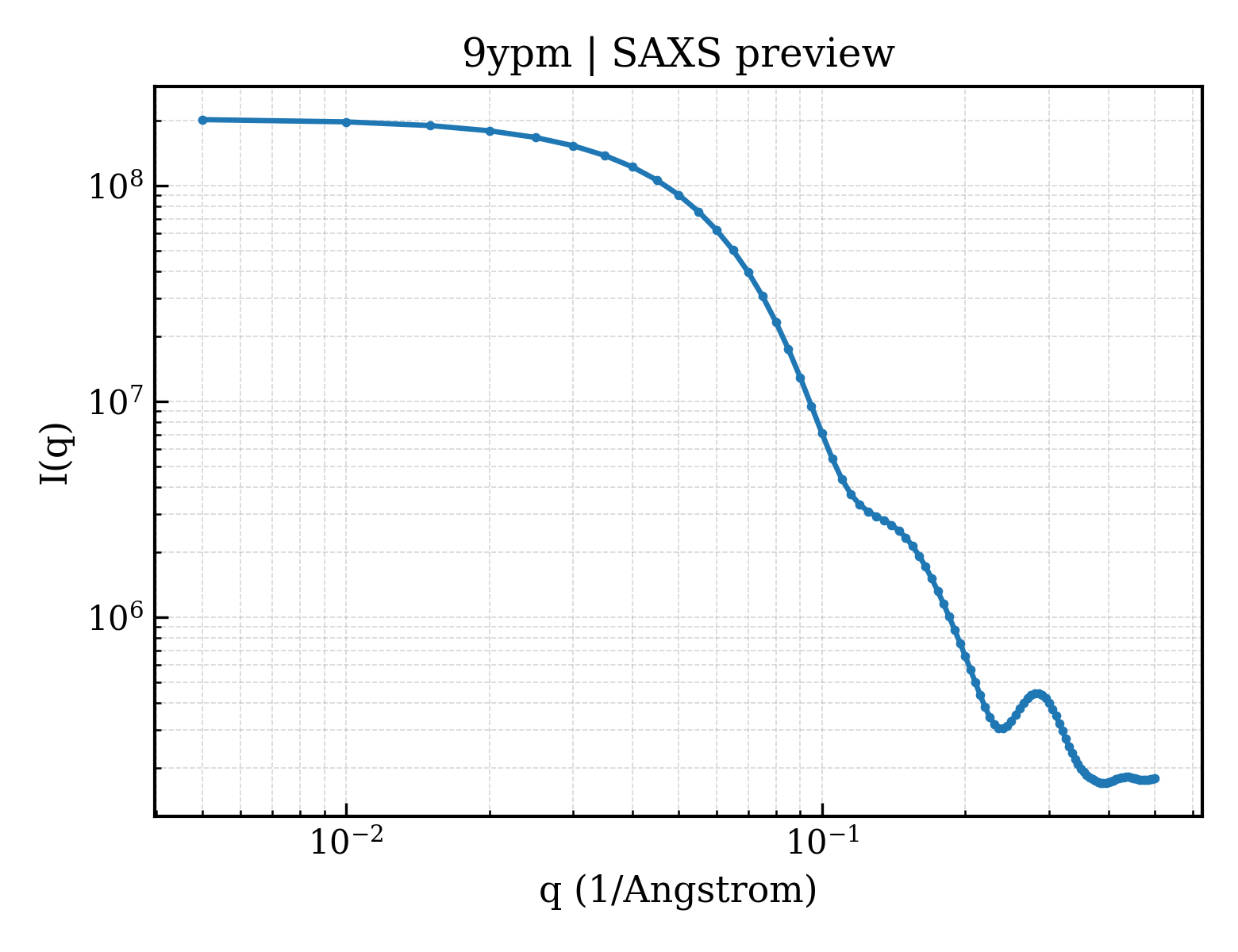
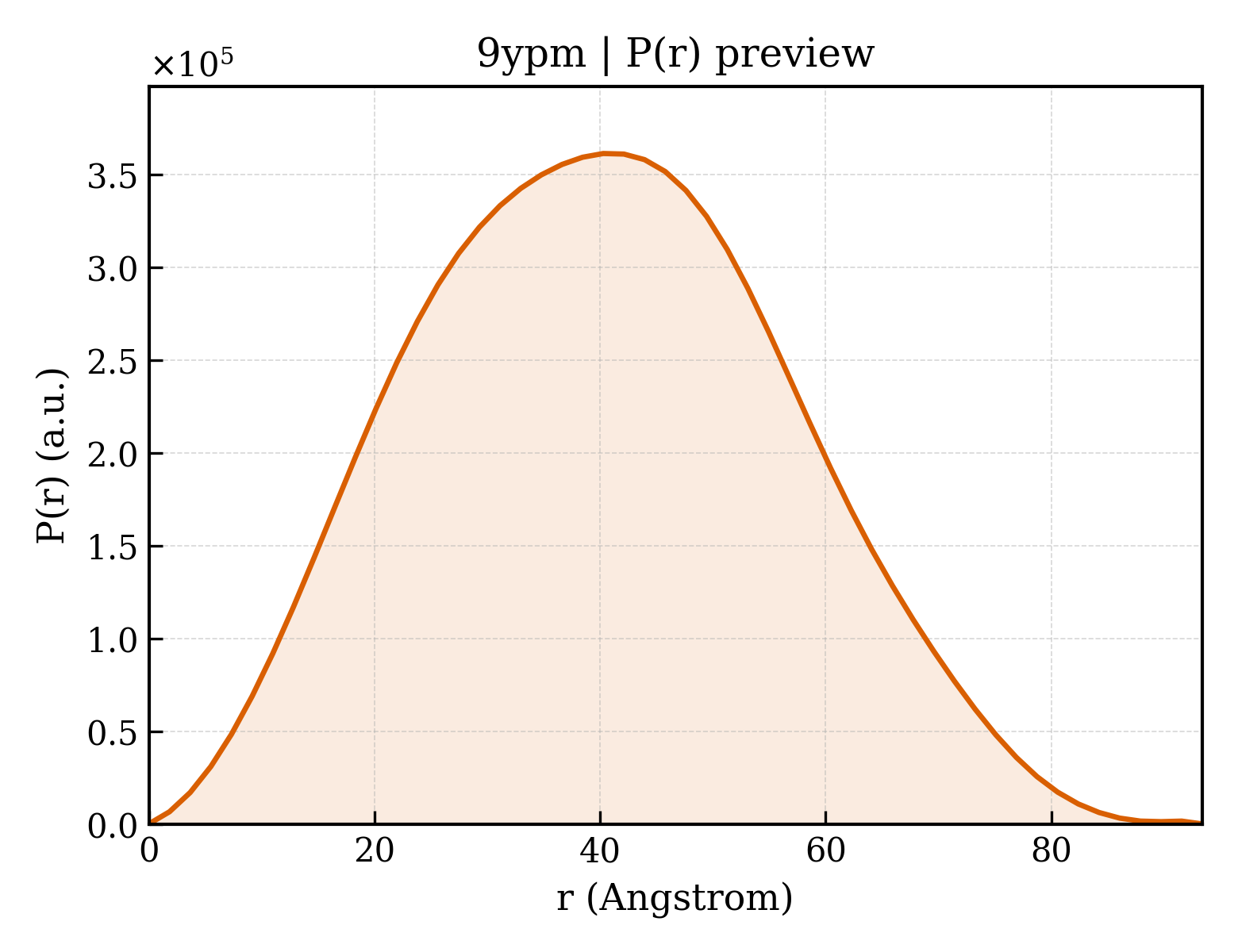
| 1 | Protein monomer Monomer Protein × 1 PDB declaration: monomeric(1) Consistent with protein copy count | Chain A; UniProt 1–225 | Not recorded | SO4 SULFATE ION × 4 CL CHLORIDE ION × 4 FE2 FE (II) ION × 2 GOL GLYCEROL × 12 | X-RAY DIFFRACTION X-ray crystallization conditions:VAPOR DIFFUSION, HANGING DROP;pH 4.6;298 K;MboA crystals were prepared via the hanging drop vapor diffusion method in an Coy anaerobic chamber with an atmosphere of < 5 ppm of O2. Hanging drops were prepared by combining equal volumes of MboA (9 mg/mL) protein solution, reservoir solution (0.6 M lithium sulfate and 0.1 M sodium acetate, pH 4.6), and 0.2 uL of a 1:1000 dilution microseed master stock generated from apo crystals for a total drop volume of 2.2 uL. The Fe(II) + substrate bound structure was prepared by incubating apo crystals with 0.4 uL of 100 mM LAR peptide and 100 mM Fe2+ in 0.1 M sodium acetate, pH 4.6 for two hours prior to looping. Crystals were cryoprotected with the addition of 0.75 uL of 40% glycerol in 0.1 M sodium acetate, pH 4.6 and flash frozen in LN2 | Resolution 2.45 Å R-free 0.195 |
| 2 | Protein monomer Monomer Protein × 1 PDB declaration: monomeric(1) Consistent with protein copy count | Chain B; UniProt 1–225 | Not recorded | SO4 SULFATE ION × 5 CL CHLORIDE ION × 4 FE2 FE (II) ION × 2 GOL GLYCEROL × 7 ACT ACETATE ION × 1 | X-RAY DIFFRACTION X-ray crystallization conditions:VAPOR DIFFUSION, HANGING DROP;pH 4.6;298 K;MboA crystals were prepared via the hanging drop vapor diffusion method in an Coy anaerobic chamber with an atmosphere of < 5 ppm of O2. Hanging drops were prepared by combining equal volumes of MboA (9 mg/mL) protein solution, reservoir solution (0.6 M lithium sulfate and 0.1 M sodium acetate, pH 4.6), and 0.2 uL of a 1:1000 dilution microseed master stock generated from apo crystals for a total drop volume of 2.2 uL. The Fe(II) + substrate bound structure was prepared by incubating apo crystals with 0.4 uL of 100 mM LAR peptide and 100 mM Fe2+ in 0.1 M sodium acetate, pH 4.6 for two hours prior to looping. Crystals were cryoprotected with the addition of 0.75 uL of 40% glycerol in 0.1 M sodium acetate, pH 4.6 and flash frozen in LN2 | Resolution 2.45 Å R-free 0.195 |
| 3 | Protein monomer Monomer Protein × 1 PDB declaration: monomeric(1) Consistent with protein copy count | Chain C; UniProt 1–225 | Not recorded | SO4 SULFATE ION × 3 CL CHLORIDE ION × 2 FE2 FE (II) ION × 2 GOL GLYCEROL × 10 ACT ACETATE ION × 2 | X-RAY DIFFRACTION X-ray crystallization conditions:VAPOR DIFFUSION, HANGING DROP;pH 4.6;298 K;MboA crystals were prepared via the hanging drop vapor diffusion method in an Coy anaerobic chamber with an atmosphere of < 5 ppm of O2. Hanging drops were prepared by combining equal volumes of MboA (9 mg/mL) protein solution, reservoir solution (0.6 M lithium sulfate and 0.1 M sodium acetate, pH 4.6), and 0.2 uL of a 1:1000 dilution microseed master stock generated from apo crystals for a total drop volume of 2.2 uL. The Fe(II) + substrate bound structure was prepared by incubating apo crystals with 0.4 uL of 100 mM LAR peptide and 100 mM Fe2+ in 0.1 M sodium acetate, pH 4.6 for two hours prior to looping. Crystals were cryoprotected with the addition of 0.75 uL of 40% glycerol in 0.1 M sodium acetate, pH 4.6 and flash frozen in LN2 | Resolution 2.45 Å R-free 0.195 |
| 4 | Protein monomer Monomer Protein × 1 PDB declaration: monomeric(1) Consistent with protein copy count | Chain D; UniProt 1–225 | Not recorded | SO4 SULFATE ION × 3 CL CHLORIDE ION × 2 FE2 FE (II) ION × 2 GOL GLYCEROL × 7 | X-RAY DIFFRACTION X-ray crystallization conditions:VAPOR DIFFUSION, HANGING DROP;pH 4.6;298 K;MboA crystals were prepared via the hanging drop vapor diffusion method in an Coy anaerobic chamber with an atmosphere of < 5 ppm of O2. Hanging drops were prepared by combining equal volumes of MboA (9 mg/mL) protein solution, reservoir solution (0.6 M lithium sulfate and 0.1 M sodium acetate, pH 4.6), and 0.2 uL of a 1:1000 dilution microseed master stock generated from apo crystals for a total drop volume of 2.2 uL. The Fe(II) + substrate bound structure was prepared by incubating apo crystals with 0.4 uL of 100 mM LAR peptide and 100 mM Fe2+ in 0.1 M sodium acetate, pH 4.6 for two hours prior to looping. Crystals were cryoprotected with the addition of 0.75 uL of 40% glycerol in 0.1 M sodium acetate, pH 4.6 and flash frozen in LN2 | Resolution 2.45 Å R-free 0.195 |
Other States of the Same Protein in the Database
Each row is a biological assembly of the same UniProt protein in another PDB entry. The “Difference from current entry” column identifies evidence-level differences; no tag means the currently parsed fields agree.
2 other PDB entries and 8 assemblies. Open the comparison page and filter oligomeric states
View Construct and Data Evidence
| UniProt name | A0A244EXR3_PSESX |
| Isoform | — |
| PDB entities | 1 |
| Chains and sequence ranges | Author chain A; PDBConstruct 26–250; UniProt 1–225 Author chain B; PDBConstruct 26–250; UniProt 1–225 Author chain C; PDBConstruct 26–250; UniProt 1–225 Author chain D; PDBConstruct 26–250; UniProt 1–225 |