2. Measurements Reported by SASBDB Reported Results
Metric
Method
Value
Uncertainty
Unit
dmax
P(r)
14.1
—
nm
i0
Guinier
0.1055
0.0001
—
i0
P(r)
0.1068
—
—
mw
Experimental
38.2
—
kDa
porod_volume
Porod
54.7
—
nm³
rg
Guinier
3.75
0.01
nm
rg
P(r)
3.931
0.001
nm
These values are reported by SASBDB; SAXSdb did not recalculate them from the experimental curve.
▼
3. Source Fits and Models Source Fits & Models
This entry has no source fit record.
▶
4. Source File Index Source Files
Category
Path
Download status
Size
curve
sasbdb/entries/25/sasdb25/source/SASDB25.dat
downloaded
39837
full_entry_zip
sasbdb/entries/25/sasdb25/source/SASDB25.zip
downloaded
40224
pddf
sasbdb/entries/25/sasdb25/source/SASDB25.out
downloaded
68448
sascif
sasbdb/entries/25/sasdb25/source/SASDB25.sascif
downloaded
93872
summary
sasbdb/entries/25/sasdb25/source/summary.json
downloaded
7901
▶
5. Experiment Description and Publication Experiment & Publication
Original Experiment Description
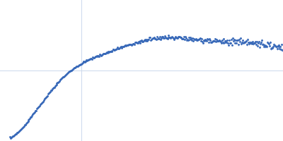
X-ray synchrotron radiation scattering data from solutions of human cardiac myosin binding protein-C, domains C5-C6-C7 in 25 mM TrisHCl 250 mM NaCl, 2 mM TCEP, 0.02% sodium azide were collected on the SAXS/WAXS beam line of the Australian Synchrotron (Melbourne, Australia) using a Pilatus 1M-W pixel detector (I(s) vs s; s = 4π sin θ/λ, where 2θ is the scattering angle and λ=0.10332 nm). Different solute concentrations in the range 0.70-3.40 mg/ml were measured using a total exposure time of 44 s (recorded as 44 x 1 s frames). The data were normalized to the intensity of the transmitted beam and radially averaged and the scattering from the matched solvent-blank was subtracted. The data presented here represent the SAXS profile extrapolated to zero concentration calculated from the solute concentration series.
Related Publication
Title
Clinically Linked Mutations in the Central Domains of Cardiac Myosin-Binding Protein C with Distinct Phenotypes Show Differential Structural Effects.
Authors
Nadvi NA, Michie KA, Kwan AH, Guss JM, Trewhella J
DOI / PMID
10.1016/j.str.2015.11.001 / 26688216
▼
6. Complete Source Record Complete Source Record
The following content comes directly from the SASBDB entry. Missing values are shown as “—”; SAXSdb does not infer undeclared units.
School of Molecular Bioscience, University of Sydney.
experiment.contributor[0].affiliation[0].webpage
experiment.contributor[0].contributor_name
Jill
experiment.contributor[0].contributor_surname
Trewhella
experiment.contributor[0].orcid
null
experiment.concentration_method
null
experiment.concentration_unit
mg/ml
experiment.date
2015-04-18
experiment.storage_temperature
22.0
experiment.cell_temperature
22.0
experiment.exposure_time
1.0
experiment.number_of_frames
44
experiment.wavelength
null
experiment.sample_detector_distance
1.6
experiment.concentration_min
0.7
experiment.concentration_max
3.4
experiment.sample_volume
null
experiment.flow_rate
null
experiment.s_min
0.057
experiment.s_max
3.436
experiment.total_exposure_time
null
experiment.seccolumn
null
fits
[]
estimated_volume_method
null
pddf_software
ATSAS GNOM
pddf_software_version
null
i0_calibration_standard
null
description
null
experiment_description
X-ray synchrotron radiation scattering data from solutions of human cardiac myosin binding protein-C, domains C5-C6-C7 in 25 mM TrisHCl 250 mM NaCl, 2 mM TCEP, 0.02% sodium azide were collected on the SAXS/WAXS beam line of the Australian Synchrotron (Melbourne, Australia) using a Pilatus 1M-W pixel detector (I(s) vs s; s = 4π sin θ/λ, where 2θ is the scattering angle and λ=0.10332 nm). Different solute concentrations in the range 0.70-3.40 mg/ml were measured using a total exposure time of 44 s (recorded as 44 x 1 s frames). The data were normalized to the intensity of the transmitted beam and radially averaged and the scattering from the matched solvent-blank was subtracted. The data presented here represent the SAXS profile extrapolated to zero concentration calculated from the solute concentration series.