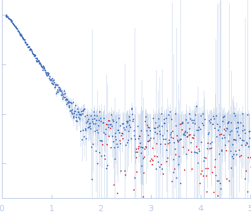
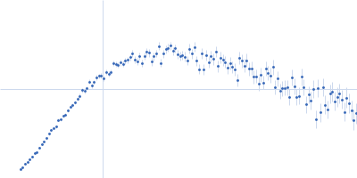
2. Measurements Reported by SASBDB Reported Results
Metric
Method
Value
Uncertainty
Unit
dmax
P(r)
16.4
—
Å
i0
Guinier
0.278134
—
arbitrary
i0
P(r)
0.2736
—
arbitrary
mw
Experimental
86.0
—
kDa
mw
Guinier I(0)
86.0
—
kDa
mw
Porod
101.0
—
kDa
porod_volume
Porod
163.0
—
ų
rg
Guinier
4.867
0.663
Å
rg
P(r)
4.87
—
Å
These values are reported by SASBDB; SAXSdb did not recalculate them from the experimental curve.
▼
3. Source Fits and Models Source Fits & Models
This entry has no source fit record.
▶
4. Source File Index Source Files
Category
Path
Download status
Size
curve
sasbdb/entries/v4/sasdxv4/source/SASDXV4.dat
downloaded
39701
full_entry_zip
sasbdb/entries/v4/sasdxv4/source/SASDXV4.zip
downloaded
22422
pddf
sasbdb/entries/v4/sasdxv4/source/SASDXV4.out
downloaded
27489
sascif
sasbdb/entries/v4/sasdxv4/source/SASDXV4.sascif
not_available
—
summary
sasbdb/entries/v4/sasdxv4/source/summary.json
downloaded
7773
▶
5. Experiment Description and Publication Experiment & Publication
Original Experiment Description
SAXS data from solutions of EGFR kinase domain duplication mutant in 20 mM HEPES, 250 mM NaCl, 250 mM KCl, pH 8 were collected on the Rigaku MicroMax-007HF instrument (Yale University, New Haven, United States) using a Rigaku PSAXS Nano detector at a sample-detector distance of 0.5 m and at a wavelength of λ = 0.1542 nm (I(s) vs s, where s = 4πsinθ/λ, and 2θ is the scattering angle). One solute concentration of 2.60 mg/ml was measured at 4°C. Three successive 7200 second frames were collected. The data were normalized to the intensity of the transmitted beam and radially averaged; the scattering of the solvent-blank was subtracted.
▼
6. Complete Source Record Complete Source Record
The following content comes directly from the SASBDB entry. Missing values are shown as “—”; SAXSdb does not infer undeclared units.
EGFR kinase domain duplication mutant. The purified construct consists of amino acids 643-976 appended to amino acids 651-993 using mature protein numbering (UniProt Q504U8 numbering: 651-993 followed by a duplicated 643-977 region). The protein construct used for SAXS contains an additional N-terminal non-native 22 amino acid extension and polyhistidine tag (HHHHHHSSGVDLGTENLYFQSM).
experiment.sample.buffer.name
20 mM HEPES, 250 mM NaCl, 250 mM KCl
experiment.sample.buffer.concentration_unit
null
experiment.sample.buffer.comment
null
experiment.sample.buffer.additive
null
experiment.sample.buffer.concentration
null
experiment.sample.buffer.pka
null
experiment.sample.buffer.ph
8.0
experiment.sample.buffer.deuteration
null
experiment.sample.purity_method
null
experiment.sample.name
EGFR kinase domain duplication mutant (KDD) at 2.6 mg/ml
SAXS data from solutions of EGFR kinase domain duplication mutant in 20 mM HEPES, 250 mM NaCl, 250 mM KCl, pH 8 were collected on the Rigaku MicroMax-007HF instrument (Yale University, New Haven, United States) using a Rigaku PSAXS Nano detector at a sample-detector distance of 0.5 m and at a wavelength of λ = 0.1542 nm (I(s) vs s, where s = 4πsinθ/λ, and 2θ is the scattering angle). One solute concentration of 2.60 mg/ml was measured at 4°C. Three successive 7200 second frames were collected. The data were normalized to the intensity of the transmitted beam and radially averaged; the scattering of the solvent-blank was subtracted.