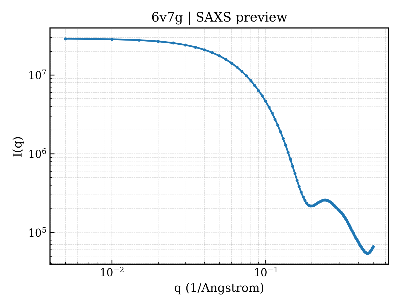
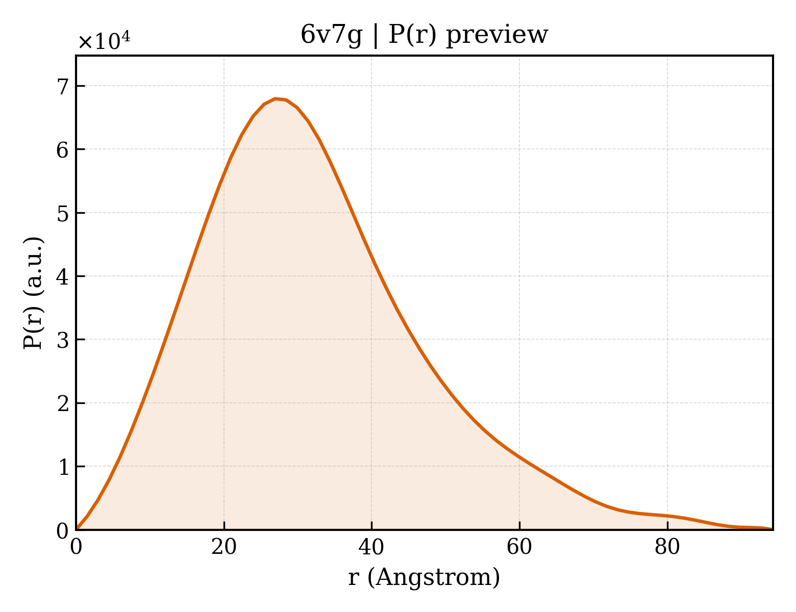
Canavalin
OrganismNot specified
State in the Current Structure
| Assembly | Oligomeric State | Construct | Mutations and Modifications | Ligands, Ions and Associated Components | Method and Experimental Conditions | Structure Quality |
|---|---|---|---|---|---|---|
| 1 | Protein homooligomer Homooligomer Protein × 3 PDB declaration: trimeric(3) Consistent with protein copy count | Chain A; UniProt 1–445 | Non-standard monomer:Yes (specific site not provided by mmCIF) | ACT ACETATE ION × 3 BEZ BENZOIC ACID × 3 CIT CITRIC ACID × 3 | X-RAY DIFFRACTION X-ray crystallization conditions:VAPOR DIFFUSION, SITTING DROP;pH 6;298 K;Vapor diffusion in sitting drop Cryschem plates. Reservoirs were 1.0 M sodium citrate titrated with acetic acid to pH6.0. Drops were initially equal amounts of the reservoir solution with a protein stock solution of 30 mg/ml canavalin in water with a trace of ammonium hydroxide. At room temperature crystallization time was about three weeks. | Resolution 1.40 Å R-free 0.188 |
Other States of the Same Protein in the Database
Each row is a biological assembly of the same UniProt protein in another PDB entry. The “Difference from current entry” column identifies evidence-level differences; no tag means the currently parsed fields agree.
| Other PDB | Difference from Current Entry 6V7G | Assembly / Oligomeric State | Construct | Mutations and Modifications | Ligands, Ions and Non-polymers | Method and Experimental Conditions | Structure Quality |
|---|---|---|---|---|---|---|---|
| 1CAU DETERMINATION OF THREE CRYSTAL STRUCTURES OF CANAVALIN BY MOLECULAR REPLACEMENT Deposited 1993-07-08 | Different construct Different mutation/modification Different oligomeric state Different ligand/ion Different experimental conditions Different structure-quality metrics | Assembly 1 Protein homooligomer Homooligomer;Protein × 6 PDB declaration: hexameric |
Chain A
44–224(181 aa)
Chain B
241–424(184 aa)
|
Not recorded | No recorded non-water small molecule | X-RAY DIFFRACTION mmCIF provides none of the parsed conditions | Resolution 2.30 Å |
| 1CAV THE THREE-DIMENSIONAL STRUCTURE OF CANAVALIN FROM JACK BEAN (CANAVALIA ENSIFORMIS) Deposited 1993-05-27 | Different construct Different mutation/modification Different oligomeric state Different ligand/ion Different experimental conditions Different structure-quality metrics | Assembly 1 Protein homooligomer Homooligomer;Protein × 6 PDB declaration: hexameric |
Chain A
44–224(181 aa)
Chain B
241–424(184 aa)
|
Not recorded | No recorded non-water small molecule | X-RAY DIFFRACTION mmCIF provides none of the parsed conditions | Resolution 2.60 Å |
| 1CAW DETERMINATION OF THREE CRYSTAL STRUCTURES OF CANAVALIN BY MOLECULAR REPLACEMENT Deposited 1993-06-02 | Different construct Different mutation/modification Different oligomeric state Different ligand/ion Different experimental conditions Different structure-quality metrics | Assembly 1 Protein homooligomer Homooligomer;Protein × 6 PDB declaration: hexameric |
Chain A
44–224(181 aa)
Chain B
241–424(184 aa)
|
Not recorded | No recorded non-water small molecule | X-RAY DIFFRACTION mmCIF provides none of the parsed conditions | Resolution 2.60 Å |
| 1CAX DETERMINATION OF THREE CRYSTAL STRUCTURES OF CANAVALIN BY MOLECULAR REPLACEMENT Deposited 1993-06-10 | Different construct Different mutation/modification Different oligomeric state Different ligand/ion Different experimental conditions Different structure-quality metrics | Assembly 1 Protein homooligomer Homooligomer;Protein × 6 PDB declaration: hexameric |
Chain A
44–224(181 aa)
Chain B
241–424(184 aa)
Chain C
44–224(181 aa)
Chain D
241–424(184 aa)
Chain E
44–224(181 aa)
Chain F
241–424(184 aa)
|
Not recorded | No recorded non-water small molecule | X-RAY DIFFRACTION mmCIF provides none of the parsed conditions | Resolution 2.60 Å |
| 1DGR Refined crystal structure of canavalin from jack bean Deposited 1999-11-25 | Different construct Different mutation/modification Different oligomeric state Different ligand/ion Different experimental conditions Different structure-quality metrics | Assembly 1 Protein homooligomer Homooligomer;Protein × 9 PDB declaration: nonameric |
Chain A
46–223(178 aa)
Fragment:RESIDUES 46-223
Chain B
46–223(178 aa)
Fragment:RESIDUES 46-223
Chain C
46–223(178 aa)
Fragment:RESIDUES 46-223
Chain M
331–423(93 aa)
Fragment:RESIDUES 331-423
Chain N
246–324(79 aa)
Fragment:RESIDUES 246-324
Chain V
246–324(79 aa)
Fragment:RESIDUES 246-324
Chain W
331–423(93 aa)
Fragment:RESIDUES 331-423
Chain X
246–324(79 aa)
Fragment:RESIDUES 246-324
Chain Y
331–423(93 aa)
Fragment:RESIDUES 331-423
|
Not recorded | PO4 PHOSPHATE ION × 3 |
X-RAY DIFFRACTION
X-ray crystallization conditions
VAPOR DIFFUSION, SITTING DROP;pH 6.8;277 K;Dulbeccos phosphate buffered saline,
ammonium hydroxide, pH 6.8, VAPOR DIFFUSION, SITTING DROP, temperature 277K
|
Resolution 2.60 Å R-free 0.226 |
| 1DGW Structure of the rhombohedral crystal of canavalin from jack bean Deposited 1999-11-26 | Different construct Different mutation/modification Different oligomeric state Different ligand/ion Different experimental conditions Different structure-quality metrics | Assembly 1 Protein homooligomer Homooligomer;Protein × 9 PDB declaration: nonameric |
Chain A
46–223(178 aa)
Fragment:RESIDUES 46-223
Chain X
246–324(79 aa)
Fragment:RESIDUES 246-324
Chain Y
331–423(93 aa)
Fragment:RESIDUES 331-423
|
Not recorded | PO4 PHOSPHATE ION × 3 |
X-RAY DIFFRACTION
X-ray crystallization conditions
LIQUID DIFFUSION;pH 6.8;293 K;DULBECCO'S PHOSPHATE BUFFERED SALINE, AMMONIUM HYDROXIDE (TRACE), MICROGRAVITY, pH 6.8, LIQUID DIFFUSION, temperature 293K
|
Resolution 1.70 Å R-free 0.252 |
| 2CAU CANAVALIN FROM JACK BEAN Deposited 1998-11-20 | Different mutation/modification Different ligand/ion Different experimental conditions Different structure-quality metrics | Assembly 1 Protein homooligomer Homooligomer;Protein × 3 PDB declaration: trimeric |
Chain A
1–445(445 aa)
|
Not recorded | No recorded non-water small molecule |
X-RAY DIFFRACTION
X-ray crystallization conditions
pH 6.8;THIS CRYSTAL FORM COULD BE GROWN ONLY OCCASIONALLY. BASICALLY IT WAS CRYSTALLIZED FROM PURIFIED PROTEIN AFTER TRYPSIN TREATMENT IN PHOSPHATE BUFFERED SALINE, BUT WAS DIFFICULT TO REPRODUCE., pH 6.8
|
Resolution 2.10 Å R-free 0.235 |
| 2CAV CANAVALIN FROM JACK BEAN Deposited 1998-11-20 | Different mutation/modification Different ligand/ion Different experimental conditions Different structure-quality metrics | Assembly 1 Protein homooligomer Homooligomer;Protein × 3 PDB declaration: trimeric |
Chain A
1–445(445 aa)
|
Not recorded | No recorded non-water small molecule |
X-RAY DIFFRACTION
X-ray crystallization conditions
VAPOR DIFFUSION;pH 6.8;30 - 40 MG/ML PROTEIN SOLUTION WAS PREPARED BY DISSOLVING 4-TIME RECRYSTALLIZED CANAVALIN IN DISTILLED WATER PLUS TRACE NH4OH. RESERVOIR SOLUTION CONTAINED 2.0 % NACL IN 50 MM PHOSPHATE BUFFER AT PH 6.8. CRYSTALS WERE OBTAINED BY MIXING PROTEIN AND RESERVOIR SOLUTION IN SITTING DROPS FOLLOWED BY VAPOR DIFFUSION AGAINST THE RESERVOIR., vapor diffusion
|
Resolution 2.00 Å R-free 0.264 |
| 6V7J The C2221 crystal form of canavalin at 173 K Deposited 2019-12-08 | Different construct Different mutation/modification Different ligand/ion Different experimental conditions Different structure-quality metrics | Assembly 1 Protein homooligomer Homooligomer;Protein × 3 PDB declaration: trimeric |
Chain A
1–445(445 aa)
Chain B
1–445(445 aa)
Chain C
1–445(445 aa)
|
Non-standard monomer:Yes (specific site not provided by mmCIF) Non-standard monomer:Yes (specific site not provided by mmCIF) Non-standard monomer:Yes (specific site not provided by mmCIF) | BEZ BENZOIC ACID × 3 GOL GLYCEROL × 3 CA CALCIUM ION × 3 |
X-RAY DIFFRACTION
X-ray crystallization conditions
VAPOR DIFFUSION, SITTING DROP;pH 6.5;298 K;Vapor diffusion in Cryschem sitting drop plates at room temperature. Reservoirs were Dulbeccos Phosphate buffered saline at pH 6.5. Drops were equal amounts of the reservoir and a 40 mg/ml solution of the protein dissolved in water with a trace of ammonium hydroxide. Crystallization time was 24 to 48 hours.
|
Resolution 2.00 Å R-free 0.247 |
| 6V7L The structure of the P212121 crystal form of canavalin at 173 K Deposited 2019-12-08 | Different construct Different mutation/modification Different ligand/ion Different experimental conditions Different structure-quality metrics | Assembly 1 Protein homooligomer Homooligomer;Protein × 3 PDB declaration: trimeric |
Chain A
1–445(445 aa)
Chain B
1–445(445 aa)
Chain C
1–445(445 aa)
|
Not recorded | BEZ BENZOIC ACID × 3 |
X-RAY DIFFRACTION
X-ray crystallization conditions
VAPOR DIFFUSION, SITTING DROP;pH 7.5;298 K;Crystals were grown by sitting drop vapor diffusion from 18% PEG 3350 in 0.1 M HEPES reservoirs. The drops were equal amounts of a 40 mg/ml solution of precanavalin (not treated with any exogenous protease) in water. Crystals grew only after six to eight weeks at room temperature.
|
Resolution 2.80 Å R-free 0.252 |
10 other PDB entries and 10 assemblies. Open the comparison page and filter oligomeric states
View Construct and Data Evidence
| UniProt name | CANA_CANEN |
| Isoform | — |
| PDB entities | 1 |
| Chains and sequence ranges | Author chain A; PDBConstruct 1–445; UniProt 1–445 |