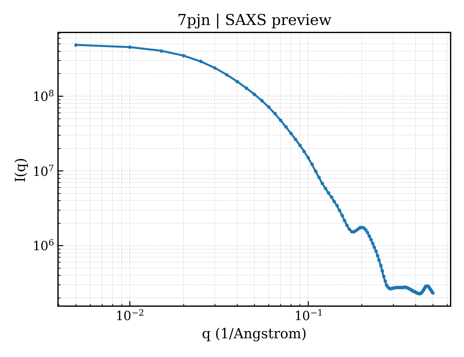
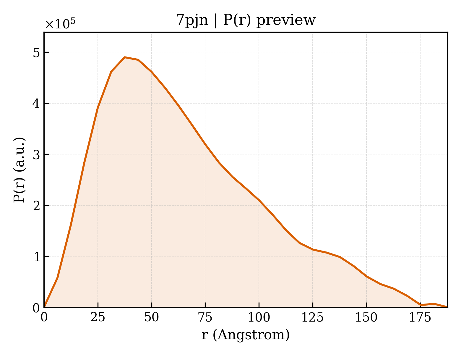
Isocitrate dehydrogenase [NADP] cytoplasmic
Homo sapiens
State in the Current Structure
| Assembly | Oligomeric State | Construct | Mutations and Modifications | Ligands, Ions and Associated Components | Method and Experimental Conditions | Structure Quality |
|---|---|---|---|---|---|---|
| 1 | Protein homooligomer Homooligomer Protein × 2 PDB declaration: dimeric(2) Consistent with protein copy count | Chain A; UniProt 1–414 Chain D; UniProt 1–414 | Mutation:R132C, S280F Non-standard monomer:Yes (specific site not provided by mmCIF) | 7SU (E)-3-(1-(5-(2-fluoropropan-2-yl)-3-(2,4,6-trichlorophenyl)isoxazole-4-carbonyl)-3-methyl-1H-indol-4-yl)acrylic acid × 2 CIT CITRIC ACID × 3 GOL GLYCEROL × 2 NDP NADPH DIHYDRO-NICOTINAMIDE-ADENINE-DINUCLEOTIDE PHOSPHATE × 2 | X-RAY DIFFRACTION X-ray crystallization conditions:VAPOR DIFFUSION, SITTING DROP;pH 7;293 K;2 M Ammonium citrate tribasic, DTT 2 mM | Resolution 2.45 Å R-free 0.228 |
| 2 | Protein homooligomer Homooligomer Protein × 2 PDB declaration: dimeric(2) Consistent with protein copy count | Chain B; UniProt 1–414 Chain C; UniProt 1–414 | Mutation:R132C, S280F Non-standard monomer:Yes (specific site not provided by mmCIF) | 7SU (E)-3-(1-(5-(2-fluoropropan-2-yl)-3-(2,4,6-trichlorophenyl)isoxazole-4-carbonyl)-3-methyl-1H-indol-4-yl)acrylic acid × 2 CIT CITRIC ACID × 2 GOL GLYCEROL × 4 NDP NADPH DIHYDRO-NICOTINAMIDE-ADENINE-DINUCLEOTIDE PHOSPHATE × 2 | X-RAY DIFFRACTION X-ray crystallization conditions:VAPOR DIFFUSION, SITTING DROP;pH 7;293 K;2 M Ammonium citrate tribasic, DTT 2 mM | Resolution 2.45 Å R-free 0.228 |
Other States of the Same Protein in the Database
Each row is a biological assembly of the same UniProt protein in another PDB entry. The “Difference from current entry” column identifies evidence-level differences; no tag means the currently parsed fields agree.
| Other PDB | Difference from Current Entry 7PJN | Assembly / Oligomeric State | Construct | Mutations and Modifications | Ligands, Ions and Non-polymers | Method and Experimental Conditions | Structure Quality |
|---|---|---|---|---|---|---|---|
| 1T09 Crystal structure of human cytosolic NADP(+)-dependent isocitrate dehydrogenase in complex NADP Deposited 2004-04-08 | Different mutation/modification Different ligand/ion Different experimental conditions Different structure-quality metrics | Assembly 1 Protein homooligomer Homooligomer;Protein × 2 PDB declaration: dimeric |
Chain A
1–414(414 aa)
Chain B
1–414(414 aa)
|
Not recorded | NAP NADP NICOTINAMIDE-ADENINE-DINUCLEOTIDE PHOSPHATE × 2 |
X-RAY DIFFRACTION
X-ray crystallization conditions
VAPOR DIFFUSION, HANGING DROP;pH 6.5;277 K;PEG 20000, MES, pH 6.5, VAPOR DIFFUSION, HANGING DROP, temperature 277K
|
Resolution 2.70 Å R-free 0.270 |
| 1T0L Crystal structure of human cytosolic NADP(+)-dependent isocitrate dehydrogenase in complex with NADP, isocitrate, and calcium(2+) Deposited 2004-04-10 | Different mutation/modification Different ligand/ion Different experimental conditions Different structure-quality metrics | Assembly 1 Protein homooligomer Homooligomer;Protein × 2 PDB declaration: dimeric |
Chain A
1–414(414 aa)
Chain B
1–414(414 aa)
|
Not recorded | CA CALCIUM ION × 2 NAP NADP NICOTINAMIDE-ADENINE-DINUCLEOTIDE PHOSPHATE × 2 ICT ISOCITRIC ACID × 2 |
X-RAY DIFFRACTION
X-ray crystallization conditions
VAPOR DIFFUSION, HANGING DROP;pH 5.9;293 K;PEG 6000, MES, NADP, isocitrate, calcium chloride, pH 5.9, VAPOR DIFFUSION, HANGING DROP, temperature 293K
|
Resolution 2.41 Å R-free 0.253 |
| 1T0L Crystal structure of human cytosolic NADP(+)-dependent isocitrate dehydrogenase in complex with NADP, isocitrate, and calcium(2+) Deposited 2004-04-10 | Different mutation/modification Different ligand/ion Different experimental conditions Different structure-quality metrics | Assembly 2 Protein homooligomer Homooligomer;Protein × 2 PDB declaration: dimeric |
Chain C
1–414(414 aa)
Chain D
1–414(414 aa)
|
Not recorded | CA CALCIUM ION × 2 NAP NADP NICOTINAMIDE-ADENINE-DINUCLEOTIDE PHOSPHATE × 2 ICT ISOCITRIC ACID × 2 |
X-RAY DIFFRACTION
X-ray crystallization conditions
VAPOR DIFFUSION, HANGING DROP;pH 5.9;293 K;PEG 6000, MES, NADP, isocitrate, calcium chloride, pH 5.9, VAPOR DIFFUSION, HANGING DROP, temperature 293K
|
Resolution 2.41 Å R-free 0.253 |
| 3INM Crystal structure of human cytosolic NADP(+)-dependent isocitrate dehydrogenase R132H mutant in complex with NADPH, ALPHA-KETOGLUTARATE and CALCIUM(2+) Deposited 2009-08-12 | Different mutation/modification Different ligand/ion Different experimental conditions Different structure-quality metrics | Assembly 1 Protein homooligomer Homooligomer;Protein × 2 PDB declaration: dimeric |
Chain A
1–414(414 aa)
Chain B
1–414(414 aa)
|
Mutation:R132H Mutation:R132H | NDP NADPH DIHYDRO-NICOTINAMIDE-ADENINE-DINUCLEOTIDE PHOSPHATE × 2 AKG 2-OXOGLUTARIC ACID × 2 CA CALCIUM ION × 2 NA SODIUM ION × 2 GOL GLYCEROL × 2 |
X-RAY DIFFRACTION
X-ray crystallization conditions
VAPOR DIFFUSION, HANGING DROP;pH 6.5;293 K;Protein component: 8 mg/ml IDH, 20mM Tris-HCl pH 7.5, 100mM sodium chloride,
10mM NADPH, 10mM calcium chloride, 75mM alpha-ketoglutaric acid sodium salt.
Precipitant: 100mM MES pH 6.5, 20% PEG 6000. Ratio of protein component to
precipitant in initial hanging drop: 2:1., VAPOR DIFFUSION, HANGING DROP, temperature 293K
|
Resolution 2.10 Å R-free 0.262 |
| 3INM Crystal structure of human cytosolic NADP(+)-dependent isocitrate dehydrogenase R132H mutant in complex with NADPH, ALPHA-KETOGLUTARATE and CALCIUM(2+) Deposited 2009-08-12 | Different construct Different mutation/modification Different ligand/ion Different experimental conditions Different structure-quality metrics | Assembly 2 Protein homooligomer Homooligomer;Protein × 2 PDB declaration: dimeric |
Chain C
1–414(414 aa)
|
Mutation:R132H | NDP NADPH DIHYDRO-NICOTINAMIDE-ADENINE-DINUCLEOTIDE PHOSPHATE × 2 AKG 2-OXOGLUTARIC ACID × 2 CA CALCIUM ION × 2 NA SODIUM ION × 2 GOL GLYCEROL × 2 |
X-RAY DIFFRACTION
X-ray crystallization conditions
VAPOR DIFFUSION, HANGING DROP;pH 6.5;293 K;Protein component: 8 mg/ml IDH, 20mM Tris-HCl pH 7.5, 100mM sodium chloride,
10mM NADPH, 10mM calcium chloride, 75mM alpha-ketoglutaric acid sodium salt.
Precipitant: 100mM MES pH 6.5, 20% PEG 6000. Ratio of protein component to
precipitant in initial hanging drop: 2:1., VAPOR DIFFUSION, HANGING DROP, temperature 293K
|
Resolution 2.10 Å R-free 0.262 |
| 3MAP Crystal structure of homodimeric R132H mutant of human cytosolic NADP(+)-dependent isocitrate dehydrogenase in complex with NADP and isocitrate Deposited 2010-03-24 | Different mutation/modification Different ligand/ion Different experimental conditions Different structure-quality metrics | Assembly 1 Protein homooligomer Homooligomer;Protein × 2 PDB declaration: dimeric |
Chain A
1–414(414 aa)
Chain B
1–414(414 aa)
|
Mutation:R132H Mutation:R132H | NAP NADP NICOTINAMIDE-ADENINE-DINUCLEOTIDE PHOSPHATE × 2 ICT ISOCITRIC ACID × 2 |
X-RAY DIFFRACTION
X-ray crystallization conditions
VAPOR DIFFUSION, HANGING DROP;pH 8.5;293 K;0.1M Tris-HCl, 2.0M (NH4)2SO4, pH 8.5, VAPOR DIFFUSION, HANGING DROP, temperature 293K
|
Resolution 2.80 Å R-free 0.269 |
| 3MAR Crystal structure of homodimeric R132H mutant of human cytosolic NADP(+)-dependent isocitrate dehydrogenase in complex with NADP Deposited 2010-03-24 | Different mutation/modification Different ligand/ion Different experimental conditions Different structure-quality metrics | Assembly 1 Protein homooligomer Homooligomer;Protein × 2 PDB declaration: dimeric |
Chain A
1–414(414 aa)
Chain B
1–414(414 aa)
|
Mutation:R132H Mutation:R132H | NAP NADP NICOTINAMIDE-ADENINE-DINUCLEOTIDE PHOSPHATE × 2 |
X-RAY DIFFRACTION
X-ray crystallization conditions
VAPOR DIFFUSION, HANGING DROP;pH 8.2;277 K;1.0M Na2HPO4/KH2PO4, pH 8.2, vapor diffusion, hanging drop, temperature 277K
|
Resolution 3.41 Å R-free 0.289 |
| 3MAS Crystal structure of heterodimeric R132H mutant of human cytosolic NADP(+)-dependent isocitrate dehydrogenase in complex with NADP and isocitrate Deposited 2010-03-24 | Different mutation/modification Different ligand/ion Different experimental conditions Different structure-quality metrics | Assembly 1 Protein homooligomer Homooligomer;Protein × 2 PDB declaration: dimeric |
Chain A
1–414(414 aa)
Chain B
1–414(414 aa)
|
Mutation:R132H | NAP NADP NICOTINAMIDE-ADENINE-DINUCLEOTIDE PHOSPHATE × 2 ICT ISOCITRIC ACID × 2 |
X-RAY DIFFRACTION
X-ray crystallization conditions
VAPOR DIFFUSION, HANGING DROP;pH 7.5;293 K;0.1M HEPES-Na, 2% PEG 400, 2.0M (NH4)2SO4, pH 7.5, vapor diffusion, hanging drop, temperature 293K
|
Resolution 3.20 Å R-free 0.283 |
| 4I3K Crystal structure of a metabolic reductase with 1-hydroxy-6-(4-hydroxybenzyl)-4-methylpyridin-2(1H)-one Deposited 2012-11-26 | Different mutation/modification Different ligand/ion Different experimental conditions Different structure-quality metrics | Assembly 1 Protein homooligomer Homooligomer;Protein × 2 PDB declaration: dimeric |
Chain A
1–414(414 aa)
Chain B
1–414(414 aa)
|
Mutation:R132H Mutation:R132H | NDP NADPH DIHYDRO-NICOTINAMIDE-ADENINE-DINUCLEOTIDE PHOSPHATE × 2 SO4 SULFATE ION × 4 1BX 1-hydroxy-6-(4-hydroxybenzyl)-4-methylpyridin-2(1H)-one × 1 |
X-RAY DIFFRACTION
X-ray crystallization conditions
VAPOR DIFFUSION, HANGING DROP;pH 5.6;293 K;10 mM NADPH and 10 mM CaCl2 in a solution of 1.75 M (NH4)2SO4 and 0.1 M NaAc (pH 5.6), VAPOR DIFFUSION, HANGING DROP, temperature 293K
|
Resolution 3.31 Å R-free 0.277 |
| 4I3L Crystal structure of a metabolic reductase with 6-benzyl-1-hydroxy-4-methylpyridin-2(1H)-one Deposited 2012-11-26 | Different mutation/modification Different ligand/ion Different experimental conditions Different structure-quality metrics | Assembly 1 Protein homooligomer Homooligomer;Protein × 2 PDB declaration: dimeric |
Chain A
1–414(414 aa)
Chain B
1–414(414 aa)
|
Mutation:R132H Mutation:R132H | NDP NADPH DIHYDRO-NICOTINAMIDE-ADENINE-DINUCLEOTIDE PHOSPHATE × 2 SO4 SULFATE ION × 2 1BZ 6-benzyl-1-hydroxy-4-methylpyridin-2(1H)-one × 1 |
X-RAY DIFFRACTION
X-ray crystallization conditions
VAPOR DIFFUSION, HANGING DROP;pH 5.7;293 K;10 mM NADPH and 10 mM CaCl2 in a solution of 1.75 M (NH4)2SO4 and 0.1 M NaAc (pH 5.7), VAPOR DIFFUSION, HANGING DROP, temperature 293K
|
Resolution 3.29 Å R-free 0.293 |
| 4KZO Crystal Structure Analysis of human IDH1 mutants in complex with NADP+ and Ca2+/alpha-Ketoglutarate Deposited 2013-05-30 | Different mutation/modification Different ligand/ion Different experimental conditions Different structure-quality metrics | Assembly 1 Protein homooligomer Homooligomer;Protein × 2 PDB declaration: dimeric |
Chain A
1–414(414 aa)
Chain B
1–414(414 aa)
|
Not recorded | CA CALCIUM ION × 2 NAP NADP NICOTINAMIDE-ADENINE-DINUCLEOTIDE PHOSPHATE × 2 AKG 2-OXOGLUTARIC ACID × 2 EDO 1,2-ETHANEDIOL × 1 |
X-RAY DIFFRACTION
X-ray crystallization conditions
VAPOR DIFFUSION;pH 6.9;298 K;16-20% PEG MME 2000, 100mM MES, pH 6.9, vapor diffusion, temperature 298K
|
Resolution 2.20 Å R-free 0.221 |
| 4KZO Crystal Structure Analysis of human IDH1 mutants in complex with NADP+ and Ca2+/alpha-Ketoglutarate Deposited 2013-05-30 | Different construct Different mutation/modification Different ligand/ion Different experimental conditions Different structure-quality metrics | Assembly 2 Protein homooligomer Homooligomer;Protein × 2 PDB declaration: dimeric |
Chain C
1–414(414 aa)
|
Not recorded | CA CALCIUM ION × 2 NAP NADP NICOTINAMIDE-ADENINE-DINUCLEOTIDE PHOSPHATE × 2 AKG 2-OXOGLUTARIC ACID × 2 |
X-RAY DIFFRACTION
X-ray crystallization conditions
VAPOR DIFFUSION;pH 6.9;298 K;16-20% PEG MME 2000, 100mM MES, pH 6.9, vapor diffusion, temperature 298K
|
Resolution 2.20 Å R-free 0.221 |
| 4L03 Crystal Structure Analysis of human IDH1 mutants in complex with NADP+ and Ca2+/alpha-Ketoglutarate Deposited 2013-05-30 | Different mutation/modification Different ligand/ion Different experimental conditions Different structure-quality metrics | Assembly 1 Protein homooligomer Homooligomer;Protein × 2 PDB declaration: dimeric |
Chain A
1–414(414 aa)
Chain B
1–414(414 aa)
|
Not recorded | NAP NADP NICOTINAMIDE-ADENINE-DINUCLEOTIDE PHOSPHATE × 2 CA CALCIUM ION × 2 EDO 1,2-ETHANEDIOL × 1 AKG 2-OXOGLUTARIC ACID × 2 |
X-RAY DIFFRACTION
X-ray crystallization conditions
VAPOR DIFFUSION;pH 6.9;298 K;16-20% PEG MME 2000, 100mM MES, pH 6.9, vapor diffusion, temperature 298K
|
Resolution 2.10 Å R-free 0.230 |
| 4L03 Crystal Structure Analysis of human IDH1 mutants in complex with NADP+ and Ca2+/alpha-Ketoglutarate Deposited 2013-05-30 | Different construct Different mutation/modification Different ligand/ion Different experimental conditions Different structure-quality metrics | Assembly 2 Protein homooligomer Homooligomer;Protein × 2 PDB declaration: dimeric |
Chain C
1–414(414 aa)
|
Not recorded | NAP NADP NICOTINAMIDE-ADENINE-DINUCLEOTIDE PHOSPHATE × 2 CA CALCIUM ION × 2 AKG 2-OXOGLUTARIC ACID × 2 |
X-RAY DIFFRACTION
X-ray crystallization conditions
VAPOR DIFFUSION;pH 6.9;298 K;16-20% PEG MME 2000, 100mM MES, pH 6.9, vapor diffusion, temperature 298K
|
Resolution 2.10 Å R-free 0.230 |
| 4L04 Crystal Structure Analysis of human IDH1 mutants in complex with NADP+ and Ca2+/alpha-Ketoglutarate Deposited 2013-05-30 | Different mutation/modification Different ligand/ion Different experimental conditions Different structure-quality metrics | Assembly 1 Protein homooligomer Homooligomer;Protein × 2 PDB declaration: dimeric |
Chain A
1–414(414 aa)
Chain B
1–414(414 aa)
|
Not recorded | AKG 2-OXOGLUTARIC ACID × 2 CA CALCIUM ION × 2 NAP NADP NICOTINAMIDE-ADENINE-DINUCLEOTIDE PHOSPHATE × 2 |
X-RAY DIFFRACTION
X-ray crystallization conditions
VAPOR DIFFUSION;pH 6.9;298 K;16-20% PEG MME 2000, 100mM MES, pH 6.9, vapor diffusion, temperature 298K
|
Resolution 2.87 Å R-free 0.259 |
| 4L04 Crystal Structure Analysis of human IDH1 mutants in complex with NADP+ and Ca2+/alpha-Ketoglutarate Deposited 2013-05-30 | Different mutation/modification Different ligand/ion Different experimental conditions Different structure-quality metrics | Assembly 2 Protein homooligomer Homooligomer;Protein × 2 PDB declaration: dimeric |
Chain C
1–414(414 aa)
Chain D
1–414(414 aa)
|
Not recorded | AKG 2-OXOGLUTARIC ACID × 2 CA CALCIUM ION × 2 NAP NADP NICOTINAMIDE-ADENINE-DINUCLEOTIDE PHOSPHATE × 2 |
X-RAY DIFFRACTION
X-ray crystallization conditions
VAPOR DIFFUSION;pH 6.9;298 K;16-20% PEG MME 2000, 100mM MES, pH 6.9, vapor diffusion, temperature 298K
|
Resolution 2.87 Å R-free 0.259 |
| 4L04 Crystal Structure Analysis of human IDH1 mutants in complex with NADP+ and Ca2+/alpha-Ketoglutarate Deposited 2013-05-30 | Different mutation/modification Different ligand/ion Different experimental conditions Different structure-quality metrics | Assembly 3 Protein homooligomer Homooligomer;Protein × 2 PDB declaration: dimeric |
Chain E
1–414(414 aa)
Chain F
1–414(414 aa)
|
Not recorded | AKG 2-OXOGLUTARIC ACID × 2 CA CALCIUM ION × 2 NAP NADP NICOTINAMIDE-ADENINE-DINUCLEOTIDE PHOSPHATE × 2 |
X-RAY DIFFRACTION
X-ray crystallization conditions
VAPOR DIFFUSION;pH 6.9;298 K;16-20% PEG MME 2000, 100mM MES, pH 6.9, vapor diffusion, temperature 298K
|
Resolution 2.87 Å R-free 0.259 |
| 4L06 Crystal Structure Analysis of human IDH1 mutants in complex with NADP+ and Ca2+/alpha-Ketoglutarate Deposited 2013-05-30 | Different mutation/modification Different ligand/ion Different experimental conditions Different structure-quality metrics | Assembly 1 Protein homooligomer Homooligomer;Protein × 2 PDB declaration: dimeric |
Chain A
1–414(414 aa)
Chain B
1–414(414 aa)
|
Mutation:Y139D Mutation:Y139D | CA CALCIUM ION × 2 NAP NADP NICOTINAMIDE-ADENINE-DINUCLEOTIDE PHOSPHATE × 2 AKG 2-OXOGLUTARIC ACID × 2 |
X-RAY DIFFRACTION
X-ray crystallization conditions
VAPOR DIFFUSION;pH 6.9;298 K;16-20% PEG MME 2000, 100mM MES, pH 6.9, vapor diffusion, temperature 298K
|
Resolution 2.28 Å R-free 0.270 |
| 4L06 Crystal Structure Analysis of human IDH1 mutants in complex with NADP+ and Ca2+/alpha-Ketoglutarate Deposited 2013-05-30 | Different mutation/modification Different ligand/ion Different experimental conditions Different structure-quality metrics | Assembly 2 Protein homooligomer Homooligomer;Protein × 2 PDB declaration: dimeric |
Chain C
1–414(414 aa)
Chain D
1–414(414 aa)
|
Mutation:Y139D Mutation:Y139D | CA CALCIUM ION × 2 NAP NADP NICOTINAMIDE-ADENINE-DINUCLEOTIDE PHOSPHATE × 2 AKG 2-OXOGLUTARIC ACID × 2 |
X-RAY DIFFRACTION
X-ray crystallization conditions
VAPOR DIFFUSION;pH 6.9;298 K;16-20% PEG MME 2000, 100mM MES, pH 6.9, vapor diffusion, temperature 298K
|
Resolution 2.28 Å R-free 0.270 |
| 4L06 Crystal Structure Analysis of human IDH1 mutants in complex with NADP+ and Ca2+/alpha-Ketoglutarate Deposited 2013-05-30 | Different mutation/modification Different ligand/ion Different experimental conditions Different structure-quality metrics | Assembly 3 Protein homooligomer Homooligomer;Protein × 2 PDB declaration: dimeric |
Chain E
1–414(414 aa)
Chain F
1–414(414 aa)
|
Mutation:Y139D Mutation:Y139D | CA CALCIUM ION × 2 NAP NADP NICOTINAMIDE-ADENINE-DINUCLEOTIDE PHOSPHATE × 2 AKG 2-OXOGLUTARIC ACID × 2 |
X-RAY DIFFRACTION
X-ray crystallization conditions
VAPOR DIFFUSION;pH 6.9;298 K;16-20% PEG MME 2000, 100mM MES, pH 6.9, vapor diffusion, temperature 298K
|
Resolution 2.28 Å R-free 0.270 |
| 4UMX IDH1 R132H in complex with cpd 1 Deposited 2014-05-22 | Different mutation/modification Different ligand/ion Different experimental conditions Different structure-quality metrics | Assembly 1 Protein homooligomer Homooligomer;Protein × 2 PDB declaration: dimeric |
Chain A
1–414(414 aa)
Chain B
1–414(414 aa)
|
Mutation:R132H Mutation:R132H | VVS 2,6-bis(1H-imidazol-1-ylmethyl)-4-(2,4,4-trimethylpentan-2-yl)phenol × 1 NAP NADP NICOTINAMIDE-ADENINE-DINUCLEOTIDE PHOSPHATE × 2 GOL GLYCEROL × 1 |
X-RAY DIFFRACTION
X-ray crystallization conditions
pH 8;PEG 8K 26%, TRIS 100MM PH 8, MALONATE NA 200MM
|
Resolution 1.88 Å R-free 0.211 |
| 4UMY IDH1 R132H in complex with cpd 1 Deposited 2014-05-22 | Different mutation/modification Different ligand/ion Different experimental conditions Different structure-quality metrics | Assembly 1 Protein homooligomer Homooligomer;Protein × 2 PDB declaration: dimeric |
Chain A
1–414(414 aa)
Chain B
1–414(414 aa)
|
Mutation:YES Mutation:YES | SO4 SULFATE ION × 4 GOL GLYCEROL × 1 NAP NADP NICOTINAMIDE-ADENINE-DINUCLEOTIDE PHOSPHATE × 2 |
X-RAY DIFFRACTION
X-ray crystallization conditions
pH 8;PEG 5000 MME 22% - BIS-TRIS 100MM PH 6.5 - AMMONIUM SULFATE 220 MM
|
Resolution 2.07 Å R-free 0.224 |
| 4XRX Crystal structure of a metabolic reductase with (E)-5-((1-methyl-5-oxo-2-thioxoimidazolidin-4-ylidene)methyl)pyridin-2(1H)-one Deposited 2015-01-21 | Different mutation/modification Different ligand/ion Different experimental conditions Different structure-quality metrics | Assembly 1 Protein homooligomer Homooligomer;Protein × 2 PDB declaration: dimeric |
Chain A
1–414(414 aa)
Chain B
1–414(414 aa)
|
Mutation:R132H Mutation:R132H | NDP NADPH DIHYDRO-NICOTINAMIDE-ADENINE-DINUCLEOTIDE PHOSPHATE × 2 42V 5-[(E)-(1-methyl-5-oxo-2-thioxoimidazolidin-4-ylidene)methyl]pyridin-2(1H)-one × 1 |
X-RAY DIFFRACTION
X-ray crystallization conditions
EVAPORATION;pH 5.6;293 K;10 mM NADPH and 10 mM CaCl2 in a solution of 1.75 M (NH4)2SO4 and 0.1 M NaAc (pH 5.6), VAPOR DIFFUSION, HANGING DROP, temperature 293K
|
Resolution 3.20 Å R-free 0.277 |
| 4XS3 Crystal structure of a metabolic reductase with (E)-1-benzyl-5-((1-methyl-5-oxo-2-thioxoimidazolidin-4-ylidene)methyl)pyridin-2(1H)-one Deposited 2015-01-21 | Different mutation/modification Different ligand/ion Different experimental conditions Different structure-quality metrics | Assembly 1 Protein homooligomer Homooligomer;Protein × 2 PDB declaration: dimeric |
Chain A
1–414(414 aa)
Chain B
1–414(414 aa)
|
Mutation:R132H Mutation:R132H | 42W (E)-1-benzyl-5-((1-methyl-5-oxo-2-thioxoimidazolidin-4-ylidene)methyl)pyridin-2(1H)-one × 2 NAP NADP NICOTINAMIDE-ADENINE-DINUCLEOTIDE PHOSPHATE × 2 |
X-RAY DIFFRACTION
X-ray crystallization conditions
EVAPORATION;pH 5.6;293 K;10 mM NADPH and 10 mM CaCl2 in a solution of 1.75 M (NH4)2SO4 and 0.1 M NaAc (pH 5.6), VAPOR DIFFUSION, HANGING DROP, temperature 293K
|
Resolution 3.29 Å R-free 0.283 |
| 5DE1 Crystal structure of human IDH1 in complex with GSK321A Deposited 2015-08-25 | Different construct Different mutation/modification Different ligand/ion Different experimental conditions Different structure-quality metrics | Assembly 1 Protein homooligomer Homooligomer;Protein × 2 PDB declaration: dimeric |
Chain A
2–414(413 aa)
Chain B
2–414(413 aa)
|
Mutation:R132H Mutation:R132H | NAP NADP NICOTINAMIDE-ADENINE-DINUCLEOTIDE PHOSPHATE × 2 59D (7R)-1-(4-fluorobenzyl)-N-{3-[(1S)-1-hydroxyethyl]phenyl}-7-methyl-5-(1H-pyrrol-2-ylcarbonyl)-4,5,6,7-tetrahydro-1H-pyrazolo[4,3-c]pyridine-3-carboxamide × 2 |
X-RAY DIFFRACTION
X-ray crystallization conditions
VAPOR DIFFUSION, SITTING DROP;pH 7;298 K;18-23% PEG3350, 0.2M ammonium sulfate, 0.1M Bis-Tris, pH 7.0, 10mM NADP+
|
Resolution 2.25 Å R-free 0.258 |
| 5K10 Cryo-EM structure of isocitrate dehydrogenase (IDH1) Deposited 2016-05-17 | Different construct Different mutation/modification Different ligand/ion Different experimental method Different experimental conditions Different structure-quality metrics | Assembly 1 Protein homooligomer Homooligomer;Protein × 2 PDB declaration: dimeric |
Chain A
3–413(411 aa)
Chain B
3–413(411 aa)
|
Not recorded | NDP NADPH DIHYDRO-NICOTINAMIDE-ADENINE-DINUCLEOTIDE PHOSPHATE × 2 |
ELECTRON MICROSCOPY
cryo-EM buffer
pH 7.5
cryo-EM vitrification conditions
Cryogen ETHANE;Plunged into liquid ethane (LEICA EM GP)
|
Resolution 3.80 Å |
| 5K11 Cryo-EM structure of isocitrate dehydrogenase (IDH1) in inhibitor-bound state Deposited 2016-05-17 | Different construct Different mutation/modification Different ligand/ion Different experimental method Different experimental conditions Different structure-quality metrics | Assembly 1 Protein homooligomer Homooligomer;Protein × 2 PDB declaration: dimeric |
Chain A
3–413(411 aa)
Chain B
3–413(411 aa)
|
Not recorded | NDP NADPH DIHYDRO-NICOTINAMIDE-ADENINE-DINUCLEOTIDE PHOSPHATE × 2 |
ELECTRON MICROSCOPY
cryo-EM buffer
pH 7.5
cryo-EM vitrification conditions
Cryogen ETHANE;Plunged into liquid ethane (LEICA EM GP)
|
Resolution 3.80 Å |
| 5L57 Crystal structure of Iso-citrate Dehydrogenase R132H in complex with a novel inhibitor (compound 13a) Deposited 2016-05-28 | Different construct Different mutation/modification Different ligand/ion Different experimental conditions Different structure-quality metrics | Assembly 1 Protein homooligomer Homooligomer;Protein × 2 PDB declaration: dimeric |
Chain A
1–414(414 aa)
|
Not recorded | NDP NADPH DIHYDRO-NICOTINAMIDE-ADENINE-DINUCLEOTIDE PHOSPHATE × 2 6N3 (1~{R},5~{S})-3-[6-(3-methylbutoxy)-5-[[(1~{R},3~{S})-5-oxidanyl-2-adamantyl]carbamoyl]pyridin-2-yl]-3-azabicyclo[3.1.0]hexane-6-carboxylic acid × 2 |
X-RAY DIFFRACTION
X-ray crystallization conditions
VAPOR DIFFUSION, SITTING DROP;pH 8.5;277 K;0.12M Ethylene Glycols, 0.1M Bicine pH 8.5, 50% (40% v/v PEG 500 MME, 20% w/v PEG 20K) [Morpheus A9] 200 plus 200nl drops (Mosquito)
|
Resolution 2.69 Å R-free 0.256 |
| 5L58 Crystal structure of Iso-citrate Dehydrogenase 1 [IDH1 (R132H)] in complex with a novel inhibitor (Compound 2) Deposited 2016-05-28 | Different construct Different mutation/modification Different ligand/ion Different experimental conditions Different structure-quality metrics | Assembly 1 Protein homooligomer Homooligomer;Protein × 2 PDB declaration: dimeric |
Chain A
1–414(414 aa)
|
Not recorded | NDP NADPH DIHYDRO-NICOTINAMIDE-ADENINE-DINUCLEOTIDE PHOSPHATE × 2 6MX 2-[(3~{R})-1-[6-cyclohexylsulfanyl-5-[[(1~{R},3~{S})-5-oxidanyl-2-adamantyl]carbamoyl]pyridin-2-yl]pyrrolidin-3-yl]ethanoic acid × 2 |
X-RAY DIFFRACTION
X-ray crystallization conditions
VAPOR DIFFUSION, SITTING DROP;pH 8.5;277 K;200nl of protein with 200nl of a reservoir solution of 0.06M Divalents, 0.1M Tris / Bicine pH 8.5, 50% (40% v/v PEG 500 MME, 20% w/v PEG 20K) [Morpheus E9]
|
Resolution 3.04 Å R-free 0.264 |
| 5LGE Crystal Structure of human IDH1 mutant (R132H) in complex with NADP+ and an Inhibitor related to BAY 1436032 Deposited 2016-07-07 | Different mutation/modification Different ligand/ion Different experimental conditions Different structure-quality metrics | Assembly 1 Protein homooligomer Homooligomer;Protein × 2 PDB declaration: dimeric |
Chain A
1–414(414 aa)
Chain B
1–414(414 aa)
|
Mutation:R132H Mutation:R132H | NAP NADP NICOTINAMIDE-ADENINE-DINUCLEOTIDE PHOSPHATE × 2 EDO 1,2-ETHANEDIOL × 1 6VN 2-[(4-propan-2-ylphenyl)amino]-1-[(1~{S},5~{S})-3,3,5-trimethylcyclohexyl]benzimidazole-5-carboxylic acid × 1 |
X-RAY DIFFRACTION
X-ray crystallization conditions
VAPOR DIFFUSION;pH 7;293 K;PROTEIN BUFFER: 15 MG/ML. PROTEIN IN 25 MM HEPES, 300 MM NACL, 5 MM BETA-ME, COMPLETE PROTEASE INHIBITOR MIXTURE, PH 7.7 RESERVOIR: 100
MM BIS-TRIS, PH 7.0, 200 MM CA-ACETATE, 20.0 %(W/V) PEG 3350
|
Resolution 2.70 Å R-free 0.230 |
| 5LGE Crystal Structure of human IDH1 mutant (R132H) in complex with NADP+ and an Inhibitor related to BAY 1436032 Deposited 2016-07-07 | Different mutation/modification Different ligand/ion Different experimental conditions Different structure-quality metrics | Assembly 2 Protein homooligomer Homooligomer;Protein × 2 PDB declaration: dimeric |
Chain C
1–414(414 aa)
Chain D
1–414(414 aa)
|
Mutation:R132H Mutation:R132H | NAP NADP NICOTINAMIDE-ADENINE-DINUCLEOTIDE PHOSPHATE × 2 EDO 1,2-ETHANEDIOL × 2 6VN 2-[(4-propan-2-ylphenyl)amino]-1-[(1~{S},5~{S})-3,3,5-trimethylcyclohexyl]benzimidazole-5-carboxylic acid × 1 ACT ACETATE ION × 1 |
X-RAY DIFFRACTION
X-ray crystallization conditions
VAPOR DIFFUSION;pH 7;293 K;PROTEIN BUFFER: 15 MG/ML. PROTEIN IN 25 MM HEPES, 300 MM NACL, 5 MM BETA-ME, COMPLETE PROTEASE INHIBITOR MIXTURE, PH 7.7 RESERVOIR: 100
MM BIS-TRIS, PH 7.0, 200 MM CA-ACETATE, 20.0 %(W/V) PEG 3350
|
Resolution 2.70 Å R-free 0.230 |
| 5SUN IDH1 R132H in complex with IDH146 Deposited 2016-08-03 | Different mutation/modification Different ligand/ion Different experimental conditions Different structure-quality metrics | Assembly 1 Protein homooligomer Homooligomer;Protein × 2 PDB declaration: dimeric |
Chain A
1–414(414 aa)
Chain B
1–414(414 aa)
|
Mutation:R132H Mutation:R132H | NDP NADPH DIHYDRO-NICOTINAMIDE-ADENINE-DINUCLEOTIDE PHOSPHATE × 2 70Q 3-benzyl-N-[3-(dimethylsulfamoyl)phenyl]-4-oxo-3,4-dihydrophthalazine-1-carboxamide × 2 SO4 SULFATE ION × 4 EDO 1,2-ETHANEDIOL × 2 DMS DIMETHYL SULFOXIDE × 2 |
X-RAY DIFFRACTION
X-ray crystallization conditions
EVAPORATION;pH 7.5;293 K;27% PEG3350, 0.22M lithium sulfate
|
Resolution 2.48 Å R-free 0.221 |
| 5SVF IDH1 R132H in complex with IDH125 Deposited 2016-08-05 | Different mutation/modification Different ligand/ion Different experimental conditions Different structure-quality metrics | Assembly 1 Protein homooligomer Homooligomer;Protein × 2 PDB declaration: dimeric |
Chain A
1–414(414 aa)
Chain C
1–414(414 aa)
|
Mutation:R132H Mutation:R132H | NDP NADPH DIHYDRO-NICOTINAMIDE-ADENINE-DINUCLEOTIDE PHOSPHATE × 2 70P (4S)-3-(2-{[(1S)-1-phenylethyl]amino}pyrimidin-4-yl)-4-(propan-2-yl)-1,3-oxazolidin-2-one × 2 FLC CITRATE ANION × 2 |
X-RAY DIFFRACTION
X-ray crystallization conditions
VAPOR DIFFUSION, HANGING DROP;293 K;0.1M Bis-Tris pH6.5, 1.45M tri-sodium citrate dihydrate
|
Resolution 2.34 Å R-free 0.223 |
| 5SVF IDH1 R132H in complex with IDH125 Deposited 2016-08-05 | Different mutation/modification Different ligand/ion Different experimental conditions Different structure-quality metrics | Assembly 2 Protein homooligomer Homooligomer;Protein × 2 PDB declaration: dimeric |
Chain B
1–414(414 aa)
Chain D
1–414(414 aa)
|
Mutation:R132H Mutation:R132H | NDP NADPH DIHYDRO-NICOTINAMIDE-ADENINE-DINUCLEOTIDE PHOSPHATE × 2 70P (4S)-3-(2-{[(1S)-1-phenylethyl]amino}pyrimidin-4-yl)-4-(propan-2-yl)-1,3-oxazolidin-2-one × 2 FLC CITRATE ANION × 2 |
X-RAY DIFFRACTION
X-ray crystallization conditions
VAPOR DIFFUSION, HANGING DROP;293 K;0.1M Bis-Tris pH6.5, 1.45M tri-sodium citrate dihydrate
|
Resolution 2.34 Å R-free 0.223 |
| 5TQH IDH1 R132H mutant in complex with IDH889 Deposited 2016-10-24 | Different mutation/modification Different ligand/ion Different experimental conditions Different structure-quality metrics | Assembly 1 Protein homooligomer Homooligomer;Protein × 2 PDB declaration: dimeric |
Chain A
1–414(414 aa)
Chain C
1–414(414 aa)
|
Mutation:R132H Mutation:R132H | FLC CITRATE ANION × 2 7J2 (4S)-3-[2-({(1S)-1-[5-(4-fluoro-3-methylphenyl)pyrimidin-2-yl]ethyl}amino)pyrimidin-4-yl]-4-(propan-2-yl)-1,3-oxazolidin-2-one × 2 NDP NADPH DIHYDRO-NICOTINAMIDE-ADENINE-DINUCLEOTIDE PHOSPHATE × 2 |
X-RAY DIFFRACTION
X-ray crystallization conditions
VAPOR DIFFUSION, HANGING DROP;294 K;co-crystallization with protein concentration at 10mG/mL and compound at 250uM; reservoir solution contains 1.6M tri-ammonium citrate and 0.1M Bis-Tris (pH5.5)
|
Resolution 2.20 Å R-free 0.225 |
| 5TQH IDH1 R132H mutant in complex with IDH889 Deposited 2016-10-24 | Different mutation/modification Different ligand/ion Different experimental conditions Different structure-quality metrics | Assembly 2 Protein homooligomer Homooligomer;Protein × 2 PDB declaration: dimeric |
Chain B
1–414(414 aa)
Chain D
1–414(414 aa)
|
Mutation:R132H Mutation:R132H | FLC CITRATE ANION × 2 7J2 (4S)-3-[2-({(1S)-1-[5-(4-fluoro-3-methylphenyl)pyrimidin-2-yl]ethyl}amino)pyrimidin-4-yl]-4-(propan-2-yl)-1,3-oxazolidin-2-one × 2 NDP NADPH DIHYDRO-NICOTINAMIDE-ADENINE-DINUCLEOTIDE PHOSPHATE × 2 |
X-RAY DIFFRACTION
X-ray crystallization conditions
VAPOR DIFFUSION, HANGING DROP;294 K;co-crystallization with protein concentration at 10mG/mL and compound at 250uM; reservoir solution contains 1.6M tri-ammonium citrate and 0.1M Bis-Tris (pH5.5)
|
Resolution 2.20 Å R-free 0.225 |
| 5YFM Human isocitrate dehydrogenase 1 bound with NADP Deposited 2017-09-21 | Different mutation/modification Different ligand/ion Different experimental conditions Different structure-quality metrics | Assembly 1 Protein homooligomer Homooligomer;Protein × 2 PDB declaration: dimeric |
Chain A
1–414(414 aa)
Chain B
1–414(414 aa)
|
Not recorded | NAP NADP NICOTINAMIDE-ADENINE-DINUCLEOTIDE PHOSPHATE × 2 MG MAGNESIUM ION × 2 |
X-RAY DIFFRACTION
X-ray crystallization conditions
VAPOR DIFFUSION, HANGING DROP;pH 8.5;298 K;0.2 M Magnesium Chloride, 0.1 M Tris pH 8.5, 20% PEG 8000
|
Resolution 2.40 Å R-free 0.239 |
| 5YFM Human isocitrate dehydrogenase 1 bound with NADP Deposited 2017-09-21 | Different construct Different mutation/modification Different ligand/ion Different experimental conditions Different structure-quality metrics | Assembly 2 Protein homooligomer Homooligomer;Protein × 2 PDB declaration: dimeric |
Chain C
1–414(414 aa)
|
Not recorded | NAP NADP NICOTINAMIDE-ADENINE-DINUCLEOTIDE PHOSPHATE × 2 MG MAGNESIUM ION × 2 |
X-RAY DIFFRACTION
X-ray crystallization conditions
VAPOR DIFFUSION, HANGING DROP;pH 8.5;298 K;0.2 M Magnesium Chloride, 0.1 M Tris pH 8.5, 20% PEG 8000
|
Resolution 2.40 Å R-free 0.239 |
| 5YFN Human isocitrate dehydrogenase 1 bound with isocitrate Deposited 2017-09-21 | Different mutation/modification Different ligand/ion Different experimental conditions Different structure-quality metrics | Assembly 1 Protein homooligomer Homooligomer;Protein × 2 PDB declaration: dimeric |
Chain A
1–414(414 aa)
Chain B
1–414(414 aa)
|
Not recorded | MG MAGNESIUM ION × 2 ICT ISOCITRIC ACID × 2 K POTASSIUM ION × 2 |
X-RAY DIFFRACTION
X-ray crystallization conditions
VAPOR DIFFUSION, HANGING DROP;298 K;0.1M HEPES sodium pH7.5, 0.8 M sodium phosphate, 0.8 M potassium phosphate
|
Resolution 2.50 Å R-free 0.226 |
| 6ADG Crystal Structures of IDH1 R132H in complex with AG-881 Deposited 2018-08-01 | Different mutation/modification Different ligand/ion Different experimental conditions Different structure-quality metrics | Assembly 1 Protein homooligomer Homooligomer;Protein × 2 PDB declaration: dimeric |
Chain A
1–414(414 aa)
Chain B
1–414(414 aa)
|
Mutation:R132H Mutation:R132H | NDP NADPH DIHYDRO-NICOTINAMIDE-ADENINE-DINUCLEOTIDE PHOSPHATE × 2 MG MAGNESIUM ION × 2 9UO 6-(6-chloropyridin-2-yl)-N2,N4-bis[(2R)-1,1,1-trifluoropropan-2-yl]-1,3,5-triazine-2,4-diamine × 1 |
X-RAY DIFFRACTION
X-ray crystallization conditions
VAPOR DIFFUSION, HANGING DROP;pH 8.5;293 K;0.2M Magnesium chloride hexahydrate, 0.1M Tris pH 8.5, 25%(w/v) Polyethylene glycol 3,350, 0.1M Cesium chloride
|
Resolution 3.00 Å R-free 0.244 |
| 6ADG Crystal Structures of IDH1 R132H in complex with AG-881 Deposited 2018-08-01 | Different construct Different mutation/modification Different ligand/ion Different experimental conditions Different structure-quality metrics | Assembly 2 Protein homooligomer Homooligomer;Protein × 2 PDB declaration: dimeric |
Chain C
1–414(414 aa)
|
Mutation:R132H | 9UO 6-(6-chloropyridin-2-yl)-N2,N4-bis[(2R)-1,1,1-trifluoropropan-2-yl]-1,3,5-triazine-2,4-diamine × 2 |
X-RAY DIFFRACTION
X-ray crystallization conditions
VAPOR DIFFUSION, HANGING DROP;pH 8.5;293 K;0.2M Magnesium chloride hexahydrate, 0.1M Tris pH 8.5, 25%(w/v) Polyethylene glycol 3,350, 0.1M Cesium chloride
|
Resolution 3.00 Å R-free 0.244 |
| 6B0Z IDH1 R132H mutant in complex with IDH305 Deposited 2017-09-15 | Different mutation/modification Different ligand/ion Different experimental conditions Different structure-quality metrics | Assembly 1 Protein homooligomer Homooligomer;Protein × 2 PDB declaration: dimeric |
Chain A
1–414(414 aa)
Chain C
1–414(414 aa)
|
Mutation:R132H Mutation:R132H | NDP NADPH DIHYDRO-NICOTINAMIDE-ADENINE-DINUCLEOTIDE PHOSPHATE × 2 C81 (4R)-4-[(1S)-1-fluoroethyl]-3-[2-({(1S)-1-[4-methyl-2'-(trifluoromethyl)[3,4'-bipyridin]-6-yl]ethyl}amino)pyrimidin-4-yl]-1,3-oxazolidin-2-one × 3 FLC CITRATE ANION × 2 |
X-RAY DIFFRACTION
X-ray crystallization conditions
VAPOR DIFFUSION, HANGING DROP;pH 6.5;294 K;co-crystallization of protein (20-24mG/mL) /compound complex at 1/1.5 molar ratio;
reservoir solution: 1.5-1.8M Tri Ammonium Citrate, 0.1M Bis-Tris pH6.5
|
Resolution 2.33 Å R-free 0.235 |
| 6B0Z IDH1 R132H mutant in complex with IDH305 Deposited 2017-09-15 | Different mutation/modification Different ligand/ion Different experimental conditions Different structure-quality metrics | Assembly 2 Protein homooligomer Homooligomer;Protein × 2 PDB declaration: dimeric |
Chain B
1–414(414 aa)
Chain D
1–414(414 aa)
|
Mutation:R132H Mutation:R132H | NDP NADPH DIHYDRO-NICOTINAMIDE-ADENINE-DINUCLEOTIDE PHOSPHATE × 2 C81 (4R)-4-[(1S)-1-fluoroethyl]-3-[2-({(1S)-1-[4-methyl-2'-(trifluoromethyl)[3,4'-bipyridin]-6-yl]ethyl}amino)pyrimidin-4-yl]-1,3-oxazolidin-2-one × 3 FLC CITRATE ANION × 2 |
X-RAY DIFFRACTION
X-ray crystallization conditions
VAPOR DIFFUSION, HANGING DROP;pH 6.5;294 K;co-crystallization of protein (20-24mG/mL) /compound complex at 1/1.5 molar ratio;
reservoir solution: 1.5-1.8M Tri Ammonium Citrate, 0.1M Bis-Tris pH6.5
|
Resolution 2.33 Å R-free 0.235 |
| 6BKX Novel Modes of Inhibition of Wild-Type IDH1: Direct Covalent Modification of His315 with Cmpd1 Deposited 2017-11-09 | Different construct Different mutation/modification Different ligand/ion Different experimental conditions Different structure-quality metrics | Assembly 1 Protein homooligomer Homooligomer;Protein × 2 PDB declaration: dimeric |
Chain C
1–414(414 aa)
|
Not recorded | CA CALCIUM ION × 2 ICT ISOCITRIC ACID × 2 DWP (6aS,7S,9R,10aS)-7,10a-dimethyl-8-oxo-2-(phenylamino)-5,6,6a,7,8,9,10,10a-octahydrobenzo[h]quinazoline-9-carbonitrile × 2 |
X-RAY DIFFRACTION
X-ray crystallization conditions
VAPOR DIFFUSION;pH 7.4;290 K;20% PEG 3350, 0.2M Potassium/Sodium tartrate
|
Resolution 1.65 Å R-free 0.187 |
| 6BKX Novel Modes of Inhibition of Wild-Type IDH1: Direct Covalent Modification of His315 with Cmpd1 Deposited 2017-11-09 | Different mutation/modification Different ligand/ion Different experimental conditions Different structure-quality metrics | Assembly 2 Protein homooligomer Homooligomer;Protein × 2 PDB declaration: dimeric |
Chain A
1–414(414 aa)
Chain B
1–414(414 aa)
|
Not recorded | CA CALCIUM ION × 2 ICT ISOCITRIC ACID × 2 DWP (6aS,7S,9R,10aS)-7,10a-dimethyl-8-oxo-2-(phenylamino)-5,6,6a,7,8,9,10,10a-octahydrobenzo[h]quinazoline-9-carbonitrile × 2 |
X-RAY DIFFRACTION
X-ray crystallization conditions
VAPOR DIFFUSION;pH 7.4;290 K;20% PEG 3350, 0.2M Potassium/Sodium tartrate
|
Resolution 1.65 Å R-free 0.187 |
| 6BKY Novel Binding Modes of Inhibition of Wild-Type IDH1: Allosteric Inhibition with Cmpd2 Deposited 2017-11-09 | Different mutation/modification Different ligand/ion Different experimental conditions Different structure-quality metrics | Assembly 1 Protein homooligomer Homooligomer;Protein × 2 PDB declaration: dimeric |
Chain C
1–414(414 aa)
Chain D
1–414(414 aa)
|
Not recorded | LMR (2S)-2-hydroxybutanedioic acid × 1 MG MAGNESIUM ION × 2 K32 4,5,6,7-TETRABROMO-1H,3H-BENZIMIDAZOL-2-ONE × 1 |
X-RAY DIFFRACTION
X-ray crystallization conditions
VAPOR DIFFUSION;pH 7;290 K;20% PEG3350, 0.15M D,L-Malic acid pH 7.0
|
Resolution 2.17 Å R-free 0.221 |
| 6BKY Novel Binding Modes of Inhibition of Wild-Type IDH1: Allosteric Inhibition with Cmpd2 Deposited 2017-11-09 | Different mutation/modification Different ligand/ion Different experimental conditions Different structure-quality metrics | Assembly 2 Protein homooligomer Homooligomer;Protein × 2 PDB declaration: dimeric |
Chain E
1–414(414 aa)
Chain F
1–414(414 aa)
|
Not recorded | LMR (2S)-2-hydroxybutanedioic acid × 1 MG MAGNESIUM ION × 1 K32 4,5,6,7-TETRABROMO-1H,3H-BENZIMIDAZOL-2-ONE × 1 |
X-RAY DIFFRACTION
X-ray crystallization conditions
VAPOR DIFFUSION;pH 7;290 K;20% PEG3350, 0.15M D,L-Malic acid pH 7.0
|
Resolution 2.17 Å R-free 0.221 |
| 6BKY Novel Binding Modes of Inhibition of Wild-Type IDH1: Allosteric Inhibition with Cmpd2 Deposited 2017-11-09 | Different mutation/modification Different ligand/ion Different experimental conditions Different structure-quality metrics | Assembly 3 Protein homooligomer Homooligomer;Protein × 2 PDB declaration: dimeric |
Chain A
1–414(414 aa)
Chain B
1–414(414 aa)
|
Not recorded | LMR (2S)-2-hydroxybutanedioic acid × 1 MG MAGNESIUM ION × 1 K32 4,5,6,7-TETRABROMO-1H,3H-BENZIMIDAZOL-2-ONE × 1 |
X-RAY DIFFRACTION
X-ray crystallization conditions
VAPOR DIFFUSION;pH 7;290 K;20% PEG3350, 0.15M D,L-Malic acid pH 7.0
|
Resolution 2.17 Å R-free 0.221 |
| 6BKZ Novel Modes of Inhibition of Wild-Type IDH1: Non-equivalent Allosteric Inhibition with Cmpd3 Deposited 2017-11-09 | Different mutation/modification Different ligand/ion Different experimental conditions Different structure-quality metrics | Assembly 1 Protein homooligomer Homooligomer;Protein × 2 PDB declaration: dimeric |
Chain A
1–414(414 aa)
Chain B
1–414(414 aa)
|
Not recorded | NAP NADP NICOTINAMIDE-ADENINE-DINUCLEOTIDE PHOSPHATE × 2 DWM (7R)-1-[(4-fluorophenyl)methyl]-N-{3-[(1R)-1-hydroxyethyl]phenyl}-7-methyl-5-(1H-pyrrole-2-carbonyl)-4,5,6,7-tetrahydro-1H-pyrazolo[4,3-c]pyridine-3-carboxamide × 2 |
X-RAY DIFFRACTION
X-ray crystallization conditions
VAPOR DIFFUSION;290 K;18% PEG8K, 0.2M Calcium Acetate, 0.1M Na Cacodylate pH6.5
|
Resolution 2.01 Å R-free 0.204 |
| 6BL0 Novel Modes of Inhibition of Wild-Type IDH1:Direct Covalent Modification of His315 with Cmpd11 Deposited 2017-11-09 | Different mutation/modification Different ligand/ion Different experimental conditions Different structure-quality metrics | Assembly 1 Protein homooligomer Homooligomer;Protein × 2 PDB declaration: dimeric |
Chain A
1–414(414 aa)
Chain B
1–414(414 aa)
|
Not recorded | MG MAGNESIUM ION × 2 ICT ISOCITRIC ACID × 2 DWJ (5aS,6S,8S,9aS)-2-(benzenecarbonyl)-6-methyl-7-oxo-9a-phenyl-4,5,5a,6,7,8,9,9a-octahydro-2H-benzo[g]indazole-8-carbonitrile × 1 NAP NADP NICOTINAMIDE-ADENINE-DINUCLEOTIDE PHOSPHATE × 1 |
X-RAY DIFFRACTION
X-ray crystallization conditions
VAPOR DIFFUSION;290 K;20% PEG 1500, 0.2M Sodium Chloride, 5% Ethylene Glycol, 0.1M HEPES pH 7.5
|
Resolution 2.17 Å R-free 0.227 |
| 6BL0 Novel Modes of Inhibition of Wild-Type IDH1:Direct Covalent Modification of His315 with Cmpd11 Deposited 2017-11-09 | Different construct Different mutation/modification Different ligand/ion Different experimental conditions Different structure-quality metrics | Assembly 2 Protein homooligomer Homooligomer;Protein × 2 PDB declaration: dimeric |
Chain C
1–414(414 aa)
|
Not recorded | MG MAGNESIUM ION × 2 ICT ISOCITRIC ACID × 2 NAP NADP NICOTINAMIDE-ADENINE-DINUCLEOTIDE PHOSPHATE × 2 |
X-RAY DIFFRACTION
X-ray crystallization conditions
VAPOR DIFFUSION;290 K;20% PEG 1500, 0.2M Sodium Chloride, 5% Ethylene Glycol, 0.1M HEPES pH 7.5
|
Resolution 2.17 Å R-free 0.227 |
| 6BL1 Novel Modes of Inhibition of Wild-Type IDH1: Direct Covalent Modification of His315 with Cmpd13 Deposited 2017-11-09 | Different mutation/modification Different ligand/ion Different experimental conditions Different structure-quality metrics | Assembly 1 Protein homooligomer Homooligomer;Protein × 2 PDB declaration: dimeric |
Chain A
1–414(414 aa)
Chain B
1–414(414 aa)
|
Not recorded | ICT ISOCITRIC ACID × 2 DWG (6aS,7S,9S,10aS)-7-methyl-8-oxo-10a-phenyl-2-(phenylamino)-5,6,6a,7,8,9,10,10a-octahydrobenzo[h]quinazoline-9-carbonitrile × 2 CA CALCIUM ION × 2 |
X-RAY DIFFRACTION
X-ray crystallization conditions
VAPOR DIFFUSION;pH 7.4;290 K;2% PEG3350, 0.2M Potassium/Sodium tartrate
|
Resolution 2.02 Å R-free 0.223 |
| 6BL1 Novel Modes of Inhibition of Wild-Type IDH1: Direct Covalent Modification of His315 with Cmpd13 Deposited 2017-11-09 | Different construct Different mutation/modification Different ligand/ion Different experimental conditions Different structure-quality metrics | Assembly 2 Protein homooligomer Homooligomer;Protein × 2 PDB declaration: dimeric |
Chain C
1–414(414 aa)
|
Not recorded | ICT ISOCITRIC ACID × 2 DWG (6aS,7S,9S,10aS)-7-methyl-8-oxo-10a-phenyl-2-(phenylamino)-5,6,6a,7,8,9,10,10a-octahydrobenzo[h]quinazoline-9-carbonitrile × 2 CA CALCIUM ION × 2 |
X-RAY DIFFRACTION
X-ray crystallization conditions
VAPOR DIFFUSION;pH 7.4;290 K;2% PEG3350, 0.2M Potassium/Sodium tartrate
|
Resolution 2.02 Å R-free 0.223 |
| 6BL2 Novel Modes of Inhibition of Wild-Type IDH1: Direct Covalent Modification of His315 with Cmpd15 Deposited 2017-11-09 | Different mutation/modification Different ligand/ion Different experimental conditions Different structure-quality metrics | Assembly 1 Protein homooligomer Homooligomer;Protein × 2 PDB declaration: dimeric |
Chain A
1–414(414 aa)
Chain C
1–414(414 aa)
|
Not recorded | CA CALCIUM ION × 2 ICT ISOCITRIC ACID × 2 DWS 3-[(6aS,7S,9S,10aS)-9-cyano-7-methyl-8-oxo-2-(phenylamino)-6,6a,7,8,9,10-hexahydrobenzo[h]quinazolin-10a(5H)-yl]benzoic acid × 2 |
X-RAY DIFFRACTION
X-ray crystallization conditions
VAPOR DIFFUSION;290 K;15% PEG3350, 0.1M Succinic acid pH 7.0
|
Resolution 1.92 Å R-free 0.211 |
| 6BL2 Novel Modes of Inhibition of Wild-Type IDH1: Direct Covalent Modification of His315 with Cmpd15 Deposited 2017-11-09 | Different construct Different mutation/modification Different ligand/ion Different experimental conditions Different structure-quality metrics | Assembly 2 Protein homooligomer Homooligomer;Protein × 2 PDB declaration: dimeric |
Chain B
1–414(414 aa)
|
Not recorded | CA CALCIUM ION × 2 ICT ISOCITRIC ACID × 2 DWS 3-[(6aS,7S,9S,10aS)-9-cyano-7-methyl-8-oxo-2-(phenylamino)-6,6a,7,8,9,10-hexahydrobenzo[h]quinazolin-10a(5H)-yl]benzoic acid × 2 |
X-RAY DIFFRACTION
X-ray crystallization conditions
VAPOR DIFFUSION;290 K;15% PEG3350, 0.1M Succinic acid pH 7.0
|
Resolution 1.92 Å R-free 0.211 |
| 6IO0 Human IDH1 R132C mutant complexed with compound A. Deposited 2018-10-29 | Different mutation/modification Different ligand/ion Different experimental conditions Different structure-quality metrics | Assembly 1 Protein homooligomer Homooligomer;Protein × 2 PDB declaration: dimeric |
Chain A
1–414(414 aa)
Chain B
1–414(414 aa)
|
Mutation:R132C Mutation:R132C | AOU (2E)-3-{3-[3-(2,6-dichlorophenyl)-5-(propan-2-yl)-1,2-oxazole-4-carbonyl]-1-methyl-1H-indol-7-yl}prop-2-enoic acid × 2 NDP NADPH DIHYDRO-NICOTINAMIDE-ADENINE-DINUCLEOTIDE PHOSPHATE × 2 CIT CITRIC ACID × 3 GOL GLYCEROL × 7 |
X-RAY DIFFRACTION
X-ray crystallization conditions
VAPOR DIFFUSION, SITTING DROP;pH 7;293 K;1.5M ammonium citrate tribasic (pH 7.0), 2.5mM DTT. micro-seeding.
|
Resolution 2.20 Å R-free 0.255 |
| 6O2Y Crystal structure of IDH1 R132H mutant in complex with compound 24 Deposited 2019-02-25 | Different mutation/modification Different ligand/ion Different experimental conditions Different structure-quality metrics | Assembly 1 Protein homooligomer Homooligomer;Protein × 2 PDB declaration: dimeric |
Chain A
1–414(414 aa)
Chain B
1–414(414 aa)
|
Mutation:R132H Mutation:R132H | LJY 4-{[(6-chloro-2-oxo-1,2-dihydroquinolin-3-yl)methyl]amino}-2-methoxybenzonitrile × 2 NAP NADP NICOTINAMIDE-ADENINE-DINUCLEOTIDE PHOSPHATE × 2 BME BETA-MERCAPTOETHANOL × 1 |
X-RAY DIFFRACTION
X-ray crystallization conditions
VAPOR DIFFUSION, SITTING DROP;293 K;20% PEG MME 5000, 0.1 M Bicine pH 8.5, and 0.2 M Sodium formate.
|
Resolution 2.80 Å R-free 0.275 |
| 6O2Y Crystal structure of IDH1 R132H mutant in complex with compound 24 Deposited 2019-02-25 | Different construct Different mutation/modification Different ligand/ion Different experimental conditions Different structure-quality metrics | Assembly 2 Protein homooligomer Homooligomer;Protein × 2 PDB declaration: dimeric |
Chain C
1–414(414 aa)
|
Mutation:R132H | LJY 4-{[(6-chloro-2-oxo-1,2-dihydroquinolin-3-yl)methyl]amino}-2-methoxybenzonitrile × 2 NAP NADP NICOTINAMIDE-ADENINE-DINUCLEOTIDE PHOSPHATE × 2 |
X-RAY DIFFRACTION
X-ray crystallization conditions
VAPOR DIFFUSION, SITTING DROP;293 K;20% PEG MME 5000, 0.1 M Bicine pH 8.5, and 0.2 M Sodium formate.
|
Resolution 2.80 Å R-free 0.275 |
| 6O2Z Crystal structure of IDH1 R132H mutant in complex with compound 32 Deposited 2019-02-25 | Different mutation/modification Different ligand/ion Different experimental conditions Different structure-quality metrics | Assembly 1 Protein homooligomer Homooligomer;Protein × 2 PDB declaration: dimeric |
Chain A
1–414(414 aa)
Chain B
1–414(414 aa)
|
Mutation:R132H Mutation:R132H | LJV 6-{[(6-chloro-2-oxo-1,2-dihydroquinolin-3-yl)methyl]amino}-2-methylpyridine-3-carbonitrile × 2 NAP NADP NICOTINAMIDE-ADENINE-DINUCLEOTIDE PHOSPHATE × 2 GOL GLYCEROL × 2 |
X-RAY DIFFRACTION
X-ray crystallization conditions
VAPOR DIFFUSION, SITTING DROP;293 K;1.8 M Tri-ammonium citrate pH 6.5
|
Resolution 2.50 Å R-free 0.234 |
| 6PAY Structure of HsICDH1:Mg(II):ICT:NADPH(50%) complex reveals structural basis for observation of half-sites reactivity Deposited 2019-06-12 | Different mutation/modification Different ligand/ion Different experimental conditions Different structure-quality metrics | Assembly 1 Protein homooligomer Homooligomer;Protein × 2 PDB declaration: dimeric |
Chain A
1–414(414 aa)
Chain D
1–414(414 aa)
|
Not recorded | ICT ISOCITRIC ACID × 2 MG MAGNESIUM ION × 2 NDP NADPH DIHYDRO-NICOTINAMIDE-ADENINE-DINUCLEOTIDE PHOSPHATE × 1 FMT FORMIC ACID × 5 |
X-RAY DIFFRACTION
X-ray crystallization conditions
VAPOR DIFFUSION, HANGING DROP;pH 5;298 K;20% PEG3350, 10% Tacsimate, pH 5.0
|
Resolution 2.20 Å R-free 0.242 |
| 6PAY Structure of HsICDH1:Mg(II):ICT:NADPH(50%) complex reveals structural basis for observation of half-sites reactivity Deposited 2019-06-12 | Different mutation/modification Different ligand/ion Different experimental conditions Different structure-quality metrics | Assembly 2 Protein homooligomer Homooligomer;Protein × 2 PDB declaration: dimeric |
Chain B
1–414(414 aa)
Chain C
1–414(414 aa)
|
Not recorded | ICT ISOCITRIC ACID × 2 MG MAGNESIUM ION × 2 FMT FORMIC ACID × 2 |
X-RAY DIFFRACTION
X-ray crystallization conditions
VAPOR DIFFUSION, HANGING DROP;pH 5;298 K;20% PEG3350, 10% Tacsimate, pH 5.0
|
Resolution 2.20 Å R-free 0.242 |
| 6Q6F Crystal structure of IDH1 R132H in complex with HMS101 Deposited 2018-12-10 | Different mutation/modification Different ligand/ion Different experimental conditions Different structure-quality metrics | Assembly 1 Protein homooligomer Homooligomer;Protein × 2 PDB declaration: dimeric |
Chain A
1–414(414 aa)
Chain B
1–414(414 aa)
|
Mutation:R132H Mutation:R132H | HJQ (2~{R})-2-[2-[(3~{R})-3-(4-fluorophenyl)pyrrolidin-1-yl]ethyl]-1,4-dimethyl-piperazine × 2 |
X-RAY DIFFRACTION
X-ray crystallization conditions
VAPOR DIFFUSION, SITTING DROP;277 K;0.2M di-ammonium citrate, 20% PEG 3350
|
Resolution 3.30 Å R-free 0.288 |
| 6U4J Crystal structure of IDH1 R132H mutant in complex with FT-2102 Deposited 2019-08-25 | Different mutation/modification Different ligand/ion Different experimental conditions Different structure-quality metrics | Assembly 1 Protein homooligomer Homooligomer;Protein × 2 PDB declaration: dimeric |
Chain A
1–414(414 aa)
Chain B
1–414(414 aa)
|
Mutation:R132H Mutation:R132H | NAP NADP NICOTINAMIDE-ADENINE-DINUCLEOTIDE PHOSPHATE × 2 CL CHLORIDE ION × 2 PWV 5-{[(1S)-1-(6-chloro-2-oxo-1,2-dihydroquinolin-3-yl)ethyl]amino}-1-methyl-6-oxo-1,6-dihydropyridine-2-carbonitrile × 2 FLC CITRATE ANION × 1 |
X-RAY DIFFRACTION
X-ray crystallization conditions
VAPOR DIFFUSION, HANGING DROP;293 K;20% PEG 3000, 0.1 M Sodium citrate
|
Resolution 2.11 Å R-free 0.220 |
| 6VEI Crystal Structure of Human Cytosolic Isocitrate Dehydrogenase (IDH1) R132H Mutant in Complex with NADPH and AG-881 (Vorasidenib) Inhibitor Deposited 2020-01-02 | Different mutation/modification Different ligand/ion Different experimental conditions Different structure-quality metrics | Assembly 1 Protein homooligomer Homooligomer;Protein × 2 PDB declaration: dimeric |
Chain A
1–414(414 aa)
Chain B
1–414(414 aa)
|
Not recorded | NDP NADPH DIHYDRO-NICOTINAMIDE-ADENINE-DINUCLEOTIDE PHOSPHATE × 2 SO4 SULFATE ION × 1 33O 3,6,9,12,15,18,21,24,27,30,33,36-dodecaoxaoctatriacontane-1,38-diol × 1 PEG DI(HYDROXYETHYL)ETHER × 4 ACT ACETATE ION × 1 MLA MALONIC ACID × 1 9UO 6-(6-chloropyridin-2-yl)-N2,N4-bis[(2R)-1,1,1-trifluoropropan-2-yl]-1,3,5-triazine-2,4-diamine × 1 MLT D-MALATE × 1 |
X-RAY DIFFRACTION
X-ray crystallization conditions
VAPOR DIFFUSION, SITTING DROP;pH 4;293 K;20% PEG 3350, 8% Tacsimate pH 4.0
|
Resolution 2.10 Å R-free 0.195 |
| 6VG0 CRYSTAL STRUCTURE OF HUMAN CYTOSOLIC ISOCITRATE DEHYDROGENASE (IDH1) R132H MUTANT IN COMPLEX WITH NADPH and AGI-15056 Deposited 2020-01-07 | Different mutation/modification Different ligand/ion Different experimental conditions Different structure-quality metrics | Assembly 1 Protein homooligomer Homooligomer;Protein × 2 PDB declaration: dimeric |
Chain A
1–414(414 aa)
Chain B
1–414(414 aa)
|
Not recorded | NDP NADPH DIHYDRO-NICOTINAMIDE-ADENINE-DINUCLEOTIDE PHOSPHATE × 2 QWM N~2~,N~4~-bis[(1R)-1-cyclopropylethyl]-6-[6-(trifluoromethyl)pyridin-2-yl]-1,3,5-triazine-2,4-diamine × 1 |
X-RAY DIFFRACTION
X-ray crystallization conditions
VAPOR DIFFUSION, HANGING DROP;pH 5.6;291 K;0.25 M ammonium sulfate, 0.1M sodium citrate pH 5.6, 23% PEG 4000
|
Resolution 2.66 Å R-free 0.264 |
| 6VG0 CRYSTAL STRUCTURE OF HUMAN CYTOSOLIC ISOCITRATE DEHYDROGENASE (IDH1) R132H MUTANT IN COMPLEX WITH NADPH and AGI-15056 Deposited 2020-01-07 | Different construct Different mutation/modification Different ligand/ion Different experimental conditions Different structure-quality metrics | Assembly 2 Protein homooligomer Homooligomer;Protein × 2 PDB declaration: dimeric |
Chain C
1–414(414 aa)
|
Not recorded | NDP NADPH DIHYDRO-NICOTINAMIDE-ADENINE-DINUCLEOTIDE PHOSPHATE × 2 QWM N~2~,N~4~-bis[(1R)-1-cyclopropylethyl]-6-[6-(trifluoromethyl)pyridin-2-yl]-1,3,5-triazine-2,4-diamine × 2 |
X-RAY DIFFRACTION
X-ray crystallization conditions
VAPOR DIFFUSION, HANGING DROP;pH 5.6;291 K;0.25 M ammonium sulfate, 0.1M sodium citrate pH 5.6, 23% PEG 4000
|
Resolution 2.66 Å R-free 0.264 |
| 7PJM Crystal Structure of Ivosidenib-resistant IDH1 variant R132C S280F in complex with NADPH and Ca2+/2-Oxoglutarate Deposited 2021-08-24 | Different ligand/ion Different experimental conditions Different structure-quality metrics | Assembly 1 Protein homooligomer Homooligomer;Protein × 2 PDB declaration: dimeric |
Chain A
1–414(414 aa)
Chain B
1–414(414 aa)
|
Mutation:R132C, S280F Mutation:R132C, S280F | AKG 2-OXOGLUTARIC ACID × 2 GOL GLYCEROL × 2 NDP NADPH DIHYDRO-NICOTINAMIDE-ADENINE-DINUCLEOTIDE PHOSPHATE × 2 CA CALCIUM ION × 2 CL CHLORIDE ION × 2 |
X-RAY DIFFRACTION
X-ray crystallization conditions
VAPOR DIFFUSION, SITTING DROP;pH 7;293 K;15-20 % PEG 3350, Calcium acetate hydrate, BISTRIS
|
Resolution 2.10 Å R-free 0.210 |
| 7PJM Crystal Structure of Ivosidenib-resistant IDH1 variant R132C S280F in complex with NADPH and Ca2+/2-Oxoglutarate Deposited 2021-08-24 | Different construct Different mutation/modification Different ligand/ion Different experimental conditions Different structure-quality metrics | Assembly 2 Protein homooligomer Homooligomer;Protein × 2 PDB declaration: dimeric |
Chain C
1–414(414 aa)
|
Mutation:R132C, S280F | AKG 2-OXOGLUTARIC ACID × 2 GOL GLYCEROL × 4 NDP NADPH DIHYDRO-NICOTINAMIDE-ADENINE-DINUCLEOTIDE PHOSPHATE × 2 CA CALCIUM ION × 2 CL CHLORIDE ION × 2 |
X-RAY DIFFRACTION
X-ray crystallization conditions
VAPOR DIFFUSION, SITTING DROP;pH 7;293 K;15-20 % PEG 3350, Calcium acetate hydrate, BISTRIS
|
Resolution 2.10 Å R-free 0.210 |
| 8BAY Crystal Structure of IDH1 variant R132C S280F in complex with NADPH, Ca2+ and 3-butyl-2-oxoglutarate Deposited 2022-10-12 | Different ligand/ion Different experimental conditions Different structure-quality metrics | Assembly 1 Protein homooligomer Homooligomer;Protein × 2 PDB declaration: dimeric |
Chain A
1–414(414 aa)
Chain B
1–414(414 aa)
|
Mutation:R132C, S280F Mutation:R132C, S280F | GOL GLYCEROL × 3 NDP NADPH DIHYDRO-NICOTINAMIDE-ADENINE-DINUCLEOTIDE PHOSPHATE × 2 QDC (R)-3-butyl-2-oxopentanedioic acid × 2 QD8 (S)-3-butyl-2-oxopentanedioic acid × 2 CA CALCIUM ION × 2 |
X-RAY DIFFRACTION
X-ray crystallization conditions
VAPOR DIFFUSION, HANGING DROP;pH 7;293 K;15-20 % PEG 3350, Calcium acetate hydrate, BISTRIS
|
Resolution 2.35 Å R-free 0.223 |
| 8BAY Crystal Structure of IDH1 variant R132C S280F in complex with NADPH, Ca2+ and 3-butyl-2-oxoglutarate Deposited 2022-10-12 | Different construct Different mutation/modification Different ligand/ion Different experimental conditions Different structure-quality metrics | Assembly 2 Protein homooligomer Homooligomer;Protein × 2 PDB declaration: dimeric |
Chain C
1–414(414 aa)
|
Mutation:R132C, S280F | GOL GLYCEROL × 4 NDP NADPH DIHYDRO-NICOTINAMIDE-ADENINE-DINUCLEOTIDE PHOSPHATE × 2 QDC (R)-3-butyl-2-oxopentanedioic acid × 2 QD8 (S)-3-butyl-2-oxopentanedioic acid × 2 CA CALCIUM ION × 2 |
X-RAY DIFFRACTION
X-ray crystallization conditions
VAPOR DIFFUSION, HANGING DROP;pH 7;293 K;15-20 % PEG 3350, Calcium acetate hydrate, BISTRIS
|
Resolution 2.35 Å R-free 0.223 |
| 8HB9 Crystal Structure of Human IDH1 R132H Mutant in Complex with NADPH and Compound IHMT-IDH1-053 Deposited 2022-10-27 | Different mutation/modification Different ligand/ion Different experimental conditions Different structure-quality metrics | Assembly 1 Protein homooligomer Homooligomer;Protein × 2 PDB declaration: dimeric |
Chain AAA
1–414(414 aa)
Chain DDD
1–414(414 aa)
|
Not recorded | R1R [2-[2-[[1-[4-[(1S)-1-[[5-fluoranyl-4-[(4S)-2-oxidanylidene-4-propan-2-yl-1,3-oxazolidin-3-yl]pyrimidin-2-yl]amino]ethyl]phenyl]piperidin-4-yl]sulfamoyl]ethylsulfanylmethyl]-3-oxidanylidene-propyl]-trimethyl-azanium × 2 NDP NADPH DIHYDRO-NICOTINAMIDE-ADENINE-DINUCLEOTIDE PHOSPHATE × 2 GOL GLYCEROL × 5 PEG DI(HYDROXYETHYL)ETHER × 5 |
X-RAY DIFFRACTION
X-ray crystallization conditions
VAPOR DIFFUSION, SITTING DROP;315 K;24% PEG 3350, 8% Tacsimate pH 6.0
|
Resolution 2.80 Å R-free 0.243 |
| 8HB9 Crystal Structure of Human IDH1 R132H Mutant in Complex with NADPH and Compound IHMT-IDH1-053 Deposited 2022-10-27 | Different mutation/modification Different ligand/ion Different experimental conditions Different structure-quality metrics | Assembly 2 Protein homooligomer Homooligomer;Protein × 2 PDB declaration: dimeric |
Chain AAA
1–414(414 aa)
Chain DDD
1–414(414 aa)
|
Not recorded | R1R [2-[2-[[1-[4-[(1S)-1-[[5-fluoranyl-4-[(4S)-2-oxidanylidene-4-propan-2-yl-1,3-oxazolidin-3-yl]pyrimidin-2-yl]amino]ethyl]phenyl]piperidin-4-yl]sulfamoyl]ethylsulfanylmethyl]-3-oxidanylidene-propyl]-trimethyl-azanium × 2 NDP NADPH DIHYDRO-NICOTINAMIDE-ADENINE-DINUCLEOTIDE PHOSPHATE × 2 GOL GLYCEROL × 5 PEG DI(HYDROXYETHYL)ETHER × 5 |
X-RAY DIFFRACTION
X-ray crystallization conditions
VAPOR DIFFUSION, SITTING DROP;315 K;24% PEG 3350, 8% Tacsimate pH 6.0
|
Resolution 2.80 Å R-free 0.243 |
| 8HB9 Crystal Structure of Human IDH1 R132H Mutant in Complex with NADPH and Compound IHMT-IDH1-053 Deposited 2022-10-27 | Different mutation/modification Different ligand/ion Different experimental conditions Different structure-quality metrics | Assembly 3 Protein homooligomer Homooligomer;Protein × 2 PDB declaration: dimeric |
Chain BBB
1–414(414 aa)
Chain CCC
1–414(414 aa)
|
Not recorded | R1R [2-[2-[[1-[4-[(1S)-1-[[5-fluoranyl-4-[(4S)-2-oxidanylidene-4-propan-2-yl-1,3-oxazolidin-3-yl]pyrimidin-2-yl]amino]ethyl]phenyl]piperidin-4-yl]sulfamoyl]ethylsulfanylmethyl]-3-oxidanylidene-propyl]-trimethyl-azanium × 2 NDP NADPH DIHYDRO-NICOTINAMIDE-ADENINE-DINUCLEOTIDE PHOSPHATE × 2 GOL GLYCEROL × 2 |
X-RAY DIFFRACTION
X-ray crystallization conditions
VAPOR DIFFUSION, SITTING DROP;315 K;24% PEG 3350, 8% Tacsimate pH 6.0
|
Resolution 2.80 Å R-free 0.243 |
| 8T7D Crystal structure of wild type IDH1 bound to compound 1 Deposited 2023-06-20 | Different mutation/modification Different ligand/ion Different experimental conditions Different structure-quality metrics | Assembly 1 Protein homooligomer Homooligomer;Protein × 2 PDB declaration: dimeric |
Chain A
1–414(414 aa)
Chain B
1–414(414 aa)
|
Not recorded | ZT3 N-(4-tert-butylphenyl)-7,8-dimethyl-5,11-dihydro-6H-pyrido[2,3-b][1,5]benzodiazepine-6-carboxamide × 2 |
X-RAY DIFFRACTION
X-ray crystallization conditions
VAPOR DIFFUSION, HANGING DROP;293 K;0.1M Sodium Cacodylate, pH6.5, 0.2M NaCl, 2M Ammonium Sulfate
|
Resolution 3.44 Å R-free 0.267 |
| 8T7D Crystal structure of wild type IDH1 bound to compound 1 Deposited 2023-06-20 | Different mutation/modification Different ligand/ion Different experimental conditions Different structure-quality metrics | Assembly 2 Protein homooligomer Homooligomer;Protein × 2 PDB declaration: dimeric |
Chain C
1–414(414 aa)
Chain D
1–414(414 aa)
|
Not recorded | ZT3 N-(4-tert-butylphenyl)-7,8-dimethyl-5,11-dihydro-6H-pyrido[2,3-b][1,5]benzodiazepine-6-carboxamide × 2 |
X-RAY DIFFRACTION
X-ray crystallization conditions
VAPOR DIFFUSION, HANGING DROP;293 K;0.1M Sodium Cacodylate, pH6.5, 0.2M NaCl, 2M Ammonium Sulfate
|
Resolution 3.44 Å R-free 0.267 |
| 8T7N Crystal structure of the R132H mutant of IDH1 bound to compound 1 Deposited 2023-06-20 | Different mutation/modification Different ligand/ion Different experimental conditions Different structure-quality metrics | Assembly 1 Protein homooligomer Homooligomer;Protein × 2 PDB declaration: dimeric |
Chain A
1–414(414 aa)
Chain B
1–414(414 aa)
|
Mutation:R132H Mutation:R132H | NAP NADP NICOTINAMIDE-ADENINE-DINUCLEOTIDE PHOSPHATE × 2 ZT3 N-(4-tert-butylphenyl)-7,8-dimethyl-5,11-dihydro-6H-pyrido[2,3-b][1,5]benzodiazepine-6-carboxamide × 2 |
X-RAY DIFFRACTION
X-ray crystallization conditions
VAPOR DIFFUSION, HANGING DROP;293 K;25% PEG3350, 0.45 M Di-Ammonium Tartrate
|
Resolution 2.26 Å R-free 0.254 |
| 8T7O Crystal structure of the R132H mutant of IDH1 bound to AG-120 Deposited 2023-06-20 | Different mutation/modification Different ligand/ion Different experimental conditions Different structure-quality metrics | Assembly 1 Protein homooligomer Homooligomer;Protein × 2 PDB declaration: dimeric |
Chain A
1–414(414 aa)
Chain B
1–414(414 aa)
|
Mutation:R132H Mutation:R132H | NAP NADP NICOTINAMIDE-ADENINE-DINUCLEOTIDE PHOSPHATE × 2 IV3 ivosidenib × 1 |
X-RAY DIFFRACTION
X-ray crystallization conditions
VAPOR DIFFUSION, HANGING DROP;293 K;25% PEG3350, 0.45 M Di-Ammonium Tartrate
|
Resolution 2.05 Å R-free 0.255 |
| 8VH9 Crystal Structure of Human IDH1 R132Q in complex with NADPH Deposited 2023-12-31 | Different mutation/modification Different ligand/ion Different experimental conditions Different structure-quality metrics | Assembly 1 Protein homooligomer Homooligomer;Protein × 2 PDB declaration: dimeric |
Chain A
1–414(414 aa)
Chain B
1–414(414 aa)
|
Mutation:R132Q Mutation:R132Q | GOL GLYCEROL × 5 NDP NADPH DIHYDRO-NICOTINAMIDE-ADENINE-DINUCLEOTIDE PHOSPHATE × 2 CIT CITRIC ACID × 1 |
X-RAY DIFFRACTION
X-ray crystallization conditions
VAPOR DIFFUSION, HANGING DROP;pH 7;277.15 K;200 mM ammonium citrate tribasic pH 7.0 and 26% (w/v) PEG 3350
|
Resolution 2.13 Å R-free 0.215 |
| 8VHA Crystal Structure of Human IDH1 R132Q in complex with NADPH and Alpha-Ketoglutarate Deposited 2023-12-31 | Different mutation/modification Different ligand/ion Different experimental conditions Different structure-quality metrics | Assembly 1 Protein homooligomer Homooligomer;Protein × 2 PDB declaration: dimeric |
Chain A
1–414(414 aa)
Chain B
1–414(414 aa)
|
Mutation:R132Q Mutation:R132Q | EE1 (3~{S})-3-[(4~{S})-3-aminocarbonyl-1-[(2~{R},3~{R},4~{S},5~{R})-5-[[[[(2~{R},3~{R},4~{R},5~{R})-5-(6-aminopurin-9-yl)-3-oxidanyl-4-phosphonooxy-oxolan-2-yl]methoxy-oxidanyl-phosphoryl]oxy-oxidanyl-phosphoryl]oxymethyl]-3,4-bis(oxidanyl)oxolan-2-yl]-4~{H}-pyridin-4-yl]-2-oxidanylidene-pentanedioic acid × 1 NO3 NITRATE ION × 2 CA CALCIUM ION × 2 NDP NADPH DIHYDRO-NICOTINAMIDE-ADENINE-DINUCLEOTIDE PHOSPHATE × 1 AKG 2-OXOGLUTARIC ACID × 1 |
X-RAY DIFFRACTION
X-ray crystallization conditions
VAPOR DIFFUSION, SITTING DROP;pH 6.8;277.15 K;160mM NaNO3 and 20% W/V PEG 3350
|
Resolution 2.28 Å R-free 0.222 |
| 8VHA Crystal Structure of Human IDH1 R132Q in complex with NADPH and Alpha-Ketoglutarate Deposited 2023-12-31 | Different mutation/modification Different ligand/ion Different experimental conditions Different structure-quality metrics | Assembly 2 Protein homooligomer Homooligomer;Protein × 2 PDB declaration: dimeric |
Chain C
1–414(414 aa)
Chain D
1–414(414 aa)
|
Mutation:R132Q Mutation:R132Q | EE1 (3~{S})-3-[(4~{S})-3-aminocarbonyl-1-[(2~{R},3~{R},4~{S},5~{R})-5-[[[[(2~{R},3~{R},4~{R},5~{R})-5-(6-aminopurin-9-yl)-3-oxidanyl-4-phosphonooxy-oxolan-2-yl]methoxy-oxidanyl-phosphoryl]oxy-oxidanyl-phosphoryl]oxymethyl]-3,4-bis(oxidanyl)oxolan-2-yl]-4~{H}-pyridin-4-yl]-2-oxidanylidene-pentanedioic acid × 1 NO3 NITRATE ION × 1 CA CALCIUM ION × 2 NDP NADPH DIHYDRO-NICOTINAMIDE-ADENINE-DINUCLEOTIDE PHOSPHATE × 1 GOL GLYCEROL × 1 |
X-RAY DIFFRACTION
X-ray crystallization conditions
VAPOR DIFFUSION, SITTING DROP;pH 6.8;277.15 K;160mM NaNO3 and 20% W/V PEG 3350
|
Resolution 2.28 Å R-free 0.222 |
| 8VHB Crystal Structure of Human IDH1 R132Q in complex with NADPH and Alpha-Ketoglutarate Deposited 2023-12-31 | Different mutation/modification Different ligand/ion Different experimental conditions Different structure-quality metrics | Assembly 1 Protein homooligomer Homooligomer;Protein × 2 PDB declaration: dimeric |
Chain A
1–414(414 aa)
Chain B
1–414(414 aa)
|
Mutation:R132Q Mutation:R132Q | AKG 2-OXOGLUTARIC ACID × 1 NDP NADPH DIHYDRO-NICOTINAMIDE-ADENINE-DINUCLEOTIDE PHOSPHATE × 1 GOL GLYCEROL × 5 SCN THIOCYANATE ION × 10 CL CHLORIDE ION × 2 CA CALCIUM ION × 2 EE1 (3~{S})-3-[(4~{S})-3-aminocarbonyl-1-[(2~{R},3~{R},4~{S},5~{R})-5-[[[[(2~{R},3~{R},4~{R},5~{R})-5-(6-aminopurin-9-yl)-3-oxidanyl-4-phosphonooxy-oxolan-2-yl]methoxy-oxidanyl-phosphoryl]oxy-oxidanyl-phosphoryl]oxymethyl]-3,4-bis(oxidanyl)oxolan-2-yl]-4~{H}-pyridin-4-yl]-2-oxidanylidene-pentanedioic acid × 1 |
X-RAY DIFFRACTION
X-ray crystallization conditions
VAPOR DIFFUSION, HANGING DROP;pH 6.9;277.15 K;200mM NaSCN and 21%(w/v) PEG 3350
|
Resolution 1.89 Å R-free 0.206 |
| 8VHB Crystal Structure of Human IDH1 R132Q in complex with NADPH and Alpha-Ketoglutarate Deposited 2023-12-31 | Different mutation/modification Different ligand/ion Different experimental conditions Different structure-quality metrics | Assembly 2 Protein homooligomer Homooligomer;Protein × 2 PDB declaration: dimeric |
Chain C
1–414(414 aa)
Chain D
1–414(414 aa)
|
Mutation:R132Q Mutation:R132Q | AKG 2-OXOGLUTARIC ACID × 1 NDP NADPH DIHYDRO-NICOTINAMIDE-ADENINE-DINUCLEOTIDE PHOSPHATE × 1 GOL GLYCEROL × 3 SCN THIOCYANATE ION × 5 CL CHLORIDE ION × 2 CA CALCIUM ION × 2 EE1 (3~{S})-3-[(4~{S})-3-aminocarbonyl-1-[(2~{R},3~{R},4~{S},5~{R})-5-[[[[(2~{R},3~{R},4~{R},5~{R})-5-(6-aminopurin-9-yl)-3-oxidanyl-4-phosphonooxy-oxolan-2-yl]methoxy-oxidanyl-phosphoryl]oxy-oxidanyl-phosphoryl]oxymethyl]-3,4-bis(oxidanyl)oxolan-2-yl]-4~{H}-pyridin-4-yl]-2-oxidanylidene-pentanedioic acid × 1 PEG DI(HYDROXYETHYL)ETHER × 1 |
X-RAY DIFFRACTION
X-ray crystallization conditions
VAPOR DIFFUSION, HANGING DROP;pH 6.9;277.15 K;200mM NaSCN and 21%(w/v) PEG 3350
|
Resolution 1.89 Å R-free 0.206 |
| 8VHC Crystal Structure of Human IDH1 R132Q in complex with NADPH Deposited 2023-12-31 | Different mutation/modification Different ligand/ion Different experimental conditions Different structure-quality metrics | Assembly 1 Protein homooligomer Homooligomer;Protein × 2 PDB declaration: dimeric |
Chain A
1–414(414 aa)
Chain B
1–414(414 aa)
|
Mutation:R132Q Mutation:R132Q | GOL GLYCEROL × 2 NDP NADPH DIHYDRO-NICOTINAMIDE-ADENINE-DINUCLEOTIDE PHOSPHATE × 2 SO4 SULFATE ION × 2 |
X-RAY DIFFRACTION
X-ray crystallization conditions
VAPOR DIFFUSION, HANGING DROP;pH 6.5;277.15 K;100mM Bis-Tris, 220mM Ammonium Sulfate, and 20%(w/v) PEG 3350
|
Resolution 2.44 Å R-free 0.240 |
| 8VHD Crystal Structure of Human IDH1 R132Q in complex with NADPH and Isocitrate Deposited 2023-12-31 | Different mutation/modification Different ligand/ion Different experimental conditions Different structure-quality metrics | Assembly 1 Protein homooligomer Homooligomer;Protein × 2 PDB declaration: dimeric |
Chain A
1–414(414 aa)
Chain B
1–414(414 aa)
|
Mutation:R132Q Mutation:R132Q | NAP NADP NICOTINAMIDE-ADENINE-DINUCLEOTIDE PHOSPHATE × 2 IOD IODIDE ION × 2 CA CALCIUM ION × 2 GOL GLYCEROL × 1 |
X-RAY DIFFRACTION
X-ray crystallization conditions
VAPOR DIFFUSION, HANGING DROP;pH 6.5;277.15 K;100mM Bis-tris propane, 200mM Sodium Iodide, 24%(w/v) PEG 3350
|
Resolution 2.38 Å R-free 0.220 |
| 8VHD Crystal Structure of Human IDH1 R132Q in complex with NADPH and Isocitrate Deposited 2023-12-31 | Different mutation/modification Different ligand/ion Different experimental conditions Different structure-quality metrics | Assembly 2 Protein homooligomer Homooligomer;Protein × 2 PDB declaration: dimeric |
Chain C
1–414(414 aa)
Chain D
1–414(414 aa)
|
Mutation:R132Q Mutation:R132Q | NAP NADP NICOTINAMIDE-ADENINE-DINUCLEOTIDE PHOSPHATE × 2 IOD IODIDE ION × 2 CA CALCIUM ION × 2 GOL GLYCEROL × 3 ICT ISOCITRIC ACID × 1 |
X-RAY DIFFRACTION
X-ray crystallization conditions
VAPOR DIFFUSION, HANGING DROP;pH 6.5;277.15 K;100mM Bis-tris propane, 200mM Sodium Iodide, 24%(w/v) PEG 3350
|
Resolution 2.38 Å R-free 0.220 |
| 8VHE Crystal Structure of Human IDH1 R132Q in Complex with NADPH-TCEP Adduct Deposited 2023-12-31 | Different mutation/modification Different ligand/ion Different experimental conditions Different structure-quality metrics | Assembly 1 Protein homooligomer Homooligomer;Protein × 2 PDB declaration: dimeric |
Chain A
1–414(414 aa)
Chain B
1–414(414 aa)
|
Mutation:R132Q Mutation:R132Q | A1AAX 3,3',3''-({(4R)-1-[(2R,3R,4S,5R)-5-({[(S)-{[(S)-{[(2R,3R,4R,5R)-5-(6-amino-9H-purin-9-yl)-3-hydroxy-4-(phosphonooxy)oxolan-2-yl]methoxy}(hydroxy)phosphoryl]oxy}(hydroxy)phosphoryl]oxy}methyl)-3,4-dihydroxyoxolan-2-yl]-3-carbamoyl-1,4-dihydropyridin-4-yl}-lambda~5~-phosphanetriyl)tripropanoic acid × 2 GOL GLYCEROL × 6 CL CHLORIDE ION × 2 CA CALCIUM ION × 2 SCN THIOCYANATE ION × 1 |
X-RAY DIFFRACTION
X-ray crystallization conditions
VAPOR DIFFUSION, HANGING DROP;pH 7.4;277.15 K;200 mM KSCN, 24% (w/v) PEG 6000, and 5 mM TCEP
|
Resolution 2.16 Å R-free 0.223 |
| 8VHE Crystal Structure of Human IDH1 R132Q in Complex with NADPH-TCEP Adduct Deposited 2023-12-31 | Different mutation/modification Different ligand/ion Different experimental conditions Different structure-quality metrics | Assembly 2 Protein homooligomer Homooligomer;Protein × 2 PDB declaration: dimeric |
Chain C
1–414(414 aa)
Chain D
1–414(414 aa)
|
Mutation:R132Q Mutation:R132Q | A1AAX 3,3',3''-({(4R)-1-[(2R,3R,4S,5R)-5-({[(S)-{[(S)-{[(2R,3R,4R,5R)-5-(6-amino-9H-purin-9-yl)-3-hydroxy-4-(phosphonooxy)oxolan-2-yl]methoxy}(hydroxy)phosphoryl]oxy}(hydroxy)phosphoryl]oxy}methyl)-3,4-dihydroxyoxolan-2-yl]-3-carbamoyl-1,4-dihydropyridin-4-yl}-lambda~5~-phosphanetriyl)tripropanoic acid × 2 GOL GLYCEROL × 3 CL CHLORIDE ION × 2 CA CALCIUM ION × 2 SCN THIOCYANATE ION × 2 |
X-RAY DIFFRACTION
X-ray crystallization conditions
VAPOR DIFFUSION, HANGING DROP;pH 7.4;277.15 K;200 mM KSCN, 24% (w/v) PEG 6000, and 5 mM TCEP
|
Resolution 2.16 Å R-free 0.223 |
| 9B81 Crystal structure of wild type IDH1 bound to compound 4 Deposited 2024-03-28 | Different mutation/modification Different ligand/ion Different experimental conditions Different structure-quality metrics | Assembly 1 Protein homooligomer Homooligomer;Protein × 2 PDB declaration: dimeric |
Chain A
1–414(414 aa)
Chain B
1–414(414 aa)
|
Not recorded | A1AI4 2-methyl-2-(6-{4-[(2S)-1,1,1-trifluoro-2-hydroxypropan-2-yl]benzoyl}-6,11-dihydro-5H-pyrido[2,3-b][1,5]benzodiazepin-8-yl)propanenitrile × 2 NAP NADP NICOTINAMIDE-ADENINE-DINUCLEOTIDE PHOSPHATE × 2 GOL GLYCEROL × 1 |
X-RAY DIFFRACTION
X-ray crystallization conditions
VAPOR DIFFUSION, SITTING DROP;293 K;0.1M Sodium Cacodylate, pH6.5, 0.2M NaCl, 2M Ammonium Sulfate
|
Resolution 2.56 Å R-free 0.233 |
| 9M82 Crystal Structure of Human IDH1 in Complex with Kinsenoside Deposited 2025-03-11 | Different mutation/modification Different ligand/ion Different experimental conditions Different structure-quality metrics | Assembly 1 Protein homooligomer Homooligomer;Protein × 2 PDB declaration: dimeric |
Chain A
1–414(414 aa)
Chain B
1–414(414 aa)
|
Not recorded | A1EOQ Kinsenoside × 2 |
X-RAY DIFFRACTION
X-ray crystallization conditions
VAPOR DIFFUSION, HANGING DROP;pH 6;289.15 K;0.5 M Ammonium Sulfate; 0.1 M Mes, pH6.0 15% PEG4000
|
Resolution 3.08 Å R-free 0.301 |
| 9YHA Cryo-EM structure of IDH1 R132H Deposited 2025-09-30 | Different mutation/modification Different ligand/ion Different experimental method Different experimental conditions Different structure-quality metrics | Assembly 1 Protein homooligomer Homooligomer;Protein × 2 PDB declaration: dimeric |
Chain A
1–414(414 aa)
Chain B
1–414(414 aa)
|
Mutation:R132H Mutation:R132H | NAP NADP NICOTINAMIDE-ADENINE-DINUCLEOTIDE PHOSPHATE × 2 |
ELECTRON MICROSCOPY
cryo-EM buffer
pH 7.5
cryo-EM vitrification conditions
Cryogen ETHANE
|
Resolution 2.69 Å |
| 9YHB Cryo-EM structure of IDH1 R132H C269S Deposited 2025-09-30 | Different mutation/modification Different ligand/ion Different experimental method Different experimental conditions Different structure-quality metrics | Assembly 1 Protein homooligomer Homooligomer;Protein × 2 PDB declaration: dimeric |
Chain A
1–414(414 aa)
Chain B
1–414(414 aa)
|
Mutation:R132H, C269S Mutation:R132H, C269S | NAP NADP NICOTINAMIDE-ADENINE-DINUCLEOTIDE PHOSPHATE × 2 |
ELECTRON MICROSCOPY
cryo-EM buffer
pH 7.5
cryo-EM vitrification conditions
Cryogen ETHANE
|
Resolution 2.85 Å |
59 other PDB entries and 91 assemblies. Open the comparison page and filter oligomeric states
View Construct and Data Evidence
| UniProt name | IDHC_HUMAN |
| Isoform | — |
| PDB entities | 1, 2 |
| Chains and sequence ranges | Author chain A; PDBConstruct 1–414; UniProt 1–414 Author chain C; PDBConstruct 1–414; UniProt 1–414 Author chain D; PDBConstruct 1–414; UniProt 1–414 Author chain B; PDBConstruct 1–414; UniProt 1–414 |