2. Measurements Reported by SASBDB Reported Results
Metric
Method
Value
Uncertainty
Unit
i0
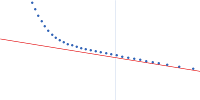
Guinier
794.0
—
—
mw
Experimental
55.0
—
kDa
rg
Guinier
2.4
—
Å
These values are reported by SASBDB; SAXSdb did not recalculate them from the experimental curve.
▼
3. Source Fits and Models Source Fits & Models
This entry has no source fit record.
▶
4. Source File Index Source Files
Category
Path
Download status
Size
curve
sasbdb/entries/u5/sasdcu5/source/SASDCU5.dat
downloaded
8658
full_entry_zip
sasbdb/entries/u5/sasdcu5/source/SASDCU5.zip
downloaded
22114
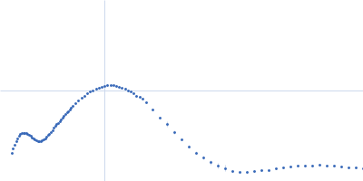
pddf
sasbdb/entries/u5/sasdcu5/source/SASDCU5.out
not_listed
—
sascif
sasbdb/entries/u5/sasdcu5/source/SASDCU5.sascif
downloaded
17457
summary
sasbdb/entries/u5/sasdcu5/source/summary.json
downloaded
6859
▶
5. Experiment Description and Publication Experiment & Publication
Original Experiment Description
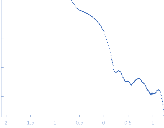
Synchrotron SAXS data from solutions of ground Doryteuthis squid lens (100% layer) diluted in phosphate buffered saline, pH 7, were collected on the X9A camera on the storage ring National Synchrotron Light Source (NSLS; Brookhaven, NY, USA) using a 20Hz Pilatus 300K detector at a wavelength of λ = 0.1 nm (I(s) vs s, where s = 4πsin θ/λ and 2θ is the scattering angle). The data were normalized to the intensity of the transmitted beam and radially averaged, and the scattering of the solvent blank (PBS) was subtracted from the diluted lens data. The dataset shown in this entry was collected from a suspension containing about 18 mg/ml of lens protein (refer to the full entry zip archive for the complete dilution series of 9, 4.5, 2.25, 1.13, 0.56, 0.28, 0.14 and 0.07 mg/ml lens protein). The lens contains about 40 unique S-crystallins isoforms that vary in molecular weight due to length variation in a disordered region encoded in the central exon of the coding sequence such that loops form protruding from the folded protein. S-crystallins are dimeric and globular, such that each dimer has two of these disordered loops, which form linking structures between particles in the system. The full set of sequences describing the protein polydispersity in this system is available at GenBank BioProject PRJNA386124.
Related Publication
Title
Eye patches: Protein assembly of index-gradient squid lenses.
Authors
Cai J, Townsend JP, Dodson TC, Heiney PA, Sweeney AM
DOI / PMID
10.1126/science.aal2674 / 28798124
▼
6. Complete Source Record Complete Source Record
The following content comes directly from the SASBDB entry. Missing values are shown as “—”; SAXSdb does not infer undeclared units.
Synchrotron SAXS data from solutions of ground Doryteuthis squid lens (100% layer) diluted in phosphate buffered saline, pH 7, were collected on the X9A camera on the storage ring National Synchrotron Light Source (NSLS; Brookhaven, NY, USA) using a 20Hz Pilatus 300K detector at a wavelength of λ = 0.1 nm (I(s) vs s, where s = 4πsin θ/λ and 2θ is the scattering angle). The data were normalized to the intensity of the transmitted beam and radially averaged, and the scattering of the solvent blank (PBS) was subtracted from the diluted lens data. The dataset shown in this entry was collected from a suspension containing about 18 mg/ml of lens protein (refer to the full entry zip archive for the complete dilution series of 9, 4.5, 2.25, 1.13, 0.56, 0.28, 0.14 and 0.07 mg/ml lens protein). The lens contains about 40 unique S-crystallins isoforms that vary in molecular weight due to length variation in a disordered region encoded in the central exon of the coding sequence such that loops form protruding from the folded protein. S-crystallins are dimeric and globular, such that each dimer has two of these disordered loops, which form linking structures between particles in the system. The full set of sequences describing the protein polydispersity in this system is available at GenBank BioProject PRJNA386124.