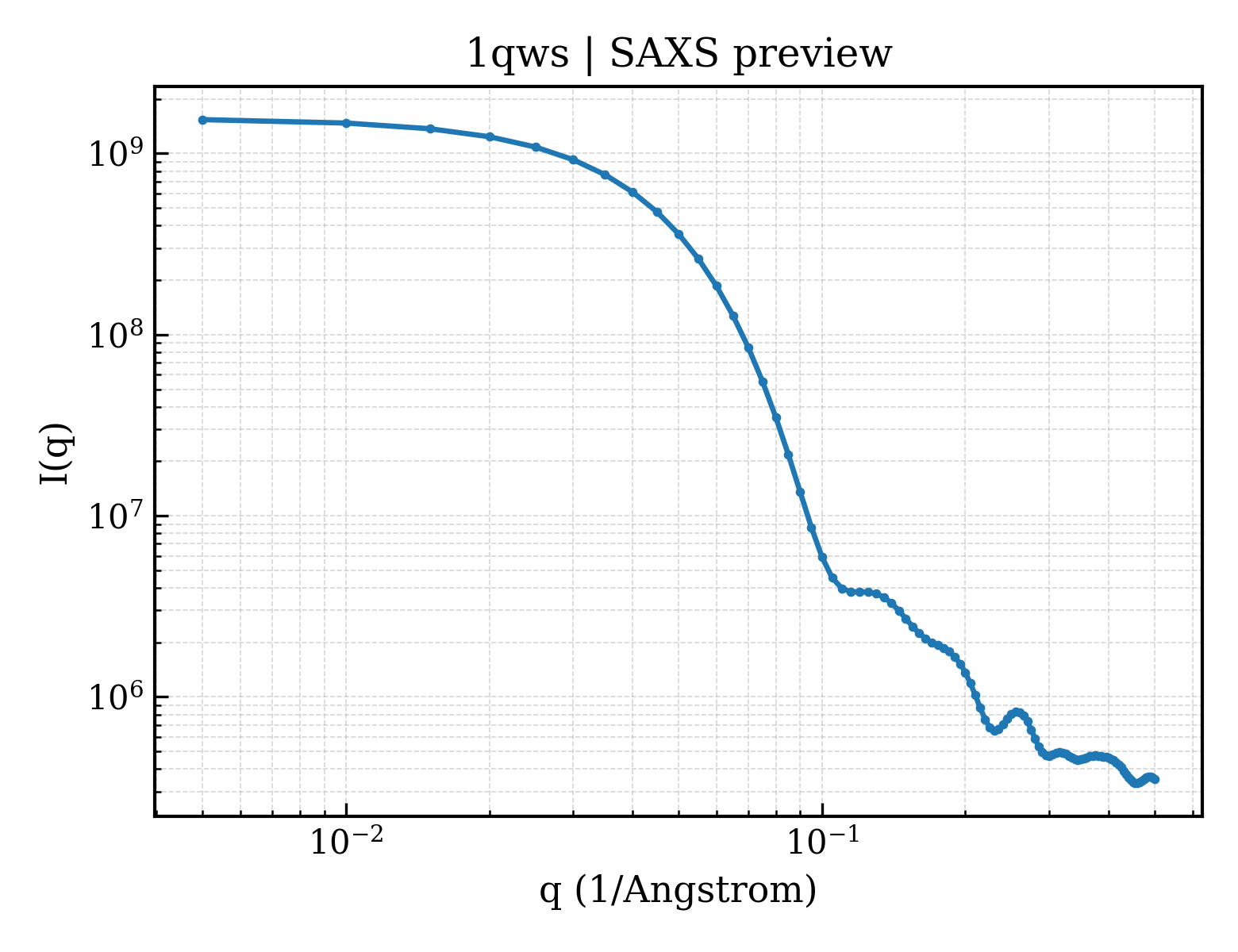
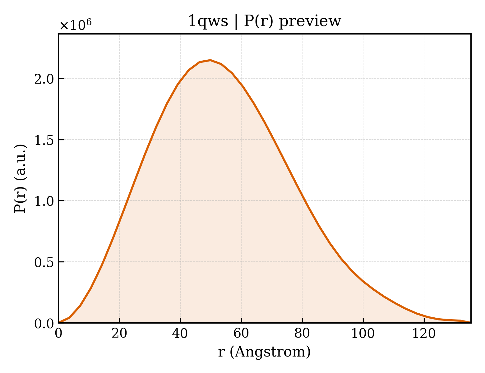
Catalase HPII
Escherichia coli
State in the Current Structure
| Assembly | Oligomeric State | Construct | Mutations and Modifications | Ligands, Ions and Associated Components | Method and Experimental Conditions | Structure Quality |
|---|---|---|---|---|---|---|
| 1 | Protein homooligomer Homooligomer Protein × 4 PDB declaration: tetrameric(4) Consistent with protein copy count | Chain A; UniProt 1–753 Chain B; UniProt 1–753 Chain C; UniProt 1–753 Chain D; UniProt 1–753 | Mutation:D181N | HEM PROTOPORPHYRIN IX CONTAINING FE × 4 | X-RAY DIFFRACTION X-ray crystallization conditions:VAPOR DIFFUSION, HANGING DROP;pH 9;293 K;15% PEG 3350, 1.6M LiCl, 0.1M Tris, pH 9.0, VAPOR DIFFUSION, HANGING DROP, temperature 293K | Resolution 1.90 Å R-free 0.220 |
Other States of the Same Protein in the Database
Each row is a biological assembly of the same UniProt protein in another PDB entry. The “Difference from current entry” column identifies evidence-level differences; no tag means the currently parsed fields agree.
| Other PDB | Difference from Current Entry 1QWS | Assembly / Oligomeric State | Construct | Mutations and Modifications | Ligands, Ions and Non-polymers | Method and Experimental Conditions | Structure Quality |
|---|---|---|---|---|---|---|---|
| 1CF9 Structure of the mutant VAL169CYS of catalase HPII from Escherichia coli Deposited 1999-03-24 | Different construct Different mutation/modification Different experimental conditions Different structure-quality metrics | Assembly 1 Protein homooligomer Homooligomer;Protein × 4 PDB declaration: tetrameric |
Chain A
27–753(727 aa)
Chain B
27–753(727 aa)
Chain C
27–753(727 aa)
Chain D
27–753(727 aa)
|
Mutation:V169C Mutation:V169C Mutation:V169C Mutation:V169C | HEM PROTOPORPHYRIN IX CONTAINING FE × 4 |
X-RAY DIFFRACTION
X-ray crystallization conditions
pH 9;pH 9
|
Resolution 1.80 Å R-free 0.237 |
| 1GG9 CRYSTAL STRUCTURE OF CATALASE HPII FROM ESCHERICHIA COLI, HIS128ASN VARIANT. Deposited 2000-08-11 | Different mutation/modification Different experimental conditions Different structure-quality metrics | Assembly 1 Protein homooligomer Homooligomer;Protein × 4 PDB declaration: tetrameric |
Chain A
1–753(753 aa)
Chain B
1–753(753 aa)
Chain C
1–753(753 aa)
Chain D
1–753(753 aa)
|
Mutation:H128N Mutation:H128N Mutation:H128N Mutation:H128N | HEM PROTOPORPHYRIN IX CONTAINING FE × 4 |
X-RAY DIFFRACTION
X-ray crystallization conditions
VAPOR DIFFUSION, HANGING DROP;pH 9;297 K;PEG 3350, LiCl, Tris-HCl, pH 9.0, vapor diffusion/hanging drop, temperature 297.0K
|
Resolution 1.89 Å R-free 0.188 |
| 1GGE CRYSTAL STRUCTURE OF CATALASE HPII FROM ESCHERICHIA COLI, NATIVE STRUCTURE AT 1.9 A RESOLUTION. Deposited 2000-08-16 | Different mutation/modification Different ligand/ion Different experimental conditions Different structure-quality metrics | Assembly 1 Protein homooligomer Homooligomer;Protein × 4 PDB declaration: tetrameric |
Chain A
1–753(753 aa)
Chain B
1–753(753 aa)
Chain C
1–753(753 aa)
Chain D
1–753(753 aa)
|
Not recorded | HDD CIS-HEME D HYDROXYCHLORIN GAMMA-SPIROLACTONE × 4 |
X-RAY DIFFRACTION
X-ray crystallization conditions
VAPOR DIFFUSION, HANGING DROP;pH 9;297 K;PEG 3350, LiCl, Tris-HCl, pH 9.00, VAPOR DIFFUSION, HANGING DROP, temperature 297.0K
|
Resolution 1.89 Å R-free 0.202 |
| 1GGF CRYSTAL STRUCTURE OF CATALASE HPII FROM ESCHERICHIA COLI, VARIANT HIS128ASN, COMPLEX WITH HYDROGEN PEROXIDE. Deposited 2000-08-21 | Different mutation/modification Different ligand/ion Different experimental conditions Different structure-quality metrics | Assembly 1 Protein homooligomer Homooligomer;Protein × 4 PDB declaration: tetrameric |
Chain A
1–753(753 aa)
Chain B
1–753(753 aa)
Chain C
1–753(753 aa)
Chain D
1–753(753 aa)
|
Mutation:H128N Mutation:H128N Mutation:H128N Mutation:H128N | HEM PROTOPORPHYRIN IX CONTAINING FE × 4 PEO HYDROGEN PEROXIDE × 13 |
X-RAY DIFFRACTION
X-ray crystallization conditions
pH 9;297 K;PEG 3350, LiCl, Tris-HCl.
Before data collection the protein crystal was soaked
during a few seconds in the solution with 2M hydrogen peroxide., pH 9.0, temperature 297.0K
|
Resolution 2.28 Å R-free 0.254 |
| 1GGH CRYSTAL STRUCTURE OF CATALASE HPII FROM ESCHERICHIA COLI, HIS128ALA VARIANT. Deposited 2000-08-21 | Different mutation/modification Different experimental conditions Different structure-quality metrics | Assembly 1 Protein homooligomer Homooligomer;Protein × 4 PDB declaration: tetrameric |
Chain A
1–753(753 aa)
Chain B
1–753(753 aa)
Chain C
1–753(753 aa)
Chain D
1–753(753 aa)
|
Mutation:H128A Mutation:H128A Mutation:H128A Mutation:H128A | HEM PROTOPORPHYRIN IX CONTAINING FE × 4 |
X-RAY DIFFRACTION
X-ray crystallization conditions
VAPOR DIFFUSION, HANGING DROP;pH 9;297 K;PEG 3350, LiCl, Tris-HCl, pH 9.0, vapor diffusion/hanging drop, temperature 297.0K
|
Resolution 2.15 Å R-free 0.206 |
| 1GGJ CRYSTAL STRUCTURE OF CATALASE HPII FROM ESCHERICHIA COLI, ASN201ALA VARIANT. Deposited 2000-08-21 | Different mutation/modification Different ligand/ion Different experimental conditions Different structure-quality metrics | Assembly 1 Protein homooligomer Homooligomer;Protein × 4 PDB declaration: tetrameric |
Chain A
1–753(753 aa)
Chain B
1–753(753 aa)
Chain C
1–753(753 aa)
Chain D
1–753(753 aa)
|
Mutation:N201A Mutation:N201A Mutation:N201A Mutation:N201A | HDD CIS-HEME D HYDROXYCHLORIN GAMMA-SPIROLACTONE × 4 |
X-RAY DIFFRACTION
X-ray crystallization conditions
VAPOR DIFFUSION, HANGING DROP;pH 9;297 K;PEG 3350, LiCl, Tris-HCl, pH 9.0, vapor diffusion/hanging drop, temperature 297.0K
|
Resolution 1.92 Å R-free 0.235 |
| 1GGK CRYSTAL STRUCTURE OF CATALASE HPII FROM ESCHERICHIA COLI, ASN201HIS VARIANT. Deposited 2000-08-21 | Different mutation/modification Different experimental conditions Different structure-quality metrics | Assembly 1 Protein homooligomer Homooligomer;Protein × 4 PDB declaration: tetrameric |
Chain A
1–753(753 aa)
Chain B
1–753(753 aa)
Chain C
1–753(753 aa)
Chain D
1–753(753 aa)
|
Mutation:N201H Mutation:N201H Mutation:N201H Mutation:N201H | HEM PROTOPORPHYRIN IX CONTAINING FE × 4 |
X-RAY DIFFRACTION
X-ray crystallization conditions
VAPOR DIFFUSION, HANGING DROP;pH 9;297 K;PEG 3350, LiCl, Tris-HCl, pH 9.0, vapor diffusion/hanging drop, temperature 297.0K
|
Resolution 2.26 Å R-free 0.208 |
| 1IPH STRUCTURE OF CATALASE HPII FROM ESCHERICHIA COLI Deposited 1995-12-31 | Different mutation/modification Different experimental conditions Different structure-quality metrics | Assembly 1 Protein homooligomer Homooligomer;Protein × 4 PDB declaration: tetrameric |
Chain A
1–753(753 aa)
Chain B
1–753(753 aa)
Chain C
1–753(753 aa)
Chain D
1–753(753 aa)
|
Not recorded | HEM PROTOPORPHYRIN IX CONTAINING FE × 4 | X-RAY DIFFRACTION mmCIF provides none of the parsed conditions | Resolution 2.80 Å |
| 1P7Y Crystal structure of the D181A variant of catalase HPII from E. coli Deposited 2003-05-06 | Different mutation/modification Different experimental conditions Different structure-quality metrics | Assembly 1 Protein homooligomer Homooligomer;Protein × 4 PDB declaration: tetrameric |
Chain A
1–753(753 aa)
Chain B
1–753(753 aa)
Chain C
1–753(753 aa)
Chain D
1–753(753 aa)
|
Mutation:D181A Mutation:D181A Mutation:D181A Mutation:D181A | HEM PROTOPORPHYRIN IX CONTAINING FE × 4 |
X-RAY DIFFRACTION
X-ray crystallization conditions
VAPOR DIFFUSION, HANGING DROP;pH 9;295 K;PEG 3350, LiCl, Tris, pH 9.0, VAPOR DIFFUSION, HANGING DROP, temperature 295K
|
Resolution 2.40 Å R-free 0.225 |
| 1P7Z Crystal structure of the D181S variant of catalase HPII from E. coli Deposited 2003-05-06 | Different mutation/modification Different experimental conditions Different structure-quality metrics | Assembly 1 Protein homooligomer Homooligomer;Protein × 4 PDB declaration: tetrameric |
Chain A
1–753(753 aa)
Chain B
1–753(753 aa)
Chain C
1–753(753 aa)
Chain D
1–753(753 aa)
|
Mutation:D181S Mutation:D181S Mutation:D181S Mutation:D181S | HEM PROTOPORPHYRIN IX CONTAINING FE × 4 |
X-RAY DIFFRACTION
X-ray crystallization conditions
VAPOR DIFFUSION, HANGING DROP;pH 9;295 K;PEG 3350, LiCl, Tris, pH 9.0, VAPOR DIFFUSION, HANGING DROP, temperature 295.0K
|
Resolution 2.21 Å R-free 0.211 |
| 1P80 Crystal structure of the D181Q variant of catalase HPII from E. coli Deposited 2003-05-06 | Different mutation/modification Different experimental conditions Different structure-quality metrics | Assembly 1 Protein homooligomer Homooligomer;Protein × 4 PDB declaration: tetrameric |
Chain A
1–753(753 aa)
Chain B
1–753(753 aa)
Chain C
1–753(753 aa)
Chain D
1–753(753 aa)
|
Mutation:D181Q Mutation:D181Q Mutation:D181Q Mutation:D181Q | HEM PROTOPORPHYRIN IX CONTAINING FE × 4 |
X-RAY DIFFRACTION
X-ray crystallization conditions
VAPOR DIFFUSION, HANGING DROP;pH 9;298 K;PEG 3350, LiCl, Tris, pH 9.0, VAPOR DIFFUSION, HANGING DROP, temperature 298.0K
|
Resolution 1.65 Å R-free 0.202 |
| 1P81 Crystal structure of the D181E variant of catalase HPII from E. coli Deposited 2003-05-06 | Different mutation/modification Different ligand/ion Different experimental conditions Different structure-quality metrics | Assembly 1 Protein homooligomer Homooligomer;Protein × 4 PDB declaration: tetrameric |
Chain A
1–753(753 aa)
Chain B
1–753(753 aa)
Chain C
1–753(753 aa)
Chain D
1–753(753 aa)
|
Mutation:D181E Mutation:D181E Mutation:D181E Mutation:D181E | HDD CIS-HEME D HYDROXYCHLORIN GAMMA-SPIROLACTONE × 4 |
X-RAY DIFFRACTION
X-ray crystallization conditions
VAPOR DIFFUSION, HANGING DROP;pH 9;295 K;PEG 3350, LiCl, Tris, pH 9.0, VAPOR DIFFUSION, HANGING DROP, temperature 295.0K
|
Resolution 1.81 Å R-free 0.216 |
| 1QF7 STRUCTURE OF THE MUTANT HIS392GLN OF CATALASE HPII FROM E. COLI Deposited 1999-03-26 | Different mutation/modification Different experimental conditions Different structure-quality metrics | Assembly 1 Protein homooligomer Homooligomer;Protein × 4 PDB declaration: tetrameric |
Chain A
1–753(753 aa)
Chain B
1–753(753 aa)
Chain C
1–753(753 aa)
Chain D
1–753(753 aa)
|
Mutation:H392Q Mutation:H392Q Mutation:H392Q Mutation:H392Q | HEM PROTOPORPHYRIN IX CONTAINING FE × 4 |
X-RAY DIFFRACTION
X-ray crystallization conditions
pH 9;pH 9
|
Resolution 2.20 Å R-free 0.210 |
| 1YE9 Crystal structure of proteolytically truncated catalase HPII from E. coli Deposited 2004-12-28 | Different construct Different mutation/modification Different oligomeric state Different ligand/ion Different experimental conditions Different structure-quality metrics | Assembly 1 Protein homooligomer Homooligomer;Protein × 8 PDB declaration: octameric |
Chain A
75–300(226 aa)
Fragment:proteolytic fragment, residues 75-300
Chain B
75–300(226 aa)
Fragment:proteolytic fragment, residues 75-300
Chain C
75–300(226 aa)
Fragment:proteolytic fragment, residues 75-300
Chain D
75–300(226 aa)
Fragment:proteolytic fragment, residues 75-300
Chain E
309–567(259 aa)
Fragment:proteolytic fragment, residues 309-567
Chain F
309–567(259 aa)
Fragment:proteolytic fragment, residues 309-567
Chain G
309–567(259 aa)
Fragment:proteolytic fragment, residues 309-567
Chain H
309–567(259 aa)
Fragment:proteolytic fragment, residues 309-567
|
Not recorded | HDD CIS-HEME D HYDROXYCHLORIN GAMMA-SPIROLACTONE × 4 |
X-RAY DIFFRACTION
X-ray crystallization conditions
VAPOR DIFFUSION, HANGING DROP;pH 7;298 K;50mM TrisHCl, 8% PEG 20000, 8% PEG MME 550, 0.2M KSCN, 0.1M dithiothreitol, pH 7.0, VAPOR DIFFUSION, HANGING DROP, temperature 298K
|
Resolution 2.80 Å R-free 0.269 |
| 1YE9 Crystal structure of proteolytically truncated catalase HPII from E. coli Deposited 2004-12-28 | Different construct Different mutation/modification Different oligomeric state Different ligand/ion Different experimental conditions Different structure-quality metrics | Assembly 2 Protein homooligomer Homooligomer;Protein × 8 PDB declaration: octameric |
Chain I
75–300(226 aa)
Fragment:proteolytic fragment, residues 75-300
Chain J
75–300(226 aa)
Fragment:proteolytic fragment, residues 75-300
Chain K
75–300(226 aa)
Fragment:proteolytic fragment, residues 75-300
Chain L
75–300(226 aa)
Fragment:proteolytic fragment, residues 75-300
Chain M
309–567(259 aa)
Fragment:proteolytic fragment, residues 309-567
Chain N
309–567(259 aa)
Fragment:proteolytic fragment, residues 309-567
Chain O
309–567(259 aa)
Fragment:proteolytic fragment, residues 309-567
Chain P
309–567(259 aa)
Fragment:proteolytic fragment, residues 309-567
|
Not recorded | HDD CIS-HEME D HYDROXYCHLORIN GAMMA-SPIROLACTONE × 4 |
X-RAY DIFFRACTION
X-ray crystallization conditions
VAPOR DIFFUSION, HANGING DROP;pH 7;298 K;50mM TrisHCl, 8% PEG 20000, 8% PEG MME 550, 0.2M KSCN, 0.1M dithiothreitol, pH 7.0, VAPOR DIFFUSION, HANGING DROP, temperature 298K
|
Resolution 2.80 Å R-free 0.269 |
| 28WU Crystal Structure of Catalase HPII (KatE) from Escherichia coli Deposited 2026-02-25 | Different mutation/modification Different experimental conditions Different structure-quality metrics | Assembly 1 Protein homooligomer Homooligomer;Protein × 4 PDB declaration: tetrameric |
Chain A
1–753(753 aa)
Chain B
1–753(753 aa)
Chain C
1–753(753 aa)
Chain D
1–753(753 aa)
|
Not recorded | HEM PROTOPORPHYRIN IX CONTAINING FE × 4 |
X-RAY DIFFRACTION
X-ray crystallization conditions
VAPOR DIFFUSION, HANGING DROP;pH 7.75;293 K;0.1M Bis-Tris-Propane pH 7.75, 0.2M Na-acetate, 20% PEG3350.
|
Resolution 2.50 Å R-free 0.224 |
| 3P9P Structure of I274V variant of E. coli KatE Deposited 2010-10-18 | Different mutation/modification Different ligand/ion Different experimental conditions Different structure-quality metrics | Assembly 1 Protein homooligomer Homooligomer;Protein × 4 PDB declaration: tetrameric |
Chain A
1–753(753 aa)
Chain B
1–753(753 aa)
Chain C
1–753(753 aa)
Chain D
1–753(753 aa)
|
Mutation:I274V Mutation:I274V Mutation:I274V Mutation:I274V | HEM PROTOPORPHYRIN IX CONTAINING FE × 8 HDE CIS-HEME D HYDROXYCHLORIN GAMMA-SPIROLACTONE 17R, 18S × 4 HDD CIS-HEME D HYDROXYCHLORIN GAMMA-SPIROLACTONE × 4 |
X-RAY DIFFRACTION
X-ray crystallization conditions
VAPOR DIFFUSION, HANGING DROP;pH 9;293 K;17% PEG3350, 1.6 M LiCl, 0.1 M Tris, pH 9.0, vapor diffusion, hanging drop, temperature 293K
|
Resolution 1.50 Å R-free 0.185 |
| 3P9Q Structure of I274C variant of E. coli KatE Deposited 2010-10-18 | Different mutation/modification Different ligand/ion Different experimental conditions Different structure-quality metrics | Assembly 1 Protein homooligomer Homooligomer;Protein × 4 PDB declaration: tetrameric |
Chain A
1–753(753 aa)
Chain B
1–753(753 aa)
Chain C
1–753(753 aa)
Chain D
1–753(753 aa)
|
Mutation:I274C Mutation:I274C Mutation:I274C Mutation:I274C | HDD CIS-HEME D HYDROXYCHLORIN GAMMA-SPIROLACTONE × 4 HDE CIS-HEME D HYDROXYCHLORIN GAMMA-SPIROLACTONE 17R, 18S × 4 H2S HYDROSULFURIC ACID × 4 |
X-RAY DIFFRACTION
X-ray crystallization conditions
VAPOR DIFFUSION, HANGING DROP;pH 9;293 K;17% PEG3350, 1.6 M LiCl, 0.1 M Tris, pH 9.0, vapor diffusion, hanging drop, temperature 293K
|
Resolution 1.48 Å R-free 0.177 |
| 3P9R Structure of I274G variant of E. coli KatE Deposited 2010-10-18 | Different mutation/modification Different ligand/ion Different experimental conditions Different structure-quality metrics | Assembly 1 Protein homooligomer Homooligomer;Protein × 4 PDB declaration: tetrameric |
Chain A
1–753(753 aa)
Chain B
1–753(753 aa)
Chain C
1–753(753 aa)
Chain D
1–753(753 aa)
|
Mutation:I274G Non-standard monomer:Yes (specific site not provided by mmCIF) Mutation:I274G Non-standard monomer:Yes (specific site not provided by mmCIF) Mutation:I274G Non-standard monomer:Yes (specific site not provided by mmCIF) Mutation:I274G Non-standard monomer:Yes (specific site not provided by mmCIF) | HDD CIS-HEME D HYDROXYCHLORIN GAMMA-SPIROLACTONE × 4 |
X-RAY DIFFRACTION
X-ray crystallization conditions
VAPOR DIFFUSION, HANGING DROP;pH 9;293 K;17% PEG3350, 1.6 M LiCl, 0.1 M Tris, pH 9.0, vapor diffusion, hanging drop, temperature 293K
|
Resolution 1.90 Å R-free 0.184 |
| 3P9S Structure of I274A variant of E. coli KatE Deposited 2010-10-18 | Different mutation/modification Different ligand/ion Different experimental conditions Different structure-quality metrics | Assembly 1 Protein homooligomer Homooligomer;Protein × 4 PDB declaration: tetrameric |
Chain A
1–753(753 aa)
Chain B
1–753(753 aa)
Chain C
1–753(753 aa)
Chain D
1–753(753 aa)
|
Mutation:I274A Non-standard monomer:Yes (specific site not provided by mmCIF) Mutation:I274A Non-standard monomer:Yes (specific site not provided by mmCIF) Mutation:I274A Non-standard monomer:Yes (specific site not provided by mmCIF) Mutation:I274A Non-standard monomer:Yes (specific site not provided by mmCIF) | HDD CIS-HEME D HYDROXYCHLORIN GAMMA-SPIROLACTONE × 4 HDE CIS-HEME D HYDROXYCHLORIN GAMMA-SPIROLACTONE 17R, 18S × 4 |
X-RAY DIFFRACTION
X-ray crystallization conditions
VAPOR DIFFUSION, HANGING DROP;pH 9;293 K;17% PEG3350, 1.6 M LiCl, 0.1 M Tris, pH 9.0, vapor diffusion, hanging drop, temperature 293K
|
Resolution 1.90 Å R-free 0.191 |
| 3PQ2 Structure of I274C variant of E. coli KatE[] - Images 1-6 Deposited 2010-11-25 | Different mutation/modification Different ligand/ion Different experimental conditions Different structure-quality metrics | Assembly 1 Protein homooligomer Homooligomer;Protein × 4 PDB declaration: tetrameric |
Chain A
1–753(753 aa)
Chain B
1–753(753 aa)
Chain C
1–753(753 aa)
Chain D
1–753(753 aa)
|
Mutation:I274C Mutation:I274C Mutation:I274C Mutation:I274C | HDD CIS-HEME D HYDROXYCHLORIN GAMMA-SPIROLACTONE × 4 HDE CIS-HEME D HYDROXYCHLORIN GAMMA-SPIROLACTONE 17R, 18S × 4 H2S HYDROSULFURIC ACID × 4 |
X-RAY DIFFRACTION
X-ray crystallization conditions
VAPOR DIFFUSION, HANGING DROP;pH 9;293 K;17% PEG3350, 1.6 M LiCl, 0.1 M Tris, pH 9.0, VAPOR DIFFUSION, HANGING DROP, temperature 293K
|
Resolution 1.79 Å R-free 0.189 |
| 3PQ3 Structure of I274C variant of E. coli KatE[] - Images 7-12 Deposited 2010-11-25 | Different mutation/modification Different ligand/ion Different experimental conditions Different structure-quality metrics | Assembly 1 Protein homooligomer Homooligomer;Protein × 4 PDB declaration: tetrameric |
Chain A
1–753(753 aa)
Chain B
1–753(753 aa)
Chain C
1–753(753 aa)
Chain D
1–753(753 aa)
|
Mutation:I274C Mutation:I274C Mutation:I274C Mutation:I274C | HDD CIS-HEME D HYDROXYCHLORIN GAMMA-SPIROLACTONE × 4 HDE CIS-HEME D HYDROXYCHLORIN GAMMA-SPIROLACTONE 17R, 18S × 4 H2S HYDROSULFURIC ACID × 4 |
X-RAY DIFFRACTION
X-ray crystallization conditions
VAPOR DIFFUSION, HANGING DROP;pH 9;293 K;17% PEG3350, 1.6 M LiCl, 0.1 M Tris, pH 9.0, VAPOR DIFFUSION, HANGING DROP, temperature 293K
|
Resolution 1.79 Å R-free 0.189 |
| 3PQ4 Structure of I274C variant of E. coli KatE[] - Images 13-18 Deposited 2010-11-25 | Different mutation/modification Different ligand/ion Different experimental conditions Different structure-quality metrics | Assembly 1 Protein homooligomer Homooligomer;Protein × 4 PDB declaration: tetrameric |
Chain A
1–753(753 aa)
Chain B
1–753(753 aa)
Chain C
1–753(753 aa)
Chain D
1–753(753 aa)
|
Mutation:I274C Mutation:I274C Mutation:I274C Mutation:I274C | HDD CIS-HEME D HYDROXYCHLORIN GAMMA-SPIROLACTONE × 4 HDE CIS-HEME D HYDROXYCHLORIN GAMMA-SPIROLACTONE 17R, 18S × 4 H2S HYDROSULFURIC ACID × 4 |
X-RAY DIFFRACTION
X-ray crystallization conditions
VAPOR DIFFUSION, HANGING DROP;pH 9;293 K;17% PEG3350, 1.6 M LiCl, 0.1 M Tris, pH 9.0, VAPOR DIFFUSION, HANGING DROP, temperature 293K
|
Resolution 1.79 Å R-free 0.189 |
| 3PQ5 Structure of I274C variant of E. coli KatE[] - Images 19-24 Deposited 2010-11-25 | Different mutation/modification Different ligand/ion Different experimental conditions Different structure-quality metrics | Assembly 1 Protein homooligomer Homooligomer;Protein × 4 PDB declaration: tetrameric |
Chain A
1–753(753 aa)
Chain B
1–753(753 aa)
Chain C
1–753(753 aa)
Chain D
1–753(753 aa)
|
Mutation:I274C Mutation:I274C Mutation:I274C Mutation:I274C | HDD CIS-HEME D HYDROXYCHLORIN GAMMA-SPIROLACTONE × 4 HDE CIS-HEME D HYDROXYCHLORIN GAMMA-SPIROLACTONE 17R, 18S × 4 H2S HYDROSULFURIC ACID × 4 |
X-RAY DIFFRACTION
X-ray crystallization conditions
VAPOR DIFFUSION, HANGING DROP;pH 9;293 K;17% PEG3350, 1.6 M LiCl, 0.1 M Tris, pH 9.0, VAPOR DIFFUSION, HANGING DROP, temperature 293K
|
Resolution 1.80 Å R-free 0.190 |
| 3PQ6 Structure of I274C variant of E. coli KatE[] - Images 25-30 Deposited 2010-11-25 | Different mutation/modification Different ligand/ion Different experimental conditions Different structure-quality metrics | Assembly 1 Protein homooligomer Homooligomer;Protein × 4 PDB declaration: tetrameric |
Chain A
1–753(753 aa)
Chain B
1–753(753 aa)
Chain C
1–753(753 aa)
Chain D
1–753(753 aa)
|
Mutation:I274C Mutation:I274C Mutation:I274C Mutation:I274C | HDD CIS-HEME D HYDROXYCHLORIN GAMMA-SPIROLACTONE × 4 HDE CIS-HEME D HYDROXYCHLORIN GAMMA-SPIROLACTONE 17R, 18S × 4 H2S HYDROSULFURIC ACID × 4 |
X-RAY DIFFRACTION
X-ray crystallization conditions
VAPOR DIFFUSION, HANGING DROP;pH 9;293 K;17% PEG3350, 1.6 M LiCl, 0.1 M Tris, pH 9.0, VAPOR DIFFUSION, HANGING DROP, temperature 293K
|
Resolution 1.80 Å R-free 0.191 |
| 3PQ7 Structure of I274C variant of E. coli KatE[] - Images 31-36 Deposited 2010-11-25 | Different mutation/modification Different ligand/ion Different experimental conditions Different structure-quality metrics | Assembly 1 Protein homooligomer Homooligomer;Protein × 4 PDB declaration: tetrameric |
Chain A
1–753(753 aa)
Chain B
1–753(753 aa)
Chain C
1–753(753 aa)
Chain D
1–753(753 aa)
|
Mutation:I274C Mutation:I274C Mutation:I274C Mutation:I274C | HDD CIS-HEME D HYDROXYCHLORIN GAMMA-SPIROLACTONE × 4 HDE CIS-HEME D HYDROXYCHLORIN GAMMA-SPIROLACTONE 17R, 18S × 4 H2S HYDROSULFURIC ACID × 4 |
X-RAY DIFFRACTION
X-ray crystallization conditions
VAPOR DIFFUSION, HANGING DROP;pH 9;293 K;17% PEG3350, 1.6 M LiCl, 0.1 M Tris, pH 9.0, VAPOR DIFFUSION, HANGING DROP, temperature 293K
|
Resolution 1.80 Å R-free 0.190 |
| 3PQ8 Structure of I274C variant of E. coli KatE[] - Images 37-42 Deposited 2010-11-25 | Different mutation/modification Different ligand/ion Different experimental conditions Different structure-quality metrics | Assembly 1 Protein homooligomer Homooligomer;Protein × 4 PDB declaration: tetrameric |
Chain A
1–753(753 aa)
Chain B
1–753(753 aa)
Chain C
1–753(753 aa)
Chain D
1–753(753 aa)
|
Mutation:I274C Mutation:I274C Mutation:I274C Mutation:I274C | HDD CIS-HEME D HYDROXYCHLORIN GAMMA-SPIROLACTONE × 4 HDE CIS-HEME D HYDROXYCHLORIN GAMMA-SPIROLACTONE 17R, 18S × 4 H2S HYDROSULFURIC ACID × 4 |
X-RAY DIFFRACTION
X-ray crystallization conditions
VAPOR DIFFUSION, HANGING DROP;pH 9;293 K;17% PEG3350, 1.6 M LiCl, 0.1 M Tris, pH 9.0, VAPOR DIFFUSION, HANGING DROP, temperature 293K
|
Resolution 1.80 Å R-free 0.190 |
| 3TTT Structure of F413Y variant of E. coli KatE Deposited 2011-09-15 | Different mutation/modification Different experimental conditions Different structure-quality metrics | Assembly 1 Protein homooligomer Homooligomer;Protein × 4 PDB declaration: tetrameric |
Chain A
1–753(753 aa)
Chain B
1–753(753 aa)
Chain C
1–753(753 aa)
Chain D
1–753(753 aa)
|
Mutation:F413Y Mutation:F413Y Mutation:F413Y Mutation:F413Y | HEM PROTOPORPHYRIN IX CONTAINING FE × 4 |
X-RAY DIFFRACTION
X-ray crystallization conditions
VAPOR DIFFUSION, HANGING DROP;pH 9;293 K;17% PEG3350, 1.6 M lithium chloride, 0.1 M Tris, pH 9.0, VAPOR DIFFUSION, HANGING DROP, temperature 293K
|
Resolution 1.58 Å R-free 0.189 |
| 3TTU Structure of F413Y/H128N double variant of E. coli KatE Deposited 2011-09-15 | Different mutation/modification Different experimental conditions Different structure-quality metrics | Assembly 1 Protein homooligomer Homooligomer;Protein × 4 PDB declaration: tetrameric |
Chain A
1–753(753 aa)
Chain B
1–753(753 aa)
Chain C
1–753(753 aa)
Chain D
1–753(753 aa)
|
Mutation:F413Y/H128N Mutation:F413Y/H128N Mutation:F413Y/H128N Mutation:F413Y/H128N | HEM PROTOPORPHYRIN IX CONTAINING FE × 4 |
X-RAY DIFFRACTION
X-ray crystallization conditions
VAPOR DIFFUSION, HANGING DROP;pH 9;293 K;17% PEG3350, 1.6 M lithium chloride, 0.1 M Tris, pH 9.0, VAPOR DIFFUSION, HANGING DROP, temperature 293K
|
Resolution 1.89 Å R-free 0.193 |
| 3TTV Structure of the F413E variant of E. coli KatE Deposited 2011-09-15 | Different mutation/modification Different experimental conditions Different structure-quality metrics | Assembly 1 Protein homooligomer Homooligomer;Protein × 4 PDB declaration: tetrameric |
Chain A
1–753(753 aa)
Chain B
1–753(753 aa)
Chain C
1–753(753 aa)
Chain D
1–753(753 aa)
|
Mutation:F413Y/T115A Non-standard monomer:Yes (specific site not provided by mmCIF) Mutation:F413Y/T115A Non-standard monomer:Yes (specific site not provided by mmCIF) Mutation:F413Y/T115A Non-standard monomer:Yes (specific site not provided by mmCIF) Mutation:F413Y/T115A Non-standard monomer:Yes (specific site not provided by mmCIF) | HEM PROTOPORPHYRIN IX CONTAINING FE × 4 |
X-RAY DIFFRACTION
X-ray crystallization conditions
VAPOR DIFFUSION, HANGING DROP;pH 9;293 K;17% PEG3350, 1.6 M lithium chloride, 0.1 M Tris, pH 9.0, VAPOR DIFFUSION, HANGING DROP, temperature 293K
|
Resolution 1.45 Å R-free 0.172 |
| 3TTW Structure of the F413E variant of E. coli KatE Deposited 2011-09-15 | Different mutation/modification Different experimental conditions Different structure-quality metrics | Assembly 1 Protein homooligomer Homooligomer;Protein × 4 PDB declaration: tetrameric |
Chain A
1–753(753 aa)
Chain B
1–753(753 aa)
Chain C
1–753(753 aa)
Chain D
1–753(753 aa)
|
Mutation:F413E Non-standard monomer:Yes (specific site not provided by mmCIF) Mutation:F413E Non-standard monomer:Yes (specific site not provided by mmCIF) Mutation:F413E Non-standard monomer:Yes (specific site not provided by mmCIF) Mutation:F413E Non-standard monomer:Yes (specific site not provided by mmCIF) | HEM PROTOPORPHYRIN IX CONTAINING FE × 4 |
X-RAY DIFFRACTION
X-ray crystallization conditions
VAPOR DIFFUSION, HANGING DROP;pH 9;293 K;17% PEG3350, 1.6 M lithium chloride, 0.1 M Tris, pH 9.0, VAPOR DIFFUSION, HANGING DROP, temperature 293K
|
Resolution 1.62 Å R-free 0.199 |
| 3TTX Structure of the F413K variant of E. coli KatE Deposited 2011-09-15 | Different mutation/modification Different experimental conditions Different structure-quality metrics | Assembly 1 Protein homooligomer Homooligomer;Protein × 4 PDB declaration: tetrameric |
Chain A
1–753(753 aa)
Chain B
1–753(753 aa)
Chain C
1–753(753 aa)
Chain D
1–753(753 aa)
|
Mutation:F413K Non-standard monomer:Yes (specific site not provided by mmCIF) Mutation:F413K Non-standard monomer:Yes (specific site not provided by mmCIF) Mutation:F413K Non-standard monomer:Yes (specific site not provided by mmCIF) Mutation:F413K Non-standard monomer:Yes (specific site not provided by mmCIF) | HEM PROTOPORPHYRIN IX CONTAINING FE × 4 |
X-RAY DIFFRACTION
X-ray crystallization conditions
VAPOR DIFFUSION, HANGING DROP;pH 9;293 K;17% PEG3350, 1.6 M lithium chloride, 0.1 M Tris, pH 9.0, VAPOR DIFFUSION, HANGING DROP, temperature 293K
|
Resolution 1.74 Å R-free 0.185 |
| 3VU3 Crystal structure of the Hfq and catalase HPII complex Deposited 2012-06-15 | Different construct Different mutation/modification Different oligomeric state Different experimental conditions Different structure-quality metrics | Assembly 1 Protein heterocomplex Heteromer;Protein × 28 PDB declaration: 28-meric |
Chain A
1–753(753 aa)
|
Not recorded | HEM PROTOPORPHYRIN IX CONTAINING FE × 4 |
X-RAY DIFFRACTION
X-ray crystallization conditions
VAPOR DIFFUSION, HANGING DROP;pH 9;293 K;0.1M Tris-HCl, 0.18M NaCl, 10% PEG4000, pH 9.0, VAPOR DIFFUSION, HANGING DROP, temperature 293K
|
Resolution 2.85 Å R-free 0.246 |
| 4BFL Structure of natively expressed catalase HPII Deposited 2013-03-19 | Different mutation/modification Different ligand/ion Different experimental conditions Different structure-quality metrics | Assembly 1 Protein homooligomer Homooligomer;Protein × 4 PDB declaration: tetrameric |
Chain A
1–753(753 aa)
Chain B
1–753(753 aa)
Chain C
1–753(753 aa)
Chain D
1–753(753 aa)
|
Not recorded | HEM PROTOPORPHYRIN IX CONTAINING FE × 4 EDO 1,2-ETHANEDIOL × 3 |
X-RAY DIFFRACTION
X-ray crystallization conditions
pH 6.5;10 % PEG 20000, 20% PEG 500MME, 30 MM SODIUM NITRATE, 30 MM DISODIUM HYDROGENPHOSPHATE, 30 MM AMMONIUM SULFATE, AND 100 MM MES/IMIDAZOLE PH 6.5
|
Resolution 1.64 Å R-free 0.202 |
| 4ENP Structure of E530A variant E. coli KatE Deposited 2012-04-13 | Different mutation/modification Different experimental conditions Different structure-quality metrics | Assembly 1 Protein homooligomer Homooligomer;Protein × 4 PDB declaration: tetrameric |
Chain A
1–753(753 aa)
Chain B
1–753(753 aa)
Chain C
1–753(753 aa)
Chain D
1–753(753 aa)
|
Mutation:E530A Mutation:E530A Mutation:E530A Mutation:E530A | HEM PROTOPORPHYRIN IX CONTAINING FE × 4 |
X-RAY DIFFRACTION
X-ray crystallization conditions
VAPOR DIFFUSION, HANGING DROP;pH 9;293 K;17% PEG3350, 1.6 M LiCl, 0.1 M Tris, pH 9.0, vapor diffusion, hanging drop, temperature 293K
|
Resolution 1.50 Å R-free 0.162 |
| 4ENQ Structure of E530D variant E. coli KatE Deposited 2012-04-13 | Different mutation/modification Different ligand/ion Different experimental conditions Different structure-quality metrics | Assembly 1 Protein homooligomer Homooligomer;Protein × 4 PDB declaration: tetrameric |
Chain A
1–753(753 aa)
Chain B
1–753(753 aa)
Chain C
1–753(753 aa)
Chain D
1–753(753 aa)
|
Mutation:E530D Non-standard monomer:Yes (specific site not provided by mmCIF) Mutation:E530D Non-standard monomer:Yes (specific site not provided by mmCIF) Mutation:E530D Non-standard monomer:Yes (specific site not provided by mmCIF) Mutation:E530D Non-standard monomer:Yes (specific site not provided by mmCIF) | HDD CIS-HEME D HYDROXYCHLORIN GAMMA-SPIROLACTONE × 4 |
X-RAY DIFFRACTION
X-ray crystallization conditions
VAPOR DIFFUSION, HANGING DROP;pH 9;293 K;17% PEG3350, 1.6 M LiCl, 0.1 M Tris, pH 9.0, vapor diffusion, hanging drop, temperature 293K
|
Resolution 1.90 Å R-free 0.207 |
| 4ENR Structure of E530I variant E. coli KatE Deposited 2012-04-13 | Different mutation/modification Different experimental conditions Different structure-quality metrics | Assembly 1 Protein homooligomer Homooligomer;Protein × 4 PDB declaration: tetrameric |
Chain A
1–753(753 aa)
Chain B
1–753(753 aa)
Chain C
1–753(753 aa)
Chain D
1–753(753 aa)
|
Mutation:E530I Non-standard monomer:Yes (specific site not provided by mmCIF) Mutation:E530I Non-standard monomer:Yes (specific site not provided by mmCIF) Mutation:E530I Non-standard monomer:Yes (specific site not provided by mmCIF) Mutation:E530I Non-standard monomer:Yes (specific site not provided by mmCIF) | HEM PROTOPORPHYRIN IX CONTAINING FE × 4 |
X-RAY DIFFRACTION
X-ray crystallization conditions
VAPOR DIFFUSION, HANGING DROP;pH 9;293 K;17% PEG3350, 1.6 M LiCl, 0.1 M Tris, pH 9.0, vapor diffusion, hanging drop, temperature 293K
|
Resolution 1.60 Å R-free 0.193 |
| 4ENS Structure of E530Q variant of E. coli KatE Deposited 2012-04-13 | Different mutation/modification Different ligand/ion Different experimental conditions Different structure-quality metrics | Assembly 1 Protein homooligomer Homooligomer;Protein × 4 PDB declaration: tetrameric |
Chain A
1–753(753 aa)
Chain B
1–753(753 aa)
Chain C
1–753(753 aa)
Chain D
1–753(753 aa)
|
Mutation:E530Q Mutation:E530Q Mutation:E530Q Mutation:E530Q | HDD CIS-HEME D HYDROXYCHLORIN GAMMA-SPIROLACTONE × 4 HEM PROTOPORPHYRIN IX CONTAINING FE × 4 |
X-RAY DIFFRACTION
X-ray crystallization conditions
VAPOR DIFFUSION, HANGING DROP;pH 9;293 K;17% PEG3350, 1.6 M LiCl, 0.1 M Tris, pH 9.0, vapor diffusion, hanging drop, temperature 293K
|
Resolution 1.60 Å R-free 0.184 |
| 4ENT Structure of the S234A variant of E. coli KatE Deposited 2012-04-13 | Different mutation/modification Different ligand/ion Different experimental conditions Different structure-quality metrics | Assembly 1 Protein homooligomer Homooligomer;Protein × 4 PDB declaration: tetrameric |
Chain A
1–753(753 aa)
Chain B
1–753(753 aa)
Chain C
1–753(753 aa)
Chain D
1–753(753 aa)
|
Mutation:S234A Non-standard monomer:Yes (specific site not provided by mmCIF) Mutation:S234A Non-standard monomer:Yes (specific site not provided by mmCIF) Mutation:S234A Non-standard monomer:Yes (specific site not provided by mmCIF) Mutation:S234A Non-standard monomer:Yes (specific site not provided by mmCIF) | HDD CIS-HEME D HYDROXYCHLORIN GAMMA-SPIROLACTONE × 4 HEM PROTOPORPHYRIN IX CONTAINING FE × 4 |
X-RAY DIFFRACTION
X-ray crystallization conditions
VAPOR DIFFUSION, HANGING DROP;pH 9;293 K;17% PEG3350, 1.6 M LiCl, 0.1 M Tris, pH 9.0, vapor diffusion, hanging drop, temperature 293K
|
Resolution 1.70 Å R-free 0.197 |
| 4ENU Structure of the S234D variant of E. coli KatE Deposited 2012-04-13 | Different mutation/modification Different ligand/ion Different experimental conditions Different structure-quality metrics | Assembly 1 Protein homooligomer Homooligomer;Protein × 4 PDB declaration: tetrameric |
Chain A
1–753(753 aa)
Chain B
1–753(753 aa)
Chain C
1–753(753 aa)
Chain D
1–753(753 aa)
|
Mutation:S234D Non-standard monomer:Yes (specific site not provided by mmCIF) Mutation:S234D Non-standard monomer:Yes (specific site not provided by mmCIF) Mutation:S234D Non-standard monomer:Yes (specific site not provided by mmCIF) Mutation:S234D Non-standard monomer:Yes (specific site not provided by mmCIF) | HDD CIS-HEME D HYDROXYCHLORIN GAMMA-SPIROLACTONE × 4 |
X-RAY DIFFRACTION
X-ray crystallization conditions
VAPOR DIFFUSION, HANGING DROP;pH 9;293 K;17% PEG3350, 1.6 M LiCl, 0.1 M Tris, pH 9.0, vapor diffusion, hanging drop, temperature 293K
|
Resolution 1.70 Å R-free 0.170 |
| 4ENV Structure of the S234I variant of E. coli KatE Deposited 2012-04-13 | Different mutation/modification Different ligand/ion Different experimental conditions Different structure-quality metrics | Assembly 1 Protein homooligomer Homooligomer;Protein × 4 PDB declaration: tetrameric |
Chain A
1–753(753 aa)
Chain B
1–753(753 aa)
Chain C
1–753(753 aa)
Chain D
1–753(753 aa)
|
Mutation:S234I Mutation:S234I Mutation:S234I Mutation:S234I | HDD CIS-HEME D HYDROXYCHLORIN GAMMA-SPIROLACTONE × 4 HEM PROTOPORPHYRIN IX CONTAINING FE × 4 |
X-RAY DIFFRACTION
X-ray crystallization conditions
VAPOR DIFFUSION, HANGING DROP;pH 9;293 K;17% PEG3350, 1.6 M LiCl, 0.1 M Tris, pH 9.0, vapor diffusion, hanging drop, temperature 293K
|
Resolution 1.70 Å R-free 0.207 |
| 4ENW Structure of the S234N variant of E. coli KatE Deposited 2012-04-13 | Different mutation/modification Different ligand/ion Different experimental conditions Different structure-quality metrics | Assembly 1 Protein homooligomer Homooligomer;Protein × 4 PDB declaration: tetrameric |
Chain A
1–753(753 aa)
Chain B
1–753(753 aa)
Chain C
1–753(753 aa)
Chain D
1–753(753 aa)
|
Mutation:S234N Mutation:S234N Mutation:S234N Mutation:S234N | HDD CIS-HEME D HYDROXYCHLORIN GAMMA-SPIROLACTONE × 4 |
X-RAY DIFFRACTION
X-ray crystallization conditions
VAPOR DIFFUSION, HANGING DROP;pH 9;293 K;17% PEG3350, 1.6 M LiCl, 0.1 M Tris, pH 9.0, vapor diffusion, hanging drop, temperature 293K
|
Resolution 1.90 Å R-free 0.221 |
| 5BV2 Crystal structure of E. coli HPII catalase variant Deposited 2015-06-04 | Different mutation/modification Different ligand/ion Different experimental conditions Different structure-quality metrics | Assembly 1 Protein homooligomer Homooligomer;Protein × 4 PDB declaration: tetrameric |
Chain P
1–753(753 aa)
Chain Q
1–753(753 aa)
Chain R
1–753(753 aa)
Chain S
1–753(753 aa)
|
Not recorded | HDD CIS-HEME D HYDROXYCHLORIN GAMMA-SPIROLACTONE × 4 GOL GLYCEROL × 11 EDO 1,2-ETHANEDIOL × 39 PGE TRIETHYLENE GLYCOL × 3 PEG DI(HYDROXYETHYL)ETHER × 10 SO4 SULFATE ION × 2 MG MAGNESIUM ION × 2 PG4 TETRAETHYLENE GLYCOL × 1 |
X-RAY DIFFRACTION
X-ray crystallization conditions
VAPOR DIFFUSION, HANGING DROP;pH 7;298 K;2% PEG20000, 15% PEG5000 MME, 0.1 M potassium chloride, 0.1 M manganese acetate, 0.1 M HEPES
|
Resolution 1.53 Å R-free 0.132 |
| 6BY0 Crystal structure of catalase HPII from E. coli in space group P1 Deposited 2017-12-19 | Different mutation/modification Different experimental conditions Different structure-quality metrics | Assembly 1 Protein homooligomer Homooligomer;Protein × 4 PDB declaration: tetrameric |
Chain A
1–753(753 aa)
Chain B
1–753(753 aa)
Chain C
1–753(753 aa)
Chain D
1–753(753 aa)
|
Not recorded | HEM PROTOPORPHYRIN IX CONTAINING FE × 4 |
X-RAY DIFFRACTION
X-ray crystallization conditions
VAPOR DIFFUSION, SITTING DROP;292 K;0.085 M sodium HEPES pH 7.5, 17% w/v PEG 4000, 15% v/v glycerol, 8.5% v/v isopropanol or 0.1 M HEPES pH 7.0, 20% w/v PEG 6000, 1.0 M lithium chloride
|
Resolution 2.93 Å R-free 0.236 |
| 6ZTV Crystal Structure of catalase HPII from Escherichia coli (serendipitously crystallized) Deposited 2020-07-20 | Different mutation/modification Different ligand/ion Different experimental conditions Different structure-quality metrics | Assembly 1 Protein homooligomer Homooligomer;Protein × 4 PDB declaration: tetrameric |
Chain A
1–753(753 aa)
Chain B
1–753(753 aa)
Chain C
1–753(753 aa)
Chain D
1–753(753 aa)
|
Mutation:S99N Non-standard monomer:Yes (specific site not provided by mmCIF) Mutation:S99N Non-standard monomer:Yes (specific site not provided by mmCIF) Mutation:S99N Non-standard monomer:Yes (specific site not provided by mmCIF) Mutation:S99N Non-standard monomer:Yes (specific site not provided by mmCIF) | HDD CIS-HEME D HYDROXYCHLORIN GAMMA-SPIROLACTONE × 4 GOL GLYCEROL × 1 EDO 1,2-ETHANEDIOL × 1 |
X-RAY DIFFRACTION
X-ray crystallization conditions
VAPOR DIFFUSION, SITTING DROP;pH 7.5;292 K;0.2 M NaCl, 0.1 M Tris pH 7.5, 20% w/v PEG 4000, 10% MPD
|
Resolution 1.78 Å R-free 0.229 |
| 6ZTW Crystal Structure of catalase HPII from Escherichia coli (serendipitously crystallized) Deposited 2020-07-20 | Different mutation/modification Different ligand/ion Different experimental conditions Different structure-quality metrics | Assembly 1 Protein homooligomer Homooligomer;Protein × 4 PDB declaration: tetrameric |
Chain A
1–753(753 aa)
Chain B
1–753(753 aa)
Chain C
1–753(753 aa)
Chain D
1–753(753 aa)
|
Mutation:S99N Non-standard monomer:Yes (specific site not provided by mmCIF) Mutation:S99N Non-standard monomer:Yes (specific site not provided by mmCIF) Mutation:S99N Non-standard monomer:Yes (specific site not provided by mmCIF) Mutation:S99N Non-standard monomer:Yes (specific site not provided by mmCIF) | HDD CIS-HEME D HYDROXYCHLORIN GAMMA-SPIROLACTONE × 4 TRS 2-AMINO-2-HYDROXYMETHYL-PROPANE-1,3-DIOL × 4 GOL GLYCEROL × 4 MPD (4S)-2-METHYL-2,4-PENTANEDIOL × 1 EDO 1,2-ETHANEDIOL × 4 |
X-RAY DIFFRACTION
X-ray crystallization conditions
VAPOR DIFFUSION, SITTING DROP;pH 7.5;292 K;0.1 M Tris pH 7.5, 20% w/v PEG 4000
|
Resolution 1.84 Å R-free 0.184 |
| 6ZTW Crystal Structure of catalase HPII from Escherichia coli (serendipitously crystallized) Deposited 2020-07-20 | Different mutation/modification Different ligand/ion Different experimental conditions Different structure-quality metrics | Assembly 2 Protein homooligomer Homooligomer;Protein × 4 PDB declaration: tetrameric |
Chain E
1–753(753 aa)
Chain F
1–753(753 aa)
Chain G
1–753(753 aa)
Chain H
1–753(753 aa)
|
Mutation:S99N Non-standard monomer:Yes (specific site not provided by mmCIF) Mutation:S99N Non-standard monomer:Yes (specific site not provided by mmCIF) Mutation:S99N Non-standard monomer:Yes (specific site not provided by mmCIF) Mutation:S99N Non-standard monomer:Yes (specific site not provided by mmCIF) | HDD CIS-HEME D HYDROXYCHLORIN GAMMA-SPIROLACTONE × 4 TRS 2-AMINO-2-HYDROXYMETHYL-PROPANE-1,3-DIOL × 5 GOL GLYCEROL × 2 EDO 1,2-ETHANEDIOL × 3 |
X-RAY DIFFRACTION
X-ray crystallization conditions
VAPOR DIFFUSION, SITTING DROP;pH 7.5;292 K;0.1 M Tris pH 7.5, 20% w/v PEG 4000
|
Resolution 1.84 Å R-free 0.184 |
| 6ZTX Crystal Structure of catalase HPII from Escherichia coli (serendipitously crystallized) Deposited 2020-07-20 | Different mutation/modification Different ligand/ion Different experimental conditions Different structure-quality metrics | Assembly 1 Protein homooligomer Homooligomer;Protein × 4 PDB declaration: tetrameric |
Chain A
1–753(753 aa)
Chain B
1–753(753 aa)
Chain C
1–753(753 aa)
Chain D
1–753(753 aa)
|
Mutation:R37S, S99D, K372N, R521S Mutation:R37S, S99D, K372N, R521S Mutation:R37S, S99D, K372N, R521S Mutation:R37S, S99D, K372N, R521S | GOL GLYCEROL × 4 EDO 1,2-ETHANEDIOL × 7 HDD CIS-HEME D HYDROXYCHLORIN GAMMA-SPIROLACTONE × 4 |
X-RAY DIFFRACTION
X-ray crystallization conditions
VAPOR DIFFUSION, SITTING DROP;pH 8.5;292 K;0.2 M lithium sulfate monohydrate, 0.1 M Tris pH 8.5, 25% w/v polyethylene glycol 3350
|
Resolution 1.30 Å R-free 0.162 |
46 other PDB entries and 48 assemblies. Open the comparison page and filter oligomeric states
View Construct and Data Evidence
| UniProt name | CATE_ECOLI |
| Isoform | — |
| PDB entities | 1 |
| Chains and sequence ranges | Author chain A; PDBConstruct 1–753; UniProt 1–753 Author chain B; PDBConstruct 1–753; UniProt 1–753 Author chain C; PDBConstruct 1–753; UniProt 1–753 Author chain D; PDBConstruct 1–753; UniProt 1–753 |