Low affinity immunoglobulin epsilon Fc receptor
Homo sapiens
State in the Current Structure
| Assembly | Oligomeric State | Construct | Mutations and Modifications | Ligands, Ions and Associated Components | Method and Experimental Conditions | Structure Quality |
|---|---|---|---|---|---|---|
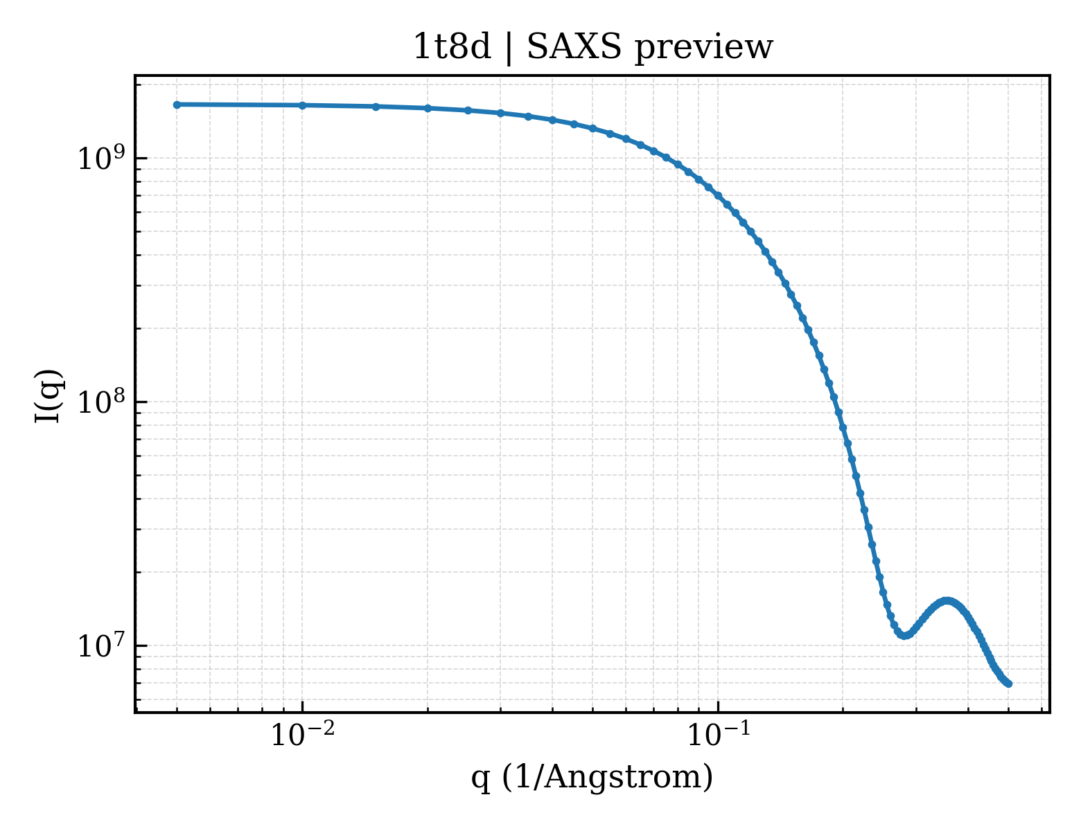
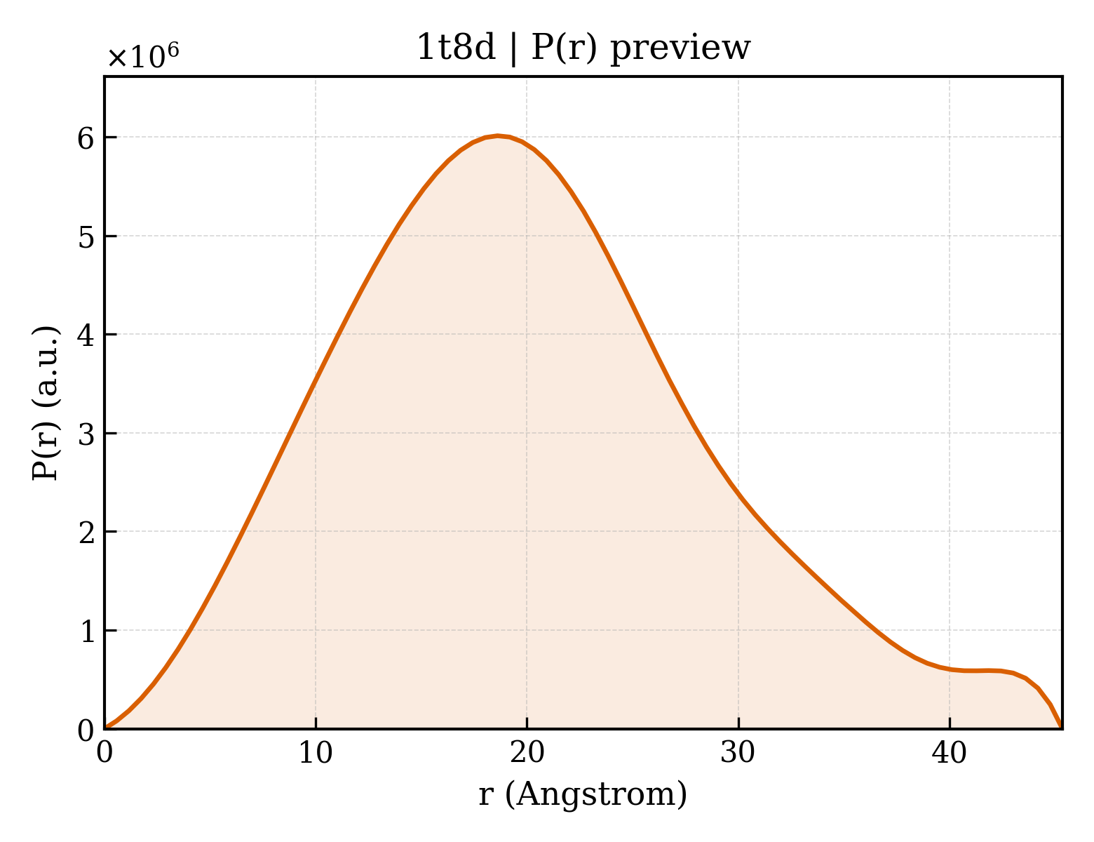
| 1 | Protein monomer Monomer Protein × 1 PDB declaration: monomeric(1) Consistent with protein copy count | Chain A; UniProt 156–298 | Fragment:derCD23 | No other associated polymer | SOLUTION NMR NMR measurement conditions:pH 6.8;308 K;Ionic strength (raw mmCIF value) 162 mM;Pressure 1 NMR sample composition:25 mM Tris, 125 mM NaCl, 4 mM CaCl2, 0.02% NaN3, 2 mM [U-100% 13C; U-100% 15N] soluble CD23 | Resolution not provided |
Other States of the Same Protein in the Database
Each row is a biological assembly of the same UniProt protein in another PDB entry. The “Difference from current entry” column identifies evidence-level differences; no tag means the currently parsed fields agree.
| Other PDB | Difference from Current Entry 1T8D | Assembly / Oligomeric State | Construct | Mutations and Modifications | Ligands, Ions and Non-polymers | Method and Experimental Conditions | Structure Quality |
|---|---|---|---|---|---|---|---|
| 1T8C Structure of the C-type lectin domain of CD23 Deposited 2004-05-12 | Different experimental conditions | Assembly 1 Protein monomer Monomer;Protein × 1 PDB declaration: monomeric |
Chain A
156–298(143 aa)
Fragment:derCD23
|
Not recorded | No recorded non-water small molecule |
SOLUTION NMR
NMR measurement conditions
pH 6.8;308 K;Ionic strength (raw mmCIF value) 162 mM;Pressure 1
NMR sample composition
25mM Tris, 125mM NaCl, 4mM CaCl2, 0.02% NaN3, 2mM soluble CD23
|
Resolution not provided |
| 2H2R Crystal structure of the human CD23 Lectin domain, apo form Deposited 2006-05-19 | Different construct Different mutation/modification Different oligomeric state Different experimental method Different experimental conditions Different structure-quality metrics | Assembly 1 Protein homooligomer Homooligomer;Protein × 2 PDB declaration: dimeric |
Chain A
150–321(172 aa)
Fragment:lectin domain
Chain B
150–321(172 aa)
Fragment:lectin domain
|
Mutation:H213R, G256S Mutation:H213R, G256S | No recorded non-water small molecule |
X-RAY DIFFRACTION
X-ray crystallization conditions
VAPOR DIFFUSION, HANGING DROP;pH 6.2;293 K;1.2 M sodium monobasic phosphate, 0.8 M potassium dibasic phosphate, 0.1 M Caps buffer pH 10.5, and 0.2 M
lithium sulfate; final pH 6.2 , VAPOR DIFFUSION, HANGING DROP, temperature 293K
|
Resolution 1.50 Å R-free 0.165 |
| 2H2T CD23 Lectin domain, Calcium 2+-bound Deposited 2006-05-19 | Different construct Different mutation/modification Different ligand/ion Different experimental method Different experimental conditions Different structure-quality metrics | Assembly 1 Protein monomer Monomer;Protein × 1 PDB declaration: monomeric |
Chain B
150–321(172 aa)
Fragment:lectin domain
|
Mutation:H213R, G256S | CA CALCIUM ION × 1 |
X-RAY DIFFRACTION
X-ray crystallization conditions
VAPOR DIFFUSION, HANGING DROP;pH 6;293 K;17% (w/v) PEG 4000, 100 mM Mes buffer, pH 6.0, VAPOR DIFFUSION, HANGING DROP, temperature 293K
|
Resolution 1.30 Å R-free 0.200 |
| 4EZM Crystal structure of the human IgE-Fc(epsilon)3-4 bound to its B cell receptor derCD23 Deposited 2012-05-03 | Different construct Different mutation/modification Different oligomeric state Different experimental method Different experimental conditions Different structure-quality metrics | Assembly 1 Other combination Heteromer;Protein × 4 PDB declaration: tetrameric |
Chain G
156–298(143 aa)
Fragment:Human derCD23, UNP residues 156-298
Chain H
156–298(143 aa)
Fragment:Human derCD23, UNP residues 156-298
|
Not recorded | No recorded non-water small molecule |
X-RAY DIFFRACTION
X-ray crystallization conditions
VAPOR DIFFUSION;pH 7.5;298 K;3% PEG 8,000, 0.1 M Tris-HCl pH 7.5, VAPOR DIFFUSION, temperature 298K
|
Resolution 3.10 Å R-free 0.283 |
| 4EZM Crystal structure of the human IgE-Fc(epsilon)3-4 bound to its B cell receptor derCD23 Deposited 2012-05-03 | Different construct Different mutation/modification Different oligomeric state Different ligand/ion Different experimental method Different experimental conditions Different structure-quality metrics | Assembly 2 Other combination Heteromer;Protein × 4 PDB declaration: tetrameric |
Chain I
156–298(143 aa)
Fragment:Human derCD23, UNP residues 156-298
Chain J
156–298(143 aa)
Fragment:Human derCD23, UNP residues 156-298
|
Not recorded | MAN alpha-D-mannopyranose × 2 |
X-RAY DIFFRACTION
X-ray crystallization conditions
VAPOR DIFFUSION;pH 7.5;298 K;3% PEG 8,000, 0.1 M Tris-HCl pH 7.5, VAPOR DIFFUSION, temperature 298K
|
Resolution 3.10 Å R-free 0.283 |
| 4EZM Crystal structure of the human IgE-Fc(epsilon)3-4 bound to its B cell receptor derCD23 Deposited 2012-05-03 | Different construct Different mutation/modification Different oligomeric state Different experimental method Different experimental conditions Different structure-quality metrics | Assembly 3 Other combination Heteromer;Protein × 4 PDB declaration: tetrameric |
Chain K
156–298(143 aa)
Fragment:Human derCD23, UNP residues 156-298
Chain L
156–298(143 aa)
Fragment:Human derCD23, UNP residues 156-298
|
Not recorded | No recorded non-water small molecule |
X-RAY DIFFRACTION
X-ray crystallization conditions
VAPOR DIFFUSION;pH 7.5;298 K;3% PEG 8,000, 0.1 M Tris-HCl pH 7.5, VAPOR DIFFUSION, temperature 298K
|
Resolution 3.10 Å R-free 0.283 |
| 4G96 Crystal structure of calcium2+-free wild-type CD23 lectin domain Deposited 2012-07-23 | Different construct Different ligand/ion Different experimental method Different experimental conditions Different structure-quality metrics | Assembly 1 Protein monomer Monomer;Protein × 1 PDB declaration: monomeric |
Chain A
156–298(143 aa)
Fragment:UNP residues 156-298
|
Not recorded | SO4 SULFATE ION × 1 |
X-RAY DIFFRACTION
X-ray crystallization conditions
VAPOR DIFFUSION, SITTING DROP;pH 4.7;291 K;18.5% PEG 6000, 2% 1,6-hexanediol, 0.05M ammonium sulphate, 0.1M sodium acetate pH 4.7, VAPOR DIFFUSION, SITTING DROP, temperature 291K
|
Resolution 2.25 Å R-free 0.236 |
| 4G96 Crystal structure of calcium2+-free wild-type CD23 lectin domain Deposited 2012-07-23 | Different construct Different ligand/ion Different experimental method Different experimental conditions Different structure-quality metrics | Assembly 2 Protein monomer Monomer;Protein × 1 PDB declaration: monomeric |
Chain B
156–298(143 aa)
Fragment:UNP residues 156-298
|
Not recorded | SO4 SULFATE ION × 1 EDO 1,2-ETHANEDIOL × 1 |
X-RAY DIFFRACTION
X-ray crystallization conditions
VAPOR DIFFUSION, SITTING DROP;pH 4.7;291 K;18.5% PEG 6000, 2% 1,6-hexanediol, 0.05M ammonium sulphate, 0.1M sodium acetate pH 4.7, VAPOR DIFFUSION, SITTING DROP, temperature 291K
|
Resolution 2.25 Å R-free 0.236 |
| 4G96 Crystal structure of calcium2+-free wild-type CD23 lectin domain Deposited 2012-07-23 | Different construct Different experimental method Different experimental conditions Different structure-quality metrics | Assembly 3 Protein monomer Monomer;Protein × 1 PDB declaration: monomeric |
Chain C
156–298(143 aa)
Fragment:UNP residues 156-298
|
Not recorded | No recorded non-water small molecule |
X-RAY DIFFRACTION
X-ray crystallization conditions
VAPOR DIFFUSION, SITTING DROP;pH 4.7;291 K;18.5% PEG 6000, 2% 1,6-hexanediol, 0.05M ammonium sulphate, 0.1M sodium acetate pH 4.7, VAPOR DIFFUSION, SITTING DROP, temperature 291K
|
Resolution 2.25 Å R-free 0.236 |
| 4G96 Crystal structure of calcium2+-free wild-type CD23 lectin domain Deposited 2012-07-23 | Different construct Different experimental method Different experimental conditions Different structure-quality metrics | Assembly 4 Protein monomer Monomer;Protein × 1 PDB declaration: monomeric |
Chain D
156–298(143 aa)
Fragment:UNP residues 156-298
|
Not recorded | No recorded non-water small molecule |
X-RAY DIFFRACTION
X-ray crystallization conditions
VAPOR DIFFUSION, SITTING DROP;pH 4.7;291 K;18.5% PEG 6000, 2% 1,6-hexanediol, 0.05M ammonium sulphate, 0.1M sodium acetate pH 4.7, VAPOR DIFFUSION, SITTING DROP, temperature 291K
|
Resolution 2.25 Å R-free 0.236 |
| 4G9A Crystal structure of calcium2+-bound wild-type CD23 lectin domain Deposited 2012-07-23 | Different construct Different ligand/ion Different experimental method Different experimental conditions Different structure-quality metrics | Assembly 1 Protein monomer Monomer;Protein × 1 PDB declaration: monomeric |
Chain A
156–298(143 aa)
Fragment:UNP residues 156-298
|
Not recorded | GOL GLYCEROL × 1 CA CALCIUM ION × 1 |
X-RAY DIFFRACTION
X-ray crystallization conditions
VAPOR DIFFUSION, SITTING DROP;pH 4.7;291 K;18.5% PEG 6000, 2% 1,6-hexanediol, 0.05M ammonium sulphate, 0.1M sodium acetate pH 4.7, VAPOR DIFFUSION, SITTING DROP, temperature 291K
|
Resolution 2.00 Å R-free 0.205 |
| 4G9A Crystal structure of calcium2+-bound wild-type CD23 lectin domain Deposited 2012-07-23 | Different construct Different ligand/ion Different experimental method Different experimental conditions Different structure-quality metrics | Assembly 2 Protein monomer Monomer;Protein × 1 PDB declaration: monomeric |
Chain B
156–298(143 aa)
Fragment:UNP residues 156-298
|
Not recorded | GOL GLYCEROL × 1 CA CALCIUM ION × 1 |
X-RAY DIFFRACTION
X-ray crystallization conditions
VAPOR DIFFUSION, SITTING DROP;pH 4.7;291 K;18.5% PEG 6000, 2% 1,6-hexanediol, 0.05M ammonium sulphate, 0.1M sodium acetate pH 4.7, VAPOR DIFFUSION, SITTING DROP, temperature 291K
|
Resolution 2.00 Å R-free 0.205 |
| 4G9A Crystal structure of calcium2+-bound wild-type CD23 lectin domain Deposited 2012-07-23 | Different construct Different ligand/ion Different experimental method Different experimental conditions Different structure-quality metrics | Assembly 3 Protein monomer Monomer;Protein × 1 PDB declaration: monomeric |
Chain C
156–298(143 aa)
Fragment:UNP residues 156-298
|
Not recorded | CA CALCIUM ION × 1 |
X-RAY DIFFRACTION
X-ray crystallization conditions
VAPOR DIFFUSION, SITTING DROP;pH 4.7;291 K;18.5% PEG 6000, 2% 1,6-hexanediol, 0.05M ammonium sulphate, 0.1M sodium acetate pH 4.7, VAPOR DIFFUSION, SITTING DROP, temperature 291K
|
Resolution 2.00 Å R-free 0.205 |
| 4G9A Crystal structure of calcium2+-bound wild-type CD23 lectin domain Deposited 2012-07-23 | Different construct Different ligand/ion Different experimental method Different experimental conditions Different structure-quality metrics | Assembly 4 Protein monomer Monomer;Protein × 1 PDB declaration: monomeric |
Chain D
156–298(143 aa)
Fragment:UNP residues 156-298
|
Not recorded | GOL GLYCEROL × 1 CA CALCIUM ION × 1 |
X-RAY DIFFRACTION
X-ray crystallization conditions
VAPOR DIFFUSION, SITTING DROP;pH 4.7;291 K;18.5% PEG 6000, 2% 1,6-hexanediol, 0.05M ammonium sulphate, 0.1M sodium acetate pH 4.7, VAPOR DIFFUSION, SITTING DROP, temperature 291K
|
Resolution 2.00 Å R-free 0.205 |
| 4GI0 Crystal structure of CD23 lectin domain mutant E249A Deposited 2012-08-08 | Different construct Different mutation/modification Different oligomeric state Different ligand/ion Different experimental method Different experimental conditions Different structure-quality metrics | Assembly 1 Protein homooligomer Homooligomer;Protein × 3 PDB declaration: trimeric |
Chain A
156–298(143 aa)
Fragment:UNP residues 156-298
Chain B
156–298(143 aa)
Fragment:UNP residues 156-298
Chain C
156–298(143 aa)
Fragment:UNP residues 156-298
|
Mutation:E249A Mutation:E249A Mutation:E249A | GOL GLYCEROL × 1 |
X-RAY DIFFRACTION
X-ray crystallization conditions
VAPOR DIFFUSION, SITTING DROP;pH 8.5;291 K;0.2M potassium thiocyanate, 0.1M bis tris propane pH 8.5, 20% PEG 3350, VAPOR DIFFUSION, SITTING DROP, temperature 291K
|
Resolution 2.27 Å R-free 0.251 |
| 4GJ0 Crystal structure of CD23 lectin domain mutant S252A Deposited 2012-08-09 | Different construct Different ligand/ion Different experimental method Different experimental conditions Different structure-quality metrics | Assembly 1 Protein monomer Monomer;Protein × 1 PDB declaration: monomeric |
Chain A
156–298(143 aa)
Fragment:UNP residues 156-298
|
Not recorded | GOL GLYCEROL × 1 SO4 SULFATE ION × 2 |
X-RAY DIFFRACTION
X-ray crystallization conditions
VAPOR DIFFUSION, SITTING DROP;pH 4.7;291 K;18.5% PEG 6000, 2% 1,6-hexanediol, 0.05M ammonium sulphate, 0.1M sodium acetate pH 4.7, VAPOR DIFFUSION, SITTING DROP, temperature 291K
|
Resolution 1.95 Å R-free 0.212 |
| 4GJ0 Crystal structure of CD23 lectin domain mutant S252A Deposited 2012-08-09 | Different construct Different ligand/ion Different experimental method Different experimental conditions Different structure-quality metrics | Assembly 2 Protein monomer Monomer;Protein × 1 PDB declaration: monomeric |
Chain B
156–298(143 aa)
Fragment:UNP residues 156-298
|
Not recorded | GOL GLYCEROL × 1 SO4 SULFATE ION × 2 |
X-RAY DIFFRACTION
X-ray crystallization conditions
VAPOR DIFFUSION, SITTING DROP;pH 4.7;291 K;18.5% PEG 6000, 2% 1,6-hexanediol, 0.05M ammonium sulphate, 0.1M sodium acetate pH 4.7, VAPOR DIFFUSION, SITTING DROP, temperature 291K
|
Resolution 1.95 Å R-free 0.212 |
| 4GJ0 Crystal structure of CD23 lectin domain mutant S252A Deposited 2012-08-09 | Different construct Different ligand/ion Different experimental method Different experimental conditions Different structure-quality metrics | Assembly 3 Protein monomer Monomer;Protein × 1 PDB declaration: monomeric |
Chain C
156–298(143 aa)
Fragment:UNP residues 156-298
|
Not recorded | GOL GLYCEROL × 1 |
X-RAY DIFFRACTION
X-ray crystallization conditions
VAPOR DIFFUSION, SITTING DROP;pH 4.7;291 K;18.5% PEG 6000, 2% 1,6-hexanediol, 0.05M ammonium sulphate, 0.1M sodium acetate pH 4.7, VAPOR DIFFUSION, SITTING DROP, temperature 291K
|
Resolution 1.95 Å R-free 0.212 |
| 4GJ0 Crystal structure of CD23 lectin domain mutant S252A Deposited 2012-08-09 | Different construct Different ligand/ion Different experimental method Different experimental conditions Different structure-quality metrics | Assembly 4 Protein monomer Monomer;Protein × 1 PDB declaration: monomeric |
Chain D
156–298(143 aa)
Fragment:UNP residues 156-298
|
Not recorded | GOL GLYCEROL × 1 |
X-RAY DIFFRACTION
X-ray crystallization conditions
VAPOR DIFFUSION, SITTING DROP;pH 4.7;291 K;18.5% PEG 6000, 2% 1,6-hexanediol, 0.05M ammonium sulphate, 0.1M sodium acetate pH 4.7, VAPOR DIFFUSION, SITTING DROP, temperature 291K
|
Resolution 1.95 Å R-free 0.212 |
| 4GJX Crystal structure of CD23 lectin domain mutant D258A Deposited 2012-08-10 | Different construct Different mutation/modification Different ligand/ion Different experimental method Different experimental conditions Different structure-quality metrics | Assembly 1 Protein monomer Monomer;Protein × 1 PDB declaration: monomeric |
Chain A
156–298(143 aa)
Fragment:UNP residues 156-298
|
Mutation:D258A | GOL GLYCEROL × 1 |
X-RAY DIFFRACTION
X-ray crystallization conditions
VAPOR DIFFUSION, SITTING DROP;pH 5;291 K;18% ethanol, 4% PEG 400, 0.1 M sodium acetate pH 5.0, VAPOR DIFFUSION, SITTING DROP, temperature 291K
|
Resolution 2.80 Å R-free 0.251 |
| 4GJX Crystal structure of CD23 lectin domain mutant D258A Deposited 2012-08-10 | Different construct Different mutation/modification Different experimental method Different experimental conditions Different structure-quality metrics | Assembly 2 Protein monomer Monomer;Protein × 1 PDB declaration: monomeric |
Chain B
156–298(143 aa)
Fragment:UNP residues 156-298
|
Mutation:D258A | No recorded non-water small molecule |
X-RAY DIFFRACTION
X-ray crystallization conditions
VAPOR DIFFUSION, SITTING DROP;pH 5;291 K;18% ethanol, 4% PEG 400, 0.1 M sodium acetate pH 5.0, VAPOR DIFFUSION, SITTING DROP, temperature 291K
|
Resolution 2.80 Å R-free 0.251 |
| 4GJX Crystal structure of CD23 lectin domain mutant D258A Deposited 2012-08-10 | Different construct Different mutation/modification Different experimental method Different experimental conditions Different structure-quality metrics | Assembly 3 Protein monomer Monomer;Protein × 1 PDB declaration: monomeric |
Chain C
156–298(143 aa)
Fragment:UNP residues 156-298
|
Mutation:D258A | No recorded non-water small molecule |
X-RAY DIFFRACTION
X-ray crystallization conditions
VAPOR DIFFUSION, SITTING DROP;pH 5;291 K;18% ethanol, 4% PEG 400, 0.1 M sodium acetate pH 5.0, VAPOR DIFFUSION, SITTING DROP, temperature 291K
|
Resolution 2.80 Å R-free 0.251 |
| 4GJX Crystal structure of CD23 lectin domain mutant D258A Deposited 2012-08-10 | Different construct Different mutation/modification Different ligand/ion Different experimental method Different experimental conditions Different structure-quality metrics | Assembly 4 Protein monomer Monomer;Protein × 1 PDB declaration: monomeric |
Chain D
156–298(143 aa)
Fragment:UNP residues 156-298
|
Mutation:D258A | GOL GLYCEROL × 1 |
X-RAY DIFFRACTION
X-ray crystallization conditions
VAPOR DIFFUSION, SITTING DROP;pH 5;291 K;18% ethanol, 4% PEG 400, 0.1 M sodium acetate pH 5.0, VAPOR DIFFUSION, SITTING DROP, temperature 291K
|
Resolution 2.80 Å R-free 0.251 |
| 4GJX Crystal structure of CD23 lectin domain mutant D258A Deposited 2012-08-10 | Different construct Different mutation/modification Different experimental method Different experimental conditions Different structure-quality metrics | Assembly 5 Protein monomer Monomer;Protein × 1 PDB declaration: monomeric |
Chain E
156–298(143 aa)
Fragment:UNP residues 156-298
|
Mutation:D258A | No recorded non-water small molecule |
X-RAY DIFFRACTION
X-ray crystallization conditions
VAPOR DIFFUSION, SITTING DROP;pH 5;291 K;18% ethanol, 4% PEG 400, 0.1 M sodium acetate pH 5.0, VAPOR DIFFUSION, SITTING DROP, temperature 291K
|
Resolution 2.80 Å R-free 0.251 |
| 4GJX Crystal structure of CD23 lectin domain mutant D258A Deposited 2012-08-10 | Different construct Different mutation/modification Different experimental method Different experimental conditions Different structure-quality metrics | Assembly 6 Protein monomer Monomer;Protein × 1 PDB declaration: monomeric |
Chain F
156–298(143 aa)
Fragment:UNP residues 156-298
|
Mutation:D258A | No recorded non-water small molecule |
X-RAY DIFFRACTION
X-ray crystallization conditions
VAPOR DIFFUSION, SITTING DROP;pH 5;291 K;18% ethanol, 4% PEG 400, 0.1 M sodium acetate pH 5.0, VAPOR DIFFUSION, SITTING DROP, temperature 291K
|
Resolution 2.80 Å R-free 0.251 |
| 4GJX Crystal structure of CD23 lectin domain mutant D258A Deposited 2012-08-10 | Different construct Different mutation/modification Different experimental method Different experimental conditions Different structure-quality metrics | Assembly 7 Protein monomer Monomer;Protein × 1 PDB declaration: monomeric |
Chain G
156–298(143 aa)
Fragment:UNP residues 156-298
|
Mutation:D258A | No recorded non-water small molecule |
X-RAY DIFFRACTION
X-ray crystallization conditions
VAPOR DIFFUSION, SITTING DROP;pH 5;291 K;18% ethanol, 4% PEG 400, 0.1 M sodium acetate pH 5.0, VAPOR DIFFUSION, SITTING DROP, temperature 291K
|
Resolution 2.80 Å R-free 0.251 |
| 4GJX Crystal structure of CD23 lectin domain mutant D258A Deposited 2012-08-10 | Different construct Different mutation/modification Different experimental method Different experimental conditions Different structure-quality metrics | Assembly 8 Protein monomer Monomer;Protein × 1 PDB declaration: monomeric |
Chain H
156–298(143 aa)
Fragment:UNP residues 156-298
|
Mutation:D258A | No recorded non-water small molecule |
X-RAY DIFFRACTION
X-ray crystallization conditions
VAPOR DIFFUSION, SITTING DROP;pH 5;291 K;18% ethanol, 4% PEG 400, 0.1 M sodium acetate pH 5.0, VAPOR DIFFUSION, SITTING DROP, temperature 291K
|
Resolution 2.80 Å R-free 0.251 |
| 4GK1 Crystal structure of CD23 lectin domain mutant D270A Deposited 2012-08-10 | Different construct Different mutation/modification Different ligand/ion Different experimental method Different experimental conditions Different structure-quality metrics | Assembly 1 Protein monomer Monomer;Protein × 1 PDB declaration: monomeric |
Chain A
156–298(143 aa)
Fragment:UNP residues 156-298
|
Mutation:D270A | GOL GLYCEROL × 2 SO4 SULFATE ION × 3 |
X-RAY DIFFRACTION
X-ray crystallization conditions
VAPOR DIFFUSION, SITTING DROP;pH 5.5;291 K;30% PEG 4000, 0.3M ammonium sulphate, pH 5.5, VAPOR DIFFUSION, SITTING DROP, temperature 291K
|
Resolution 2.24 Å R-free 0.226 |
| 4GK1 Crystal structure of CD23 lectin domain mutant D270A Deposited 2012-08-10 | Different construct Different mutation/modification Different ligand/ion Different experimental method Different experimental conditions Different structure-quality metrics | Assembly 2 Protein monomer Monomer;Protein × 1 PDB declaration: monomeric |
Chain B
156–298(143 aa)
Fragment:UNP residues 156-298
|
Mutation:D270A | GOL GLYCEROL × 1 SO4 SULFATE ION × 5 |
X-RAY DIFFRACTION
X-ray crystallization conditions
VAPOR DIFFUSION, SITTING DROP;pH 5.5;291 K;30% PEG 4000, 0.3M ammonium sulphate, pH 5.5, VAPOR DIFFUSION, SITTING DROP, temperature 291K
|
Resolution 2.24 Å R-free 0.226 |
| 4GK1 Crystal structure of CD23 lectin domain mutant D270A Deposited 2012-08-10 | Different construct Different mutation/modification Different ligand/ion Different experimental method Different experimental conditions Different structure-quality metrics | Assembly 3 Protein monomer Monomer;Protein × 1 PDB declaration: monomeric |
Chain C
156–298(143 aa)
Fragment:UNP residues 156-298
|
Mutation:D270A | GOL GLYCEROL × 2 SO4 SULFATE ION × 4 |
X-RAY DIFFRACTION
X-ray crystallization conditions
VAPOR DIFFUSION, SITTING DROP;pH 5.5;291 K;30% PEG 4000, 0.3M ammonium sulphate, pH 5.5, VAPOR DIFFUSION, SITTING DROP, temperature 291K
|
Resolution 2.24 Å R-free 0.226 |
| 4GK1 Crystal structure of CD23 lectin domain mutant D270A Deposited 2012-08-10 | Different construct Different mutation/modification Different ligand/ion Different experimental method Different experimental conditions Different structure-quality metrics | Assembly 4 Protein monomer Monomer;Protein × 1 PDB declaration: monomeric |
Chain D
156–298(143 aa)
Fragment:UNP residues 156-298
|
Mutation:D270A | GOL GLYCEROL × 2 SO4 SULFATE ION × 2 |
X-RAY DIFFRACTION
X-ray crystallization conditions
VAPOR DIFFUSION, SITTING DROP;pH 5.5;291 K;30% PEG 4000, 0.3M ammonium sulphate, pH 5.5, VAPOR DIFFUSION, SITTING DROP, temperature 291K
|
Resolution 2.24 Å R-free 0.226 |
| 4GK1 Crystal structure of CD23 lectin domain mutant D270A Deposited 2012-08-10 | Different construct Different mutation/modification Different ligand/ion Different experimental method Different experimental conditions Different structure-quality metrics | Assembly 5 Protein monomer Monomer;Protein × 1 PDB declaration: monomeric |
Chain E
156–298(143 aa)
Fragment:UNP residues 156-298
|
Mutation:D270A | GOL GLYCEROL × 1 SO4 SULFATE ION × 4 |
X-RAY DIFFRACTION
X-ray crystallization conditions
VAPOR DIFFUSION, SITTING DROP;pH 5.5;291 K;30% PEG 4000, 0.3M ammonium sulphate, pH 5.5, VAPOR DIFFUSION, SITTING DROP, temperature 291K
|
Resolution 2.24 Å R-free 0.226 |
| 4GK1 Crystal structure of CD23 lectin domain mutant D270A Deposited 2012-08-10 | Different construct Different mutation/modification Different ligand/ion Different experimental method Different experimental conditions Different structure-quality metrics | Assembly 6 Protein monomer Monomer;Protein × 1 PDB declaration: monomeric |
Chain F
156–298(143 aa)
Fragment:UNP residues 156-298
|
Mutation:D270A | GOL GLYCEROL × 3 SO4 SULFATE ION × 6 |
X-RAY DIFFRACTION
X-ray crystallization conditions
VAPOR DIFFUSION, SITTING DROP;pH 5.5;291 K;30% PEG 4000, 0.3M ammonium sulphate, pH 5.5, VAPOR DIFFUSION, SITTING DROP, temperature 291K
|
Resolution 2.24 Å R-free 0.226 |
| 4GK1 Crystal structure of CD23 lectin domain mutant D270A Deposited 2012-08-10 | Different construct Different mutation/modification Different ligand/ion Different experimental method Different experimental conditions Different structure-quality metrics | Assembly 7 Protein monomer Monomer;Protein × 1 PDB declaration: monomeric |
Chain G
156–298(143 aa)
Fragment:UNP residues 156-298
|
Mutation:D270A | GOL GLYCEROL × 1 SO4 SULFATE ION × 1 |
X-RAY DIFFRACTION
X-ray crystallization conditions
VAPOR DIFFUSION, SITTING DROP;pH 5.5;291 K;30% PEG 4000, 0.3M ammonium sulphate, pH 5.5, VAPOR DIFFUSION, SITTING DROP, temperature 291K
|
Resolution 2.24 Å R-free 0.226 |
| 4GKO Crystal structure of the calcium2+-bound human IgE-Fc(epsilon)3-4 bound to its B cell receptor derCD23 Deposited 2012-08-13 | Different construct Different mutation/modification Different oligomeric state Different ligand/ion Different experimental method Different experimental conditions Different structure-quality metrics | Assembly 1 Other combination Heteromer;Protein × 4 PDB declaration: tetrameric |
Chain G
156–298(143 aa)
Fragment:UNP residues 156-298
Chain H
156–298(143 aa)
Fragment:UNP residues 156-298
|
Not recorded | CA CALCIUM ION × 2 |
X-RAY DIFFRACTION
X-ray crystallization conditions
VAPOR DIFFUSION, SITTING DROP;pH 8.5;291 K;0.2 M sodium acetate trihydrate, 0.1 M Tris pH 8.5, 16% PEG 4000, 10 mM CaCl2, VAPOR DIFFUSION, SITTING DROP, temperature 291K
|
Resolution 3.30 Å R-free 0.312 |
| 4GKO Crystal structure of the calcium2+-bound human IgE-Fc(epsilon)3-4 bound to its B cell receptor derCD23 Deposited 2012-08-13 | Different construct Different mutation/modification Different oligomeric state Different ligand/ion Different experimental method Different experimental conditions Different structure-quality metrics | Assembly 2 Other combination Heteromer;Protein × 4 PDB declaration: tetrameric |
Chain I
156–298(143 aa)
Fragment:UNP residues 156-298
Chain J
156–298(143 aa)
Fragment:UNP residues 156-298
|
Not recorded | MAN alpha-D-mannopyranose × 2 CA CALCIUM ION × 2 |
X-RAY DIFFRACTION
X-ray crystallization conditions
VAPOR DIFFUSION, SITTING DROP;pH 8.5;291 K;0.2 M sodium acetate trihydrate, 0.1 M Tris pH 8.5, 16% PEG 4000, 10 mM CaCl2, VAPOR DIFFUSION, SITTING DROP, temperature 291K
|
Resolution 3.30 Å R-free 0.312 |
| 4GKO Crystal structure of the calcium2+-bound human IgE-Fc(epsilon)3-4 bound to its B cell receptor derCD23 Deposited 2012-08-13 | Different construct Different mutation/modification Different oligomeric state Different ligand/ion Different experimental method Different experimental conditions Different structure-quality metrics | Assembly 3 Other combination Heteromer;Protein × 4 PDB declaration: tetrameric |
Chain K
156–298(143 aa)
Fragment:UNP residues 156-298
Chain L
156–298(143 aa)
Fragment:UNP residues 156-298
|
Not recorded | CA CALCIUM ION × 2 |
X-RAY DIFFRACTION
X-ray crystallization conditions
VAPOR DIFFUSION, SITTING DROP;pH 8.5;291 K;0.2 M sodium acetate trihydrate, 0.1 M Tris pH 8.5, 16% PEG 4000, 10 mM CaCl2, VAPOR DIFFUSION, SITTING DROP, temperature 291K
|
Resolution 3.30 Å R-free 0.312 |
| 4J6J Crystal structure of calcium2+-free wild-type CD23 lectin domain (crystal form A) Deposited 2013-02-11 | Different construct Different experimental method Different experimental conditions Different structure-quality metrics | Assembly 1 Protein monomer Monomer;Protein × 1 PDB declaration: monomeric |
Chain A
156–298(143 aa)
Fragment:Soluble head domain of the B-cell receptor CD23 (UNP Residues 156-298)
|
Not recorded | No recorded non-water small molecule |
X-RAY DIFFRACTION
X-ray crystallization conditions
VAPOR DIFFUSION, SITTING DROP;pH 4.7;298 K;16 % (w/v) PEG 6,000, 2 % (v/v) 1,6-hexanediol, 0.05 M ammonium sulfate and 0.1 M sodium acetate pH 4.7 , VAPOR DIFFUSION, SITTING DROP, temperature 298K
|
Resolution 1.90 Å R-free 0.217 |
| 4J6J Crystal structure of calcium2+-free wild-type CD23 lectin domain (crystal form A) Deposited 2013-02-11 | Different construct Different experimental method Different experimental conditions Different structure-quality metrics | Assembly 2 Protein monomer Monomer;Protein × 1 PDB declaration: monomeric |
Chain B
156–298(143 aa)
Fragment:Soluble head domain of the B-cell receptor CD23 (UNP Residues 156-298)
|
Not recorded | No recorded non-water small molecule |
X-RAY DIFFRACTION
X-ray crystallization conditions
VAPOR DIFFUSION, SITTING DROP;pH 4.7;298 K;16 % (w/v) PEG 6,000, 2 % (v/v) 1,6-hexanediol, 0.05 M ammonium sulfate and 0.1 M sodium acetate pH 4.7 , VAPOR DIFFUSION, SITTING DROP, temperature 298K
|
Resolution 1.90 Å R-free 0.217 |
| 4J6J Crystal structure of calcium2+-free wild-type CD23 lectin domain (crystal form A) Deposited 2013-02-11 | Different construct Different ligand/ion Different experimental method Different experimental conditions Different structure-quality metrics | Assembly 3 Protein monomer Monomer;Protein × 1 PDB declaration: monomeric |
Chain C
156–298(143 aa)
Fragment:Soluble head domain of the B-cell receptor CD23 (UNP Residues 156-298)
|
Not recorded | SO4 SULFATE ION × 1 |
X-RAY DIFFRACTION
X-ray crystallization conditions
VAPOR DIFFUSION, SITTING DROP;pH 4.7;298 K;16 % (w/v) PEG 6,000, 2 % (v/v) 1,6-hexanediol, 0.05 M ammonium sulfate and 0.1 M sodium acetate pH 4.7 , VAPOR DIFFUSION, SITTING DROP, temperature 298K
|
Resolution 1.90 Å R-free 0.217 |
| 4J6J Crystal structure of calcium2+-free wild-type CD23 lectin domain (crystal form A) Deposited 2013-02-11 | Different construct Different ligand/ion Different experimental method Different experimental conditions Different structure-quality metrics | Assembly 4 Protein monomer Monomer;Protein × 1 PDB declaration: monomeric |
Chain D
156–298(143 aa)
Fragment:Soluble head domain of the B-cell receptor CD23 (UNP Residues 156-298)
|
Not recorded | SO4 SULFATE ION × 1 |
X-RAY DIFFRACTION
X-ray crystallization conditions
VAPOR DIFFUSION, SITTING DROP;pH 4.7;298 K;16 % (w/v) PEG 6,000, 2 % (v/v) 1,6-hexanediol, 0.05 M ammonium sulfate and 0.1 M sodium acetate pH 4.7 , VAPOR DIFFUSION, SITTING DROP, temperature 298K
|
Resolution 1.90 Å R-free 0.217 |
| 4J6K Crystal structure of calcium2+-free wild-type CD23 lectin domain (crystal form B) Deposited 2013-02-11 | Different construct Different experimental method Different experimental conditions Different structure-quality metrics | Assembly 1 Protein monomer Monomer;Protein × 1 PDB declaration: monomeric |
Chain A
156–298(143 aa)
Fragment:Soluble head domain of the B-cell receptor CD23 (UNP Residues 156-298)
|
Not recorded | No recorded non-water small molecule |
X-RAY DIFFRACTION
X-ray crystallization conditions
VAPOR DIFFUSION, SITTING DROP;pH 4.5;298 K;18 % (w/v) PEG 6,000, 2 % (v/v) 1,6-hexanediol, 0.05 M ammonium sulfate and 0.1 M sodium acetate pH 4.5, VAPOR DIFFUSION, SITTING DROP, temperature 298K
|
Resolution 2.30 Å R-free 0.218 |
| 4J6K Crystal structure of calcium2+-free wild-type CD23 lectin domain (crystal form B) Deposited 2013-02-11 | Different construct Different experimental method Different experimental conditions Different structure-quality metrics | Assembly 2 Protein monomer Monomer;Protein × 1 PDB declaration: monomeric |
Chain B
156–298(143 aa)
Fragment:Soluble head domain of the B-cell receptor CD23 (UNP Residues 156-298)
|
Not recorded | No recorded non-water small molecule |
X-RAY DIFFRACTION
X-ray crystallization conditions
VAPOR DIFFUSION, SITTING DROP;pH 4.5;298 K;18 % (w/v) PEG 6,000, 2 % (v/v) 1,6-hexanediol, 0.05 M ammonium sulfate and 0.1 M sodium acetate pH 4.5, VAPOR DIFFUSION, SITTING DROP, temperature 298K
|
Resolution 2.30 Å R-free 0.218 |
| 4J6K Crystal structure of calcium2+-free wild-type CD23 lectin domain (crystal form B) Deposited 2013-02-11 | Different construct Different experimental method Different experimental conditions Different structure-quality metrics | Assembly 3 Protein monomer Monomer;Protein × 1 PDB declaration: monomeric |
Chain C
156–298(143 aa)
Fragment:Soluble head domain of the B-cell receptor CD23 (UNP Residues 156-298)
|
Not recorded | No recorded non-water small molecule |
X-RAY DIFFRACTION
X-ray crystallization conditions
VAPOR DIFFUSION, SITTING DROP;pH 4.5;298 K;18 % (w/v) PEG 6,000, 2 % (v/v) 1,6-hexanediol, 0.05 M ammonium sulfate and 0.1 M sodium acetate pH 4.5, VAPOR DIFFUSION, SITTING DROP, temperature 298K
|
Resolution 2.30 Å R-free 0.218 |
| 4J6K Crystal structure of calcium2+-free wild-type CD23 lectin domain (crystal form B) Deposited 2013-02-11 | Different construct Different ligand/ion Different experimental method Different experimental conditions Different structure-quality metrics | Assembly 4 Protein monomer Monomer;Protein × 1 PDB declaration: monomeric |
Chain D
156–298(143 aa)
Fragment:Soluble head domain of the B-cell receptor CD23 (UNP Residues 156-298)
|
Not recorded | SO4 SULFATE ION × 1 |
X-RAY DIFFRACTION
X-ray crystallization conditions
VAPOR DIFFUSION, SITTING DROP;pH 4.5;298 K;18 % (w/v) PEG 6,000, 2 % (v/v) 1,6-hexanediol, 0.05 M ammonium sulfate and 0.1 M sodium acetate pH 4.5, VAPOR DIFFUSION, SITTING DROP, temperature 298K
|
Resolution 2.30 Å R-free 0.218 |
| 4J6K Crystal structure of calcium2+-free wild-type CD23 lectin domain (crystal form B) Deposited 2013-02-11 | Different construct Different experimental method Different experimental conditions Different structure-quality metrics | Assembly 5 Protein monomer Monomer;Protein × 1 PDB declaration: monomeric |
Chain E
156–298(143 aa)
Fragment:Soluble head domain of the B-cell receptor CD23 (UNP Residues 156-298)
|
Not recorded | No recorded non-water small molecule |
X-RAY DIFFRACTION
X-ray crystallization conditions
VAPOR DIFFUSION, SITTING DROP;pH 4.5;298 K;18 % (w/v) PEG 6,000, 2 % (v/v) 1,6-hexanediol, 0.05 M ammonium sulfate and 0.1 M sodium acetate pH 4.5, VAPOR DIFFUSION, SITTING DROP, temperature 298K
|
Resolution 2.30 Å R-free 0.218 |
| 4J6K Crystal structure of calcium2+-free wild-type CD23 lectin domain (crystal form B) Deposited 2013-02-11 | Different construct Different ligand/ion Different experimental method Different experimental conditions Different structure-quality metrics | Assembly 6 Protein monomer Monomer;Protein × 1 PDB declaration: monomeric |
Chain F
156–298(143 aa)
Fragment:Soluble head domain of the B-cell receptor CD23 (UNP Residues 156-298)
|
Not recorded | SO4 SULFATE ION × 1 |
X-RAY DIFFRACTION
X-ray crystallization conditions
VAPOR DIFFUSION, SITTING DROP;pH 4.5;298 K;18 % (w/v) PEG 6,000, 2 % (v/v) 1,6-hexanediol, 0.05 M ammonium sulfate and 0.1 M sodium acetate pH 4.5, VAPOR DIFFUSION, SITTING DROP, temperature 298K
|
Resolution 2.30 Å R-free 0.218 |
| 4J6K Crystal structure of calcium2+-free wild-type CD23 lectin domain (crystal form B) Deposited 2013-02-11 | Different construct Different ligand/ion Different experimental method Different experimental conditions Different structure-quality metrics | Assembly 7 Protein monomer Monomer;Protein × 1 PDB declaration: monomeric |
Chain G
156–298(143 aa)
Fragment:Soluble head domain of the B-cell receptor CD23 (UNP Residues 156-298)
|
Not recorded | SO4 SULFATE ION × 1 |
X-RAY DIFFRACTION
X-ray crystallization conditions
VAPOR DIFFUSION, SITTING DROP;pH 4.5;298 K;18 % (w/v) PEG 6,000, 2 % (v/v) 1,6-hexanediol, 0.05 M ammonium sulfate and 0.1 M sodium acetate pH 4.5, VAPOR DIFFUSION, SITTING DROP, temperature 298K
|
Resolution 2.30 Å R-free 0.218 |
| 4J6K Crystal structure of calcium2+-free wild-type CD23 lectin domain (crystal form B) Deposited 2013-02-11 | Different construct Different ligand/ion Different experimental method Different experimental conditions Different structure-quality metrics | Assembly 8 Protein monomer Monomer;Protein × 1 PDB declaration: monomeric |
Chain H
156–298(143 aa)
Fragment:Soluble head domain of the B-cell receptor CD23 (UNP Residues 156-298)
|
Not recorded | SO4 SULFATE ION × 1 |
X-RAY DIFFRACTION
X-ray crystallization conditions
VAPOR DIFFUSION, SITTING DROP;pH 4.5;298 K;18 % (w/v) PEG 6,000, 2 % (v/v) 1,6-hexanediol, 0.05 M ammonium sulfate and 0.1 M sodium acetate pH 4.5, VAPOR DIFFUSION, SITTING DROP, temperature 298K
|
Resolution 2.30 Å R-free 0.218 |
| 4J6L Crystal structure of calcium2+-free wild-type CD23 lectin domain (crystal form C) Deposited 2013-02-11 | Different construct Different experimental method Different experimental conditions Different structure-quality metrics | Assembly 1 Protein monomer Monomer;Protein × 1 PDB declaration: monomeric |
Chain A
156–298(143 aa)
Fragment:Soluble head domain of the B-cell receptor CD23 (UNP Residues 156-298)
|
Not recorded | No recorded non-water small molecule |
X-RAY DIFFRACTION
X-ray crystallization conditions
VAPOR DIFFUSION, SITTING DROP;pH 5.5;298 K;25 % (w/v) PEG 4,000, 0.15 M ammonium sulfate and 0.1 M MES pH 5.5, VAPOR DIFFUSION, SITTING DROP, temperature 298K
|
Resolution 3.15 Å R-free 0.253 |
| 4J6L Crystal structure of calcium2+-free wild-type CD23 lectin domain (crystal form C) Deposited 2013-02-11 | Different construct Different experimental method Different experimental conditions Different structure-quality metrics | Assembly 2 Protein monomer Monomer;Protein × 1 PDB declaration: monomeric |
Chain B
156–298(143 aa)
Fragment:Soluble head domain of the B-cell receptor CD23 (UNP Residues 156-298)
|
Not recorded | No recorded non-water small molecule |
X-RAY DIFFRACTION
X-ray crystallization conditions
VAPOR DIFFUSION, SITTING DROP;pH 5.5;298 K;25 % (w/v) PEG 4,000, 0.15 M ammonium sulfate and 0.1 M MES pH 5.5, VAPOR DIFFUSION, SITTING DROP, temperature 298K
|
Resolution 3.15 Å R-free 0.253 |
| 4J6L Crystal structure of calcium2+-free wild-type CD23 lectin domain (crystal form C) Deposited 2013-02-11 | Different construct Different experimental method Different experimental conditions Different structure-quality metrics | Assembly 3 Protein monomer Monomer;Protein × 1 PDB declaration: monomeric |
Chain C
156–298(143 aa)
Fragment:Soluble head domain of the B-cell receptor CD23 (UNP Residues 156-298)
|
Not recorded | No recorded non-water small molecule |
X-RAY DIFFRACTION
X-ray crystallization conditions
VAPOR DIFFUSION, SITTING DROP;pH 5.5;298 K;25 % (w/v) PEG 4,000, 0.15 M ammonium sulfate and 0.1 M MES pH 5.5, VAPOR DIFFUSION, SITTING DROP, temperature 298K
|
Resolution 3.15 Å R-free 0.253 |
| 4J6L Crystal structure of calcium2+-free wild-type CD23 lectin domain (crystal form C) Deposited 2013-02-11 | Different construct Different experimental method Different experimental conditions Different structure-quality metrics | Assembly 4 Protein monomer Monomer;Protein × 1 PDB declaration: monomeric |
Chain D
156–298(143 aa)
Fragment:Soluble head domain of the B-cell receptor CD23 (UNP Residues 156-298)
|
Not recorded | No recorded non-water small molecule |
X-RAY DIFFRACTION
X-ray crystallization conditions
VAPOR DIFFUSION, SITTING DROP;pH 5.5;298 K;25 % (w/v) PEG 4,000, 0.15 M ammonium sulfate and 0.1 M MES pH 5.5, VAPOR DIFFUSION, SITTING DROP, temperature 298K
|
Resolution 3.15 Å R-free 0.253 |
| 4J6L Crystal structure of calcium2+-free wild-type CD23 lectin domain (crystal form C) Deposited 2013-02-11 | Different construct Different experimental method Different experimental conditions Different structure-quality metrics | Assembly 5 Protein monomer Monomer;Protein × 1 PDB declaration: monomeric |
Chain E
156–298(143 aa)
Fragment:Soluble head domain of the B-cell receptor CD23 (UNP Residues 156-298)
|
Not recorded | No recorded non-water small molecule |
X-RAY DIFFRACTION
X-ray crystallization conditions
VAPOR DIFFUSION, SITTING DROP;pH 5.5;298 K;25 % (w/v) PEG 4,000, 0.15 M ammonium sulfate and 0.1 M MES pH 5.5, VAPOR DIFFUSION, SITTING DROP, temperature 298K
|
Resolution 3.15 Å R-free 0.253 |
| 4J6L Crystal structure of calcium2+-free wild-type CD23 lectin domain (crystal form C) Deposited 2013-02-11 | Different construct Different experimental method Different experimental conditions Different structure-quality metrics | Assembly 6 Protein monomer Monomer;Protein × 1 PDB declaration: monomeric |
Chain F
156–298(143 aa)
Fragment:Soluble head domain of the B-cell receptor CD23 (UNP Residues 156-298)
|
Not recorded | No recorded non-water small molecule |
X-RAY DIFFRACTION
X-ray crystallization conditions
VAPOR DIFFUSION, SITTING DROP;pH 5.5;298 K;25 % (w/v) PEG 4,000, 0.15 M ammonium sulfate and 0.1 M MES pH 5.5, VAPOR DIFFUSION, SITTING DROP, temperature 298K
|
Resolution 3.15 Å R-free 0.253 |
| 4J6L Crystal structure of calcium2+-free wild-type CD23 lectin domain (crystal form C) Deposited 2013-02-11 | Different construct Different experimental method Different experimental conditions Different structure-quality metrics | Assembly 7 Protein monomer Monomer;Protein × 1 PDB declaration: monomeric |
Chain G
156–298(143 aa)
Fragment:Soluble head domain of the B-cell receptor CD23 (UNP Residues 156-298)
|
Not recorded | No recorded non-water small molecule |
X-RAY DIFFRACTION
X-ray crystallization conditions
VAPOR DIFFUSION, SITTING DROP;pH 5.5;298 K;25 % (w/v) PEG 4,000, 0.15 M ammonium sulfate and 0.1 M MES pH 5.5, VAPOR DIFFUSION, SITTING DROP, temperature 298K
|
Resolution 3.15 Å R-free 0.253 |
| 4J6L Crystal structure of calcium2+-free wild-type CD23 lectin domain (crystal form C) Deposited 2013-02-11 | Different construct Different experimental method Different experimental conditions Different structure-quality metrics | Assembly 8 Protein monomer Monomer;Protein × 1 PDB declaration: monomeric |
Chain H
156–298(143 aa)
Fragment:Soluble head domain of the B-cell receptor CD23 (UNP Residues 156-298)
|
Not recorded | No recorded non-water small molecule |
X-RAY DIFFRACTION
X-ray crystallization conditions
VAPOR DIFFUSION, SITTING DROP;pH 5.5;298 K;25 % (w/v) PEG 4,000, 0.15 M ammonium sulfate and 0.1 M MES pH 5.5, VAPOR DIFFUSION, SITTING DROP, temperature 298K
|
Resolution 3.15 Å R-free 0.253 |
| 4J6M Crystal structure of calcium2+-free wild-type CD23 lectin domain (crystal form D) Deposited 2013-02-11 | Different construct Different ligand/ion Different experimental method Different experimental conditions Different structure-quality metrics | Assembly 1 Protein monomer Monomer;Protein × 1 PDB declaration: monomeric |
Chain A
156–298(143 aa)
Fragment:Soluble head domain of the B-cell receptor CD23 (UNP Residues 156-298)
|
Not recorded | SO4 SULFATE ION × 1 |
X-RAY DIFFRACTION
X-ray crystallization conditions
VAPOR DIFFUSION, SITTING DROP;pH 5.5;298 K;22.5 % (w/v) PEG 3,350, 0.2 M ammonium sulfate and 0.1 M MES pH 5.5, VAPOR DIFFUSION, SITTING DROP, temperature 298K
|
Resolution 2.48 Å R-free 0.243 |
| 4J6M Crystal structure of calcium2+-free wild-type CD23 lectin domain (crystal form D) Deposited 2013-02-11 | Different construct Different ligand/ion Different experimental method Different experimental conditions Different structure-quality metrics | Assembly 2 Protein monomer Monomer;Protein × 1 PDB declaration: monomeric |
Chain B
156–298(143 aa)
Fragment:Soluble head domain of the B-cell receptor CD23 (UNP Residues 156-298)
|
Not recorded | SO4 SULFATE ION × 1 |
X-RAY DIFFRACTION
X-ray crystallization conditions
VAPOR DIFFUSION, SITTING DROP;pH 5.5;298 K;22.5 % (w/v) PEG 3,350, 0.2 M ammonium sulfate and 0.1 M MES pH 5.5, VAPOR DIFFUSION, SITTING DROP, temperature 298K
|
Resolution 2.48 Å R-free 0.243 |
| 4J6M Crystal structure of calcium2+-free wild-type CD23 lectin domain (crystal form D) Deposited 2013-02-11 | Different construct Different ligand/ion Different experimental method Different experimental conditions Different structure-quality metrics | Assembly 3 Protein monomer Monomer;Protein × 1 PDB declaration: monomeric |
Chain C
156–298(143 aa)
Fragment:Soluble head domain of the B-cell receptor CD23 (UNP Residues 156-298)
|
Not recorded | SO4 SULFATE ION × 1 |
X-RAY DIFFRACTION
X-ray crystallization conditions
VAPOR DIFFUSION, SITTING DROP;pH 5.5;298 K;22.5 % (w/v) PEG 3,350, 0.2 M ammonium sulfate and 0.1 M MES pH 5.5, VAPOR DIFFUSION, SITTING DROP, temperature 298K
|
Resolution 2.48 Å R-free 0.243 |
| 4J6M Crystal structure of calcium2+-free wild-type CD23 lectin domain (crystal form D) Deposited 2013-02-11 | Different construct Different experimental method Different experimental conditions Different structure-quality metrics | Assembly 4 Protein monomer Monomer;Protein × 1 PDB declaration: monomeric |
Chain D
156–298(143 aa)
Fragment:Soluble head domain of the B-cell receptor CD23 (UNP Residues 156-298)
|
Not recorded | No recorded non-water small molecule |
X-RAY DIFFRACTION
X-ray crystallization conditions
VAPOR DIFFUSION, SITTING DROP;pH 5.5;298 K;22.5 % (w/v) PEG 3,350, 0.2 M ammonium sulfate and 0.1 M MES pH 5.5, VAPOR DIFFUSION, SITTING DROP, temperature 298K
|
Resolution 2.48 Å R-free 0.243 |
| 4J6M Crystal structure of calcium2+-free wild-type CD23 lectin domain (crystal form D) Deposited 2013-02-11 | Different construct Different experimental method Different experimental conditions Different structure-quality metrics | Assembly 5 Protein monomer Monomer;Protein × 1 PDB declaration: monomeric |
Chain E
156–298(143 aa)
Fragment:Soluble head domain of the B-cell receptor CD23 (UNP Residues 156-298)
|
Not recorded | No recorded non-water small molecule |
X-RAY DIFFRACTION
X-ray crystallization conditions
VAPOR DIFFUSION, SITTING DROP;pH 5.5;298 K;22.5 % (w/v) PEG 3,350, 0.2 M ammonium sulfate and 0.1 M MES pH 5.5, VAPOR DIFFUSION, SITTING DROP, temperature 298K
|
Resolution 2.48 Å R-free 0.243 |
| 4J6M Crystal structure of calcium2+-free wild-type CD23 lectin domain (crystal form D) Deposited 2013-02-11 | Different construct Different ligand/ion Different experimental method Different experimental conditions Different structure-quality metrics | Assembly 6 Protein monomer Monomer;Protein × 1 PDB declaration: monomeric |
Chain F
156–298(143 aa)
Fragment:Soluble head domain of the B-cell receptor CD23 (UNP Residues 156-298)
|
Not recorded | SO4 SULFATE ION × 1 |
X-RAY DIFFRACTION
X-ray crystallization conditions
VAPOR DIFFUSION, SITTING DROP;pH 5.5;298 K;22.5 % (w/v) PEG 3,350, 0.2 M ammonium sulfate and 0.1 M MES pH 5.5, VAPOR DIFFUSION, SITTING DROP, temperature 298K
|
Resolution 2.48 Å R-free 0.243 |
| 4J6M Crystal structure of calcium2+-free wild-type CD23 lectin domain (crystal form D) Deposited 2013-02-11 | Different construct Different experimental method Different experimental conditions Different structure-quality metrics | Assembly 7 Protein monomer Monomer;Protein × 1 PDB declaration: monomeric |
Chain G
156–298(143 aa)
Fragment:Soluble head domain of the B-cell receptor CD23 (UNP Residues 156-298)
|
Not recorded | No recorded non-water small molecule |
X-RAY DIFFRACTION
X-ray crystallization conditions
VAPOR DIFFUSION, SITTING DROP;pH 5.5;298 K;22.5 % (w/v) PEG 3,350, 0.2 M ammonium sulfate and 0.1 M MES pH 5.5, VAPOR DIFFUSION, SITTING DROP, temperature 298K
|
Resolution 2.48 Å R-free 0.243 |
| 4J6M Crystal structure of calcium2+-free wild-type CD23 lectin domain (crystal form D) Deposited 2013-02-11 | Different construct Different experimental method Different experimental conditions Different structure-quality metrics | Assembly 8 Protein monomer Monomer;Protein × 1 PDB declaration: monomeric |
Chain H
156–298(143 aa)
Fragment:Soluble head domain of the B-cell receptor CD23 (UNP Residues 156-298)
|
Not recorded | No recorded non-water small molecule |
X-RAY DIFFRACTION
X-ray crystallization conditions
VAPOR DIFFUSION, SITTING DROP;pH 5.5;298 K;22.5 % (w/v) PEG 3,350, 0.2 M ammonium sulfate and 0.1 M MES pH 5.5, VAPOR DIFFUSION, SITTING DROP, temperature 298K
|
Resolution 2.48 Å R-free 0.243 |
| 4J6N Crystal structure of calcium2+-free wild-type CD23 lectin domain (crystal form E) Deposited 2013-02-11 | Different construct Different experimental method Different experimental conditions Different structure-quality metrics | Assembly 1 Protein monomer Monomer;Protein × 1 PDB declaration: monomeric |
Chain A
156–298(143 aa)
Fragment:Soluble head domain of the B-cell receptor CD23 (UNP Residues 156-298)
|
Not recorded | No recorded non-water small molecule |
X-RAY DIFFRACTION
X-ray crystallization conditions
VAPOR DIFFUSION, SITTING DROP;pH 4.25;298 K;30 % (w/v) PEG 1,500 and 0.1 M Malic acid: MES: Tris (MMT) buffer pH 4.25, VAPOR DIFFUSION, SITTING DROP, temperature 298K
|
Resolution 2.85 Å R-free 0.317 |
| 4J6N Crystal structure of calcium2+-free wild-type CD23 lectin domain (crystal form E) Deposited 2013-02-11 | Different construct Different experimental method Different experimental conditions Different structure-quality metrics | Assembly 2 Protein monomer Monomer;Protein × 1 PDB declaration: monomeric |
Chain B
156–298(143 aa)
Fragment:Soluble head domain of the B-cell receptor CD23 (UNP Residues 156-298)
|
Not recorded | No recorded non-water small molecule |
X-RAY DIFFRACTION
X-ray crystallization conditions
VAPOR DIFFUSION, SITTING DROP;pH 4.25;298 K;30 % (w/v) PEG 1,500 and 0.1 M Malic acid: MES: Tris (MMT) buffer pH 4.25, VAPOR DIFFUSION, SITTING DROP, temperature 298K
|
Resolution 2.85 Å R-free 0.317 |
| 4J6P Crystal structure of calcium2+-free wild-type CD23 lectin domain (crystal form F) Deposited 2013-02-11 | Different construct Different experimental method Different experimental conditions Different structure-quality metrics | Assembly 1 Protein monomer Monomer;Protein × 1 PDB declaration: monomeric |
Chain A
156–298(143 aa)
Fragment:Soluble head domain of the B-cell receptor CD23 (UNP Residues 156-298)
|
Not recorded | No recorded non-water small molecule |
X-RAY DIFFRACTION
X-ray crystallization conditions
VAPOR DIFFUSION, SITTING DROP;pH 4.75;298 K;22.5 % (w/v) PEG 4,000 and 0.1 M sodium citrate pH 4.75, VAPOR DIFFUSION, SITTING DROP, temperature 298K
|
Resolution 1.90 Å R-free 0.214 |
| 4J6P Crystal structure of calcium2+-free wild-type CD23 lectin domain (crystal form F) Deposited 2013-02-11 | Different construct Different experimental method Different experimental conditions Different structure-quality metrics | Assembly 2 Protein monomer Monomer;Protein × 1 PDB declaration: monomeric |
Chain B
156–298(143 aa)
Fragment:Soluble head domain of the B-cell receptor CD23 (UNP Residues 156-298)
|
Not recorded | No recorded non-water small molecule |
X-RAY DIFFRACTION
X-ray crystallization conditions
VAPOR DIFFUSION, SITTING DROP;pH 4.75;298 K;22.5 % (w/v) PEG 4,000 and 0.1 M sodium citrate pH 4.75, VAPOR DIFFUSION, SITTING DROP, temperature 298K
|
Resolution 1.90 Å R-free 0.214 |
| 4J6P Crystal structure of calcium2+-free wild-type CD23 lectin domain (crystal form F) Deposited 2013-02-11 | Different construct Different experimental method Different experimental conditions Different structure-quality metrics | Assembly 3 Protein monomer Monomer;Protein × 1 PDB declaration: monomeric |
Chain C
156–298(143 aa)
Fragment:Soluble head domain of the B-cell receptor CD23 (UNP Residues 156-298)
|
Not recorded | No recorded non-water small molecule |
X-RAY DIFFRACTION
X-ray crystallization conditions
VAPOR DIFFUSION, SITTING DROP;pH 4.75;298 K;22.5 % (w/v) PEG 4,000 and 0.1 M sodium citrate pH 4.75, VAPOR DIFFUSION, SITTING DROP, temperature 298K
|
Resolution 1.90 Å R-free 0.214 |
| 4J6P Crystal structure of calcium2+-free wild-type CD23 lectin domain (crystal form F) Deposited 2013-02-11 | Different construct Different experimental method Different experimental conditions Different structure-quality metrics | Assembly 4 Protein monomer Monomer;Protein × 1 PDB declaration: monomeric |
Chain D
156–298(143 aa)
Fragment:Soluble head domain of the B-cell receptor CD23 (UNP Residues 156-298)
|
Not recorded | No recorded non-water small molecule |
X-RAY DIFFRACTION
X-ray crystallization conditions
VAPOR DIFFUSION, SITTING DROP;pH 4.75;298 K;22.5 % (w/v) PEG 4,000 and 0.1 M sodium citrate pH 4.75, VAPOR DIFFUSION, SITTING DROP, temperature 298K
|
Resolution 1.90 Å R-free 0.214 |
| 4J6Q Crystal structure of calcium2+-free wild-type CD23 lectin domain (crystal form G) Deposited 2013-02-11 | Different construct Different experimental method Different experimental conditions Different structure-quality metrics | Assembly 1 Protein monomer Monomer;Protein × 1 PDB declaration: monomeric |
Chain A
156–298(143 aa)
Fragment:Soluble head domain of the B-cell receptor CD23 (UNP Residues 156-298)
|
Not recorded | No recorded non-water small molecule |
X-RAY DIFFRACTION
X-ray crystallization conditions
VAPOR DIFFUSION, SITTING DROP;pH 4.5;298 K;3.7 M NaCl and 0.1 M citric acid pH 4.5, VAPOR DIFFUSION, SITTING DROP, temperature 298K
|
Resolution 2.54 Å R-free 0.239 |
| 4J6Q Crystal structure of calcium2+-free wild-type CD23 lectin domain (crystal form G) Deposited 2013-02-11 | Different construct Different oligomeric state Different experimental method Different experimental conditions Different structure-quality metrics | Assembly 2 Protein homooligomer Homooligomer;Protein × 2 PDB declaration: dimeric |
Chain A
156–298(143 aa)
Fragment:Soluble head domain of the B-cell receptor CD23 (UNP Residues 156-298)
|
Not recorded | No recorded non-water small molecule |
X-RAY DIFFRACTION
X-ray crystallization conditions
VAPOR DIFFUSION, SITTING DROP;pH 4.5;298 K;3.7 M NaCl and 0.1 M citric acid pH 4.5, VAPOR DIFFUSION, SITTING DROP, temperature 298K
|
Resolution 2.54 Å R-free 0.239 |
| 4KI1 Primitive triclinic crystal form of the human IgE-Fc(epsilon)3-4 bound to its B cell receptor derCD23 Deposited 2013-05-01 | Different construct Different mutation/modification Different oligomeric state Different experimental method Different experimental conditions Different structure-quality metrics | Assembly 1 Other combination Heteromer;Protein × 4 PDB declaration: tetrameric |
Chain E
156–298(143 aa)
Fragment:HUMAN DERCD23, UNP residues 156-298
Chain F
156–298(143 aa)
Fragment:HUMAN DERCD23, UNP residues 156-298
|
Not recorded | No recorded non-water small molecule |
X-RAY DIFFRACTION
X-ray crystallization conditions
VAPOR DIFFUSION, SITTING DROP;pH 7.5;291 K;26 % (w/v) PEG 1,500 and 20 % (v/v) glycerol, pH 7.5, VAPOR DIFFUSION, SITTING DROP, temperature 291K
|
Resolution 3.20 Å R-free 0.266 |
| 4KI1 Primitive triclinic crystal form of the human IgE-Fc(epsilon)3-4 bound to its B cell receptor derCD23 Deposited 2013-05-01 | Different construct Different mutation/modification Different oligomeric state Different experimental method Different experimental conditions Different structure-quality metrics | Assembly 2 Other combination Heteromer;Protein × 4 PDB declaration: tetrameric |
Chain G
156–298(143 aa)
Fragment:HUMAN DERCD23, UNP residues 156-298
Chain H
156–298(143 aa)
Fragment:HUMAN DERCD23, UNP residues 156-298
|
Not recorded | No recorded non-water small molecule |
X-RAY DIFFRACTION
X-ray crystallization conditions
VAPOR DIFFUSION, SITTING DROP;pH 7.5;291 K;26 % (w/v) PEG 1,500 and 20 % (v/v) glycerol, pH 7.5, VAPOR DIFFUSION, SITTING DROP, temperature 291K
|
Resolution 3.20 Å R-free 0.266 |
| 5LGK Crystal structure of the human IgE-Fc bound to its B cell receptor derCD23 Deposited 2016-07-07 | Different construct Different oligomeric state Different ligand/ion Different experimental method Different experimental conditions Different structure-quality metrics | Assembly 1 Other combination Heteromer;Protein × 3 PDB declaration: trimeric |
Chain E
165–284(120 aa)
|
Not recorded | NAG 2-acetamido-2-deoxy-beta-D-glucopyranose × 1 |
X-RAY DIFFRACTION
X-ray crystallization conditions
VAPOR DIFFUSION, SITTING DROP;294 K;15% (w/v) PEG 8,000, 0.1 M sodium citrate, 0.05 M ammonium sulfate
|
Resolution 3.50 Å R-free 0.278 |
| 5LGK Crystal structure of the human IgE-Fc bound to its B cell receptor derCD23 Deposited 2016-07-07 | Different construct Different oligomeric state Different ligand/ion Different experimental method Different experimental conditions Different structure-quality metrics | Assembly 2 Other combination Heteromer;Protein × 3 PDB declaration: trimeric |
Chain F
165–284(120 aa)
|
Not recorded | NAG 2-acetamido-2-deoxy-beta-D-glucopyranose × 1 |
X-RAY DIFFRACTION
X-ray crystallization conditions
VAPOR DIFFUSION, SITTING DROP;294 K;15% (w/v) PEG 8,000, 0.1 M sodium citrate, 0.05 M ammonium sulfate
|
Resolution 3.50 Å R-free 0.278 |
| 6Y0L Crystal structure of human CD23 lectin domain N225D, K229E, S252N, T251N, R253G, S254G mutant Deposited 2020-02-09 | Different construct Different mutation/modification Different ligand/ion Different experimental method Different experimental conditions Different structure-quality metrics | Assembly 1 Protein monomer Monomer;Protein × 1 PDB declaration: monomeric |
Chain A
156–298(143 aa)
|
Mutation:N225D, K229E, S252N, T251N, R253G, S254G | GOL GLYCEROL × 3 SO4 SULFATE ION × 1 |
X-RAY DIFFRACTION
X-ray crystallization conditions
VAPOR DIFFUSION, SITTING DROP;291 K;2.0M ammonium sulphate, 0.2M sodium chloride, 0.1M sodium-cacodylate, 8mM CaCl2
|
Resolution 1.65 Å R-free 0.205 |
| 6Y0M Crystal structure of human CD23 lectin domain N225D, K229E, S252N, T251N mutant Deposited 2020-02-09 | Different construct Different mutation/modification Different experimental method Different experimental conditions Different structure-quality metrics | Assembly 1 Protein monomer Monomer;Protein × 1 PDB declaration: monomeric |
Chain A
156–298(143 aa)
|
Mutation:N225D, K229E, S252N, T251N | No recorded non-water small molecule |
X-RAY DIFFRACTION
X-ray crystallization conditions
VAPOR DIFFUSION, SITTING DROP;pH 6.5;291 K;0.1M sodium-cacodylate pH 6.5, 1M tri-sodium-citrate
Crystals were soaked with a solution of 0.1M sodium-cacodylate at pH 7.1, 1.4M tri-sodium-citrate and 10mM CaCl2 for four days before harvesting which also served as a cryoprotectant.
|
Resolution 1.50 Å R-free 0.212 |
22 other PDB entries and 79 assemblies. Open the comparison page and filter oligomeric states
View Construct and Data Evidence
| UniProt name | FCER2_HUMAN |
| Isoform | — |
| PDB entities | 1 |
| Chains and sequence ranges | Author chain A; PDBConstruct 1–143; UniProt 156–298 |