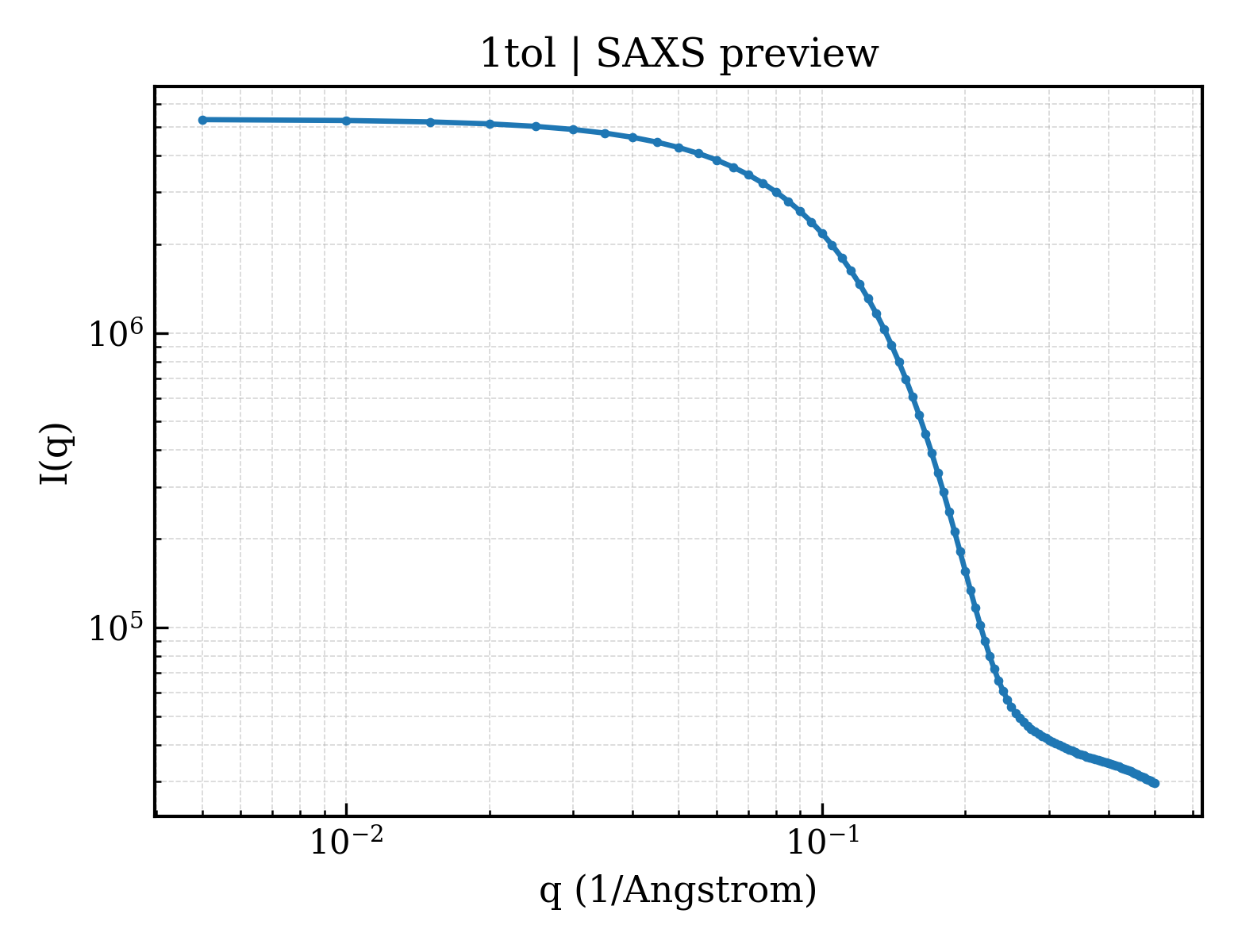
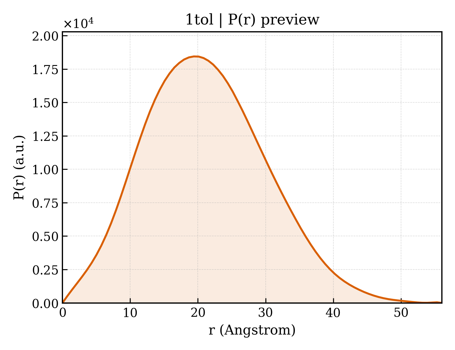
|
1S62
Solution structure of the Escherichia coli TolA C-terminal domain
Deposited 2004-01-22
|
Different construct
Different oligomeric state
Different experimental method
Different experimental conditions
Different structure-quality metrics
|
Assembly 1
Protein monomer
Monomer;Protein × 1
PDB declaration: monomeric
|
Chain A
325–421(97 aa)
Fragment:C-terminal domain (residues 325-421)
|
Not recorded
|
No recorded non-water small molecule
|
SOLUTION NMR
NMR measurement conditions
pH 6.8;300 K;Ionic strength (raw mmCIF value) 100mN NaCl, 50mM NaPO4;Pressure ambient
NMR measurement conditions
pH 6.8;300 K;Ionic strength (raw mmCIF value) 50mN NaCl, 50mM NaPO4;Pressure ambient
NMR measurement conditions
pH 6.8;300 K;Ionic strength (raw mmCIF value) 500mN NaCl, 50mM NaPO4;Pressure ambient
NMR sample composition
1.4mM U-15N, 100mN NaCl, 50mM NaPO4, 90% H2O, 10% D2O | 90% H2O/10% D2O
NMR sample composition
0.5mM U-15N,13C, 50mN NaCl, 50mM NaPO4, Complete protease inhibitor (Boehringer), 90% H2O, 10% D2O | 90% H2O/10% D2O
NMR sample composition
0.7mM U-15N, 500mN NaCl, 50mM NaPO4, 90% H2O, 10% D2O | 90% H2O/10% D2O
|
Resolution not provided
|
|
3QDP
Structural characterization of the interaction of colicin A, colicin N, and TolB with TolAIII translocon
Deposited 2011-01-19
|
Different construct
Different mutation/modification
Different oligomeric state
Different ligand/ion
Different experimental conditions
Different structure-quality metrics
|
Assembly 1
Protein monomer
Monomer;Protein × 1
PDB declaration: monomeric
|
Chain A
302–421(120 aa)
Fragment:TolA domain III, residues 302-421
|
Non-standard monomer:Yes (specific site not provided by mmCIF)
|
NO3 NITRATE ION × 2
|
X-RAY DIFFRACTION
X-ray crystallization conditions
VAPOR DIFFUSION, SITTING DROP;pH 5.28;293 K;0.2 M NaNO3, 2.2 M ammonium sulphate, pH 5.28, VAPOR DIFFUSION, SITTING DROP, temperature 293K
|
Resolution 2.15 Å
R-free 0.225
|
|
3QDR
Structural characterization of the interaction of colicin A, colicin N, and TolB with the TolAIII translocon
Deposited 2011-01-19
|
Different construct
Different mutation/modification
Different oligomeric state
Different ligand/ion
Different experimental conditions
Different structure-quality metrics
|
Assembly 1
Protein heterocomplex
Heteromer;Protein × 2
PDB declaration: dimeric
|
Chain A
302–421(120 aa)
Fragment:TolA domain III, residues 302-421
|
Non-standard monomer:Yes (specific site not provided by mmCIF)
|
TRS 2-AMINO-2-HYDROXYMETHYL-PROPANE-1,3-DIOL × 1
|
X-RAY DIFFRACTION
X-ray crystallization conditions
VAPOR DIFFUSION, SITTING DROP;pH 7.5;293 K;0.2 M K2HPO4, 2.2 M ammonium sulphate, pH 7.5, VAPOR DIFFUSION, SITTING DROP, temperature 293K
|
Resolution 2.65 Å
R-free 0.273
|
|
9DDM
E. coli TolAQR conformation I
Deposited 2024-08-28
|
Different construct
Different mutation/modification
Different oligomeric state
Different experimental method
Different experimental conditions
Different structure-quality metrics
|
Assembly 1
Protein heterocomplex
Heteromer;Protein × 9
PDB declaration: nonameric
|
Chain F
2–421(420 aa)
Chain G
2–421(420 aa)
|
Not recorded
|
No recorded non-water small molecule
|
ELECTRON MICROSCOPY
cryo-EM buffer
pH 7.5
cryo-EM vitrification conditions
Cryogen ETHANE
|
Resolution 2.94 Å
|
|
9DDN
E. coli TolAQR conformation II
Deposited 2024-08-28
|
Different construct
Different mutation/modification
Different oligomeric state
Different experimental method
Different experimental conditions
Different structure-quality metrics
|
Assembly 1
Protein heterocomplex
Heteromer;Protein × 9
PDB declaration: nonameric
|
Chain F
2–421(420 aa)
Chain G
2–421(420 aa)
|
Not recorded
|
No recorded non-water small molecule
|
ELECTRON MICROSCOPY
cryo-EM buffer
pH 7.5
cryo-EM vitrification conditions
Cryogen ETHANE
|
Resolution 3.18 Å
|
|
9K49
Cryo-EM structure of inner membrane TolQRA complex in CYMAL-6-Neopentyl Glycol detergent micelles
Deposited 2024-10-21
|
Different construct
Different oligomeric state
Different experimental method
Different experimental conditions
Different structure-quality metrics
|
Assembly 1
Protein heterocomplex
Heteromer;Protein × 8
PDB declaration: octameric
|
Chain H
1–421(421 aa)
|
Not recorded
|
No recorded non-water small molecule
|
ELECTRON MICROSCOPY
cryo-EM buffer
pH 7.5;Phosphate-buffered saline (PBS) buffer at pH 7.5 (20 mM PBS pH 7.5, 300 mM NaCl) with 20 mM Imidazole and 0.04 mM CYMAL-6 Neopentyl Glycol detergent
cryo-EM vitrification conditions
Cryogen ETHANE
|
Resolution 3.60 Å
|
|
9KCH
Cryo-EM structure of inner membrane TolQRA complex in CYMAL-6-Neopentyl Glycol detergent micelles
Deposited 2024-11-01
|
Different construct
Different oligomeric state
Different experimental method
Different experimental conditions
Different structure-quality metrics
|
Assembly 1
Protein heterocomplex
Heteromer;Protein × 8
PDB declaration: octameric
|
Chain H
1–421(421 aa)
|
Not recorded
|
No recorded non-water small molecule
|
ELECTRON MICROSCOPY
cryo-EM buffer
pH 7.5;Phosphate-buffered saline (PBS) buffer at pH 7.5 (20 mM PBS pH 7.5, 300 mM NaCl) with 20 mM Imidazole and 0.04 mM CYMAL-6 Neopentyl Glycol detergent
cryo-EM vitrification conditions
Cryogen ETHANE
|
Resolution 4.19 Å
|
|
9KPZ
Structure of TolQRA complex at pH 5.4 from E.coli
Deposited 2024-11-24
|
Different construct
Different mutation/modification
Different oligomeric state
Different ligand/ion
Different experimental method
Different experimental conditions
Different structure-quality metrics
|
Assembly 1
Insufficient information
Heteromer;Protein × 12
PDB declaration: dodecameric
|
Chain H
2–421(420 aa)
Chain I
2–421(420 aa)
Chain J
2–421(420 aa)
Chain K
2–421(420 aa)
Chain L
2–421(420 aa)
|
Not recorded
|
3PE 1,2-Distearoyl-sn-glycerophosphoethanolamine × 7
|
ELECTRON MICROSCOPY
cryo-EM buffer
pH 5.4
cryo-EM vitrification conditions
Cryogen ETHANE
|
Resolution 3.18 Å
|
|
9KQ0
Structure of TolQRA complex at pH 8.0 from E.coli
Deposited 2024-11-24
|
Different construct
Different mutation/modification
Different oligomeric state
Different experimental method
Different experimental conditions
Different structure-quality metrics
|
Assembly 1
Insufficient information
Heteromer;Protein × 12
PDB declaration: dodecameric
|
Chain H
2–421(420 aa)
Chain I
2–421(420 aa)
Chain J
2–421(420 aa)
Chain K
2–421(420 aa)
Chain L
2–421(420 aa)
|
Not recorded
|
No recorded non-water small molecule
|
ELECTRON MICROSCOPY
cryo-EM buffer
pH 8
cryo-EM vitrification conditions
Cryogen ETHANE
|
Resolution 3.60 Å
|
|
9QUQ
cryo-EM structure of TolQR conformation2 in SMA nanodiscs
Deposited 2025-04-11
|
Different construct
Different oligomeric state
Different experimental method
Different experimental conditions
Different structure-quality metrics
|
Assembly 1
Protein heterocomplex
Heteromer;Protein × 8
PDB declaration: octameric
|
Chain H
1–421(421 aa)
|
Not recorded
|
No recorded non-water small molecule
|
ELECTRON MICROSCOPY
cryo-EM buffer
pH 7.8
cryo-EM vitrification conditions
Cryogen ETHANE
|
Resolution 3.28 Å
|
|
9QVD
cryo-EM structure of TolQRA in nanodiscs
Deposited 2025-04-11
|
Different construct
Different mutation/modification
Different oligomeric state
Different experimental method
Different experimental conditions
Different structure-quality metrics
|
Assembly 1
Protein heterocomplex
Heteromer;Protein × 10
PDB declaration: decameric
|
Chain H
1–421(421 aa)
Chain I
1–421(421 aa)
Chain J
1–421(421 aa)
|
Not recorded
|
No recorded non-water small molecule
|
ELECTRON MICROSCOPY
cryo-EM buffer
pH 7.8
cryo-EM vitrification conditions
Cryogen ETHANE
|
Resolution 3.52 Å
|