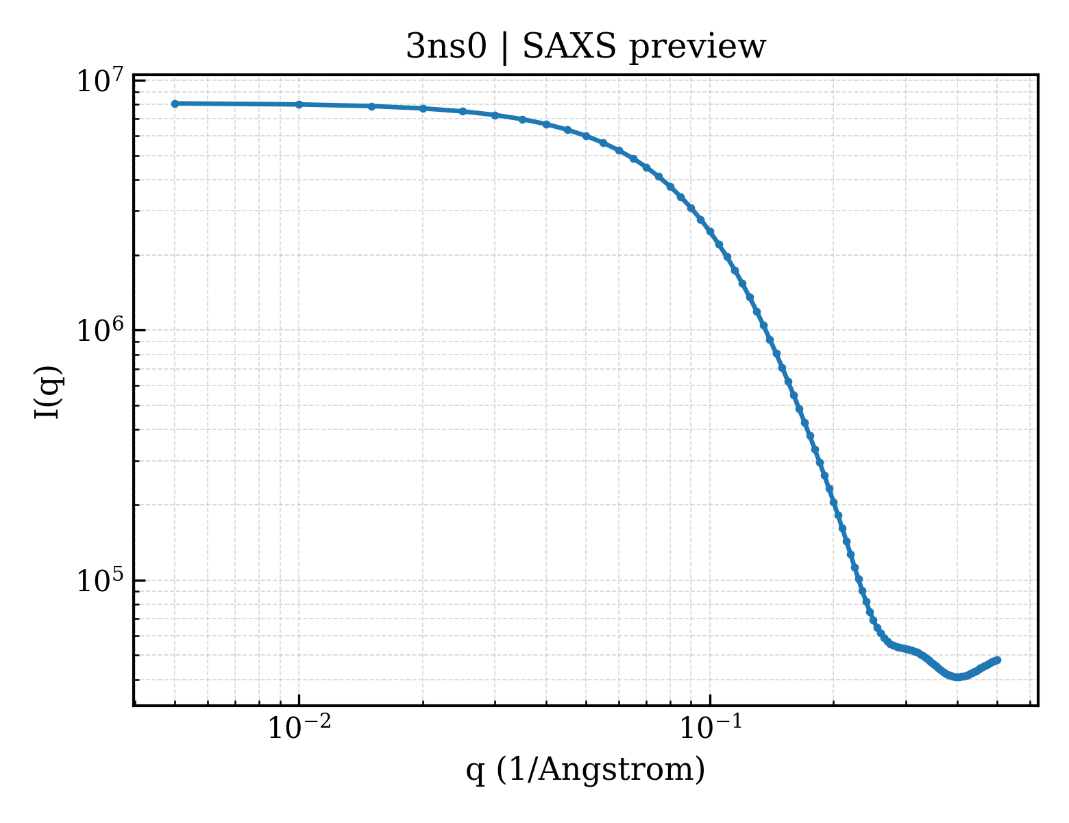
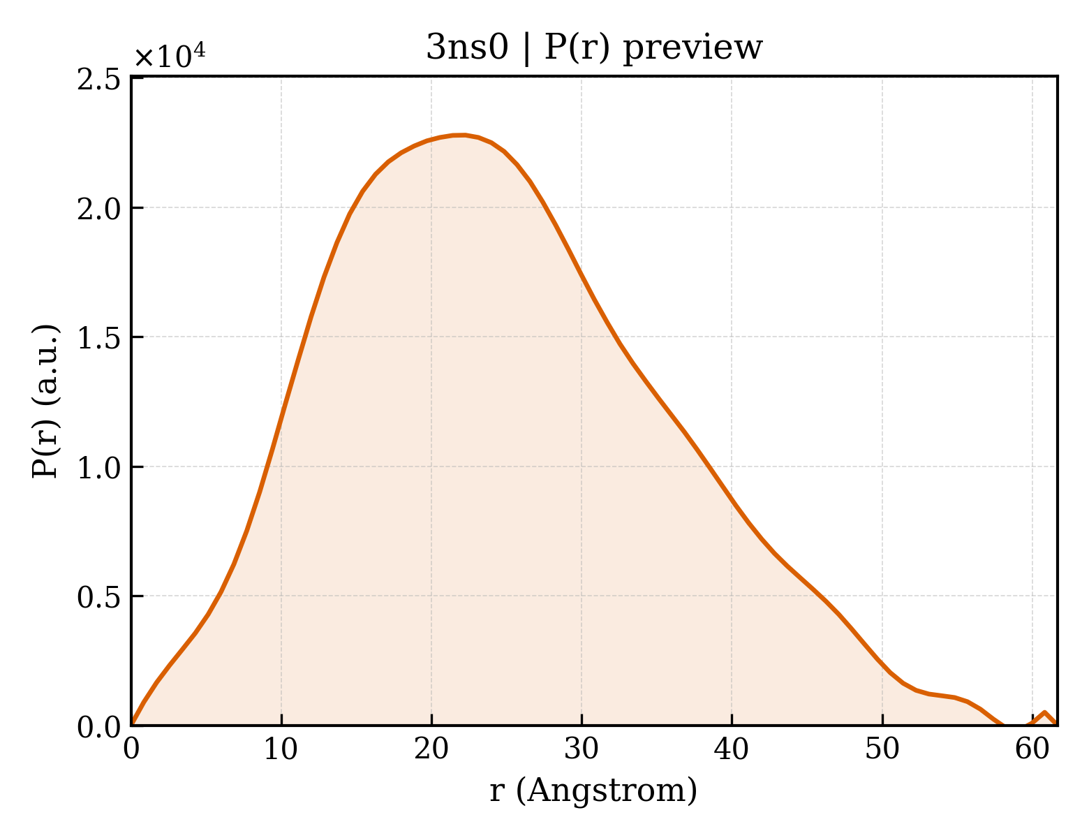
Bacteriorhodopsin
OrganismNot specified
State in the Current Structure
| Assembly | Oligomeric State | Construct | Mutations and Modifications | Ligands, Ions and Associated Components | Method and Experimental Conditions | Structure Quality |
|---|---|---|---|---|---|---|
| 1 | Protein homooligomer Homooligomer Protein × 3 PDB declaration: trimeric(3) Consistent with protein copy count | Chain A; UniProt 14–261 | Fragment:UNP residues 14-261 | RET RETINAL × 3 LI1 1-[2,6,10.14-TETRAMETHYL-HEXADECAN-16-YL]-2-[2,10,14-TRIMETHYLHEXADECAN-16-YL]GLYCEROL × 27 | X-RAY DIFFRACTION X-ray crystallization conditions:pH 5.6;293 K;monoolein meso phase, K/Na-Pi as a precipitant, pH 5.6, temperature 293K | Resolution 1.78 Å R-free 0.229 |
Other States of the Same Protein in the Database
Each row is a biological assembly of the same UniProt protein in another PDB entry. The “Difference from current entry” column identifies evidence-level differences; no tag means the currently parsed fields agree.
| Other PDB | Difference from Current Entry 3NS0 | Assembly / Oligomeric State | Construct | Mutations and Modifications | Ligands, Ions and Non-polymers | Method and Experimental Conditions | Structure Quality |
|---|---|---|---|---|---|---|---|
| 1AP9 X-RAY STRUCTURE OF BACTERIORHODOPSIN FROM MICROCRYSTALS GROWN IN LIPIDIC CUBIC PHASES Deposited 1997-07-26 | Different construct Different ligand/ion Different experimental conditions Different structure-quality metrics | Assembly 1 Protein homooligomer Homooligomer;Protein × 3 PDB declaration: trimeric |
Chain A
14–261(248 aa)
|
Not recorded | RET RETINAL × 3 |
X-RAY DIFFRACTION
X-ray crystallization conditions
pH 5.6;PROTEIN FROM THE PURPLE MEMBRANE WAS DELIPIDATED AND SOLUBILIZED IN OCTYL GLUCOSIDE. PROTEIN WAS CRYSTALLIZED FROM 60 - 70% (W/W) MONOOLEIN, 0.7 - 4.0 M NA/K - PHOSPHATE IN A PHOSPHATE BUFFER AT PH 5.6, AT 20C AND IN THE DARK. THE MIXTURE WAS CENTRIFUGED AT 10000G FOR 150 MN PRIOR TO CRYSTALLISATION.
|
Resolution 2.35 Å R-free 0.317 |
| 1AT9 STRUCTURE OF BACTERIORHODOPSIN AT 3.0 ANGSTROM DETERMINED BY ELECTRON CRYSTALLOGRAPHY Deposited 1997-08-20 | Different construct Different mutation/modification Different ligand/ion Different experimental method Different experimental conditions Different structure-quality metrics | Assembly 1 Protein homooligomer Homooligomer;Protein × 3 PDB declaration: trimeric |
Chain A
15–261(247 aa)
|
Non-standard monomer:Yes (specific site not provided by mmCIF) | RET RETINAL × 3 |
ELECTRON CRYSTALLOGRAPHY
cryo-EM buffer
pH 5.5;0.4 M citric acid Na2HPO4, 3% trehalose
cryo-EM vitrification conditions
Cryogen ETHANE;delay of 10 seconds before rapid freezing
|
Resolution 2.80 Å |
| 1BCT THREE-DIMENSIONAL STRUCTURE OF PROTEOLYTIC FRAGMENT 163-231 OF BACTERIOOPSIN DETERMINED FROM NUCLEAR MAGNETIC RESONANCE DATA IN SOLUTION Deposited 1993-07-07 | Different construct Different oligomeric state Different ligand/ion Different experimental method Different experimental conditions Different structure-quality metrics | Assembly 1 Protein monomer Monomer;Protein × 1 PDB declaration: monomeric |
Chain A
176–244(69 aa)
|
Not recorded | No recorded non-water small molecule | SOLUTION NMR mmCIF provides none of the parsed conditions | Resolution not provided |
| 1BHA THREE-DIMENSIONAL STRUCTURE OF (1-71) BACTERIOOPSIN SOLUBILIZED IN METHANOL-CHLOROFORM AND SDS MICELLES DETERMINED BY 15N-1H HETERONUCLEAR NMR SPECTROSCOPY Deposited 1993-10-11 | Different construct Different oligomeric state Different ligand/ion Different experimental method Different experimental conditions Different structure-quality metrics | Assembly 1 Protein monomer Monomer;Protein × 1 PDB declaration: monomeric |
Chain A
14–84(71 aa)
|
Not recorded | No recorded non-water small molecule | SOLUTION NMR mmCIF provides none of the parsed conditions | Resolution not provided |
| 1BHB Three-dimensional structure of (1-71) bacterioopsin solubilized in methanol-chloroform and SDS micelles determined by 15N-1H heteronuclear NMR spectroscopy Deposited 1993-10-11 | Different construct Different oligomeric state Different ligand/ion Different experimental method Different experimental conditions Different structure-quality metrics | Assembly 1 Protein monomer Monomer;Protein × 1 PDB declaration: monomeric |
Chain A
14–84(71 aa)
|
Not recorded | No recorded non-water small molecule | SOLUTION NMR mmCIF provides none of the parsed conditions | Resolution not provided |
| 1BM1 CRYSTAL STRUCTURE OF BACTERIORHODOPSIN IN THE LIGHT-ADAPTED STATE Deposited 1998-07-28 | Different construct Different mutation/modification Different ligand/ion Different experimental conditions Different structure-quality metrics | Assembly 1 Protein homooligomer Homooligomer;Protein × 3 PDB declaration: trimeric |
Chain A
14–253(240 aa)
|
Non-standard monomer:Yes (specific site not provided by mmCIF) | RET RETINAL × 3 DPG PHOSPHORIC ACID 2,3-BIS-(3,7,11,15-TETRAMETHYL-HEXADECYLOXY)-PROPYL ESTER 2-HYDROXO-3-PHOSPHONOOXY-PROPYL ESTER × 3 |
X-RAY DIFFRACTION
X-ray crystallization conditions
pH 5.2;pH 5.2
|
Resolution 3.50 Å R-free 0.311 |
| 1BRD Model for the structure of Bacteriorhodopsin based on high-resolution Electron Cryo-microscopy Deposited 1990-05-23 | Different construct Different mutation/modification Different ligand/ion Different experimental method Different experimental conditions Different structure-quality metrics | Assembly 1 Protein homooligomer Homooligomer;Protein × 3 PDB declaration: trimeric |
Chain A
15–261(247 aa)
|
Non-standard monomer:Yes (specific site not provided by mmCIF) | RET RETINAL × 3 |
ELECTRON CRYSTALLOGRAPHY
cryo-EM buffer
pH 5.2;0.1 mM potassium phosphate, 6 mM octyl glucoside, 0.2 mM trimethylammonium chloride
|
Resolution 3.50 Å |
| 1BRR X-RAY STRUCTURE OF THE BACTERIORHODOPSIN TRIMER/LIPID COMPLEX Deposited 1998-07-28 | Different construct Different mutation/modification Different oligomeric state Different ligand/ion Different experimental conditions Different structure-quality metrics | Assembly 1 Other combination Homooligomer;Protein × 3 PDB declaration: trimeric |
Chain A
14–260(247 aa)
Chain B
14–260(247 aa)
Chain C
14–260(247 aa)
|
Non-standard monomer:Yes (specific site not provided by mmCIF) Non-standard monomer:Yes (specific site not provided by mmCIF) Non-standard monomer:Yes (specific site not provided by mmCIF) | RET RETINAL × 3 ARC 3,7,11,15-TETRAMETHYL-HEXADECAN-1-OL × 10 GOL GLYCEROL × 3 BGC beta-D-glucopyranose × 1 OCT N-OCTANE × 1 |
X-RAY DIFFRACTION
X-ray crystallization conditions
pH 5.2;SEE REFERENCE 2, pH 5.2
|
Resolution 2.90 Å R-free 0.299 |
| 1BRX BACTERIORHODOPSIN/LIPID COMPLEX Deposited 1998-05-28 | Different construct Different mutation/modification Different ligand/ion Different experimental conditions Different structure-quality metrics | Assembly 1 Protein homooligomer Homooligomer;Protein × 3 PDB declaration: trimeric |
Chain A
14–253(240 aa)
|
Non-standard monomer:Yes (specific site not provided by mmCIF) | RET RETINAL × 3 |
X-RAY DIFFRACTION
X-ray crystallization conditions
pH 5.6;pH 5.6
|
Resolution 2.30 Å R-free 0.298 |
| 1C3W BACTERIORHODOPSIN/LIPID COMPLEX AT 1.55 A RESOLUTION Deposited 1999-07-28 | Different construct Different ligand/ion Different experimental conditions Different structure-quality metrics | Assembly 1 Protein homooligomer Homooligomer;Protein × 3 PDB declaration: trimeric |
Chain A
5–231(227 aa)
|
Not recorded | LI1 1-[2,6,10.14-TETRAMETHYL-HEXADECAN-16-YL]-2-[2,10,14-TRIMETHYLHEXADECAN-16-YL]GLYCEROL × 39 SQU 2,10,23-TRIMETHYL-TETRACOSANE × 3 RET RETINAL × 3 |
X-RAY DIFFRACTION
X-ray crystallization conditions
CUBIC LIPID PHASE;pH 5.6;293 K;MO:WATER:PHOSPHATE, pH 5.6, CUBIC LIPID PHASE, temperature 20K
|
Resolution 1.55 Å R-free 0.225 |
| 1C8R BACTERIORHODOPSIN D96N BR STATE AT 2.0 A RESOLUTION Deposited 1999-07-29 | Different construct Different mutation/modification Different ligand/ion Different experimental conditions Different structure-quality metrics | Assembly 1 Protein homooligomer Homooligomer;Protein × 3 PDB declaration: trimeric |
Chain A
14–262(249 aa)
Fragment:;"BR" STATE INTERMEDIATE
;
|
Mutation:YES | LI1 1-[2,6,10.14-TETRAMETHYL-HEXADECAN-16-YL]-2-[2,10,14-TRIMETHYLHEXADECAN-16-YL]GLYCEROL × 39 SQU 2,10,23-TRIMETHYL-TETRACOSANE × 3 RET RETINAL × 3 |
X-RAY DIFFRACTION
X-ray crystallization conditions
CUBIC LIPID PHASE;pH 5.6;296 K;MO:WATER:PHOSPHATE, pH 5.6, CUBIC LIPID PHASE, temperature 296K
|
Resolution 1.80 Å R-free 0.182 |
| 1C8S BACTERIORHODOPSIN D96N LATE M STATE INTERMEDIATE Deposited 1999-07-29 | Different construct Different mutation/modification Different ligand/ion Different experimental conditions Different structure-quality metrics | Assembly 1 Protein homooligomer Homooligomer;Protein × 3 PDB declaration: trimeric |
Chain A
5–222(218 aa)
|
Mutation:D96N | LI1 1-[2,6,10.14-TETRAMETHYL-HEXADECAN-16-YL]-2-[2,10,14-TRIMETHYLHEXADECAN-16-YL]GLYCEROL × 33 SQU 2,10,23-TRIMETHYL-TETRACOSANE × 3 RET RETINAL × 3 |
X-RAY DIFFRACTION
X-ray crystallization conditions
CUBIC LIPID PHASE;pH 5.6;296 K;MO:WATER:PHOSPHATE, pH 5.6, CUBIC LIPID PHASE, temperature 296K
|
Resolution 2.00 Å R-free 0.219 |
| 1CWQ M INTERMEDIATE STRUCTURE OF THE WILD TYPE BACTERIORHODOPSIN IN COMBINATION WITH THE GROUND STATE STRUCTURE Deposited 1999-08-26 | Different construct Different mutation/modification Different oligomeric state Different ligand/ion Different experimental conditions Different structure-quality metrics | Assembly 1 Protein homooligomer Homooligomer;Protein × 6 PDB declaration: hexameric |
Chain A
2–247(246 aa)
Chain B
2–247(246 aa)
|
Not recorded | RET RETINAL × 6 UND UNDECANE × 24 OCT N-OCTANE × 48 HEX HEXANE × 36 TRD TRIDECANE × 18 |
X-RAY DIFFRACTION
X-ray crystallization conditions
pH 5.6;CUBIC LIPID PHASE 2.5M PHOSPHATE, pH 5.60
|
Resolution 2.25 Å R-free 0.236 |
| 1DZE Structure of the M Intermediate of Bacteriorhodopsin trapped at 100K Deposited 2000-02-25 | Different construct Different oligomeric state Different ligand/ion Different experimental conditions Different structure-quality metrics | Assembly 1 Other combination Homooligomer;Protein × 3 PDB declaration: trimeric |
Chain A
14–261(248 aa)
|
Not recorded | RET RETINAL × 3 L1P 3-PHOSPHORYL-[1,2-DI-PHYTANYL]GLYCEROL × 3 L2P 2,3-DI-PHYTANYL-GLYCEROL × 6 L3P 2,3-DI-O-PHYTANLY-3-SN-GLYCERO-1-PHOSPHORYL-3'-SN-GLYCEROL-1'-PHOSPHATE × 3 L4P 3-[GLYCEROLYLPHOSPHONYL]-[1,2-DI-PHYTANYL]GLYCEROL × 3 |
X-RAY DIFFRACTION
X-ray crystallization conditions
VAPOR DIFFUSION;pH 5.2;THE CRYSTALLIZATION CONSISTS OF TWO STEPS. FIRST, A MIXTURE OF 5 MG/ML PURPLE MEMBRANE, 0.25% OTG, 1 M AMMONIUM SULFATE, 0.16 M NACL, 0.04 M SODIUM CITRATE (PH5.2) , 0.04% NAN3 WAS INCUBATED AND 15% TREHALOSE AT 305K FOR 5 DAYS. THIS RESULTED IN THE FORMATION OF SPHERICAL VESICLES WITH A DIAMETER OF 50 NM. AFTER SEDIMENTAL MATERIALS WERE REMOVED BY CENTRIFUGATION (4000G X 10 MIN), A SUSPENSION OF THE SPHERICAL VESICLES WAS COOLED TO 278K AND CONCENTRATED BY VAPOR DIFFUSION AGAINST A RESERVOIR SOLUTION CONTAINING 2.0 M AMMONIUM SULFATE 0.08M SODIUM CITRATE (PH 5.2) AND 30% TREHALOSE. INCUBATION FOR A COUPLE OF MONTHS YIELDED HEXAGONAL CRYSTALS.
|
Resolution 2.50 Å R-free 0.283 |
| 1E0P L intermediate of bacteriorhodopsin Deposited 2000-04-04 | Different construct Different ligand/ion Different experimental conditions Different structure-quality metrics | Assembly 1 Protein homooligomer Homooligomer;Protein × 3 PDB declaration: trimeric |
Chain A
18–245(228 aa)
|
Not recorded | RET RETINAL × 3 |
X-RAY DIFFRACTION
X-ray crystallization conditions
pH 5.6;PROTEIN FROM THE PURPLE MEMBRANE WAS RESOLVED IN OCTYL GLUC, pH 5.60
|
Resolution 2.10 Å R-free 0.288 |
| 1F4Z BACTERIORHODOPSIN-M PHOTOINTERMEDIATE STATE OF THE E204Q MUTANT AT 1.8 ANGSTROM RESOLUTION Deposited 2000-06-10 | Different construct Different mutation/modification Different ligand/ion Different experimental conditions Different structure-quality metrics | Assembly 1 Protein homooligomer Homooligomer;Protein × 3 PDB declaration: trimeric |
Chain A
18–244(227 aa)
|
Mutation:E204Q | LI1 1-[2,6,10.14-TETRAMETHYL-HEXADECAN-16-YL]-2-[2,10,14-TRIMETHYLHEXADECAN-16-YL]GLYCEROL × 39 SQU 2,10,23-TRIMETHYL-TETRACOSANE × 3 RET RETINAL × 3 |
X-RAY DIFFRACTION
X-ray crystallization conditions
Cubic lipid phase;pH 5.6;295 K;MO:WATER:PHOSPHATE, pH 5.6, Cubic lipid phase, temperature 295K
|
Resolution 1.80 Å R-free 0.203 |
| 1F50 BACTERIORHODOPSIN-BR STATE OF THE E204Q MUTANT AT 1.7 ANGSTROM RESOLUTION Deposited 2000-06-10 | Different construct Different mutation/modification Different ligand/ion Different experimental conditions Different structure-quality metrics | Assembly 1 Protein homooligomer Homooligomer;Protein × 3 PDB declaration: trimeric |
Chain A
18–244(227 aa)
|
Mutation:E204Q | LI1 1-[2,6,10.14-TETRAMETHYL-HEXADECAN-16-YL]-2-[2,10,14-TRIMETHYLHEXADECAN-16-YL]GLYCEROL × 39 SQU 2,10,23-TRIMETHYL-TETRACOSANE × 3 RET RETINAL × 3 |
X-RAY DIFFRACTION
X-ray crystallization conditions
Cubic lipid phase;pH 5.6;295 K;MO:WATER:PHOSPHATE, pH 5.6, Cubic lipid phase, temperature 295K
|
Resolution 1.70 Å R-free 0.191 |
| 1FBB CRYSTAL STRUCTURE OF NATIVE CONFORMATION OF BACTERIORHODOPSIN Deposited 2000-07-15 | Different construct Different ligand/ion Different experimental method Different experimental conditions Different structure-quality metrics | Assembly 1 Protein homooligomer Homooligomer;Protein × 3 PDB declaration: trimeric |
Chain A
14–261(248 aa)
|
Not recorded | RET RETINAL × 3 |
ELECTRON CRYSTALLOGRAPHY
X-ray crystallization conditions
naturally occurring in vivo;pH 7;310 K;crystal size is increased by fusion and annealing using detergents, pH 7, naturally occurring in vivo, temperature 37K
|
Resolution 3.20 Å R-free 0.310 |
| 1FBK CRYSTAL STRUCTURE OF CYTOPLASMICALLY OPEN CONFORMATION OF BACTERIORHODOPSIN Deposited 2000-07-15 | Different construct Different mutation/modification Different ligand/ion Different experimental method Different experimental conditions Different structure-quality metrics | Assembly 1 Protein homooligomer Homooligomer;Protein × 3 PDB declaration: trimeric |
Chain A
14–261(248 aa)
|
Mutation:D96G,F171C,F219L | RET RETINAL × 3 |
ELECTRON CRYSTALLOGRAPHY
X-ray crystallization conditions
naturally occurring in vivo;pH 7;310 K;crystals are increased in size by fusion and annealing using detergents, pH 7, naturally occurring in vivo,
temperature 37K
|
Resolution 3.20 Å R-free 0.321 |
| 1IW6 Crystal Structure of the Ground State of Bacteriorhodopsin Deposited 2002-04-22 | Different construct Different oligomeric state Different ligand/ion Different experimental conditions Different structure-quality metrics | Assembly 1 Other combination Homooligomer;Protein × 3 PDB declaration: trimeric |
Chain A
14–261(248 aa)
|
Not recorded | RET RETINAL × 3 L3P 2,3-DI-O-PHYTANLY-3-SN-GLYCERO-1-PHOSPHORYL-3'-SN-GLYCEROL-1'-PHOSPHATE × 12 L2P 2,3-DI-PHYTANYL-GLYCEROL × 3 |
X-RAY DIFFRACTION
X-ray crystallization conditions
VAPOR DIFFUSION, SITTING DROP;pH 5.2;293 K;ammonium sulfate, pH 5.2, VAPOR DIFFUSION, SITTING DROP, temperature 293K
|
Resolution 2.30 Å R-free 0.275 |
| 1IW9 Crystal Structure of the M Intermediate of Bacteriorhodopsin Deposited 2002-04-25 | Different construct Different oligomeric state Different ligand/ion Different experimental conditions Different structure-quality metrics | Assembly 1 Other combination Homooligomer;Protein × 3 PDB declaration: trimeric |
Chain A
14–261(248 aa)
|
Not recorded | RET RETINAL × 3 L3P 2,3-DI-O-PHYTANLY-3-SN-GLYCERO-1-PHOSPHORYL-3'-SN-GLYCEROL-1'-PHOSPHATE × 12 L2P 2,3-DI-PHYTANYL-GLYCEROL × 3 |
X-RAY DIFFRACTION
X-ray crystallization conditions
VAPOR DIFFUSION, SITTING DROP;pH 5.2;293 K;ammonium sulfate, pH 5.2, VAPOR DIFFUSION, SITTING DROP, temperature 293K
|
Resolution 2.50 Å R-free 0.271 |
| 1IXF Crystal Structure of the K intermediate of bacteriorhodopsin Deposited 2002-06-20 | Different construct Different oligomeric state Different ligand/ion Different experimental conditions Different structure-quality metrics | Assembly 1 Other combination Homooligomer;Protein × 3 PDB declaration: trimeric |
Chain A
14–261(248 aa)
|
Not recorded | RET RETINAL × 3 L3P 2,3-DI-O-PHYTANLY-3-SN-GLYCERO-1-PHOSPHORYL-3'-SN-GLYCEROL-1'-PHOSPHATE × 12 L2P 2,3-DI-PHYTANYL-GLYCEROL × 3 |
X-RAY DIFFRACTION
X-ray crystallization conditions
VAPOR DIFFUSION, SITTING DROP;pH 5.2;293 K;ammonium sulfate, pH 5.2, VAPOR DIFFUSION, SITTING DROP, temperature 293K
|
Resolution 2.60 Å R-free 0.321 |
| 1JV6 BACTERIORHODOPSIN D85S/F219L DOUBLE MUTANT AT 2.00 ANGSTROM RESOLUTION Deposited 2001-08-28 | Different construct Different mutation/modification Different oligomeric state Different ligand/ion Different experimental conditions Different structure-quality metrics | Assembly 1 Protein monomer Monomer;Protein × 1 PDB declaration: monomeric |
Chain A
14–262(249 aa)
|
Mutation:D85S,F219L | RET RETINAL × 1 LI1 1-[2,6,10.14-TETRAMETHYL-HEXADECAN-16-YL]-2-[2,10,14-TRIMETHYLHEXADECAN-16-YL]GLYCEROL × 8 |
X-RAY DIFFRACTION
X-ray crystallization conditions
cubic lipid phase;pH 5.6;293 K;monoolein, octyl-beta-D-glucopyranoside, pH 5.6, cubic lipid phase, temperature 293K
|
Resolution 2.00 Å R-free 0.240 |
| 1JV7 BACTERIORHODOPSIN O-LIKE INTERMEDIATE STATE OF THE D85S MUTANT AT 2.25 ANGSTROM RESOLUTION Deposited 2001-08-28 | Different construct Different mutation/modification Different oligomeric state Different ligand/ion Different experimental conditions Different structure-quality metrics | Assembly 1 Protein monomer Monomer;Protein × 1 PDB declaration: monomeric |
Chain A
14–262(249 aa)
|
Mutation:D85S | RET RETINAL × 1 LI1 1-[2,6,10.14-TETRAMETHYL-HEXADECAN-16-YL]-2-[2,10,14-TRIMETHYLHEXADECAN-16-YL]GLYCEROL × 9 |
X-RAY DIFFRACTION
X-ray crystallization conditions
cubic lipid phase;pH 5.6;293 K;monoolein, octyl-beta-D-glucopyranoside, pH 5.6, cubic lipid phase, temperature 293K
|
Resolution 2.25 Å R-free 0.244 |
| 1KG8 X-ray structure of an early-M intermediate of bacteriorhodopsin Deposited 2001-11-26 | Different construct Different ligand/ion Different experimental conditions Different structure-quality metrics | Assembly 1 Protein homooligomer Homooligomer;Protein × 3 PDB declaration: trimeric |
Chain A
13–243(231 aa)
|
Not recorded | RET RETINAL × 3 LI1 1-[2,6,10.14-TETRAMETHYL-HEXADECAN-16-YL]-2-[2,10,14-TRIMETHYLHEXADECAN-16-YL]GLYCEROL × 9 |
X-RAY DIFFRACTION
X-ray crystallization conditions
mono-olein cubic phase;pH 5.6;293 K;3.0M Na/K Phosphate, pH 5.6, mono-olein cubic phase, temperature 293K
|
Resolution 2.00 Å R-free 0.217 |
| 1KG9 Structure of a "mock-trapped" early-M intermediate of bacteriorhosopsin Deposited 2001-11-26 | Different construct Different experimental conditions Different structure-quality metrics | Assembly 1 Protein homooligomer Homooligomer;Protein × 3 PDB declaration: trimeric |
Chain A
13–243(231 aa)
|
Not recorded | RET RETINAL × 3 LI1 1-[2,6,10.14-TETRAMETHYL-HEXADECAN-16-YL]-2-[2,10,14-TRIMETHYLHEXADECAN-16-YL]GLYCEROL × 27 |
X-RAY DIFFRACTION
X-ray crystallization conditions
mono-olein cubic phase;pH 5.6;293 K;3.0M Na/K Phosphate, pH 5.6, mono-olein cubic phase, temperature 293K
|
Resolution 1.81 Å R-free 0.212 |
| 1KGB structure of ground-state bacteriorhodopsin Deposited 2001-11-26 | Different construct Different ligand/ion Different experimental conditions Different structure-quality metrics | Assembly 1 Protein homooligomer Homooligomer;Protein × 3 PDB declaration: trimeric |
Chain A
13–243(231 aa)
|
Not recorded | RET RETINAL × 3 LI1 1-[2,6,10.14-TETRAMETHYL-HEXADECAN-16-YL]-2-[2,10,14-TRIMETHYLHEXADECAN-16-YL]GLYCEROL × 39 |
X-RAY DIFFRACTION
X-ray crystallization conditions
mono-olein cubic phase;pH 5.6;3.0M Na/K Phosphate, pH 5.6, mono-olein cubic phase, temperature 100K
|
Resolution 1.65 Å R-free 0.188 |
| 1KME CRYSTAL STRUCTURE OF BACTERIORHODOPSIN CRYSTALLIZED FROM BICELLES Deposited 2001-12-14 | Different construct Different oligomeric state Different ligand/ion Different experimental conditions Different structure-quality metrics | Assembly 1 Protein monomer Monomer;Protein × 1 PDB declaration: monomeric |
Chain A
14–244(231 aa)
Fragment:Residues 14-244
|
Not recorded | RET RETINAL × 1 SQU 2,10,23-TRIMETHYL-TETRACOSANE × 2 |
X-RAY DIFFRACTION
X-ray crystallization conditions
VAPOR DIFFUSION, HANGING DROP, Bicelle Method;pH 3.5;310 K;sodium phosphate, DMPC, Chapso, pH 3.5, VAPOR DIFFUSION, HANGING DROP, Bicelle Method, temperature 310.0K
|
Resolution 2.00 Å R-free 0.275 |
| 1KME CRYSTAL STRUCTURE OF BACTERIORHODOPSIN CRYSTALLIZED FROM BICELLES Deposited 2001-12-14 | Different construct Different oligomeric state Different ligand/ion Different experimental conditions Different structure-quality metrics | Assembly 2 Protein monomer Monomer;Protein × 1 PDB declaration: monomeric |
Chain B
14–244(231 aa)
Fragment:Residues 14-244
|
Not recorded | RET RETINAL × 1 BGC beta-D-glucopyranose × 2 |
X-RAY DIFFRACTION
X-ray crystallization conditions
VAPOR DIFFUSION, HANGING DROP, Bicelle Method;pH 3.5;310 K;sodium phosphate, DMPC, Chapso, pH 3.5, VAPOR DIFFUSION, HANGING DROP, Bicelle Method, temperature 310.0K
|
Resolution 2.00 Å R-free 0.275 |
| 1L0M Solution structure of Bacteriorhodopsin Deposited 2002-02-11 | Different construct Different mutation/modification Different oligomeric state Different ligand/ion Different experimental method Different experimental conditions Different structure-quality metrics | Assembly 1 Protein monomer Monomer;Protein × 1 PDB declaration: monomeric |
Chain A
20–231(212 aa)
|
Mutation:part of helix G absent | No recorded non-water small molecule |
SOLUTION NMR
NMR measurement conditions
pH 6;293 K;Pressure 1
NMR sample composition
chmeically synthesized peptides | DMSO
|
Resolution not provided |
| 1M0K BACTERIORHODOPSIN K INTERMEDIATE AT 1.43 A RESOLUTION Deposited 2002-06-13 | Different construct Different ligand/ion Different experimental conditions Different structure-quality metrics | Assembly 1 Protein homooligomer Homooligomer;Protein × 3 PDB declaration: trimeric |
Chain A
1–262(262 aa)
|
Not recorded | RET RETINAL × 3 LI1 1-[2,6,10.14-TETRAMETHYL-HEXADECAN-16-YL]-2-[2,10,14-TRIMETHYLHEXADECAN-16-YL]GLYCEROL × 39 SQU 2,10,23-TRIMETHYL-TETRACOSANE × 3 |
X-RAY DIFFRACTION
X-ray crystallization conditions
cubic lipid phase;pH 5.6;295 K;cubic lipid phase with mono-olein and potassium phosphate, pH 5.60, temperature 295K
|
Resolution 1.43 Å R-free 0.176 |
| 1M0L BACTERIORHODOPSIN/LIPID COMPLEX AT 1.47 A RESOLUTION Deposited 2002-06-13 | Different construct Different ligand/ion Different experimental conditions Different structure-quality metrics | Assembly 1 Protein homooligomer Homooligomer;Protein × 3 PDB declaration: trimeric |
Chain A
1–262(262 aa)
|
Not recorded | LI1 1-[2,6,10.14-TETRAMETHYL-HEXADECAN-16-YL]-2-[2,10,14-TRIMETHYLHEXADECAN-16-YL]GLYCEROL × 39 SQU 2,10,23-TRIMETHYL-TETRACOSANE × 3 RET RETINAL × 3 |
X-RAY DIFFRACTION
X-ray crystallization conditions
CUBIC LIPID PHASE;pH 5.6;295 K;CUBIC LIPID PHASE WITH MONO-OLEIN, PRECIPITANT PHOSPHATE, pH 5.60, temperature 295K
|
Resolution 1.47 Å R-free 0.179 |
| 1M0M BACTERIORHODOPSIN M1 INTERMEDIATE AT 1.43 A RESOLUTION Deposited 2002-06-13 | Different construct Different ligand/ion Different experimental conditions Different structure-quality metrics | Assembly 1 Protein homooligomer Homooligomer;Protein × 3 PDB declaration: trimeric |
Chain A
1–262(262 aa)
|
Not recorded | RET RETINAL × 3 LI1 1-[2,6,10.14-TETRAMETHYL-HEXADECAN-16-YL]-2-[2,10,14-TRIMETHYLHEXADECAN-16-YL]GLYCEROL × 39 SQU 2,10,23-TRIMETHYL-TETRACOSANE × 3 |
X-RAY DIFFRACTION
X-ray crystallization conditions
CUBIC LIPID PHASE;pH 5.6;295 K;cubic lipid phase with mono-olein and potassium phosphate, pH 5.60, CUBIC LIPID PHASE, temperature 295K
|
Resolution 1.43 Å R-free 0.213 |
| 1MGY Structure of the D85S mutant of bacteriorhodopsin with bromide bound Deposited 2002-08-16 | Different construct Different mutation/modification Different oligomeric state Different ligand/ion Different experimental conditions Different structure-quality metrics | Assembly 1 Protein monomer Monomer;Protein × 1 PDB declaration: monomeric |
Chain A
14–262(249 aa)
|
Mutation:D85S | BR BROMIDE ION × 2 K POTASSIUM ION × 1 RET RETINAL × 1 LI1 1-[2,6,10.14-TETRAMETHYL-HEXADECAN-16-YL]-2-[2,10,14-TRIMETHYLHEXADECAN-16-YL]GLYCEROL × 8 |
X-RAY DIFFRACTION
X-ray crystallization conditions
connected bilayer gel;pH 4.6;293 K;Sodium Acetate, PEG 4000, potassium chloride, mono-olein, pH 4.6, connected bilayer gel, temperature 293K
|
Resolution 2.00 Å R-free 0.236 |
| 1O0A BACTERIORHODOPSIN L INTERMEDIATE AT 1.62 A RESOLUTION Deposited 2003-02-20 | Different construct Different ligand/ion Different experimental conditions Different structure-quality metrics | Assembly 1 Protein homooligomer Homooligomer;Protein × 3 PDB declaration: trimeric |
Chain A
14–262(249 aa)
|
Not recorded | RET RETINAL × 3 LI1 1-[2,6,10.14-TETRAMETHYL-HEXADECAN-16-YL]-2-[2,10,14-TRIMETHYLHEXADECAN-16-YL]GLYCEROL × 39 SQU 2,10,23-TRIMETHYL-TETRACOSANE × 3 |
X-RAY DIFFRACTION
X-ray crystallization conditions
CUBIC LIPID PHASE;pH 5.6;295 K;POTASSIUM [PHOSPHATE, pH 5.60, CUBIC LIPID PHASE, temperature 295K
|
Resolution 1.62 Å R-free 0.211 |
| 1P8H BACTERIORHODOPSIN M1 INTERMEDIATE PRODUCED AT ROOM TEMPERATURE Deposited 2003-05-07 | Different construct Different ligand/ion Different experimental conditions Different structure-quality metrics | Assembly 1 Protein homooligomer Homooligomer;Protein × 3 PDB declaration: trimeric |
Chain A
14–262(249 aa)
|
Not recorded | RET RETINAL × 3 LI1 1-[2,6,10.14-TETRAMETHYL-HEXADECAN-16-YL]-2-[2,10,14-TRIMETHYLHEXADECAN-16-YL]GLYCEROL × 39 SQU 2,10,23-TRIMETHYL-TETRACOSANE × 3 |
X-RAY DIFFRACTION
X-ray crystallization conditions
CUBIC LIPID PHASE;pH 5.6;295 K;mono-olein, potassium phosphate, pH 5.60, CUBIC LIPID PHASE, temperature 295K
|
Resolution 1.52 Å R-free 0.207 |
| 1P8I F219L BACTERIORHODOPSIN MUTANT Deposited 2003-05-07 | Different construct Different mutation/modification Different ligand/ion Different experimental conditions Different structure-quality metrics | Assembly 1 Protein homooligomer Homooligomer;Protein × 3 PDB declaration: trimeric |
Chain A
14–262(249 aa)
|
Mutation:F219L | RET RETINAL × 3 LI1 1-[2,6,10.14-TETRAMETHYL-HEXADECAN-16-YL]-2-[2,10,14-TRIMETHYLHEXADECAN-16-YL]GLYCEROL × 39 SQU 2,10,23-TRIMETHYL-TETRACOSANE × 3 |
X-RAY DIFFRACTION
X-ray crystallization conditions
CUBIC LIPID PHASE;pH 5.6;295 K;MONO-OLEIN, POTASSIUM PHOSPHATE, pH 5.60, CUBIC LIPID PHASE, temperature 295K
|
Resolution 1.86 Å R-free 0.220 |
| 1P8U BACTERIORHODOPSIN N' INTERMEDIATE AT 1.62 A RESOLUTION Deposited 2003-05-07 | Different construct Different mutation/modification Different ligand/ion Different experimental conditions Different structure-quality metrics | Assembly 1 Protein homooligomer Homooligomer;Protein × 3 PDB declaration: trimeric |
Chain A
14–262(249 aa)
|
Mutation:V49A | RET RETINAL × 3 LI1 1-[2,6,10.14-TETRAMETHYL-HEXADECAN-16-YL]-2-[2,10,14-TRIMETHYLHEXADECAN-16-YL]GLYCEROL × 39 SQU 2,10,23-TRIMETHYL-TETRACOSANE × 3 |
X-RAY DIFFRACTION
X-ray crystallization conditions
CUBIC LIPID PHASE;pH 5.6;295 K;MONO-OLEIN, POTASSIUM PHOSPHATE, pH 5.60, CUBIC LIPID PHASE, temperature 295K
|
Resolution 1.62 Å R-free 0.205 |
| 1PXR Structure of Pro50Ala mutant of Bacteriorhodopsin Deposited 2003-07-06 | Different construct Different mutation/modification Different oligomeric state Different ligand/ion Different experimental conditions Different structure-quality metrics | Assembly 1 Protein monomer Monomer;Protein × 1 PDB declaration: monomeric |
Chain A
14–262(249 aa)
|
Mutation:P50A | RET RETINAL × 1 |
X-RAY DIFFRACTION
X-ray crystallization conditions
VAPOR DIFFUSION, HANGING DROP;pH 3.5;310 K;sodium phosphate, hexanediol, DMPC, Chapso, pH 3.5, VAPOR DIFFUSION, HANGING DROP, temperature 310K
|
Resolution 1.70 Å R-free 0.245 |
| 1PXR Structure of Pro50Ala mutant of Bacteriorhodopsin Deposited 2003-07-06 | Different construct Different mutation/modification Different oligomeric state Different ligand/ion Different experimental conditions Different structure-quality metrics | Assembly 2 Protein monomer Monomer;Protein × 1 PDB declaration: monomeric |
Chain B
14–262(249 aa)
|
Mutation:P50A | RET RETINAL × 1 |
X-RAY DIFFRACTION
X-ray crystallization conditions
VAPOR DIFFUSION, HANGING DROP;pH 3.5;310 K;sodium phosphate, hexanediol, DMPC, Chapso, pH 3.5, VAPOR DIFFUSION, HANGING DROP, temperature 310K
|
Resolution 1.70 Å R-free 0.245 |
| 1PXS Structure of Met56Ala mutant of Bacteriorhodopsin Deposited 2003-07-06 | Different construct Different mutation/modification Different oligomeric state Different ligand/ion Different experimental conditions Different structure-quality metrics | Assembly 1 Protein monomer Monomer;Protein × 1 PDB declaration: monomeric |
Chain A
14–262(249 aa)
|
Mutation:M56A | RET RETINAL × 1 |
X-RAY DIFFRACTION
X-ray crystallization conditions
VAPOR DIFFUSION, HANGING DROP;pH 3.5;310 K;sodium phosphate, hexanediol, DMPC, Chapso, pH 3.5, VAPOR DIFFUSION, HANGING DROP, temperature 310K
|
Resolution 2.20 Å R-free 0.251 |
| 1PXS Structure of Met56Ala mutant of Bacteriorhodopsin Deposited 2003-07-06 | Different construct Different mutation/modification Different oligomeric state Different ligand/ion Different experimental conditions Different structure-quality metrics | Assembly 2 Protein monomer Monomer;Protein × 1 PDB declaration: monomeric |
Chain B
14–262(249 aa)
|
Mutation:M56A | RET RETINAL × 1 |
X-RAY DIFFRACTION
X-ray crystallization conditions
VAPOR DIFFUSION, HANGING DROP;pH 3.5;310 K;sodium phosphate, hexanediol, DMPC, Chapso, pH 3.5, VAPOR DIFFUSION, HANGING DROP, temperature 310K
|
Resolution 2.20 Å R-free 0.251 |
| 1PY6 Bacteriorhodopsin crystallized from bicells Deposited 2003-07-08 | Different construct Different oligomeric state Different ligand/ion Different experimental conditions Different structure-quality metrics | Assembly 1 Protein monomer Monomer;Protein × 1 PDB declaration: monomeric |
Chain A
14–262(249 aa)
|
Not recorded | RET RETINAL × 1 |
X-RAY DIFFRACTION
X-ray crystallization conditions
VAPOR DIFFUSION, HANGING DROP;pH 3.5;310 K;DMPC, Chapso, Sodium Phosphate, 1,6 Hexane Diol, pH 3.5, VAPOR DIFFUSION, HANGING DROP, temperature 310K
|
Resolution 1.80 Å R-free 0.249 |
| 1PY6 Bacteriorhodopsin crystallized from bicells Deposited 2003-07-08 | Different construct Different oligomeric state Different ligand/ion Different experimental conditions Different structure-quality metrics | Assembly 2 Protein monomer Monomer;Protein × 1 PDB declaration: monomeric |
Chain B
14–262(249 aa)
|
Not recorded | RET RETINAL × 1 |
X-RAY DIFFRACTION
X-ray crystallization conditions
VAPOR DIFFUSION, HANGING DROP;pH 3.5;310 K;DMPC, Chapso, Sodium Phosphate, 1,6 Hexane Diol, pH 3.5, VAPOR DIFFUSION, HANGING DROP, temperature 310K
|
Resolution 1.80 Å R-free 0.249 |
| 1Q5I Crystal structure of bacteriorhodopsin mutant P186A crystallized from bicelles Deposited 2003-08-07 | Different construct Different mutation/modification Different oligomeric state Different ligand/ion Different experimental conditions Different structure-quality metrics | Assembly 1 Protein monomer Monomer;Protein × 1 PDB declaration: monomeric |
Chain A
14–262(249 aa)
|
Mutation:P186A | RET RETINAL × 1 |
X-RAY DIFFRACTION
X-ray crystallization conditions
VAPOR DIFFUSION, HANGING DROP;pH 4;310 K;DMPC, CHAPSO, sodium phosphate, hexanediol, pH 4.0, VAPOR DIFFUSION, HANGING DROP, temperature 310.0K
|
Resolution 2.30 Å R-free 0.256 |
| 1Q5I Crystal structure of bacteriorhodopsin mutant P186A crystallized from bicelles Deposited 2003-08-07 | Different construct Different mutation/modification Different oligomeric state Different ligand/ion Different experimental conditions Different structure-quality metrics | Assembly 2 Protein monomer Monomer;Protein × 1 PDB declaration: monomeric |
Chain B
14–262(249 aa)
|
Mutation:P186A | RET RETINAL × 1 |
X-RAY DIFFRACTION
X-ray crystallization conditions
VAPOR DIFFUSION, HANGING DROP;pH 4;310 K;DMPC, CHAPSO, sodium phosphate, hexanediol, pH 4.0, VAPOR DIFFUSION, HANGING DROP, temperature 310.0K
|
Resolution 2.30 Å R-free 0.256 |
| 1Q5J Crystal structure of bacteriorhodopsin mutant P91A crystallized from bicelles Deposited 2003-08-07 | Different construct Different mutation/modification Different oligomeric state Different ligand/ion Different experimental conditions Different structure-quality metrics | Assembly 1 Protein monomer Monomer;Protein × 1 PDB declaration: monomeric |
Chain A
14–262(249 aa)
|
Mutation:P91A | RET RETINAL × 1 |
X-RAY DIFFRACTION
X-ray crystallization conditions
VAPOR DIFFUSION, HANGING DROP;pH 3.7;310 K;DMPC, CHAPSO, sodium phosphate, hexanediol, pH 3.7, VAPOR DIFFUSION, HANGING DROP, temperature 310K
|
Resolution 2.10 Å R-free 0.263 |
| 1Q5J Crystal structure of bacteriorhodopsin mutant P91A crystallized from bicelles Deposited 2003-08-07 | Different construct Different mutation/modification Different oligomeric state Different ligand/ion Different experimental conditions Different structure-quality metrics | Assembly 2 Protein monomer Monomer;Protein × 1 PDB declaration: monomeric |
Chain B
14–262(249 aa)
|
Mutation:P91A | RET RETINAL × 1 |
X-RAY DIFFRACTION
X-ray crystallization conditions
VAPOR DIFFUSION, HANGING DROP;pH 3.7;310 K;DMPC, CHAPSO, sodium phosphate, hexanediol, pH 3.7, VAPOR DIFFUSION, HANGING DROP, temperature 310K
|
Resolution 2.10 Å R-free 0.263 |
| 1QHJ X-RAY STRUCTURE OF BACTERIORHODOPSIN GROWN IN LIPIDIC CUBIC PHASES Deposited 1999-05-04 | Different construct Different ligand/ion Different experimental conditions Different structure-quality metrics | Assembly 1 Protein homooligomer Homooligomer;Protein × 3 PDB declaration: trimeric |
Chain A
14–261(248 aa)
|
Not recorded | RET RETINAL × 3 PH1 1,2-[DI-2,6,10,14-TETRAMETHYL-HEXADECAN-16-OXY]-PROPANE × 27 |
X-RAY DIFFRACTION
X-ray crystallization conditions
pH 5.6;PROTEIN FROM THE PURPLE MEMBRANE WAS DELIPIDATED AND RESOLVED IN OCTYL
GLUCOSIDE. PROTEIN WAS CRYSTALLIZED FROM 60 - 70% (W/W) MONOOLEIN, 0.7 - 4.0 M
NA/K - PHOSPHATE IN A PHOSPHATE BUFFER AT PH 5.6, AT 20C AND IN THE DARK. THE
MIXTURE WAS CENTRIFUGED AT 10000G FOR 150 MN PRIOR TO CRYSTALLISATION.
|
Resolution 1.90 Å R-free 0.245 |
| 1QKO HIGH RESOLUTION X-RAY STRUCTURE OF AN EARLY INTERMEDIATE IN THE BACTERIORHODOPSIN PHOTOCYCLE Deposited 1999-07-30 | Different construct Different ligand/ion Different experimental conditions Different structure-quality metrics | Assembly 1 Protein homooligomer Homooligomer;Protein × 3 PDB declaration: trimeric |
Chain A
14–261(248 aa)
|
Not recorded | RET RETINAL × 3 |
X-RAY DIFFRACTION
X-ray crystallization conditions
pH 5.6;293 K;PROTEIN FROM THE PURPLE MEMBRANE WAS DELIPIDATED AND SOLUBILIZED IN OCTYL GLUCOSIDE. PROTEIN WAS CRYSTALLIZED FROM 60 - 70% (W/W) MONOOLEIN, 0.7 - 4.0 M NA/K - PHOSPHATE IN A PHOSPHATE BUFFER AT PH 5.6, AT 20C AND IN THE DARK. THE MIXTURE WAS CENTRIFUGED AT 10000G FOR 150 MN PRIOR TO CRYSTALLISATION.
|
Resolution 2.10 Å R-free 0.303 |
| 1QKP HIGH RESOLUTION X-RAY STRUCTURE OF AN EARLY INTERMEDIATE IN THE BACTERIORHODOPSIN PHOTOCYCLE Deposited 1999-07-30 | Different construct Different ligand/ion Different experimental conditions Different structure-quality metrics | Assembly 1 Protein homooligomer Homooligomer;Protein × 3 PDB declaration: trimeric |
Chain A
14–261(248 aa)
|
Not recorded | RET RETINAL × 3 |
X-RAY DIFFRACTION
X-ray crystallization conditions
LIPIDIC CUBIC PHASE;pH 5.6;293 K;PROTEIN FROM THE PURPLE MEMBRANE WAS DELIPIDATED AND SOLUBILIZED IN OCTYL GLUCOSIDE. PROTEIN WAS CRYSTALLIZED FROM 60 - 70% (W/W) MONOOLEIN, 0.7 - 4.0 M NA/K - PHOSPHATE IN A PHOSPHATE BUFFER AT PH 5.6, AT 20C AND IN THE DARK. THE MIXTURE WAS CENTRIFUGED AT 10000G FOR 150 MN PRIOR TO CRYSTALLISATION.
|
Resolution 2.10 Å R-free 0.255 |
| 1QM8 Structure of Bacteriorhodopsin at 100 K Deposited 1999-09-22 | Different construct Different oligomeric state Different ligand/ion Different experimental conditions Different structure-quality metrics | Assembly 1 Other combination Homooligomer;Protein × 3 PDB declaration: trimeric |
Chain A
14–261(248 aa)
|
Not recorded | RET RETINAL × 3 L1P 3-PHOSPHORYL-[1,2-DI-PHYTANYL]GLYCEROL × 3 L2P 2,3-DI-PHYTANYL-GLYCEROL × 6 L3P 2,3-DI-O-PHYTANLY-3-SN-GLYCERO-1-PHOSPHORYL-3'-SN-GLYCEROL-1'-PHOSPHATE × 3 L4P 3-[GLYCEROLYLPHOSPHONYL]-[1,2-DI-PHYTANYL]GLYCEROL × 3 |
X-RAY DIFFRACTION
X-ray crystallization conditions
VAPOR DIFFUSION;pH 5.2;THE CRYSTALLIZATION CONSISTS OF TWO STEPS. FIRST, A MIXTURE OF 5 MG/ML PURPLE MEMBRANE, 0.25% OTG, 1 M AMMONIUM SULFATE, 0.16 M NACL, 0.04 M SODIUM CITRATE (PH5.2) , 0.04% NAN3 WAS INCUBATED AND 15% TREHALOSE AT 305K FOR 5 DAYS. THIS RESULTED IN THE FORMATION OF SPHERICAL VESICLES WITH A DIAMETER OF 50 NM. AFTER SEDIMENTAL MATERIALS WERE REMOVED BY CENTRIFUGATION (4000G X 10 MIN), A SUSPENSION OF THE SPHERICAL VESICLES WAS COOLED TO 278K AND CONCENTRATED BY VAPOR DIFFUSION AGAINST A RESERVOIR SOLUTION CONTAINING 2.0 M AMMONIUM SULFATE 0.08M SODIUM CITRATE (PH 5.2) AND 30% TREHALOSE. INCUBATION FOR A COUPLE OF MONTHS YIELDED HEXAGONAL CRYSTALS.
|
Resolution 2.50 Å R-free 0.278 |
| 1R2N NMR structure of the all-trans retinal in dark-adapted Bacteriorhodopsin Deposited 2003-09-29 | Different construct Different oligomeric state Different ligand/ion Different experimental method Different experimental conditions Different structure-quality metrics | Assembly 1 Protein monomer Monomer;Protein × 1 PDB declaration: monomeric |
Chain A
14–262(249 aa)
|
Not recorded | RET RETINAL × 1 |
SOLUTION NMR
NMR measurement conditions
pH 6.5;308 K;Ionic strength (raw mmCIF value) 1;Pressure 1
NMR measurement conditions
pH 6.5;313 K;Ionic strength (raw mmCIF value) 1;Pressure 1
NMR measurement conditions
pH 6.5;318 K;Ionic strength (raw mmCIF value) 1;Pressure 1
NMR sample composition
residue specifically labeled purple membrane (12-20 mg Bacteriorhodopsin) suspended in 1% deuterated dodecal maltoside, 10 mM potassium phosphate buffer in D2O | D2O
|
Resolution not provided |
| 1R84 NMR structure of the 13-cis-15-syn retinal in dark_adapted bacteriorhodopsin Deposited 2003-10-23 | Different construct Different oligomeric state Different ligand/ion Different experimental method Different experimental conditions Different structure-quality metrics | Assembly 1 Protein monomer Monomer;Protein × 1 PDB declaration: monomeric |
Chain A
14–245(232 aa)
Fragment:residues 1-232
|
Not recorded | RET RETINAL × 1 |
SOLUTION NMR
NMR measurement conditions
pH 6.5;308 K;Ionic strength (raw mmCIF value) 1;Pressure 1
NMR measurement conditions
pH 6.5;313 K;Ionic strength (raw mmCIF value) 1;Pressure 1
NMR measurement conditions
pH 6.5;318 K;Ionic strength (raw mmCIF value) 1;Pressure 1
NMR sample composition
residue specifically labeled purple membrane (12-20mg bacteriorhodopsin) suspended in 1% deuterated dodecal maltoside, 10mm potassium phosphate buffer in D2O | 10mm potassium phosphate buffer in D2O
|
Resolution not provided |
| 1S51 Thr24Ser Bacteriorhodopsin Deposited 2004-01-19 | Different construct Different mutation/modification Different oligomeric state Different ligand/ion Different experimental conditions Different structure-quality metrics | Assembly 1 Protein monomer Monomer;Protein × 1 PDB declaration: monomeric |
Chain A
18–244(227 aa)
|
Mutation:T24S | RET RETINAL × 1 |
X-RAY DIFFRACTION
X-ray crystallization conditions
bicelle vapor diffusion hanging drop;pH 3.7;310 K;sodium phosphate, hexanediol, pH 3.7, bicelle vapor diffusion hanging drop, temperature 310K
|
Resolution 2.00 Å R-free 0.272 |
| 1S51 Thr24Ser Bacteriorhodopsin Deposited 2004-01-19 | Different construct Different mutation/modification Different oligomeric state Different ligand/ion Different experimental conditions Different structure-quality metrics | Assembly 2 Protein monomer Monomer;Protein × 1 PDB declaration: monomeric |
Chain B
18–244(227 aa)
|
Mutation:T24S | RET RETINAL × 1 |
X-RAY DIFFRACTION
X-ray crystallization conditions
bicelle vapor diffusion hanging drop;pH 3.7;310 K;sodium phosphate, hexanediol, pH 3.7, bicelle vapor diffusion hanging drop, temperature 310K
|
Resolution 2.00 Å R-free 0.272 |
| 1S52 Thr24Val Bacteriorhodopsin Deposited 2004-01-19 | Different construct Different mutation/modification Different oligomeric state Different ligand/ion Different experimental conditions Different structure-quality metrics | Assembly 1 Protein monomer Monomer;Protein × 1 PDB declaration: monomeric |
Chain A
18–244(227 aa)
|
Mutation:T24V | RET RETINAL × 1 |
X-RAY DIFFRACTION
X-ray crystallization conditions
bicelle hanging drop vapor diffusion;pH 3.7;310 K;sodium phosphate, hexanediol, pH 3.7, bicelle hanging drop vapor diffusion, temperature 310K
|
Resolution 2.30 Å R-free 0.266 |
| 1S52 Thr24Val Bacteriorhodopsin Deposited 2004-01-19 | Different construct Different mutation/modification Different oligomeric state Different ligand/ion Different experimental conditions Different structure-quality metrics | Assembly 2 Protein monomer Monomer;Protein × 1 PDB declaration: monomeric |
Chain B
18–244(227 aa)
|
Mutation:T24V | RET RETINAL × 1 |
X-RAY DIFFRACTION
X-ray crystallization conditions
bicelle hanging drop vapor diffusion;pH 3.7;310 K;sodium phosphate, hexanediol, pH 3.7, bicelle hanging drop vapor diffusion, temperature 310K
|
Resolution 2.30 Å R-free 0.266 |
| 1S53 Thr46Ser Bacteriorhodopsin Deposited 2004-01-19 | Different construct Different mutation/modification Different oligomeric state Different ligand/ion Different experimental conditions Different structure-quality metrics | Assembly 1 Protein monomer Monomer;Protein × 1 PDB declaration: monomeric |
Chain A
18–244(227 aa)
|
Mutation:T46S | RET RETINAL × 1 |
X-RAY DIFFRACTION
X-ray crystallization conditions
bicelle hanging drop vapor diffusion;pH 3.7;310 K;sodium phosphate, hexanediol, pH 3.7, bicelle hanging drop vapor diffusion, temperature 310K
|
Resolution 2.00 Å R-free 0.266 |
| 1S53 Thr46Ser Bacteriorhodopsin Deposited 2004-01-19 | Different construct Different mutation/modification Different oligomeric state Different ligand/ion Different experimental conditions Different structure-quality metrics | Assembly 2 Protein monomer Monomer;Protein × 1 PDB declaration: monomeric |
Chain B
18–244(227 aa)
|
Mutation:T46S | RET RETINAL × 1 |
X-RAY DIFFRACTION
X-ray crystallization conditions
bicelle hanging drop vapor diffusion;pH 3.7;310 K;sodium phosphate, hexanediol, pH 3.7, bicelle hanging drop vapor diffusion, temperature 310K
|
Resolution 2.00 Å R-free 0.266 |
| 1S54 Thr24Ala Bacteriorhodopsin Deposited 2004-01-19 | Different construct Different mutation/modification Different oligomeric state Different ligand/ion Different experimental conditions Different structure-quality metrics | Assembly 1 Protein monomer Monomer;Protein × 1 PDB declaration: monomeric |
Chain A
18–244(227 aa)
|
Mutation:T24A | RET RETINAL × 1 |
X-RAY DIFFRACTION
X-ray crystallization conditions
bicelle hanging drop vapor diffusion;pH 3.7;310 K;Sodium Phosphate, Hexanediol, pH 3.7, bicelle hanging drop vapor diffusion, temperature 310K
|
Resolution 2.20 Å R-free 0.287 |
| 1S54 Thr24Ala Bacteriorhodopsin Deposited 2004-01-19 | Different construct Different mutation/modification Different oligomeric state Different ligand/ion Different experimental conditions Different structure-quality metrics | Assembly 2 Protein monomer Monomer;Protein × 1 PDB declaration: monomeric |
Chain B
18–244(227 aa)
|
Mutation:T24A | RET RETINAL × 1 |
X-RAY DIFFRACTION
X-ray crystallization conditions
bicelle hanging drop vapor diffusion;pH 3.7;310 K;Sodium Phosphate, Hexanediol, pH 3.7, bicelle hanging drop vapor diffusion, temperature 310K
|
Resolution 2.20 Å R-free 0.287 |
| 1S8J Nitrate-bound D85S mutant of bacteriorhodopsin Deposited 2004-02-02 | Different construct Different mutation/modification Different oligomeric state Different ligand/ion Different experimental conditions Different structure-quality metrics | Assembly 1 Protein monomer Monomer;Protein × 1 PDB declaration: monomeric |
Chain A
14–262(249 aa)
|
Mutation:D85S | NO3 NITRATE ION × 1 RET RETINAL × 1 LI1 1-[2,6,10.14-TETRAMETHYL-HEXADECAN-16-YL]-2-[2,10,14-TRIMETHYLHEXADECAN-16-YL]GLYCEROL × 7 | X-RAY DIFFRACTION mmCIF provides none of the parsed conditions | Resolution 2.30 Å R-free 0.235 |
| 1S8L Anion-free form of the D85S mutant of bacteriorhodopsin from crystals grown in the presence of halide Deposited 2004-02-02 | Different construct Different mutation/modification Different oligomeric state Different ligand/ion Different experimental conditions Different structure-quality metrics | Assembly 1 Protein monomer Monomer;Protein × 1 PDB declaration: monomeric |
Chain A
14–262(249 aa)
|
Mutation:D85S | RET RETINAL × 1 LI1 1-[2,6,10.14-TETRAMETHYL-HEXADECAN-16-YL]-2-[2,10,14-TRIMETHYLHEXADECAN-16-YL]GLYCEROL × 8 |
X-RAY DIFFRACTION
X-ray crystallization conditions
LIPIDIC CUBIC PHASE;pH 4.6;298 K;sodium acetate, PEG 4000, KCl, pH 4.6, lipidic cubic phase, temperature 298K
|
Resolution 2.30 Å R-free 0.253 |
| 1TN0 Structure of bacterorhodopsin mutant A51P Deposited 2004-06-11 | Different construct Different mutation/modification Different oligomeric state Different ligand/ion Different experimental conditions Different structure-quality metrics | Assembly 1 Protein monomer Monomer;Protein × 1 PDB declaration: monomeric |
Chain A
14–262(249 aa)
|
Mutation:A51P | RET RETINAL × 1 |
X-RAY DIFFRACTION
X-ray crystallization conditions
VAPOR DIFFUSION, HANGING DROP;pH 4;310 K;sodium phosphate, hexanediol, DMPC/CHAPSO, pH 4, VAPOR DIFFUSION, HANGING DROP, temperature 310K
|
Resolution 2.50 Å R-free 0.262 |
| 1TN0 Structure of bacterorhodopsin mutant A51P Deposited 2004-06-11 | Different construct Different mutation/modification Different oligomeric state Different ligand/ion Different experimental conditions Different structure-quality metrics | Assembly 2 Protein monomer Monomer;Protein × 1 PDB declaration: monomeric |
Chain B
14–262(249 aa)
|
Mutation:A51P | RET RETINAL × 1 |
X-RAY DIFFRACTION
X-ray crystallization conditions
VAPOR DIFFUSION, HANGING DROP;pH 4;310 K;sodium phosphate, hexanediol, DMPC/CHAPSO, pH 4, VAPOR DIFFUSION, HANGING DROP, temperature 310K
|
Resolution 2.50 Å R-free 0.262 |
| 1TN5 Structure of bacterorhodopsin mutant K41P Deposited 2004-06-11 | Different construct Different mutation/modification Different oligomeric state Different ligand/ion Different experimental conditions Different structure-quality metrics | Assembly 1 Protein monomer Monomer;Protein × 1 PDB declaration: monomeric |
Chain A
14–262(249 aa)
|
Mutation:K41P | RET RETINAL × 1 |
X-RAY DIFFRACTION
X-ray crystallization conditions
VAPOR DIFFUSION, HANGING DROP;pH 4;310 K;sodium phosphate, hexanediol, DMPC/CHAPSO, pH 4.0, VAPOR DIFFUSION, HANGING DROP, temperature 310K
|
Resolution 2.20 Å R-free 0.299 |
| 1TN5 Structure of bacterorhodopsin mutant K41P Deposited 2004-06-11 | Different construct Different mutation/modification Different oligomeric state Different ligand/ion Different experimental conditions Different structure-quality metrics | Assembly 2 Protein monomer Monomer;Protein × 1 PDB declaration: monomeric |
Chain B
14–262(249 aa)
|
Mutation:K41P | RET RETINAL × 1 |
X-RAY DIFFRACTION
X-ray crystallization conditions
VAPOR DIFFUSION, HANGING DROP;pH 4;310 K;sodium phosphate, hexanediol, DMPC/CHAPSO, pH 4.0, VAPOR DIFFUSION, HANGING DROP, temperature 310K
|
Resolution 2.20 Å R-free 0.299 |
| 1UCQ Crystal structure of the L intermediate of bacteriorhodopsin Deposited 2003-04-17 | Different construct Different oligomeric state Different ligand/ion Different experimental conditions Different structure-quality metrics | Assembly 1 Other combination Homooligomer;Protein × 3 PDB declaration: trimeric |
Chain A
14–262(249 aa)
|
Not recorded | RET RETINAL × 3 L3P 2,3-DI-O-PHYTANLY-3-SN-GLYCERO-1-PHOSPHORYL-3'-SN-GLYCEROL-1'-PHOSPHATE × 12 L2P 2,3-DI-PHYTANYL-GLYCEROL × 3 |
X-RAY DIFFRACTION
X-ray crystallization conditions
VAPOR DIFFUSION, SITTING DROP;pH 5.2;278 K;ammonium sulfate, pH 5.2, VAPOR DIFFUSION, SITTING DROP, temperature 278.0K
|
Resolution 2.40 Å R-free 0.333 |
| 1VJM Deformation of helix C in the low-temperature L-intermediate of bacteriorhodopsin Deposited 2004-03-12 | Different construct Different ligand/ion Different experimental conditions Different structure-quality metrics | Assembly 1 Protein homooligomer Homooligomer;Protein × 3 PDB declaration: trimeric |
Chain A
14–262(249 aa)
|
Not recorded | RET RETINAL × 3 |
X-RAY DIFFRACTION
X-ray crystallization conditions
LIPIDIC CUBIC PHASE;pH 5.6;293 K;Soerensen salt, pH 5.6, Lipidic Cubic Phase, temperature 293K
|
Resolution 2.30 Å R-free 0.298 |
| 1VJM Deformation of helix C in the low-temperature L-intermediate of bacteriorhodopsin Deposited 2004-03-12 | Different construct Different oligomeric state Different ligand/ion Different experimental conditions Different structure-quality metrics | Assembly 2 Protein monomer Monomer;Protein × 1 PDB declaration: monomeric |
Chain A
14–262(249 aa)
|
Not recorded | RET RETINAL × 1 |
X-RAY DIFFRACTION
X-ray crystallization conditions
LIPIDIC CUBIC PHASE;pH 5.6;293 K;Soerensen salt, pH 5.6, Lipidic Cubic Phase, temperature 293K
|
Resolution 2.30 Å R-free 0.298 |
| 1X0I Crystal Structure of the Acid Blue Form of Bacteriorhodopsin Deposited 2005-03-23 | Different construct Different ligand/ion Different experimental conditions Different structure-quality metrics | Assembly 1 Protein homooligomer Homooligomer;Protein × 3 PDB declaration: trimeric |
Chain 1
14–261(248 aa)
|
Not recorded | SO4 SULFATE ION × 3 RET RETINAL × 3 L3P 2,3-DI-O-PHYTANLY-3-SN-GLYCERO-1-PHOSPHORYL-3'-SN-GLYCEROL-1'-PHOSPHATE × 9 L2P 2,3-DI-PHYTANYL-GLYCEROL × 3 |
X-RAY DIFFRACTION
X-ray crystallization conditions
VAPOR DIFFUSION, SITTING DROP;pH 2;277 K;ammonium sulfate, pH 2.00, VAPOR DIFFUSION, SITTING DROP, temperature 277K
|
Resolution 2.30 Å R-free 0.282 |
| 1X0K Crystal Structure of Bacteriorhodopsin at pH 10 Deposited 2005-03-23 | Different construct Different oligomeric state Different ligand/ion Different experimental conditions Different structure-quality metrics | Assembly 1 Other combination Homooligomer;Protein × 3 PDB declaration: trimeric |
Chain 1
14–261(248 aa)
|
Not recorded | RET RETINAL × 3 L3P 2,3-DI-O-PHYTANLY-3-SN-GLYCERO-1-PHOSPHORYL-3'-SN-GLYCEROL-1'-PHOSPHATE × 12 L2P 2,3-DI-PHYTANYL-GLYCEROL × 3 |
X-RAY DIFFRACTION
X-ray crystallization conditions
VAPOR DIFFUSION, SITTING DROP;pH 10;300 K;ammonium sulfate, pH 10.00, VAPOR DIFFUSION, SITTING DROP, temperature 300K
|
Resolution 2.60 Å R-free 0.309 |
| 1X0S Crystal structure of the 13-cis isomer of bacteriorhodopsin Deposited 2005-03-28 | Different construct Different oligomeric state Different ligand/ion Different experimental conditions Different structure-quality metrics | Assembly 1 Other combination Homooligomer;Protein × 3 PDB declaration: trimeric |
Chain A
14–261(248 aa)
|
Not recorded | SO4 SULFATE ION × 3 RET RETINAL × 3 L3P 2,3-DI-O-PHYTANLY-3-SN-GLYCERO-1-PHOSPHORYL-3'-SN-GLYCEROL-1'-PHOSPHATE × 12 L2P 2,3-DI-PHYTANYL-GLYCEROL × 3 |
X-RAY DIFFRACTION
X-ray crystallization conditions
VAPOR DIFFUSION, SITTING DROP;pH 5.2;283 K;ammonium sulfate, sodium chloride, citrate, pH 5.2, VAPOR DIFFUSION, SITTING DROP, temperature 283K
|
Resolution 2.50 Å R-free 0.305 |
| 1XJI Bacteriorhodopsin crystallized in bicelles at room temperature Deposited 2004-09-23 | Different construct Different oligomeric state Different ligand/ion Different experimental conditions Different structure-quality metrics | Assembly 1 Protein monomer Monomer;Protein × 1 PDB declaration: monomeric |
Chain A
15–261(247 aa)
|
Not recorded | RET RETINAL × 1 D12 DODECANE × 1 D10 DECANE × 6 C14 TETRADECANE × 1 OCT N-OCTANE × 3 CPS 3-[(3-CHOLAMIDOPROPYL)DIMETHYLAMMONIO]-1-PROPANESULFONATE × 1 |
X-RAY DIFFRACTION
X-ray crystallization conditions
Bicelles vapor diffusion hanging drop;pH 3.7;292 K;Sodium Phosphate, Hexanediol, pH 3.7, Bicelles vapor diffusion hanging drop, temperature 292K
|
Resolution 2.20 Å R-free 0.256 |
| 2AT9 STRUCTURE OF BACTERIORHODOPSIN AT 3.0 ANGSTROM BY ELECTRON CRYSTALLOGRAPHY Deposited 1998-12-17 | Different construct Different mutation/modification Different ligand/ion Different experimental method Different experimental conditions Different structure-quality metrics | Assembly 1 Protein homooligomer Homooligomer;Protein × 3 PDB declaration: trimeric |
Chain A
14–253(240 aa)
|
Non-standard monomer:Yes (specific site not provided by mmCIF) | RET RETINAL × 3 2DP 3-[[3-METHYLPHOSPHONO-GLYCEROLYL]PHOSPHONYL]-[1,2-DI[2,6,10,14-TETRAMETHYL-HEXADECAN-16-YL]GLYCEROL × 24 | ELECTRON CRYSTALLOGRAPHY mmCIF provides none of the parsed conditions | Resolution 3.00 Å R-free 0.330 |
| 2BRD CRYSTAL STRUCTURE OF BACTERIORHODOPSIN IN PURPLE MEMBRANE Deposited 1995-12-27 | Different construct Different mutation/modification Different ligand/ion Different experimental method Different experimental conditions Different structure-quality metrics | Assembly 1 Protein homooligomer Homooligomer;Protein × 3 PDB declaration: trimeric |
Chain A
14–253(240 aa)
|
Non-standard monomer:Yes (specific site not provided by mmCIF) | DPG PHOSPHORIC ACID 2,3-BIS-(3,7,11,15-TETRAMETHYL-HEXADECYLOXY)-PROPYL ESTER 2-HYDROXO-3-PHOSPHONOOXY-PROPYL ESTER × 30 RET RETINAL × 3 | ELECTRON CRYSTALLOGRAPHY mmCIF provides none of the parsed conditions | Resolution 3.50 Å |
| 2I1X Bacteriorhodopsin/lipid complex, D96A mutant Deposited 2006-08-15 | Different construct Different mutation/modification Different ligand/ion Different experimental conditions Different structure-quality metrics | Assembly 1 Protein homooligomer Homooligomer;Protein × 3 PDB declaration: trimeric |
Chain A
14–262(249 aa)
|
Mutation:D96A | RET RETINAL × 3 LI1 1-[2,6,10.14-TETRAMETHYL-HEXADECAN-16-YL]-2-[2,10,14-TRIMETHYLHEXADECAN-16-YL]GLYCEROL × 39 SQU 2,10,23-TRIMETHYL-TETRACOSANE × 3 |
X-RAY DIFFRACTION
X-ray crystallization conditions
CUBIC LIPID PHASE;pH 5.6;295 K;MO:WATER:PHOSPHATE, pH 5.60, CUBIC LIPID PHASE, temperature 295K
|
Resolution 2.00 Å R-free 0.299 |
| 2I20 Bacteriorhodopsin/lipid complex, M state of D96A mutant Deposited 2006-08-15 | Different construct Different mutation/modification Different ligand/ion Different experimental conditions Different structure-quality metrics | Assembly 1 Protein homooligomer Homooligomer;Protein × 3 PDB declaration: trimeric |
Chain A
14–262(249 aa)
|
Mutation:D96A | RET RETINAL × 3 LI1 1-[2,6,10.14-TETRAMETHYL-HEXADECAN-16-YL]-2-[2,10,14-TRIMETHYLHEXADECAN-16-YL]GLYCEROL × 39 SQU 2,10,23-TRIMETHYL-TETRACOSANE × 3 |
X-RAY DIFFRACTION
X-ray crystallization conditions
CUBIC LIPID PHASE;pH 8.5;295 K;MO:WATER:PHOSPHATE, pH 8.50, CUBIC LIPID PHASE, temperature 295K
|
Resolution 2.08 Å R-free 0.260 |
| 2I21 Bacteriorhodopsin/lipid complex, T46V mutant Deposited 2006-08-15 | Different construct Different mutation/modification Different ligand/ion Different experimental conditions Different structure-quality metrics | Assembly 1 Protein homooligomer Homooligomer;Protein × 3 PDB declaration: trimeric |
Chain A
14–262(249 aa)
|
Mutation:T46V | RET RETINAL × 3 LI1 1-[2,6,10.14-TETRAMETHYL-HEXADECAN-16-YL]-2-[2,10,14-TRIMETHYLHEXADECAN-16-YL]GLYCEROL × 39 SQU 2,10,23-TRIMETHYL-TETRACOSANE × 3 |
X-RAY DIFFRACTION
X-ray crystallization conditions
CUBIC LIPID PHASE;pH 5.6;295 K;MO:WATER:PHOSPHATE, pH 5.6, CUBIC LIPID PHASE, temperature 295K
|
Resolution 1.84 Å R-free 0.260 |
| 2NTU Bacteriorhodopsin, wild type, before illumination Deposited 2006-11-08 | Different construct Different ligand/ion Different experimental conditions Different structure-quality metrics | Assembly 1 Protein homooligomer Homooligomer;Protein × 3 PDB declaration: trimeric |
Chain A
14–262(249 aa)
|
Not recorded | RET RETINAL × 3 |
X-RAY DIFFRACTION
X-ray crystallization conditions
pH 5.6;295 K;MO:WATER:PHOSPHATE, PH 5.6, CUBIC LIPID PHASE, TEMPERATURE 295K
|
Resolution 1.53 Å R-free 0.188 |
| 2NTW Bacteriorhodopsin, wild type, after illumination to produce the L intermediate Deposited 2006-11-08 | Different construct Different ligand/ion Different experimental conditions Different structure-quality metrics | Assembly 1 Protein homooligomer Homooligomer;Protein × 3 PDB declaration: trimeric |
Chain A
14–262(249 aa)
|
Not recorded | RET RETINAL × 3 |
X-RAY DIFFRACTION
X-ray crystallization conditions
pH 5.6;295 K;MO:WATER:PHOSPHATE, PH 5.6, CUBIC LIPID PHASE, TEMPERATURE 295K
|
Resolution 1.53 Å R-free 0.195 |
| 2WJK Bacteriorhodopsin mutant E204D Deposited 2009-05-27 | Different construct Different mutation/modification Different ligand/ion Different experimental conditions Different structure-quality metrics | Assembly 1 Protein homooligomer Homooligomer;Protein × 3 PDB declaration: trimeric |
Chain A
14–262(249 aa)
|
Mutation:E204D | RET RETINAL × 3 LI1 1-[2,6,10.14-TETRAMETHYL-HEXADECAN-16-YL]-2-[2,10,14-TRIMETHYLHEXADECAN-16-YL]GLYCEROL × 3 |
X-RAY DIFFRACTION
X-ray crystallization conditions
LIPIDIC CUBIC PHASE;pH 5.6;LIPID CUBIC PHASE AFTER LUECKE ET AL., pH 5.6
|
Resolution 2.30 Å |
| 2WJL Bacteriorhodopsin mutant E194D Deposited 2009-05-27 | Different construct Different mutation/modification Different ligand/ion Different experimental conditions Different structure-quality metrics | Assembly 1 Protein homooligomer Homooligomer;Protein × 3 PDB declaration: trimeric |
Chain A
14–262(249 aa)
|
Mutation:E194D | RET RETINAL × 3 |
X-RAY DIFFRACTION
X-ray crystallization conditions
LIPIDIC CUBIC PHASE;pH 5.6;LIPID CUBIC PHASE AFTER LUECKE ET AL., pH 5.6
|
Resolution 2.15 Å |
| 2ZFE Crystal structure of bacteriorhodopsin-xenon complex Deposited 2007-12-31 | Different construct Different oligomeric state Different ligand/ion Different experimental conditions Different structure-quality metrics | Assembly 1 Other combination Homooligomer;Protein × 3 PDB declaration: trimeric |
Chain A
1–262(262 aa)
|
Not recorded | RET RETINAL × 3 L3P 2,3-DI-O-PHYTANLY-3-SN-GLYCERO-1-PHOSPHORYL-3'-SN-GLYCEROL-1'-PHOSPHATE × 3 L2P 2,3-DI-PHYTANYL-GLYCEROL × 3 L1P 3-PHOSPHORYL-[1,2-DI-PHYTANYL]GLYCEROL × 9 XE XENON × 3 |
X-RAY DIFFRACTION
X-ray crystallization conditions
pH 5.2;283 K;2.2M ammonium sulfate, 0.1M sodium citrate, 0.16M potasium chloride, pH5.2, membrane fusion method, temperature 283K
|
Resolution 2.50 Å R-free 0.279 |
| 2ZZL Structure of bacteriorhodopsin's M intermediate at pH 7 Deposited 2009-02-18 | Different construct Different oligomeric state Different ligand/ion Different experimental conditions Different structure-quality metrics | Assembly 1 Other combination Homooligomer;Protein × 3 PDB declaration: trimeric |
Chain A
1–262(262 aa)
|
Not recorded | RET RETINAL × 3 L3P 2,3-DI-O-PHYTANLY-3-SN-GLYCERO-1-PHOSPHORYL-3'-SN-GLYCEROL-1'-PHOSPHATE × 3 L2P 2,3-DI-PHYTANYL-GLYCEROL × 3 L1P 3-PHOSPHORYL-[1,2-DI-PHYTANYL]GLYCEROL × 9 SOG octyl 1-thio-beta-D-glucopyranoside × 3 |
X-RAY DIFFRACTION
X-ray crystallization conditions
Membrane fusion method;pH 5.2;293 K;2.2M ammonium sulfate, 0.1M sodium citrate, 0.16M potasium chloride, 3M ammonium sulfate, 0.1M HEPES pH 7, liquid propane, Membrane fusion method, temperature 293K
|
Resolution 2.03 Å R-free 0.238 |
| 3COC Crystal Structure of D115A mutant of Bacteriorhodopsin Deposited 2008-03-27 | Different construct Different mutation/modification Different oligomeric state Different ligand/ion Different experimental conditions Different structure-quality metrics | Assembly 1 Protein monomer Monomer;Protein × 1 PDB declaration: monomeric |
Chain A
14–262(249 aa)
|
Mutation:D128A | RET RETINAL × 1 |
X-RAY DIFFRACTION
X-ray crystallization conditions
pH 4.5;310 K;1.4 M NaH2PO4 (pH 3.7), 0.8 M NaH2PO4 (pH 4.5), and 0.12 M hexanediol, bicelle method, temperature 310K
|
Resolution 2.31 Å R-free 0.275 |
| 3COC Crystal Structure of D115A mutant of Bacteriorhodopsin Deposited 2008-03-27 | Different construct Different mutation/modification Different oligomeric state Different ligand/ion Different experimental conditions Different structure-quality metrics | Assembly 2 Protein monomer Monomer;Protein × 1 PDB declaration: monomeric |
Chain B
14–262(249 aa)
|
Mutation:D128A | RET RETINAL × 1 |
X-RAY DIFFRACTION
X-ray crystallization conditions
pH 4.5;310 K;1.4 M NaH2PO4 (pH 3.7), 0.8 M NaH2PO4 (pH 4.5), and 0.12 M hexanediol, bicelle method, temperature 310K
|
Resolution 2.31 Å R-free 0.275 |
| 3COD Crystal Structure of T90A/D115A mutant of Bacteriorhodopsin Deposited 2008-03-27 | Different construct Different mutation/modification Different oligomeric state Different ligand/ion Different experimental conditions Different structure-quality metrics | Assembly 1 Protein monomer Monomer;Protein × 1 PDB declaration: monomeric |
Chain A
14–262(249 aa)
|
Mutation:T103A, D128A | RET RETINAL × 1 |
X-RAY DIFFRACTION
X-ray crystallization conditions
pH 3.7;310 K;2.4 M NaH2PO4 (pH 3.7), 3.5% triethylene glycerol, and 0.15 M hexanediol, bicelle method, temperature 310K
|
Resolution 2.70 Å R-free 0.287 |
| 3COD Crystal Structure of T90A/D115A mutant of Bacteriorhodopsin Deposited 2008-03-27 | Different construct Different mutation/modification Different oligomeric state Different ligand/ion Different experimental conditions Different structure-quality metrics | Assembly 2 Protein monomer Monomer;Protein × 1 PDB declaration: monomeric |
Chain B
14–262(249 aa)
|
Mutation:T103A, D128A | RET RETINAL × 1 |
X-RAY DIFFRACTION
X-ray crystallization conditions
pH 3.7;310 K;2.4 M NaH2PO4 (pH 3.7), 3.5% triethylene glycerol, and 0.15 M hexanediol, bicelle method, temperature 310K
|
Resolution 2.70 Å R-free 0.287 |
| 3HAN Crystal structure of bacteriorhodopsin mutant V49A crystallized from bicelles Deposited 2009-05-02 | Different construct Different mutation/modification Different oligomeric state Different ligand/ion Different experimental conditions Different structure-quality metrics | Assembly 1 Protein monomer Monomer;Protein × 1 PDB declaration: monomeric |
Chain A
14–262(249 aa)
|
Mutation:V49A | RET RETINAL × 1 |
X-RAY DIFFRACTION
X-ray crystallization conditions
hanging drop, bicelle method;310 K;1 M sodium phosphate (pH 3.9), 1 M sodium phosphate (pH 4.5), 180 mM 1,6-hexanediol, 3.5% triethyleneglycol, PFPC used as cryoprotectant, hanging drop, bicelle method, temperature 310K
|
Resolution 2.75 Å R-free 0.282 |
| 3HAO Crystal structure of bacteriorhodopsin mutant L94A crystallized from bicelles Deposited 2009-05-02 | Different construct Different mutation/modification Different oligomeric state Different ligand/ion Different experimental conditions Different structure-quality metrics | Assembly 1 Protein monomer Monomer;Protein × 1 PDB declaration: monomeric |
Chain A
14–262(249 aa)
|
Mutation:L94A | RET RETINAL × 1 |
X-RAY DIFFRACTION
X-ray crystallization conditions
hanging drop, bicelle method;pH 3.7;310 K;350ul 4M NaPi, 2.5ul 6M 1.6-hexanediol, 17.5ul 100% triethylene glycol, 130ul H2O, pH 3.7, hanging drop, bicelle method, temperature 310K
|
Resolution 2.49 Å R-free 0.245 |
| 3HAO Crystal structure of bacteriorhodopsin mutant L94A crystallized from bicelles Deposited 2009-05-02 | Different construct Different mutation/modification Different oligomeric state Different ligand/ion Different experimental conditions Different structure-quality metrics | Assembly 2 Protein monomer Monomer;Protein × 1 PDB declaration: monomeric |
Chain B
14–262(249 aa)
|
Mutation:L94A | RET RETINAL × 1 |
X-RAY DIFFRACTION
X-ray crystallization conditions
hanging drop, bicelle method;pH 3.7;310 K;350ul 4M NaPi, 2.5ul 6M 1.6-hexanediol, 17.5ul 100% triethylene glycol, 130ul H2O, pH 3.7, hanging drop, bicelle method, temperature 310K
|
Resolution 2.49 Å R-free 0.245 |
| 3HAP Crystal structure of bacteriorhodopsin mutant L111A crystallized from bicelles Deposited 2009-05-02 | Different construct Different mutation/modification Different oligomeric state Different ligand/ion Different experimental conditions Different structure-quality metrics | Assembly 1 Protein monomer Monomer;Protein × 1 PDB declaration: monomeric |
Chain A
14–262(249 aa)
|
Mutation:L111A | RET RETINAL × 1 D12 DODECANE × 8 D10 DECANE × 4 R16 HEXADECANE × 1 CPS 3-[(3-CHOLAMIDOPROPYL)DIMETHYLAMMONIO]-1-PROPANESULFONATE × 1 HP6 HEPTANE × 1 DD9 nonane × 1 |
X-RAY DIFFRACTION
X-ray crystallization conditions
hanging drop, bicelle method;pH 4;310 K;400ul 4M NaPi, 30ul 6M 1,6-hexanediol, 35ul 100% triethylene glycol, 535 ul H2O, pH 4.0, hanging drop, bicelle method, temperature 310K
|
Resolution 1.60 Å R-free 0.192 |
| 3HAQ Crystal structure of bacteriorhodopsin mutant I148A crystallized from bicelles Deposited 2009-05-02 | Different construct Different mutation/modification Different oligomeric state Different ligand/ion Different experimental conditions Different structure-quality metrics | Assembly 1 Protein monomer Monomer;Protein × 1 PDB declaration: monomeric |
Chain A
14–262(249 aa)
|
Mutation:I148A | RET RETINAL × 1 HP6 HEPTANE × 1 DD9 nonane × 3 D12 DODECANE × 2 D10 DECANE × 1 R16 HEXADECANE × 1 CPS 3-[(3-CHOLAMIDOPROPYL)DIMETHYLAMMONIO]-1-PROPANESULFONATE × 1 |
X-RAY DIFFRACTION
X-ray crystallization conditions
hanging drop, bicelle method;pH 3.9;310 K;2.2 M sodium phosphate, 180 mM 1,6-hexanediol, 3.5 % triethylene glycol, PFPC used as cryoprotectant, pH 3.9, hanging drop, bicelle method, temperature 310K
|
Resolution 2.30 Å R-free 0.236 |
| 3HAR Crystal structure of bacteriorhodopsin mutant I148V crystallized from bicelles Deposited 2009-05-02 | Different construct Different mutation/modification Different oligomeric state Different ligand/ion Different experimental conditions Different structure-quality metrics | Assembly 1 Protein monomer Monomer;Protein × 1 PDB declaration: monomeric |
Chain A
14–262(249 aa)
|
Mutation:I148V | RET RETINAL × 1 CPS 3-[(3-CHOLAMIDOPROPYL)DIMETHYLAMMONIO]-1-PROPANESULFONATE × 1 |
X-RAY DIFFRACTION
X-ray crystallization conditions
hanging drop, bicelle method;pH 3.8;310 K;1.92 M sodium phosphate, 180 mM 1,6-hexanediol, 3.5 % triethylene glycol, PFPC used as cryoprotectant, pH 3.8, hanging drop, bicelle method, temperature 310K
|
Resolution 1.70 Å R-free 0.206 |
| 3HAS Crystal structure of bacteriorhodopsin mutant L152A crystallized from bicelles Deposited 2009-05-02 | Different construct Different mutation/modification Different oligomeric state Different ligand/ion Different experimental conditions Different structure-quality metrics | Assembly 1 Protein monomer Monomer;Protein × 1 PDB declaration: monomeric |
Chain A
14–262(249 aa)
|
Mutation:L152A | RET RETINAL × 1 D12 DODECANE × 3 OCT N-OCTANE × 1 D10 DECANE × 2 R16 HEXADECANE × 1 HP6 HEPTANE × 1 CPS 3-[(3-CHOLAMIDOPROPYL)DIMETHYLAMMONIO]-1-PROPANESULFONATE × 1 |
X-RAY DIFFRACTION
X-ray crystallization conditions
hanging drop, bicelle method;pH 4;310 K;1.68 M sodium phosphate, 180 mM 1,6-hexanediol, 3.5 % triethylene glycol, PFPC used as cryoprotectant, pH 4.0, hanging drop, bicelle method, temperature 310K
|
Resolution 1.90 Å R-free 0.202 |
| 3MBV Structure of bacterirhodopsin crystallized in betta-XylOC(16+4) meso phase Deposited 2010-03-26 | Different ligand/ion Different experimental conditions Different structure-quality metrics | Assembly 1 Protein homooligomer Homooligomer;Protein × 3 PDB declaration: trimeric |
Chain A
14–261(248 aa)
Fragment:UNP residues 14-261
|
Not recorded | RET RETINAL × 3 BXC (3R,7R,11R)-3,7,11,15-tetramethylhexadecyl alpha-D-ribopyranoside × 3 |
X-RAY DIFFRACTION
X-ray crystallization conditions
in meso crystallization;298 K;in meso crystallization, temperature 298K
|
Resolution 2.00 Å R-free 0.203 |
| 3NSB Structure of bacteriorhodopsin ground state before and after X-ray modification Deposited 2010-07-01 | Different construct Different structure-quality metrics | Assembly 1 Protein homooligomer Homooligomer;Protein × 3 PDB declaration: trimeric |
Chain A
14–261(248 aa)
Fragment:UNP RESIDUES 14-261
|
Not recorded | RET RETINAL × 3 LI1 1-[2,6,10.14-TETRAMETHYL-HEXADECAN-16-YL]-2-[2,10,14-TRIMETHYLHEXADECAN-16-YL]GLYCEROL × 27 |
X-RAY DIFFRACTION
X-ray crystallization conditions
pH 5.6;293 K;monoolein meso phase, K/Na-Pi as a precipitant, pH 5.6, temperature 293K
|
Resolution 1.78 Å R-free 0.225 |
| 3T45 Crystal structure of bacteriorhodopsin mutant A215T, a phototaxis signaling mutant at 3.0 A resolution Deposited 2011-07-25 | Different construct Different mutation/modification Different ligand/ion Different experimental conditions Different structure-quality metrics | Assembly 1 Protein homooligomer Homooligomer;Protein × 3 PDB declaration: trimeric |
Chain A
20–244(225 aa)
Chain B
20–244(225 aa)
Chain C
20–244(225 aa)
|
Mutation:A228T Mutation:A228T Mutation:A228T | RET RETINAL × 3 LI1 1-[2,6,10.14-TETRAMETHYL-HEXADECAN-16-YL]-2-[2,10,14-TRIMETHYLHEXADECAN-16-YL]GLYCEROL × 11 |
X-RAY DIFFRACTION
X-ray crystallization conditions
VAPOR DIFFUSION, HANGING DROP;pH 3.6;301 K;3.0M MONOSODIUM PHOSPHATE pH 3.6, vapor diffusion, hanging drop, bicelles, temperature 301K, VAPOR DIFFUSION, HANGING DROP
|
Resolution 3.01 Å R-free 0.288 |
| 3UTV Crystal structure of bacteriorhodopsin mutant Y57F Deposited 2011-11-26 | Different construct Different mutation/modification Different oligomeric state Different ligand/ion Different experimental conditions Different structure-quality metrics | Assembly 1 Protein monomer Monomer;Protein × 1 PDB declaration: monomeric |
Chain A
14–262(249 aa)
|
Mutation:Y57F | RET RETINAL × 1 MC3 1,2-DIMYRISTOYL-RAC-GLYCERO-3-PHOSPHOCHOLINE × 1 |
X-RAY DIFFRACTION
X-ray crystallization conditions
BICELLES, VAPOR DIFFUSION, HANGING DROP;pH 4;310 K;0.65M sodium phosphate, 0.95% triethylene glycerol, 0.008M 1,6-hexanediol, 4.3% DMPC, 1.5% CHAPSO, BICELLES, VAPOR DIFFUSION, HANGING DROP, temperature 310K
|
Resolution 2.06 Å R-free 0.210 |
| 3UTV Crystal structure of bacteriorhodopsin mutant Y57F Deposited 2011-11-26 | Different construct Different mutation/modification Different oligomeric state Different ligand/ion Different experimental conditions Different structure-quality metrics | Assembly 2 Protein homooligomer Homooligomer;Protein × 2 PDB declaration: dimeric |
Chain A
14–262(249 aa)
|
Mutation:Y57F | RET RETINAL × 2 MC3 1,2-DIMYRISTOYL-RAC-GLYCERO-3-PHOSPHOCHOLINE × 2 |
X-RAY DIFFRACTION
X-ray crystallization conditions
BICELLES, VAPOR DIFFUSION, HANGING DROP;pH 4;310 K;0.65M sodium phosphate, 0.95% triethylene glycerol, 0.008M 1,6-hexanediol, 4.3% DMPC, 1.5% CHAPSO, BICELLES, VAPOR DIFFUSION, HANGING DROP, temperature 310K
|
Resolution 2.06 Å R-free 0.210 |
| 3UTW Crystal structure of bacteriorhodopsin mutant P50A/Y57F Deposited 2011-11-27 | Different construct Different mutation/modification Different oligomeric state Different ligand/ion Different experimental conditions Different structure-quality metrics | Assembly 1 Protein monomer Monomer;Protein × 1 PDB declaration: monomeric |
Chain A
14–262(249 aa)
|
Mutation:P50A, Y57F | RET RETINAL × 1 BOG octyl beta-D-glucopyranoside × 1 MC3 1,2-DIMYRISTOYL-RAC-GLYCERO-3-PHOSPHOCHOLINE × 1 |
X-RAY DIFFRACTION
X-ray crystallization conditions
BICELLES, VAPOR DIFFUSION, HANGING DROP;pH 4;310 K;0.65M sodium phosphate, 0.95% triethylene glycerol, 0.008M 1,6-hexanediol, 4.3% DMPC, 1.5% CHAPSO , BICELLES, VAPOR DIFFUSION, HANGING DROP, temperature 310K
|
Resolution 2.40 Å R-free 0.246 |
| 3UTW Crystal structure of bacteriorhodopsin mutant P50A/Y57F Deposited 2011-11-27 | Different construct Different mutation/modification Different oligomeric state Different ligand/ion Different experimental conditions Different structure-quality metrics | Assembly 2 Protein homooligomer Homooligomer;Protein × 2 PDB declaration: dimeric |
Chain A
14–262(249 aa)
|
Mutation:P50A, Y57F | RET RETINAL × 2 BOG octyl beta-D-glucopyranoside × 2 MC3 1,2-DIMYRISTOYL-RAC-GLYCERO-3-PHOSPHOCHOLINE × 2 |
X-RAY DIFFRACTION
X-ray crystallization conditions
BICELLES, VAPOR DIFFUSION, HANGING DROP;pH 4;310 K;0.65M sodium phosphate, 0.95% triethylene glycerol, 0.008M 1,6-hexanediol, 4.3% DMPC, 1.5% CHAPSO , BICELLES, VAPOR DIFFUSION, HANGING DROP, temperature 310K
|
Resolution 2.40 Å R-free 0.246 |
| 3UTX Crystal structure of bacteriorhodopsin mutant T46A Deposited 2011-11-27 | Different construct Different mutation/modification Different oligomeric state Different ligand/ion Different experimental conditions Different structure-quality metrics | Assembly 1 Protein monomer Monomer;Protein × 1 PDB declaration: monomeric |
Chain A
14–262(249 aa)
|
Mutation:T46A | RET RETINAL × 1 |
X-RAY DIFFRACTION
X-ray crystallization conditions
BICELLES, VAPOR DIFFUSION, HANGING DROP;pH 4;310 K;0.65M sodium phosphate, 0.95% triethylene glycerol, 0.008M 1,6-hexanediol, 4.3% DMPC, 1.5% CHAPSO, BICELLES, VAPOR DIFFUSION, HANGING DROP, temperature 310K
|
Resolution 2.47 Å R-free 0.249 |
| 3UTX Crystal structure of bacteriorhodopsin mutant T46A Deposited 2011-11-27 | Different construct Different mutation/modification Different oligomeric state Different ligand/ion Different experimental conditions Different structure-quality metrics | Assembly 2 Protein monomer Monomer;Protein × 1 PDB declaration: monomeric |
Chain B
14–262(249 aa)
|
Mutation:T46A | RET RETINAL × 1 D12 DODECANE × 1 |
X-RAY DIFFRACTION
X-ray crystallization conditions
BICELLES, VAPOR DIFFUSION, HANGING DROP;pH 4;310 K;0.65M sodium phosphate, 0.95% triethylene glycerol, 0.008M 1,6-hexanediol, 4.3% DMPC, 1.5% CHAPSO, BICELLES, VAPOR DIFFUSION, HANGING DROP, temperature 310K
|
Resolution 2.47 Å R-free 0.249 |
| 3UTX Crystal structure of bacteriorhodopsin mutant T46A Deposited 2011-11-27 | Different construct Different mutation/modification Different oligomeric state Different ligand/ion Different experimental conditions Different structure-quality metrics | Assembly 3 Protein homooligomer Homooligomer;Protein × 2 PDB declaration: dimeric |
Chain A
14–262(249 aa)
Chain B
14–262(249 aa)
|
Mutation:T46A Mutation:T46A | RET RETINAL × 2 D12 DODECANE × 1 |
X-RAY DIFFRACTION
X-ray crystallization conditions
BICELLES, VAPOR DIFFUSION, HANGING DROP;pH 4;310 K;0.65M sodium phosphate, 0.95% triethylene glycerol, 0.008M 1,6-hexanediol, 4.3% DMPC, 1.5% CHAPSO, BICELLES, VAPOR DIFFUSION, HANGING DROP, temperature 310K
|
Resolution 2.47 Å R-free 0.249 |
| 3UTY Crystal structure of bacteriorhodopsin mutant P50A/T46A Deposited 2011-11-27 | Different construct Different mutation/modification Different oligomeric state Different ligand/ion Different experimental conditions Different structure-quality metrics | Assembly 1 Protein monomer Monomer;Protein × 1 PDB declaration: monomeric |
Chain A
14–262(249 aa)
|
Mutation:P50A, T46A | RET RETINAL × 1 D12 DODECANE × 1 |
X-RAY DIFFRACTION
X-ray crystallization conditions
BICELLES, VAPOR DIFFUSION, HANGING DROP;pH 4;310 K;0.65M sodium phosphate, 0.95% triethylene glycerol, 0.008M 1,6-hexanediol, 4.3% DMPC, 1.5% CHAPSO, BICELLES, VAPOR DIFFUSION, HANGING DROP, temperature 310K
|
Resolution 2.37 Å R-free 0.239 |
| 3UTY Crystal structure of bacteriorhodopsin mutant P50A/T46A Deposited 2011-11-27 | Different construct Different mutation/modification Different oligomeric state Different ligand/ion Different experimental conditions Different structure-quality metrics | Assembly 2 Protein monomer Monomer;Protein × 1 PDB declaration: monomeric |
Chain B
14–262(249 aa)
|
Mutation:P50A, T46A | RET RETINAL × 1 |
X-RAY DIFFRACTION
X-ray crystallization conditions
BICELLES, VAPOR DIFFUSION, HANGING DROP;pH 4;310 K;0.65M sodium phosphate, 0.95% triethylene glycerol, 0.008M 1,6-hexanediol, 4.3% DMPC, 1.5% CHAPSO, BICELLES, VAPOR DIFFUSION, HANGING DROP, temperature 310K
|
Resolution 2.37 Å R-free 0.239 |
| 3UTY Crystal structure of bacteriorhodopsin mutant P50A/T46A Deposited 2011-11-27 | Different construct Different mutation/modification Different oligomeric state Different ligand/ion Different experimental conditions Different structure-quality metrics | Assembly 3 Protein homooligomer Homooligomer;Protein × 2 PDB declaration: dimeric |
Chain A
14–262(249 aa)
Chain B
14–262(249 aa)
|
Mutation:P50A, T46A Mutation:P50A, T46A | RET RETINAL × 2 D12 DODECANE × 1 |
X-RAY DIFFRACTION
X-ray crystallization conditions
BICELLES, VAPOR DIFFUSION, HANGING DROP;pH 4;310 K;0.65M sodium phosphate, 0.95% triethylene glycerol, 0.008M 1,6-hexanediol, 4.3% DMPC, 1.5% CHAPSO, BICELLES, VAPOR DIFFUSION, HANGING DROP, temperature 310K
|
Resolution 2.37 Å R-free 0.239 |
| 3VHZ Crystal structure of the trans isomer of the L93A mutant of bacteriorhodopsin Deposited 2011-09-13 | Different construct Different mutation/modification Different oligomeric state Different ligand/ion Different experimental conditions Different structure-quality metrics | Assembly 1 Other combination Homooligomer;Protein × 3 PDB declaration: trimeric |
Chain A
1–262(262 aa)
|
Mutation:L93A | RET RETINAL × 3 SOG octyl 1-thio-beta-D-glucopyranoside × 3 L2P 2,3-DI-PHYTANYL-GLYCEROL × 3 |
X-RAY DIFFRACTION
X-ray crystallization conditions
membrane fusion method;pH 5.2;283 K;membrane fusion method, 2.3M ammonium sulfate, 0.05M Na-citrate, pH 5.2, temperature 283K
|
Resolution 2.30 Å R-free 0.284 |
| 3VI0 Crystal structure of the O intermediate of the L93A mutant of bacteriorhodopsin Deposited 2011-09-13 | Different construct Different mutation/modification Different oligomeric state Different ligand/ion Different experimental conditions Different structure-quality metrics | Assembly 1 Other combination Homooligomer;Protein × 3 PDB declaration: trimeric |
Chain A
1–262(262 aa)
|
Mutation:L93A | RET RETINAL × 3 SOG octyl 1-thio-beta-D-glucopyranoside × 3 L2P 2,3-DI-PHYTANYL-GLYCEROL × 3 |
X-RAY DIFFRACTION
X-ray crystallization conditions
VAPOR DIFFUSION, SITTING DROP;pH 5.2;283 K;membrane fusion method, 2.3 M ammonium sulfate, 0.05M Na-citrate, pH 5.2, VAPOR DIFFUSION, SITTING DROP, temperature 283K
|
Resolution 2.30 Å R-free 0.348 |
| 4FPD Deprotonation of D96 in bacteriorhodopsin opens the proton uptake pathway Deposited 2012-06-22 | Different construct Different mutation/modification Different ligand/ion Different experimental conditions Different structure-quality metrics | Assembly 1 Protein homooligomer Homooligomer;Protein × 3 PDB declaration: trimeric |
Chain A
1–262(262 aa)
|
Mutation:D96G, F171C, F219L | RET RETINAL × 3 CL CHLORIDE ION × 3 LI1 1-[2,6,10.14-TETRAMETHYL-HEXADECAN-16-YL]-2-[2,10,14-TRIMETHYLHEXADECAN-16-YL]GLYCEROL × 24 |
X-RAY DIFFRACTION
X-ray crystallization conditions
EVAPORATION;pH 5.5;298 K;95% MONOMETHYL-DOPE, 5% DOPE-MPEG350, 67% HYDRATION WITH DETERGENT/RETINAL/MEMBRANE SUSPENSION, CONTINUOUS LIPID BI-LAYER GEL, TEMPERATURE 295K, pH 5.5, temperature 298K, EVAPORATION
|
Resolution 2.65 Å R-free 0.240 |
| 4FPD Deprotonation of D96 in bacteriorhodopsin opens the proton uptake pathway Deposited 2012-06-22 | Different construct Different mutation/modification Different ligand/ion Different experimental conditions Different structure-quality metrics | Assembly 2 Protein homooligomer Homooligomer;Protein × 3 PDB declaration: trimeric |
Chain A
1–262(262 aa)
|
Mutation:D96G, F171C, F219L | RET RETINAL × 3 CL CHLORIDE ION × 3 LI1 1-[2,6,10.14-TETRAMETHYL-HEXADECAN-16-YL]-2-[2,10,14-TRIMETHYLHEXADECAN-16-YL]GLYCEROL × 24 |
X-RAY DIFFRACTION
X-ray crystallization conditions
EVAPORATION;pH 5.5;298 K;95% MONOMETHYL-DOPE, 5% DOPE-MPEG350, 67% HYDRATION WITH DETERGENT/RETINAL/MEMBRANE SUSPENSION, CONTINUOUS LIPID BI-LAYER GEL, TEMPERATURE 295K, pH 5.5, temperature 298K, EVAPORATION
|
Resolution 2.65 Å R-free 0.240 |
| 4HWL Crystal Structure Analysis of the Bacteriorhodopsin in Facial Amphiphile-7 DMPC Bicelle Deposited 2012-11-08 | Different construct Different oligomeric state Different ligand/ion Different experimental conditions Different structure-quality metrics | Assembly 1 Protein monomer Monomer;Protein × 1 PDB declaration: monomeric |
Chain A
1–262(262 aa)
|
Not recorded | RET RETINAL × 1 HEX HEXANE × 2 HP6 HEPTANE × 3 |
X-RAY DIFFRACTION
X-ray crystallization conditions
VAPOR DIFFUSION, SITTING DROP;pH 3.4;300.15 K;3.6M Sodium Phosphate (pH 3.4), 3.5% Triethyleneglycol, 180mM Hexanediol, VAPOR DIFFUSION, SITTING DROP, temperature 300.15K
|
Resolution 2.00 Å R-free 0.172 |
| 4HWL Crystal Structure Analysis of the Bacteriorhodopsin in Facial Amphiphile-7 DMPC Bicelle Deposited 2012-11-08 | Different construct Different oligomeric state Different ligand/ion Different experimental conditions Different structure-quality metrics | Assembly 2 Protein monomer Monomer;Protein × 1 PDB declaration: monomeric |
Chain B
1–262(262 aa)
|
Not recorded | RET RETINAL × 1 HEX HEXANE × 2 GOL GLYCEROL × 1 |
X-RAY DIFFRACTION
X-ray crystallization conditions
VAPOR DIFFUSION, SITTING DROP;pH 3.4;300.15 K;3.6M Sodium Phosphate (pH 3.4), 3.5% Triethyleneglycol, 180mM Hexanediol, VAPOR DIFFUSION, SITTING DROP, temperature 300.15K
|
Resolution 2.00 Å R-free 0.172 |
| 4HWL Crystal Structure Analysis of the Bacteriorhodopsin in Facial Amphiphile-7 DMPC Bicelle Deposited 2012-11-08 | Different construct Different mutation/modification Different oligomeric state Different ligand/ion Different experimental conditions Different structure-quality metrics | Assembly 3 Protein homooligomer Homooligomer;Protein × 2 PDB declaration: dimeric |
Chain A
1–262(262 aa)
Chain B
1–262(262 aa)
|
Not recorded | RET RETINAL × 2 HEX HEXANE × 4 HP6 HEPTANE × 3 GOL GLYCEROL × 1 |
X-RAY DIFFRACTION
X-ray crystallization conditions
VAPOR DIFFUSION, SITTING DROP;pH 3.4;300.15 K;3.6M Sodium Phosphate (pH 3.4), 3.5% Triethyleneglycol, 180mM Hexanediol, VAPOR DIFFUSION, SITTING DROP, temperature 300.15K
|
Resolution 2.00 Å R-free 0.172 |
| 4HYX Crystal Structure Analysis of the Bacteriorhodopsin in Facial Amphiphile-4 DMPC Bicelle Deposited 2012-11-14 | Different construct Different oligomeric state Different ligand/ion Different experimental conditions Different structure-quality metrics | Assembly 1 Protein monomer Monomer;Protein × 1 PDB declaration: monomeric |
Chain A
1–262(262 aa)
|
Not recorded | RET RETINAL × 1 OCT N-OCTANE × 2 D10 DECANE × 5 |
X-RAY DIFFRACTION
X-ray crystallization conditions
VAPOR DIFFUSION, SITTING DROP;pH 3.4;300.15 K;3.6M Sodium Phosphate (pH 3.4), 3.5% Triethyleneglycol, 180mM Hexanediol, VAPOR DIFFUSION, SITTING DROP, temperature 300.15K
|
Resolution 1.99 Å R-free 0.214 |
| 4HYX Crystal Structure Analysis of the Bacteriorhodopsin in Facial Amphiphile-4 DMPC Bicelle Deposited 2012-11-14 | Different construct Different oligomeric state Different ligand/ion Different experimental conditions Different structure-quality metrics | Assembly 2 Protein monomer Monomer;Protein × 1 PDB declaration: monomeric |
Chain B
1–262(262 aa)
|
Not recorded | RET RETINAL × 1 OCT N-OCTANE × 2 D10 DECANE × 3 HEX HEXANE × 1 C14 TETRADECANE × 1 GOL GLYCEROL × 1 |
X-RAY DIFFRACTION
X-ray crystallization conditions
VAPOR DIFFUSION, SITTING DROP;pH 3.4;300.15 K;3.6M Sodium Phosphate (pH 3.4), 3.5% Triethyleneglycol, 180mM Hexanediol, VAPOR DIFFUSION, SITTING DROP, temperature 300.15K
|
Resolution 1.99 Å R-free 0.214 |
| 4MD1 Orange species of bacteriorhodopsin from Halobacterium salinarum Deposited 2013-08-22 | Different construct Different ligand/ion Different experimental conditions Different structure-quality metrics | Assembly 1 Protein homooligomer Homooligomer;Protein × 3 PDB declaration: trimeric |
Chain A
14–261(248 aa)
|
Not recorded | L2P 2,3-DI-PHYTANYL-GLYCEROL × 54 SQL (6E,10E,14E,18E)-2,6,10,15,19,23-hexamethyltetracosa-2,6,10,14,18,22-hexaene × 3 RET RETINAL × 3 |
X-RAY DIFFRACTION
X-ray crystallization conditions
in meso;pH 5.6;295 K;Crystals were obtained in a lipidic cubic phase of monoolein, pH 5.6, in meso, temperature 295K
|
Resolution 1.73 Å R-free 0.190 |
| 4MD2 Ground state of bacteriorhodopsin from Halobacterium salinarum Deposited 2013-08-22 | Different construct Different ligand/ion Different experimental conditions Different structure-quality metrics | Assembly 1 Protein homooligomer Homooligomer;Protein × 3 PDB declaration: trimeric |
Chain A
14–261(248 aa)
|
Not recorded | L2P 2,3-DI-PHYTANYL-GLYCEROL × 54 SQL (6E,10E,14E,18E)-2,6,10,15,19,23-hexamethyltetracosa-2,6,10,14,18,22-hexaene × 3 RET RETINAL × 3 |
X-RAY DIFFRACTION
X-ray crystallization conditions
in meso;pH 5.6;295 K;Crystals were obtained in a lipidic cubic phase of monoolein, pH 5.6, in meso, temperature 295K
|
Resolution 1.73 Å R-free 0.188 |
| 4OV0 Structure of Bacteriorhdopsin Transferred from Amphipol A8-35 to a Lipidic Mesophase Deposited 2014-02-19 | Different construct Different ligand/ion Different experimental conditions Different structure-quality metrics | Assembly 1 Protein homooligomer Homooligomer;Protein × 3 PDB declaration: trimeric |
Chain A
14–262(249 aa)
|
Not recorded | RET RETINAL × 3 |
X-RAY DIFFRACTION
X-ray crystallization conditions
VAPOR DIFFUSION, HANGING DROP;pH 5.6;295 K;Monooleoyl, amphipol A8-35, 1-2M sodium/potassium phosphate, pH 5.6, VAPOR DIFFUSION, HANGING DROP, temperature 295K
|
Resolution 2.00 Å R-free 0.209 |
| 4X31 Room temperature structure of bacteriorhodopsin from lipidic cubic phase obtained with serial millisecond crystallography using synchrotron radiation Deposited 2014-11-27 | Different construct Different ligand/ion Different experimental conditions Different structure-quality metrics | Assembly 1 Protein homooligomer Homooligomer;Protein × 3 PDB declaration: trimeric |
Chain A
18–246(229 aa)
Fragment:UNP RESIDUES 18-246
|
Not recorded | RET RETINAL × 3 LI1 1-[2,6,10.14-TETRAMETHYL-HEXADECAN-16-YL]-2-[2,10,14-TRIMETHYLHEXADECAN-16-YL]GLYCEROL × 15 |
X-RAY DIFFRACTION
X-ray crystallization conditions
LIPIDIC CUBIC PHASE;pH 5.6;294 K;Precipitant: 27%-39% PEG 2000, 100 mM phosphate buffer pH 5.6.
MAG 9.9 used for LCP formation.
Crystallization setup in Hamilton syringe with tube of LCP surrounded by precipitant.
|
Resolution 2.40 Å R-free 0.249 |
| 4X32 Bacteriorhodopsin ground state structure collected in cryo conditions from crystals obtained in LCP with PEG as a precipitant. Deposited 2014-11-27 | Different construct Different ligand/ion Different experimental conditions Different structure-quality metrics | Assembly 1 Protein homooligomer Homooligomer;Protein × 3 PDB declaration: trimeric |
Chain A
18–245(228 aa)
Fragment:UNP RESIDUES 18-245
|
Not recorded | RET RETINAL × 3 LI1 1-[2,6,10.14-TETRAMETHYL-HEXADECAN-16-YL]-2-[2,10,14-TRIMETHYLHEXADECAN-16-YL]GLYCEROL × 24 |
X-RAY DIFFRACTION
X-ray crystallization conditions
LIPIDIC CUBIC PHASE;pH 5.6;294 K;27% PEG2000, 100 mM phosphate buffer pH 5.6
|
Resolution 1.90 Å R-free 0.214 |
| 4XXJ Crystal Structure of Escherichia coli-Expressed Halobacterium salinarum Bacteriorhodopsin in the Trimeric Form Deposited 2015-01-30 | Different construct Different mutation/modification Different ligand/ion Different experimental conditions Different structure-quality metrics | Assembly 1 Protein homooligomer Homooligomer;Protein × 3 PDB declaration: trimeric |
Chain A
14–262(249 aa)
Fragment:UNP residues 14-262
Chain B
14–262(249 aa)
Fragment:UNP residues 14-262
Chain C
14–262(249 aa)
Fragment:UNP residues 14-262
|
Non-standard monomer:Yes (specific site not provided by mmCIF) Non-standard monomer:Yes (specific site not provided by mmCIF) Non-standard monomer:Yes (specific site not provided by mmCIF) | LFA EICOSANE × 33 MPG [(Z)-octadec-9-enyl] (2R)-2,3-bis(oxidanyl)propanoate × 3 |
X-RAY DIFFRACTION
X-ray crystallization conditions
LIPIDIC CUBIC PHASE;293 K;The crystals were grown using in meso crystallization technique
|
Resolution 1.90 Å R-free 0.214 |
| 5A44 Structure of Bacteriorhodopsin obtained from 20um crystals by multi crystal data collection Deposited 2015-06-05 | Different construct Different ligand/ion Different experimental conditions Different structure-quality metrics | Assembly 1 Protein homooligomer Homooligomer;Protein × 3 PDB declaration: trimeric |
Chain A
14–261(248 aa)
Fragment:UNP RESIDUES 14-261
|
Not recorded | RET RETINAL × 3 LI1 1-[2,6,10.14-TETRAMETHYL-HEXADECAN-16-YL]-2-[2,10,14-TRIMETHYLHEXADECAN-16-YL]GLYCEROL × 27 HP6 HEPTANE × 3 | X-RAY DIFFRACTION mmCIF provides none of the parsed conditions | Resolution 2.29 Å R-free 0.239 |
| 5A45 Structure of Bacteriorhodopsin obtained from 5um crystals by multi crystal data collection Deposited 2015-06-05 | Different construct Different ligand/ion Different experimental conditions Different structure-quality metrics | Assembly 1 Protein homooligomer Homooligomer;Protein × 3 PDB declaration: trimeric |
Chain A
14–261(248 aa)
Fragment:RESIDUES 14-261
|
Not recorded | RET RETINAL × 3 | X-RAY DIFFRACTION mmCIF provides none of the parsed conditions | Resolution 2.57 Å R-free 0.218 |
| 5B34 Serial Femtosecond Crystallography (SFX) of Ground State Bacteriorhodopsin Crystallized from Bicelles in Complex with Iodine-labeled Detergent HAD13a Determined Using 7-keV X-ray Free Electron Laser (XFEL) at SACLA Deposited 2016-02-10 | Different construct Different oligomeric state Different ligand/ion Different experimental conditions Different structure-quality metrics | Assembly 1 Protein monomer Monomer;Protein × 1 PDB declaration: monomeric |
Chain A
14–262(249 aa)
|
Not recorded | RET RETINAL × 1 4QL 2,4,6-tris(iodanyl)-5-(octanoylamino)benzene-1,3-dicarboxylic acid × 2 OCT N-OCTANE × 2 D12 DODECANE × 4 D10 DECANE × 3 R16 HEXADECANE × 1 HP6 HEPTANE × 3 DD9 nonane × 2 |
X-RAY DIFFRACTION
X-ray crystallization conditions
BATCH MODE;293 K;25%(w/v) DMPC/CHAPSO bicelles, 3.2 M NaH2PO4, 3.5%(w/v) triethylene glycol, 180 mM 1,6-hexanediol, 4 mM HAD13a
|
Resolution 2.10 Å R-free 0.216 |
| 5B35 Serial Femtosecond Crystallography (SFX) of Ground State Bacteriorhodopsin Crystallized from Bicelles Determined Using 7-keV X-ray Free Electron Laser (XFEL) at SACLA Deposited 2016-02-10 | Different construct Different oligomeric state Different ligand/ion Different experimental conditions Different structure-quality metrics | Assembly 1 Protein monomer Monomer;Protein × 1 PDB declaration: monomeric |
Chain A
14–262(249 aa)
|
Not recorded | RET RETINAL × 1 4QM (3R,5S,7R,8R,9S,10S,12S,13R,14S,17R)-10,13-dimethyl-17-[(2R)-pentan-2-yl]-2,3,4,5,6,7,8,9,11,12,14,15,16,17-tetradecahydro-1H-cyclopenta[a]phenanthrene-3,7,12-triol × 1 OCT N-OCTANE × 1 D10 DECANE × 5 D12 DODECANE × 3 R16 HEXADECANE × 2 HP6 HEPTANE × 2 DD9 nonane × 2 |
X-RAY DIFFRACTION
X-ray crystallization conditions
BATCH MODE;293 K;25%(w/v) DMPC/CHAPSO bicelles, 3.2 M NaH2PO4, 3.5%(w/v) triethylene glycol, 180 mM 1,6-hexanediol
|
Resolution 2.35 Å R-free 0.220 |
| 5B6V A three dimensional movie of structural changes in bacteriorhodopsin: resting state structure Deposited 2016-06-02 | Different ligand/ion Different experimental conditions Different structure-quality metrics | Assembly 1 Protein homooligomer Homooligomer;Protein × 3 PDB declaration: trimeric |
Chain A
14–261(248 aa)
Fragment:UNP residues 14-261
|
Not recorded | RET RETINAL × 3 L2P 2,3-DI-PHYTANYL-GLYCEROL × 9 TRD TRIDECANE × 3 D10 DECANE × 3 HP6 HEPTANE × 3 OCT N-OCTANE × 9 MYS PENTADECANE × 6 UND UNDECANE × 6 DD9 nonane × 3 C14 TETRADECANE × 3 |
X-RAY DIFFRACTION
X-ray crystallization conditions
LIPIDIC CUBIC PHASE;293 K;0.1M Na/K phosphate pH5.4 , 30% PEG 2000
|
Resolution 2.00 Å R-free 0.175 |
| 5B6W A three dimensional movie of structural changes in bacteriorhodopsin: structure obtained 16 ns after photoexcitation Deposited 2016-06-02 | Different ligand/ion Different experimental conditions Different structure-quality metrics | Assembly 1 Protein homooligomer Homooligomer;Protein × 3 PDB declaration: trimeric |
Chain A
14–261(248 aa)
Fragment:UNP residues 14-261
|
Not recorded | RET RETINAL × 3 L2P 2,3-DI-PHYTANYL-GLYCEROL × 9 TRD TRIDECANE × 3 D10 DECANE × 3 HP6 HEPTANE × 3 OCT N-OCTANE × 9 MYS PENTADECANE × 6 UND UNDECANE × 6 DD9 nonane × 3 C14 TETRADECANE × 3 |
X-RAY DIFFRACTION
X-ray crystallization conditions
LIPIDIC CUBIC PHASE;293 K;0.1M Na/K phosphate pH5.4, 30% PEG 2000
|
Resolution 2.10 Å R-free 0.175 |
| 5B6X A three dimensional movie of structural changes in bacteriorhodopsin: structure obtained 760 ns after photoexcitation Deposited 2016-06-02 | Different ligand/ion Different experimental conditions Different structure-quality metrics | Assembly 1 Protein homooligomer Homooligomer;Protein × 3 PDB declaration: trimeric |
Chain A
14–261(248 aa)
Fragment:UNP residues 14-261
|
Not recorded | RET RETINAL × 3 L2P 2,3-DI-PHYTANYL-GLYCEROL × 9 TRD TRIDECANE × 3 D10 DECANE × 3 HP6 HEPTANE × 3 OCT N-OCTANE × 9 MYS PENTADECANE × 6 UND UNDECANE × 6 DD9 nonane × 3 C14 TETRADECANE × 3 |
X-RAY DIFFRACTION
X-ray crystallization conditions
LIPIDIC CUBIC PHASE;293 K;0.1M Na/K phosphate pH5.4, 30% PEG 2000
|
Resolution 2.10 Å R-free 0.173 |
| 5B6Y A three dimensional movie of structural changes in bacteriorhodopsin: structure obtained 36.2 us after photoexcitation Deposited 2016-06-02 | Different ligand/ion Different experimental conditions Different structure-quality metrics | Assembly 1 Protein homooligomer Homooligomer;Protein × 3 PDB declaration: trimeric |
Chain A
14–261(248 aa)
Fragment:UNP residues 14-261
|
Not recorded | RET RETINAL × 3 L2P 2,3-DI-PHYTANYL-GLYCEROL × 9 TRD TRIDECANE × 3 D10 DECANE × 3 HP6 HEPTANE × 3 OCT N-OCTANE × 9 MYS PENTADECANE × 6 UND UNDECANE × 6 DD9 nonane × 3 C14 TETRADECANE × 3 |
X-RAY DIFFRACTION
X-ray crystallization conditions
LIPIDIC CUBIC PHASE;293 K;0.1M Na/K phosphate pH5.4, 30% PEG 2000
|
Resolution 2.10 Å R-free 0.182 |
| 5B6Z A three dimensional movie of structural changes in bacteriorhodopsin: structure obtained 1.725 ms us after photoexcitation Deposited 2016-06-02 | Different ligand/ion Different experimental conditions Different structure-quality metrics | Assembly 1 Protein homooligomer Homooligomer;Protein × 3 PDB declaration: trimeric |
Chain A
14–261(248 aa)
Fragment:UNP residues 14-261
|
Not recorded | RET RETINAL × 3 L2P 2,3-DI-PHYTANYL-GLYCEROL × 9 TRD TRIDECANE × 3 D10 DECANE × 3 HP6 HEPTANE × 3 OCT N-OCTANE × 9 MYS PENTADECANE × 6 UND UNDECANE × 6 DD9 nonane × 3 C14 TETRADECANE × 3 |
X-RAY DIFFRACTION
X-ray crystallization conditions
LIPIDIC CUBIC PHASE;293 K;0.1M Na/K phosphate pH5.4, 30% PEG 2000
|
Resolution 2.10 Å R-free 0.186 |
| 5BR2 Structure of bacteriorhodopsin crystallized from ND-MSP1 Deposited 2015-05-29 | Different construct Different ligand/ion Different experimental conditions Different structure-quality metrics | Assembly 1 Protein homooligomer Homooligomer;Protein × 3 PDB declaration: trimeric |
Chain A
14–262(249 aa)
|
Not recorded | RET RETINAL × 3 |
X-RAY DIFFRACTION
X-ray crystallization conditions
LIPIDIC CUBIC PHASE;293 K;The crystals were grown using the in meso crystallization method
|
Resolution 1.80 Å R-free 0.237 |
| 5BR5 Structure of bacteriorhodopsin crystallized from ND-MSP1E3D1 Deposited 2015-05-29 | Different construct Different ligand/ion Different experimental conditions Different structure-quality metrics | Assembly 1 Protein homooligomer Homooligomer;Protein × 3 PDB declaration: trimeric |
Chain A
14–262(249 aa)
|
Not recorded | RET RETINAL × 3 |
X-RAY DIFFRACTION
X-ray crystallization conditions
LIPIDIC CUBIC PHASE;293 K;The crystals were grown using the in meso crystallization method
|
Resolution 2.00 Å R-free 0.249 |
| 5H2H A three dimensional movie of structural changes in bacteriorhodopsin: structure obtained 40 ns after photoexcitation Deposited 2016-10-15 | Different ligand/ion Different experimental conditions Different structure-quality metrics | Assembly 1 Protein homooligomer Homooligomer;Protein × 3 PDB declaration: trimeric |
Chain A
14–261(248 aa)
Fragment:UNP residues 14-261
|
Not recorded | RET RETINAL × 3 L2P 2,3-DI-PHYTANYL-GLYCEROL × 9 TRD TRIDECANE × 3 D10 DECANE × 3 HP6 HEPTANE × 3 OCT N-OCTANE × 9 MYS PENTADECANE × 6 UND UNDECANE × 6 DD9 nonane × 3 C14 TETRADECANE × 3 |
X-RAY DIFFRACTION
X-ray crystallization conditions
LIPIDIC CUBIC PHASE;pH 5.4;293 K;0.1M Na/K phosphate pH5.4, 30% PEG 2000
|
Resolution 2.10 Å R-free 0.174 |
| 5H2I A three dimensional movie of structural changes in bacteriorhodopsin: structure obtained 110 ns after photoexcitation Deposited 2016-10-15 | Different ligand/ion Different experimental conditions Different structure-quality metrics | Assembly 1 Protein homooligomer Homooligomer;Protein × 3 PDB declaration: trimeric |
Chain A
14–261(248 aa)
Fragment:UNP residues 14-261
|
Not recorded | RET RETINAL × 3 L2P 2,3-DI-PHYTANYL-GLYCEROL × 9 TRD TRIDECANE × 3 D10 DECANE × 3 HP6 HEPTANE × 3 OCT N-OCTANE × 9 MYS PENTADECANE × 6 UND UNDECANE × 6 DD9 nonane × 3 C14 TETRADECANE × 3 |
X-RAY DIFFRACTION
X-ray crystallization conditions
LIPIDIC CUBIC PHASE;pH 5.4;293 K;0.1M Na/K phosphate pH5.4, 30% PEG 2000
|
Resolution 2.10 Å R-free 0.175 |
| 5H2J A three dimensional movie of structural changes in bacteriorhodopsin: structure obtained 290 ns after photoexcitation Deposited 2016-10-15 | Different ligand/ion Different experimental conditions Different structure-quality metrics | Assembly 1 Protein homooligomer Homooligomer;Protein × 3 PDB declaration: trimeric |
Chain A
14–261(248 aa)
Fragment:UNP residues 14-261
|
Not recorded | RET RETINAL × 3 L2P 2,3-DI-PHYTANYL-GLYCEROL × 9 TRD TRIDECANE × 3 D10 DECANE × 3 HP6 HEPTANE × 3 OCT N-OCTANE × 9 MYS PENTADECANE × 6 UND UNDECANE × 6 DD9 nonane × 3 C14 TETRADECANE × 3 |
X-RAY DIFFRACTION
X-ray crystallization conditions
LIPIDIC CUBIC PHASE;pH 5.4;293 K;0.1M Na/K phosphate pH5.4, 30% PEG 2000
|
Resolution 2.10 Å R-free 0.173 |
| 5H2K A three dimensional movie of structural changes in bacteriorhodopsin: structure obtained 2 us after photoexcitation Deposited 2016-10-15 | Different ligand/ion Different experimental conditions Different structure-quality metrics | Assembly 1 Protein homooligomer Homooligomer;Protein × 3 PDB declaration: trimeric |
Chain A
14–261(248 aa)
Fragment:UNP residues 14-261
|
Not recorded | RET RETINAL × 3 L2P 2,3-DI-PHYTANYL-GLYCEROL × 9 TRD TRIDECANE × 3 D10 DECANE × 3 HP6 HEPTANE × 3 OCT N-OCTANE × 9 MYS PENTADECANE × 6 UND UNDECANE × 6 DD9 nonane × 3 C14 TETRADECANE × 3 |
X-RAY DIFFRACTION
X-ray crystallization conditions
LIPIDIC CUBIC PHASE;pH 5.4;293 K;0.1M Na/K phosphate pH5.4, 30% PEG 2000
|
Resolution 2.10 Å R-free 0.178 |
| 5H2L A three dimensional movie of structural changes in bacteriorhodopsin: structure obtained 5.25 us after photoexcitation Deposited 2016-10-15 | Different ligand/ion Different experimental conditions Different structure-quality metrics | Assembly 1 Protein homooligomer Homooligomer;Protein × 3 PDB declaration: trimeric |
Chain A
14–261(248 aa)
Fragment:UNP residues 14-261
|
Not recorded | RET RETINAL × 3 L2P 2,3-DI-PHYTANYL-GLYCEROL × 9 TRD TRIDECANE × 3 D10 DECANE × 3 HP6 HEPTANE × 3 OCT N-OCTANE × 9 MYS PENTADECANE × 6 UND UNDECANE × 6 DD9 nonane × 3 C14 TETRADECANE × 3 |
X-RAY DIFFRACTION
X-ray crystallization conditions
LIPIDIC CUBIC PHASE;pH 5.4;293 K;0.1M Na/K phosphate pH5.4, 30% PEG 2000
|
Resolution 2.10 Å R-free 0.179 |
| 5H2M A three dimensional movie of structural changes in bacteriorhodopsin: structure obtained 13.8 us after photoexcitation Deposited 2016-10-15 | Different ligand/ion Different experimental conditions Different structure-quality metrics | Assembly 1 Protein homooligomer Homooligomer;Protein × 3 PDB declaration: trimeric |
Chain A
14–261(248 aa)
Fragment:UNP residues 14-261
|
Not recorded | RET RETINAL × 3 L2P 2,3-DI-PHYTANYL-GLYCEROL × 9 TRD TRIDECANE × 3 D10 DECANE × 3 HP6 HEPTANE × 3 OCT N-OCTANE × 9 MYS PENTADECANE × 6 UND UNDECANE × 6 DD9 nonane × 3 C14 TETRADECANE × 3 |
X-RAY DIFFRACTION
X-ray crystallization conditions
LIPIDIC CUBIC PHASE;pH 5.4;293 K;0.1M Na/K phosphate pH5.4, 30% PEG 2000
|
Resolution 2.10 Å R-free 0.183 |
| 5H2N A three dimensional movie of structural changes in bacteriorhodopsin: structure obtained 95.2 us after photoexcitation Deposited 2016-10-15 | Different ligand/ion Different experimental conditions Different structure-quality metrics | Assembly 1 Protein homooligomer Homooligomer;Protein × 3 PDB declaration: trimeric |
Chain A
14–261(248 aa)
Fragment:UNP residues 14-261
|
Not recorded | RET RETINAL × 3 L2P 2,3-DI-PHYTANYL-GLYCEROL × 9 TRD TRIDECANE × 3 D10 DECANE × 3 HP6 HEPTANE × 3 OCT N-OCTANE × 9 MYS PENTADECANE × 6 UND UNDECANE × 6 DD9 nonane × 3 C14 TETRADECANE × 3 |
X-RAY DIFFRACTION
X-ray crystallization conditions
LIPIDIC CUBIC PHASE;pH 5.4;293 K;0.1M Na/K phosphate pH5.4, 30% PEG 2000
|
Resolution 2.10 Å R-free 0.179 |
| 5H2O A three dimensional movie of structural changes in bacteriorhodopsin: structure obtained 250 us after photoexcitation Deposited 2016-10-15 | Different ligand/ion Different experimental conditions Different structure-quality metrics | Assembly 1 Protein homooligomer Homooligomer;Protein × 3 PDB declaration: trimeric |
Chain A
14–261(248 aa)
Fragment:UNP residues 14-261
|
Not recorded | RET RETINAL × 3 L2P 2,3-DI-PHYTANYL-GLYCEROL × 9 TRD TRIDECANE × 3 D10 DECANE × 3 HP6 HEPTANE × 3 OCT N-OCTANE × 9 MYS PENTADECANE × 6 UND UNDECANE × 6 DD9 nonane × 3 C14 TETRADECANE × 3 |
X-RAY DIFFRACTION
X-ray crystallization conditions
LIPIDIC CUBIC PHASE;pH 5.4;293 K;0.1M Na/K phosphate pH5.4, 30% PEG 2000
|
Resolution 2.10 Å R-free 0.184 |
| 5H2P A three dimensional movie of structural changes in bacteriorhodopsin: structure obtained 657 us after photoexcitation Deposited 2016-10-15 | Different ligand/ion Different experimental conditions Different structure-quality metrics | Assembly 1 Protein homooligomer Homooligomer;Protein × 3 PDB declaration: trimeric |
Chain A
14–261(248 aa)
Fragment:UNP residues 14-261
|
Not recorded | RET RETINAL × 3 L2P 2,3-DI-PHYTANYL-GLYCEROL × 9 TRD TRIDECANE × 3 D10 DECANE × 3 HP6 HEPTANE × 3 OCT N-OCTANE × 9 MYS PENTADECANE × 6 UND UNDECANE × 6 DD9 nonane × 3 C14 TETRADECANE × 3 |
X-RAY DIFFRACTION
X-ray crystallization conditions
LIPIDIC CUBIC PHASE;pH 5.4;293 K;0.1M Na/K phosphate pH5.4, 30% PEG 2000
|
Resolution 2.10 Å R-free 0.178 |
| 5J7A Bacteriorhodopsin ground state structure obtained with Serial Femtosecond Crystallography Deposited 2016-04-06 | Different construct Different ligand/ion Different experimental conditions Different structure-quality metrics | Assembly 1 Protein homooligomer Homooligomer;Protein × 3 PDB declaration: trimeric |
Chain A
18–244(227 aa)
|
Not recorded | RET RETINAL × 3 LI1 1-[2,6,10.14-TETRAMETHYL-HEXADECAN-16-YL]-2-[2,10,14-TRIMETHYLHEXADECAN-16-YL]GLYCEROL × 15 |
X-RAY DIFFRACTION
X-ray crystallization conditions
LIPIDIC CUBIC PHASE;pH 5.6;293 K;Precipitant: 29%-38% PEG 2000, 100 mM phosphate buffer pH 5.6. MAG 9.9 used for LCP formation. Crystallization setup in Hamilton syringe with tube of LCP surrounded by precipitant.
|
Resolution 2.30 Å R-free 0.218 |
| 5VN7 Structure of bacteriorhodopsin from crystals grown at 20 deg Celcius using GlyNCOC15+4 as an LCP host lipid Deposited 2017-04-28 | Different construct Different mutation/modification Different oligomeric state Different ligand/ion Different experimental conditions Different structure-quality metrics | Assembly 1 Protein monomer Monomer;Protein × 1 PDB declaration: monomeric |
Chain A
1–262(262 aa)
|
Non-standard monomer:Yes (specific site not provided by mmCIF) | No recorded non-water small molecule |
X-RAY DIFFRACTION
X-ray crystallization conditions
LIPIDIC CUBIC PHASE;pH 5.6;293 K;1.9-3.0 M sodium/potassium phosphate buffer (pH5.6), 3.5% v/v methylpentanediol and 0.5% w/v OG
|
Resolution 2.70 Å R-free 0.225 |
| 5VN7 Structure of bacteriorhodopsin from crystals grown at 20 deg Celcius using GlyNCOC15+4 as an LCP host lipid Deposited 2017-04-28 | Different construct Different mutation/modification Different oligomeric state Different ligand/ion Different experimental conditions Different structure-quality metrics | Assembly 2 Protein monomer Monomer;Protein × 1 PDB declaration: monomeric |
Chain B
1–262(262 aa)
|
Non-standard monomer:Yes (specific site not provided by mmCIF) | No recorded non-water small molecule |
X-RAY DIFFRACTION
X-ray crystallization conditions
LIPIDIC CUBIC PHASE;pH 5.6;293 K;1.9-3.0 M sodium/potassium phosphate buffer (pH5.6), 3.5% v/v methylpentanediol and 0.5% w/v OG
|
Resolution 2.70 Å R-free 0.225 |
| 5VN9 Structure of bacteriorhodopsin from crystals grown at 4 deg C using GlyNCOC15+4 as an LCP host lipid Deposited 2017-04-28 | Different construct Different mutation/modification Different oligomeric state Different ligand/ion Different experimental conditions Different structure-quality metrics | Assembly 1 Protein monomer Monomer;Protein × 1 PDB declaration: monomeric |
Chain A
1–262(262 aa)
|
Non-standard monomer:Yes (specific site not provided by mmCIF) | No recorded non-water small molecule |
X-RAY DIFFRACTION
X-ray crystallization conditions
LIPIDIC CUBIC PHASE;pH 5.6;277 K;1.9-3.0 M sodium/potassium phosphate buffer (pH5.6), 3.5% v/v methylpentanediol and 0.5% w/v OG
|
Resolution 2.59 Å R-free 0.265 |
| 5VN9 Structure of bacteriorhodopsin from crystals grown at 4 deg C using GlyNCOC15+4 as an LCP host lipid Deposited 2017-04-28 | Different construct Different mutation/modification Different oligomeric state Different ligand/ion Different experimental conditions Different structure-quality metrics | Assembly 2 Protein monomer Monomer;Protein × 1 PDB declaration: monomeric |
Chain B
1–262(262 aa)
|
Non-standard monomer:Yes (specific site not provided by mmCIF) | No recorded non-water small molecule |
X-RAY DIFFRACTION
X-ray crystallization conditions
LIPIDIC CUBIC PHASE;pH 5.6;277 K;1.9-3.0 M sodium/potassium phosphate buffer (pH5.6), 3.5% v/v methylpentanediol and 0.5% w/v OG
|
Resolution 2.59 Å R-free 0.265 |
| 5ZIL Crystal structure of bacteriorhodopsin at 1.29 A resolution Deposited 2018-03-16 | Different construct Different ligand/ion Different experimental conditions Different structure-quality metrics | Assembly 1 Protein homooligomer Homooligomer;Protein × 3 PDB declaration: trimeric |
Chain A
18–246(229 aa)
|
Not recorded | RET RETINAL × 3 L2P 2,3-DI-PHYTANYL-GLYCEROL × 12 |
X-RAY DIFFRACTION
X-ray crystallization conditions
LIPIDIC CUBIC PHASE;pH 5.6;293 K;MO, 2.0-2.5 M PHOSPHATE
|
Resolution 1.29 Å R-free 0.164 |
| 5ZIM Crystal structure of bacteriorhodopsin at 1.25 A resolution Deposited 2018-03-16 | Different construct Different ligand/ion Different experimental conditions Different structure-quality metrics | Assembly 1 Protein homooligomer Homooligomer;Protein × 3 PDB declaration: trimeric |
Chain A
18–245(228 aa)
|
Not recorded | RET RETINAL × 3 L2P 2,3-DI-PHYTANYL-GLYCEROL × 12 |
X-RAY DIFFRACTION
X-ray crystallization conditions
LIPIDIC CUBIC PHASE;293 K;MO, 2.0-2.5 M phosphate
|
Resolution 1.25 Å R-free 0.148 |
| 5ZIN Crystal structure of bacteriorhodopsin at 1.27 A resolution Deposited 2018-03-16 | Different construct Different ligand/ion Different experimental conditions Different structure-quality metrics | Assembly 1 Protein homooligomer Homooligomer;Protein × 3 PDB declaration: trimeric |
Chain A
18–245(228 aa)
|
Not recorded | RET RETINAL × 3 L2P 2,3-DI-PHYTANYL-GLYCEROL × 9 |
X-RAY DIFFRACTION
X-ray crystallization conditions
LIPIDIC CUBIC PHASE;293 K;MO, 2.0-2.5 M phosphate
|
Resolution 1.27 Å R-free 0.163 |
| 6G7H Retinal isomerization in bacteriorhodopsin revealed by a femtosecond X-ray laser: resting state structure Deposited 2018-04-06 | Different construct Different ligand/ion Different experimental conditions Different structure-quality metrics | Assembly 1 Protein homooligomer Homooligomer;Protein × 3 PDB declaration: trimeric |
Chain A
14–261(248 aa)
|
Not recorded | RET RETINAL × 3 LI1 1-[2,6,10.14-TETRAMETHYL-HEXADECAN-16-YL]-2-[2,10,14-TRIMETHYLHEXADECAN-16-YL]GLYCEROL × 21 OLC (2R)-2,3-dihydroxypropyl (9Z)-octadec-9-enoate × 12 |
X-RAY DIFFRACTION
X-ray crystallization conditions
LIPIDIC CUBIC PHASE;pH 5.6;294 K;100 mM Na/K Phosphate buffer pH 5.6
30 % PEG 2000
|
Resolution 1.50 Å R-free 0.143 |
| 6G7I Retinal isomerization in bacteriorhodopsin revealed by a femtosecond X-ray laser: 49-406 fs state structure Deposited 2018-04-06 | Different construct Different ligand/ion Different experimental conditions Different structure-quality metrics | Assembly 1 Protein homooligomer Homooligomer;Protein × 3 PDB declaration: trimeric |
Chain A
1–262(262 aa)
|
Not recorded | RET RETINAL × 3 LI1 1-[2,6,10.14-TETRAMETHYL-HEXADECAN-16-YL]-2-[2,10,14-TRIMETHYLHEXADECAN-16-YL]GLYCEROL × 21 OLC (2R)-2,3-dihydroxypropyl (9Z)-octadec-9-enoate × 12 |
X-RAY DIFFRACTION
X-ray crystallization conditions
LIPIDIC CUBIC PHASE;pH 5.6;294 K;30 % Peg 2000, Na+ / K+ Phosphate Buffer pH 5.6
|
Resolution 1.90 Å R-free 0.231 |
| 6G7J Retinal isomerization in bacteriorhodopsin revealed by a femtosecond X-ray laser: 457-646 fs state structure Deposited 2018-04-06 | Different construct Different ligand/ion Different experimental conditions Different structure-quality metrics | Assembly 1 Protein homooligomer Homooligomer;Protein × 3 PDB declaration: trimeric |
Chain A
1–262(262 aa)
|
Not recorded | RET RETINAL × 3 LI1 1-[2,6,10.14-TETRAMETHYL-HEXADECAN-16-YL]-2-[2,10,14-TRIMETHYLHEXADECAN-16-YL]GLYCEROL × 21 OLC (2R)-2,3-dihydroxypropyl (9Z)-octadec-9-enoate × 12 |
X-RAY DIFFRACTION
X-ray crystallization conditions
LIPIDIC CUBIC PHASE;pH 5.6;294 K;100 mM Na/K Phosphate buffer pH 5.6
30 % PEG 2000
|
Resolution 1.90 Å R-free 0.264 |
| 6G7K Retinal isomerization in bacteriorhodopsin revealed by a femtosecond X-ray laser: 10 ps state structure Deposited 2018-04-06 | Different construct Different ligand/ion Different experimental conditions Different structure-quality metrics | Assembly 1 Protein homooligomer Homooligomer;Protein × 3 PDB declaration: trimeric |
Chain A
1–262(262 aa)
|
Not recorded | RET RETINAL × 3 LI1 1-[2,6,10.14-TETRAMETHYL-HEXADECAN-16-YL]-2-[2,10,14-TRIMETHYLHEXADECAN-16-YL]GLYCEROL × 21 OLC (2R)-2,3-dihydroxypropyl (9Z)-octadec-9-enoate × 12 |
X-RAY DIFFRACTION
X-ray crystallization conditions
LIPIDIC CUBIC PHASE;pH 5.6;294 K;100 mM Na/K Phosphate buffer pH 5.6
30 % PEG 2000
|
Resolution 1.90 Å R-free 0.298 |
| 6G7L Retinal isomerization in bacteriorhodopsin revealed by a femtosecond X-ray laser: 8.3 ms state structure Deposited 2018-04-06 | Different construct Different ligand/ion Different experimental conditions Different structure-quality metrics | Assembly 1 Protein homooligomer Homooligomer;Protein × 3 PDB declaration: trimeric |
Chain A
1–262(262 aa)
|
Not recorded | RET RETINAL × 3 LI1 1-[2,6,10.14-TETRAMETHYL-HEXADECAN-16-YL]-2-[2,10,14-TRIMETHYLHEXADECAN-16-YL]GLYCEROL × 21 OLC (2R)-2,3-dihydroxypropyl (9Z)-octadec-9-enoate × 12 |
X-RAY DIFFRACTION
X-ray crystallization conditions
LIPIDIC CUBIC PHASE;pH 5.6;294 K;100 mM Na/K Phosphate buffer pH 5.6
30 % PEG 2000
|
Resolution 1.90 Å R-free 0.272 |
| 6GA1 Bacteriorhodopsin, dark state, cell 1 Deposited 2018-04-11 | Different construct Different ligand/ion Different experimental conditions Different structure-quality metrics | Assembly 1 Protein homooligomer Homooligomer;Protein × 3 PDB declaration: trimeric |
Chain A
14–262(249 aa)
|
Not recorded | RET RETINAL × 3 L2P 2,3-DI-PHYTANYL-GLYCEROL × 9 TRD TRIDECANE × 3 D10 DECANE × 3 HP6 HEPTANE × 3 OCT N-OCTANE × 9 MYS PENTADECANE × 3 UND UNDECANE × 6 DD9 nonane × 3 C14 TETRADECANE × 3 |
X-RAY DIFFRACTION
X-ray crystallization conditions
LIPIDIC CUBIC PHASE;pH 5.6;293 K;32% (w/v) PEG 2000, 0.1 M K2HPO4 /NaH2PO4, in hamilton syringes
|
Resolution 1.70 Å R-free 0.214 |
| 6GA2 Bacteriorhodopsin, dark state, cell 2 Deposited 2018-04-11 | Different construct Different ligand/ion Different experimental conditions Different structure-quality metrics | Assembly 1 Protein homooligomer Homooligomer;Protein × 3 PDB declaration: trimeric |
Chain A
14–262(249 aa)
|
Not recorded | RET RETINAL × 3 L2P 2,3-DI-PHYTANYL-GLYCEROL × 9 TRD TRIDECANE × 3 D10 DECANE × 3 HP6 HEPTANE × 3 OCT N-OCTANE × 9 MYS PENTADECANE × 3 UND UNDECANE × 6 DD9 nonane × 3 C14 TETRADECANE × 3 |
X-RAY DIFFRACTION
X-ray crystallization conditions
LIPIDIC CUBIC PHASE;pH 5.6;293 K;32% (w/v) PEG 2000, 0.1 M K2HPO4 /NaH2PO4
|
Resolution 1.80 Å R-free 0.216 |
| 6GA3 Bacteriorhodopsin, 33 ms state, ensemble refinement Deposited 2018-04-11 | Different construct Different ligand/ion Different experimental conditions Different structure-quality metrics | Assembly 1 Protein homooligomer Homooligomer;Protein × 3 PDB declaration: trimeric |
Chain A
14–262(249 aa)
|
Not recorded | RET RETINAL × 3 L2P 2,3-DI-PHYTANYL-GLYCEROL × 9 TRD TRIDECANE × 3 D10 DECANE × 3 HP6 HEPTANE × 3 OCT N-OCTANE × 9 MYS PENTADECANE × 3 UND UNDECANE × 6 DD9 nonane × 3 C14 TETRADECANE × 3 |
X-RAY DIFFRACTION
X-ray crystallization conditions
LIPIDIC CUBIC PHASE;pH 5.6;293 K;32% (w/v) PEG 2000, 0.1 M K2HPO4 /NaH2PO4
|
Resolution 2.10 Å R-free 0.244 |
| 6GA4 Bacteriorhodopsin, 1 ps state, real-space refined against 15% extrapolated map Deposited 2018-04-11 | Different construct Different ligand/ion Different experimental conditions Different structure-quality metrics | Assembly 1 Protein homooligomer Homooligomer;Protein × 3 PDB declaration: trimeric |
Chain A
14–262(249 aa)
|
Not recorded | RET RETINAL × 3 L2P 2,3-DI-PHYTANYL-GLYCEROL × 9 TRD TRIDECANE × 3 D10 DECANE × 3 HP6 HEPTANE × 3 OCT N-OCTANE × 9 MYS PENTADECANE × 3 UND UNDECANE × 6 DD9 nonane × 3 C14 TETRADECANE × 3 |
X-RAY DIFFRACTION
X-ray crystallization conditions
LIPIDIC CUBIC PHASE;pH 5.6;293 K;32% (w/v) PEG 2000, 0.1 M K2HPO4 /NaH2PO4
|
Resolution 1.80 Å R-free 0.218 |
| 6GA5 Bacteriorhodopsin, 3 ps state, REAL-SPACE REFINEMED AGAINST 10% EXTRAPOLATED MAP Deposited 2018-04-11 | Different construct Different ligand/ion Different experimental conditions Different structure-quality metrics | Assembly 1 Protein homooligomer Homooligomer;Protein × 3 PDB declaration: trimeric |
Chain A
14–262(249 aa)
|
Not recorded | RET RETINAL × 3 L2P 2,3-DI-PHYTANYL-GLYCEROL × 9 TRD TRIDECANE × 3 D10 DECANE × 3 HP6 HEPTANE × 3 OCT N-OCTANE × 9 MYS PENTADECANE × 3 UND UNDECANE × 6 DD9 nonane × 3 C14 TETRADECANE × 3 |
X-RAY DIFFRACTION
X-ray crystallization conditions
LIPIDIC CUBIC PHASE;pH 5.6;293 K;32% (w/v) PEG 2000, 0.1 M K2HPO4 /NaH2PO4
|
Resolution 1.90 Å R-free 0.221 |
| 6GA6 Bacteriorhodopsin, 10 ps state, real-space refined against 10% extrapolated map Deposited 2018-04-11 | Different construct Different ligand/ion Different experimental conditions Different structure-quality metrics | Assembly 1 Protein homooligomer Homooligomer;Protein × 3 PDB declaration: trimeric |
Chain A
14–262(249 aa)
|
Not recorded | RET RETINAL × 3 L2P 2,3-DI-PHYTANYL-GLYCEROL × 9 TRD TRIDECANE × 3 D10 DECANE × 3 HP6 HEPTANE × 3 OCT N-OCTANE × 9 MYS PENTADECANE × 3 UND UNDECANE × 6 DD9 nonane × 3 C14 TETRADECANE × 3 |
X-RAY DIFFRACTION
X-ray crystallization conditions
LIPIDIC CUBIC PHASE;pH 5.6;293 K;32% (w/v) PEG 2000, 0.1 M K2HPO4 /NaH2PO4
|
Resolution 1.80 Å R-free 0.216 |
| 6GA7 BACTERIORHODOPSIN, 240FS STATE, REAL-SPACE REFINED AGAINST 10% EXTRAPOLATED MAP Deposited 2018-04-11 | Different construct Different ligand/ion Different experimental conditions Different structure-quality metrics | Assembly 1 Protein homooligomer Homooligomer;Protein × 3 PDB declaration: trimeric |
Chain A
14–247(234 aa)
|
Not recorded | RET RETINAL × 3 L2P 2,3-DI-PHYTANYL-GLYCEROL × 9 TRD TRIDECANE × 3 D10 DECANE × 3 HP6 HEPTANE × 3 OCT N-OCTANE × 9 MYS PENTADECANE × 3 UND UNDECANE × 6 DD9 nonane × 3 C14 TETRADECANE × 3 |
X-RAY DIFFRACTION
X-ray crystallization conditions
LIPIDIC CUBIC PHASE;pH 5.6;293 K;32% (w/v) PEG 2000, 0.1 M K2HPO4 /NaH2PO4
|
Resolution 1.80 Å R-free 0.232 |
| 6GA8 BACTERIORHODOPSIN, 330 FS STATE, REAL-SPACE REFINED AGAINST 15% EXTRAPOLATED STRUCTURE FACTORS Deposited 2018-04-11 | Different construct Different ligand/ion Different experimental conditions Different structure-quality metrics | Assembly 1 Protein homooligomer Homooligomer;Protein × 3 PDB declaration: trimeric |
Chain A
14–262(249 aa)
|
Not recorded | RET RETINAL × 3 L2P 2,3-DI-PHYTANYL-GLYCEROL × 9 TRD TRIDECANE × 3 D10 DECANE × 3 HP6 HEPTANE × 3 OCT N-OCTANE × 9 MYS PENTADECANE × 3 UND UNDECANE × 6 DD9 nonane × 3 C14 TETRADECANE × 3 |
X-RAY DIFFRACTION
X-ray crystallization conditions
LIPIDIC CUBIC PHASE;pH 5.6;293 K;32% (w/v) PEG 2000, 0.1 M K2HPO4 /NaH2PO4
|
Resolution 1.80 Å R-free 0.214 |
| 6GA9 BACTERIORHODOPSIN, 390 FS STATE, REAL-SPACE REFINED AGAINST 15% EXTRAPOLATED STRUCTURE FACTORS Deposited 2018-04-11 | Different construct Different ligand/ion Different experimental conditions Different structure-quality metrics | Assembly 1 Protein homooligomer Homooligomer;Protein × 3 PDB declaration: trimeric |
Chain A
14–262(249 aa)
|
Not recorded | RET RETINAL × 3 L2P 2,3-DI-PHYTANYL-GLYCEROL × 9 TRD TRIDECANE × 3 D10 DECANE × 3 HP6 HEPTANE × 3 OCT N-OCTANE × 9 MYS PENTADECANE × 3 UND UNDECANE × 6 DD9 nonane × 3 C14 TETRADECANE × 3 |
X-RAY DIFFRACTION
X-ray crystallization conditions
LIPIDIC CUBIC PHASE;pH 5.6;293 K;32% (w/v) PEG 2000, 0.1 M K2HPO4 /NaH2PO4
|
Resolution 1.80 Å R-free 0.228 |
| 6GAA BACTERIORHODOPSIN, 430 FS STATE, REAL-SPACE REFINED AGAINST 15% EXTRAPOLATED STRUCTURE FACTORS Deposited 2018-04-11 | Different construct Different ligand/ion Different experimental conditions Different structure-quality metrics | Assembly 1 Protein homooligomer Homooligomer;Protein × 3 PDB declaration: trimeric |
Chain A
14–262(249 aa)
|
Not recorded | RET RETINAL × 3 L2P 2,3-DI-PHYTANYL-GLYCEROL × 9 TRD TRIDECANE × 3 D10 DECANE × 3 HP6 HEPTANE × 3 OCT N-OCTANE × 9 MYS PENTADECANE × 3 UND UNDECANE × 6 DD9 nonane × 3 C14 TETRADECANE × 3 |
X-RAY DIFFRACTION
X-ray crystallization conditions
LIPIDIC CUBIC PHASE;pH 5.6;293 K;32% (w/v) PEG 2000, 0.1 M K2HPO4 /NaH2PO4
|
Resolution 1.80 Å R-free 0.214 |
| 6GAB BACTERIORHODOPSIN, 460 FS STATE, REAL-SPACE REFINED AGAINST 15% EXTRAPOLATED STRUCTURE FACTORS Deposited 2018-04-11 | Different construct Different ligand/ion Different experimental conditions Different structure-quality metrics | Assembly 1 Protein homooligomer Homooligomer;Protein × 3 PDB declaration: trimeric |
Chain A
14–262(249 aa)
|
Not recorded | RET RETINAL × 3 L2P 2,3-DI-PHYTANYL-GLYCEROL × 9 TRD TRIDECANE × 3 D10 DECANE × 3 HP6 HEPTANE × 3 OCT N-OCTANE × 9 MYS PENTADECANE × 3 UND UNDECANE × 6 DD9 nonane × 3 C14 TETRADECANE × 3 |
X-RAY DIFFRACTION
X-ray crystallization conditions
LIPIDIC CUBIC PHASE;pH 5.6;293 K;32% (w/v) PEG 2000, 0.1 M K2HPO4 /NaH2PO4
|
Resolution 1.80 Å R-free 0.228 |
| 6GAC BACTERIORHODOPSIN, 490 FS STATE, REAL-SPACE REFINED AGAINST 15% EXTRAPOLATED STRUCTURE FACTORS Deposited 2018-04-11 | Different construct Different ligand/ion Different experimental conditions Different structure-quality metrics | Assembly 1 Protein homooligomer Homooligomer;Protein × 3 PDB declaration: trimeric |
Chain A
14–262(249 aa)
|
Not recorded | RET RETINAL × 3 L2P 2,3-DI-PHYTANYL-GLYCEROL × 9 TRD TRIDECANE × 3 D10 DECANE × 3 HP6 HEPTANE × 3 OCT N-OCTANE × 9 MYS PENTADECANE × 3 UND UNDECANE × 6 DD9 nonane × 3 C14 TETRADECANE × 3 |
X-RAY DIFFRACTION
X-ray crystallization conditions
LIPIDIC CUBIC PHASE;pH 5.8;293 K;32% (w/v) PEG 2000, 0.1 M K2HPO4 /NaH2PO4
|
Resolution 1.80 Å R-free 0.213 |
| 6GAD BACTERIORHODOPSIN, 530 FS STATE, REAL-SPACE REFINED AGAINST 15% EXTRAPOLATED STRUCTURE FACTORS Deposited 2018-04-11 | Different construct Different ligand/ion Different experimental conditions Different structure-quality metrics | Assembly 1 Protein homooligomer Homooligomer;Protein × 3 PDB declaration: trimeric |
Chain A
14–262(249 aa)
|
Not recorded | RET RETINAL × 3 L2P 2,3-DI-PHYTANYL-GLYCEROL × 9 TRD TRIDECANE × 3 D10 DECANE × 3 HP6 HEPTANE × 3 OCT N-OCTANE × 9 MYS PENTADECANE × 3 UND UNDECANE × 6 DD9 nonane × 3 C14 TETRADECANE × 3 |
X-RAY DIFFRACTION
X-ray crystallization conditions
LIPIDIC CUBIC PHASE;pH 5.8;293 K;32% (w/v) PEG 2000, 0.1 M K2HPO4 /NaH2PO4
|
Resolution 1.80 Å R-free 0.230 |
| 6GAE BACTERIORHODOPSIN, 560 FS STATE, REAL-SPACE REFINED AGAINST 15% EXTRAPOLATED STRUCTURE FACTORS Deposited 2018-04-11 | Different construct Different ligand/ion Different experimental conditions Different structure-quality metrics | Assembly 1 Protein homooligomer Homooligomer;Protein × 3 PDB declaration: trimeric |
Chain A
14–262(249 aa)
|
Not recorded | RET RETINAL × 3 L2P 2,3-DI-PHYTANYL-GLYCEROL × 9 TRD TRIDECANE × 3 D10 DECANE × 3 HP6 HEPTANE × 3 OCT N-OCTANE × 9 MYS PENTADECANE × 3 UND UNDECANE × 6 DD9 nonane × 3 C14 TETRADECANE × 3 |
X-RAY DIFFRACTION
X-ray crystallization conditions
LIPIDIC CUBIC PHASE;pH 5.8;293 K;32% (w/v) PEG 2000, 0.1 M K2HPO4 /NaH2PO4
|
Resolution 1.80 Å R-free 0.212 |
| 6GAF BACTERIORHODOPSIN, 590 FS STATE, REAL-SPACE REFINED AGAINST 15% EXTRAPOLATED STRUCTURE FACTORS Deposited 2018-04-11 | Different construct Different ligand/ion Different experimental conditions Different structure-quality metrics | Assembly 1 Protein homooligomer Homooligomer;Protein × 3 PDB declaration: trimeric |
Chain A
14–262(249 aa)
|
Not recorded | RET RETINAL × 3 L2P 2,3-DI-PHYTANYL-GLYCEROL × 9 TRD TRIDECANE × 3 D10 DECANE × 3 HP6 HEPTANE × 3 OCT N-OCTANE × 9 MYS PENTADECANE × 3 UND UNDECANE × 6 DD9 nonane × 3 C14 TETRADECANE × 3 |
X-RAY DIFFRACTION
X-ray crystallization conditions
LIPIDIC CUBIC PHASE;pH 5.8;293 K;32% (w/v) PEG 2000, 0.1 M K2HPO4 /NaH2PO4
|
Resolution 1.80 Å R-free 0.227 |
| 6GAG BACTERIORHODOPSIN, 630 FS STATE, REAL-SPACE REFINED AGAINST 15% EXTRAPOLATED STRUCTURE FACTORS Deposited 2018-04-11 | Different construct Different ligand/ion Different experimental conditions Different structure-quality metrics | Assembly 1 Protein homooligomer Homooligomer;Protein × 3 PDB declaration: trimeric |
Chain A
14–262(249 aa)
|
Not recorded | RET RETINAL × 3 L2P 2,3-DI-PHYTANYL-GLYCEROL × 9 TRD TRIDECANE × 3 D10 DECANE × 3 HP6 HEPTANE × 3 OCT N-OCTANE × 9 MYS PENTADECANE × 3 UND UNDECANE × 6 DD9 nonane × 3 C14 TETRADECANE × 3 |
X-RAY DIFFRACTION
X-ray crystallization conditions
LIPIDIC CUBIC PHASE;pH 5.8;293 K;32% (w/v) PEG 2000, 0.1 M K2HPO4 /NaH2PO4
|
Resolution 1.80 Å R-free 0.207 |
| 6GAH BACTERIORHODOPSIN, 680 FS STATE, REAL-SPACE REFINED AGAINST 15% EXTRAPOLATED STRUCTURE FACTORS Deposited 2018-04-11 | Different construct Different ligand/ion Different experimental conditions Different structure-quality metrics | Assembly 1 Protein homooligomer Homooligomer;Protein × 3 PDB declaration: trimeric |
Chain A
14–262(249 aa)
|
Not recorded | RET RETINAL × 3 L2P 2,3-DI-PHYTANYL-GLYCEROL × 9 TRD TRIDECANE × 3 D10 DECANE × 3 HP6 HEPTANE × 3 OCT N-OCTANE × 9 MYS PENTADECANE × 3 UND UNDECANE × 6 DD9 nonane × 3 C14 TETRADECANE × 3 |
X-RAY DIFFRACTION
X-ray crystallization conditions
LIPIDIC CUBIC PHASE;pH 5.8;293 K;32% (w/v) PEG 2000, 0.1 M K2HPO4 /NaH2PO4
|
Resolution 1.80 Å R-free 0.226 |
| 6GAI BACTERIORHODOPSIN, 740 FS STATE, REAL-SPACE REFINED AGAINST 15% EXTRAPOLATED STRUCTURE FACTORS Deposited 2018-04-11 | Different construct Different ligand/ion Different experimental conditions Different structure-quality metrics | Assembly 1 Protein homooligomer Homooligomer;Protein × 3 PDB declaration: trimeric |
Chain A
14–262(249 aa)
|
Not recorded | RET RETINAL × 3 L2P 2,3-DI-PHYTANYL-GLYCEROL × 9 TRD TRIDECANE × 3 D10 DECANE × 3 HP6 HEPTANE × 3 OCT N-OCTANE × 9 MYS PENTADECANE × 3 UND UNDECANE × 6 DD9 nonane × 3 C14 TETRADECANE × 3 |
X-RAY DIFFRACTION
X-ray crystallization conditions
LIPIDIC CUBIC PHASE;pH 5.8;293 K;32% (w/v) PEG 2000, 0.1 M K2HPO4 /NaH2PO4
|
Resolution 1.80 Å R-free 0.213 |
| 6RMK Bacteriorhodopsin, dark state, cell 2, refined using the same protocol as sub-ps time delays Deposited 2019-05-07 | Different construct Different ligand/ion Different experimental conditions Different structure-quality metrics | Assembly 1 Protein homooligomer Homooligomer;Protein × 3 PDB declaration: trimeric |
Chain A
17–247(231 aa)
|
Not recorded | RET RETINAL × 3 |
X-RAY DIFFRACTION
X-ray crystallization conditions
LIPIDIC CUBIC PHASE;293 K;32% (W/V) PEG 2000, 0.1 M K2HPO4 /NAH2PO4, IN HAMILTON SYRINGES, PH 5.6, LIPIDIC CUBIC PHASE,
|
Resolution 1.80 Å R-free 0.249 |
| 6RNJ TR-SMX closed state structure (0-5ms) of bacteriorhodopsin Deposited 2019-05-08 | Different construct Different ligand/ion Different experimental conditions Different structure-quality metrics | Assembly 1 Protein homooligomer Homooligomer;Protein × 3 PDB declaration: trimeric |
Chain A
18–246(229 aa)
|
Not recorded | RET RETINAL × 3 |
X-RAY DIFFRACTION
X-ray crystallization conditions
LIPIDIC CUBIC PHASE;pH 5.6;294 K;100 mM Na/K Phosphate buffer pH 5.6 30 % PEG 2000
|
Resolution 2.60 Å R-free 0.301 |
| 6RPH TR-SMX open state structure (10-15ms) of bacteriorhodopsin Deposited 2019-05-14 | Different construct Different ligand/ion Different experimental conditions Different structure-quality metrics | Assembly 1 Protein homooligomer Homooligomer;Protein × 3 PDB declaration: trimeric |
Chain A
1–240(240 aa)
|
Not recorded | RET RETINAL × 3 |
X-RAY DIFFRACTION
X-ray crystallization conditions
LIPIDIC CUBIC PHASE;pH 5.6;294 K;100 mM Na/K Phosphate buffer pH 5.6 30 % PEG 2000
|
Resolution 2.60 Å R-free 0.360 |
| 6RQO Steady-state-SMX activated state structure of bacteriorhodopsin Deposited 2019-05-16 | Different construct Different ligand/ion Different experimental conditions Different structure-quality metrics | Assembly 1 Protein homooligomer Homooligomer;Protein × 3 PDB declaration: trimeric |
Chain A
18–240(223 aa)
|
Not recorded | RET RETINAL × 3 |
X-RAY DIFFRACTION
X-ray crystallization conditions
LIPIDIC CUBIC PHASE;pH 5.6;294 K;100 mM Na/K Phosphate buffer pH 5.6 30 % PEG 2000
|
Resolution 2.00 Å R-free 0.246 |
| 6RQP Steady-state-SMX dark state structure of bacteriorhodopsin Deposited 2019-05-16 | Different construct Different ligand/ion Different experimental conditions Different structure-quality metrics | Assembly 1 Protein homooligomer Homooligomer;Protein × 3 PDB declaration: trimeric |
Chain A
18–246(229 aa)
|
Not recorded | RET RETINAL × 3 LI1 1-[2,6,10.14-TETRAMETHYL-HEXADECAN-16-YL]-2-[2,10,14-TRIMETHYLHEXADECAN-16-YL]GLYCEROL × 12 OLC (2R)-2,3-dihydroxypropyl (9Z)-octadec-9-enoate × 9 |
X-RAY DIFFRACTION
X-ray crystallization conditions
LIPIDIC CUBIC PHASE;pH 5.6;294 K;100 mM Na/K Phosphate buffer pH 5.6 30 % PEG 2000
|
Resolution 1.80 Å R-free 0.198 |
| 7Q35 Crystal structure of the mutant bacteriorhodopsin pressurized with krypton Deposited 2021-10-27 | Different construct Different oligomeric state Different ligand/ion Different experimental conditions Different structure-quality metrics | Assembly 1 Protein monomer Monomer;Protein × 1 PDB declaration: monomeric |
Chain A
14–262(249 aa)
|
Not recorded | RET RETINAL × 1 LFA EICOSANE × 11 OLA OLEIC ACID × 3 HEX HEXANE × 3 SO4 SULFATE ION × 1 KR KRYPTON × 35 |
X-RAY DIFFRACTION
X-ray crystallization conditions
LIPIDIC CUBIC PHASE;293 K;3.0 M Ammonium Sulfate,0.1 M Sodium
Acetate, pH 4.6
|
Resolution 2.00 Å R-free 0.232 |
| 7Q38 Crystal structure of the mutant bacteriorhodopsin pressurized with argon Deposited 2021-10-27 | Different construct Different oligomeric state Different ligand/ion Different experimental conditions Different structure-quality metrics | Assembly 1 Protein monomer Monomer;Protein × 1 PDB declaration: monomeric |
Chain A
14–262(249 aa)
|
Not recorded | RET RETINAL × 1 LFA EICOSANE × 10 OLC (2R)-2,3-dihydroxypropyl (9Z)-octadec-9-enoate × 1 OLA OLEIC ACID × 3 HEX HEXANE × 4 SO4 SULFATE ION × 2 AR ARGON × 47 |
X-RAY DIFFRACTION
X-ray crystallization conditions
LIPIDIC CUBIC PHASE;293 K;3.0 M Ammonium Sulfate,0.1 M Sodium
Acetate, pH 4.6
|
Resolution 1.65 Å R-free 0.205 |
| 7VSO Serial Femtosecond Crystallography (SFX) of Ground State Bacteriorhodopsin Crystallized from Bicelles in Complex with HAD16 Determined Using 7-keV X-ray Free Electron Laser (XFEL) at SACLA Deposited 2021-10-27 | Different construct Different oligomeric state Different ligand/ion Different experimental conditions Different structure-quality metrics | Assembly 1 Protein homooligomer Homooligomer;Protein × 2 PDB declaration: dimeric |
Chain A
14–262(249 aa)
|
Not recorded | RET RETINAL × 2 7YH 2-[[(2R)-2-(3-bromanyl-5-iodanyl-phenyl)carbonyloxy-3-tetradecanoyloxy-propoxy]-oxidanyl-phosphoryl]oxyethyl-trimethyl-azanium × 2 IOD IODIDE ION × 2 CPS 3-[(3-CHOLAMIDOPROPYL)DIMETHYLAMMONIO]-1-PROPANESULFONATE × 2 OCT N-OCTANE × 4 HP6 HEPTANE × 10 D12 DODECANE × 6 R16 HEXADECANE × 6 D10 DECANE × 4 DD9 nonane × 2 |
X-RAY DIFFRACTION
X-ray crystallization conditions
BATCH MODE;293 K;25%(W/V) DMPC/CHAPSO BICELLES, 3.2 M
REMARK 280 NAH2PO4, 3.5%(W/V) TRIETHYLENE GLYCOL, 180 MM 1,6-HEXANEDIOL, 4 MM HAD16
|
Resolution 2.35 Å R-free 0.210 |
| 7XJC Crystal structure of bacteriorhodopsin in the ground and K states after green laser irradiation Deposited 2022-04-16 | Different construct Different ligand/ion Different experimental conditions Different structure-quality metrics | Assembly 1 Protein homooligomer Homooligomer;Protein × 3 PDB declaration: trimeric |
Chain A
18–247(230 aa)
|
Not recorded | RET RETINAL × 3 L2P 2,3-DI-PHYTANYL-GLYCEROL × 42 SQU 2,10,23-TRIMETHYL-TETRACOSANE × 12 |
X-RAY DIFFRACTION
X-ray crystallization conditions
LIPIDIC CUBIC PHASE;pH 5.6;293 K;MO, 2.0-2.5 M Na/K phosphate pH 5.6
|
Resolution 1.33 Å R-free 0.172 |
| 7XJD Crystal structure of bacteriorhodopsin in the ground state by red laser irradiation Deposited 2022-04-16 | Different construct Different ligand/ion Different experimental conditions Different structure-quality metrics | Assembly 1 Protein homooligomer Homooligomer;Protein × 3 PDB declaration: trimeric |
Chain A
18–247(230 aa)
|
Not recorded | RET RETINAL × 3 L2P 2,3-DI-PHYTANYL-GLYCEROL × 42 SQU 2,10,23-TRIMETHYL-TETRACOSANE × 12 |
X-RAY DIFFRACTION
X-ray crystallization conditions
LIPIDIC CUBIC PHASE;pH 5.6;293 K;MO, 2.0-2.5 M PHOSPHATE
|
Resolution 1.33 Å R-free 0.176 |
| 7XJE Crystal structure of bacteriorhodopsin in the K state refined against the extrapolated dataset Deposited 2022-04-16 | Different construct Different ligand/ion Different experimental conditions Different structure-quality metrics | Assembly 1 Protein homooligomer Homooligomer;Protein × 3 PDB declaration: trimeric |
Chain A
18–247(230 aa)
|
Not recorded | RET RETINAL × 3 L2P 2,3-DI-PHYTANYL-GLYCEROL × 30 |
X-RAY DIFFRACTION
X-ray crystallization conditions
LIPIDIC CUBIC PHASE;293 K;MO, 2.0-2.5 M PHOSPHATE
|
Resolution 1.33 Å R-free 0.293 |
| 7Z09 Crystal structure of the ground state of bacteriorhodopsin at 1.05 Angstrom resolution Deposited 2022-02-22 | Different construct Different mutation/modification Different oligomeric state Different ligand/ion Different experimental conditions Different structure-quality metrics | Assembly 1 Protein monomer Monomer;Protein × 1 PDB declaration: monomeric |
Chain A
14–261(248 aa)
|
Non-standard monomer:Yes (specific site not provided by mmCIF) | LFA EICOSANE × 24 OLC (2R)-2,3-dihydroxypropyl (9Z)-octadec-9-enoate × 3 OLA OLEIC ACID × 1 |
X-RAY DIFFRACTION
X-ray crystallization conditions
LIPIDIC CUBIC PHASE;pH 5.6;293 K;Na2HPO4 (5%) and KH2PO4 (95%)
|
Resolution 1.05 Å R-free 0.189 |
| 7Z0A Crystal structure of the ground state of bacteriorhodopsin at 1.22 Angstrom resolution Deposited 2022-02-22 | Different construct Different mutation/modification Different oligomeric state Different ligand/ion Different experimental conditions Different structure-quality metrics | Assembly 1 Protein monomer Monomer;Protein × 1 PDB declaration: monomeric |
Chain A
14–261(248 aa)
|
Non-standard monomer:Yes (specific site not provided by mmCIF) | LFA EICOSANE × 24 OLC (2R)-2,3-dihydroxypropyl (9Z)-octadec-9-enoate × 3 OLA OLEIC ACID × 1 |
X-RAY DIFFRACTION
X-ray crystallization conditions
LIPIDIC CUBIC PHASE;293 K;Na2HPO4 (5%) and KH2PO4 (95%)
|
Resolution 1.22 Å R-free 0.167 |
| 7Z0C Crystal structure of the K state of bacteriorhodopsin at 1.53 Angstrom resolution Deposited 2022-02-22 | Different construct Different mutation/modification Different oligomeric state Different ligand/ion Different experimental conditions Different structure-quality metrics | Assembly 1 Protein monomer Monomer;Protein × 1 PDB declaration: monomeric |
Chain A
14–261(248 aa)
|
Non-standard monomer:Yes (specific site not provided by mmCIF) | LFA EICOSANE × 24 OLC (2R)-2,3-dihydroxypropyl (9Z)-octadec-9-enoate × 3 OLA OLEIC ACID × 1 |
X-RAY DIFFRACTION
X-ray crystallization conditions
LIPIDIC CUBIC PHASE;293 K;Na2HPO4 (5%) and KH2PO4 (95%)
|
Resolution 1.53 Å R-free 0.171 |
| 7Z0D Crystal structure of the L state of bacteriorhodopsin at 1.20 Angstrom resolution Deposited 2022-02-22 | Different construct Different mutation/modification Different oligomeric state Different ligand/ion Different experimental conditions Different structure-quality metrics | Assembly 1 Protein monomer Monomer;Protein × 1 PDB declaration: monomeric |
Chain A
14–261(248 aa)
|
Non-standard monomer:Yes (specific site not provided by mmCIF) | LFA EICOSANE × 23 OLC (2R)-2,3-dihydroxypropyl (9Z)-octadec-9-enoate × 3 OLA OLEIC ACID × 1 |
X-RAY DIFFRACTION
X-ray crystallization conditions
LIPIDIC CUBIC PHASE;293 K;Na2HPO4 (5%) and KH2PO4 (95%)
|
Resolution 1.20 Å R-free 0.174 |
| 7Z0E Crystal structure of the M state of bacteriorhodopsin at 1.22 Angstrom resolution Deposited 2022-02-22 | Different construct Different mutation/modification Different oligomeric state Different ligand/ion Different experimental conditions Different structure-quality metrics | Assembly 1 Protein monomer Monomer;Protein × 1 PDB declaration: monomeric |
Chain P
14–261(248 aa)
|
Non-standard monomer:Yes (specific site not provided by mmCIF) | OLC (2R)-2,3-dihydroxypropyl (9Z)-octadec-9-enoate × 1 LFA EICOSANE × 19 L2P 2,3-DI-PHYTANYL-GLYCEROL × 1 SQL (6E,10E,14E,18E)-2,6,10,15,19,23-hexamethyltetracosa-2,6,10,14,18,22-hexaene × 1 |
X-RAY DIFFRACTION
X-ray crystallization conditions
LIPIDIC CUBIC PHASE;293 K;Na2HPO4 (5%) and KH2PO4 (95%)
|
Resolution 1.22 Å R-free 0.195 |
| 9F9B Laser excitation effects on BR: reprocessed dark dataset recorded from Nango et al. Deposited 2024-05-07 | Different construct Different oligomeric state Different ligand/ion Different experimental conditions Different structure-quality metrics | Assembly 1 Protein monomer Monomer;Protein × 1 PDB declaration: monomeric |
Chain A
18–247(230 aa)
|
Not recorded | LI1 1-[2,6,10.14-TETRAMETHYL-HEXADECAN-16-YL]-2-[2,10,14-TRIMETHYLHEXADECAN-16-YL]GLYCEROL × 13 OLC (2R)-2,3-dihydroxypropyl (9Z)-octadec-9-enoate × 4 RET RETINAL × 1 |
X-RAY DIFFRACTION
X-ray crystallization conditions
LIPIDIC CUBIC PHASE;294 K;100 mM Na/K Phosphate buffer pH 5.4 30 % PEG 2000
|
Resolution 2.00 Å R-free 0.185 |
| 9F9C Laser excitation effects on BR: Reprocessed Extrapolated 16 ns Light dataset recorded at 0.04 GW/cm2 from Nango et al. Deposited 2024-05-07 | Different construct Different oligomeric state Different ligand/ion Different experimental conditions Different structure-quality metrics | Assembly 1 Protein monomer Monomer;Protein × 1 PDB declaration: monomeric |
Chain A
18–247(230 aa)
|
Not recorded | LI1 1-[2,6,10.14-TETRAMETHYL-HEXADECAN-16-YL]-2-[2,10,14-TRIMETHYLHEXADECAN-16-YL]GLYCEROL × 13 OLC (2R)-2,3-dihydroxypropyl (9Z)-octadec-9-enoate × 4 RET RETINAL × 1 |
X-RAY DIFFRACTION
X-ray crystallization conditions
LIPIDIC CUBIC PHASE;293 K;0.1M Na/K phosphate pH5.4, 30% PEG 2000
|
Resolution 2.00 Å R-free 0.259 |
| 9F9D Laser excitation effects on BR: Dark dataset recorded at SwissFEL Deposited 2024-05-07 | Different construct Different oligomeric state Different ligand/ion Different experimental conditions Different structure-quality metrics | Assembly 1 Protein monomer Monomer;Protein × 1 PDB declaration: monomeric |
Chain A
18–247(230 aa)
|
Not recorded | LI1 1-[2,6,10.14-TETRAMETHYL-HEXADECAN-16-YL]-2-[2,10,14-TRIMETHYLHEXADECAN-16-YL]GLYCEROL × 12 OLC (2R)-2,3-dihydroxypropyl (9Z)-octadec-9-enoate × 5 RET RETINAL × 1 |
X-RAY DIFFRACTION
X-ray crystallization conditions
LIPIDIC CUBIC PHASE;294 K;100 mM Na/K Phosphate buffer pH 5.6 30 % PEG 2000
|
Resolution 1.70 Å R-free 0.172 |
| 9F9E Laser excitation effects on BR: Extrapolated 6 ps Light dataset recorded at 342 GW/cm2 at SwissFEL Deposited 2024-05-07 | Different construct Different oligomeric state Different ligand/ion Different experimental conditions Different structure-quality metrics | Assembly 1 Protein monomer Monomer;Protein × 1 PDB declaration: monomeric |
Chain A
18–247(230 aa)
|
Not recorded | LI1 1-[2,6,10.14-TETRAMETHYL-HEXADECAN-16-YL]-2-[2,10,14-TRIMETHYLHEXADECAN-16-YL]GLYCEROL × 11 OLC (2R)-2,3-dihydroxypropyl (9Z)-octadec-9-enoate × 4 RET RETINAL × 1 |
X-RAY DIFFRACTION
X-ray crystallization conditions
LIPIDIC CUBIC PHASE;294 K;100 mM Na/K Phosphate buffer pH 5.6 30 % PEG 2000
|
Resolution 1.80 Å R-free 0.304 |
| 9F9F Laser excitation effects on BR: Extrapolated 6 ps Light dataset recorded at 2493 GW/cm2 at SwissFEL Deposited 2024-05-07 | Different construct Different oligomeric state Different ligand/ion Different experimental conditions Different structure-quality metrics | Assembly 1 Protein monomer Monomer;Protein × 1 PDB declaration: monomeric |
Chain A
18–247(230 aa)
|
Not recorded | LI1 1-[2,6,10.14-TETRAMETHYL-HEXADECAN-16-YL]-2-[2,10,14-TRIMETHYLHEXADECAN-16-YL]GLYCEROL × 11 OLC (2R)-2,3-dihydroxypropyl (9Z)-octadec-9-enoate × 4 RET RETINAL × 1 |
X-RAY DIFFRACTION
X-ray crystallization conditions
LIPIDIC CUBIC PHASE;293 K;100 mM Na/K Phosphate buffer pH 5.6 30 % PEG 2000
|
Resolution 1.80 Å R-free 0.259 |
| 9F9G Laser excitation effects on BR: Reprocessed Dark from Nogly et al. Deposited 2024-05-07 | Different construct Different ligand/ion Different experimental conditions Different structure-quality metrics | Assembly 1 Protein homooligomer Homooligomer;Protein × 3 PDB declaration: trimeric |
Chain A
17–247(231 aa)
|
Not recorded | LI1 1-[2,6,10.14-TETRAMETHYL-HEXADECAN-16-YL]-2-[2,10,14-TRIMETHYLHEXADECAN-16-YL]GLYCEROL × 36 OLC (2R)-2,3-dihydroxypropyl (9Z)-octadec-9-enoate × 15 RET RETINAL × 3 |
X-RAY DIFFRACTION
X-ray crystallization conditions
LIPIDIC CUBIC PHASE;pH 5.6;294 K;100 mM Na/K Phosphate buffer pH 5.6 30 % PEG 2000
|
Resolution 1.44 Å R-free 0.172 |
| 9F9H Laser excitation effects on BR: Reprocessed Extrapolated 10ps Light dataset recorded at 525 GW/cm2 from Nogly et al. Deposited 2024-05-07 | Different construct Different oligomeric state Different ligand/ion Different experimental conditions Different structure-quality metrics | Assembly 1 Protein monomer Monomer;Protein × 1 PDB declaration: monomeric |
Chain A
18–247(230 aa)
|
Not recorded | LI1 1-[2,6,10.14-TETRAMETHYL-HEXADECAN-16-YL]-2-[2,10,14-TRIMETHYLHEXADECAN-16-YL]GLYCEROL × 12 OLC (2R)-2,3-dihydroxypropyl (9Z)-octadec-9-enoate × 4 RET RETINAL × 1 |
X-RAY DIFFRACTION
X-ray crystallization conditions
LIPIDIC CUBIC PHASE;pH 5.6;294 K;100 mM Na/K Phosphate buffer pH 5.6 30 % PEG 2000
|
Resolution 1.66 Å R-free 0.268 |
| 9F9I Laser excitation effects on BR: Dark dataset recorded at SACLA Deposited 2024-05-07 | Different construct Different oligomeric state Different ligand/ion Different experimental conditions Different structure-quality metrics | Assembly 1 Protein monomer Monomer;Protein × 1 PDB declaration: monomeric |
Chain A
18–247(230 aa)
|
Not recorded | LI1 1-[2,6,10.14-TETRAMETHYL-HEXADECAN-16-YL]-2-[2,10,14-TRIMETHYLHEXADECAN-16-YL]GLYCEROL × 12 OLC (2R)-2,3-dihydroxypropyl (9Z)-octadec-9-enoate × 5 RET RETINAL × 1 |
X-RAY DIFFRACTION
X-ray crystallization conditions
LIPIDIC CUBIC PHASE;pH 5.6;294 K;100 mM Na/K Phosphate buffer pH 5.6 30 % PEG 2000
|
Resolution 1.60 Å R-free 0.178 |
| 9F9J Laser excitation effects on BR: Extrapolated 10ps Light dataset recorded at 1281 GW/cm2 at SACLA (+100ps, 1ns, 10ns) Deposited 2024-05-07 | Different construct Different oligomeric state Different ligand/ion Different experimental conditions Different structure-quality metrics | Assembly 1 Protein monomer Monomer;Protein × 1 PDB declaration: monomeric |
Chain A
18–247(230 aa)
|
Not recorded | LI1 1-[2,6,10.14-TETRAMETHYL-HEXADECAN-16-YL]-2-[2,10,14-TRIMETHYLHEXADECAN-16-YL]GLYCEROL × 11 OLC (2R)-2,3-dihydroxypropyl (9Z)-octadec-9-enoate × 5 RET RETINAL × 1 |
X-RAY DIFFRACTION
X-ray crystallization conditions
LIPIDIC CUBIC PHASE;293 K;0.1M Na/K phosphate pH5.4, 30% PEG 2000
|
Resolution 1.76 Å R-free 0.282 |
173 other PDB entries and 201 assemblies. Open the comparison page and filter oligomeric states
View Construct and Data Evidence
| UniProt name | BACR_HALSA |
| Isoform | — |
| PDB entities | 1 |
| Chains and sequence ranges | Author chain A; PDBConstruct 1–248; UniProt 14–261 |