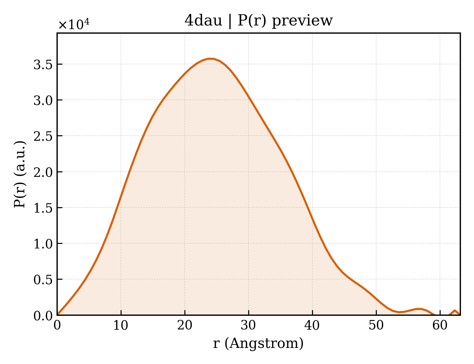
14-3-3 protein sigma
Homo sapiens
State in the Current Structure
| Assembly | Oligomeric State | Construct | Mutations and Modifications | Ligands, Ions and Associated Components | Method and Experimental Conditions | Structure Quality |
|---|---|---|---|---|---|---|
| 1 | Protein heterocomplex Heteromer Protein × 4 PDB declaration: tetrameric(4) Consistent with protein copy count | Chain A; UniProt 1–231 | Fragment:UNP residues 1-231 | Peptidylarginine Deiminase type VI × 2 (Q330K5) | X-RAY DIFFRACTION X-ray crystallization conditions:VAPOR DIFFUSION, HANGING DROP;pH 7.5;277 K;26.6% v/v PEG400, 0.19 M calcium chloride, 5% v/v glycerol, 0.095 M HEPES sodium, pH 7.5, VAPOR DIFFUSION, HANGING DROP, temperature 277K | Resolution 2.00 Å R-free 0.228 |
Other States of the Same Protein in the Database
Each row is a biological assembly of the same UniProt protein in another PDB entry. The “Difference from current entry” column identifies evidence-level differences; no tag means the currently parsed fields agree.
| Other PDB | Difference from Current Entry 4DAU | Assembly / Oligomeric State | Construct | Mutations and Modifications | Ligands, Ions and Non-polymers | Method and Experimental Conditions | Structure Quality |
|---|---|---|---|---|---|---|---|
| 1YWT Crystal structure of the human sigma isoform of 14-3-3 in complex with a mode-1 phosphopeptide Deposited 2005-02-18 | Different construct Different mutation/modification Different ligand/ion Different experimental conditions Different structure-quality metrics | Assembly 1 Protein heterocomplex Heteromer;Protein × 4 PDB declaration: tetrameric |
Chain A
1–248(248 aa)
Chain B
1–248(248 aa)
|
Not recorded | CA CALCIUM ION × 1 |
X-RAY DIFFRACTION
X-ray crystallization conditions
VAPOR DIFFUSION, HANGING DROP;pH 7;291 K;peg 3350, sodium flouride, calcium chloride, pH 7, VAPOR DIFFUSION, HANGING DROP, temperature 291K
|
Resolution 2.40 Å R-free 0.283 |
| 1YWT Crystal structure of the human sigma isoform of 14-3-3 in complex with a mode-1 phosphopeptide Deposited 2005-02-18 | Different construct Different mutation/modification Different oligomeric state Different ligand/ion Different experimental conditions Different structure-quality metrics | Assembly 2 Protein heterocomplex Heteromer;Protein × 8 PDB declaration: octameric |
Chain A
1–248(248 aa)
Chain B
1–248(248 aa)
|
Not recorded | CA CALCIUM ION × 2 |
X-RAY DIFFRACTION
X-ray crystallization conditions
VAPOR DIFFUSION, HANGING DROP;pH 7;291 K;peg 3350, sodium flouride, calcium chloride, pH 7, VAPOR DIFFUSION, HANGING DROP, temperature 291K
|
Resolution 2.40 Å R-free 0.283 |
| 1YZ5 The crystal structure of 14-3-3-sigma at 2.8 angstrom resolution Deposited 2005-02-28 | Different construct Different mutation/modification Different oligomeric state Different experimental conditions Different structure-quality metrics | Assembly 1 Protein homooligomer Homooligomer;Protein × 2 PDB declaration: dimeric |
Chain A
1–248(248 aa)
Chain B
1–248(248 aa)
|
Not recorded | No recorded non-water small molecule |
X-RAY DIFFRACTION
X-ray crystallization conditions
VAPOR DIFFUSION, SITTING DROP;pH 9;278 K;40% PEG 4000, 0.6M ammonium sulfate, 0.1M Tris/HCl, pH 9, VAPOR DIFFUSION, SITTING DROP, temperature 278K
|
Resolution 2.80 Å R-free 0.292 |
| 3IQJ Crystal Structure of human 14-3-3 sigma in Complex with Raf1 peptide (10mer) Deposited 2009-08-20 | Different mutation/modification Different ligand/ion Different experimental conditions Different structure-quality metrics | Assembly 1 Protein heterocomplex Heteromer;Protein × 4 PDB declaration: tetrameric |
Chain A
1–231(231 aa)
Fragment:UNP residues 1-231
|
Non-standard monomer:Yes (specific site not provided by mmCIF) | CL CHLORIDE ION × 2 MG MAGNESIUM ION × 6 |
X-RAY DIFFRACTION
X-ray crystallization conditions
VAPOR DIFFUSION, HANGING DROP;pH 7.5;277 K;0.1M Hepes/NaOH ph 7.5, 0.2M CaCl2, 28% PEG 400, 5% glycerol, 2mM DTT, VAPOR DIFFUSION, HANGING DROP, temperature 277K
|
Resolution 1.15 Å R-free 0.153 |
| 3IQU Crystal Structure of human 14-3-3 sigma in Complex with Raf1 peptide (6mer) Deposited 2009-08-21 | Different mutation/modification Different ligand/ion Different experimental conditions Different structure-quality metrics | Assembly 1 Protein heterocomplex Heteromer;Protein × 4 PDB declaration: tetrameric |
Chain A
1–231(231 aa)
Fragment:UNP residues 1-231
|
Non-standard monomer:Yes (specific site not provided by mmCIF) | CL CHLORIDE ION × 2 MG MAGNESIUM ION × 6 |
X-RAY DIFFRACTION
X-ray crystallization conditions
VAPOR DIFFUSION, HANGING DROP;pH 7.5;277 K;0.1M Hepes/NaOH ph 7.5, 0.2M CaCl2, 28% PEG 400, 5% glycerol, 2mM DTT, VAPOR DIFFUSION, HANGING DROP, temperature 277K
|
Resolution 1.05 Å R-free 0.151 |
| 3IQV Crystal Structure of human 14-3-3 sigma in Complex with Raf1 peptide (6mer) and stabilisator Fusicoccin Deposited 2009-08-21 | Different mutation/modification Different ligand/ion Different experimental conditions Different structure-quality metrics | Assembly 1 Protein heterocomplex Heteromer;Protein × 4 PDB declaration: tetrameric |
Chain A
1–231(231 aa)
Fragment:UNP residues 1-231
|
Non-standard monomer:Yes (specific site not provided by mmCIF) | FSC FUSICOCCIN × 2 CL CHLORIDE ION × 4 MG MAGNESIUM ION × 8 |
X-RAY DIFFRACTION
X-ray crystallization conditions
VAPOR DIFFUSION, HANGING DROP;pH 7.5;277 K;20mM Hepes/NaOH pH 7.5, 2mM MgCl2 and 2 mM DTT, set up for crystallization in 0.1 M Hepes/NaOH ph 7.5, 0.2 M CaCl2, 28% PEG 400, 5% glycerol, 2mM DTT, VAPOR DIFFUSION, HANGING DROP, temperature 277K
|
Resolution 1.20 Å R-free 0.153 |
| 3LW1 Binary complex of 14-3-3 sigma and p53 pT387-peptide Deposited 2010-02-23 | Different construct Different mutation/modification Different ligand/ion Different experimental conditions Different structure-quality metrics | Assembly 1 Protein heterocomplex Heteromer;Protein × 4 PDB declaration: tetrameric |
Chain A
1–248(248 aa)
|
Non-standard monomer:Yes (specific site not provided by mmCIF) | MG MAGNESIUM ION × 6 CL CHLORIDE ION × 2 GOL GLYCEROL × 2 |
X-RAY DIFFRACTION
X-ray crystallization conditions
VAPOR DIFFUSION, HANGING DROP;pH 7.5;277 K;0.1M HEPES, 0.2M calcium chloride, 28% PEG 400, 5% glycerol, 2mM DTT, pH 7.5, VAPOR DIFFUSION, HANGING DROP, temperature 277K
|
Resolution 1.28 Å R-free 0.151 |
| 3MHR 14-3-3 sigma in complex with YAP pS127-peptide Deposited 2010-04-08 | Different construct Different mutation/modification Different ligand/ion Different experimental conditions Different structure-quality metrics | Assembly 1 Protein heterocomplex Heteromer;Protein × 4 PDB declaration: tetrameric |
Chain A
1–231(231 aa)
Fragment:UNP RESIDUES 1-231
|
Non-standard monomer:Yes (specific site not provided by mmCIF) | MG MAGNESIUM ION × 8 CL CHLORIDE ION × 8 CA CALCIUM ION × 2 GOL GLYCEROL × 6 |
X-RAY DIFFRACTION
X-ray crystallization conditions
VAPOR DIFFUSION, HANGING DROP;pH 7.4;277 K;0.095M HEPES Na-Salt pH7.4, 25.6% PEG 400, 0.19M CaCl2, 5% Glycerol, VAPOR DIFFUSION, HANGING DROP, temperature 277K
|
Resolution 1.15 Å R-free 0.153 |
| 3O8I Structure of 14-3-3 isoform sigma in complex with a C-Raf1 peptide and a stabilizing small molecule fragment Deposited 2010-08-03 | Different construct Different ligand/ion Different experimental conditions Different structure-quality metrics | Assembly 1 Protein heterocomplex Heteromer;Protein × 4 PDB declaration: tetrameric |
Chain A
1–231(231 aa)
Fragment:C-terminal deletion
|
Not recorded | M1T 6,6-dihydroxy-1-methoxyhexan-2-one × 2 |
X-RAY DIFFRACTION
X-ray crystallization conditions
VAPOR DIFFUSION, HANGING DROP;pH 7.5;277.15 K;0.095M HEPES Na, 26.6%(v/v) PEG400, 0.19M CaCl2, 5%(v/v) Glycerol, pH 7.5, VAPOR DIFFUSION, HANGING DROP, temperature 277.15K
|
Resolution 2.00 Å R-free 0.239 |
| 3P1N Crystal structure of human 14-3-3 sigma in complex with TASK-3 peptide Deposited 2010-09-30 | Different construct Different mutation/modification Different ligand/ion Different experimental conditions Different structure-quality metrics | Assembly 1 Protein heterocomplex Heteromer;Protein × 4 PDB declaration: tetrameric |
Chain A
1–231(231 aa)
|
Non-standard monomer:Yes (specific site not provided by mmCIF) | MG MAGNESIUM ION × 6 CL CHLORIDE ION × 2 |
X-RAY DIFFRACTION
X-ray crystallization conditions
VAPOR DIFFUSION, HANGING DROP;pH 7.4;277 K;0.095M HEPES Na-Salt, 25.6% PEG 400, 0.19M CaCl2, 5% Glycerol, pH 7.4, VAPOR DIFFUSION, HANGING DROP, temperature 277K
|
Resolution 1.40 Å R-free 0.174 |
| 3P1O Crystal structure of human 14-3-3 sigma in complex with TASK-3 peptide and stabilisator Fusicoccin A Deposited 2010-09-30 | Different construct Different mutation/modification Different ligand/ion Different experimental conditions Different structure-quality metrics | Assembly 1 Protein heterocomplex Heteromer;Protein × 4 PDB declaration: tetrameric |
Chain A
1–231(231 aa)
|
Non-standard monomer:Yes (specific site not provided by mmCIF) | FSC FUSICOCCIN × 2 CL CHLORIDE ION × 2 CA CALCIUM ION × 2 MG MAGNESIUM ION × 8 |
X-RAY DIFFRACTION
X-ray crystallization conditions
VAPOR DIFFUSION, HANGING DROP;pH 7.4;277 K;0.095M HEPES Na-Salt, 25.6% PEG 400, 0.19M CaCl2, 5% Glycerol, pH 7.4, VAPOR DIFFUSION, HANGING DROP, temperature 277K
|
Resolution 1.90 Å R-free 0.234 |
| 3P1P Crystal structure of human 14-3-3 sigma C38N/N166H in complex with TASK-3 peptide Deposited 2010-09-30 | Different construct Different mutation/modification Different ligand/ion Different experimental conditions Different structure-quality metrics | Assembly 1 Protein heterocomplex Heteromer;Protein × 4 PDB declaration: tetrameric |
Chain A
1–231(231 aa)
|
Mutation:C38N, N166H | CL CHLORIDE ION × 2 MG MAGNESIUM ION × 6 |
X-RAY DIFFRACTION
X-ray crystallization conditions
VAPOR DIFFUSION, HANGING DROP;pH 7.4;277.15 K;0.095M HEPES Na-Salt, 25.6% PEG 400, 0.19M CaCl2, 5% Glycerol, pH 7.4, VAPOR DIFFUSION, HANGING DROP, temperature 277.15K
|
Resolution 1.95 Å R-free 0.227 |
| 3P1Q Crystal structure of human 14-3-3 sigma C38N/N166H in complex with TASK-3 Peptide and stabilizer fusicoccin A Deposited 2010-09-30 | Different construct Different mutation/modification Different ligand/ion Different experimental conditions Different structure-quality metrics | Assembly 1 Protein heterocomplex Heteromer;Protein × 4 PDB declaration: tetrameric |
Chain A
1–231(231 aa)
|
Mutation:C38N,N166H | FSC FUSICOCCIN × 2 CL CHLORIDE ION × 2 MG MAGNESIUM ION × 4 CA CALCIUM ION × 2 |
X-RAY DIFFRACTION
X-ray crystallization conditions
VAPOR DIFFUSION, HANGING DROP;pH 7.4;277.15 K;0.095M HEPES Na-Salt, 25.6% PEG 400, 0.19M CaCl2, 5% Glycerol, pH 7.4, VAPOR DIFFUSION, HANGING DROP, temperature 277.15K
|
Resolution 1.70 Å R-free 0.217 |
| 3P1R Crystal structure of human 14-3-3 sigma C38V/N166H in complex with TASK-3 peptide Deposited 2010-09-30 | Different construct Different mutation/modification Different ligand/ion Different experimental conditions Different structure-quality metrics | Assembly 1 Protein heterocomplex Heteromer;Protein × 4 PDB declaration: tetrameric |
Chain A
1–231(231 aa)
|
Mutation:C38V, N166H | MG MAGNESIUM ION × 8 CL CHLORIDE ION × 8 CA CALCIUM ION × 2 |
X-RAY DIFFRACTION
X-ray crystallization conditions
VAPOR DIFFUSION, HANGING DROP;pH 7.4;277 K;0.095M HEPES Na-Salt, 25.6% PEG 400, 0.19M CaCl2, 5% Glycerol, pH 7.4, VAPOR DIFFUSION, HANGING DROP, temperature 277K
|
Resolution 1.70 Å R-free 0.194 |
| 3P1S Crystal structure of human 14-3-3 sigma C38N/N166H in complex with TASK-3 peptide and stabilizer fusicoccin A Deposited 2010-09-30 | Different construct Different mutation/modification Different ligand/ion Different experimental conditions Different structure-quality metrics | Assembly 1 Protein heterocomplex Heteromer;Protein × 4 PDB declaration: tetrameric |
Chain A
1–231(231 aa)
|
Mutation:C38V, N166H | FSC FUSICOCCIN × 2 CL CHLORIDE ION × 2 MG MAGNESIUM ION × 4 |
X-RAY DIFFRACTION
X-ray crystallization conditions
VAPOR DIFFUSION, HANGING DROP;pH 7.4;277.15 K;0.095M HEPES Na-Salt, 25.6% PEG 400, 0.19M CaCl2, 5% Glycerol, pH 7.4, VAPOR DIFFUSION, HANGING DROP, temperature 277.15K
|
Resolution 1.65 Å R-free 0.201 |
| 3SMK Crystal structure of human 14-3-3 sigma C38V/N166H in complex with TASK-3 peptide and stabilizer Cotylenin A Deposited 2011-06-28 | Different construct Different mutation/modification Different ligand/ion Different experimental conditions Different structure-quality metrics | Assembly 1 Protein heterocomplex Heteromer;Protein × 4 PDB declaration: tetrameric |
Chain A
1–231(231 aa)
|
Mutation:C38V N166H | CW7 Cotylenin A × 2 MG MAGNESIUM ION × 2 |
X-RAY DIFFRACTION
X-ray crystallization conditions
VAPOR DIFFUSION, HANGING DROP;pH 7.4;277 K;0.095M HEPES Na-Salt pH7.4, 25.6% PEG 400, 0.19M CaCl2, 5% Glycerol, VAPOR DIFFUSION, HANGING DROP, temperature 277K
|
Resolution 2.10 Å R-free 0.237 |
| 3SML Crystal structure of human 14-3-3 sigma C38N/N166H in complex with TASK-3 peptide and stabilizer Fusicoccin A aglycone Deposited 2011-06-28 | Different construct Different mutation/modification Different ligand/ion Different experimental conditions Different structure-quality metrics | Assembly 1 Protein heterocomplex Heteromer;Protein × 4 PDB declaration: tetrameric |
Chain A
1–231(231 aa)
|
Mutation:C38N N166H | FW1 Fusicoccin A aglycone × 2 CL CHLORIDE ION × 2 MG MAGNESIUM ION × 8 |
X-RAY DIFFRACTION
X-ray crystallization conditions
VAPOR DIFFUSION, HANGING DROP;pH 7.4;277 K;0.095M HEPES Na-Salt pH7.4, 25.6% PEG 400, 0.19M CaCl2, 5% Glycerol, VAPOR DIFFUSION, HANGING DROP, temperature 277K
|
Resolution 1.90 Å R-free 0.206 |
| 3SMM Crystal structure of human 14-3-3 sigma C38N/N166H in complex with task-3 peptide and stabilizer Fusicoccin J aglycone Deposited 2011-06-28 | Different construct Different mutation/modification Different ligand/ion Different experimental conditions Different structure-quality metrics | Assembly 1 Protein heterocomplex Heteromer;Protein × 4 PDB declaration: tetrameric |
Chain A
1–231(231 aa)
|
Mutation:C38N N166H | FJA Fusicoccin J aglycone × 2 MG MAGNESIUM ION × 6 CL CHLORIDE ION × 2 |
X-RAY DIFFRACTION
X-ray crystallization conditions
VAPOR DIFFUSION, HANGING DROP;pH 7.4;277 K;0.095M HEPES Na-Salt pH7.4, 25.6% PEG 400, 0.19M CaCl2, 5% Glycerol, VAPOR DIFFUSION, HANGING DROP, temperature 277K
|
Resolution 2.00 Å R-free 0.216 |
| 3SMN Crystal structure of human 14-3-3 sigma C38N/N166H in complex with task-3 peptide and stabilizer Fusicoccin A-THF Deposited 2011-06-28 | Different construct Different mutation/modification Different ligand/ion Different experimental conditions Different structure-quality metrics | Assembly 1 Protein heterocomplex Heteromer;Protein × 4 PDB declaration: tetrameric |
Chain A
1–231(231 aa)
|
Mutation:C38N N166H | FC7 Fusicoccin A-THF × 2 MG MAGNESIUM ION × 4 CA CALCIUM ION × 4 CL CHLORIDE ION × 4 |
X-RAY DIFFRACTION
X-ray crystallization conditions
VAPOR DIFFUSION, HANGING DROP;pH 7.4;277 K;0.095M HEPES Na-Salt pH7.4, 25.6% PEG 400, 0.19M CaCl2, 5% Glycerol, VAPOR DIFFUSION, HANGING DROP, temperature 277K
|
Resolution 2.00 Å R-free 0.203 |
| 3SMO Crystal structure of human 14-3-3 sigma C38V/N166H in complex with TASK-3 peptide and stabilizer Fusicoccin J aglycone Deposited 2011-06-28 | Different construct Different mutation/modification Different ligand/ion Different experimental conditions Different structure-quality metrics | Assembly 1 Protein heterocomplex Heteromer;Protein × 4 PDB declaration: tetrameric |
Chain A
1–231(231 aa)
|
Mutation:C38V N166H | FJA Fusicoccin J aglycone × 2 MG MAGNESIUM ION × 6 |
X-RAY DIFFRACTION
X-ray crystallization conditions
VAPOR DIFFUSION, HANGING DROP;pH 7.4;277 K;0.095M HEPES Na-Salt pH7.4, 25.6% PEG 400, 0.19M CaCl2, 5% Glycerol, VAPOR DIFFUSION, HANGING DROP, temperature 277K
|
Resolution 1.80 Å R-free 0.203 |
| 3SP5 Crystal structure of human 14-3-3 sigma C38V/N166H in complex with TASK-3 peptide and stabilizer Cotylenol Deposited 2011-07-01 | Different construct Different mutation/modification Different ligand/ion Different experimental conditions Different structure-quality metrics | Assembly 1 Protein heterocomplex Heteromer;Protein × 4 PDB declaration: tetrameric |
Chain A
1–231(231 aa)
|
Mutation:C38V N166H | CX7 Cotylenol × 2 MG MAGNESIUM ION × 4 |
X-RAY DIFFRACTION
X-ray crystallization conditions
VAPOR DIFFUSION, HANGING DROP;pH 7.4;277 K;0.095M HEPES Na-Salt pH7.4, 25.6% PEG 400, 0.19M CaCl2, 5% Glycerol, VAPOR DIFFUSION, HANGING DROP, temperature 277K
|
Resolution 1.80 Å R-free 0.211 |
| 3SP5 Crystal structure of human 14-3-3 sigma C38V/N166H in complex with TASK-3 peptide and stabilizer Cotylenol Deposited 2011-07-01 | Different construct Different mutation/modification Different oligomeric state Different ligand/ion Different experimental conditions Different structure-quality metrics | Assembly 2 Protein heterocomplex Heteromer;Protein × 2 PDB declaration: dimeric |
Chain A
1–231(231 aa)
|
Mutation:C38V N166H | CX7 Cotylenol × 1 MG MAGNESIUM ION × 2 |
X-RAY DIFFRACTION
X-ray crystallization conditions
VAPOR DIFFUSION, HANGING DROP;pH 7.4;277 K;0.095M HEPES Na-Salt pH7.4, 25.6% PEG 400, 0.19M CaCl2, 5% Glycerol, VAPOR DIFFUSION, HANGING DROP, temperature 277K
|
Resolution 1.80 Å R-free 0.211 |
| 3SPR Crystal structure of human 14-3-3 sigma C38V/N166H in complex with TASK-3 peptide and stabilizer FC-THF Deposited 2011-07-03 | Different construct Different mutation/modification Different ligand/ion Different experimental conditions Different structure-quality metrics | Assembly 1 Protein heterocomplex Heteromer;Protein × 4 PDB declaration: tetrameric |
Chain A
1–231(231 aa)
|
Mutation:C38V N166H | FC7 Fusicoccin A-THF × 2 CL CHLORIDE ION × 2 MG MAGNESIUM ION × 6 |
X-RAY DIFFRACTION
X-ray crystallization conditions
VAPOR DIFFUSION, HANGING DROP;pH 7.4;277 K;0.095M HEPES Na-Salt pH7.4, 25.6% PEG 400, 0.19M CaCl2, 5% Glycerol, VAPOR DIFFUSION, HANGING DROP, temperature 277K
|
Resolution 1.99 Å R-free 0.250 |
| 3T0L Small-molecule inhibitors of 14-3-3 protein-protein interactions from virtual screening Deposited 2011-07-20 | Different oligomeric state Different ligand/ion Different experimental conditions Different structure-quality metrics | Assembly 1 Protein homooligomer Homooligomer;Protein × 2 PDB declaration: dimeric |
Chain A
1–231(231 aa)
Fragment:UNP residues 1-231
|
Not recorded | 1CT [2-(2-oxo-2-{[2-(trifluoromethyl)phenyl]amino}ethoxy)phenyl]phosphonic acid × 2 GOL GLYCEROL × 4 CL CHLORIDE ION × 6 MG MAGNESIUM ION × 4 |
X-RAY DIFFRACTION
X-ray crystallization conditions
VAPOR DIFFUSION, HANGING DROP;pH 7.7;277 K;0.095M HEPES Na salt, 0.19M calcium chloride, 5% glycerol, 28% PEG 400, pH 7.7, VAPOR DIFFUSION, HANGING DROP, temperature 277K
|
Resolution 1.60 Å R-free 0.190 |
| 3T0M Small-molecule inhibitors of 14-3-3 protein-protein interactions from virtual screening Deposited 2011-07-20 | Different oligomeric state Different ligand/ion Different experimental conditions Different structure-quality metrics | Assembly 1 Protein homooligomer Homooligomer;Protein × 2 PDB declaration: dimeric |
Chain A
1–231(231 aa)
Fragment:UNP residues 1-231
|
Not recorded | 2CT (2-{2-[(2,5-dimethoxyphenyl)amino]-2-oxoethoxy}phenyl)phosphonic acid × 2 GOL GLYCEROL × 4 CL CHLORIDE ION × 4 MG MAGNESIUM ION × 6 |
X-RAY DIFFRACTION
X-ray crystallization conditions
VAPOR DIFFUSION, HANGING DROP;pH 7.5;277 K;0.095M HEPES Na salt, 0.19M calcium chloride, 5% glycerol, 25.6% PEG 400, pH 7.5, VAPOR DIFFUSION, HANGING DROP, temperature 277K
|
Resolution 1.62 Å R-free 0.198 |
| 3U9X Covalent attachment of pyridoxal-phosphate derivatives to 14-3-3 proteins Deposited 2011-10-20 | Different mutation/modification Different oligomeric state Different ligand/ion Different experimental conditions Different structure-quality metrics | Assembly 1 Protein homooligomer Homooligomer;Protein × 2 PDB declaration: dimeric |
Chain A
1–231(231 aa)
Fragment:UNP residues 1-231
|
Non-standard monomer:Yes (specific site not provided by mmCIF) | MG MAGNESIUM ION × 4 CL CHLORIDE ION × 6 GOL GLYCEROL × 2 |
X-RAY DIFFRACTION
X-ray crystallization conditions
VAPOR DIFFUSION, HANGING DROP;pH 7.5;277 K;0.095M HEPES Na, 26.6% PEG400, 0.1M Calcium chloride, 5% Glycerol, pH 7.5, VAPOR DIFFUSION, HANGING DROP, temperature 277K
|
Resolution 1.80 Å R-free 0.188 |
| 3UX0 Crystal structure of human 14-3-3 sigma in complex with TASK-3 peptide and stabilizer Fusicoccin H Deposited 2011-12-03 | Different construct Different mutation/modification Different ligand/ion Different experimental conditions Different structure-quality metrics | Assembly 1 Protein heterocomplex Heteromer;Protein × 4 PDB declaration: tetrameric |
Chain A
1–231(231 aa)
|
Non-standard monomer:Yes (specific site not provided by mmCIF) | MG MAGNESIUM ION × 8 CL CHLORIDE ION × 2 GOL GLYCEROL × 2 0DV (4R,5R,6R,6aS,9S,9aE,10aR)-5-hydroxy-9-(hydroxymethyl)-6,10a-dimethyl-3-(propan-2-yl)-1,2,4,5,6,6a,7,8,9,10a-decahydrod icyclopenta[a,d][8]annulen-4-yl alpha-D-gulopyranoside × 2 |
X-RAY DIFFRACTION
X-ray crystallization conditions
VAPOR DIFFUSION, HANGING DROP;pH 7.5;277 K;0.095M HEPES Na, 0.19M calcium chloride, 5% glycerol, 26.6% PEG400, pH 7.5, VAPOR DIFFUSION, HANGING DROP, temperature 277K
|
Resolution 1.75 Å R-free 0.192 |
| 4DAT Structure of 14-3-3 sigma in complex with PADI6 14-3-3 binding motif II Deposited 2012-01-13 | Different ligand/ion Different structure-quality metrics | Assembly 1 Protein heterocomplex Heteromer;Protein × 4 PDB declaration: tetrameric |
Chain A
1–231(231 aa)
Fragment:UNP residues 1-231
|
Not recorded | MG MAGNESIUM ION × 2 |
X-RAY DIFFRACTION
X-ray crystallization conditions
VAPOR DIFFUSION, HANGING DROP;pH 7.5;277 K;26.6% v/v PEG400, 0.19 M calcium chloride, 5% v/v glycerol, 0.095 M HEPES sodium, pH 7.5, VAPOR DIFFUSION, HANGING DROP, temperature 277K
|
Resolution 1.40 Å R-free 0.183 |
| 4DHM Small-molecule inhibitors of 14-3-3 protein-protein interactions from virtual screening Deposited 2012-01-30 | Different oligomeric state Different ligand/ion Different experimental conditions Different structure-quality metrics | Assembly 1 Protein homooligomer Homooligomer;Protein × 2 PDB declaration: dimeric |
Chain A
1–231(231 aa)
Fragment:UNP residues 1-231
|
Not recorded | MG MAGNESIUM ION × 6 CL CHLORIDE ION × 6 GOL GLYCEROL × 2 0KB [2-(2-oxo-2-{[3-(trifluoromethyl)phenyl]amino}ethoxy)phenyl]phosphonic acid × 2 |
X-RAY DIFFRACTION
X-ray crystallization conditions
VAPOR DIFFUSION, HANGING DROP;pH 7.7;277 K;0.095M HEPES Na, 0.19M calcium chloride, 5% glycerol, 28% PEG400, pH 7.7, VAPOR DIFFUSION, HANGING DROP, temperature 277K
|
Resolution 1.70 Å R-free 0.187 |
| 4DHN Small-molecule inhibitors of 14-3-3 protein-protein interactions from virtual screening Deposited 2012-01-30 | Different oligomeric state Different ligand/ion Different experimental conditions Different structure-quality metrics | Assembly 1 Protein homooligomer Homooligomer;Protein × 2 PDB declaration: dimeric |
Chain A
1–231(231 aa)
Fragment:UNP residues 1-231
|
Not recorded | MG MAGNESIUM ION × 6 CL CHLORIDE ION × 4 GOL GLYCEROL × 2 0KC {2-[2-(cyclopentylamino)-2-oxoethoxy]phenyl}phosphonic acid × 2 |
X-RAY DIFFRACTION
X-ray crystallization conditions
VAPOR DIFFUSION, HANGING DROP;pH 7.3;277 K;0.095M HEPES Na, 0.19M calcium chloride, 5% glycerol, 28% PEG400, pH 7.3, VAPOR DIFFUSION, HANGING DROP, temperature 277K
|
Resolution 1.80 Å R-free 0.185 |
| 4DHO Small-molecule inhibitors of 14-3-3 protein-protein interactions from virtual screening Deposited 2012-01-30 | Different oligomeric state Different ligand/ion Different experimental conditions Different structure-quality metrics | Assembly 1 Protein homooligomer Homooligomer;Protein × 2 PDB declaration: dimeric |
Chain A
1–231(231 aa)
Fragment:UNP residues 1-231
|
Not recorded | MG MAGNESIUM ION × 8 CL CHLORIDE ION × 4 GOL GLYCEROL × 2 Y09 (2-{2-[(3-methoxyphenyl)amino]-2-oxoethoxy}phenyl)phosphonic acid × 2 |
X-RAY DIFFRACTION
X-ray crystallization conditions
VAPOR DIFFUSION, HANGING DROP;pH 7.1;277 K;0.095M HEPES Na, 0.19M calcium chloride, 5% glycerol, 28% PEG400, pH 7.1, VAPOR DIFFUSION, HANGING DROP, temperature 277K
|
Resolution 1.70 Å R-free 0.203 |
| 4DHP Small-molecule inhibitors of 14-3-3 protein-protein interactions from virtual screening Deposited 2012-01-30 | Different mutation/modification Different oligomeric state Different ligand/ion Different experimental conditions Different structure-quality metrics | Assembly 1 Protein homooligomer Homooligomer;Protein × 2 PDB declaration: dimeric |
Chain A
1–231(231 aa)
Fragment:UNP residues 1-231
|
Non-standard monomer:Yes (specific site not provided by mmCIF) | MG MAGNESIUM ION × 10 CL CHLORIDE ION × 4 GOL GLYCEROL × 2 Y07 [2-(2-{[2-chloro-5-(trifluoromethyl)phenyl]amino}-2-oxoethoxy)phenyl]phosphonic acid × 2 |
X-RAY DIFFRACTION
X-ray crystallization conditions
VAPOR DIFFUSION, HANGING DROP;pH 7.5;277 K;0.095M HEPES Na, 0.19M calcium chloride, 5% glycerol, 26% PEG400, pH 7.5, VAPOR DIFFUSION, HANGING DROP, temperature 277K
|
Resolution 1.75 Å R-free 0.196 |
| 4DHQ Small-molecule inhibitors of 14-3-3 protein-protein interactions from virtual screening Deposited 2012-01-30 | Different mutation/modification Different oligomeric state Different ligand/ion Different experimental conditions Different structure-quality metrics | Assembly 1 Protein homooligomer Homooligomer;Protein × 2 PDB declaration: dimeric |
Chain A
1–231(231 aa)
Fragment:UNP residues 1-231
|
Non-standard monomer:Yes (specific site not provided by mmCIF) | MG MAGNESIUM ION × 10 CL CHLORIDE ION × 4 GOL GLYCEROL × 2 Y06 (2-{2-[(3-chlorophenyl)amino]-2-oxoethoxy}phenyl)phosphonic acid × 2 |
X-RAY DIFFRACTION
X-ray crystallization conditions
VAPOR DIFFUSION, HANGING DROP;pH 7.1;277 K;0.095M HEPES Na, 0.19M calcium chloride, 5% glycerol, 27% PEG400, pH 7.1, VAPOR DIFFUSION, HANGING DROP, temperature 277K
|
Resolution 1.75 Å R-free 0.189 |
| 4DHR Small-molecule inhibitors of 14-3-3 protein-protein interactions from virtual screening Deposited 2012-01-30 | Different mutation/modification Different oligomeric state Different ligand/ion Different experimental conditions Different structure-quality metrics | Assembly 1 Protein homooligomer Homooligomer;Protein × 2 PDB declaration: dimeric |
Chain A
1–231(231 aa)
Fragment:UNP residues 1-231
|
Non-standard monomer:Yes (specific site not provided by mmCIF) | MG MAGNESIUM ION × 10 CL CHLORIDE ION × 4 GOL GLYCEROL × 2 Y04 (2-{2-[(2-chlorophenyl)amino]-2-oxoethoxy}phenyl)phosphonic acid × 2 |
X-RAY DIFFRACTION
X-ray crystallization conditions
VAPOR DIFFUSION, HANGING DROP;pH 7.1;277 K;0.095M HEPES Na, 0.19M calcium chloride, 5% glycerol, 28% PEG400, pH 7.1, VAPOR DIFFUSION, HANGING DROP, temperature 277K
|
Resolution 1.40 Å R-free 0.158 |
| 4DHS Small-molecule inhibitors of 14-3-3 protein-protein interactions from virtual screening Deposited 2012-01-30 | Different mutation/modification Different oligomeric state Different ligand/ion Different experimental conditions Different structure-quality metrics | Assembly 1 Protein homooligomer Homooligomer;Protein × 2 PDB declaration: dimeric |
Chain A
1–231(231 aa)
Fragment:UNP residues 1-231
|
Non-standard monomer:Yes (specific site not provided by mmCIF) | MG MAGNESIUM ION × 6 CL CHLORIDE ION × 4 GOL GLYCEROL × 2 Y03 (2-{2-[(3,5-dichlorophenyl)amino]-2-oxoethoxy}phenyl)phosphonic acid × 2 |
X-RAY DIFFRACTION
X-ray crystallization conditions
VAPOR DIFFUSION, HANGING DROP;pH 7.5;277 K;0.095M HEPES Na, 0.19M calcium chloride, 5% glycerol, 28% PEG400, pH 7.5, VAPOR DIFFUSION, HANGING DROP, temperature 277K
|
Resolution 1.74 Å R-free 0.193 |
| 4DHT Small-molecule inhibitors of 14-3-3 protein-protein interactions from virtual screening Deposited 2012-01-30 | Different construct Different oligomeric state Different ligand/ion Different experimental conditions Different structure-quality metrics | Assembly 1 Protein homooligomer Homooligomer;Protein × 2 PDB declaration: dimeric |
Chain A
1–231(231 aa)
Fragment:UNP resiudes 1-231
|
Not recorded | MG MAGNESIUM ION × 4 CL CHLORIDE ION × 4 GOL GLYCEROL × 2 0KG {2-[2-(cyclohexylamino)-2-oxoethoxy]phenyl}phosphonic acid × 2 |
X-RAY DIFFRACTION
X-ray crystallization conditions
VAPOR DIFFUSION, HANGING DROP;pH 7.3;277 K;0.095M HEPES Na, 0.19M calcium chloride, 5% glycerol, 28% PEG400, pH 7.3, VAPOR DIFFUSION, HANGING DROP, temperature 277K
|
Resolution 1.80 Å R-free 0.206 |
| 4DHU Small-molecule inhibitors of 14-3-3 protein-protein interactions from virtual screening Deposited 2012-01-30 | Different construct Different mutation/modification Different oligomeric state Different ligand/ion Different experimental conditions Different structure-quality metrics | Assembly 1 Protein homooligomer Homooligomer;Protein × 2 PDB declaration: dimeric |
Chain A
1–231(231 aa)
Fragment:UNP resiudes 1-231
|
Non-standard monomer:Yes (specific site not provided by mmCIF) | MG MAGNESIUM ION × 10 CL CHLORIDE ION × 4 GOL GLYCEROL × 2 0KH (2-{2-[(2,3-dichlorophenyl)amino]-2-oxoethoxy}phenyl)phosphonic acid × 2 |
X-RAY DIFFRACTION
X-ray crystallization conditions
VAPOR DIFFUSION, SITTING DROP;pH 7.1;277 K;0.095M HEPES Na, 0.19M calcium chloride, 5% glycerol, 27% PEG400, pH 7.1, VAPOR DIFFUSION, SITTING DROP, temperature 277K
|
Resolution 1.67 Å R-free 0.176 |
| 4FL5 Crystal structure of human 14-3-3 sigma in complex with a Tau-protein peptide surrounding pS214 Deposited 2012-06-14 | Different construct Different mutation/modification Different ligand/ion Different experimental conditions Different structure-quality metrics | Assembly 1 Protein heterocomplex Heteromer;Protein × 4 PDB declaration: tetrameric |
Chain A
1–231(231 aa)
Chain B
1–231(231 aa)
|
Non-standard monomer:Yes (specific site not provided by mmCIF) Non-standard monomer:Yes (specific site not provided by mmCIF) | CL CHLORIDE ION × 2 MG MAGNESIUM ION × 4 CA CALCIUM ION × 6 GOL GLYCEROL × 3 |
X-RAY DIFFRACTION
X-ray crystallization conditions
VAPOR DIFFUSION, HANGING DROP;pH 7.5;277 K;0.1 M Hepes/NaOH pH 7.5, 0.2 M CaCl2, 28% PEG 400, 5% glycerol, 2 mM DTT, VAPOR DIFFUSION, HANGING DROP, temperature 277K
|
Resolution 1.90 Å R-free 0.212 |
| 4FR3 Crystal structure of human 14-3-3 sigma in complex with TASK-3 peptide and stabilizer 16-O-Me-FC-H Deposited 2012-06-26 | Different construct Different mutation/modification Different ligand/ion Different experimental conditions Different structure-quality metrics | Assembly 1 Protein heterocomplex Heteromer;Protein × 4 PDB declaration: tetrameric |
Chain A
1–231(231 aa)
|
Non-standard monomer:Yes (specific site not provided by mmCIF) | 0V4 (4R,5R,6R,6aS,9S,9aE,10aR)-5-hydroxy-9-(methoxymethyl)-6,10a-dimethyl-3-(propan-2-yl)-1,2,4,5,6,6a,7,8,9,10a-decahydrodicyclopenta[a,d][8]annulen-4-yl alpha-D-glucopyranoside × 2 MG MAGNESIUM ION × 4 |
X-RAY DIFFRACTION
X-ray crystallization conditions
VAPOR DIFFUSION, HANGING DROP;pH 7.4;277 K;0.095M HEPES Na-Salt pH7.4, 25.6% PEG 400, 0.19M CaCl2, 5% Glycerol, Vapor Diffusion, Hanging Drop, temperature 277K
|
Resolution 1.90 Å R-free 0.209 |
| 4IEA 14-3-3 isoform sigma in complex with a phosphorylated C-RAF peptide Deposited 2012-12-13 | Different experimental conditions Different structure-quality metrics | Assembly 1 Protein heterocomplex Heteromer;Protein × 4 PDB declaration: tetrameric |
Chain A
1–231(231 aa)
Fragment:UNP residues 1-231
|
Not recorded | No recorded non-water small molecule |
X-RAY DIFFRACTION
X-ray crystallization conditions
VAPOR DIFFUSION, HANGING DROP;pH 7.4;277 K;95 mM Na-HEPES pH 7.4, 25.6 % PEG 400, 190 mM CaCl2, 5 % Glycerol, VAPOR DIFFUSION, HANGING DROP, temperature 277K
|
Resolution 1.70 Å R-free 0.208 |
| 4JC3 14-3-3 protein interaction with Estrogen Receptor Alpha provides a novel drug target interface Deposited 2013-02-21 | Different ligand/ion Different experimental conditions Different structure-quality metrics | Assembly 1 Protein heterocomplex Heteromer;Protein × 4 PDB declaration: tetrameric |
Chain A
1–231(231 aa)
Fragment:UNP residues 1-231
|
Not recorded | MG MAGNESIUM ION × 6 |
X-RAY DIFFRACTION
X-ray crystallization conditions
VAPOR DIFFUSION, SITTING DROP;pH 7.5;277.15 K;0.095M HEPES Na, 0.19M calcium chloride, 5% glycerol, 26.6% PEG400, pH 7.5, VAPOR DIFFUSION, SITTING DROP, temperature 277.15K
|
Resolution 2.05 Å R-free 0.235 |
| 4JDD 14-3-3 protein interaction with Estrogen Receptor Alpha provides a novel drug target interface Deposited 2013-02-25 | Different ligand/ion Different experimental conditions Different structure-quality metrics | Assembly 1 Protein heterocomplex Heteromer;Protein × 4 PDB declaration: tetrameric |
Chain A
1–231(231 aa)
Fragment:UNP residues 1-231
|
Not recorded | FSC FUSICOCCIN × 2 |
X-RAY DIFFRACTION
X-ray crystallization conditions
VAPOR DIFFUSION, SITTING DROP;pH 7.5;277.15 K;0.095M HEPES Na, 0.19M calcium chloride, 5% glycerol, 26.6% PEG400, pH 7.5, VAPOR DIFFUSION, SITTING DROP, temperature 277.15K
|
Resolution 2.10 Å R-free 0.232 |
| 4QLI A novel phospho-switch in the linker region of the snail zinc finger protein which regulates 14-3-3 association, DNA binding and epithelial-mesenchymal differentiation Deposited 2014-06-12 | Different ligand/ion Different experimental conditions Different structure-quality metrics | Assembly 1 Protein heterocomplex Heteromer;Protein × 4 PDB declaration: tetrameric |
Chain A
1–231(231 aa)
Fragment:UNP residues 1-231
|
Not recorded | MG MAGNESIUM ION × 4 GOL GLYCEROL × 2 |
X-RAY DIFFRACTION
X-ray crystallization conditions
VAPOR DIFFUSION, SITTING DROP;pH 7.5;277.15 K;0.1 M HEPES, pH 7.5, 0.2 M calcium chloride, 28% PEG400, 5% glycerol, VAPOR DIFFUSION, SITTING DROP, temperature 277.15K
|
Resolution 1.45 Å R-free 0.203 |
| 4Y32 Crystal structure of C-terminal modified Tau peptide-hybrid 109B with 14-3-3sigma Deposited 2015-02-10 | Different construct Different mutation/modification Different ligand/ion Different experimental conditions Different structure-quality metrics | Assembly 1 Protein heterocomplex Heteromer;Protein × 4 PDB declaration: tetrameric |
Chain A
1–231(231 aa)
Chain B
1–231(231 aa)
|
Not recorded | 49F (2S)-2-(2-methoxyethyl)pyrrolidine × 1 |
X-RAY DIFFRACTION
X-ray crystallization conditions
VAPOR DIFFUSION, HANGING DROP;pH 7.1;277 K;0.1 M Hepes/NaOH pH 7.1, 0.19 M CaCl2, 24% PEG 400, 5% glycerol
|
Resolution 1.70 Å R-free 0.238 |
| 4Y5I Crystal structure of C-terminal modified Tau peptide-hybrid 126B with 14-3-3sigma Deposited 2015-02-11 | Different construct Different mutation/modification Different ligand/ion Different experimental conditions Different structure-quality metrics | Assembly 1 Protein heterocomplex Heteromer;Protein × 4 PDB declaration: tetrameric |
Chain A
1–231(231 aa)
Fragment:UNP residues 1-231
Chain B
1–231(231 aa)
Fragment:UNP residues 1-231
|
Not recorded | CL CHLORIDE ION × 2 |
X-RAY DIFFRACTION
X-ray crystallization conditions
VAPOR DIFFUSION, HANGING DROP;pH 7.1;277 K;0.1 M Hepes/NaOH pH 7.1, 0.19 M CaCl2, 24% PEG 400, 5% glycerol
|
Resolution 1.40 Å R-free 0.172 |
| 5BTV Crystal structure of human 14-3-3 sigma in complex with a Tau-protein peptide surrounding pS324 Deposited 2015-06-03 | Different construct Different mutation/modification Different ligand/ion Different experimental conditions Different structure-quality metrics | Assembly 1 Protein heterocomplex Heteromer;Protein × 4 PDB declaration: tetrameric |
Chain A
1–231(231 aa)
|
Non-standard monomer:Yes (specific site not provided by mmCIF) | MG MAGNESIUM ION × 4 CA CALCIUM ION × 2 CL CHLORIDE ION × 2 |
X-RAY DIFFRACTION
X-ray crystallization conditions
VAPOR DIFFUSION, HANGING DROP;277 K;0.1 M Hepes/NaOH pH 7.5, 0.2 M CaCl2, 28% PEG 400, 5% glycerol, 2 mM DTT, VAPOR DIFFUSION, HANGING DROP, temperature 277K
|
Resolution 1.70 Å R-free 0.203 |
| 5HF3 Crystal structure of C-terminal modified Tau peptide-hybrid 201D with 14-3-3sigma Deposited 2016-01-06 | Different construct Different mutation/modification Different experimental conditions Different structure-quality metrics | Assembly 1 Protein heterocomplex Heteromer;Protein × 4 PDB declaration: tetrameric |
Chain A
1–231(231 aa)
|
Non-standard monomer:Yes (specific site not provided by mmCIF) | No recorded non-water small molecule |
X-RAY DIFFRACTION
X-ray crystallization conditions
VAPOR DIFFUSION, HANGING DROP;pH 7.1;277 K;0.1 M Hepes/NaOH pH 7.1, 0.19 M CaCl2, 24% PEG 400, 5% glycerol
|
Resolution 1.80 Å R-free 0.205 |
| 5LTW Complex of human 14-3-3 sigma CLU1 mutant with phosphorylated heat shock protein B6 Deposited 2016-09-07 | Different construct Different mutation/modification Different ligand/ion Different experimental conditions Different structure-quality metrics | Assembly 1 Protein heterocomplex Heteromer;Protein × 4 PDB declaration: tetrameric |
Chain A
1–231(231 aa)
Chain B
1–231(231 aa)
|
Mutation:K162A, K163A, E164A Mutation:K162A, K163A, E164A | TRS 2-AMINO-2-HYDROXYMETHYL-PROPANE-1,3-DIOL × 2 NI NICKEL (II) ION × 1 |
X-RAY DIFFRACTION
X-ray crystallization conditions
VAPOR DIFFUSION, HANGING DROP;pH 7.5;277 K;0.1 M HEPES (pH 7.5), 0.2 M LiCl, 17% PEG 6000 and 2 mM DTT
|
Resolution 4.50 Å R-free 0.256 |
| 5LTW Complex of human 14-3-3 sigma CLU1 mutant with phosphorylated heat shock protein B6 Deposited 2016-09-07 | Different construct Different mutation/modification Different ligand/ion Different experimental conditions Different structure-quality metrics | Assembly 2 Protein heterocomplex Heteromer;Protein × 4 PDB declaration: tetrameric |
Chain E
1–231(231 aa)
Chain F
1–231(231 aa)
|
Mutation:K162A, K163A, E164A Mutation:K162A, K163A, E164A | TRS 2-AMINO-2-HYDROXYMETHYL-PROPANE-1,3-DIOL × 2 NI NICKEL (II) ION × 1 |
X-RAY DIFFRACTION
X-ray crystallization conditions
VAPOR DIFFUSION, HANGING DROP;pH 7.5;277 K;0.1 M HEPES (pH 7.5), 0.2 M LiCl, 17% PEG 6000 and 2 mM DTT
|
Resolution 4.50 Å R-free 0.256 |
| 5LTW Complex of human 14-3-3 sigma CLU1 mutant with phosphorylated heat shock protein B6 Deposited 2016-09-07 | Different construct Different mutation/modification Different ligand/ion Different experimental conditions Different structure-quality metrics | Assembly 3 Protein heterocomplex Heteromer;Protein × 4 PDB declaration: tetrameric |
Chain I
1–231(231 aa)
Chain J
1–231(231 aa)
|
Mutation:K162A, K163A, E164A Mutation:K162A, K163A, E164A | TRS 2-AMINO-2-HYDROXYMETHYL-PROPANE-1,3-DIOL × 2 NI NICKEL (II) ION × 1 |
X-RAY DIFFRACTION
X-ray crystallization conditions
VAPOR DIFFUSION, HANGING DROP;pH 7.5;277 K;0.1 M HEPES (pH 7.5), 0.2 M LiCl, 17% PEG 6000 and 2 mM DTT
|
Resolution 4.50 Å R-free 0.256 |
| 5LU1 Human 14-3-3 sigma CLU3 mutant complexed with short HSPB6 phosphopeptide Deposited 2016-09-07 | Different construct Different mutation/modification Different ligand/ion Different experimental conditions Different structure-quality metrics | Assembly 1 Protein heterocomplex Heteromer;Protein × 4 PDB declaration: tetrameric |
Chain A
1–231(231 aa)
Fragment:UNP residues 1-231
Chain B
1–231(231 aa)
Fragment:UNP residues 1-231
|
Mutation:CLU3 Mutation:CLU3 | TRS 2-AMINO-2-HYDROXYMETHYL-PROPANE-1,3-DIOL × 2 |
X-RAY DIFFRACTION
X-ray crystallization conditions
VAPOR DIFFUSION, SITTING DROP;pH 6.5;293 K;0.1 M Bis-Tris propane (pH 6.5), 0.2 M NaNO3 and 20% PEG 3350
|
Resolution 2.40 Å R-free 0.214 |
| 5LU1 Human 14-3-3 sigma CLU3 mutant complexed with short HSPB6 phosphopeptide Deposited 2016-09-07 | Different construct Different mutation/modification Different ligand/ion Different experimental conditions Different structure-quality metrics | Assembly 2 Protein heterocomplex Heteromer;Protein × 4 PDB declaration: tetrameric |
Chain E
1–231(231 aa)
Fragment:UNP residues 1-231
Chain F
1–231(231 aa)
Fragment:UNP residues 1-231
|
Mutation:CLU3 Mutation:CLU3 | TRS 2-AMINO-2-HYDROXYMETHYL-PROPANE-1,3-DIOL × 2 |
X-RAY DIFFRACTION
X-ray crystallization conditions
VAPOR DIFFUSION, SITTING DROP;pH 6.5;293 K;0.1 M Bis-Tris propane (pH 6.5), 0.2 M NaNO3 and 20% PEG 3350
|
Resolution 2.40 Å R-free 0.214 |
| 5LU2 Human 14-3-3 sigma complexed with long HSPB6 phosphopeptide Deposited 2016-09-07 | Different construct Different mutation/modification Different experimental conditions Different structure-quality metrics | Assembly 1 Protein heterocomplex Heteromer;Protein × 4 PDB declaration: tetrameric |
Chain A
1–231(231 aa)
Fragment:UNP residues 1-231
Chain B
1–231(231 aa)
Fragment:UNP residues 1-231
|
Not recorded | No recorded non-water small molecule |
X-RAY DIFFRACTION
X-ray crystallization conditions
VAPOR DIFFUSION, SITTING DROP;pH 7.5;277 K;0.1 M HEPES (pH 7.5), 0.2 M MgCl2 and 30% PEG 400
|
Resolution 2.50 Å R-free 0.250 |
| 5MHC Crystal structure of 14-3-3sigma and a p53 C-terminal 12-mer synthetic phosphopeptide Deposited 2016-11-24 | Different construct Different ligand/ion Different experimental conditions Different structure-quality metrics | Assembly 1 Protein heterocomplex Heteromer;Protein × 4 PDB declaration: tetrameric |
Chain A
1–231(231 aa)
|
Not recorded | CA CALCIUM ION × 2 MG MAGNESIUM ION × 2 |
X-RAY DIFFRACTION
X-ray crystallization conditions
VAPOR DIFFUSION, HANGING DROP;277 K;0.095 M HEPES pH 7.5, 26% PEG400, 0.19 M CaCl2, 5% glycerol
|
Resolution 1.20 Å R-free 0.165 |
| 5MOC Crystal structure of 14-3-3sigma and a p53 C-terminal 12-mer synthetic phosphopeptide Deposited 2016-12-14 | Different construct Different ligand/ion Different experimental conditions Different structure-quality metrics | Assembly 1 Protein heterocomplex Heteromer;Protein × 4 PDB declaration: tetrameric |
Chain A
1–231(231 aa)
|
Not recorded | CL CHLORIDE ION × 2 MG MAGNESIUM ION × 2 |
X-RAY DIFFRACTION
X-ray crystallization conditions
VAPOR DIFFUSION, HANGING DROP;277 K;0.095 M HEPES, pH 7.1, 29% PEG 400, 0.19 M CaCl2, 5% glycerol
|
Resolution 1.80 Å R-free 0.208 |
| 5MXO Crystal structure of 14-3-3sigma and a p53 C-terminal 12-mer synthetic phosphopeptide stabilized by Fusicoccin-A Deposited 2017-01-24 | Different construct Different ligand/ion Different experimental conditions Different structure-quality metrics | Assembly 1 Protein heterocomplex Heteromer;Protein × 4 PDB declaration: tetrameric |
Chain A
1–231(231 aa)
|
Not recorded | FSC FUSICOCCIN × 2 CL CHLORIDE ION × 2 MG MAGNESIUM ION × 2 |
X-RAY DIFFRACTION
X-ray crystallization conditions
VAPOR DIFFUSION, HANGING DROP;277 K;0.095 M HEPES, pH 7.1, 29% PEG 400, 0.19 M CaCl2, 5% glycerol
|
Resolution 1.20 Å R-free 0.182 |
| 5MY9 Crystal structure of human 14-3-3 sigma in complex with LRRK2 peptide pS935 Deposited 2017-01-26 | Different construct Different ligand/ion Different experimental conditions Different structure-quality metrics | Assembly 1 Protein heterocomplex Heteromer;Protein × 4 PDB declaration: tetrameric |
Chain A
1–231(231 aa)
|
Not recorded | CA CALCIUM ION × 6 CL CHLORIDE ION × 2 |
X-RAY DIFFRACTION
X-ray crystallization conditions
VAPOR DIFFUSION, SITTING DROP;pH 7.4;277 K;HEPES, NaCl, DTT
|
Resolution 1.33 Å R-free 0.201 |
| 5MYC Crystal structure of human 14-3-3 sigma in complex with LRRK2 peptide pS910 Deposited 2017-01-26 | Different construct Different ligand/ion Different experimental conditions Different structure-quality metrics | Assembly 1 Protein heterocomplex Heteromer;Protein × 4 PDB declaration: tetrameric |
Chain A
1–231(231 aa)
|
Not recorded | CA CALCIUM ION × 4 CL CHLORIDE ION × 2 NA SODIUM ION × 2 |
X-RAY DIFFRACTION
X-ray crystallization conditions
VAPOR DIFFUSION, SITTING DROP;pH 7.3;277 K;HEPES, NaCl2, PEG400, Glycerol
|
Resolution 1.46 Å R-free 0.189 |
| 5N5R 14-3-3 sigma in complex with TAZ pS89 peptide and fragment NV1 Deposited 2017-02-14 | Different construct Different ligand/ion Different experimental conditions Different structure-quality metrics | Assembly 1 Protein heterocomplex Heteromer;Protein × 4 PDB declaration: tetrameric |
Chain A
1–231(231 aa)
|
Not recorded | MG MAGNESIUM ION × 6 CL CHLORIDE ION × 2 NV1 2-azanyl-~{N}-(2,6-dimethylphenyl)-~{N}-propan-2-yl-ethanamide × 2 |
X-RAY DIFFRACTION
X-ray crystallization conditions
VAPOR DIFFUSION, SITTING DROP;pH 7.1;277 K;0.095M Na-HEPES pH 7.1, 28% PEG400, 0.19M Calcium chloride, 5% Glycerol
|
Resolution 1.80 Å R-free 0.182 |
| 5N5T 14-3-3 sigma in complex with TAZ pS89 peptide and fragment NV2 Deposited 2017-02-14 | Different construct Different ligand/ion Different experimental conditions Different structure-quality metrics | Assembly 1 Protein heterocomplex Heteromer;Protein × 4 PDB declaration: tetrameric |
Chain A
1–231(231 aa)
|
Not recorded | MG MAGNESIUM ION × 4 CL CHLORIDE ION × 2 8OB 4-[3,5-bis(chloranyl)pyridin-2-yl]oxyphenol × 2 |
X-RAY DIFFRACTION
X-ray crystallization conditions
VAPOR DIFFUSION, SITTING DROP;pH 7.1;277 K;0.095 M Na-HEPES pH 7.1, 28% PEG400, 0.19 M Calcium chloride, 5% Glycerol
|
Resolution 1.80 Å R-free 0.184 |
| 5N5W 14-3-3 sigma in complex with TAZ pS89 peptide and fragment NV3 Deposited 2017-02-14 | Different construct Different ligand/ion Different experimental conditions Different structure-quality metrics | Assembly 1 Protein heterocomplex Heteromer;Protein × 4 PDB declaration: tetrameric |
Chain A
1–231(231 aa)
|
Not recorded | MG MAGNESIUM ION × 6 CL CHLORIDE ION × 4 CA CALCIUM ION × 2 8OE 4-[3,5-bis(chloranyl)pyridin-2-yl]oxyaniline × 2 |
X-RAY DIFFRACTION
X-ray crystallization conditions
VAPOR DIFFUSION, SITTING DROP;pH 7.1;277 K;0.095 M Na-HEPES pH 7.1, 28% PEG400, 0.19 M Calcium chloride, 5% Glycerol
|
Resolution 1.37 Å R-free 0.165 |
| 5N75 14-3-3 sigma in complex with TAZ pS89 peptide Deposited 2017-02-19 | Different construct Different ligand/ion Different experimental conditions Different structure-quality metrics | Assembly 1 Protein heterocomplex Heteromer;Protein × 4 PDB declaration: tetrameric |
Chain A
1–231(231 aa)
|
Not recorded | MG MAGNESIUM ION × 6 CL CHLORIDE ION × 6 |
X-RAY DIFFRACTION
X-ray crystallization conditions
VAPOR DIFFUSION, SITTING DROP;pH 7.1;277 K;0.095 M HEPES Na pH 7.1, 28% PEG400, 0.19M Calcium chloride, 5% Glycerol
|
Resolution 1.80 Å R-free 0.186 |
| 5OEG Molecular tweezers modulate 14-3-3 protein-protein interactions Deposited 2017-07-07 | Different construct Different oligomeric state Different ligand/ion Different experimental conditions Different structure-quality metrics | Assembly 1 Protein homooligomer Homooligomer;Protein × 2 PDB declaration: dimeric |
Chain A
1–231(231 aa)
|
Not recorded | 9SZ (1R,5S,9S,16R,20R,24S,28S,35R)-3,22-Bis(dihydroxyphosphoryloxy)tridecacyclo[22.14.1.15,20.19,16.128,35.02,23.04,21.06,19.08,17.010,15.025,38.027,36.029,34]dotetraconta-2(23),3,6,8(17),10,12,14,18,21,25,27(36),29,31,33,37-pentadecaene × 2 CL CHLORIDE ION × 2 |
X-RAY DIFFRACTION
X-ray crystallization conditions
VAPOR DIFFUSION;pH 7;277.15 K;0.1 M Tris/HCl, 10% PEG 8000, 500 mM MgCl2, pH 7
|
Resolution 3.15 Å R-free 0.266 |
| 5OEH Molecular tweezers modulate 14-3-3 protein-protein interactions. Deposited 2017-07-07 | Different construct Different oligomeric state Different ligand/ion Different experimental conditions Different structure-quality metrics | Assembly 1 Protein homooligomer Homooligomer;Protein × 2 PDB declaration: dimeric |
Chain A
1–231(231 aa)
|
Not recorded | 9SZ (1R,5S,9S,16R,20R,24S,28S,35R)-3,22-Bis(dihydroxyphosphoryloxy)tridecacyclo[22.14.1.15,20.19,16.128,35.02,23.04,21.06,19.08,17.010,15.025,38.027,36.029,34]dotetraconta-2(23),3,6,8(17),10,12,14,18,21,25,27(36),29,31,33,37-pentadecaene × 2 CL CHLORIDE ION × 2 GOL GLYCEROL × 2 |
X-RAY DIFFRACTION
X-ray crystallization conditions
VAPOR DIFFUSION;277.15 K;0.09 M HEPES sodium salt, 1.26 M Tri-Sodium Citrate, 10 % Glycerol, pH 7.5
|
Resolution 2.35 Å R-free 0.264 |
| 5OK9 CH1 chimera of human 14-3-3 sigma with the HSPB6 phosphopeptide in a conformation with swapped phosphopeptides Deposited 2017-07-25 | Different construct Different mutation/modification Different oligomeric state Different ligand/ion Different experimental conditions Different structure-quality metrics | Assembly 1 Insufficient information Homooligomer;Protein × 4 PDB declaration: tetrameric |
Chain A
1–231(231 aa)
Chain B
1–231(231 aa)
Chain E
1–231(231 aa)
Chain F
1–231(231 aa)
|
Non-standard monomer:Yes (specific site not provided by mmCIF) Non-standard monomer:Yes (specific site not provided by mmCIF) Non-standard monomer:Yes (specific site not provided by mmCIF) Non-standard monomer:Yes (specific site not provided by mmCIF) | PEG DI(HYDROXYETHYL)ETHER × 5 |
X-RAY DIFFRACTION
X-ray crystallization conditions
VAPOR DIFFUSION, SITTING DROP;293 K;0.1 M MMT
(malateMES-Tris)
buffer (pH 4)
and 25%
PEG 1500
|
Resolution 2.35 Å R-free 0.240 |
| 5OKF CH1 chimera of human 14-3-3 sigma with the HSPB6 phosphopeptide in a conformation with self-bound phosphopeptides Deposited 2017-07-25 | Different construct Different mutation/modification Different oligomeric state Different ligand/ion Different experimental conditions Different structure-quality metrics | Assembly 1 Insufficient information Homooligomer;Protein × 2 PDB declaration: dimeric |
Chain A
1–231(231 aa)
Fragment:;Phosphopeptide, UNP Residues 12-19,Phosphopeptide, UNP Residues 12-19,Phosphopeptide, UNP Residues 12-19,Phosphopeptide, UNP Residues 12-19,Phosphopeptide, UNP Residues 12-19,Phosphopeptide, UNP Residues 12-19,Phosphopeptide, UNP Residues 12-19,Phosphopeptide, UNP Residues 12-19,Phosphopeptide, UNP Residues 12-19,Phosphopeptide, UNP Residues 12-19,Phosphopeptide, UNP Residues 12-19,Phosphopeptide, UNP Residues 12-19,Phosphopeptide, UNP Residues 12-19,Phosphopeptide, UNP Residues 12-19,Phosphopeptide, UNP Residues 12-19,Phosphopeptide, UNP Residues 12-19
;
Chain B
1–231(231 aa)
Fragment:;Phosphopeptide, UNP Residues 12-19,Phosphopeptide, UNP Residues 12-19,Phosphopeptide, UNP Residues 12-19,Phosphopeptide, UNP Residues 12-19,Phosphopeptide, UNP Residues 12-19,Phosphopeptide, UNP Residues 12-19,Phosphopeptide, UNP Residues 12-19,Phosphopeptide, UNP Residues 12-19,Phosphopeptide, UNP Residues 12-19,Phosphopeptide, UNP Residues 12-19,Phosphopeptide, UNP Residues 12-19,Phosphopeptide, UNP Residues 12-19,Phosphopeptide, UNP Residues 12-19,Phosphopeptide, UNP Residues 12-19,Phosphopeptide, UNP Residues 12-19,Phosphopeptide, UNP Residues 12-19
;
|
Non-standard monomer:Yes (specific site not provided by mmCIF) Non-standard monomer:Yes (specific site not provided by mmCIF) | CD CADMIUM ION × 11 |
X-RAY DIFFRACTION
X-ray crystallization conditions
VAPOR DIFFUSION, SITTING DROP;293 K;0.1 M HEPES
buffer (pH 7.5),
1 M Naacetate, and 50
mM cadmium
sulfate
|
Resolution 3.20 Å R-free 0.279 |
| 5OKF CH1 chimera of human 14-3-3 sigma with the HSPB6 phosphopeptide in a conformation with self-bound phosphopeptides Deposited 2017-07-25 | Different construct Different mutation/modification Different oligomeric state Different ligand/ion Different experimental conditions Different structure-quality metrics | Assembly 2 Insufficient information Homooligomer;Protein × 2 PDB declaration: dimeric |
Chain C
1–231(231 aa)
Fragment:;Phosphopeptide, UNP Residues 12-19,Phosphopeptide, UNP Residues 12-19,Phosphopeptide, UNP Residues 12-19,Phosphopeptide, UNP Residues 12-19,Phosphopeptide, UNP Residues 12-19,Phosphopeptide, UNP Residues 12-19,Phosphopeptide, UNP Residues 12-19,Phosphopeptide, UNP Residues 12-19,Phosphopeptide, UNP Residues 12-19,Phosphopeptide, UNP Residues 12-19,Phosphopeptide, UNP Residues 12-19,Phosphopeptide, UNP Residues 12-19,Phosphopeptide, UNP Residues 12-19,Phosphopeptide, UNP Residues 12-19,Phosphopeptide, UNP Residues 12-19,Phosphopeptide, UNP Residues 12-19
;
Chain D
1–231(231 aa)
Fragment:;Phosphopeptide, UNP Residues 12-19,Phosphopeptide, UNP Residues 12-19,Phosphopeptide, UNP Residues 12-19,Phosphopeptide, UNP Residues 12-19,Phosphopeptide, UNP Residues 12-19,Phosphopeptide, UNP Residues 12-19,Phosphopeptide, UNP Residues 12-19,Phosphopeptide, UNP Residues 12-19,Phosphopeptide, UNP Residues 12-19,Phosphopeptide, UNP Residues 12-19,Phosphopeptide, UNP Residues 12-19,Phosphopeptide, UNP Residues 12-19,Phosphopeptide, UNP Residues 12-19,Phosphopeptide, UNP Residues 12-19,Phosphopeptide, UNP Residues 12-19,Phosphopeptide, UNP Residues 12-19
;
|
Non-standard monomer:Yes (specific site not provided by mmCIF) Non-standard monomer:Yes (specific site not provided by mmCIF) | CD CADMIUM ION × 6 |
X-RAY DIFFRACTION
X-ray crystallization conditions
VAPOR DIFFUSION, SITTING DROP;293 K;0.1 M HEPES
buffer (pH 7.5),
1 M Naacetate, and 50
mM cadmium
sulfate
|
Resolution 3.20 Å R-free 0.279 |
| 5OM0 CH2 chimera of human 14-3-3 sigma with the Gli1 phosphopeptide around Ser640 Deposited 2017-07-28 | Different construct Different mutation/modification Different oligomeric state Different ligand/ion Different experimental conditions Different structure-quality metrics | Assembly 1 Insufficient information Homooligomer;Protein × 2 PDB declaration: dimeric |
Chain A
1–231(231 aa)
Chain B
1–231(231 aa)
|
Non-standard monomer:Yes (specific site not provided by mmCIF) Non-standard monomer:Yes (specific site not provided by mmCIF) | SO4 SULFATE ION × 4 TRS 2-AMINO-2-HYDROXYMETHYL-PROPANE-1,3-DIOL × 2 |
X-RAY DIFFRACTION
X-ray crystallization conditions
VAPOR DIFFUSION, SITTING DROP;293 K;0.1 M bis-Tris
(pH 6.5), 2 M
ammonium
sulfate
|
Resolution 3.20 Å R-free 0.267 |
| 5OM0 CH2 chimera of human 14-3-3 sigma with the Gli1 phosphopeptide around Ser640 Deposited 2017-07-28 | Different construct Different mutation/modification Different oligomeric state Different ligand/ion Different experimental conditions Different structure-quality metrics | Assembly 2 Insufficient information Homooligomer;Protein × 2 PDB declaration: dimeric |
Chain A
1–231(231 aa)
Chain B
1–231(231 aa)
|
Non-standard monomer:Yes (specific site not provided by mmCIF) Non-standard monomer:Yes (specific site not provided by mmCIF) | SO4 SULFATE ION × 4 TRS 2-AMINO-2-HYDROXYMETHYL-PROPANE-1,3-DIOL × 2 |
X-RAY DIFFRACTION
X-ray crystallization conditions
VAPOR DIFFUSION, SITTING DROP;293 K;0.1 M bis-Tris
(pH 6.5), 2 M
ammonium
sulfate
|
Resolution 3.20 Å R-free 0.267 |
| 5OMA CH3 chimera of human 14-3-3 sigma with the StARD1 peptide including Ser57 Deposited 2017-07-28 | Different construct Different mutation/modification Different oligomeric state Different ligand/ion Different experimental conditions Different structure-quality metrics | Assembly 1 Insufficient information Heteromer;Protein × 3 PDB declaration: trimeric |
Chain A
1–231(231 aa)
Chain C
1–231(231 aa)
|
Non-standard monomer:Yes (specific site not provided by mmCIF) Non-standard monomer:Yes (specific site not provided by mmCIF) | PO4 PHOSPHATE ION × 1 |
X-RAY DIFFRACTION
X-ray crystallization conditions
VAPOR DIFFUSION, SITTING DROP;293 K;0.1 M bis-Trispropane pH 6.5, 0.2
M ammonium sulfate,
25% PEG 3350
|
Resolution 3.90 Å R-free 0.248 |
| 5OMA CH3 chimera of human 14-3-3 sigma with the StARD1 peptide including Ser57 Deposited 2017-07-28 | Different construct Different mutation/modification Different oligomeric state Different ligand/ion Different experimental conditions Different structure-quality metrics | Assembly 2 Insufficient information Homooligomer;Protein × 2 PDB declaration: dimeric |
Chain B
1–231(231 aa)
Chain D
1–231(231 aa)
|
Non-standard monomer:Yes (specific site not provided by mmCIF) Non-standard monomer:Yes (specific site not provided by mmCIF) | TRS 2-AMINO-2-HYDROXYMETHYL-PROPANE-1,3-DIOL × 1 SO4 SULFATE ION × 1 |
X-RAY DIFFRACTION
X-ray crystallization conditions
VAPOR DIFFUSION, SITTING DROP;293 K;0.1 M bis-Trispropane pH 6.5, 0.2
M ammonium sulfate,
25% PEG 3350
|
Resolution 3.90 Å R-free 0.248 |
| 6FAU Crystal structure of C-terminal modified Tau peptide-hybrid 4.2e-I with 14-3-3sigma Deposited 2017-12-18 | Different construct Different mutation/modification Different ligand/ion Different experimental conditions Different structure-quality metrics | Assembly 1 Protein heterocomplex Heteromer;Protein × 4 PDB declaration: tetrameric |
Chain A
1–231(231 aa)
Chain C
1–231(231 aa)
|
Not recorded | NA SODIUM ION × 7 D3W (2~{R})-2-[(~{R})-(2-methoxyphenyl)-phenyl-methyl]pyrrolidine × 2 CL CHLORIDE ION × 2 |
X-RAY DIFFRACTION
X-ray crystallization conditions
VAPOR DIFFUSION, SITTING DROP;277 K;25% PEG400, 10 mM HEPES pH 7.1, 5% glycerol, 0.19 M CaCl2
|
Resolution 1.25 Å R-free 0.164 |
| 6FAV Crystal structure of C-terminal modified Tau peptide-hybrid 4.2f-I with 14-3-3sigma Deposited 2017-12-18 | Different construct Different mutation/modification Different ligand/ion Different experimental conditions Different structure-quality metrics | Assembly 1 Protein heterocomplex Heteromer;Protein × 4 PDB declaration: tetrameric |
Chain A
1–231(231 aa)
Chain C
1–231(231 aa)
|
Not recorded | NA SODIUM ION × 5 D3Q (2~{R})-2-[(~{R})-(3-methoxyphenyl)-phenyl-methyl]pyrrolidine × 2 CL CHLORIDE ION × 2 |
X-RAY DIFFRACTION
X-ray crystallization conditions
VAPOR DIFFUSION, SITTING DROP;pH 7.1;277 K;26% PEG400, 10 mM HEPES pH 7.1, 5% glycerol, 0.19 M CaCl2
|
Resolution 1.40 Å R-free 0.171 |
| 6FAW Crystal structure of C-terminal modified Tau peptide-hybrid 4.2c-I with 14-3-3sigma Deposited 2017-12-18 | Different construct Different mutation/modification Different ligand/ion Different experimental conditions Different structure-quality metrics | Assembly 1 Protein heterocomplex Heteromer;Protein × 4 PDB declaration: tetrameric |
Chain A
1–231(231 aa)
Chain C
1–231(231 aa)
|
Not recorded | CL CHLORIDE ION × 2 MG MAGNESIUM ION × 1 NA SODIUM ION × 2 D3K (2~{R})-2-[(~{R})-[2-(2-methoxyethoxy)phenyl]-phenyl-methyl]pyrrolidine × 2 |
X-RAY DIFFRACTION
X-ray crystallization conditions
VAPOR DIFFUSION, SITTING DROP;pH 7.5;277 K;25% PEG400, 10 mM HEPES pH 7.5, 5% glycerol, 0.19 M CaCl 2
|
Resolution 1.40 Å R-free 0.171 |
| 6FBB Crystal structure of 14-3-3 sigma in complex with wild-type Shroom3 Deposited 2017-12-18 | Different construct Different ligand/ion Different experimental conditions Different structure-quality metrics | Assembly 1 Protein heterocomplex Heteromer;Protein × 4 PDB declaration: tetrameric |
Chain A
1–231(231 aa)
|
Not recorded | MG MAGNESIUM ION × 4 CL CHLORIDE ION × 4 |
X-RAY DIFFRACTION
X-ray crystallization conditions
VAPOR DIFFUSION, HANGING DROP;277.15 K;0.095 M HEPES pH7.3
0.19 M CaCl2
26% PEG400
5% glycerol
|
Resolution 1.30 Å R-free 0.166 |
| 6FBW Crystal structure of C-terminal modified Tau peptide-hybrid 4.2f-II with 14-3-3sigma Deposited 2017-12-20 | Different construct Different mutation/modification Different ligand/ion Different experimental conditions Different structure-quality metrics | Assembly 1 Protein heterocomplex Heteromer;Protein × 4 PDB declaration: tetrameric |
Chain A
1–231(231 aa)
Chain C
1–231(231 aa)
|
Not recorded | CL CHLORIDE ION × 2 NA SODIUM ION × 4 D4K (2~{R})-2-[(~{S})-(3-methoxyphenyl)-phenyl-methyl]pyrrolidine × 2 |
X-RAY DIFFRACTION
X-ray crystallization conditions
VAPOR DIFFUSION, SITTING DROP;pH 7.3;277 K;24% PEG400, 10 mM HEPES pH 7.3, 5% glycerol, 0.19 M CaCl2
|
Resolution 1.45 Å R-free 0.178 |
| 6FBY Crystal structure of C-terminal modified Tau peptide-hybrid 4.2b with 14-3-3sigma Deposited 2017-12-20 | Different construct Different mutation/modification Different ligand/ion Different experimental conditions Different structure-quality metrics | Assembly 1 Protein heterocomplex Heteromer;Protein × 4 PDB declaration: tetrameric |
Chain A
1–231(231 aa)
Chain C
1–231(231 aa)
|
Not recorded | CL CHLORIDE ION × 2 NA SODIUM ION × 4 D4H (2~{R})-2-[(~{S})-(3-methylphenyl)-phenyl-methyl]pyrrolidine × 2 |
X-RAY DIFFRACTION
X-ray crystallization conditions
VAPOR DIFFUSION, SITTING DROP;pH 7.1;277 K;24% PEG400, 10 mM HEPES pH 7.1, 5% glycerol, 0.19 M CaCl 2
|
Resolution 1.50 Å R-free 0.180 |
| 6FCP Crystal structure of 14-3-3 sigma in complex with Shroom3 P1244L Deposited 2017-12-20 | Different construct Different ligand/ion Different experimental conditions Different structure-quality metrics | Assembly 1 Protein heterocomplex Heteromer;Protein × 4 PDB declaration: tetrameric |
Chain A
1–231(231 aa)
|
Not recorded | MG MAGNESIUM ION × 4 CL CHLORIDE ION × 4 |
X-RAY DIFFRACTION
X-ray crystallization conditions
VAPOR DIFFUSION, SITTING DROP;277 K;0.095 M HEPES pH7.3
0.19M CaCl2
26% v/v PEG400
5% v/v glycerol
|
Resolution 1.45 Å R-free 0.158 |
| 6FI4 Crystal structure of C-terminal modified Tau peptide-hybrid 3.2e with 14-3-3sigma Deposited 2018-01-17 | Different construct Different ligand/ion Different experimental conditions Different structure-quality metrics | Assembly 1 Protein heterocomplex Heteromer;Protein × 4 PDB declaration: tetrameric |
Chain A
1–231(231 aa)
|
Not recorded | CA CALCIUM ION × 2 CL CHLORIDE ION × 2 NA SODIUM ION × 2 60H (2~{S})-2-(diphenylmethyl)pyrrolidine × 2 |
X-RAY DIFFRACTION
X-ray crystallization conditions
VAPOR DIFFUSION, SITTING DROP;pH 7.1;277 K;25% PEG400, 20 mM HEPES pH 7.1, 5% glycerol, 0.19 M CaCl2
|
Resolution 2.00 Å R-free 0.229 |
| 6FI5 Crystal structure of C-terminal modified Tau peptide-hybrid 3.2d with 14-3-3sigma Deposited 2018-01-17 | Different construct Different ligand/ion Different experimental conditions Different structure-quality metrics | Assembly 1 Protein heterocomplex Heteromer;Protein × 4 PDB declaration: tetrameric |
Chain A
1–231(231 aa)
|
Not recorded | CL CHLORIDE ION × 2 MG MAGNESIUM ION × 2 NA SODIUM ION × 8 60H (2~{S})-2-(diphenylmethyl)pyrrolidine × 2 |
X-RAY DIFFRACTION
X-ray crystallization conditions
VAPOR DIFFUSION, SITTING DROP;277 K;25% PEG400, 20 mM HEPES, pH 7.1, 5% glycerol, 0.19 M CaCl2
|
Resolution 1.70 Å R-free 0.227 |
| 6G6X 14-3-3sigma in complex with a P129beta3P mutated YAP pS127 phosphopeptide Deposited 2018-04-03 | Different construct Different ligand/ion Different experimental conditions Different structure-quality metrics | Assembly 1 Protein heterocomplex Heteromer;Protein × 4 PDB declaration: tetrameric |
Chain A
1–231(231 aa)
|
Not recorded | CL CHLORIDE ION × 8 NA SODIUM ION × 4 |
X-RAY DIFFRACTION
X-ray crystallization conditions
VAPOR DIFFUSION, SITTING DROP;277 K;0.095 M HEPES, pH 7.1, 29% PEG 400, 0.19 M CaCl2, 5% glycerol
|
Resolution 1.13 Å R-free 0.168 |
| 6G8I 14-3-3sigma in complex with a R124beta3R mutated YAP pS127 phosphopeptide Deposited 2018-04-09 | Different construct Different ligand/ion Different experimental conditions Different structure-quality metrics | Assembly 1 Protein heterocomplex Heteromer;Protein × 4 PDB declaration: tetrameric |
Chain A
1–231(231 aa)
|
Not recorded | CL CHLORIDE ION × 2 MG MAGNESIUM ION × 2 NA SODIUM ION × 4 |
X-RAY DIFFRACTION
X-ray crystallization conditions
VAPOR DIFFUSION, SITTING DROP;277 K;0.095 M HEPES pH 7.5, 26% PEG400, 0.19 M CaCl2, 5% glycerol
|
Resolution 1.60 Å R-free 0.173 |
| 6G8J 14-3-3sigma in complex with a A130beta3A mutated YAP pS127 phosphopeptide Deposited 2018-04-09 | Different construct Different ligand/ion Different experimental conditions Different structure-quality metrics | Assembly 1 Protein heterocomplex Heteromer;Protein × 4 PDB declaration: tetrameric |
Chain A
1–231(231 aa)
|
Not recorded | CL CHLORIDE ION × 2 MG MAGNESIUM ION × 2 NA SODIUM ION × 2 |
X-RAY DIFFRACTION
X-ray crystallization conditions
VAPOR DIFFUSION, SITTING DROP;277 K;0.095 M HEPES, pH 7.1, 29% PEG 400, 0.19 M CaCl2, 5% glycerol
|
Resolution 1.47 Å R-free 0.173 |
| 6G8K 14-3-3sigma in complex with a S131beta3S mutated YAP pS127 phosphopeptide Deposited 2018-04-09 | Different construct Different ligand/ion Different experimental conditions Different structure-quality metrics | Assembly 1 Protein heterocomplex Heteromer;Protein × 4 PDB declaration: tetrameric |
Chain A
1–231(231 aa)
|
Not recorded | CL CHLORIDE ION × 4 MG MAGNESIUM ION × 2 NA SODIUM ION × 2 |
X-RAY DIFFRACTION
X-ray crystallization conditions
VAPOR DIFFUSION, SITTING DROP;277 K;0.095 M HEPES, pH 7.1, 29% PEG 400, 0.19 M CaCl2, 5% glycerol
|
Resolution 1.25 Å R-free 0.160 |
| 6G8L 14-3-3sigma in complex with a L132beta3L mutated YAP pS127 phosphopeptide Deposited 2018-04-09 | Different construct Different ligand/ion Different experimental conditions Different structure-quality metrics | Assembly 1 Protein heterocomplex Heteromer;Protein × 4 PDB declaration: tetrameric |
Chain A
1–231(231 aa)
|
Not recorded | CL CHLORIDE ION × 2 MG MAGNESIUM ION × 2 NA SODIUM ION × 2 |
X-RAY DIFFRACTION
X-ray crystallization conditions
VAPOR DIFFUSION, SITTING DROP;277 K;0.095 M HEPES, pH 7.1, 29% PEG 400, 0.19 M CaCl2, 5% glycerol
|
Resolution 1.37 Å R-free 0.157 |
| 6G8P 14-3-3sigma in complex with a P129beta3P and L132beta3L mutated YAP pS127 phosphopeptide Deposited 2018-04-09 | Different construct Different ligand/ion Different experimental conditions Different structure-quality metrics | Assembly 1 Protein heterocomplex Heteromer;Protein × 4 PDB declaration: tetrameric |
Chain A
1–231(231 aa)
|
Not recorded | CA CALCIUM ION × 4 CL CHLORIDE ION × 2 |
X-RAY DIFFRACTION
X-ray crystallization conditions
VAPOR DIFFUSION, SITTING DROP;277 K;0.095 M HEPES, pH 7.1, 29% PEG 400, 0.19 M CaCl2, 5% glycerol
|
Resolution 1.90 Å R-free 0.159 |
| 6G8Q 14-3-3sigma in complex with a A130beta3A and Q133beta3Q mutated YAP pS127 phosphopeptide Deposited 2018-04-09 | Different construct Different ligand/ion Different experimental conditions Different structure-quality metrics | Assembly 1 Protein heterocomplex Heteromer;Protein × 4 PDB declaration: tetrameric |
Chain A
1–231(231 aa)
|
Not recorded | CA CALCIUM ION × 2 CL CHLORIDE ION × 4 NA SODIUM ION × 6 |
X-RAY DIFFRACTION
X-ray crystallization conditions
VAPOR DIFFUSION, SITTING DROP;277 K;0.095 M HEPES, pH 7.1, 29% PEG 400, 0.19 M CaCl2, 5% glycerol
|
Resolution 1.85 Å R-free 0.175 |
| 6GHP 14-3-3sigma in complex with a TASK3 peptide stabilized by semi-synthetic natural product FC-NAc Deposited 2018-05-08 | Different construct Different mutation/modification Different ligand/ion Different experimental conditions Different structure-quality metrics | Assembly 1 Protein heterocomplex Heteromer;Protein × 4 PDB declaration: tetrameric |
Chain A
1–231(231 aa)
|
Non-standard monomer:Yes (specific site not provided by mmCIF) | EZ5 ~{N}-[(2~{S})-2-[(1~{E},3~{R},4~{S},8~{R},9~{R},10~{R},11~{S},14~{S})-14-(methoxymethyl)-3,10-dimethyl-8-[(2~{S},3~{R},4~{S},5~{S},6~{R})-6-(2-methylbut-3-en-2-yloxymethyl)-3,4,5-tris(oxidanyl)oxan-2-yl]oxy-4,9-bis(oxidanyl)-6-tricyclo[9.3.0.0^{3,7}]tetradeca-1,6-dienyl]propyl]ethanamide × 2 CL CHLORIDE ION × 2 NA SODIUM ION × 4 |
X-RAY DIFFRACTION
X-ray crystallization conditions
VAPOR DIFFUSION, HANGING DROP;pH 7.1;278 K;0.095 M HEPES, pH 7.1, 26% v/v PEG400, 0.19 M CaCl2, 5% v/v glycerol
|
Resolution 1.95 Å R-free 0.231 |
| 6HHP Ternary complex of Estrogen Receptor alpha peptide and 14-3-3 sigma C42 mutant bound to disulfide fragment PPI stabilizer 1 Deposited 2018-08-28 | Different construct Different mutation/modification Different ligand/ion Different experimental conditions Different structure-quality metrics | Assembly 1 Protein heterocomplex Heteromer;Protein × 4 PDB declaration: tetrameric |
Chain A
1–231(231 aa)
|
Mutation:C38N, N42C | MG MAGNESIUM ION × 6 G4Z (1~{R})-2-(4-chloranylphenoxy)-2-methyl-1-[methyl(2-sulfanylethyl)amino]propan-1-ol × 2 |
X-RAY DIFFRACTION
X-ray crystallization conditions
VAPOR DIFFUSION, SITTING DROP;pH 7.1;278 K;0.095M Hepes, 0.19M CaCl2, 5% glycerol, 26% PEG 400
|
Resolution 1.80 Å R-free 0.215 |
| 6HKB Ternary complex of Estrogen Receptor alpha peptide and 14-3-3 sigma C42 mutant bound to disulfide fragment PPI stabilizer 3 Deposited 2018-09-06 | Different construct Different mutation/modification Different ligand/ion Different experimental conditions Different structure-quality metrics | Assembly 1 Protein heterocomplex Heteromer;Protein × 4 PDB declaration: tetrameric |
Chain A
1–231(231 aa)
|
Mutation:C38N, N42C | MG MAGNESIUM ION × 6 G8Q (1~{R},2~{S})-2-[methyl-[(~{R})-(2-methylpropan-2-yl)oxy-oxidanyl-methyl]amino]-2-phenyl-1-(2-sulfanylethylamino)ethanol × 2 |
X-RAY DIFFRACTION
X-ray crystallization conditions
VAPOR DIFFUSION, SITTING DROP;pH 7.1;278 K;0.095M Hepes, 0.19M CaCl2, 5% glycerol, 26% PEG400
|
Resolution 1.70 Å R-free 0.200 |
| 6HKF Ternary complex of Estrogen Receptor alpha peptide and 14-3-3 sigma C42 mutant bound to disulfide fragment PPI stabilizer 4 Deposited 2018-09-06 | Different construct Different mutation/modification Different ligand/ion Different experimental conditions Different structure-quality metrics | Assembly 1 Protein heterocomplex Heteromer;Protein × 4 PDB declaration: tetrameric |
Chain A
1–231(231 aa)
|
Mutation:C38N, N42C | MG MAGNESIUM ION × 6 G8T (1~{S})-2,2-diphenyl-1-(2-sulfanylethylamino)propan-1-ol × 2 |
X-RAY DIFFRACTION
X-ray crystallization conditions
VAPOR DIFFUSION, SITTING DROP;pH 7.1;278 K;0.095M Hepes, 0.19M CaCl2, 5% glycerol, 26% PEG 400
|
Resolution 1.80 Å R-free 0.211 |
| 6HMT Ternary complex of Estrogen Receptor alpha peptide and 14-3-3 sigma C42 mutant bound to disulfide fragment PPI stabilizer 2 Deposited 2018-09-12 | Different construct Different mutation/modification Different ligand/ion Different experimental conditions Different structure-quality metrics | Assembly 1 Protein heterocomplex Heteromer;Protein × 4 PDB declaration: tetrameric |
Chain A
1–231(231 aa)
|
Mutation:C38N, N42C | MG MAGNESIUM ION × 2 GEH 2-(4-chloranylphenoxy)-2-methyl-~{N}-(2-sulfanylethyl)propanamide × 2 |
X-RAY DIFFRACTION
X-ray crystallization conditions
VAPOR DIFFUSION, SITTING DROP;pH 7.1;278 K;0.095M Hepes, 0.19M CaCl2, 5% glycerol, 26% PEG 400
|
Resolution 1.10 Å R-free 0.184 |
| 6HMU Ternary complex of Estrogen Receptor alpha peptide and 14-3-3 sigma C45 mutant bound to disulfide fragment PPI stabilizer 6 Deposited 2018-09-12 | Different construct Different mutation/modification Different ligand/ion Different experimental conditions Different structure-quality metrics | Assembly 1 Protein heterocomplex Heteromer;Protein × 4 PDB declaration: tetrameric |
Chain A
1–231(231 aa)
|
Mutation:C38N, S45C | MG MAGNESIUM ION × 4 GE8 2-(4-chloranylphenoxy)-2-methyl-~{N}-(3-sulfanylpropyl)propanamide × 2 |
X-RAY DIFFRACTION
X-ray crystallization conditions
VAPOR DIFFUSION, SITTING DROP;pH 7.1;278 K;0.095M Hepes, 0.19M CaCl2, 5% glycerol, 26% PEG 400
|
Resolution 1.20 Å R-free 0.206 |
| 6HN2 Ternary complex of Estrogen Receptor alpha peptide and 14-3-3 sigma C42 mutant bound to disulfide fragment PPI stabilizer 5 Deposited 2018-09-13 | Different construct Different mutation/modification Different ligand/ion Different experimental conditions Different structure-quality metrics | Assembly 1 Protein heterocomplex Heteromer;Protein × 4 PDB declaration: tetrameric |
Chain A
1–231(231 aa)
|
Mutation:C38N, N42C | MG MAGNESIUM ION × 4 GF8 2-(3,4-dichlorophenyl)-~{N}-(2-sulfanylethyl)ethanamide × 2 |
X-RAY DIFFRACTION
X-ray crystallization conditions
VAPOR DIFFUSION, SITTING DROP;pH 7.1;278 K;0.095M Hepes, 0.19M CaCl2, 5% glycerol, 26% PEG 400
|
Resolution 1.70 Å R-free 0.208 |
| 6NV2 14-3-3 sigma with RelA/p65 binding site pS45 in complex with DP005 Deposited 2019-02-04 | Different construct Different mutation/modification Different ligand/ion Different experimental conditions Different structure-quality metrics | Assembly 1 Protein heterocomplex Heteromer;Protein × 4 PDB declaration: tetrameric |
Chain A
1–231(231 aa)
|
Non-standard monomer:Yes (specific site not provided by mmCIF) | CL CHLORIDE ION × 2 0V4 (4R,5R,6R,6aS,9S,9aE,10aR)-5-hydroxy-9-(methoxymethyl)-6,10a-dimethyl-3-(propan-2-yl)-1,2,4,5,6,6a,7,8,9,10a-decahydrodicyclopenta[a,d][8]annulen-4-yl alpha-D-glucopyranoside × 2 |
X-RAY DIFFRACTION
X-ray crystallization conditions
VAPOR DIFFUSION, HANGING DROP;pH 7.1;277 K;0.095 M HEPES Na pH 7.1, 27% PEG400, 0.19M Calcium chloride, 5% Glycerol
|
Resolution 1.13 Å R-free 0.179 |
| 6QDR Crystal structure of 14-3-3sigma in complex with a PAK6 pT99 phosphopeptide Deposited 2019-01-02 | Different construct Different ligand/ion Different experimental conditions Different structure-quality metrics | Assembly 1 Protein heterocomplex Heteromer;Protein × 4 PDB declaration: tetrameric |
Chain A
1–231(231 aa)
|
Not recorded | CA CALCIUM ION × 4 CL CHLORIDE ION × 2 MG MAGNESIUM ION × 2 NA SODIUM ION × 6 |
X-RAY DIFFRACTION
X-ray crystallization conditions
VAPOR DIFFUSION, SITTING DROP;277 K;0.19 M CaCl2, 5% v/v glycerol, 28 % PEG400, 0.095 M HEPES, pH 7.1
|
Resolution 1.61 Å R-free 0.182 |
| 6QDS Crystal structure of 14-3-3sigma in complex with a PAK6 pT99 phosphopeptide stabilized by semi-synthetic fusicoccane FC-NCPC Deposited 2019-01-02 | Different construct Different ligand/ion Different experimental conditions Different structure-quality metrics | Assembly 1 Protein heterocomplex Heteromer;Protein × 4 PDB declaration: tetrameric |
Chain A
1–231(231 aa)
|
Not recorded | HY5 FC-NCPC × 2 CL CHLORIDE ION × 2 NA SODIUM ION × 4 |
X-RAY DIFFRACTION
X-ray crystallization conditions
VAPOR DIFFUSION, SITTING DROP;277 K;0.19 M CaCl2, 5 % v/v glycerol, 28 % PEG 400, 0.095 M HEPES, pH 7.1
|
Resolution 1.72 Å R-free 0.198 |
| 6QDT Crystal structure of 14-3-3sigma in complex with a RapGef2 pT740 phosphopeptide Deposited 2019-01-02 | Different construct Different ligand/ion Different experimental conditions Different structure-quality metrics | Assembly 1 Protein heterocomplex Heteromer;Protein × 4 PDB declaration: tetrameric |
Chain A
1–231(231 aa)
|
Not recorded | CA CALCIUM ION × 2 CL CHLORIDE ION × 2 NA SODIUM ION × 10 |
X-RAY DIFFRACTION
X-ray crystallization conditions
VAPOR DIFFUSION, SITTING DROP;277 K;0.19 M CaCl2, 5% v/v glycerol, 28 % PEG 400, 0.095 M HEPES, pH 7.1
|
Resolution 1.70 Å R-free 0.192 |
| 6QDU Crystal structure of 14-3-3sigma in complex with a RapGef2 pT740 phosphopeptide inhibited by semi-synthetic fusicoccane FC-NCPC Deposited 2019-01-02 | Different construct Different oligomeric state Different ligand/ion Different experimental conditions Different structure-quality metrics | Assembly 1 Protein homooligomer Homooligomer;Protein × 2 PDB declaration: dimeric |
Chain A
1–231(231 aa)
|
Not recorded | HY5 FC-NCPC × 2 TPO PHOSPHOTHREONINE × 2 CL CHLORIDE ION × 2 NA SODIUM ION × 4 |
X-RAY DIFFRACTION
X-ray crystallization conditions
VAPOR DIFFUSION, SITTING DROP;277 K;0.19 M CaCl2, 5% v/v glycerol, 28 % PEG 400, 0.095 M HEPES, pH 7.1
|
Resolution 1.63 Å R-free 0.196 |
| 6QHL 14-3-3 sigma with RelA/p65 binding site pS45 Deposited 2019-01-16 | Different construct Different ligand/ion Different experimental conditions Different structure-quality metrics | Assembly 1 Protein heterocomplex Heteromer;Protein × 4 PDB declaration: tetrameric |
Chain A
1–248(248 aa)
|
Not recorded | GOL GLYCEROL × 4 |
X-RAY DIFFRACTION
X-ray crystallization conditions
VAPOR DIFFUSION, HANGING DROP;pH 7.1;277 K;0.095 M HEPES Na pH 7.1, 28% PEG400, 0.19M Calcium chloride, 5% Glycerol
|
Resolution 1.20 Å R-free 0.164 |
| 6QHM 14-3-3 sigma with RelA/p65 binding site pS281 Deposited 2019-01-16 | Different construct Different mutation/modification Different experimental conditions Different structure-quality metrics | Assembly 1 Protein heterocomplex Heteromer;Protein × 4 PDB declaration: tetrameric |
Chain A
1–248(248 aa)
|
Non-standard monomer:Yes (specific site not provided by mmCIF) | No recorded non-water small molecule |
X-RAY DIFFRACTION
X-ray crystallization conditions
VAPOR DIFFUSION, HANGING DROP;277 K;0.095 M HEPES Na pH 7.1, 28% PEG400, 0.19M Calcium chloride, 5% Glycerol
|
Resolution 1.25 Å R-free 0.160 |
| 6QIU Crystal structure of 14-3-3 sigma in complex with Ataxin-1 Ser776 phosphopeptide Deposited 2019-01-21 | Different construct Different ligand/ion Different experimental conditions Different structure-quality metrics | Assembly 1 Protein heterocomplex Heteromer;Protein × 4 PDB declaration: tetrameric |
Chain A
1–231(231 aa)
|
Not recorded | MG MAGNESIUM ION × 2 CL CHLORIDE ION × 2 |
X-RAY DIFFRACTION
X-ray crystallization conditions
VAPOR DIFFUSION, HANGING DROP;277 K;0.095 M Hepes (pH 7.1, 7.3, 7.5, 7.7), 0.19 M CaCl2, 5% glycerol, 24-29% PEG 400.
|
Resolution 1.80 Å R-free 0.193 |
| 6QZR 14-3-3 sigma in complex with FOXO1 pT24 peptide Deposited 2019-03-12 | Different construct Different mutation/modification Different ligand/ion Different experimental conditions Different structure-quality metrics | Assembly 1 Protein heterocomplex Heteromer;Protein × 4 PDB declaration: tetrameric |
Chain A
1–248(248 aa)
Chain C
1–248(248 aa)
|
Non-standard monomer:Yes (specific site not provided by mmCIF) Non-standard monomer:Yes (specific site not provided by mmCIF) | B3P 2-[3-(2-HYDROXY-1,1-DIHYDROXYMETHYL-ETHYLAMINO)-PROPYLAMINO]-2-HYDROXYMETHYL-PROPANE-1,3-DIOL × 1 GOL GLYCEROL × 2 |
X-RAY DIFFRACTION
X-ray crystallization conditions
VAPOR DIFFUSION, SITTING DROP;pH 7;277 K;0.1M Bis-Tris propane pH 7, 0.2M Sodium nitrate, 28% (w/v) PEG400, 5% (w/v) glycerol
|
Resolution 2.30 Å R-free 0.267 |
| 6QZR 14-3-3 sigma in complex with FOXO1 pT24 peptide Deposited 2019-03-12 | Different construct Different mutation/modification Different ligand/ion Different experimental conditions Different structure-quality metrics | Assembly 2 Protein heterocomplex Heteromer;Protein × 4 PDB declaration: tetrameric |
Chain B
1–248(248 aa)
Chain D
1–248(248 aa)
|
Non-standard monomer:Yes (specific site not provided by mmCIF) Non-standard monomer:Yes (specific site not provided by mmCIF) | B3P 2-[3-(2-HYDROXY-1,1-DIHYDROXYMETHYL-ETHYLAMINO)-PROPYLAMINO]-2-HYDROXYMETHYL-PROPANE-1,3-DIOL × 1 GOL GLYCEROL × 2 |
X-RAY DIFFRACTION
X-ray crystallization conditions
VAPOR DIFFUSION, SITTING DROP;pH 7;277 K;0.1M Bis-Tris propane pH 7, 0.2M Sodium nitrate, 28% (w/v) PEG400, 5% (w/v) glycerol
|
Resolution 2.30 Å R-free 0.267 |
| 6QZR 14-3-3 sigma in complex with FOXO1 pT24 peptide Deposited 2019-03-12 | Different construct Different mutation/modification Different ligand/ion Different experimental conditions Different structure-quality metrics | Assembly 3 Protein heterocomplex Heteromer;Protein × 4 PDB declaration: tetrameric |
Chain E
1–248(248 aa)
Chain H
1–248(248 aa)
|
Non-standard monomer:Yes (specific site not provided by mmCIF) Non-standard monomer:Yes (specific site not provided by mmCIF) | B3P 2-[3-(2-HYDROXY-1,1-DIHYDROXYMETHYL-ETHYLAMINO)-PROPYLAMINO]-2-HYDROXYMETHYL-PROPANE-1,3-DIOL × 1 GOL GLYCEROL × 2 |
X-RAY DIFFRACTION
X-ray crystallization conditions
VAPOR DIFFUSION, SITTING DROP;pH 7;277 K;0.1M Bis-Tris propane pH 7, 0.2M Sodium nitrate, 28% (w/v) PEG400, 5% (w/v) glycerol
|
Resolution 2.30 Å R-free 0.267 |
| 6QZR 14-3-3 sigma in complex with FOXO1 pT24 peptide Deposited 2019-03-12 | Different construct Different mutation/modification Different ligand/ion Different experimental conditions Different structure-quality metrics | Assembly 4 Protein heterocomplex Heteromer;Protein × 4 PDB declaration: tetrameric |
Chain F
1–248(248 aa)
Chain G
1–248(248 aa)
|
Non-standard monomer:Yes (specific site not provided by mmCIF) Non-standard monomer:Yes (specific site not provided by mmCIF) | B3P 2-[3-(2-HYDROXY-1,1-DIHYDROXYMETHYL-ETHYLAMINO)-PROPYLAMINO]-2-HYDROXYMETHYL-PROPANE-1,3-DIOL × 1 GOL GLYCEROL × 1 |
X-RAY DIFFRACTION
X-ray crystallization conditions
VAPOR DIFFUSION, SITTING DROP;pH 7;277 K;0.1M Bis-Tris propane pH 7, 0.2M Sodium nitrate, 28% (w/v) PEG400, 5% (w/v) glycerol
|
Resolution 2.30 Å R-free 0.267 |
| 6QZS 14-3-3 sigma in complex with FOXO1 pS256 peptide Deposited 2019-03-12 | Different construct Different mutation/modification Different ligand/ion Different experimental conditions Different structure-quality metrics | Assembly 1 Protein heterocomplex Heteromer;Protein × 4 PDB declaration: tetrameric |
Chain A
1–248(248 aa)
Chain B
1–248(248 aa)
|
Non-standard monomer:Yes (specific site not provided by mmCIF) Non-standard monomer:Yes (specific site not provided by mmCIF) | GOL GLYCEROL × 2 B3P 2-[3-(2-HYDROXY-1,1-DIHYDROXYMETHYL-ETHYLAMINO)-PROPYLAMINO]-2-HYDROXYMETHYL-PROPANE-1,3-DIOL × 1 |
X-RAY DIFFRACTION
X-ray crystallization conditions
VAPOR DIFFUSION, SITTING DROP;pH 7;277.15 K;0.1 M Bis-Tris propane, pH7
0.2 M Sodium citrate tribasic dihydrate
20 % w/v PEG 3350
10 % w/v Glycerol
|
Resolution 1.90 Å R-free 0.216 |
| 6R5L Fragment AZ-006 binding at the p53pT387/14-3-3 sigma interface Deposited 2019-03-25 | Different construct Different ligand/ion Different experimental conditions Different structure-quality metrics | Assembly 1 Protein heterocomplex Heteromer;Protein × 4 PDB declaration: tetrameric |
Chain A
1–231(231 aa)
|
Not recorded | MG MAGNESIUM ION × 4 CL CHLORIDE ION × 2 GOL GLYCEROL × 2 JT2 4-[[(2~{S})-1-azanylpropan-2-yl]amino]-6-(sulfanylmethyl)-1-benzothiophene-2-carboximidamide × 2 |
X-RAY DIFFRACTION
X-ray crystallization conditions
VAPOR DIFFUSION, HANGING DROP;277 K;Hepes pH 7.5, PEG 400, glycerol, Calcium chloride, DTT
|
Resolution 1.88 Å R-free 0.238 |
| 6RHC Fragment AZ-003 binding at the TAZpS89/14-3-3 sigma interface Deposited 2019-04-19 | Different construct Different ligand/ion Different experimental conditions Different structure-quality metrics | Assembly 1 Protein heterocomplex Heteromer;Protein × 4 PDB declaration: tetrameric |
Chain A
1–231(231 aa)
|
Not recorded | MG MAGNESIUM ION × 6 CL CHLORIDE ION × 4 K48 5-azanyl-4-phenyl-thiophene-2-carboximidamide × 2 |
X-RAY DIFFRACTION
X-ray crystallization conditions
VAPOR DIFFUSION, HANGING DROP;pH 7.5;277.15 K;Protein and peptide were mixed at a 1:2 molar stoichiometry with a final protein concentration of 12 mg/mL in crystallization buffer. This was used during hanging-drop crystallization in a 1:1
ratio with 0.1 M HEPES pH 7.5, 0.2 M CaCl2, 5% glycerol, 2 mM BME and 28% PEG400. Crystals were grown within 10 days at 4 C and fragment soaking was performed on crystals of 10 days and older by adding 0.2 uL of a 100 mM stock solution in dimethyl sulfoxide to 2 uL drops containing multiple crystals.
|
Resolution 1.20 Å R-free 0.197 |
| 6RJL Fragment AZ-018 binding at the TAZpS89/14-3-3 sigma interface Deposited 2019-04-27 | Different construct Different ligand/ion Different experimental conditions Different structure-quality metrics | Assembly 1 Protein heterocomplex Heteromer;Protein × 4 PDB declaration: tetrameric |
Chain A
1–248(248 aa)
|
Not recorded | K5Z 5-(3-azanylpropyl)-4-phenyl-thiophene-2-carboximidamide × 2 |
X-RAY DIFFRACTION
X-ray crystallization conditions
VAPOR DIFFUSION, HANGING DROP;pH 7.1;277 K;0.095 M Na-HEPES pH 7.1, 27% PEG400, 0.19 M Calcium chloride, 5% Glycerol
|
Resolution 1.28 Å R-free 0.197 |
| 6RJQ Fragment AZ-006 binding at the TAZpS89/14-3-3 sigma interface Deposited 2019-04-29 | Different construct Different ligand/ion Different experimental conditions Different structure-quality metrics | Assembly 1 Protein heterocomplex Heteromer;Protein × 4 PDB declaration: tetrameric |
Chain A
1–248(248 aa)
|
Not recorded | JT2 4-[[(2~{S})-1-azanylpropan-2-yl]amino]-6-(sulfanylmethyl)-1-benzothiophene-2-carboximidamide × 4 |
X-RAY DIFFRACTION
X-ray crystallization conditions
VAPOR DIFFUSION, HANGING DROP;pH 7.1;277 K;0.095 M Na-HEPES pH 7.1, 27% PEG400, 0.19 M Calcium chloride, 5% Glycerol
|
Resolution 1.89 Å R-free 0.232 |
| 6RJZ Fragment AZ-015 binding at the p53pT387/14-3-3 sigma interface Deposited 2019-04-29 | Different construct Different mutation/modification Different ligand/ion Different experimental conditions Different structure-quality metrics | Assembly 1 Protein heterocomplex Heteromer;Protein × 4 PDB declaration: tetrameric |
Chain A
1–248(248 aa)
|
Non-standard monomer:Yes (specific site not provided by mmCIF) | MG MAGNESIUM ION × 4 CL CHLORIDE ION × 2 GOL GLYCEROL × 2 K6B 7-(6-azanylpyridin-2-yl)-1-benzothiophene-2-carboximidamide × 2 |
X-RAY DIFFRACTION
X-ray crystallization conditions
VAPOR DIFFUSION, HANGING DROP;pH 7.5;277 K;0.1M Hepes, pH7.5, 27%PEG, 5% Glycerol, 0.2M Calcium Chloride, 2mM DTT.
|
Resolution 1.58 Å R-free 0.218 |
| 6RK8 Fragment AZ-014 binding at the p53pT387/14-3-3 sigma interface Deposited 2019-04-30 | Different construct Different mutation/modification Different ligand/ion Different experimental conditions Different structure-quality metrics | Assembly 1 Protein heterocomplex Heteromer;Protein × 4 PDB declaration: tetrameric |
Chain A
1–248(248 aa)
|
Non-standard monomer:Yes (specific site not provided by mmCIF) | MG MAGNESIUM ION × 4 CL CHLORIDE ION × 2 K6N 7-(3-azanyl-4-methyl-pyrazol-1-yl)-1-benzothiophene-2-carboximidamide × 2 |
X-RAY DIFFRACTION
X-ray crystallization conditions
VAPOR DIFFUSION, HANGING DROP;pH 7.5;277 K;0.1M Hepes, pH7.5, 27%PEG, 5% Glycerol, 0.2M Calcium Chloride, 2mM DTT.
|
Resolution 1.60 Å R-free 0.229 |
| 6RKI Fragment AZ-023 binding at the p53pT387/14-3-3 sigma interface Deposited 2019-04-30 | Different construct Different ligand/ion Different experimental conditions Different structure-quality metrics | Assembly 1 Protein heterocomplex Heteromer;Protein × 4 PDB declaration: tetrameric |
Chain A
1–248(248 aa)
|
Not recorded | MG MAGNESIUM ION × 4 CL CHLORIDE ION × 2 K6T 5-[(3-aminophenyl)amino]-4-phenyl-thiophene-2-carboximidamide × 2 |
X-RAY DIFFRACTION
X-ray crystallization conditions
VAPOR DIFFUSION, HANGING DROP;pH 7.5;277 K;0.1M Hepes, pH7.5, 27%PEG, 5% Glycerol, 0.2M Calcium Chloride, 2mM DTT.
|
Resolution 1.88 Å R-free 0.206 |
| 6RKK Fragment AZ-021 binding at the p53pT387/14-3-3 sigma interface Deposited 2019-04-30 | Different construct Different ligand/ion Different experimental conditions Different structure-quality metrics | Assembly 1 Protein heterocomplex Heteromer;Protein × 4 PDB declaration: tetrameric |
Chain A
1–248(248 aa)
|
Not recorded | MG MAGNESIUM ION × 4 CL CHLORIDE ION × 2 GOL GLYCEROL × 2 K6W 4-phenyl-5-(phenylmethyl)thiophene-2-carboximidamide × 2 |
X-RAY DIFFRACTION
X-ray crystallization conditions
VAPOR DIFFUSION, HANGING DROP;pH 7.5;277 K;0.1M Hepes, pH7.5, 27%PEG, 5% Glycerol, 0.2M Calcium Chloride, 2mM DTT.
|
Resolution 1.88 Å R-free 0.272 |
| 6RKM Fragment AZ-022 binding at the p53pT387/14-3-3 sigma interface Deposited 2019-04-30 | Different construct Different ligand/ion Different experimental conditions Different structure-quality metrics | Assembly 1 Protein heterocomplex Heteromer;Protein × 4 PDB declaration: tetrameric |
Chain A
1–248(248 aa)
|
Not recorded | MG MAGNESIUM ION × 2 CL CHLORIDE ION × 2 GOL GLYCEROL × 2 K6Z 4-phenyl-5-phenylazanyl-thiophene-2-carboximidamide × 4 |
X-RAY DIFFRACTION
X-ray crystallization conditions
VAPOR DIFFUSION, HANGING DROP;pH 7.5;277 K;0.1M Hepes, pH7.5, 27%PEG, 5% Glycerol, 0.2M Calcium Chloride, 2mM DTT.
|
Resolution 1.88 Å R-free 0.230 |
| 6RL3 Fragment AZ-003 binding at the p53pT387/14-3-3 sigma interface Deposited 2019-05-01 | Different construct Different mutation/modification Different ligand/ion Different experimental conditions Different structure-quality metrics | Assembly 1 Protein heterocomplex Heteromer;Protein × 4 PDB declaration: tetrameric |
Chain A
1–248(248 aa)
|
Non-standard monomer:Yes (specific site not provided by mmCIF) | MG MAGNESIUM ION × 4 CL CHLORIDE ION × 2 GOL GLYCEROL × 2 K48 5-azanyl-4-phenyl-thiophene-2-carboximidamide × 2 |
X-RAY DIFFRACTION
X-ray crystallization conditions
VAPOR DIFFUSION, HANGING DROP;pH 7.5;277 K;0.1M Hepes, pH7.5, 27%PEG, 5% Glycerol, 0.2M Calcium Chloride, 2mM DTT.
|
Resolution 1.30 Å R-free 0.192 |
| 6RL4 Fragment AZ-025 binding at the p53pT387/14-3-3 sigma interface Deposited 2019-05-01 | Different construct Different mutation/modification Different ligand/ion Different experimental conditions Different structure-quality metrics | Assembly 1 Protein heterocomplex Heteromer;Protein × 4 PDB declaration: tetrameric |
Chain A
1–248(248 aa)
|
Non-standard monomer:Yes (specific site not provided by mmCIF) | MG MAGNESIUM ION × 4 CL CHLORIDE ION × 2 K7N 5-[1-(2-azanylethyl)imidazol-4-yl]-4-phenyl-thiophene-2-carboximidamide × 2 |
X-RAY DIFFRACTION
X-ray crystallization conditions
VAPOR DIFFUSION, HANGING DROP;pH 7.5;277 K;0.1M Hepes, pH7.5, 27%PEG, 5% Glycerol, 0.2M Calcium Chloride, 2mM DTT.
|
Resolution 1.60 Å R-free 0.180 |
| 6RL6 Fragment AZ-024 binding at the p53pT387/14-3-3 sigma interface Deposited 2019-05-01 | Different construct Different mutation/modification Different ligand/ion Different experimental conditions Different structure-quality metrics | Assembly 1 Protein heterocomplex Heteromer;Protein × 4 PDB declaration: tetrameric |
Chain A
1–248(248 aa)
|
Non-standard monomer:Yes (specific site not provided by mmCIF) | MG MAGNESIUM ION × 2 CL CHLORIDE ION × 2 K7Q ~{N}-(2-azanylethyl)-2-carbamimidoyl-7-methoxy-1-benzothiophene-4-carboxamide × 2 |
X-RAY DIFFRACTION
X-ray crystallization conditions
VAPOR DIFFUSION, HANGING DROP;pH 7.5;277 K;0.1M Hepes, pH7.5, 27%PEG, 5% Glycerol, 0.2M Calcium Chloride, 2mM DTT.
|
Resolution 1.60 Å R-free 0.183 |
| 6RM5 Fragment AZ-016 binding at the p53pT387/14-3-3 sigma interface Deposited 2019-05-05 | Different construct Different ligand/ion Different experimental conditions Different structure-quality metrics | Assembly 1 Protein heterocomplex Heteromer;Protein × 4 PDB declaration: tetrameric |
Chain A
1–248(248 aa)
|
Not recorded | MG MAGNESIUM ION × 6 CL CHLORIDE ION × 2 K8W 7-(6-azanyl-5-methyl-pyridin-2-yl)-1-benzothiophene-2-carboximidamide × 4 |
X-RAY DIFFRACTION
X-ray crystallization conditions
VAPOR DIFFUSION, HANGING DROP;pH 7.5;277 K;0.1M Hepes, pH7.5, 27%PEG, 5% Glycerol, 0.2M Calcium Chloride, 2mM DTT.
|
Resolution 1.88 Å R-free 0.207 |
| 6RM7 Fragment AZ-026 binding at the p53pT387/14-3-3 sigma interface Deposited 2019-05-05 | Different construct Different mutation/modification Different ligand/ion Different experimental conditions Different structure-quality metrics | Assembly 1 Protein heterocomplex Heteromer;Protein × 4 PDB declaration: tetrameric |
Chain A
1–248(248 aa)
|
Non-standard monomer:Yes (specific site not provided by mmCIF) | MG MAGNESIUM ION × 4 CL CHLORIDE ION × 2 K92 5-[3-(2-azanylethyl)imidazol-4-yl]-4-phenyl-thiophene-2-carboximidamide × 2 |
X-RAY DIFFRACTION
X-ray crystallization conditions
VAPOR DIFFUSION, HANGING DROP;pH 7.5;277 K;0.1M Hepes, pH7.5, 27%PEG, 5% Glycerol, 0.2M Calcium Chloride, 2mM DTT.
|
Resolution 1.60 Å R-free 0.169 |
| 6RP6 Fragment AZ-019 binding at the TAZpS89/14-3-3 sigma interface Deposited 2019-05-14 | Different construct Different ligand/ion Different experimental conditions Different structure-quality metrics | Assembly 1 Protein heterocomplex Heteromer;Protein × 4 PDB declaration: tetrameric |
Chain A
1–231(231 aa)
|
Not recorded | KDK 4-phenyl-5-(piperidin-4-ylmethyl)thiophene-2-carboximidamide × 2 CA CALCIUM ION × 2 NA SODIUM ION × 2 |
X-RAY DIFFRACTION
X-ray crystallization conditions
VAPOR DIFFUSION, HANGING DROP;pH 7.1;277 K;0.095 M Na-HEPES pH 7.1, 27% PEG400, 0.19 M Calcium chloride, 5% Glycerol
|
Resolution 1.89 Å R-free 0.220 |
| 6RWH Fragment AZ-007 binding at a primary and secondary binding site of the the p53pT387/14-3-3 sigma complex Deposited 2019-06-05 | Different construct Different ligand/ion Different experimental conditions Different structure-quality metrics | Assembly 1 Protein heterocomplex Heteromer;Protein × 4 PDB declaration: tetrameric |
Chain A
1–248(248 aa)
|
Not recorded | KLB 5-(1~{H}-imidazol-5-yl)-4-phenyl-thiophene-2-carboximidamide × 4 CA CALCIUM ION × 2 MG MAGNESIUM ION × 4 |
X-RAY DIFFRACTION
X-ray crystallization conditions
VAPOR DIFFUSION, HANGING DROP;277 K;0.1M Hepes, pH7.5, 27%PEG, 5% Glycerol, 0.2M Calcium Chloride, 2mM DTT.
|
Resolution 1.68 Å R-free 0.227 |
| 6RWI Fragment AZ-002 binding at the p53pT387/14-3-3 sigma interface Deposited 2019-06-05 | Different construct Different ligand/ion Different experimental conditions Different structure-quality metrics | Assembly 1 Protein heterocomplex Heteromer;Protein × 4 PDB declaration: tetrameric |
Chain A
1–248(248 aa)
|
Not recorded | KLE 5-phenylthiophene-2-carboximidamide × 2 CA CALCIUM ION × 2 MG MAGNESIUM ION × 4 |
X-RAY DIFFRACTION
X-ray crystallization conditions
VAPOR DIFFUSION, HANGING DROP;pH 7.5;277 K;0.1M Hepes, pH7.5, 27%PEG, 5% Glycerol, 0.2M Calcium Chloride, 2mM DTT.
|
Resolution 1.65 Å R-free 0.204 |
| 6RWS Fragment AZ-009 binding at the p53pT387/14-3-3 sigma interface Deposited 2019-06-06 | Different construct Different ligand/ion Different experimental conditions Different structure-quality metrics | Assembly 1 Protein heterocomplex Heteromer;Protein × 4 PDB declaration: tetrameric |
Chain A
1–248(248 aa)
|
Not recorded | KLZ 4-chloranyl-6-(sulfanylmethyl)-1-benzothiophene-2-carboximidamide × 2 MG MAGNESIUM ION × 2 |
X-RAY DIFFRACTION
X-ray crystallization conditions
VAPOR DIFFUSION, HANGING DROP;pH 7.5;277 K;0.1M Hepes, pH7.5, 27%PEG, 5% Glycerol, 0.2M Calcium Chloride, 2mM DTT.
|
Resolution 1.53 Å R-free 0.200 |
| 6RWU Fragment AZ-010 binding at the p53pT387/14-3-3 sigma interface Deposited 2019-06-06 | Different construct Different ligand/ion Different experimental conditions Different structure-quality metrics | Assembly 1 Protein heterocomplex Heteromer;Protein × 4 PDB declaration: tetrameric |
Chain A
1–248(248 aa)
|
Not recorded | KM8 ~{N}-[3-(5-carbamimidoylthiophen-3-yl)phenyl]propanamide × 2 |
X-RAY DIFFRACTION
X-ray crystallization conditions
VAPOR DIFFUSION, HANGING DROP;pH 7.5;277 K;0.1M Hepes, pH7.5, 27%PEG, 5% Glycerol, 0.2M Calcium Chloride, 2mM DTT.
|
Resolution 1.46 Å R-free 0.221 |
| 6RX2 Fragment AZ-005 binding at the p53pT387/14-3-3 sigma interface Deposited 2019-06-07 | Different construct Different mutation/modification Different ligand/ion Different experimental conditions Different structure-quality metrics | Assembly 1 Protein heterocomplex Heteromer;Protein × 4 PDB declaration: tetrameric |
Chain A
1–248(248 aa)
|
Non-standard monomer:Yes (specific site not provided by mmCIF) | MG MAGNESIUM ION × 6 CA CALCIUM ION × 2 KM2 7-[[(2~{S})-1-azanylpropan-2-yl]amino]-1-benzothiophene-2-carboximidamide × 2 |
X-RAY DIFFRACTION
X-ray crystallization conditions
VAPOR DIFFUSION, HANGING DROP;pH 7.5;277 K;0.1M Hepes, pH7.5, 27%PEG, 5% Glycerol, 0.2M Calcium Chloride, 2mM DTT.
|
Resolution 1.82 Å R-free 0.220 |
| 6S39 Fragment AZ-018 binding at the p53pT387/14-3-3 sigma interface Deposited 2019-06-24 | Different construct Different ligand/ion Different experimental conditions Different structure-quality metrics | Assembly 1 Protein heterocomplex Heteromer;Protein × 4 PDB declaration: tetrameric |
Chain A
1–248(248 aa)
|
Not recorded | K5Z 5-(3-azanylpropyl)-4-phenyl-thiophene-2-carboximidamide × 2 CA CALCIUM ION × 2 |
X-RAY DIFFRACTION
X-ray crystallization conditions
VAPOR DIFFUSION, HANGING DROP;pH 7.5;277 K;0.1M Hepes, pH7.5, 27%PEG, 5% Glycerol, 0.2M Calcium Chloride, 2mM DTT.
|
Resolution 1.88 Å R-free 0.388 |
| 6S3C Fragment AZ-019 binding at the p53pT387/14-3-3 sigma interface Deposited 2019-06-25 | Different construct Different ligand/ion Different experimental conditions Different structure-quality metrics | Assembly 1 Protein heterocomplex Heteromer;Protein × 4 PDB declaration: tetrameric |
Chain A
1–248(248 aa)
|
Not recorded | MG MAGNESIUM ION × 2 CL CHLORIDE ION × 2 GOL GLYCEROL × 2 KDK 4-phenyl-5-(piperidin-4-ylmethyl)thiophene-2-carboximidamide × 2 |
X-RAY DIFFRACTION
X-ray crystallization conditions
VAPOR DIFFUSION, HANGING DROP;pH 7.5;277 K;0.1M Hepes, pH7.5, 27%PEG, 5% Glycerol, 0.2M Calcium Chloride, 2mM DTT.
|
Resolution 2.00 Å R-free 0.252 |
| 6S40 Fragment AZ-001 binding at the p53pT387/14-3-3 sigma interface and additional sites Deposited 2019-06-26 | Different construct Different ligand/ion Different experimental conditions Different structure-quality metrics | Assembly 1 Protein heterocomplex Heteromer;Protein × 4 PDB declaration: tetrameric |
Chain A
1–248(248 aa)
|
Not recorded | MG MAGNESIUM ION × 8 CL CHLORIDE ION × 2 KTW 4-chloranyl-1-benzothiophene-2-carboximidamide × 8 GOL GLYCEROL × 2 CA CALCIUM ION × 2 |
X-RAY DIFFRACTION
X-ray crystallization conditions
VAPOR DIFFUSION, HANGING DROP;pH 7.5;277 K;0.1M Hepes, pH7.5, 27%PEG, 5% Glycerol, 0.2M Calcium Chloride, 2mM DTT.
|
Resolution 1.90 Å R-free 0.197 |
| 6S9Q Fragment AZ-004 binding at a primary and secondary site in a p53pT387/14-3-3 complex Deposited 2019-07-15 | Different construct Different ligand/ion Different experimental conditions Different structure-quality metrics | Assembly 1 Protein heterocomplex Heteromer;Protein × 4 PDB declaration: tetrameric |
Chain A
1–248(248 aa)
|
Not recorded | L1T 4-methyl-5-phenyl-thiophene-2-carboximidamide × 6 CA CALCIUM ION × 2 |
X-RAY DIFFRACTION
X-ray crystallization conditions
VAPOR DIFFUSION, HANGING DROP;pH 7.5;277 K;0.1M Hepes, pH7.5, 27%PEG, 5% Glycerol, 0.2M Calcium Chloride, 2mM DTT.
|
Resolution 1.69 Å R-free 0.226 |
| 6SIN Fragment AZ-020 binding at the p53pT387/14-3-3 sigma interface Deposited 2019-08-10 | Different construct Different ligand/ion Different experimental conditions Different structure-quality metrics | Assembly 1 Protein heterocomplex Heteromer;Protein × 4 PDB declaration: tetrameric |
Chain A
1–231(231 aa)
|
Not recorded | K65 ~{N}-[2-(5-carbamimidoylthiophen-3-yl)phenyl]prop-2-enamide × 2 |
X-RAY DIFFRACTION
X-ray crystallization conditions
VAPOR DIFFUSION, HANGING DROP;pH 7.5;277 K;0.1M Hepes, pH7.5, 27%PEG, 5% Glycerol, 0.2M Calcium Chloride, 2mM DTT
|
Resolution 1.64 Å R-free 0.211 |
| 6SIO Fragment AZ-017 binding at the p53pT387/14-3-3 sigma interface Deposited 2019-08-10 | Different construct Different ligand/ion Different experimental conditions Different structure-quality metrics | Assembly 1 Protein heterocomplex Heteromer;Protein × 4 PDB declaration: tetrameric |
Chain A
1–231(231 aa)
|
Not recorded | MG MAGNESIUM ION × 4 CL CHLORIDE ION × 2 LFQ 5-methyl-4-phenyl-thiophene-2-carboximidamide × 4 |
X-RAY DIFFRACTION
X-ray crystallization conditions
VAPOR DIFFUSION, HANGING DROP;277 K;0.1M Hepes, pH7.5, 27%PEG, 5% Glycerol, 0.2M Calcium Chloride, 2mM DTT
|
Resolution 1.60 Å R-free 0.195 |
| 6SIP Fragment AZ-011 binding at the p53pT387/14-3-3 sigma interface Deposited 2019-08-10 | Different construct Different ligand/ion Different experimental conditions Different structure-quality metrics | Assembly 1 Protein heterocomplex Heteromer;Protein × 4 PDB declaration: tetrameric |
Chain A
1–231(231 aa)
|
Not recorded | MG MAGNESIUM ION × 4 CL CHLORIDE ION × 2 LFB 7-methoxy-1-benzothiophene-2-carboximidamide × 2 |
X-RAY DIFFRACTION
X-ray crystallization conditions
VAPOR DIFFUSION, HANGING DROP;277 K;0.1M Hepes, pH7.5, 27%PEG, 5% Glycerol, 0.2M Calcium Chloride, 2mM DTT
|
Resolution 1.60 Å R-free 0.196 |
| 6SIQ Fragment AZ-012 binding at the p53pT387/14-3-3 sigma interface Deposited 2019-08-10 | Different construct Different ligand/ion Different experimental conditions Different structure-quality metrics | Assembly 1 Protein heterocomplex Heteromer;Protein × 4 PDB declaration: tetrameric |
Chain A
1–231(231 aa)
|
Not recorded | CL CHLORIDE ION × 2 LF5 4-chloranyl-7-propan-2-yloxy-1-benzothiophene-2-carboximidamide × 4 |
X-RAY DIFFRACTION
X-ray crystallization conditions
VAPOR DIFFUSION, HANGING DROP;277 K;0.1M Hepes, pH7.5, 27%PEG, 5% Glycerol, 0.2M Calcium Chloride, 2mM DTT
|
Resolution 1.60 Å R-free 0.217 |
| 6SLV Fragment AZ-013 binding at the p53pT387/14-3-3 sigma interface Deposited 2019-08-20 | Different construct Different ligand/ion Different experimental conditions Different structure-quality metrics | Assembly 1 Protein heterocomplex Heteromer;Protein × 4 PDB declaration: tetrameric |
Chain A
1–231(231 aa)
|
Not recorded | MG MAGNESIUM ION × 6 CL CHLORIDE ION × 2 LJW 4-(methylamino)-7-propan-2-yloxy-1-benzothiophene-2-carboximidamide × 2 |
X-RAY DIFFRACTION
X-ray crystallization conditions
VAPOR DIFFUSION, HANGING DROP;pH 7.5;277 K;0.1M Hepes, pH7.5, 27%PEG, 5% Glycerol, 0.2M Calcium Chloride, 2mM DTT
|
Resolution 1.90 Å R-free 0.218 |
| 6SLW Fragment AZ-004 binding at the TAZpS89/14-3-3 sigma interface Deposited 2019-08-20 | Different construct Different ligand/ion Different experimental conditions Different structure-quality metrics | Assembly 1 Protein heterocomplex Heteromer;Protein × 4 PDB declaration: tetrameric |
Chain A
1–248(248 aa)
|
Not recorded | L1T 4-methyl-5-phenyl-thiophene-2-carboximidamide × 4 |
X-RAY DIFFRACTION
X-ray crystallization conditions
VAPOR DIFFUSION, HANGING DROP;pH 7.1;277 K;0.095 M Na-HEPES pH 7.1, 27% PEG400, 0.19 M Calcium chloride, 5% Glycerol
|
Resolution 2.00 Å R-free 0.215 |
| 6SLX Fragment AZ-010 binding at the TAZpS89/14-3-3 sigma interface Deposited 2019-08-20 | Different construct Different ligand/ion Different experimental conditions Different structure-quality metrics | Assembly 1 Protein heterocomplex Heteromer;Protein × 4 PDB declaration: tetrameric |
Chain A
1–248(248 aa)
|
Not recorded | KM8 ~{N}-[3-(5-carbamimidoylthiophen-3-yl)phenyl]propanamide × 2 |
X-RAY DIFFRACTION
X-ray crystallization conditions
VAPOR DIFFUSION, HANGING DROP;277 K;0.095 M Na-HEPES pH 7.1, 27% PEG400, 0.19 M Calcium chloride, 5% Glycerol
|
Resolution 1.80 Å R-free 0.218 |
| 6T5F Human 14-3-3 sigma fused to the StARD1 peptide including phosphoserine-195 Deposited 2019-10-16 | Different construct Different mutation/modification Different experimental conditions Different structure-quality metrics | Assembly 1 Protein heterocomplex Heteromer;Protein × 4 PDB declaration: tetrameric |
Chain A
1–231(231 aa)
Fragment:Human 14-3-3 sigma protein is fused to the StARD1 peptide, though not clear which chains connect together in the structure.
Chain B
1–231(231 aa)
Fragment:Human 14-3-3 sigma protein is fused to the StARD1 peptide, though not clear which chains connect together in the structure.
|
Not recorded | No recorded non-water small molecule |
X-RAY DIFFRACTION
X-ray crystallization conditions
VAPOR DIFFUSION, SITTING DROP;283 K;20% w/v polyethylene glycol 3350
|
Resolution 2.63 Å R-free 0.260 |
| 6T5F Human 14-3-3 sigma fused to the StARD1 peptide including phosphoserine-195 Deposited 2019-10-16 | Different construct Different mutation/modification Different experimental conditions Different structure-quality metrics | Assembly 2 Protein heterocomplex Heteromer;Protein × 4 PDB declaration: tetrameric |
Chain C
1–231(231 aa)
Fragment:Human 14-3-3 sigma protein is fused to the StARD1 peptide, though not clear which chains connect together in the structure.
Chain D
1–231(231 aa)
Fragment:Human 14-3-3 sigma protein is fused to the StARD1 peptide, though not clear which chains connect together in the structure.
|
Not recorded | No recorded non-water small molecule |
X-RAY DIFFRACTION
X-ray crystallization conditions
VAPOR DIFFUSION, SITTING DROP;283 K;20% w/v polyethylene glycol 3350
|
Resolution 2.63 Å R-free 0.260 |
| 6T5H Human 14-3-3 sigma fused to the StARD1 peptide including phosphoserine-57 Deposited 2019-10-16 | Different construct Different mutation/modification Different oligomeric state Different ligand/ion Different experimental conditions Different structure-quality metrics | Assembly 1 Insufficient information Homooligomer;Protein × 2 PDB declaration: dimeric |
Chain A
1–231(231 aa)
Chain B
1–231(231 aa)
|
Non-standard monomer:Yes (specific site not provided by mmCIF) Non-standard monomer:Yes (specific site not provided by mmCIF) | PGE TRIETHYLENE GLYCOL × 3 IMD IMIDAZOLE × 1 |
X-RAY DIFFRACTION
X-ray crystallization conditions
VAPOR DIFFUSION;pH 6.5;279 K;0.09 M NPS mix (0.3 M sodium nitrate, 0.3 M sodium phosphate dibasic, 0.3 M ammonium sulfate), 0.1 M Buffer mix (1.0 M Imidazole; MES), 25% v/v MPD; 25% polyethylene glycol 1000; 25% w/v polyethylene glycol 3350, pH 6.5
|
Resolution 2.04 Å R-free 0.229 |
| 6T80 Human 14-3-3 sigma fused to the AANAT peptide including phosphoserine-205 Deposited 2019-10-23 | Different construct Different mutation/modification Different experimental conditions Different structure-quality metrics | Assembly 1 Protein heterocomplex Heteromer;Protein × 4 PDB declaration: tetrameric |
Chain A
1–231(231 aa)
Fragment:This is a fusion protein with AANAT peptide, however it is not clear which chain this is linked to.
Chain B
1–231(231 aa)
Fragment:This is a fusion protein with AANAT peptide, however it is not clear which chain this is linked to.
|
Not recorded | No recorded non-water small molecule |
X-RAY DIFFRACTION
X-ray crystallization conditions
VAPOR DIFFUSION;pH 7;293 K;0.1 M MIB mix (sodium malonate dibasic monohydrate, imidazole, boric acid), 25% w/v polyethylene glycol 1500, pH 7.0
|
Resolution 2.99 Å R-free 0.251 |
| 6T80 Human 14-3-3 sigma fused to the AANAT peptide including phosphoserine-205 Deposited 2019-10-23 | Different construct Different mutation/modification Different experimental conditions Different structure-quality metrics | Assembly 2 Protein heterocomplex Heteromer;Protein × 4 PDB declaration: tetrameric |
Chain C
1–231(231 aa)
Fragment:This is a fusion protein with AANAT peptide, however it is not clear which chain this is linked to.
Chain D
1–231(231 aa)
Fragment:This is a fusion protein with AANAT peptide, however it is not clear which chain this is linked to.
|
Not recorded | No recorded non-water small molecule |
X-RAY DIFFRACTION
X-ray crystallization conditions
VAPOR DIFFUSION;pH 7;293 K;0.1 M MIB mix (sodium malonate dibasic monohydrate, imidazole, boric acid), 25% w/v polyethylene glycol 1500, pH 7.0
|
Resolution 2.99 Å R-free 0.251 |
| 6TCH Binary complex of 14-3-3 sigma and a high-affinity non-canonical 9-mer peptide binder Deposited 2019-11-06 | Different construct Different ligand/ion Different experimental conditions Different structure-quality metrics | Assembly 1 Protein heterocomplex Heteromer;Protein × 4 PDB declaration: tetrameric |
Chain B
1–231(231 aa)
|
Not recorded | MG MAGNESIUM ION × 8 CL CHLORIDE ION × 2 |
X-RAY DIFFRACTION
X-ray crystallization conditions
VAPOR DIFFUSION, HANGING DROP;277 K;0.095 M Na-HEPES pH 7.5
26.6% (v/v) PEG400
0.19 M Ca chloride
5% (v/v) Glycerol
|
Resolution 1.80 Å R-free 0.184 |
| 6TJM Crystal structure of an Estrogen Receptor alpha 8-mer phosphopeptide in complex with 14-3-3sigma stabilized by Pyrrolidone1 Deposited 2019-11-26 | Different construct Different oligomeric state Different ligand/ion Different experimental conditions Different structure-quality metrics | Assembly 1 Protein heterocomplex Heteromer;Protein × 8 PDB declaration: octameric |
Chain A
1–231(231 aa)
|
Not recorded | MG MAGNESIUM ION × 4 NE5 5-[(2R)-3-benzoyl-4-hydroxy-2-(4-nitrophenyl)-5-oxo-2,5-dihydro-1H-pyrrol-1-yl]-2-hydroxybenzoic acid × 4 |
X-RAY DIFFRACTION
X-ray crystallization conditions
VAPOR DIFFUSION, HANGING DROP;pH 7;277 K;0.2 M Magnesium chloride hexahydrate, 0.1 M Tris, pH 7.0, 10 % v/v PEG 8000
|
Resolution 1.85 Å R-free 0.218 |
| 6TL3 Crystal structure of an Estrogen Receptor alpha 8-mer phosphopeptide in complex with 14-3-3sigma stabilized by a Pyrrolidone1 derivative Deposited 2019-11-30 | Different construct Different mutation/modification Different ligand/ion Different experimental conditions Different structure-quality metrics | Assembly 1 Protein heterocomplex Heteromer;Protein × 4 PDB declaration: tetrameric |
Chain A
1–231(231 aa)
|
Non-standard monomer:Yes (specific site not provided by mmCIF) | NJW 5-[(2~{S},3~{R})-3-[(~{R})-azanyl(phenyl)methyl]-2-(4-nitrophenyl)-4,5-bis(oxidanylidene)pyrrolidin-1-yl]-2-oxidanyl-benzoic acid × 2 |
X-RAY DIFFRACTION
X-ray crystallization conditions
VAPOR DIFFUSION, HANGING DROP;pH 7;277 K;0.2 M Magnesium chloride hexahydrate, 0.1 M Tris, pH 7.0, 10 % v/v PEG 8000
|
Resolution 2.46 Å R-free 0.259 |
| 6TLF human 14-3-3 sigma isoform in complex with IMP Deposited 2019-12-02 | Different construct Different oligomeric state Different ligand/ion Different experimental conditions Different structure-quality metrics | Assembly 1 Protein homooligomer Homooligomer;Protein × 2 PDB declaration: dimeric |
Chain A
1–248(248 aa)
|
Not recorded | IMP INOSINIC ACID × 2 SO4 SULFATE ION × 2 |
X-RAY DIFFRACTION
X-ray crystallization conditions
VAPOR DIFFUSION, HANGING DROP;pH 9;281 K;40 % wt/vol PEG 4000, 600mM ammonium sulfate, 100 mM Tris-HCl, pH 9.0
|
Resolution 2.90 Å R-free 0.246 |
| 6TLG Ligand-free state of human 14-3-3 sigma isoform Deposited 2019-12-02 | Different construct Different oligomeric state Different ligand/ion Different experimental conditions Different structure-quality metrics | Assembly 1 Protein homooligomer Homooligomer;Protein × 2 PDB declaration: dimeric |
Chain A
1–248(248 aa)
|
Not recorded | PEG DI(HYDROXYETHYL)ETHER × 2 SO4 SULFATE ION × 12 |
X-RAY DIFFRACTION
X-ray crystallization conditions
VAPOR DIFFUSION, HANGING DROP;pH 9;281 K;40 % wt/vol PEG 4000, 600mM ammonium sulfate, 100 mM Tris-HCl, pH 9.0
|
Resolution 2.40 Å R-free 0.254 |
| 6TM7 Human 14-3-3 sigma isoform in complex with PLP Deposited 2019-12-03 | Different construct Different mutation/modification Different oligomeric state Different ligand/ion Different experimental conditions Different structure-quality metrics | Assembly 1 Protein homooligomer Homooligomer;Protein × 2 PDB declaration: dimeric |
Chain A
1–248(248 aa)
Chain B
1–248(248 aa)
|
Not recorded | SO4 SULFATE ION × 2 PLP PYRIDOXAL-5'-PHOSPHATE × 1 |
X-RAY DIFFRACTION
X-ray crystallization conditions
VAPOR DIFFUSION, HANGING DROP;pH 9;281 K;40 % wt/vol PEG 4000, 600mM ammonium sulfate, 100 mM Tris-HCl, pH 9.0
|
Resolution 3.00 Å R-free 0.269 |
| 6TWZ 14-3-3 sigma complexed with a phosphorylated 16E6 peptide Deposited 2020-01-13 | Different construct Different mutation/modification Different ligand/ion Different experimental conditions Different structure-quality metrics | Assembly 1 Protein heterocomplex Heteromer;Protein × 4 PDB declaration: tetrameric |
Chain A
1–231(231 aa)
Chain B
1–231(231 aa)
|
Not recorded | TAR D(-)-TARTARIC ACID × 2 |
X-RAY DIFFRACTION
X-ray crystallization conditions
VAPOR DIFFUSION, SITTING DROP;277 K;20% PEG3350, 0.1M Bis-Tris-Propane (pH 6.5), 0.2M K/Na-tartarate
|
Resolution 2.80 Å R-free 0.236 |
| 6TWZ 14-3-3 sigma complexed with a phosphorylated 16E6 peptide Deposited 2020-01-13 | Different construct Different mutation/modification Different ligand/ion Different experimental conditions Different structure-quality metrics | Assembly 2 Protein heterocomplex Heteromer;Protein × 4 PDB declaration: tetrameric |
Chain C
1–231(231 aa)
Chain D
1–231(231 aa)
|
Not recorded | TAR D(-)-TARTARIC ACID × 2 B3P 2-[3-(2-HYDROXY-1,1-DIHYDROXYMETHYL-ETHYLAMINO)-PROPYLAMINO]-2-HYDROXYMETHYL-PROPANE-1,3-DIOL × 1 |
X-RAY DIFFRACTION
X-ray crystallization conditions
VAPOR DIFFUSION, SITTING DROP;277 K;20% PEG3350, 0.1M Bis-Tris-Propane (pH 6.5), 0.2M K/Na-tartarate
|
Resolution 2.80 Å R-free 0.236 |
| 6W0L Henipavirus W protein interacts with 14-3-3 to modulate host gene expression Deposited 2020-03-01 | Different construct Different ligand/ion Different experimental conditions Different structure-quality metrics | Assembly 1 Protein heterocomplex Heteromer;Protein × 4 PDB declaration: tetrameric |
Chain A
1–231(231 aa)
|
Not recorded | CA CALCIUM ION × 2 |
X-RAY DIFFRACTION
X-ray crystallization conditions
VAPOR DIFFUSION, HANGING DROP;pH 7.5;277.15 K;31% PEG 400, 0.1 M HEPES pH 7.5, 0.2 M CaCl2 and 2 mM DTT
|
Resolution 2.30 Å R-free 0.265 |
| 6XWD 14-3-3 sigma bound to canonical mono-phosphorylated aminopeptidase N (APN, CD13) binding motif Deposited 2020-01-23 | Different construct Different ligand/ion Different experimental conditions Different structure-quality metrics | Assembly 1 Protein heterocomplex Heteromer;Protein × 4 PDB declaration: tetrameric |
Chain A
1–231(231 aa)
|
Not recorded | NA SODIUM ION × 4 CA CALCIUM ION × 2 MG MAGNESIUM ION × 4 |
X-RAY DIFFRACTION
X-ray crystallization conditions
VAPOR DIFFUSION, SITTING DROP;277 K;0.1 M HEPES pH7.3
0.19 M CaCl2
5% glycerol
28% PEG400
|
Resolution 1.60 Å R-free 0.196 |
| 6XXC Ternary complex of 14-3-3 sigma (C38N), Estrogen Related Receptor gamma (DBD) phosphopeptide, and disulfide PPI stabilizer 4 Deposited 2020-01-27 | Different construct Different mutation/modification Different ligand/ion Different experimental conditions Different structure-quality metrics | Assembly 1 Protein heterocomplex Heteromer;Protein × 4 PDB declaration: tetrameric |
Chain A
1–231(231 aa)
|
Mutation:C38N | MG MAGNESIUM ION × 6 CL CHLORIDE ION × 2 O3W ~{N}-methyl-~{N}-(2-sulfanylethyl)benzamide × 2 |
X-RAY DIFFRACTION
X-ray crystallization conditions
VAPOR DIFFUSION, SITTING DROP;277 K;0.095 M HEPES (pH 7.3), 0.19 M CaCl2, 25% (v/v) PEG 400 and 5% (v/v) glycerol
|
Resolution 1.30 Å R-free 0.181 |
| 6XY5 Ternary complex of 14-3-3 sigma (C38N), Estrogen Related Receptor gamma (DBD) phosphopeptide, and disulfide PPI stabilizer 5 Deposited 2020-01-29 | Different construct Different mutation/modification Different ligand/ion Different experimental conditions Different structure-quality metrics | Assembly 1 Protein heterocomplex Heteromer;Protein × 4 PDB declaration: tetrameric |
Chain A
1–231(231 aa)
|
Mutation:C38N | MG MAGNESIUM ION × 4 CL CHLORIDE ION × 2 O4E 3-bromanyl-~{N}-methyl-~{N}-(2-sulfanylethyl)benzamide × 2 |
X-RAY DIFFRACTION
X-ray crystallization conditions
VAPOR DIFFUSION, HANGING DROP;277 K;0.095 M HEPES (pH 7.3), 0.19 M CaCl2, 25% (v/v) PEG 400 and 5% (v/v) glycerol
|
Resolution 1.30 Å R-free 0.177 |
| 6Y18 Ternary complex of 14-3-3 sigma (C38N), Estrogen Related Receptor gamma (DBD) phosphopeptide, and disulfide PPI stabilizer 3 Deposited 2020-02-11 | Different construct Different mutation/modification Different ligand/ion Different experimental conditions Different structure-quality metrics | Assembly 1 Protein heterocomplex Heteromer;Protein × 4 PDB declaration: tetrameric |
Chain A
1–231(231 aa)
|
Mutation:C38N | MG MAGNESIUM ION × 2 CL CHLORIDE ION × 2 O68 4-fluoranyl-~{N}-methyl-~{N}-(2-sulfanylethyl)benzamide × 2 |
X-RAY DIFFRACTION
X-ray crystallization conditions
VAPOR DIFFUSION, SITTING DROP;277 K;0.095 M HEPES (pH 7.3), 0.19 M CaCl2, 25% (v/v) PEG 400 and 5% (v/v) glycerol
|
Resolution 1.30 Å R-free 0.182 |
| 6Y1D Binary complex of 14-3-3 sigma (C38N) with the Estrogen Related Receptor gamma (DBD) phosphopeptide Deposited 2020-02-12 | Different construct Different mutation/modification Different ligand/ion Different experimental conditions Different structure-quality metrics | Assembly 1 Protein heterocomplex Heteromer;Protein × 4 PDB declaration: tetrameric |
Chain A
1–231(231 aa)
|
Mutation:C38N | MG MAGNESIUM ION × 2 CL CHLORIDE ION × 2 |
X-RAY DIFFRACTION
X-ray crystallization conditions
VAPOR DIFFUSION, SITTING DROP;277 K;0.095 M HEPES (pH 7.3), 0.19 M CaCl2, 25% (v/v) PEG 400 and 5% (v/v) glycerol
|
Resolution 1.38 Å R-free 0.187 |
| 6Y1J 14-3-3 sigma in complex with IkappaBalpha pS63 peptide Deposited 2020-02-12 | Different construct Different mutation/modification Different ligand/ion Different experimental conditions Different structure-quality metrics | Assembly 1 Protein heterocomplex Heteromer;Protein × 4 PDB declaration: tetrameric |
Chain A
1–248(248 aa)
|
Non-standard monomer:Yes (specific site not provided by mmCIF) | CA CALCIUM ION × 4 CL CHLORIDE ION × 2 MG MAGNESIUM ION × 2 GOL GLYCEROL × 14 |
X-RAY DIFFRACTION
X-ray crystallization conditions
VAPOR DIFFUSION, HANGING DROP;277 K;0.095 M Na-HEPES pH 7.1, 27% PEG400, 0.19 M Calcium chloride, 5% Glycerol
|
Resolution 1.13 Å R-free 0.183 |
| 6Y3M 14-3-3 Sigma in complex with phosphorylated ATPase peptide Deposited 2020-02-18 | Different construct Different mutation/modification Different ligand/ion Different experimental conditions Different structure-quality metrics | Assembly 1 Protein heterocomplex Heteromer;Protein × 4 PDB declaration: tetrameric |
Chain A
1–248(248 aa)
|
Non-standard monomer:Yes (specific site not provided by mmCIF) | MG MAGNESIUM ION × 6 CA CALCIUM ION × 2 |
X-RAY DIFFRACTION
X-ray crystallization conditions
VAPOR DIFFUSION, HANGING DROP;277 K;28% (v/v) PEG400, 1.25% glycerol, 0.2M CaCl, 0.1M HEPES pH 7.5, 2mM BME
|
Resolution 1.50 Å R-free 0.197 |
| 6Y3O 14-3-3 Sigma in complex with phosphorylated CAMKK2 peptide Deposited 2020-02-18 | Different construct Different mutation/modification Different ligand/ion Different experimental conditions Different structure-quality metrics | Assembly 1 Protein heterocomplex Heteromer;Protein × 4 PDB declaration: tetrameric |
Chain A
1–248(248 aa)
|
Non-standard monomer:Yes (specific site not provided by mmCIF) | MG MAGNESIUM ION × 4 CA CALCIUM ION × 2 |
X-RAY DIFFRACTION
X-ray crystallization conditions
VAPOR DIFFUSION, HANGING DROP;277 K;28% (v/v) PEG400, 1.25% glycerol, 0.2M CaCl, 0.1M HEPES pH 7.5, 2mM BME
|
Resolution 1.50 Å R-free 0.198 |
| 6Y3R 14-3-3 Sigma in complex with phosphorylated (Thr391) Gab2 peptide Deposited 2020-02-18 | Different construct Different mutation/modification Different ligand/ion Different experimental conditions Different structure-quality metrics | Assembly 1 Protein heterocomplex Heteromer;Protein × 4 PDB declaration: tetrameric |
Chain A
1–248(248 aa)
|
Non-standard monomer:Yes (specific site not provided by mmCIF) | MG MAGNESIUM ION × 4 |
X-RAY DIFFRACTION
X-ray crystallization conditions
VAPOR DIFFUSION, HANGING DROP;277 K;28% (v/v) PEG400, 1.25% glycerol, 0.2M CaCl, 0.1M HEPES pH 7.5, 2mM BME
|
Resolution 1.50 Å R-free 0.220 |
| 6Y3S 14-3-3 Sigma in complex with phosphorylated (pS210) Gab2 peptide Deposited 2020-02-18 | Different construct Different mutation/modification Different ligand/ion Different experimental conditions Different structure-quality metrics | Assembly 1 Protein heterocomplex Heteromer;Protein × 4 PDB declaration: tetrameric |
Chain A
1–248(248 aa)
|
Non-standard monomer:Yes (specific site not provided by mmCIF) | MG MAGNESIUM ION × 4 |
X-RAY DIFFRACTION
X-ray crystallization conditions
VAPOR DIFFUSION, HANGING DROP;277 K;28% (v/v) PEG400, 1.25% glycerol, 0.2M CaCl, 0.1M HEPES pH 7.5, 2mM BME
|
Resolution 1.95 Å R-free 0.211 |
| 6Y3V 14-3-3 Sigma in complex with phosphorylated c-Jun peptide Deposited 2020-02-19 | Different construct Different ligand/ion Different experimental conditions Different structure-quality metrics | Assembly 1 Protein heterocomplex Heteromer;Protein × 4 PDB declaration: tetrameric |
Chain A
1–248(248 aa)
|
Not recorded | MG MAGNESIUM ION × 4 CA CALCIUM ION × 4 |
X-RAY DIFFRACTION
X-ray crystallization conditions
VAPOR DIFFUSION, HANGING DROP;277 K;28% (v/v) PEG400, 1.25% glycerol, 0.2M CaCl, 0.1M HEPES pH 7.5, 2mM BME
|
Resolution 1.50 Å R-free 0.221 |
| 6Y3W Ternary complex of 14-3-3 sigma (C38N), Estrogen Related Receptor gamma (DBD) phosphopeptide, and disulfide PPI stabilizer 1 Deposited 2020-02-19 | Different construct Different mutation/modification Different ligand/ion Different experimental conditions Different structure-quality metrics | Assembly 1 Protein heterocomplex Heteromer;Protein × 4 PDB declaration: tetrameric |
Chain A
1–231(231 aa)
|
Mutation:C38N | MG MAGNESIUM ION × 6 CL CHLORIDE ION × 2 O85 3-fluoranyl-~{N}-methyl-~{N}-(2-sulfanylethyl)benzamide × 2 |
X-RAY DIFFRACTION
X-ray crystallization conditions
VAPOR DIFFUSION, SITTING DROP;277 K;0.095 M HEPES (pH 7.3), 0.19 M CaCl2, 25% (v/v) PEG 400 and 5% (v/v) glycerol
|
Resolution 1.34 Å R-free 0.187 |
| 6Y40 14-3-3 Sigma in complex with phosphorylated PLN peptide Deposited 2020-02-19 | Different construct Different mutation/modification Different ligand/ion Different experimental conditions Different structure-quality metrics | Assembly 1 Protein heterocomplex Heteromer;Protein × 4 PDB declaration: tetrameric |
Chain A
1–248(248 aa)
|
Non-standard monomer:Yes (specific site not provided by mmCIF) | MG MAGNESIUM ION × 6 CL CHLORIDE ION × 2 |
X-RAY DIFFRACTION
X-ray crystallization conditions
VAPOR DIFFUSION, HANGING DROP;277 K;PEG400, 1.25% glycerol, 0.2M CaCl, 0.1M HEPES pH 7.5, 2mM BME
|
Resolution 1.75 Å R-free 0.207 |
| 6Y44 14-3-3 Sigma in complex with phosphorylated SOS1 peptide Deposited 2020-02-19 | Different construct Different mutation/modification Different ligand/ion Different experimental conditions Different structure-quality metrics | Assembly 1 Protein heterocomplex Heteromer;Protein × 4 PDB declaration: tetrameric |
Chain A
1–248(248 aa)
|
Non-standard monomer:Yes (specific site not provided by mmCIF) | MG MAGNESIUM ION × 6 CL CHLORIDE ION × 8 CA CALCIUM ION × 2 |
X-RAY DIFFRACTION
X-ray crystallization conditions
VAPOR DIFFUSION, HANGING DROP;277 K;28% (v/v) PEG400, 1.25% glycerol, 0.2M CaCl, 0.1M HEPES pH 7.5, 2mM BME
|
Resolution 1.71 Å R-free 0.202 |
| 6Y58 Binary complex of 14-3-3 sigma (C38N) with the Estrogen Related Receptor gamma (LBD) phosphopeptide Deposited 2020-02-25 | Different construct Different ligand/ion Different experimental conditions Different structure-quality metrics | Assembly 1 Protein heterocomplex Heteromer;Protein × 4 PDB declaration: tetrameric |
Chain A
1–231(231 aa)
|
Not recorded | MG MAGNESIUM ION × 6 |
X-RAY DIFFRACTION
X-ray crystallization conditions
VAPOR DIFFUSION, HANGING DROP;277 K;0.095 M HEPES (pH 7.1), 0.19 M CaCl2, 27% (v/v) PEG 400 and 5% (v/v) glycerol
|
Resolution 1.90 Å R-free 0.209 |
| 6Y7T Engineered conjugation of lysine-specific molecular tweezers with ExoS derived peptidic inhibitor enhance affinity towards target protein 14-3-3 through ditopic binding Deposited 2020-03-02 | Different construct Different mutation/modification Different oligomeric state Different ligand/ion Different experimental conditions Different structure-quality metrics | Assembly 1 Protein heterocomplex Heteromer;Protein × 3 PDB declaration: TRIMERIC |
Chain F
1–248(248 aa)
Chain P
1–248(248 aa)
|
Non-standard monomer:Yes (specific site not provided by mmCIF) Non-standard monomer:Yes (specific site not provided by mmCIF) | NA SODIUM ION × 3 GOL GLYCEROL × 1 TWZ 2-[4-[2-[hydroxy-[[(1R,5S,9S,16R,20R,24S,28S,35R)-22-phosphonooxy-3-tridecacyclo[22.14.1.15,20.19,16.128,35.02,23.04,21.06,19.08,17.010,15.025,38.027,36.029,34]dotetraconta-2,4(21),6(19),7,10,12,14,17,22,25(38),26,29,31,33,36-pentadecaenyl]oxy]phosphoryl]oxyethyl]triazol-1-yl]acetic acid × 1 |
X-RAY DIFFRACTION
X-ray crystallization conditions
VAPOR DIFFUSION, HANGING DROP;pH 7;277 K;Bis-Tris Propane
0.2M sodium acetate trihydrate
20% PEG1000
10% Glycerol
pH 7
Grew at 4 degree Celsius after a few days
|
Resolution 2.50 Å R-free 0.268 |
| 6Y7T Engineered conjugation of lysine-specific molecular tweezers with ExoS derived peptidic inhibitor enhance affinity towards target protein 14-3-3 through ditopic binding Deposited 2020-03-02 | Different construct Different mutation/modification Different oligomeric state Different ligand/ion Different experimental conditions Different structure-quality metrics | Assembly 2 Protein heterocomplex Heteromer;Protein × 3 PDB declaration: TRIMERIC |
Chain A
1–248(248 aa)
Chain K
1–248(248 aa)
|
Non-standard monomer:Yes (specific site not provided by mmCIF) Non-standard monomer:Yes (specific site not provided by mmCIF) | NA SODIUM ION × 2 GOL GLYCEROL × 1 TWZ 2-[4-[2-[hydroxy-[[(1R,5S,9S,16R,20R,24S,28S,35R)-22-phosphonooxy-3-tridecacyclo[22.14.1.15,20.19,16.128,35.02,23.04,21.06,19.08,17.010,15.025,38.027,36.029,34]dotetraconta-2,4(21),6(19),7,10,12,14,17,22,25(38),26,29,31,33,36-pentadecaenyl]oxy]phosphoryl]oxyethyl]triazol-1-yl]acetic acid × 1 |
X-RAY DIFFRACTION
X-ray crystallization conditions
VAPOR DIFFUSION, HANGING DROP;pH 7;277 K;Bis-Tris Propane
0.2M sodium acetate trihydrate
20% PEG1000
10% Glycerol
pH 7
Grew at 4 degree Celsius after a few days
|
Resolution 2.50 Å R-free 0.268 |
| 6Y7T Engineered conjugation of lysine-specific molecular tweezers with ExoS derived peptidic inhibitor enhance affinity towards target protein 14-3-3 through ditopic binding Deposited 2020-03-02 | Different construct Different mutation/modification Different oligomeric state Different ligand/ion Different experimental conditions Different structure-quality metrics | Assembly 3 Protein homooligomer Homooligomer;Protein × 2 PDB declaration: DIMERIC |
Chain A
1–248(248 aa)
Chain K
1–248(248 aa)
|
Non-standard monomer:Yes (specific site not provided by mmCIF) Non-standard monomer:Yes (specific site not provided by mmCIF) | NA SODIUM ION × 2 GOL GLYCEROL × 1 |
X-RAY DIFFRACTION
X-ray crystallization conditions
VAPOR DIFFUSION, HANGING DROP;pH 7;277 K;Bis-Tris Propane
0.2M sodium acetate trihydrate
20% PEG1000
10% Glycerol
pH 7
Grew at 4 degree Celsius after a few days
|
Resolution 2.50 Å R-free 0.268 |
| 6Y8A 14-3-3 Sigma in complex with phosphorylated camkk2{pS511} peptide Deposited 2020-03-04 | Different construct Different mutation/modification Different ligand/ion Different experimental conditions Different structure-quality metrics | Assembly 1 Protein heterocomplex Heteromer;Protein × 4 PDB declaration: tetrameric |
Chain A
1–248(248 aa)
|
Non-standard monomer:Yes (specific site not provided by mmCIF) | MG MAGNESIUM ION × 6 CL CHLORIDE ION × 2 |
X-RAY DIFFRACTION
X-ray crystallization conditions
VAPOR DIFFUSION, SITTING DROP;277 K;28% PEG400, 5% glycerol, 0.2M CaCl, 0.1M HEPES pH 7.5, 2mM BME
|
Resolution 1.50 Å R-free 0.201 |
| 6Y8B 14-3-3 Sigma in complex with phosphorylated caspase{pS139} peptide Deposited 2020-03-04 | Different construct Different mutation/modification Different ligand/ion Different experimental conditions Different structure-quality metrics | Assembly 1 Protein heterocomplex Heteromer;Protein × 4 PDB declaration: tetrameric |
Chain A
1–248(248 aa)
|
Non-standard monomer:Yes (specific site not provided by mmCIF) | MG MAGNESIUM ION × 6 CL CHLORIDE ION × 2 |
X-RAY DIFFRACTION
X-ray crystallization conditions
VAPOR DIFFUSION, SITTING DROP;277 K;28% PEG400, 5% glycerol, 0.2M CaCl, 0.1M HEPES pH 7.5, 2mM BME
|
Resolution 1.54 Å R-free 0.212 |
| 6Y8D 14-3-3 Sigma in complex with phosphorylated caspase{pS164} peptide Deposited 2020-03-04 | Different construct Different mutation/modification Different ligand/ion Different experimental conditions Different structure-quality metrics | Assembly 1 Protein heterocomplex Heteromer;Protein × 4 PDB declaration: tetrameric |
Chain A
1–248(248 aa)
|
Non-standard monomer:Yes (specific site not provided by mmCIF) | MG MAGNESIUM ION × 8 CL CHLORIDE ION × 2 |
X-RAY DIFFRACTION
X-ray crystallization conditions
VAPOR DIFFUSION, SITTING DROP;277 K;28% PEG400, 5% glycerol, 0.2M CaCl, 0.1M HEPES pH 7.5, 2mM BME
|
Resolution 1.51 Å R-free 0.197 |
| 6Y8E 14-3-3 Sigma in complex with phosphorylated MLF1 peptide Deposited 2020-03-04 | Different construct Different mutation/modification Different ligand/ion Different experimental conditions Different structure-quality metrics | Assembly 1 Protein heterocomplex Heteromer;Protein × 4 PDB declaration: tetrameric |
Chain A
1–248(248 aa)
|
Non-standard monomer:Yes (specific site not provided by mmCIF) | CA CALCIUM ION × 4 MG MAGNESIUM ION × 4 CL CHLORIDE ION × 2 |
X-RAY DIFFRACTION
X-ray crystallization conditions
VAPOR DIFFUSION, SITTING DROP;277 K;28% PEG400, 5% glycerol, 0.2M CaCl, 0.1M HEPES pH 7.5, 2mM BME
|
Resolution 1.42 Å R-free 0.201 |
| 6YE9 Small-molecule inhibitor of 14-3-3 protein-protein interactions Deposited 2020-03-24 | Different construct Different oligomeric state Different ligand/ion Different experimental conditions Different structure-quality metrics | Assembly 1 Protein homooligomer Homooligomer;Protein × 2 PDB declaration: dimeric |
Chain A
1–231(231 aa)
|
Not recorded | MG MAGNESIUM ION × 6 OO8 [2-[2-oxidanylidene-2-[(phenylmethyl)amino]ethoxy]phenyl]phosphonic acid × 2 |
X-RAY DIFFRACTION
X-ray crystallization conditions
VAPOR DIFFUSION, SITTING DROP;277 K;0.095 M HEPES Na (pH 7.1), 27% (v/v) PEG 400, 0.19 M CaCL2, 5% (v/v) Glycerol
|
Resolution 1.80 Å R-free 0.219 |
| 6YIA 14-3-3 sigma in complex with SMAD2 pS465 peptide Deposited 2020-04-01 | Different construct Different ligand/ion Different experimental conditions Different structure-quality metrics | Assembly 1 Protein heterocomplex Heteromer;Protein × 4 PDB declaration: tetrameric |
Chain A
1–231(231 aa)
|
Not recorded | MG MAGNESIUM ION × 10 GOL GLYCEROL × 2 CL CHLORIDE ION × 2 CA CALCIUM ION × 2 NA SODIUM ION × 2 |
X-RAY DIFFRACTION
X-ray crystallization conditions
VAPOR DIFFUSION;277 K;0.1 M HEPES pH7.1
0.19 M CaCl2
5% glycerol
28% PEG400
|
Resolution 1.30 Å R-free 0.181 |
| 6YIB 14-3-3 sigma in complex with SMAD3 pS423 peptide Deposited 2020-04-01 | Different construct Different ligand/ion Different experimental conditions Different structure-quality metrics | Assembly 1 Protein heterocomplex Heteromer;Protein × 4 PDB declaration: tetrameric |
Chain A
1–231(231 aa)
|
Not recorded | MG MAGNESIUM ION × 6 CL CHLORIDE ION × 6 CA CALCIUM ION × 2 GOL GLYCEROL × 2 NA SODIUM ION × 2 |
X-RAY DIFFRACTION
X-ray crystallization conditions
VAPOR DIFFUSION;277 K;0.1 M HEPES pH7.5
0.19 M CaCl2
5% glycerol
28% PEG400
|
Resolution 1.70 Å R-free 0.203 |
| 6YIC 14-3-3 sigma in complex with SMAD4 pS403 peptide Deposited 2020-04-01 | Different construct Different ligand/ion Different experimental conditions Different structure-quality metrics | Assembly 1 Protein heterocomplex Heteromer;Protein × 4 PDB declaration: tetrameric |
Chain A
1–231(231 aa)
|
Not recorded | GOL GLYCEROL × 2 NA SODIUM ION × 4 MG MAGNESIUM ION × 4 CA CALCIUM ION × 2 |
X-RAY DIFFRACTION
X-ray crystallization conditions
VAPOR DIFFUSION;277 K;0.1 M HEPES pH7.5
0.19 M CaCl2
5% glycerol
28% PEG400
|
Resolution 1.60 Å R-free 0.205 |
| 6YLU 14-3-3sigma in complex with BLNKpT152 phosphopeptide crystal structure Deposited 2020-04-07 | Different construct Different ligand/ion Different experimental conditions Different structure-quality metrics | Assembly 1 Protein heterocomplex Heteromer;Protein × 4 PDB declaration: tetrameric |
Chain A
1–231(231 aa)
|
Not recorded | MG MAGNESIUM ION × 6 |
X-RAY DIFFRACTION
X-ray crystallization conditions
VAPOR DIFFUSION, SITTING DROP;pH 7.5;277.15 K;95 mM Hepes pH 7.1-7.7, 24-29% PEG400, 190 mM CaCl2, Glycerol 5%
|
Resolution 1.88 Å R-free 0.211 |
| 6YOW 14-3-3 sigma with RelA/p65 binding site pS45 and covalently bound TCF521 Deposited 2020-04-15 | Different construct Different mutation/modification Different ligand/ion Different experimental conditions Different structure-quality metrics | Assembly 1 Protein heterocomplex Heteromer;Protein × 4 PDB declaration: tetrameric |
Chain A
1–231(231 aa)
|
Non-standard monomer:Yes (specific site not provided by mmCIF) | L3Y 4-(methylsulfonyl)benzaldehyde × 2 |
X-RAY DIFFRACTION
X-ray crystallization conditions
VAPOR DIFFUSION, HANGING DROP;277 K;0.095 M HEPES Na pH 7.1, 27% PEG400, 0.19M Calcium chloride, 5% Glycerol
|
Resolution 1.23 Å R-free 0.197 |
| 6YOX 14-3-3 sigma with RelA/p65 binding site pS45 and covalently bound TCF521-027 Deposited 2020-04-15 | Different construct Different mutation/modification Different ligand/ion Different experimental conditions Different structure-quality metrics | Assembly 1 Protein heterocomplex Heteromer;Protein × 4 PDB declaration: tetrameric |
Chain A
1–248(248 aa)
|
Non-standard monomer:Yes (specific site not provided by mmCIF) | CL CHLORIDE ION × 2 L3V N-[4-(hydroxymethyl)phenyl]acetamide × 2 CA CALCIUM ION × 2 MG MAGNESIUM ION × 2 |
X-RAY DIFFRACTION
X-ray crystallization conditions
VAPOR DIFFUSION, HANGING DROP;277 K;0.095 M HEPES Na pH 7.1, 27% PEG400, 0.19M Calcium chloride, 5% Glycerol
|
Resolution 2.05 Å R-free 0.245 |
| 6YOY 14-3-3 sigma with RelA/p65 binding site pS45 and covalently bound TCF521-025 Deposited 2020-04-15 | Different construct Different mutation/modification Different ligand/ion Different experimental conditions Different structure-quality metrics | Assembly 1 Protein heterocomplex Heteromer;Protein × 4 PDB declaration: tetrameric |
Chain A
1–231(231 aa)
|
Non-standard monomer:Yes (specific site not provided by mmCIF) | CL CHLORIDE ION × 2 L3S 5-methyl-2-nitrophenol × 2 |
X-RAY DIFFRACTION
X-ray crystallization conditions
VAPOR DIFFUSION, HANGING DROP;277 K;0.095 M HEPES Na pH 7.1, 27% PEG400, 0.19M Calcium chloride, 5% Glycerol
|
Resolution 1.80 Å R-free 0.234 |
| 6YP2 14-3-3 sigma with RelA/p65 binding site pS45 and covalently bound TCF521-011 Deposited 2020-04-15 | Different construct Different mutation/modification Different ligand/ion Different experimental conditions Different structure-quality metrics | Assembly 1 Protein heterocomplex Heteromer;Protein × 4 PDB declaration: tetrameric |
Chain A
1–231(231 aa)
|
Non-standard monomer:Yes (specific site not provided by mmCIF) | CL CHLORIDE ION × 2 P5N 1-(4-methylphenyl)imidazole × 2 |
X-RAY DIFFRACTION
X-ray crystallization conditions
VAPOR DIFFUSION, HANGING DROP;277 K;0.095 M HEPES Na pH 7.1, 27% PEG400, 0.19M Calcium chloride, 5% Glycerol
|
Resolution 1.80 Å R-free 0.220 |
| 6YP3 14-3-3 sigma with RelA/p65 binding site pS45 and covalently bound TCF521-028 Deposited 2020-04-15 | Different construct Different mutation/modification Different ligand/ion Different experimental conditions Different structure-quality metrics | Assembly 1 Protein heterocomplex Heteromer;Protein × 4 PDB declaration: tetrameric |
Chain A
1–231(231 aa)
|
Non-standard monomer:Yes (specific site not provided by mmCIF) | CL CHLORIDE ION × 2 P5Q 1-(4-methylphenyl)-1,2,4-triazole × 2 |
X-RAY DIFFRACTION
X-ray crystallization conditions
VAPOR DIFFUSION, HANGING DROP;277 K;0.095 M HEPES Na pH 7.1, 27% PEG400, 0.19M Calcium chloride, 5% Glycerol
|
Resolution 1.80 Å R-free 0.215 |
| 6YP8 14-3-3 sigma with RelA/p65 binding site pS45 and covalently bound TCF521-033 Deposited 2020-04-15 | Different construct Different mutation/modification Different ligand/ion Different experimental conditions Different structure-quality metrics | Assembly 1 Protein heterocomplex Heteromer;Protein × 4 PDB declaration: tetrameric |
Chain A
1–231(231 aa)
|
Non-standard monomer:Yes (specific site not provided by mmCIF) | CL CHLORIDE ION × 2 P6Z 3-imidazol-1-ylbenzaldehyde × 2 |
X-RAY DIFFRACTION
X-ray crystallization conditions
VAPOR DIFFUSION, HANGING DROP;277 K;0.095 M HEPES Na pH 7.1, 27% PEG400, 0.19M Calcium chloride, 5% Glycerol
|
Resolution 1.80 Å R-free 0.228 |
| 6YPL 14-3-3 sigma with RelA/p65 binding site pS45 and covalently bound TCF521-037 Deposited 2020-04-16 | Different construct Different mutation/modification Different ligand/ion Different experimental conditions Different structure-quality metrics | Assembly 1 Protein heterocomplex Heteromer;Protein × 4 PDB declaration: tetrameric |
Chain A
1–231(231 aa)
|
Non-standard monomer:Yes (specific site not provided by mmCIF) | CL CHLORIDE ION × 2 P6H (2-methylsulfonylphenyl)methanol × 2 |
X-RAY DIFFRACTION
X-ray crystallization conditions
VAPOR DIFFUSION, HANGING DROP;pH 7.1;277 K;0.095 M HEPES Na pH 7.1, 27% PEG400, 0.19M Calcium chloride, 5% Glycerol
|
Resolution 1.80 Å R-free 0.229 |
| 6YPY 14-3-3 sigma with RelA/p65 binding site pS45 and covalently bound TCF521-123 Deposited 2020-04-16 | Different construct Different mutation/modification Different ligand/ion Different experimental conditions Different structure-quality metrics | Assembly 1 Protein heterocomplex Heteromer;Protein × 4 PDB declaration: tetrameric |
Chain A
1–231(231 aa)
|
Non-standard monomer:Yes (specific site not provided by mmCIF) | P7E 4-(4-ethanoylpiperazin-1-yl)sulfonylbenzaldehyde × 2 |
X-RAY DIFFRACTION
X-ray crystallization conditions
VAPOR DIFFUSION, HANGING DROP;pH 7.1;277 K;0.095 M HEPES Na pH 7.1, 27% PEG400, 0.19M Calcium chloride, 5% Glycerol
|
Resolution 1.40 Å R-free 0.193 |
| 6YQ2 14-3-3 sigma with RelA/p65 binding site pS45 and covalently bound TCF521-129 Deposited 2020-04-16 | Different construct Different mutation/modification Different ligand/ion Different experimental conditions Different structure-quality metrics | Assembly 1 Protein heterocomplex Heteromer;Protein × 4 PDB declaration: tetrameric |
Chain A
1–231(231 aa)
|
Non-standard monomer:Yes (specific site not provided by mmCIF) | P7T 4-[(2~{R},6~{S})-2,6-dimethylmorpholin-4-yl]sulfonylbenzaldehyde × 2 |
X-RAY DIFFRACTION
X-ray crystallization conditions
VAPOR DIFFUSION, HANGING DROP;pH 7.1;277 K;0.095 M HEPES Na pH 7.1, 27% PEG400, 0.19M Calcium chloride, 5% Glycerol
|
Resolution 1.40 Å R-free 0.194 |
| 6YR5 14-3-3 sigma in complex with hDMX-367 peptide Deposited 2020-04-19 | Different construct Different mutation/modification Different ligand/ion Different experimental conditions Different structure-quality metrics | Assembly 1 Protein heterocomplex Heteromer;Protein × 4 PDB declaration: tetrameric |
Chain A
1–231(231 aa)
Chain B
1–231(231 aa)
|
Not recorded | SO4 SULFATE ION × 1 |
X-RAY DIFFRACTION
X-ray crystallization conditions
VAPOR DIFFUSION, HANGING DROP;pH 7;277 K;0.1M Bis-Tris propane pH 7, 0.2 M Sodium citrate, 20% PEG3350
|
Resolution 2.25 Å R-free 0.222 |
| 6YR5 14-3-3 sigma in complex with hDMX-367 peptide Deposited 2020-04-19 | Different construct Different mutation/modification Different experimental conditions Different structure-quality metrics | Assembly 2 Protein heterocomplex Heteromer;Protein × 4 PDB declaration: tetrameric |
Chain C
1–231(231 aa)
Chain D
1–231(231 aa)
|
Not recorded | No recorded non-water small molecule |
X-RAY DIFFRACTION
X-ray crystallization conditions
VAPOR DIFFUSION, HANGING DROP;pH 7;277 K;0.1M Bis-Tris propane pH 7, 0.2 M Sodium citrate, 20% PEG3350
|
Resolution 2.25 Å R-free 0.222 |
| 6YR6 14-3-3 sigma in complex with hDM2-186 peptide Deposited 2020-04-19 | Different construct Different mutation/modification Different ligand/ion Different experimental conditions Different structure-quality metrics | Assembly 1 Protein heterocomplex Heteromer;Protein × 4 PDB declaration: tetrameric |
Chain A
1–231(231 aa)
Chain E
1–231(231 aa)
|
Not recorded | IOD IODIDE ION × 6 |
X-RAY DIFFRACTION
X-ray crystallization conditions
VAPOR DIFFUSION, SITTING DROP;pH 7.5;277 K;0.2M Sodium iodide, 0.1M Bis-Tris propane pH7.5, 20% PEG3350
|
Resolution 1.75 Å R-free 0.224 |
| 6YR6 14-3-3 sigma in complex with hDM2-186 peptide Deposited 2020-04-19 | Different construct Different mutation/modification Different ligand/ion Different experimental conditions Different structure-quality metrics | Assembly 2 Protein heterocomplex Heteromer;Protein × 4 PDB declaration: tetrameric |
Chain C
1–231(231 aa)
Chain G
1–231(231 aa)
|
Not recorded | IOD IODIDE ION × 5 B3P 2-[3-(2-HYDROXY-1,1-DIHYDROXYMETHYL-ETHYLAMINO)-PROPYLAMINO]-2-HYDROXYMETHYL-PROPANE-1,3-DIOL × 2 |
X-RAY DIFFRACTION
X-ray crystallization conditions
VAPOR DIFFUSION, SITTING DROP;pH 7.5;277 K;0.2M Sodium iodide, 0.1M Bis-Tris propane pH7.5, 20% PEG3350
|
Resolution 1.75 Å R-free 0.224 |
| 6YR7 14-3-3 sigma in complex with hDMX-342+367 peptide Deposited 2020-04-19 | Different construct Different mutation/modification Different ligand/ion Different experimental conditions Different structure-quality metrics | Assembly 1 Protein heterocomplex Heteromer;Protein × 4 PDB declaration: tetrameric |
Chain A
1–231(231 aa)
Chain B
1–231(231 aa)
|
Not recorded | B3P 2-[3-(2-HYDROXY-1,1-DIHYDROXYMETHYL-ETHYLAMINO)-PROPYLAMINO]-2-HYDROXYMETHYL-PROPANE-1,3-DIOL × 1 |
X-RAY DIFFRACTION
X-ray crystallization conditions
VAPOR DIFFUSION, SITTING DROP;pH 8;277 K;Sodium citrate tribasic dihydrate, PEG3350, Bis-Tris propane pH8
|
Resolution 2.10 Å R-free 0.246 |
| 6ZCJ 14-3-3sigma in complex with SLP76pS376 phosphopeptide crystal structure Deposited 2020-06-11 | Different construct Different ligand/ion Different experimental conditions Different structure-quality metrics | Assembly 1 Protein heterocomplex Heteromer;Protein × 4 PDB declaration: tetrameric |
Chain A
1–231(231 aa)
|
Not recorded | MG MAGNESIUM ION × 10 |
X-RAY DIFFRACTION
X-ray crystallization conditions
VAPOR DIFFUSION;pH 7.5;277.15 K;95 mM Hepes pH 7.1-7.7, 24-29% PEG400, 190 mM CaCl2, Glycerol 5%
|
Resolution 1.53 Å R-free 0.190 |
| 6ZVB 14-3-3 Sigma in complex with phosphorylated Gab2pT391 peptide - 24h incubation Deposited 2020-07-24 | Different construct Different mutation/modification Different ligand/ion Different experimental conditions Different structure-quality metrics | Assembly 1 Protein heterocomplex Heteromer;Protein × 4 PDB declaration: tetrameric |
Chain A
1–248(248 aa)
|
Non-standard monomer:Yes (specific site not provided by mmCIF) | MG MAGNESIUM ION × 4 |
X-RAY DIFFRACTION
X-ray crystallization conditions
VAPOR DIFFUSION, HANGING DROP;277 K;PEG400, 1.25% glycerol, 0.2M CaCl, 0.1M HEPES pH 7.5, 2mM BME
|
Resolution 2.51 Å R-free 0.263 |
| 6ZVC 14-3-3 Sigma in complex with phosphorylated Gab2pT391 peptide - 48h incubation Deposited 2020-07-24 | Different construct Different mutation/modification Different ligand/ion Different experimental conditions Different structure-quality metrics | Assembly 1 Protein heterocomplex Heteromer;Protein × 4 PDB declaration: tetrameric |
Chain A
1–248(248 aa)
|
Non-standard monomer:Yes (specific site not provided by mmCIF) | MG MAGNESIUM ION × 4 |
X-RAY DIFFRACTION
X-ray crystallization conditions
VAPOR DIFFUSION, HANGING DROP;277 K;PEG400, 1.25% glycerol, 0.2M CaCl, 0.1M HEPES pH 7.5, 2mM BME
|
Resolution 2.51 Å R-free 0.229 |
| 6ZVD 14-3-3 Sigma in complex with phosphorylated Gab2pT391 peptide - 96h incubation Deposited 2020-07-24 | Different construct Different mutation/modification Different ligand/ion Different experimental conditions Different structure-quality metrics | Assembly 1 Protein heterocomplex Heteromer;Protein × 4 PDB declaration: tetrameric |
Chain A
1–248(248 aa)
|
Non-standard monomer:Yes (specific site not provided by mmCIF) | MG MAGNESIUM ION × 4 |
X-RAY DIFFRACTION
X-ray crystallization conditions
VAPOR DIFFUSION, HANGING DROP;277 K;PEG400, 1.25% glycerol, 0.2M CaCl, 0.1M HEPES pH 7.5, 2mM BME
|
Resolution 2.50 Å R-free 0.234 |
| 6ZVE 14-3-3 Sigma in complex with phosphorylated Gab2pT391 peptide - 1h incubation Deposited 2020-07-24 | Different construct Different mutation/modification Different ligand/ion Different experimental conditions Different structure-quality metrics | Assembly 1 Protein heterocomplex Heteromer;Protein × 4 PDB declaration: tetrameric |
Chain A
1–248(248 aa)
|
Non-standard monomer:Yes (specific site not provided by mmCIF) | MG MAGNESIUM ION × 4 |
X-RAY DIFFRACTION
X-ray crystallization conditions
VAPOR DIFFUSION, HANGING DROP;277 K;PEG400, 1.25% glycerol, 0.2M CaCl, 0.1M HEPES pH 7.5, 2mM BME
|
Resolution 2.51 Å R-free 0.269 |
| 7AEW 14-3-3 sigma bound to bis-phosphorylated aminopeptidase N (APN, CD13) via canonical and non-canonical binding motifs Deposited 2020-09-18 | Different construct Different oligomeric state Different ligand/ion Different experimental conditions Different structure-quality metrics | Assembly 1 Protein heterocomplex Heteromer;Protein × 6 PDB declaration: hexameric |
Chain AAA
1–231(231 aa)
|
Not recorded | NA SODIUM ION × 8 CL CHLORIDE ION × 2 |
X-RAY DIFFRACTION
X-ray crystallization conditions
VAPOR DIFFUSION, SITTING DROP;277 K;0.1 M HEPES pH 7.7
0.19 M CaCl2
5% glycerol
27 % PEG400
|
Resolution 1.20 Å R-free 0.184 |
| 7AOG 14-3-3 sigma in complex with Pin1 binding site pS72 Deposited 2020-10-14 | Different construct Different mutation/modification Different ligand/ion Different experimental conditions Different structure-quality metrics | Assembly 1 Protein heterocomplex Heteromer;Protein × 4 PDB declaration: tetrameric |
Chain A
1–248(248 aa)
|
Non-standard monomer:Yes (specific site not provided by mmCIF) | CA CALCIUM ION × 4 CL CHLORIDE ION × 4 |
X-RAY DIFFRACTION
X-ray crystallization conditions
VAPOR DIFFUSION, HANGING DROP;277 K;0.095 M HEPES Na pH 7.1, 27% PEG400, 0.19M Calcium chloride, 5% Glycerol
|
Resolution 1.50 Å R-free 0.184 |
| 7AXN 14-3-3 sigma in complex with Pin1 binding site pS72 and covalently bound TCF521-026 Deposited 2020-11-10 | Different construct Different mutation/modification Different ligand/ion Different experimental conditions Different structure-quality metrics | Assembly 1 Protein heterocomplex Heteromer;Protein × 4 PDB declaration: tetrameric |
Chain A
1–248(248 aa)
|
Non-standard monomer:Yes (specific site not provided by mmCIF) | S6B 3-chloranyl-4-imidazol-1-yl-benzaldehyde × 2 CA CALCIUM ION × 8 CL CHLORIDE ION × 2 |
X-RAY DIFFRACTION
X-ray crystallization conditions
VAPOR DIFFUSION, HANGING DROP;pH 7.1;277 K;0.095 M HEPES Na pH 7.1, 27% PEG400, 0.19M Calcium chloride, 5% Glycerol
|
Resolution 1.40 Å R-free 0.182 |
| 7AYF 14-3-3 sigma with Pin1 binding site pS72 and covalently bound TCF521-110 Deposited 2020-11-12 | Different construct Different mutation/modification Different ligand/ion Different experimental conditions Different structure-quality metrics | Assembly 1 Protein heterocomplex Heteromer;Protein × 4 PDB declaration: tetrameric |
Chain A
1–248(248 aa)
|
Non-standard monomer:Yes (specific site not provided by mmCIF) | S9E 6-(2-bromanylimidazol-1-yl)pyridine-3-carbaldehyde × 2 CA CALCIUM ION × 2 |
X-RAY DIFFRACTION
X-ray crystallization conditions
VAPOR DIFFUSION, HANGING DROP;pH 7.1;277 K;0.095 M HEPES Na pH 7.1, 27% PEG400, 0.19M Calcium chloride, 5% Glycerol
|
Resolution 1.75 Å R-free 0.217 |
| 7AZ1 14-3-3 sigma with Pin1 binding site pS72 and covalently bound LvD1013 Deposited 2020-11-14 | Different construct Different mutation/modification Different ligand/ion Different experimental conditions Different structure-quality metrics | Assembly 1 Protein heterocomplex Heteromer;Protein × 4 PDB declaration: tetrameric |
Chain A
1–248(248 aa)
|
Non-standard monomer:Yes (specific site not provided by mmCIF) | SGH 1-[4-methyl-3-(trifluoromethyl)phenyl]-2-phenyl-imidazole × 2 CA CALCIUM ION × 4 CL CHLORIDE ION × 2 |
X-RAY DIFFRACTION
X-ray crystallization conditions
VAPOR DIFFUSION, HANGING DROP;pH 7.1;277 K;0.095 M HEPES Na pH 7.1, 27% PEG400, 0.19M Calcium chloride, 5% Glycerol
|
Resolution 1.15 Å R-free 0.192 |
| 7AZ2 14-3-3 sigma with Pin1 binding site pS72 and covalently bound LvD1014 Deposited 2020-11-14 | Different construct Different mutation/modification Different ligand/ion Different experimental conditions Different structure-quality metrics | Assembly 1 Protein heterocomplex Heteromer;Protein × 4 PDB declaration: tetrameric |
Chain A
1–248(248 aa)
|
Non-standard monomer:Yes (specific site not provided by mmCIF) | SFW 1-[4-methyl-2-(trifluoromethyl)phenyl]-2-phenyl-imidazole × 2 CA CALCIUM ION × 4 CL CHLORIDE ION × 2 |
X-RAY DIFFRACTION
X-ray crystallization conditions
VAPOR DIFFUSION, HANGING DROP;pH 7.1;277 K;0.095 M HEPES Na pH 7.1, 27% PEG400, 0.19M Calcium chloride, 5% Glycerol
|
Resolution 1.08 Å R-free 0.197 |
| 7B13 14-3-3sigma in complex with SHN3pS542 phosphopeptide crystal structure Deposited 2020-11-23 | Different construct Different ligand/ion Different experimental conditions Different structure-quality metrics | Assembly 1 Protein heterocomplex Heteromer;Protein × 4 PDB declaration: tetrameric |
Chain A
1–231(231 aa)
|
Not recorded | MG MAGNESIUM ION × 8 |
X-RAY DIFFRACTION
X-ray crystallization conditions
VAPOR DIFFUSION;pH 7.5;277.15 K;95 mM Hepes pH 7.1-7.7, 24-29% PEG400, 190 mM CaCl2, Glycerol 5%
|
Resolution 1.37 Å R-free 0.170 |
| 7B15 14-3-3sigma in complex with SHN3pT869 phosphopeptide crystal structure Deposited 2020-11-23 | Different construct Different ligand/ion Different experimental conditions Different structure-quality metrics | Assembly 1 Protein heterocomplex Heteromer;Protein × 4 PDB declaration: tetrameric |
Chain A
1–231(231 aa)
|
Not recorded | MG MAGNESIUM ION × 8 |
X-RAY DIFFRACTION
X-ray crystallization conditions
VAPOR DIFFUSION;pH 7.5;277.15 K;95 mM Hepes pH 7.1-7.7, 24-29% PEG400, 190 mM Cacl2, Glycerol 5%
|
Resolution 1.59 Å R-free 0.159 |
| 7B9M Cys-45-tethered stabilizer 3 of 14-3-3(sigma)/ERa PPI Deposited 2020-12-14 | Different construct Different ligand/ion Different experimental conditions Different structure-quality metrics | Assembly 1 Protein heterocomplex Heteromer;Protein × 4 PDB declaration: tetrameric |
Chain A
1–231(231 aa)
|
Not recorded | MG MAGNESIUM ION × 4 T4W 2-(2-cyanophenyl)sulfanyl-~{N}-(2-sulfanylethyl)benzamide × 2 |
X-RAY DIFFRACTION
X-ray crystallization conditions
VAPOR DIFFUSION, SITTING DROP;pH 7.1;277 K;0.095 M HEPES, 0.19 M CaCl2, 27% (v/v) PEG400, 5% (v/v) glycerol
|
Resolution 1.70 Å R-free 0.213 |
| 7B9R Cys-45-tethered stabilizer 4 of 14-3-3(sigma)/ERa PPI Deposited 2020-12-14 | Different construct Different ligand/ion Different experimental conditions Different structure-quality metrics | Assembly 1 Protein heterocomplex Heteromer;Protein × 4 PDB declaration: tetrameric |
Chain A
1–231(231 aa)
|
Not recorded | MG MAGNESIUM ION × 2 T4Q 2-(2-cyanophenyl)sulfanyl-~{N}-(3-sulfanylpropyl)benzamide × 2 |
X-RAY DIFFRACTION
X-ray crystallization conditions
VAPOR DIFFUSION, SITTING DROP;pH 7.1;277 K;0.095 M HEPES, 0.19 M CaCl2, 27% (v/v) PEG400, 5% (v/v) glycerol
|
Resolution 1.15 Å R-free 0.196 |
| 7B9T Cys-45-tethered stabilizer 5 of 14-3-3(sigma)/ERa PPI Deposited 2020-12-14 | Different construct Different ligand/ion Different experimental conditions Different structure-quality metrics | Assembly 1 Protein heterocomplex Heteromer;Protein × 4 PDB declaration: tetrameric |
Chain A
1–231(231 aa)
|
Not recorded | MG MAGNESIUM ION × 6 T4Z 2-(4-chlorophenyl)sulfanyl-~{N}-(3-sulfanylpropyl)ethanamide × 2 |
X-RAY DIFFRACTION
X-ray crystallization conditions
VAPOR DIFFUSION, SITTING DROP;pH 7.1;277 K;0.095 M HEPES, 0.19 M CaCl2, 27% (v/v) PEG400, 5% (v/v) glycerol
|
Resolution 1.15 Å R-free 0.194 |
| 7BA3 Cys-42-tethered stabilizer 6 of 14-3-3(sigma)/ERa PPI Deposited 2020-12-15 | Different construct Different ligand/ion Different experimental conditions Different structure-quality metrics | Assembly 1 Protein heterocomplex Heteromer;Protein × 4 PDB declaration: tetrameric |
Chain A
1–231(231 aa)
|
Not recorded | MG MAGNESIUM ION × 6 T5N 2-(4-bromanylphenoxy)-2-methyl-~{N}-(2-sulfanylethyl)propanamide × 2 |
X-RAY DIFFRACTION
X-ray crystallization conditions
VAPOR DIFFUSION, SITTING DROP;pH 7.1;277 K;0.095 M HEPES, 0.19 M CaCl2, 27% (v/v) PEG400, 5% (v/v) glycerol
|
Resolution 1.40 Å R-free 0.204 |
| 7BA5 Cys-42-tethered stabilizer 7 of 14-3-3(sigma)/ERa PPI Deposited 2020-12-15 | Different construct Different ligand/ion Different experimental conditions Different structure-quality metrics | Assembly 1 Protein heterocomplex Heteromer;Protein × 4 PDB declaration: tetrameric |
Chain A
1–231(231 aa)
|
Not recorded | MG MAGNESIUM ION × 6 T5T 2-(4-fluoranylphenoxy)-2-methyl-~{N}-(2-sulfanylethyl)propanamide × 2 |
X-RAY DIFFRACTION
X-ray crystallization conditions
VAPOR DIFFUSION, SITTING DROP;pH 7.1;277 K;0.095 M HEPES, 0.19 M CaCl2, 27% (v/v) PEG400, 5% (v/v) glycerol
|
Resolution 1.45 Å R-free 0.201 |
| 7BA6 Cys-42-tethered stabilizer 8 of 14-3-3(sigma)/ERa PPI Deposited 2020-12-15 | Different construct Different ligand/ion Different experimental conditions Different structure-quality metrics | Assembly 1 Protein heterocomplex Heteromer;Protein × 4 PDB declaration: tetrameric |
Chain A
1–231(231 aa)
|
Not recorded | MG MAGNESIUM ION × 4 T5Q 2-[3,5-bis(fluoranyl)phenoxy]-2-methyl-~{N}-(2-sulfanylethyl)propanamide × 2 |
X-RAY DIFFRACTION
X-ray crystallization conditions
VAPOR DIFFUSION, SITTING DROP;pH 7.1;277 K;0.095 M HEPES, 0.19 M CaCl2, 27% (v/v) PEG400, 5% (v/v) glycerol
|
Resolution 1.40 Å R-free 0.206 |
| 7BA7 Cys-42-tethered stabilizer 9 of 14-3-3(sigma)/ERa PPI Deposited 2020-12-15 | Different construct Different ligand/ion Different experimental conditions Different structure-quality metrics | Assembly 1 Protein heterocomplex Heteromer;Protein × 4 PDB declaration: tetrameric |
Chain A
1–231(231 aa)
|
Not recorded | MG MAGNESIUM ION × 6 T5W 2-[3,5-bis(chloranyl)phenoxy]-2-methyl-~{N}-(2-sulfanylethyl)propanamide × 2 |
X-RAY DIFFRACTION
X-ray crystallization conditions
VAPOR DIFFUSION, SITTING DROP;pH 7.1;277 K;0.095 M HEPES, 0.19 M CaCl2, 27% (v/v) PEG400, 5% (v/v) glycerol
|
Resolution 1.45 Å R-free 0.208 |
| 7BA8 Cys-42-tethered stabilizer 10 of 14-3-3(sigma)/ERa PPI Deposited 2020-12-15 | Different construct Different ligand/ion Different experimental conditions Different structure-quality metrics | Assembly 1 Protein heterocomplex Heteromer;Protein × 4 PDB declaration: tetrameric |
Chain A
1–231(231 aa)
|
Not recorded | MG MAGNESIUM ION × 2 T6H 2-methyl-2-phenoxy-~{N}-(2-sulfanylethyl)propanamide × 2 |
X-RAY DIFFRACTION
X-ray crystallization conditions
VAPOR DIFFUSION, SITTING DROP;pH 7.1;277 K;0.095 M HEPES, 0.19 M CaCl2, 27% (v/v) PEG400, 5% (v/v) glycerol
|
Resolution 1.20 Å R-free 0.207 |
| 7BA9 Cys-42-tethered stabilizer 11 of 14-3-3(sigma)/ERa PPI Deposited 2020-12-15 | Different construct Different ligand/ion Different experimental conditions Different structure-quality metrics | Assembly 1 Protein heterocomplex Heteromer;Protein × 4 PDB declaration: tetrameric |
Chain A
1–231(231 aa)
|
Not recorded | MG MAGNESIUM ION × 6 T6N 2-methyl-2-(4-methylphenoxy)-~{N}-(2-sulfanylethyl)propanamide × 2 |
X-RAY DIFFRACTION
X-ray crystallization conditions
VAPOR DIFFUSION, SITTING DROP;pH 7.1;277 K;0.095 M HEPES, 0.19 M CaCl2, 27% (v/v) PEG400, 5% (v/v) glycerol
|
Resolution 1.48 Å R-free 0.212 |
| 7BAA Cys-42-tethered stabilizer 12 of 14-3-3(sigma)/ERa PPI Deposited 2020-12-15 | Different construct Different ligand/ion Different experimental conditions Different structure-quality metrics | Assembly 1 Protein heterocomplex Heteromer;Protein × 4 PDB declaration: tetrameric |
Chain A
1–231(231 aa)
|
Not recorded | MG MAGNESIUM ION × 6 T5Z 2-(4-bromanyl-3-methoxy-phenoxy)-2-methyl-~{N}-(2-sulfanylethyl)propanamide × 2 |
X-RAY DIFFRACTION
X-ray crystallization conditions
VAPOR DIFFUSION, SITTING DROP;pH 7.1;277 K;0.095 M HEPES, 0.19 M CaCl2, 27% (v/v) PEG400, 5% (v/v) glycerol
|
Resolution 1.10 Å R-free 0.197 |
| 7BAB Cys-42-tethered stabilizer 13 of 14-3-3(sigma)/ERa PPI Deposited 2020-12-15 | Different construct Different ligand/ion Different experimental conditions Different structure-quality metrics | Assembly 1 Protein heterocomplex Heteromer;Protein × 4 PDB declaration: tetrameric |
Chain A
1–231(231 aa)
|
Not recorded | MG MAGNESIUM ION × 6 T6W 2-(2-chloro-4-nitrophenoxy)-2-methyl-N-(2-sulfanylethyl)propanamide × 2 |
X-RAY DIFFRACTION
X-ray crystallization conditions
VAPOR DIFFUSION, SITTING DROP;pH 7.1;277 K;0.095 M HEPES, 0.19 M CaCl2, 27% (v/v) PEG400, 5% (v/v) glycerol
|
Resolution 1.30 Å R-free 0.201 |
| 7BDP 14-3-3 sigma with Pin1 binding site pS72 and covalently bound LvD1017 Deposited 2020-12-22 | Different construct Different mutation/modification Different ligand/ion Different experimental conditions Different structure-quality metrics | Assembly 1 Protein heterocomplex Heteromer;Protein × 4 PDB declaration: tetrameric |
Chain A
1–248(248 aa)
|
Non-standard monomer:Yes (specific site not provided by mmCIF) | TJB 2-chloranyl-4-(2-phenylimidazol-1-yl)benzaldehyde × 2 MG MAGNESIUM ION × 2 |
X-RAY DIFFRACTION
X-ray crystallization conditions
VAPOR DIFFUSION, HANGING DROP;pH 7.1;277 K;0.095 M HEPES Na pH 7.1, 27% PEG400, 0.19M Calcium chloride, 5% Glycerol
|
Resolution 1.75 Å R-free 0.216 |
| 7BDT 14-3-3 sigma with Pin1 binding site pS72 and covalently bound LvD1009 Deposited 2020-12-22 | Different construct Different mutation/modification Different ligand/ion Different experimental conditions Different structure-quality metrics | Assembly 1 Protein heterocomplex Heteromer;Protein × 4 PDB declaration: tetrameric |
Chain A
1–248(248 aa)
|
Non-standard monomer:Yes (specific site not provided by mmCIF) | TJ8 2-bromanyl-4-(2-phenylimidazol-1-yl)benzaldehyde × 2 MG MAGNESIUM ION × 2 |
X-RAY DIFFRACTION
X-ray crystallization conditions
VAPOR DIFFUSION, HANGING DROP;pH 7.1;277 K;0.095 M HEPES Na pH 7.1, 27% PEG400, 0.19M Calcium chloride, 5% Glycerol
|
Resolution 1.75 Å R-free 0.202 |
| 7BDY 14-3-3 sigma with Pin1 binding site pS72 and covalently bound PC2068B Deposited 2020-12-22 | Different construct Different mutation/modification Different ligand/ion Different experimental conditions Different structure-quality metrics | Assembly 1 Protein heterocomplex Heteromer;Protein × 4 PDB declaration: tetrameric |
Chain A
1–248(248 aa)
|
Non-standard monomer:Yes (specific site not provided by mmCIF) | TJK 2-bromanyl-4-[2-(5-bromanyl-2-fluoranyl-phenyl)imidazol-1-yl]benzaldehyde × 2 CA CALCIUM ION × 2 CL CHLORIDE ION × 2 MG MAGNESIUM ION × 2 |
X-RAY DIFFRACTION
X-ray crystallization conditions
VAPOR DIFFUSION, HANGING DROP;277 K;0.095 M HEPES Na pH 7.1, 27% PEG400, 0.19M Calcium chloride, 5% Glycerol
|
Resolution 1.80 Å R-free 0.199 |
| 7BFW 14-3-3 sigma with Pin1 binding site pS72 and covalently bound PC2068A Deposited 2021-01-05 | Different construct Different mutation/modification Different ligand/ion Different experimental conditions Different structure-quality metrics | Assembly 1 Protein heterocomplex Heteromer;Protein × 4 PDB declaration: tetrameric |
Chain A
1–248(248 aa)
|
Non-standard monomer:Yes (specific site not provided by mmCIF) | TKH [4-[2-[2,4-bis(fluoranyl)phenyl]imidazol-1-yl]-2-bromanyl-phenyl]methanol × 2 CA CALCIUM ION × 2 CL CHLORIDE ION × 2 |
X-RAY DIFFRACTION
X-ray crystallization conditions
VAPOR DIFFUSION, HANGING DROP;pH 7.1;277 K;0.095 M HEPES Na pH 7.1, 27% PEG400, 0.19M Calcium chloride, 5% Glycerol
|
Resolution 1.80 Å R-free 0.243 |
| 7BG3 14-3-3 sigma with Pin1 binding site pS72 and covalently bound PC2046 Deposited 2021-01-05 | Different construct Different mutation/modification Different ligand/ion Different experimental conditions Different structure-quality metrics | Assembly 1 Protein heterocomplex Heteromer;Protein × 4 PDB declaration: tetrameric |
Chain A
1–248(248 aa)
|
Non-standard monomer:Yes (specific site not provided by mmCIF) | TKK 1-(3-bromanyl-4-methyl-phenyl)-2-(2-bromophenyl)imidazole × 2 CA CALCIUM ION × 2 CL CHLORIDE ION × 2 |
X-RAY DIFFRACTION
X-ray crystallization conditions
VAPOR DIFFUSION, HANGING DROP;pH 7.1;277 K;0.095 M HEPES Na pH 7.1, 27% PEG400, 0.19M Calcium chloride, 5% Glycerol
|
Resolution 1.40 Å R-free 0.190 |
| 7BGQ 14-3-3 sigma with Pin1 binding site pS72 and covalently bound LvD1019 Deposited 2021-01-08 | Different construct Different mutation/modification Different ligand/ion Different experimental conditions Different structure-quality metrics | Assembly 1 Protein heterocomplex Heteromer;Protein × 4 PDB declaration: tetrameric |
Chain A
1–248(248 aa)
|
Non-standard monomer:Yes (specific site not provided by mmCIF) | TL8 2-methoxy-4-(2-phenylimidazol-1-yl)benzaldehyde × 2 CA CALCIUM ION × 6 CL CHLORIDE ION × 2 |
X-RAY DIFFRACTION
X-ray crystallization conditions
VAPOR DIFFUSION, HANGING DROP;pH 7.1;277 K;0.095 M HEPES Na pH 7.1, 27% PEG400, 0.19M Calcium chloride, 5% Glycerol
|
Resolution 1.75 Å R-free 0.208 |
| 7BGR 14-3-3 sigma with Pin1 binding site pS72 and covalently bound LvD1016 Deposited 2021-01-08 | Different construct Different mutation/modification Different ligand/ion Different experimental conditions Different structure-quality metrics | Assembly 1 Protein heterocomplex Heteromer;Protein × 4 PDB declaration: tetrameric |
Chain A
1–248(248 aa)
|
Non-standard monomer:Yes (specific site not provided by mmCIF) | TQK 2-methyl-4-(2-phenylimidazol-1-yl)benzaldehyde × 2 CA CALCIUM ION × 2 CL CHLORIDE ION × 2 |
X-RAY DIFFRACTION
X-ray crystallization conditions
VAPOR DIFFUSION, HANGING DROP;pH 7.1;277 K;0.095 M HEPES Na pH 7.1, 27% PEG400, 0.19M Calcium chloride, 5% Glycerol
|
Resolution 1.80 Å R-free 0.211 |
| 7BGV 14-3-3 sigma with Pin1 binding site pS72 and covalently bound LvD1012 Deposited 2021-01-08 | Different construct Different mutation/modification Different ligand/ion Different experimental conditions Different structure-quality metrics | Assembly 1 Protein heterocomplex Heteromer;Protein × 4 PDB declaration: tetrameric |
Chain A
1–248(248 aa)
|
Non-standard monomer:Yes (specific site not provided by mmCIF) | TLK 3-methoxy-4-(2-phenylimidazol-1-yl)benzaldehyde × 2 CA CALCIUM ION × 2 CL CHLORIDE ION × 2 MG MAGNESIUM ION × 2 |
X-RAY DIFFRACTION
X-ray crystallization conditions
VAPOR DIFFUSION, HANGING DROP;pH 7.1;277 K;0.095 M HEPES Na pH 7.1, 27% PEG400, 0.19M Calcium chloride, 5% Glycerol
|
Resolution 1.68 Å R-free 0.246 |
| 7BGW 14-3-3 sigma with Pin1 binding site pS72 and covalently bound LvD1011 Deposited 2021-01-08 | Different construct Different mutation/modification Different ligand/ion Different experimental conditions Different structure-quality metrics | Assembly 1 Protein heterocomplex Heteromer;Protein × 4 PDB declaration: tetrameric |
Chain A
1–248(248 aa)
|
Non-standard monomer:Yes (specific site not provided by mmCIF) | TLQ 4-(2-phenylimidazol-1-yl)naphthalene-1-carbaldehyde × 2 CA CALCIUM ION × 4 CL CHLORIDE ION × 2 NA SODIUM ION × 2 |
X-RAY DIFFRACTION
X-ray crystallization conditions
VAPOR DIFFUSION, HANGING DROP;277 K;0.095 M HEPES Na pH 7.1, 27% PEG400, 0.19M Calcium chloride, 5% Glycerol
|
Resolution 1.90 Å R-free 0.203 |
| 7BI3 14-3-3 sigma with RelA/p65 binding site pS45 and covalently bound TCF521-179 Deposited 2021-01-12 | Different construct Different mutation/modification Different ligand/ion Different experimental conditions Different structure-quality metrics | Assembly 1 Protein heterocomplex Heteromer;Protein × 4 PDB declaration: tetrameric |
Chain A
1–248(248 aa)
|
Non-standard monomer:Yes (specific site not provided by mmCIF) | TQW 4-[(6-methoxy-3,4-dihydro-2~{H}-quinolin-1-yl)sulfonyl]benzaldehyde × 2 GOL GLYCEROL × 2 CA CALCIUM ION × 4 CL CHLORIDE ION × 2 PEG DI(HYDROXYETHYL)ETHER × 2 |
X-RAY DIFFRACTION
X-ray crystallization conditions
VAPOR DIFFUSION, HANGING DROP;pH 7.1;277 K;0.095 M HEPES Na pH 7.1, 27% PEG400, 0.19M Calcium chloride, 5% Glycerol
|
Resolution 1.20 Å R-free 0.192 |
| 7BIQ 14-3-3 sigma with RelA/p65 binding site pS45 and covalently bound TCF521-180 Deposited 2021-01-13 | Different construct Different mutation/modification Different ligand/ion Different experimental conditions Different structure-quality metrics | Assembly 1 Protein heterocomplex Heteromer;Protein × 4 PDB declaration: tetrameric |
Chain A
1–248(248 aa)
|
Non-standard monomer:Yes (specific site not provided by mmCIF) | TVB 4-[[(2~{R})-2-methyl-3,4-dihydro-2~{H}-quinolin-1-yl]sulfonyl]benzaldehyde × 2 CA CALCIUM ION × 6 CL CHLORIDE ION × 4 GOL GLYCEROL × 2 |
X-RAY DIFFRACTION
X-ray crystallization conditions
VAPOR DIFFUSION, HANGING DROP;pH 7.1;277 K;0.095 M HEPES Na pH 7.1, 27% PEG400, 0.19M Calcium chloride, 5% Glycerol
|
Resolution 1.20 Å R-free 0.179 |
| 7BIW 14-3-3 sigma with RelA/p65 binding site pS45 and covalently bound TCF521-187 Deposited 2021-01-13 | Different construct Different mutation/modification Different ligand/ion Different experimental conditions Different structure-quality metrics | Assembly 1 Protein heterocomplex Heteromer;Protein × 4 PDB declaration: tetrameric |
Chain A
1–248(248 aa)
|
Non-standard monomer:Yes (specific site not provided by mmCIF) | TW8 4-(3,4-dihydro-2~{H}-quinoxalin-1-ylsulfonyl)benzaldehyde × 2 CA CALCIUM ION × 4 CL CHLORIDE ION × 2 PEG DI(HYDROXYETHYL)ETHER × 2 |
X-RAY DIFFRACTION
X-ray crystallization conditions
VAPOR DIFFUSION, HANGING DROP;pH 7.1;277 K;0.095 M HEPES Na pH 7.1, 27% PEG400, 0.19M Calcium chloride, 5% Glycerol
|
Resolution 1.20 Å R-free 0.194 |
| 7BIY 14-3-3 sigma with RelA/p65 binding site pS45 and covalently bound TCF521-175 Deposited 2021-01-13 | Different construct Different mutation/modification Different ligand/ion Different experimental conditions Different structure-quality metrics | Assembly 1 Protein heterocomplex Heteromer;Protein × 4 PDB declaration: tetrameric |
Chain A
1–248(248 aa)
|
Non-standard monomer:Yes (specific site not provided by mmCIF) | CL CHLORIDE ION × 2 TWE 1-(4-methanoylphenyl)carbonylpiperidine-4-carbonitrile × 2 |
X-RAY DIFFRACTION
X-ray crystallization conditions
VAPOR DIFFUSION, HANGING DROP;pH 7.1;277 K;0.095 M HEPES Na pH 7.1, 27% PEG400, 0.19M Calcium chloride, 5% Glycerol
|
Resolution 1.80 Å R-free 0.227 |
| 7BJB 14-3-3 sigma with RelA/p65 binding site pS45 and covalently bound TCF521-044 Deposited 2021-01-14 | Different construct Different mutation/modification Different ligand/ion Different experimental conditions Different structure-quality metrics | Assembly 1 Protein heterocomplex Heteromer;Protein × 4 PDB declaration: tetrameric |
Chain A
1–248(248 aa)
|
Non-standard monomer:Yes (specific site not provided by mmCIF) | CL CHLORIDE ION × 2 TZK 4-(4-methylphenyl)sulfonylmorpholine × 2 GOL GLYCEROL × 2 CA CALCIUM ION × 4 MG MAGNESIUM ION × 2 |
X-RAY DIFFRACTION
X-ray crystallization conditions
VAPOR DIFFUSION, HANGING DROP;pH 7.1;277 K;0.095 M HEPES Na pH 7.1, 27% PEG400, 0.19M Calcium chloride, 5% Glycerol
|
Resolution 1.80 Å R-free 0.211 |
| 7BJF 14-3-3 sigma with RelA/p65 binding site pS45 and covalently bound TCF521-069 Deposited 2021-01-14 | Different construct Different mutation/modification Different ligand/ion Different experimental conditions Different structure-quality metrics | Assembly 1 Protein heterocomplex Heteromer;Protein × 4 PDB declaration: tetrameric |
Chain A
1–248(248 aa)
|
Non-standard monomer:Yes (specific site not provided by mmCIF) | CL CHLORIDE ION × 2 TZH 5-methyl-2-nitrophenyl cyclobutanecarboxylate × 2 |
X-RAY DIFFRACTION
X-ray crystallization conditions
VAPOR DIFFUSION, HANGING DROP;pH 7.1;277 K;0.095 M HEPES Na pH 7.1, 27% PEG400, 0.19M Calcium chloride, 5% Glycerol
|
Resolution 1.40 Å R-free 0.198 |
| 7BJL 14-3-3 sigma with RelA/p65 binding site pS45 and covalently bound TCF521-142 Deposited 2021-01-14 | Different construct Different mutation/modification Different ligand/ion Different experimental conditions Different structure-quality metrics | Assembly 1 Protein heterocomplex Heteromer;Protein × 4 PDB declaration: tetrameric |
Chain A
1–248(248 aa)
|
Non-standard monomer:Yes (specific site not provided by mmCIF) | TWQ 3-[(3S)-3-methoxypiperidin-1-yl]-4-nitrobenzaldehyde × 2 |
X-RAY DIFFRACTION
X-ray crystallization conditions
VAPOR DIFFUSION, HANGING DROP;pH 7.1;277 K;0.095 M HEPES Na pH 7.1, 27% PEG400, 0.19M Calcium chloride, 5% Glycerol
|
Resolution 1.40 Å R-free 0.193 |
| 7BJW 14-3-3 sigma with RelA/p65 binding site pS45 and covalently bound TCF521-154 Deposited 2021-01-14 | Different construct Different mutation/modification Different ligand/ion Different experimental conditions Different structure-quality metrics | Assembly 1 Protein heterocomplex Heteromer;Protein × 4 PDB declaration: tetrameric |
Chain A
1–248(248 aa)
|
Non-standard monomer:Yes (specific site not provided by mmCIF) | TZW 3-(4-hydroxypiperidin-1-yl)-4-nitrobenzaldehyde × 2 |
X-RAY DIFFRACTION
X-ray crystallization conditions
VAPOR DIFFUSION, HANGING DROP;pH 7.1;277 K;0.095 M HEPES Na pH 7.1, 27% PEG400, 0.19M Calcium chloride, 5% Glycerol
|
Resolution 1.40 Å R-free 0.192 |
| 7BKH 14-3-3 sigma with RelA/p65 binding site pS45 and covalently bound TCF521-153 Deposited 2021-01-16 | Different construct Different mutation/modification Different ligand/ion Different experimental conditions Different structure-quality metrics | Assembly 1 Protein heterocomplex Heteromer;Protein × 4 PDB declaration: tetrameric |
Chain A
1–248(248 aa)
|
Non-standard monomer:Yes (specific site not provided by mmCIF) | U1B 4-nitro-3-[(3S)-3-oxidanylpiperidin-1-yl]benzaldehyde × 4 |
X-RAY DIFFRACTION
X-ray crystallization conditions
VAPOR DIFFUSION, HANGING DROP;pH 7.1;277 K;0.095 M HEPES Na pH 7.1, 27% PEG400, 0.19M Calcium chloride, 5% Glycerol
|
Resolution 1.40 Å R-free 0.197 |
| 7BM9 Crystal structure of 14-3-3 sigma in complex with CIP2ApS904 peptide Deposited 2021-01-19 | Different construct Different mutation/modification Different ligand/ion Different experimental conditions Different structure-quality metrics | Assembly 1 Protein heterocomplex Heteromer;Protein × 4 PDB declaration: tetrameric |
Chain A
1–248(248 aa)
|
Non-standard monomer:Yes (specific site not provided by mmCIF) | CL CHLORIDE ION × 2 MG MAGNESIUM ION × 4 |
X-RAY DIFFRACTION
X-ray crystallization conditions
VAPOR DIFFUSION, SITTING DROP;277 K;0.095 M Hepes pH 7.1, 25% PEG 400, 0.19 M CaCl2 and 5% Glycerol
|
Resolution 1.80 Å R-free 0.186 |
| 7BMC Crystal structure of 14-3-3 sigma in complex with CIP2ApS904 peptide and stabilizing Fusicoccin A Deposited 2021-01-19 | Different construct Different mutation/modification Different ligand/ion Different experimental conditions Different structure-quality metrics | Assembly 1 Protein heterocomplex Heteromer;Protein × 4 PDB declaration: tetrameric |
Chain A
1–248(248 aa)
|
Non-standard monomer:Yes (specific site not provided by mmCIF) | FSC FUSICOCCIN × 2 MG MAGNESIUM ION × 8 CL CHLORIDE ION × 4 |
X-RAY DIFFRACTION
X-ray crystallization conditions
VAPOR DIFFUSION, SITTING DROP;277 K;0.095 M Hepes pH 7.1, 25% PEG 400, 0.19 M CaCl2 and 5% Glycerol
|
Resolution 2.00 Å R-free 0.220 |
| 7NFW Human 14-3-3 sigma in complex with human Estrogen Receptor alpha peptide Deposited 2021-02-08 | Different construct Different experimental conditions Different structure-quality metrics | Assembly 1 Protein heterocomplex Heteromer;Protein × 4 PDB declaration: tetrameric |
Chain A
1–248(248 aa)
|
Not recorded | No recorded non-water small molecule |
X-RAY DIFFRACTION
X-ray crystallization conditions
VAPOR DIFFUSION, SITTING DROP;pH 7;277 K;5 % Glycerol, 27.4% PEG 400, 0.19 M Calciumchloridedihydrate, 0.0678 M HEPES, 0.0272 M HEPES
|
Resolution 1.19 Å R-free 0.226 |
| 7NIF 14-3-3 sigma with Pin1 binding site pS72 and covalently bound TCF521-011 Deposited 2021-02-12 | Different construct Different mutation/modification Different ligand/ion Different experimental conditions Different structure-quality metrics | Assembly 1 Protein heterocomplex Heteromer;Protein × 4 PDB declaration: tetrameric |
Chain A
1–248(248 aa)
|
Non-standard monomer:Yes (specific site not provided by mmCIF) | P5N 1-(4-methylphenyl)imidazole × 2 CA CALCIUM ION × 4 CL CHLORIDE ION × 2 |
X-RAY DIFFRACTION
X-ray crystallization conditions
VAPOR DIFFUSION, HANGING DROP;pH 7.1;277 K;0.095 M HEPES Na pH 7.1, 27% PEG400, 0.19M Calcium chloride, 5% Glycerol
|
Resolution 1.71 Å R-free 0.239 |
| 7NIG 14-3-3 sigma with Pin1 binding site pS72 and covalently bound LvD1008 Deposited 2021-02-12 | Different construct Different mutation/modification Different ligand/ion Different experimental conditions Different structure-quality metrics | Assembly 1 Protein heterocomplex Heteromer;Protein × 4 PDB declaration: tetrameric |
Chain A
1–248(248 aa)
|
Non-standard monomer:Yes (specific site not provided by mmCIF) | UFH 2-bromanyl-4-imidazol-1-yl-benzaldehyde × 2 CA CALCIUM ION × 4 CL CHLORIDE ION × 2 |
X-RAY DIFFRACTION
X-ray crystallization conditions
VAPOR DIFFUSION, HANGING DROP;pH 7.1;277 K;0.095 M HEPES Na pH 7.1, 27% PEG400, 0.19M Calcium chloride, 5% Glycerol
|
Resolution 1.90 Å R-free 0.205 |
| 7NIX 14-3-3 sigma with AS160 binding site pT642 Deposited 2021-02-14 | Different construct Different ligand/ion Different experimental conditions Different structure-quality metrics | Assembly 1 Protein heterocomplex Heteromer;Protein × 4 PDB declaration: tetrameric |
Chain A
1–248(248 aa)
|
Not recorded | NHE 2-[N-CYCLOHEXYLAMINO]ETHANE SULFONIC ACID × 4 MG MAGNESIUM ION × 4 |
X-RAY DIFFRACTION
X-ray crystallization conditions
VAPOR DIFFUSION, SITTING DROP;pH 9.5;277 K;40% (v/v) MPD, 100 mM CHES/ Sodium hydroxide pH 9.5
|
Resolution 1.90 Å R-free 0.204 |
| 7NIZ Human 14-3-3 sigma in complex with human Estrogen Receptor alpha peptide and ligands Fusicoccin-A and WR-1065 Deposited 2021-02-14 | Different construct Different ligand/ion Different experimental conditions Different structure-quality metrics | Assembly 1 Protein heterocomplex Heteromer;Protein × 4 PDB declaration: tetrameric |
Chain A
1–248(248 aa)
|
Not recorded | FSC FUSICOCCIN × 2 UGH WR-1065 × 2 |
X-RAY DIFFRACTION
X-ray crystallization conditions
VAPOR DIFFUSION, SITTING DROP;277 K;5 %Glycerol, 0.19 M Calciumchloridedihydrate, 29% PEG 400, 0.0406 M HEPES pH 7.0, 0.0544 M HEPES pH 7.6
|
Resolution 1.48 Å R-free 0.239 |
| 7NJ6 14-3-3 sigma with Pin1 binding site pS72 and covalently bound LvD1005 Deposited 2021-02-16 | Different construct Different mutation/modification Different ligand/ion Different experimental conditions Different structure-quality metrics | Assembly 1 Protein heterocomplex Heteromer;Protein × 4 PDB declaration: tetrameric |
Chain A
1–248(248 aa)
|
Non-standard monomer:Yes (specific site not provided by mmCIF) | UG5 4-[4-(trifluoromethyl)imidazol-1-yl]benzaldehyde × 2 CA CALCIUM ION × 2 |
X-RAY DIFFRACTION
X-ray crystallization conditions
VAPOR DIFFUSION, HANGING DROP;pH 7.1;277 K;0.095 M HEPES Na pH 7.1, 27% PEG400, 0.19M Calcium chloride, 5% Glycerol
|
Resolution 1.59 Å R-free 0.207 |
| 7NJ8 14-3-3 sigma with Pin1 binding site pS72 and covalently bound LvD1007 Deposited 2021-02-16 | Different construct Different mutation/modification Different ligand/ion Different experimental conditions Different structure-quality metrics | Assembly 1 Protein heterocomplex Heteromer;Protein × 4 PDB declaration: tetrameric |
Chain A
1–248(248 aa)
|
Non-standard monomer:Yes (specific site not provided by mmCIF) | JFS [4-(1H-benzimidazol-1-yl)phenyl]methanol × 2 CA CALCIUM ION × 2 MG MAGNESIUM ION × 2 |
X-RAY DIFFRACTION
X-ray crystallization conditions
VAPOR DIFFUSION, HANGING DROP;pH 7.1;277 K;0.095 M HEPES Na pH 7.1, 27% PEG400, 0.19M Calcium chloride, 5% Glycerol
|
Resolution 1.80 Å R-free 0.200 |
| 7NJ9 14-3-3 sigma with RelA/p65 binding site pS45 and covalently bound TCF521-133 Deposited 2021-02-16 | Different construct Different mutation/modification Different ligand/ion Different experimental conditions Different structure-quality metrics | Assembly 1 Protein heterocomplex Heteromer;Protein × 4 PDB declaration: tetrameric |
Chain A
1–248(248 aa)
|
Non-standard monomer:Yes (specific site not provided by mmCIF) | UGE 4-(3,4-dihydro-2~{H}-quinolin-1-ylsulfonyl)benzaldehyde × 2 PEG DI(HYDROXYETHYL)ETHER × 2 |
X-RAY DIFFRACTION
X-ray crystallization conditions
VAPOR DIFFUSION, HANGING DROP;pH 7.1;277 K;0.095 M HEPES Na pH 7.1, 27% PEG400, 0.19M Calcium chloride, 5% Glycerol
|
Resolution 1.40 Å R-free 0.191 |
| 7NJA 14-3-3 sigma with Pin1 binding site pS72 and covalently bound LvD1006 Deposited 2021-02-16 | Different construct Different mutation/modification Different ligand/ion Different experimental conditions Different structure-quality metrics | Assembly 1 Protein heterocomplex Heteromer;Protein × 4 PDB declaration: tetrameric |
Chain A
1–248(248 aa)
|
Non-standard monomer:Yes (specific site not provided by mmCIF) | UG2 [4-(2-phenylimidazol-1-yl)phenyl]methanol × 2 CL CHLORIDE ION × 2 |
X-RAY DIFFRACTION
X-ray crystallization conditions
VAPOR DIFFUSION, HANGING DROP;pH 7.1;277 K;0.095 M HEPES Na pH 7.1, 27% PEG400, 0.19M Calcium chloride, 5% Glycerol
|
Resolution 1.75 Å R-free 0.265 |
| 7NJB 14-3-3 sigma with RelA/p65 binding site pS45 and covalently bound TCF521-132 Deposited 2021-02-16 | Different construct Different mutation/modification Different ligand/ion Different experimental conditions Different structure-quality metrics | Assembly 1 Protein heterocomplex Heteromer;Protein × 4 PDB declaration: tetrameric |
Chain A
1–248(248 aa)
|
Non-standard monomer:Yes (specific site not provided by mmCIF) | GOL GLYCEROL × 2 CL CHLORIDE ION × 2 UG8 4-piperidin-1-ylsulfonylbenzaldehyde × 2 |
X-RAY DIFFRACTION
X-ray crystallization conditions
VAPOR DIFFUSION, HANGING DROP;pH 7.1;277 K;0.095 M HEPES Na pH 7.1, 27% PEG400, 0.19M Calcium chloride, 5% Glycerol
|
Resolution 1.40 Å R-free 0.186 |
| 7NK3 14-3-3 sigma with RelA/p65 binding site pS45 and covalently bound TCF521-128 Deposited 2021-02-17 | Different construct Different mutation/modification Different ligand/ion Different experimental conditions Different structure-quality metrics | Assembly 1 Protein heterocomplex Heteromer;Protein × 4 PDB declaration: tetrameric |
Chain A
1–248(248 aa)
|
Non-standard monomer:Yes (specific site not provided by mmCIF) | UGQ 2-(4-methylphenyl)sulfonyl-3,4-dihydro-1~{H}-isoquinoline × 2 CL CHLORIDE ION × 2 CA CALCIUM ION × 2 GOL GLYCEROL × 2 |
X-RAY DIFFRACTION
X-ray crystallization conditions
VAPOR DIFFUSION, HANGING DROP;pH 7.1;277 K;0.095 M HEPES Na pH 7.1, 27% PEG400, 0.19M Calcium chloride, 5% Glycerol
|
Resolution 1.40 Å R-free 0.193 |
| 7NK5 14-3-3 sigma with RelA/p65 binding site pS45 and covalently bound TCF521-124 Deposited 2021-02-17 | Different construct Different mutation/modification Different ligand/ion Different experimental conditions Different structure-quality metrics | Assembly 1 Protein heterocomplex Heteromer;Protein × 4 PDB declaration: tetrameric |
Chain A
1–248(248 aa)
|
Non-standard monomer:Yes (specific site not provided by mmCIF) | UGN 1-(4-methylphenyl)sulfonyl-4-(2-methylpropyl)piperazine × 2 CA CALCIUM ION × 2 GOL GLYCEROL × 2 |
X-RAY DIFFRACTION
X-ray crystallization conditions
VAPOR DIFFUSION, HANGING DROP;pH 7.1;277 K;0.095 M HEPES Na pH 7.1, 27% PEG400, 0.19M Calcium chloride, 5% Glycerol
|
Resolution 1.40 Å R-free 0.190 |
| 7NLA 14-3-3 sigma with RelA/p65 binding site pS45 and covalently bound TCF521-119 Deposited 2021-02-22 | Different construct Different mutation/modification Different ligand/ion Different experimental conditions Different structure-quality metrics | Assembly 1 Protein heterocomplex Heteromer;Protein × 4 PDB declaration: tetrameric |
Chain A
1–248(248 aa)
|
Non-standard monomer:Yes (specific site not provided by mmCIF) | CL CHLORIDE ION × 2 CA CALCIUM ION × 2 UHT ~{N}-cyclohexyl-4-methanoyl-~{N}-methyl-benzenesulfonamide × 2 |
X-RAY DIFFRACTION
X-ray crystallization conditions
VAPOR DIFFUSION, HANGING DROP;pH 7.1;277 K;0.095 M HEPES Na pH 7.1, 27% PEG400, 0.19M Calcium chloride, 5% Glycerol
|
Resolution 1.40 Å R-free 0.202 |
| 7NLE 14-3-3 sigma with RelA/p65 binding site pS45 and covalently bound TCF521-118 Deposited 2021-02-22 | Different construct Different mutation/modification Different ligand/ion Different experimental conditions Different structure-quality metrics | Assembly 1 Protein heterocomplex Heteromer;Protein × 4 PDB declaration: tetrameric |
Chain A
1–248(248 aa)
|
Non-standard monomer:Yes (specific site not provided by mmCIF) | CL CHLORIDE ION × 2 GOL GLYCEROL × 2 UHW 4-(4-methylpiperazin-1-yl)sulfonylbenzaldehyde × 2 |
X-RAY DIFFRACTION
X-ray crystallization conditions
VAPOR DIFFUSION, HANGING DROP;pH 7.1;277 K;0.095 M HEPES Na pH 7.1, 27% PEG400, 0.19M Calcium chloride, 5% Glycerol
|
Resolution 1.40 Å R-free 0.227 |
| 7NM1 14-3-3 sigma with RelA/p65 binding site pS45 and covalently bound TCF521-126 Deposited 2021-02-23 | Different construct Different mutation/modification Different ligand/ion Different experimental conditions Different structure-quality metrics | Assembly 1 Protein heterocomplex Heteromer;Protein × 4 PDB declaration: tetrameric |
Chain A
1–248(248 aa)
|
Non-standard monomer:Yes (specific site not provided by mmCIF) | UHN 4-(4-pyrrolidin-1-ylpiperidin-1-yl)sulfonylbenzaldehyde × 2 CA CALCIUM ION × 2 CL CHLORIDE ION × 2 |
X-RAY DIFFRACTION
X-ray crystallization conditions
VAPOR DIFFUSION, HANGING DROP;pH 7.1;277 K;0.095 M HEPES Na pH 7.1, 27% PEG400, 0.19M Calcium chloride, 5% Glycerol
|
Resolution 1.40 Å R-free 0.213 |
| 7NM3 14-3-3 sigma with RelA/p65 binding site pS45 and covalently bound TCF521-135 Deposited 2021-02-23 | Different construct Different mutation/modification Different ligand/ion Different experimental conditions Different structure-quality metrics | Assembly 1 Protein heterocomplex Heteromer;Protein × 4 PDB declaration: tetrameric |
Chain A
1–248(248 aa)
|
Non-standard monomer:Yes (specific site not provided by mmCIF) | UHQ 4-[4-(dimethylamino)piperidin-1-yl]sulfonylbenzaldehyde × 2 GOL GLYCEROL × 4 CA CALCIUM ION × 2 |
X-RAY DIFFRACTION
X-ray crystallization conditions
VAPOR DIFFUSION, HANGING DROP;pH 7.1;277 K;0.095 M HEPES Na pH 7.1, 27% PEG400, 0.19M Calcium chloride, 5% Glycerol
|
Resolution 1.40 Å R-free 0.198 |
| 7NM9 14-3-3 sigma with RelA/p65 binding site pS45 and covalently bound TCF521-110 Deposited 2021-02-23 | Different construct Different mutation/modification Different ligand/ion Different experimental conditions Different structure-quality metrics | Assembly 1 Protein heterocomplex Heteromer;Protein × 4 PDB declaration: tetrameric |
Chain A
1–248(248 aa)
|
Non-standard monomer:Yes (specific site not provided by mmCIF) | S9E 6-(2-bromanylimidazol-1-yl)pyridine-3-carbaldehyde × 2 PEG DI(HYDROXYETHYL)ETHER × 2 |
X-RAY DIFFRACTION
X-ray crystallization conditions
VAPOR DIFFUSION, HANGING DROP;pH 7.1;277 K;0.095 M HEPES Na pH 7.1, 27% PEG400, 0.19M Calcium chloride, 5% Glycerol
|
Resolution 1.70 Å R-free 0.277 |
| 7NMA Crystal structure of 14-3-3 sigma in complex with 13mer Amot-p130 peptide Deposited 2021-02-23 | Different construct Different mutation/modification Different ligand/ion Different experimental conditions Different structure-quality metrics | Assembly 1 Protein heterocomplex Heteromer;Protein × 4 PDB declaration: tetrameric |
Chain A
1–248(248 aa)
|
Non-standard monomer:Yes (specific site not provided by mmCIF) | CL CHLORIDE ION × 2 MG MAGNESIUM ION × 2 |
X-RAY DIFFRACTION
X-ray crystallization conditions
VAPOR DIFFUSION, HANGING DROP;277.15 K;0.095 M Hepes pH 7.5, 26% PEG 400, 0.19 M CaCl2 and 5% Glycerol
|
Resolution 1.75 Å R-free 0.220 |
| 7NMH 14-3-3 sigma with RelA/p65 binding site pS45 and covalently bound TCF521-070 Deposited 2021-02-23 | Different construct Different mutation/modification Different ligand/ion Different experimental conditions Different structure-quality metrics | Assembly 1 Protein heterocomplex Heteromer;Protein × 4 PDB declaration: tetrameric |
Chain A
1–248(248 aa)
|
Non-standard monomer:Yes (specific site not provided by mmCIF) | GOL GLYCEROL × 2 CA CALCIUM ION × 2 UHZ (5-methanoyl-2-nitro-phenyl) methanesulfonate × 2 CL CHLORIDE ION × 2 PEG DI(HYDROXYETHYL)ETHER × 2 |
X-RAY DIFFRACTION
X-ray crystallization conditions
VAPOR DIFFUSION, HANGING DROP;pH 7.1;277 K;0.095 M HEPES Na pH 7.1, 27% PEG400, 0.19M Calcium chloride, 5% Glycerol
|
Resolution 1.40 Å R-free 0.206 |
| 7NMW Crystal structure of 14-3-3 sigma in complex with 13mer Amot-p130 peptide and fragment 40 Deposited 2021-02-23 | Different construct Different mutation/modification Different ligand/ion Different experimental conditions Different structure-quality metrics | Assembly 1 Protein heterocomplex Heteromer;Protein × 4 PDB declaration: tetrameric |
Chain A
1–248(248 aa)
|
Non-standard monomer:Yes (specific site not provided by mmCIF) | UJW 5-(2-azanylethyl)-4-phenyl-thiophene-2-carboximidamide × 2 CL CHLORIDE ION × 2 |
X-RAY DIFFRACTION
X-ray crystallization conditions
VAPOR DIFFUSION, HANGING DROP;277.15 K;0.095 M Hepes pH 7.5, 26% PEG 400, 0.19 M CaCl2 and 5% Glycerol
|
Resolution 1.50 Å R-free 0.180 |
| 7NMX Crystal structure of 14-3-3 sigma in complex with 13mer Amot-p130 peptide and fragment 12 Deposited 2021-02-23 | Different construct Different mutation/modification Different ligand/ion Different experimental conditions Different structure-quality metrics | Assembly 1 Protein heterocomplex Heteromer;Protein × 4 PDB declaration: tetrameric |
Chain A
1–248(248 aa)
|
Non-standard monomer:Yes (specific site not provided by mmCIF) | K7Q ~{N}-(2-azanylethyl)-2-carbamimidoyl-7-methoxy-1-benzothiophene-4-carboxamide × 2 CA CALCIUM ION × 4 CL CHLORIDE ION × 6 MG MAGNESIUM ION × 2 |
X-RAY DIFFRACTION
X-ray crystallization conditions
VAPOR DIFFUSION, HANGING DROP;277.15 K;0.095 M Hepes pH 7.5, 26% PEG 400, 0.19 M CaCl2 and 5% Glycerol.
|
Resolution 2.30 Å R-free 0.227 |
| 7NN2 Crystal structure of 14-3-3 sigma in complex with 13mer Amot-p130 peptide and fragment 41 Deposited 2021-02-24 | Different construct Different mutation/modification Different ligand/ion Different experimental conditions Different structure-quality metrics | Assembly 1 Protein heterocomplex Heteromer;Protein × 4 PDB declaration: tetrameric |
Chain A
1–248(248 aa)
|
Non-standard monomer:Yes (specific site not provided by mmCIF) | K8W 7-(6-azanyl-5-methyl-pyridin-2-yl)-1-benzothiophene-2-carboximidamide × 2 CA CALCIUM ION × 2 CL CHLORIDE ION × 2 |
X-RAY DIFFRACTION
X-ray crystallization conditions
VAPOR DIFFUSION, HANGING DROP;277.15 K;0.095 M Hepes pH 7.5, 26% PEG 400, 0.19 M CaCl2 and 5% Glycerol
|
Resolution 1.80 Å R-free 0.192 |
| 7NND Crystal structure of 14-3-3 sigma in complex with 13mer Amot-p130 peptide and fragment 09 Deposited 2021-02-24 | Different construct Different mutation/modification Different ligand/ion Different experimental conditions Different structure-quality metrics | Assembly 1 Protein heterocomplex Heteromer;Protein × 4 PDB declaration: tetrameric |
Chain A
1–248(248 aa)
|
Non-standard monomer:Yes (specific site not provided by mmCIF) | CL CHLORIDE ION × 2 MG MAGNESIUM ION × 2 K7N 5-[1-(2-azanylethyl)imidazol-4-yl]-4-phenyl-thiophene-2-carboximidamide × 2 |
X-RAY DIFFRACTION
X-ray crystallization conditions
VAPOR DIFFUSION, HANGING DROP;277.15 K;0.095 M Hepes pH 7.5, 26% PEG 400, 0.19 M CaCl2 and 5% Glycerol
|
Resolution 1.40 Å R-free 0.172 |
| 7NNE Crystal structure of 14-3-3 sigma in complex with 13mer Amot-p130 peptide and fragment 22 Deposited 2021-02-24 | Different construct Different mutation/modification Different ligand/ion Different experimental conditions Different structure-quality metrics | Assembly 1 Protein heterocomplex Heteromer;Protein × 4 PDB declaration: tetrameric |
Chain A
1–248(248 aa)
|
Non-standard monomer:Yes (specific site not provided by mmCIF) | K92 5-[3-(2-azanylethyl)imidazol-4-yl]-4-phenyl-thiophene-2-carboximidamide × 2 CL CHLORIDE ION × 2 |
X-RAY DIFFRACTION
X-ray crystallization conditions
VAPOR DIFFUSION, HANGING DROP;277.15 K;0.095 M Hepes pH 7.5, 26% PEG 400, 0.19 M CaCl2 and 5% Glycerol
|
Resolution 1.96 Å R-free 0.260 |
| 7NP2 Crystal structure of 14-3-3 sigma in complex with 20mer Amot-p130 peptide Deposited 2021-02-26 | Different construct Different mutation/modification Different ligand/ion Different experimental conditions Different structure-quality metrics | Assembly 1 Protein heterocomplex Heteromer;Protein × 4 PDB declaration: tetrameric |
Chain A
1–248(248 aa)
|
Non-standard monomer:Yes (specific site not provided by mmCIF) | MG MAGNESIUM ION × 2 CL CHLORIDE ION × 2 |
X-RAY DIFFRACTION
X-ray crystallization conditions
VAPOR DIFFUSION, HANGING DROP;277.15 K;0.095 M Hepes pH 7.5, 26% PEG 400, 0.19 M CaCl2 and 5% Glycerol
|
Resolution 1.27 Å R-free 0.181 |
| 7NPB Crystal structure of 14-3-3 sigma in complex with 20mer Amot-p130 peptide and fragment 09 Deposited 2021-02-26 | Different construct Different mutation/modification Different ligand/ion Different experimental conditions Different structure-quality metrics | Assembly 1 Protein heterocomplex Heteromer;Protein × 4 PDB declaration: tetrameric |
Chain A
1–248(248 aa)
|
Non-standard monomer:Yes (specific site not provided by mmCIF) | K7N 5-[1-(2-azanylethyl)imidazol-4-yl]-4-phenyl-thiophene-2-carboximidamide × 2 CA CALCIUM ION × 4 CL CHLORIDE ION × 2 |
X-RAY DIFFRACTION
X-ray crystallization conditions
VAPOR DIFFUSION, HANGING DROP;277.15 K;0.095 M Hepes pH 7.5, 26% PEG 400, 0.19 M CaCl2 and 5% Glycerol
|
Resolution 1.37 Å R-free 0.179 |
| 7NPG Crystal structure of 14-3-3 sigma in complex with 20mer Amot-p130 peptide and fragment 22 Deposited 2021-02-26 | Different construct Different mutation/modification Different ligand/ion Different experimental conditions Different structure-quality metrics | Assembly 1 Protein heterocomplex Heteromer;Protein × 4 PDB declaration: tetrameric |
Chain A
1–248(248 aa)
|
Non-standard monomer:Yes (specific site not provided by mmCIF) | K92 5-[3-(2-azanylethyl)imidazol-4-yl]-4-phenyl-thiophene-2-carboximidamide × 2 CA CALCIUM ION × 4 CL CHLORIDE ION × 2 MG MAGNESIUM ION × 2 |
X-RAY DIFFRACTION
X-ray crystallization conditions
VAPOR DIFFUSION, HANGING DROP;277.15 K;0.095 M Hepes pH 7.5, 26% PEG 400, 0.19 M CaCl2 and 5% Glycerol
|
Resolution 1.37 Å R-free 0.171 |
| 7NQP 14-3-3 sigma with RelA/p65 binding site pS45 and covalently bound LvD1009 Deposited 2021-03-02 | Different construct Different mutation/modification Different ligand/ion Different experimental conditions Different structure-quality metrics | Assembly 1 Protein heterocomplex Heteromer;Protein × 4 PDB declaration: tetrameric |
Chain A
1–248(248 aa)
|
Non-standard monomer:Yes (specific site not provided by mmCIF) | TJ8 2-bromanyl-4-(2-phenylimidazol-1-yl)benzaldehyde × 2 MG MAGNESIUM ION × 2 |
X-RAY DIFFRACTION
X-ray crystallization conditions
VAPOR DIFFUSION, HANGING DROP;277 K;0.095 M HEPES Na pH 7.1, 27% PEG400, 0.19M Calcium chloride, 5% Glycerol
|
Resolution 1.24 Å R-free 0.217 |
| 7NR7 14-3-3 sigma with RelA/p65 binding site pS45 and covalently bound TCF521-111 Deposited 2021-03-03 | Different construct Different mutation/modification Different ligand/ion Different experimental conditions Different structure-quality metrics | Assembly 1 Protein heterocomplex Heteromer;Protein × 4 PDB declaration: tetrameric |
Chain A
1–248(248 aa)
|
Non-standard monomer:Yes (specific site not provided by mmCIF) | UON 4-(2-chloranyl-5-methoxy-benzimidazol-1-yl)benzaldehyde × 2 UOT 4-(2-chloranyl-6-methoxy-benzimidazol-1-yl)benzaldehyde × 2 PEG DI(HYDROXYETHYL)ETHER × 2 CL CHLORIDE ION × 2 GOL GLYCEROL × 2 |
X-RAY DIFFRACTION
X-ray crystallization conditions
VAPOR DIFFUSION, HANGING DROP;pH 7.1;277 K;0.095 M HEPES Na pH 7.1, 27% PEG400, 0.19M Calcium chloride, 5% Glycerol
|
Resolution 1.40 Å R-free 0.222 |
| 7NRK 14-3-3 sigma with Pin1 binding site pS72 and covalently bound LvD1002F1 Deposited 2021-03-04 | Different construct Different mutation/modification Different ligand/ion Different experimental conditions Different structure-quality metrics | Assembly 1 Protein heterocomplex Heteromer;Protein × 4 PDB declaration: tetrameric |
Chain A
1–248(248 aa)
|
Non-standard monomer:Yes (specific site not provided by mmCIF) | UPQ 4-(4-methylimidazol-1-yl)benzaldehyde × 2 CA CALCIUM ION × 2 CL CHLORIDE ION × 2 PEG DI(HYDROXYETHYL)ETHER × 4 |
X-RAY DIFFRACTION
X-ray crystallization conditions
VAPOR DIFFUSION, HANGING DROP;pH 7.1;277 K;0.095 M HEPES Na pH 7.1, 27% PEG400, 0.19M Calcium chloride, 5% Glycerol
|
Resolution 1.75 Å R-free 0.216 |
| 7NRL 14-3-3 sigma with Pin1 binding site pS72 and covalently bound LvD1032 Deposited 2021-03-04 | Different construct Different mutation/modification Different ligand/ion Different experimental conditions Different structure-quality metrics | Assembly 1 Protein heterocomplex Heteromer;Protein × 4 PDB declaration: tetrameric |
Chain A
1–248(248 aa)
|
Non-standard monomer:Yes (specific site not provided by mmCIF) | UPK 2-(hydroxymethyl)-5-(2-phenylimidazol-1-yl)phenol × 6 GOL GLYCEROL × 4 PEG DI(HYDROXYETHYL)ETHER × 4 MG MAGNESIUM ION × 2 |
X-RAY DIFFRACTION
X-ray crystallization conditions
VAPOR DIFFUSION, HANGING DROP;pH 7.1;277 K;0.095 M HEPES Na pH 7.1, 27% PEG400, 0.19M Calcium chloride, 5% Glycerol
|
Resolution 1.80 Å R-free 0.225 |
| 7NSV 14-3-3 sigma with p65 (RelA) binding site pS45 and covalently bound PC2046 Deposited 2021-03-08 | Different construct Different mutation/modification Different ligand/ion Different experimental conditions Different structure-quality metrics | Assembly 1 Protein heterocomplex Heteromer;Protein × 4 PDB declaration: tetrameric |
Chain A
1–248(248 aa)
|
Non-standard monomer:Yes (specific site not provided by mmCIF) | UQN 1-(3-bromanyl-4-methyl-phenyl)-2-(2-bromophenyl)imidazole × 2 GOL GLYCEROL × 2 |
X-RAY DIFFRACTION
X-ray crystallization conditions
VAPOR DIFFUSION, HANGING DROP;pH 7.1;277 K;0.095 M HEPES Na pH 7.1, 27% PEG400, 0.19M Calcium chloride, 5% Glycerol
|
Resolution 1.33 Å R-free 0.213 |
| 7NV4 14-3-3 sigma with RelA/p65 binding site pS45 and covalently bound TCF521-188 Deposited 2021-03-15 | Different construct Different mutation/modification Different ligand/ion Different experimental conditions Different structure-quality metrics | Assembly 1 Protein heterocomplex Heteromer;Protein × 4 PDB declaration: tetrameric |
Chain A
1–231(231 aa)
|
Non-standard monomer:Yes (specific site not provided by mmCIF) | UST 4-(2,3-dihydroindol-1-ylsulfonyl)benzaldehyde × 2 CL CHLORIDE ION × 4 MG MAGNESIUM ION × 2 PEG DI(HYDROXYETHYL)ETHER × 2 |
X-RAY DIFFRACTION
X-ray crystallization conditions
VAPOR DIFFUSION, HANGING DROP;pH 7.1;277 K;0.095 M HEPES Na pH 7.1, 27% PEG400, 0.19M Calcium chloride, 5% Glycerol
|
Resolution 1.20 Å R-free 0.194 |
| 7NVI 14-3-3 sigma with RelA/p65 binding site pS45 and covalently bound TCF521-186 Deposited 2021-03-15 | Different construct Different mutation/modification Different ligand/ion Different experimental conditions Different structure-quality metrics | Assembly 1 Protein heterocomplex Heteromer;Protein × 4 PDB declaration: tetrameric |
Chain A
1–231(231 aa)
|
Non-standard monomer:Yes (specific site not provided by mmCIF) | USW 4-[(7-chloranyl-2,3-dihydro-1,4-benzoxazin-4-yl)sulfonyl]benzaldehyde × 2 CL CHLORIDE ION × 4 MG MAGNESIUM ION × 2 PEG DI(HYDROXYETHYL)ETHER × 2 |
X-RAY DIFFRACTION
X-ray crystallization conditions
VAPOR DIFFUSION, HANGING DROP;pH 7.1;277 K;0.095 M HEPES Na pH 7.1, 27% PEG400, 0.19M Calcium chloride, 5% Glycerol
|
Resolution 1.20 Å R-free 0.195 |
| 7NWS 14-3-3 sigma with RelA/p65 binding site pS45 and covalently bound TCF521-185 Deposited 2021-03-17 | Different construct Different mutation/modification Different ligand/ion Different experimental conditions Different structure-quality metrics | Assembly 1 Protein heterocomplex Heteromer;Protein × 4 PDB declaration: tetrameric |
Chain A
1–231(231 aa)
|
Non-standard monomer:Yes (specific site not provided by mmCIF) | UT8 4-[(7-methoxy-2,3-dihydro-1,4-benzoxazin-4-yl)sulfonyl]benzaldehyde × 2 CL CHLORIDE ION × 4 PEG DI(HYDROXYETHYL)ETHER × 2 |
X-RAY DIFFRACTION
X-ray crystallization conditions
VAPOR DIFFUSION, HANGING DROP;pH 7.1;277 K;0.095 M HEPES Na pH 7.1, 27% PEG400, 0.19M Calcium chloride, 5% Glycerol
|
Resolution 1.20 Å R-free 0.202 |
| 7NXS 14-3-3 sigma with RelA/p65 binding site pS45 and covalently bound TCF521-184 Deposited 2021-03-19 | Different construct Different mutation/modification Different ligand/ion Different experimental conditions Different structure-quality metrics | Assembly 1 Protein heterocomplex Heteromer;Protein × 4 PDB declaration: tetrameric |
Chain A
1–231(231 aa)
|
Non-standard monomer:Yes (specific site not provided by mmCIF) | UVK 4-[(6-fluoranyl-2,3-dihydro-1,4-benzoxazin-4-yl)sulfonyl]benzaldehyde × 2 CL CHLORIDE ION × 2 MG MAGNESIUM ION × 2 PEG DI(HYDROXYETHYL)ETHER × 2 |
X-RAY DIFFRACTION
X-ray crystallization conditions
VAPOR DIFFUSION, HANGING DROP;pH 7.1;277 K;0.095 M HEPES Na pH 7.1, 27% PEG400, 0.19M Calcium chloride, 5% Glycerol
|
Resolution 1.20 Å R-free 0.195 |
| 7NXT 14-3-3 sigma with RelA/p65 binding site pS45 and covalently bound TCF521-183 Deposited 2021-03-19 | Different construct Different mutation/modification Different ligand/ion Different experimental conditions Different structure-quality metrics | Assembly 1 Protein heterocomplex Heteromer;Protein × 4 PDB declaration: tetrameric |
Chain A
1–231(231 aa)
|
Non-standard monomer:Yes (specific site not provided by mmCIF) | UVE 4-(2,3-dihydro-1,4-benzoxazin-4-ylsulfonyl)benzaldehyde × 2 CL CHLORIDE ION × 4 MG MAGNESIUM ION × 2 PEG DI(HYDROXYETHYL)ETHER × 2 |
X-RAY DIFFRACTION
X-ray crystallization conditions
VAPOR DIFFUSION, HANGING DROP;pH 7.1;277 K;0.095 M HEPES Na pH 7.1, 27% PEG400, 0.19M Calcium chloride, 5% Glycerol
|
Resolution 1.20 Å R-free 0.195 |
| 7NXW 14-3-3 sigma with RelA/p65 binding site pS45 and covalently bound TCF521-182 Deposited 2021-03-19 | Different construct Different mutation/modification Different ligand/ion Different experimental conditions Different structure-quality metrics | Assembly 1 Protein heterocomplex Heteromer;Protein × 4 PDB declaration: tetrameric |
Chain A
1–231(231 aa)
|
Non-standard monomer:Yes (specific site not provided by mmCIF) | UVZ 4-[(6-chloranyl-3,4-dihydro-2~{H}-quinolin-1-yl)sulfonyl]benzaldehyde × 2 CL CHLORIDE ION × 4 MG MAGNESIUM ION × 2 PEG DI(HYDROXYETHYL)ETHER × 2 |
X-RAY DIFFRACTION
X-ray crystallization conditions
VAPOR DIFFUSION, HANGING DROP;pH 7.1;277 K;0.095 M HEPES Na pH 7.1, 27% PEG400, 0.19M Calcium chloride, 5% Glycerol
|
Resolution 1.20 Å R-free 0.199 |
| 7NXY 14-3-3 sigma with RelA/p65 binding site pS45 and covalently bound TCF521-181 Deposited 2021-03-19 | Different construct Different mutation/modification Different ligand/ion Different experimental conditions Different structure-quality metrics | Assembly 1 Protein heterocomplex Heteromer;Protein × 4 PDB declaration: tetrameric |
Chain A
1–231(231 aa)
|
Non-standard monomer:Yes (specific site not provided by mmCIF) | UVQ 4-[(6-fluoranyl-3,4-dihydro-2~{H}-quinolin-1-yl)sulfonyl]benzaldehyde × 2 CL CHLORIDE ION × 4 MG MAGNESIUM ION × 2 PEG DI(HYDROXYETHYL)ETHER × 2 |
X-RAY DIFFRACTION
X-ray crystallization conditions
VAPOR DIFFUSION, HANGING DROP;pH 7.1;277 K;0.095 M HEPES Na pH 7.1, 27% PEG400, 0.19M Calcium chloride, 5% Glycerol
|
Resolution 1.20 Å R-free 0.205 |
| 7NY4 14-3-3 sigma with RelA/p65 binding site pS45 and covalently bound TCF521-130 Deposited 2021-03-20 | Different construct Different mutation/modification Different ligand/ion Different experimental conditions Different structure-quality metrics | Assembly 1 Protein heterocomplex Heteromer;Protein × 4 PDB declaration: tetrameric |
Chain A
1–231(231 aa)
|
Non-standard monomer:Yes (specific site not provided by mmCIF) | UVN 4-(3-oxidanylidenepiperazin-1-yl)sulfonylbenzaldehyde × 2 CL CHLORIDE ION × 2 |
X-RAY DIFFRACTION
X-ray crystallization conditions
VAPOR DIFFUSION, HANGING DROP;277 K;0.095 M HEPES Na pH 7.1, 27% PEG400, 0.19M Calcium chloride, 5% Glycerol
|
Resolution 1.40 Å R-free 0.187 |
| 7NYE 14-3-3 sigma with RelA/p65 binding site pS45 and covalently bound TCF521-134 Deposited 2021-03-22 | Different construct Different mutation/modification Different ligand/ion Different experimental conditions Different structure-quality metrics | Assembly 1 Protein heterocomplex Heteromer;Protein × 4 PDB declaration: tetrameric |
Chain A
1–231(231 aa)
|
Non-standard monomer:Yes (specific site not provided by mmCIF) | UVH 4-(4-propan-2-ylpiperazin-1-yl)sulfonylbenzaldehyde × 2 GOL GLYCEROL × 2 |
X-RAY DIFFRACTION
X-ray crystallization conditions
VAPOR DIFFUSION, HANGING DROP;pH 7.1;277 K;0.095 M HEPES Na pH 7.1, 27% PEG400, 0.19M Calcium chloride, 5% Glycerol
|
Resolution 1.40 Å R-free 0.197 |
| 7NYF 14-3-3 sigma with RelA/p65 binding site pS45 and covalently bound TCF521-131 Deposited 2021-03-22 | Different construct Different mutation/modification Different ligand/ion Different experimental conditions Different structure-quality metrics | Assembly 1 Protein heterocomplex Heteromer;Protein × 4 PDB declaration: tetrameric |
Chain A
1–231(231 aa)
|
Non-standard monomer:Yes (specific site not provided by mmCIF) | UVB 4-[(3~{S})-3-oxidanylpiperidin-1-yl]sulfonylbenzaldehyde × 2 MG MAGNESIUM ION × 2 CL CHLORIDE ION × 2 |
X-RAY DIFFRACTION
X-ray crystallization conditions
VAPOR DIFFUSION, HANGING DROP;pH 7.1;277 K;0.095 M HEPES Na pH 7.1, 27% PEG400, 0.19M Calcium chloride, 5% Glycerol
|
Resolution 1.40 Å R-free 0.194 |
| 7NYG 14-3-3 sigma with RelA/p65 binding site pS45 and covalently bound TCF521-127 Deposited 2021-03-22 | Different construct Different mutation/modification Different ligand/ion Different experimental conditions Different structure-quality metrics | Assembly 1 Protein heterocomplex Heteromer;Protein × 4 PDB declaration: tetrameric |
Chain A
1–231(231 aa)
|
Non-standard monomer:Yes (specific site not provided by mmCIF) | UVT 4-[(2~{S})-2-methylpyrrolidin-1-yl]sulfonylbenzaldehyde × 2 CL CHLORIDE ION × 2 MG MAGNESIUM ION × 2 GOL GLYCEROL × 2 |
X-RAY DIFFRACTION
X-ray crystallization conditions
VAPOR DIFFUSION, HANGING DROP;pH 7.1;277 K;0.095 M HEPES Na pH 7.1, 27% PEG400, 0.19M Calcium chloride, 5% Glycerol
|
Resolution 1.40 Å R-free 0.203 |
| 7NZ6 14-3-3 sigma with RelA/p65 binding site pS45 and covalently bound TCF521-125 Deposited 2021-03-23 | Different construct Different mutation/modification Different ligand/ion Different experimental conditions Different structure-quality metrics | Assembly 1 Protein heterocomplex Heteromer;Protein × 4 PDB declaration: tetrameric |
Chain A
1–231(231 aa)
|
Non-standard monomer:Yes (specific site not provided by mmCIF) | UWH 4-[4-(2-methoxyethyl)piperazin-1-yl]sulfonylbenzaldehyde × 2 CA CALCIUM ION × 2 CL CHLORIDE ION × 2 GOL GLYCEROL × 4 |
X-RAY DIFFRACTION
X-ray crystallization conditions
VAPOR DIFFUSION, HANGING DROP;pH 7.1;277 K;0.095 M HEPES Na pH 7.1, 27% PEG400, 0.19M Calcium chloride, 5% Glycerol
|
Resolution 1.40 Å R-free 0.213 |
| 7NZG 14-3-3 sigma with RelA/p65 binding site pS45 and covalently bound TCF521-122 Deposited 2021-03-24 | Different construct Different mutation/modification Different ligand/ion Different experimental conditions Different structure-quality metrics | Assembly 1 Protein heterocomplex Heteromer;Protein × 4 PDB declaration: tetrameric |
Chain A
1–231(231 aa)
|
Non-standard monomer:Yes (specific site not provided by mmCIF) | UWQ 4-(4-methoxypiperidin-1-yl)sulfonylbenzaldehyde × 2 CA CALCIUM ION × 2 |
X-RAY DIFFRACTION
X-ray crystallization conditions
VAPOR DIFFUSION, HANGING DROP;pH 7.1;277 K;0.095 M HEPES Na pH 7.1, 27% PEG400, 0.19M Calcium chloride, 5% Glycerol
|
Resolution 1.40 Å R-free 0.188 |
| 7NZK 14-3-3 sigma with RelA/p65 binding site pS45 and covalently bound TCF521-121 Deposited 2021-03-24 | Different construct Different mutation/modification Different ligand/ion Different experimental conditions Different structure-quality metrics | Assembly 1 Protein heterocomplex Heteromer;Protein × 4 PDB declaration: tetrameric |
Chain A
1–231(231 aa)
|
Non-standard monomer:Yes (specific site not provided by mmCIF) | UWK 4-[(3~{R})-3-methoxypiperidin-1-yl]sulfonylbenzaldehyde × 2 |
X-RAY DIFFRACTION
X-ray crystallization conditions
VAPOR DIFFUSION, HANGING DROP;pH 7.1;277 K;0.095 M HEPES Na pH 7.1, 27% PEG400, 0.19M Calcium chloride, 5% Glycerol
|
Resolution 1.40 Å R-free 0.195 |
| 7NZV 14-3-3 sigma with RelA/p65 binding site pS45 and covalently bound TCF521-120 Deposited 2021-03-24 | Different construct Different mutation/modification Different ligand/ion Different experimental conditions Different structure-quality metrics | Assembly 1 Protein heterocomplex Heteromer;Protein × 4 PDB declaration: tetrameric |
Chain A
1–231(231 aa)
|
Non-standard monomer:Yes (specific site not provided by mmCIF) | UXN 4-methanoyl-N-methyl-N-(oxan-4-yl)benzenesulfonamide × 2 CL CHLORIDE ION × 2 |
X-RAY DIFFRACTION
X-ray crystallization conditions
VAPOR DIFFUSION, HANGING DROP;pH 7.1;277 K;0.095 M HEPES Na pH 7.1, 27% PEG400, 0.19M Calcium chloride, 5% Glycerol
|
Resolution 1.40 Å R-free 0.263 |
| 7O07 14-3-3sigma covalently bound to peptide (chloroacetamide-Cys interaction) Deposited 2021-03-25 | Different construct Different ligand/ion Different experimental conditions Different structure-quality metrics | Assembly 1 Protein heterocomplex Heteromer;Protein × 4 PDB declaration: tetrameric |
Chain A
1–231(231 aa)
|
Not recorded | MG MAGNESIUM ION × 8 |
X-RAY DIFFRACTION
X-ray crystallization conditions
VAPOR DIFFUSION, SITTING DROP;pH 7.3;277 K;0.095 M HEPES (pH 7.3), 0.19 M CaCl2, 26% (v/v) PEG 400 and 5% (v/v) glycerol
12 mg/mL 14-3-3sdc; 1:1.2 molar equivalents 14-3-3:peptide
|
Resolution 1.20 Å R-free 0.196 |
| 7O34 14-3-3 sigma with RelA/p65 binding site pS45 and covalently bound TCF521-043 Deposited 2021-04-01 | Different construct Different ligand/ion Different experimental conditions Different structure-quality metrics | Assembly 1 Protein heterocomplex Heteromer;Protein × 4 PDB declaration: tetrameric |
Chain A
1–231(231 aa)
|
Not recorded | V0Q [4-(4-ethylpiperazin-1-yl)sulfonylphenyl]methanol × 2 MG MAGNESIUM ION × 2 |
X-RAY DIFFRACTION
X-ray crystallization conditions
VAPOR DIFFUSION, HANGING DROP;pH 7.1;277 K;0.095 M HEPES Na pH 7.1, 27% PEG400, 0.19M Calcium chloride, 5% Glycerol
|
Resolution 1.20 Å R-free 0.206 |
| 7O3A 14-3-3 sigma with RelA/p65 binding site pS45 and covalently bound TCF521-046 Deposited 2021-04-01 | Different construct Different mutation/modification Different ligand/ion Different experimental conditions Different structure-quality metrics | Assembly 1 Protein heterocomplex Heteromer;Protein × 4 PDB declaration: tetrameric |
Chain A
1–231(231 aa)
|
Non-standard monomer:Yes (specific site not provided by mmCIF) | CL CHLORIDE ION × 4 V0T 1-[4-(hydroxymethyl)phenyl]sulfonylpiperidin-4-ol × 2 CA CALCIUM ION × 2 GOL GLYCEROL × 2 PEG DI(HYDROXYETHYL)ETHER × 2 |
X-RAY DIFFRACTION
X-ray crystallization conditions
VAPOR DIFFUSION, HANGING DROP;pH 7.1;277 K;0.095 M HEPES Na pH 7.1, 27% PEG400, 0.19M Calcium chloride, 5% Glycerol
|
Resolution 1.20 Å R-free 0.202 |
| 7O3F 14-3-3 sigma with RelA/p65 binding site pS45 and covalently bound TCF521-117 Deposited 2021-04-01 | Different construct Different mutation/modification Different ligand/ion Different experimental conditions Different structure-quality metrics | Assembly 1 Protein heterocomplex Heteromer;Protein × 4 PDB declaration: tetrameric |
Chain A
1–231(231 aa)
|
Non-standard monomer:Yes (specific site not provided by mmCIF) | V0W 1-methyl-4-(4-methylphenyl)sulfonyl-1,4-diazepane × 2 CL CHLORIDE ION × 2 MG MAGNESIUM ION × 2 GOL GLYCEROL × 6 |
X-RAY DIFFRACTION
X-ray crystallization conditions
VAPOR DIFFUSION, HANGING DROP;pH 7.1;277 K;0.095 M HEPES Na pH 7.1, 27% PEG400, 0.19M Calcium chloride, 5% Glycerol
|
Resolution 1.40 Å R-free 0.225 |
| 7O3P 14-3-3 sigma with RelA/p65 binding site pS45 and covalently bound TCF521-116 Deposited 2021-04-02 | Different construct Different mutation/modification Different ligand/ion Different experimental conditions Different structure-quality metrics | Assembly 1 Protein heterocomplex Heteromer;Protein × 4 PDB declaration: tetrameric |
Chain A
1–231(231 aa)
|
Non-standard monomer:Yes (specific site not provided by mmCIF) | CL CHLORIDE ION × 4 CA CALCIUM ION × 2 GOL GLYCEROL × 2 PEG DI(HYDROXYETHYL)ETHER × 2 V1K 2-chloranyl-6-methoxy-1-(4-methylphenyl)sulfonyl-benzimidazole × 2 |
X-RAY DIFFRACTION
X-ray crystallization conditions
VAPOR DIFFUSION, HANGING DROP;pH 7.1;277 K;0.095 M HEPES Na pH 7.1, 27% PEG400, 0.19M Calcium chloride, 5% Glycerol
|
Resolution 1.40 Å R-free 0.198 |
| 7O3Q 14-3-3 sigma with RelA/p65 binding site pS45 and covalently bound TCF521-041 Deposited 2021-04-02 | Different construct Different mutation/modification Different ligand/ion Different experimental conditions Different structure-quality metrics | Assembly 1 Protein heterocomplex Heteromer;Protein × 4 PDB declaration: tetrameric |
Chain A
1–231(231 aa)
|
Non-standard monomer:Yes (specific site not provided by mmCIF) | CL CHLORIDE ION × 6 V1Q 4-methyl-~{N}-(1-methylpyrazol-3-yl)benzenesulfonamide × 2 PEG DI(HYDROXYETHYL)ETHER × 2 |
X-RAY DIFFRACTION
X-ray crystallization conditions
VAPOR DIFFUSION, HANGING DROP;pH 7.1;277 K;0.095 M HEPES Na pH 7.1, 27% PEG400, 0.19M Calcium chloride, 5% Glycerol
|
Resolution 1.80 Å R-free 0.215 |
| 7O3R 14-3-3 sigma with RelA/p65 binding site pS45 and covalently bound TCF521-042 Deposited 2021-04-02 | Different construct Different mutation/modification Different ligand/ion Different experimental conditions Different structure-quality metrics | Assembly 1 Protein heterocomplex Heteromer;Protein × 4 PDB declaration: tetrameric |
Chain A
1–231(231 aa)
|
Non-standard monomer:Yes (specific site not provided by mmCIF) | CL CHLORIDE ION × 4 V1E ~{N},~{N},4-trimethylbenzenesulfonamide × 2 CA CALCIUM ION × 2 GOL GLYCEROL × 2 |
X-RAY DIFFRACTION
X-ray crystallization conditions
VAPOR DIFFUSION, HANGING DROP;pH 7.1;277 K;0.095 M HEPES Na pH 7.1, 27% PEG400, 0.19M Calcium chloride, 5% Glycerol
|
Resolution 1.80 Å R-free 0.215 |
| 7O3S 14-3-3 sigma with RelA/p65 binding site pS45 and covalently bound TCF521-045 Deposited 2021-04-02 | Different construct Different mutation/modification Different ligand/ion Different experimental conditions Different structure-quality metrics | Assembly 1 Protein heterocomplex Heteromer;Protein × 4 PDB declaration: tetrameric |
Chain A
1–231(231 aa)
|
Non-standard monomer:Yes (specific site not provided by mmCIF) | CL CHLORIDE ION × 4 V1H 3-methoxy-1-(4-methylphenyl)sulfonyl-azetidine × 2 MG MAGNESIUM ION × 2 |
X-RAY DIFFRACTION
X-ray crystallization conditions
VAPOR DIFFUSION, HANGING DROP;pH 7.1;277 K;0.095 M HEPES Na pH 7.1, 27% PEG400, 0.19M Calcium chloride, 5% Glycerol
|
Resolution 2.00 Å R-free 0.201 |
| 7O57 14-3-3 sigma with RelA/p65 binding site pS45 and covalently bound TCF521-157 Deposited 2021-04-08 | Different construct Different mutation/modification Different ligand/ion Different experimental conditions Different structure-quality metrics | Assembly 1 Protein heterocomplex Heteromer;Protein × 4 PDB declaration: tetrameric |
Chain A
1–231(231 aa)
|
Non-standard monomer:Yes (specific site not provided by mmCIF) | V2E 3-morpholin-4-yl-4-nitro-benzaldehyde × 2 |
X-RAY DIFFRACTION
X-ray crystallization conditions
VAPOR DIFFUSION, HANGING DROP;pH 7.1;277 K;0.095 M HEPES Na pH 7.1, 27% PEG400, 0.19M Calcium chloride, 5% Glycerol
|
Resolution 1.40 Å R-free 0.191 |
| 7O59 14-3-3 sigma with RelA/p65 binding site pS45 and covalently bound TCF521-192 Deposited 2021-04-08 | Different construct Different mutation/modification Different ligand/ion Different experimental conditions Different structure-quality metrics | Assembly 1 Protein heterocomplex Heteromer;Protein × 4 PDB declaration: tetrameric |
Chain A
1–231(231 aa)
|
Non-standard monomer:Yes (specific site not provided by mmCIF) | V2N 4-(6-methoxy-2-methyl-benzimidazol-1-yl)benzaldehyde × 2 V2K 4-(5-methoxy-2-methyl-benzimidazol-1-yl)benzaldehyde × 2 CL CHLORIDE ION × 2 |
X-RAY DIFFRACTION
X-ray crystallization conditions
VAPOR DIFFUSION, HANGING DROP;pH 7.1;277 K;0.095 M HEPES Na pH 7.1, 27% PEG400, 0.19M Calcium chloride, 5% Glycerol
|
Resolution 1.20 Å R-free 0.202 |
| 7O5A 14-3-3 sigma with RelA/p65 binding site pS45 and covalently bound TCF521-158 Deposited 2021-04-08 | Different construct Different mutation/modification Different ligand/ion Different experimental conditions Different structure-quality metrics | Assembly 1 Protein heterocomplex Heteromer;Protein × 4 PDB declaration: tetrameric |
Chain A
1–231(231 aa)
|
Non-standard monomer:Yes (specific site not provided by mmCIF) | CL CHLORIDE ION × 2 V2Q 4-(3-methoxyazetidin-1-yl)carbonylbenzaldehyde × 2 |
X-RAY DIFFRACTION
X-ray crystallization conditions
VAPOR DIFFUSION, HANGING DROP;pH 7.1;277 K;0.095 M HEPES Na pH 7.1, 27% PEG400, 0.19M Calcium chloride, 5% Glycerol
|
Resolution 1.80 Å R-free 0.239 |
| 7O5C 14-3-3 sigma with RelA/p65 binding site pS45 and covalently bound TCF521-159 Deposited 2021-04-08 | Different construct Different mutation/modification Different ligand/ion Different experimental conditions Different structure-quality metrics | Assembly 1 Protein heterocomplex Heteromer;Protein × 4 PDB declaration: tetrameric |
Chain A
1–231(231 aa)
|
Non-standard monomer:Yes (specific site not provided by mmCIF) | CL CHLORIDE ION × 2 V2T 4-methanoyl-~{N}-(1-methylpyrazol-3-yl)benzamide × 2 GOL GLYCEROL × 2 |
X-RAY DIFFRACTION
X-ray crystallization conditions
VAPOR DIFFUSION, HANGING DROP;pH 7.1;277 K;0.095 M HEPES Na pH 7.1, 27% PEG400, 0.19M Calcium chloride, 5% Glycerol
|
Resolution 1.80 Å R-free 0.227 |
| 7O5D 14-3-3 sigma with RelA/p65 binding site pS45 and covalently bound TCF521-160 Deposited 2021-04-08 | Different construct Different mutation/modification Different ligand/ion Different experimental conditions Different structure-quality metrics | Assembly 1 Protein heterocomplex Heteromer;Protein × 4 PDB declaration: tetrameric |
Chain A
1–231(231 aa)
|
Non-standard monomer:Yes (specific site not provided by mmCIF) | CL CHLORIDE ION × 4 V2W 4-methanoyl-~{N}-[(4-methoxyphenyl)methyl]benzamide × 2 MG MAGNESIUM ION × 2 GOL GLYCEROL × 2 |
X-RAY DIFFRACTION
X-ray crystallization conditions
VAPOR DIFFUSION, HANGING DROP;pH 7.1;277 K;0.095 M HEPES Na pH 7.1, 27% PEG400, 0.19M Calcium chloride, 5% Glycerol
|
Resolution 1.80 Å R-free 0.205 |
| 7O5F 14-3-3 sigma with RelA/p65 binding site pS45 and covalently bound TCF521-161 Deposited 2021-04-08 | Different construct Different mutation/modification Different ligand/ion Different experimental conditions Different structure-quality metrics | Assembly 1 Protein heterocomplex Heteromer;Protein × 4 PDB declaration: tetrameric |
Chain A
1–231(231 aa)
|
Non-standard monomer:Yes (specific site not provided by mmCIF) | CL CHLORIDE ION × 4 V32 4-morpholin-4-ylcarbonylbenzaldehyde × 2 MG MAGNESIUM ION × 2 PEG DI(HYDROXYETHYL)ETHER × 2 |
X-RAY DIFFRACTION
X-ray crystallization conditions
VAPOR DIFFUSION, HANGING DROP;pH 7.1;277 K;0.095 M HEPES Na pH 7.1, 27% PEG400, 0.19M Calcium chloride, 5% Glycerol
|
Resolution 1.80 Å R-free 0.220 |
| 7O5G 14-3-3 sigma with RelA/p65 binding site pS45 and covalently bound TCF521-164 Deposited 2021-04-08 | Different construct Different mutation/modification Different ligand/ion Different experimental conditions Different structure-quality metrics | Assembly 1 Protein heterocomplex Heteromer;Protein × 4 PDB declaration: tetrameric |
Chain A
1–231(231 aa)
|
Non-standard monomer:Yes (specific site not provided by mmCIF) | CL CHLORIDE ION × 4 V3B 4-(4-methanoylphenyl)carbonyl-~{N},~{N}-dimethyl-piperazine-1-carboxamide × 2 PEG DI(HYDROXYETHYL)ETHER × 4 MG MAGNESIUM ION × 2 |
X-RAY DIFFRACTION
X-ray crystallization conditions
VAPOR DIFFUSION, HANGING DROP;pH 7.1;277 K;0.095 M HEPES Na pH 7.1, 27% PEG400, 0.19M Calcium chloride, 5% Glycerol
|
Resolution 1.80 Å R-free 0.220 |
| 7O5O 14-3-3 sigma with RelA/p65 binding site pS45 and covalently bound TCF521-165 Deposited 2021-04-09 | Different construct Different mutation/modification Different ligand/ion Different experimental conditions Different structure-quality metrics | Assembly 1 Protein heterocomplex Heteromer;Protein × 4 PDB declaration: tetrameric |
Chain A
1–231(231 aa)
|
Non-standard monomer:Yes (specific site not provided by mmCIF) | CL CHLORIDE ION × 4 V3K 4-methanoyl-~{N}-[(1-methylpyrazol-4-yl)methyl]benzamide × 2 GOL GLYCEROL × 2 CA CALCIUM ION × 2 |
X-RAY DIFFRACTION
X-ray crystallization conditions
VAPOR DIFFUSION, HANGING DROP;pH 7.1;277 K;0.095 M HEPES Na pH 7.1, 27% PEG400, 0.19M Calcium chloride, 5% Glycerol
|
Resolution 1.80 Å R-free 0.214 |
| 7O5P 14-3-3 sigma with RelA/p65 binding site pS45 and covalently bound TCF521-166 Deposited 2021-04-09 | Different construct Different mutation/modification Different ligand/ion Different experimental conditions Different structure-quality metrics | Assembly 1 Protein heterocomplex Heteromer;Protein × 4 PDB declaration: tetrameric |
Chain A
1–231(231 aa)
|
Non-standard monomer:Yes (specific site not provided by mmCIF) | CL CHLORIDE ION × 2 V3N 4-methanoyl-~{N}-(oxan-4-ylmethyl)benzamide × 2 |
X-RAY DIFFRACTION
X-ray crystallization conditions
VAPOR DIFFUSION, HANGING DROP;pH 7.1;277 K;0.095 M HEPES Na pH 7.1, 27% PEG400, 0.19M Calcium chloride, 5% Glycerol
|
Resolution 1.80 Å R-free 0.215 |
| 7O5S 14-3-3 sigma with RelA/p65 binding site pS45 and covalently bound TCF521-167 Deposited 2021-04-09 | Different construct Different mutation/modification Different ligand/ion Different experimental conditions Different structure-quality metrics | Assembly 1 Protein heterocomplex Heteromer;Protein × 4 PDB declaration: tetrameric |
Chain A
1–231(231 aa)
|
Non-standard monomer:Yes (specific site not provided by mmCIF) | CL CHLORIDE ION × 4 V3Q 4-methanoyl-~{N}-[(3-methoxyphenyl)methyl]benzamide × 2 MG MAGNESIUM ION × 2 GOL GLYCEROL × 2 |
X-RAY DIFFRACTION
X-ray crystallization conditions
VAPOR DIFFUSION, HANGING DROP;pH 7.1;277 K;0.095 M HEPES Na pH 7.1, 27% PEG400, 0.19M Calcium chloride, 5% Glycerol
|
Resolution 1.80 Å R-free 0.213 |
| 7O5U 14-3-3 sigma with RelA/p65 binding site pS45 and covalently bound TCF521-168 Deposited 2021-04-09 | Different construct Different mutation/modification Different ligand/ion Different experimental conditions Different structure-quality metrics | Assembly 1 Protein heterocomplex Heteromer;Protein × 4 PDB declaration: tetrameric |
Chain A
1–231(231 aa)
|
Non-standard monomer:Yes (specific site not provided by mmCIF) | CL CHLORIDE ION × 4 V3T 4-methanoyl-~{N}-[(1-methylimidazol-2-yl)methyl]benzamide × 2 CA CALCIUM ION × 2 |
X-RAY DIFFRACTION
X-ray crystallization conditions
VAPOR DIFFUSION, HANGING DROP;pH 7.1;277 K;0.095 M HEPES Na pH 7.1, 27% PEG400, 0.19M Calcium chloride, 5% Glycerol
|
Resolution 1.80 Å R-free 0.223 |
| 7O5X 14-3-3 sigma with RelA/p65 binding site pS45 and covalently bound TCF521-173 Deposited 2021-04-09 | Different construct Different mutation/modification Different ligand/ion Different experimental conditions Different structure-quality metrics | Assembly 1 Protein heterocomplex Heteromer;Protein × 4 PDB declaration: tetrameric |
Chain A
1–231(231 aa)
|
Non-standard monomer:Yes (specific site not provided by mmCIF) | CL CHLORIDE ION × 4 V3W 4-(4-methoxypiperidin-1-yl)carbonylbenzaldehyde × 2 CA CALCIUM ION × 2 PEG DI(HYDROXYETHYL)ETHER × 2 |
X-RAY DIFFRACTION
X-ray crystallization conditions
VAPOR DIFFUSION, HANGING DROP;pH 7.1;277 K;0.095 M HEPES Na pH 7.1, 27% PEG400, 0.19M Calcium chloride, 5% Glycerol
|
Resolution 1.80 Å R-free 0.219 |
| 7O6F 14-3-3 sigma with RelA/p65 binding site pS45 and covalently bound TCF521-178 Deposited 2021-04-11 | Different construct Different mutation/modification Different ligand/ion Different experimental conditions Different structure-quality metrics | Assembly 1 Protein heterocomplex Heteromer;Protein × 4 PDB declaration: tetrameric |
Chain A
1–231(231 aa)
|
Non-standard monomer:Yes (specific site not provided by mmCIF) | CL CHLORIDE ION × 4 V4K 4-[4-(2-hydroxyethyl)piperidin-1-yl]carbonylbenzaldehyde × 2 CA CALCIUM ION × 2 GOL GLYCEROL × 2 |
X-RAY DIFFRACTION
X-ray crystallization conditions
VAPOR DIFFUSION, HANGING DROP;pH 7.1;277 K;0.095 M HEPES Na pH 7.1, 27% PEG400, 0.19M Calcium chloride, 5% Glycerol
|
Resolution 2.00 Å R-free 0.216 |
| 7O6G 14-3-3 sigma with RelA/p65 binding site pS45 and covalently bound TCF521-176 Deposited 2021-04-11 | Different construct Different mutation/modification Different ligand/ion Different experimental conditions Different structure-quality metrics | Assembly 1 Protein heterocomplex Heteromer;Protein × 4 PDB declaration: tetrameric |
Chain A
1–231(231 aa)
|
Non-standard monomer:Yes (specific site not provided by mmCIF) | CL CHLORIDE ION × 4 V4H 4-[(3~{R})-3-oxidanylpiperidin-1-yl]carbonylbenzaldehyde × 2 CA CALCIUM ION × 2 PEG DI(HYDROXYETHYL)ETHER × 2 GOL GLYCEROL × 2 |
X-RAY DIFFRACTION
X-ray crystallization conditions
VAPOR DIFFUSION, HANGING DROP;pH 7.1;277 K;0.095 M HEPES Na pH 7.1, 27% PEG400, 0.19M Calcium chloride, 5% Glycerol
|
Resolution 1.80 Å R-free 0.216 |
| 7O6I 14-3-3 sigma with RelA/p65 binding site pS45 and covalently bound TCF521-085 Deposited 2021-04-11 | Different construct Different mutation/modification Different ligand/ion Different experimental conditions Different structure-quality metrics | Assembly 1 Protein heterocomplex Heteromer;Protein × 4 PDB declaration: tetrameric |
Chain A
1–231(231 aa)
|
Non-standard monomer:Yes (specific site not provided by mmCIF) | CL CHLORIDE ION × 4 V4B [5-methanoyl-2-[oxidanyl(oxidanylidene)-$l^{4}-azanyl]phenyl] 2-methyl-6-(trifluoromethyl)pyridine-3-carboxylate × 2 CA CALCIUM ION × 2 |
X-RAY DIFFRACTION
X-ray crystallization conditions
VAPOR DIFFUSION, HANGING DROP;pH 7.1;277 K;0.095 M HEPES Na pH 7.1, 27% PEG400, 0.19M Calcium chloride, 5% Glycerol
|
Resolution 1.80 Å R-free 0.216 |
| 7O6J 14-3-3 sigma with RelA/p65 binding site pS45 and covalently bound TCF521-083 Deposited 2021-04-11 | Different construct Different mutation/modification Different ligand/ion Different experimental conditions Different structure-quality metrics | Assembly 1 Protein heterocomplex Heteromer;Protein × 4 PDB declaration: tetrameric |
Chain A
1–231(231 aa)
|
Non-standard monomer:Yes (specific site not provided by mmCIF) | V48 (5-methanoyl-2-nitro-phenyl) 2-(3-methoxyphenyl)ethanoate × 6 CL CHLORIDE ION × 2 |
X-RAY DIFFRACTION
X-ray crystallization conditions
VAPOR DIFFUSION, HANGING DROP;pH 7.1;277 K;0.095 M HEPES Na pH 7.1, 27% PEG400, 0.19M Calcium chloride, 5% Glycerol
|
Resolution 1.40 Å R-free 0.182 |
| 7O6K 14-3-3 sigma with RelA/p65 binding site pS45 and covalently bound TCF521-080 Deposited 2021-04-11 | Different construct Different mutation/modification Different ligand/ion Different experimental conditions Different structure-quality metrics | Assembly 1 Protein heterocomplex Heteromer;Protein × 4 PDB declaration: tetrameric |
Chain A
1–231(231 aa)
|
Non-standard monomer:Yes (specific site not provided by mmCIF) | V4N (5-methanoyl-2-nitro-phenyl) 3-chloranylbenzoate × 2 CL CHLORIDE ION × 2 CA CALCIUM ION × 2 GOL GLYCEROL × 2 |
X-RAY DIFFRACTION
X-ray crystallization conditions
VAPOR DIFFUSION, HANGING DROP;pH 7.1;277 K;0.095 M HEPES Na pH 7.1, 27% PEG400, 0.19M Calcium chloride, 5% Glycerol
|
Resolution 1.40 Å R-free 0.189 |
| 7O6M 14-3-3 sigma with RelA/p65 binding site pS45 and covalently bound TCF521-097 Deposited 2021-04-11 | Different construct Different mutation/modification Different ligand/ion Different experimental conditions Different structure-quality metrics | Assembly 1 Protein heterocomplex Heteromer;Protein × 4 PDB declaration: tetrameric |
Chain A
1–231(231 aa)
|
Non-standard monomer:Yes (specific site not provided by mmCIF) | V42 (5-methanoyl-2-nitro-phenyl) 1-methylpyrazole-4-sulfonate × 2 CA CALCIUM ION × 2 CL CHLORIDE ION × 2 |
X-RAY DIFFRACTION
X-ray crystallization conditions
VAPOR DIFFUSION, HANGING DROP;pH 7.1;277 K;0.095 M HEPES Na pH 7.1, 27% PEG400, 0.19M Calcium chloride, 5% Glycerol
|
Resolution 1.40 Å R-free 0.201 |
| 7O6O 14-3-3 sigma with RelA/p65 binding site pS45 and covalently bound TCF521-096 Deposited 2021-04-11 | Different construct Different mutation/modification Different ligand/ion Different experimental conditions Different structure-quality metrics | Assembly 1 Protein heterocomplex Heteromer;Protein × 4 PDB declaration: tetrameric |
Chain A
1–231(231 aa)
|
Non-standard monomer:Yes (specific site not provided by mmCIF) | V3Z (5-methanoyl-2-nitro-phenyl) propane-2-sulfonate × 2 CL CHLORIDE ION × 2 CA CALCIUM ION × 2 |
X-RAY DIFFRACTION
X-ray crystallization conditions
VAPOR DIFFUSION, HANGING DROP;pH 7.1;277 K;0.095 M HEPES Na pH 7.1, 27% PEG400, 0.19M Calcium chloride, 5% Glycerol
|
Resolution 1.40 Å R-free 0.209 |
| 7OB5 Crystal structure of 14-3-3 sigma in complex with LDB1 phosphopeptide Deposited 2021-04-21 | Different construct Different mutation/modification Different ligand/ion Different experimental conditions Different structure-quality metrics | Assembly 1 Protein heterocomplex Heteromer;Protein × 4 PDB declaration: tetrameric |
Chain A
1–248(248 aa)
|
Non-standard monomer:Yes (specific site not provided by mmCIF) | CA CALCIUM ION × 4 CL CHLORIDE ION × 2 MG MAGNESIUM ION × 2 GOL GLYCEROL × 2 |
X-RAY DIFFRACTION
X-ray crystallization conditions
VAPOR DIFFUSION, SITTING DROP;277.15 K;0.095 M Hepes pH 7.1, 28% PEG 400, 0.19 M CaCl2 and 5 % Glycerol
|
Resolution 1.80 Å R-free 0.171 |
| 7OB8 Crystal structure of 14-3-3 sigma in complex with LDB1 phosphopeptide and stabilizer Fusicoccin-A Deposited 2021-04-21 | Different construct Different mutation/modification Different ligand/ion Different experimental conditions Different structure-quality metrics | Assembly 1 Protein heterocomplex Heteromer;Protein × 4 PDB declaration: tetrameric |
Chain A
1–248(248 aa)
|
Non-standard monomer:Yes (specific site not provided by mmCIF) | FSC FUSICOCCIN × 2 CL CHLORIDE ION × 2 MG MAGNESIUM ION × 4 |
X-RAY DIFFRACTION
X-ray crystallization conditions
VAPOR DIFFUSION, SITTING DROP;277.15 K;0.095 M Hepes pH 7.1, 28%PEG 400, 0.19 M CaCl2 and 5 % Glycerol
|
Resolution 1.80 Å R-free 0.199 |
| 7OBC Crystal structure of 14-3-3 sigma in complex with Phosphorylated and Farnesylated Rnd3 peptide Deposited 2021-04-21 | Different construct Different mutation/modification Different ligand/ion Different experimental conditions Different structure-quality metrics | Assembly 1 Protein heterocomplex Heteromer;Protein × 4 PDB declaration: tetrameric |
Chain A
1–248(248 aa)
|
Non-standard monomer:Yes (specific site not provided by mmCIF) | CL CHLORIDE ION × 2 MG MAGNESIUM ION × 2 FAR FARNESYL × 2 |
X-RAY DIFFRACTION
X-ray crystallization conditions
VAPOR DIFFUSION, SITTING DROP;277.15 K;0.095 M Hepes pH 7.7, 24%PEG 400, 0.19 M CaCl2 and 5 % Glycerol
|
Resolution 1.90 Å R-free 0.200 |
| 7OBD Crystal structure of 14-3-3 sigma in complex with Phosphorylated and Farnesylated Rnd3 peptide and stabilizer Fusicoccin-A Deposited 2021-04-21 | Different construct Different mutation/modification Different ligand/ion Different experimental conditions Different structure-quality metrics | Assembly 1 Protein heterocomplex Heteromer;Protein × 4 PDB declaration: tetrameric |
Chain A
1–248(248 aa)
|
Non-standard monomer:Yes (specific site not provided by mmCIF) | FSC FUSICOCCIN × 2 MG MAGNESIUM ION × 4 CL CHLORIDE ION × 2 FAR FARNESYL × 2 |
X-RAY DIFFRACTION
X-ray crystallization conditions
VAPOR DIFFUSION, SITTING DROP;277.15 K;0.095 M Hepes pH 7.7, 24%PEG 400, 0.19 M CaCl2 and 5 % Glycerol
|
Resolution 2.00 Å R-free 0.210 |
| 7OBG Crystal structure of 14-3-3 sigma in complex with NPM1 phosphopeptide Deposited 2021-04-22 | Different construct Different mutation/modification Different ligand/ion Different experimental conditions Different structure-quality metrics | Assembly 1 Protein heterocomplex Heteromer;Protein × 4 PDB declaration: tetrameric |
Chain A
1–248(248 aa)
|
Non-standard monomer:Yes (specific site not provided by mmCIF) | CL CHLORIDE ION × 2 MG MAGNESIUM ION × 6 |
X-RAY DIFFRACTION
X-ray crystallization conditions
VAPOR DIFFUSION, SITTING DROP;277.15 K;0.095 M Hepes pH 7.3, 25%PEG 400, 0.19 M CaCl2 and 5 % Glycerol
|
Resolution 1.80 Å R-free 0.173 |
| 7OBH Crystal structure of 14-3-3 sigma in complex with NPM1 phosphopeptide and stabilizer Fusicoccin-A Deposited 2021-04-22 | Different construct Different mutation/modification Different ligand/ion Different experimental conditions Different structure-quality metrics | Assembly 1 Protein heterocomplex Heteromer;Protein × 4 PDB declaration: tetrameric |
Chain A
1–248(248 aa)
|
Non-standard monomer:Yes (specific site not provided by mmCIF) | FSC FUSICOCCIN × 2 CL CHLORIDE ION × 2 |
X-RAY DIFFRACTION
X-ray crystallization conditions
VAPOR DIFFUSION, SITTING DROP;277.15 K;0.095 M Hepes pH 7.3, 25%PEG 400, 0.19 M CaCl2 and 5 % Glycerol
|
Resolution 2.00 Å R-free 0.180 |
| 7OBK Crystal structure of 14-3-3 sigma in complex with PKR phosphopeptide Deposited 2021-04-22 | Different construct Different mutation/modification Different ligand/ion Different experimental conditions Different structure-quality metrics | Assembly 1 Protein heterocomplex Heteromer;Protein × 4 PDB declaration: tetrameric |
Chain A
1–248(248 aa)
|
Non-standard monomer:Yes (specific site not provided by mmCIF) | MG MAGNESIUM ION × 4 |
X-RAY DIFFRACTION
X-ray crystallization conditions
VAPOR DIFFUSION, SITTING DROP;277.15 K;0.095 M Hepes pH7.3, 25%PEG 400, 0.19 M CaCl2 and 5 % Glycerol
|
Resolution 1.80 Å R-free 0.183 |
| 7OBL Crystal structure of 14-3-3 sigma in complex with PKR phosphopeptide and stabilizer Fusicoccin-A Deposited 2021-04-22 | Different construct Different ligand/ion Different experimental conditions Different structure-quality metrics | Assembly 1 Protein heterocomplex Heteromer;Protein × 4 PDB declaration: tetrameric |
Chain A
1–248(248 aa)
|
Not recorded | FSC FUSICOCCIN × 2 MG MAGNESIUM ION × 4 |
X-RAY DIFFRACTION
X-ray crystallization conditions
VAPOR DIFFUSION, SITTING DROP;277.15 K;0.095 M Hepes pH7.3, 25%PEG 400, 0.19 M CaCl2 and 5 % Glycerol
|
Resolution 1.80 Å R-free 0.239 |
| 7OBS Crystal structure of 14-3-3 sigma in complex with RIPK2 phosphopeptide Deposited 2021-04-23 | Different construct Different mutation/modification Different ligand/ion Different experimental conditions Different structure-quality metrics | Assembly 1 Protein heterocomplex Heteromer;Protein × 4 PDB declaration: tetrameric |
Chain A
1–248(248 aa)
|
Non-standard monomer:Yes (specific site not provided by mmCIF) | CA CALCIUM ION × 2 MG MAGNESIUM ION × 6 |
X-RAY DIFFRACTION
X-ray crystallization conditions
VAPOR DIFFUSION, SITTING DROP;277.15 K;0.095 M Hepes pH7.7, 23%PEG 400, 0.19 M CaCl2 and 5 % Glycerol
|
Resolution 1.80 Å R-free 0.189 |
| 7OBT Crystal structure of 14-3-3 sigma in complex with RIPK2 phosphopeptide and stabilizer Fusicoccin-A Deposited 2021-04-23 | Different construct Different mutation/modification Different ligand/ion Different experimental conditions Different structure-quality metrics | Assembly 1 Protein heterocomplex Heteromer;Protein × 4 PDB declaration: tetrameric |
Chain A
1–248(248 aa)
|
Non-standard monomer:Yes (specific site not provided by mmCIF) | FSC FUSICOCCIN × 2 MG MAGNESIUM ION × 4 CL CHLORIDE ION × 2 |
X-RAY DIFFRACTION
X-ray crystallization conditions
VAPOR DIFFUSION, SITTING DROP;277.15 K;0.095 M Hepes pH7.7, 23%PEG 400, 0.19 M CaCl2 and 5 % Glycerol
|
Resolution 2.30 Å R-free 0.246 |
| 7OBX Crystal structure of 14-3-3 sigma in complex with SSBP4 phosphopeptide Deposited 2021-04-23 | Different construct Different mutation/modification Different ligand/ion Different experimental conditions Different structure-quality metrics | Assembly 1 Protein heterocomplex Heteromer;Protein × 4 PDB declaration: tetrameric |
Chain A
1–248(248 aa)
|
Non-standard monomer:Yes (specific site not provided by mmCIF) | MG MAGNESIUM ION × 4 CL CHLORIDE ION × 2 |
X-RAY DIFFRACTION
X-ray crystallization conditions
VAPOR DIFFUSION, SITTING DROP;277.15 K;0.095 M Hepes pH 7.3, 25%PEG 400, 0.19 M CaCl2 and 5 % Glycerol
|
Resolution 1.80 Å R-free 0.174 |
| 7OBY Crystal structure of 14-3-3 sigma in complex with SSBP4 phosphopeptide and stabilizer Fusicoccin-A Deposited 2021-04-24 | Different construct Different mutation/modification Different ligand/ion Different experimental conditions Different structure-quality metrics | Assembly 1 Protein heterocomplex Heteromer;Protein × 4 PDB declaration: tetrameric |
Chain A
1–248(248 aa)
|
Non-standard monomer:Yes (specific site not provided by mmCIF) | FSC FUSICOCCIN × 2 CL CHLORIDE ION × 2 MG MAGNESIUM ION × 4 |
X-RAY DIFFRACTION
X-ray crystallization conditions
VAPOR DIFFUSION, SITTING DROP;277.15 K;0.095 M Hepes pH7.3, 25%PEG 400, 0.19 M CaCl2 and 5 % Glycerol
|
Resolution 2.10 Å R-free 0.220 |
| 7OPW Ternary complex of 14-3-3 sigma, Estrogen Receptor alfa phosphopeptide, and WQ136 Deposited 2021-06-01 | Different construct Different ligand/ion Different experimental conditions Different structure-quality metrics | Assembly 1 Protein heterocomplex Heteromer;Protein × 4 PDB declaration: tetrameric |
Chain A
1–248(248 aa)
|
Not recorded | 0AW ~{N}-[(5-carbamimidoyl-3-phenyl-thiophen-2-yl)methyl]-2,3-dihydro-1-benzofuran-5-carboxamide × 6 MG MAGNESIUM ION × 4 CL CHLORIDE ION × 2 BME BETA-MERCAPTOETHANOL × 2 |
X-RAY DIFFRACTION
X-ray crystallization conditions
VAPOR DIFFUSION, SITTING DROP;277.15 K;0.095 M HEPES pH7.5, 0.19 M CaCl2, 5% glycerol, 26% PEG 400.
|
Resolution 1.81 Å R-free 0.229 |
| 7OQ7 Ternary complex of 14-3-3 sigma, Estrogen Receptor alfa phosphopeptide, and WQ162 Deposited 2021-06-02 | Different construct Different ligand/ion Different experimental conditions Different structure-quality metrics | Assembly 1 Protein heterocomplex Heteromer;Protein × 4 PDB declaration: tetrameric |
Chain A
1–248(248 aa)
|
Not recorded | 09W ~{N}-[(5-carbamimidoyl-3-phenyl-thiophen-2-yl)methyl]-2,3-dihydro-1-benzofuran-7-carboxamide × 4 BME BETA-MERCAPTOETHANOL × 2 CL CHLORIDE ION × 2 MG MAGNESIUM ION × 4 |
X-RAY DIFFRACTION
X-ray crystallization conditions
VAPOR DIFFUSION, SITTING DROP;277.15 K;0.095 M Hepes pH 7.5, 26%PEG 400, 0.19 M CaCl2, and 5 % Glycerol
|
Resolution 1.60 Å R-free 0.198 |
| 7OQ8 Ternary complex of 14-3-3 sigma, Estrogen Receptor alfa phosphopeptide, and WQ178 Deposited 2021-06-02 | Different construct Different mutation/modification Different ligand/ion Different experimental conditions Different structure-quality metrics | Assembly 1 Protein heterocomplex Heteromer;Protein × 4 PDB declaration: tetrameric |
Chain C
1–248(248 aa)
|
Non-standard monomer:Yes (specific site not provided by mmCIF) | MG MAGNESIUM ION × 6 CL CHLORIDE ION × 2 0B7 ~{N}-[(5-carbamimidoyl-3-phenyl-thiophen-2-yl)methyl]-1~{H}-indole-6-carboxamide × 4 |
X-RAY DIFFRACTION
X-ray crystallization conditions
VAPOR DIFFUSION, SITTING DROP;277.15 K;0.095 M Hepes pH7.5, 26%PEG 400, 0.19 M CaCl2, and 5 % Glycerol
|
Resolution 1.43 Å R-free 0.206 |
| 7OQ9 Ternary complex of 14-3-3 sigma, Pin1pS72 phosphopeptide, and WQ136 Deposited 2021-06-02 | Different construct Different mutation/modification Different ligand/ion Different experimental conditions Different structure-quality metrics | Assembly 1 Protein heterocomplex Heteromer;Protein × 4 PDB declaration: tetrameric |
Chain A
1–248(248 aa)
|
Non-standard monomer:Yes (specific site not provided by mmCIF) | 0AW ~{N}-[(5-carbamimidoyl-3-phenyl-thiophen-2-yl)methyl]-2,3-dihydro-1-benzofuran-5-carboxamide × 6 CL CHLORIDE ION × 2 |
X-RAY DIFFRACTION
X-ray crystallization conditions
VAPOR DIFFUSION, SITTING DROP;277.15 K;0.095 M Hepes pH7.1, 27%PEG 400, 0.19 M CaCl2, and 5 % Glycerol
|
Resolution 1.80 Å R-free 0.197 |
| 7OQA Ternary complex of 14-3-3 sigma, Pin1pS72 phosphopeptide, and WQ162 Deposited 2021-06-02 | Different construct Different mutation/modification Different ligand/ion Different experimental conditions Different structure-quality metrics | Assembly 1 Protein heterocomplex Heteromer;Protein × 4 PDB declaration: tetrameric |
Chain A
1–248(248 aa)
|
Non-standard monomer:Yes (specific site not provided by mmCIF) | 09W ~{N}-[(5-carbamimidoyl-3-phenyl-thiophen-2-yl)methyl]-2,3-dihydro-1-benzofuran-7-carboxamide × 4 CL CHLORIDE ION × 2 |
X-RAY DIFFRACTION
X-ray crystallization conditions
VAPOR DIFFUSION, SITTING DROP;277.15 K;0.095 M Hepes pH7.1, 27%PEG 400, 0.19 M CaCl2, and 5 % Glycerol
|
Resolution 1.80 Å R-free 0.213 |
| 7OQG Ternary complex of 14-3-3 sigma, Amot-p130 phosphopeptide, and WQ136 Deposited 2021-06-03 | Different construct Different ligand/ion Different experimental conditions Different structure-quality metrics | Assembly 1 Protein heterocomplex Heteromer;Protein × 4 PDB declaration: tetrameric |
Chain A
1–248(248 aa)
|
Not recorded | 0AW ~{N}-[(5-carbamimidoyl-3-phenyl-thiophen-2-yl)methyl]-2,3-dihydro-1-benzofuran-5-carboxamide × 6 BME BETA-MERCAPTOETHANOL × 2 MG MAGNESIUM ION × 2 |
X-RAY DIFFRACTION
X-ray crystallization conditions
VAPOR DIFFUSION, HANGING DROP;277.15 K;0.095 M Hepes pH 7.5, 26% PEG 400, 0.19 M CaCl2, and 5% Glycerol
|
Resolution 1.50 Å R-free 0.178 |
| 7OQJ Ternary complex of 14-3-3 sigma, Amot-p130 phosphopeptide, and WQ162 Deposited 2021-06-03 | Different construct Different mutation/modification Different ligand/ion Different experimental conditions Different structure-quality metrics | Assembly 1 Protein heterocomplex Heteromer;Protein × 4 PDB declaration: tetrameric |
Chain A
1–248(248 aa)
|
Non-standard monomer:Yes (specific site not provided by mmCIF) | 09W ~{N}-[(5-carbamimidoyl-3-phenyl-thiophen-2-yl)methyl]-2,3-dihydro-1-benzofuran-7-carboxamide × 6 CL CHLORIDE ION × 2 |
X-RAY DIFFRACTION
X-ray crystallization conditions
VAPOR DIFFUSION, HANGING DROP;277.15 K;0.095 M Hepes pH 7.5, 26% PEG 400, 0.19 M CaCl2, and 5% Glycerol
|
Resolution 1.40 Å R-free 0.201 |
| 7OQS Ternary complex of 14-3-3 sigma, Amot-p130 phosphopeptide, and WQ177 Deposited 2021-06-04 | Different construct Different mutation/modification Different ligand/ion Different experimental conditions Different structure-quality metrics | Assembly 1 Protein heterocomplex Heteromer;Protein × 4 PDB declaration: tetrameric |
Chain A
1–248(248 aa)
|
Non-standard monomer:Yes (specific site not provided by mmCIF) | 0BS 2-(1,3-benzodioxol-5-yl)-~{N}-[(5-carbamimidoyl-3-phenyl-thiophen-2-yl)methyl]ethanamide × 2 MG MAGNESIUM ION × 2 |
X-RAY DIFFRACTION
X-ray crystallization conditions
VAPOR DIFFUSION, HANGING DROP;277.15 K;0.095 M Hepes pH7.5, 26%PEG 400, 0.19 M CaCl2, and 5 % Glycerol
|
Resolution 1.34 Å R-free 0.195 |
| 7OQU Ternary complex of 14-3-3 sigma, Amot-p130 phosphopeptide, and WQ180 Deposited 2021-06-04 | Different construct Different mutation/modification Different ligand/ion Different experimental conditions Different structure-quality metrics | Assembly 1 Protein heterocomplex Heteromer;Protein × 4 PDB declaration: tetrameric |
Chain A
1–248(248 aa)
|
Non-standard monomer:Yes (specific site not provided by mmCIF) | 0BL (2~{S},3~{S})-~{N}-[(5-carbamimidoyl-3-phenyl-thiophen-2-yl)methyl]-3-methyl-2,3-dihydro-1-benzofuran-2-carboxamide × 2 CL CHLORIDE ION × 2 MG MAGNESIUM ION × 4 |
X-RAY DIFFRACTION
X-ray crystallization conditions
VAPOR DIFFUSION, HANGING DROP;277.15 K;0.095 M Hepes pH7.5, 26%PEG 400, 0.19 M CaCl2, and 5 % Glycerol
|
Resolution 1.40 Å R-free 0.182 |
| 7OQW Ternary complex of 14-3-3 sigma, Amot-p130 phosphopeptide, and WQ178 Deposited 2021-06-04 | Different construct Different mutation/modification Different ligand/ion Different experimental conditions Different structure-quality metrics | Assembly 1 Protein heterocomplex Heteromer;Protein × 4 PDB declaration: tetrameric |
Chain A
1–248(248 aa)
|
Non-standard monomer:Yes (specific site not provided by mmCIF) | 0B7 ~{N}-[(5-carbamimidoyl-3-phenyl-thiophen-2-yl)methyl]-1~{H}-indole-6-carboxamide × 4 MG MAGNESIUM ION × 4 |
X-RAY DIFFRACTION
X-ray crystallization conditions
VAPOR DIFFUSION, HANGING DROP;277.15 K;0.095 M Hepes pH7.5, 26%PEG 400, 0.19 M CaCl2, and 5 % Glycerol
|
Resolution 1.90 Å R-free 0.221 |
| 7OR3 Ternary complex of 14-3-3 sigma, NotchpS1917 phosphopeptide, and WQ136 Deposited 2021-06-04 | Different construct Different mutation/modification Different ligand/ion Different experimental conditions Different structure-quality metrics | Assembly 1 Protein heterocomplex Heteromer;Protein × 4 PDB declaration: tetrameric |
Chain A
1–248(248 aa)
|
Non-standard monomer:Yes (specific site not provided by mmCIF) | CL CHLORIDE ION × 2 MG MAGNESIUM ION × 2 0AW ~{N}-[(5-carbamimidoyl-3-phenyl-thiophen-2-yl)methyl]-2,3-dihydro-1-benzofuran-5-carboxamide × 6 |
X-RAY DIFFRACTION
X-ray crystallization conditions
VAPOR DIFFUSION, SITTING DROP;277.15 K;0.095 M Hepes pH7.7, 26%PEG 400, 0.19 M CaCl2, and 5 % Glycerol
|
Resolution 1.80 Å R-free 0.247 |
| 7OR5 Ternary complex of 14-3-3 sigma, NotchpS1917 phosphopeptide, and WQ162 Deposited 2021-06-04 | Different construct Different mutation/modification Different ligand/ion Different experimental conditions Different structure-quality metrics | Assembly 1 Protein heterocomplex Heteromer;Protein × 4 PDB declaration: tetrameric |
Chain A
1–248(248 aa)
|
Non-standard monomer:Yes (specific site not provided by mmCIF) | CL CHLORIDE ION × 2 09W ~{N}-[(5-carbamimidoyl-3-phenyl-thiophen-2-yl)methyl]-2,3-dihydro-1-benzofuran-7-carboxamide × 2 |
X-RAY DIFFRACTION
X-ray crystallization conditions
VAPOR DIFFUSION, SITTING DROP;277.15 K;0.095 M Hepes pH7.7, 26%PEG 400, 0.19 M CaCl2, and 5 % Glycerol
|
Resolution 1.80 Å R-free 0.186 |
| 7OR7 Ternary complex of 14-3-3 sigma, NotchpS1917 phosphopeptide, and WQ178 Deposited 2021-06-04 | Different construct Different mutation/modification Different ligand/ion Different experimental conditions Different structure-quality metrics | Assembly 1 Protein heterocomplex Heteromer;Protein × 4 PDB declaration: tetrameric |
Chain A
1–248(248 aa)
|
Non-standard monomer:Yes (specific site not provided by mmCIF) | CL CHLORIDE ION × 2 MG MAGNESIUM ION × 2 0B7 ~{N}-[(5-carbamimidoyl-3-phenyl-thiophen-2-yl)methyl]-1~{H}-indole-6-carboxamide × 2 |
X-RAY DIFFRACTION
X-ray crystallization conditions
VAPOR DIFFUSION, SITTING DROP;277.15 K;0.095 M Hepes pH7.7, 26%PEG 400, 0.19 M CaCl2, and 5 % Glycerol
|
Resolution 1.80 Å R-free 0.197 |
| 7OR8 Ternary complex of 14-3-3 sigma, p27pT198 phosphopeptide, and WQ136 Deposited 2021-06-04 | Different construct Different mutation/modification Different ligand/ion Different experimental conditions Different structure-quality metrics | Assembly 1 Protein heterocomplex Heteromer;Protein × 4 PDB declaration: tetrameric |
Chain A
1–248(248 aa)
|
Non-standard monomer:Yes (specific site not provided by mmCIF) | 0AW ~{N}-[(5-carbamimidoyl-3-phenyl-thiophen-2-yl)methyl]-2,3-dihydro-1-benzofuran-5-carboxamide × 6 MG MAGNESIUM ION × 4 CL CHLORIDE ION × 2 CA CALCIUM ION × 2 |
X-RAY DIFFRACTION
X-ray crystallization conditions
VAPOR DIFFUSION, SITTING DROP;277.15 K;0.095 M Hepes pH7.3, 26%PEG 400, 0.19 M CaCl2, and 5 % Glycerol
|
Resolution 1.80 Å R-free 0.189 |
| 7ORG Ternary complex of 14-3-3 sigma, p27pT198 phosphopeptide, and WQ162 Deposited 2021-06-05 | Different construct Different mutation/modification Different ligand/ion Different experimental conditions Different structure-quality metrics | Assembly 1 Protein heterocomplex Heteromer;Protein × 4 PDB declaration: tetrameric |
Chain A
1–248(248 aa)
|
Non-standard monomer:Yes (specific site not provided by mmCIF) | 09W ~{N}-[(5-carbamimidoyl-3-phenyl-thiophen-2-yl)methyl]-2,3-dihydro-1-benzofuran-7-carboxamide × 4 MG MAGNESIUM ION × 4 CL CHLORIDE ION × 2 |
X-RAY DIFFRACTION
X-ray crystallization conditions
VAPOR DIFFUSION, SITTING DROP;277.15 K;0.095 M Hepes pH7.3, 26%PEG 400, 0.19 M CaCl2, and 5 % Glycerol
|
Resolution 1.80 Å R-free 0.202 |
| 7ORH Ternary complex of 14-3-3 sigma, p27pT198 phosphopeptide, and WQ178 Deposited 2021-06-05 | Different construct Different mutation/modification Different ligand/ion Different experimental conditions Different structure-quality metrics | Assembly 1 Protein heterocomplex Heteromer;Protein × 4 PDB declaration: tetrameric |
Chain A
1–248(248 aa)
|
Non-standard monomer:Yes (specific site not provided by mmCIF) | 0B7 ~{N}-[(5-carbamimidoyl-3-phenyl-thiophen-2-yl)methyl]-1~{H}-indole-6-carboxamide × 4 MG MAGNESIUM ION × 4 CL CHLORIDE ION × 2 |
X-RAY DIFFRACTION
X-ray crystallization conditions
VAPOR DIFFUSION, SITTING DROP;277.15 K;0.095 M Hepes pH7.3, 26%PEG 400, 0.19 M CaCl2, and 5 % Glycerol
|
Resolution 1.80 Å R-free 0.213 |
| 7ORS Ternary complex of 14-3-3 sigma, p27pT198 phosphopeptide, and WQ147 Deposited 2021-06-06 | Different construct Different mutation/modification Different ligand/ion Different experimental conditions Different structure-quality metrics | Assembly 1 Protein heterocomplex Heteromer;Protein × 4 PDB declaration: tetrameric |
Chain A
1–248(248 aa)
|
Non-standard monomer:Yes (specific site not provided by mmCIF) | MG MAGNESIUM ION × 6 CL CHLORIDE ION × 2 0I2 (4~{S})-~{N}-[(5-carbamimidoyl-3-phenyl-thiophen-2-yl)methyl]oxepane-4-carboxamide × 2 |
X-RAY DIFFRACTION
X-ray crystallization conditions
VAPOR DIFFUSION, SITTING DROP;277.15 K;0.095 M Hepes pH7.3, 26%PEG 400, 0.19 M CaCl2, and 5 % Glycerol
|
Resolution 1.80 Å R-free 0.180 |
| 7ORT Ternary complex of 14-3-3 sigma, p27pT198 phosphopeptide, and WQ176 Deposited 2021-06-06 | Different construct Different mutation/modification Different ligand/ion Different experimental conditions Different structure-quality metrics | Assembly 1 Protein heterocomplex Heteromer;Protein × 4 PDB declaration: tetrameric |
Chain A
1–248(248 aa)
|
Non-standard monomer:Yes (specific site not provided by mmCIF) | 0HP ~{N}-[(5-carbamimidoyl-3-phenyl-thiophen-2-yl)methyl]-2-(1~{H}-indol-6-yl)ethanamide × 2 MG MAGNESIUM ION × 4 CL CHLORIDE ION × 2 |
X-RAY DIFFRACTION
X-ray crystallization conditions
VAPOR DIFFUSION, SITTING DROP;277.15 K;0.095 M Hepes pH7.3, 26%PEG 400, 0.19 M CaCl2, and 5 % Glycerol
|
Resolution 2.33 Å R-free 0.228 |
| 7PWT Crystal structure of 14-3-3 sigma in complex with a C-terminal Estrogen Receptor alpha phosphopeptide, stabilised by pyrrolidone derivative 228 Deposited 2021-10-07 | Different construct Different ligand/ion Different experimental conditions Different structure-quality metrics | Assembly 1 Protein heterocomplex Heteromer;Protein × 4 PDB declaration: tetrameric |
Chain A
1–231(231 aa)
|
Not recorded | MG MAGNESIUM ION × 2 PJN (5R)-4-benzoyl-3-hydroxy-1-(2-hydroxyphenyl)-5-(4-nitrophenyl)-1,5-dihydro-2H-pyrrol-2-one × 2 |
X-RAY DIFFRACTION
X-ray crystallization conditions
VAPOR DIFFUSION, SITTING DROP;277 K;1 : 1 (v/v) 0.1 M Tris, pH 7.0, 0.2 magnesium chloride hexahydrate and 10 % v/v PEG 8000
|
Resolution 2.31 Å R-free 0.254 |
| 7PWZ Crystal structure of 14-3-3 sigma in complex with a C-terminal Estrogen Receptoralpha phosphopeptide, stabilised by Pyrrolidone1 derivative 228 Deposited 2021-10-07 | Different construct Different ligand/ion Different experimental conditions Different structure-quality metrics | Assembly 1 Protein heterocomplex Heteromer;Protein × 4 PDB declaration: tetrameric |
Chain A
1–231(231 aa)
|
Not recorded | MG MAGNESIUM ION × 2 8CQ 2-oxidanyl-5-[(2~{R})-4-oxidanyl-5-oxidanylidene-2-(1-oxidanylidene-3~{H}-2-benzofuran-5-yl)-3-(phenylcarbonyl)-2~{H}-pyrrol-1-yl]benzoic acid × 2 |
X-RAY DIFFRACTION
X-ray crystallization conditions
VAPOR DIFFUSION, SITTING DROP;277 K;1 : 1 (v/v) 0.1 M Tris, pH 7.0, 0.2 magnesium chloride hexahydrate and 10 % v/v PEG 8000
|
Resolution 2.50 Å R-free 0.298 |
| 7QIK SARS-CoV-2 Nucleocapsid phosphopeptide 193-200 bound to human 14-3-3 sigma Deposited 2021-12-15 | Different construct Different mutation/modification Different ligand/ion Different experimental conditions Different structure-quality metrics | Assembly 1 Protein heterocomplex Heteromer;Protein × 4 PDB declaration: tetrameric |
Chain A
1–231(231 aa)
Fragment:UNP residues 1-231
Chain B
1–231(231 aa)
Fragment:UNP residues 1-231
|
Not recorded | PEG DI(HYDROXYETHYL)ETHER × 4 GOL GLYCEROL × 2 BR BROMIDE ION × 4 TRS 2-AMINO-2-HYDROXYMETHYL-PROPANE-1,3-DIOL × 1 |
X-RAY DIFFRACTION
X-ray crystallization conditions
VAPOR DIFFUSION, SITTING DROP;293 K;0.2 M NaBr, 100 mM BisTris propane pH 7.5, 20% PEG 3350
|
Resolution 2.01 Å R-free 0.226 |
| 7QIP SARS-CoV-2 Nucleocapsid phosphopeptide 201-210 bound to human 14-3-3 sigma Deposited 2021-12-15 | Different construct Different mutation/modification Different ligand/ion Different experimental conditions Different structure-quality metrics | Assembly 1 Protein heterocomplex Heteromer;Protein × 4 PDB declaration: tetrameric |
Chain A
1–231(231 aa)
Chain B
1–231(231 aa)
|
Not recorded | CL CHLORIDE ION × 3 |
X-RAY DIFFRACTION
X-ray crystallization conditions
VAPOR DIFFUSION, SITTING DROP;293 K;0.2 M AmSO4, 100 mM BisTris pH 5.5, 25% PEG 3350
|
Resolution 2.65 Å R-free 0.266 |
| 7ZMU 14-3-3s binding to non-natural peptide 2d Deposited 2022-04-19 | Different construct Different ligand/ion Different experimental conditions Different structure-quality metrics | Assembly 1 Protein heterocomplex Heteromer;Protein × 4 PDB declaration: tetrameric |
Chain A
1–231(231 aa)
|
Not recorded | MG MAGNESIUM ION × 4 |
X-RAY DIFFRACTION
X-ray crystallization conditions
VAPOR DIFFUSION, SITTING DROP;pH 7.1;277 K;95 mM HEPES pH 7.1, 0.19 M CaCl2, 5% glycerol and 27% PEG400
|
Resolution 1.60 Å R-free 0.294 |
| 7ZMW 14-3-3s binding to non-natural peptide 2c Deposited 2022-04-19 | Different construct Different ligand/ion Different experimental conditions Different structure-quality metrics | Assembly 1 Protein heterocomplex Heteromer;Protein × 4 PDB declaration: tetrameric |
Chain A
1–231(231 aa)
|
Not recorded | MG MAGNESIUM ION × 4 |
X-RAY DIFFRACTION
X-ray crystallization conditions
VAPOR DIFFUSION, HANGING DROP;pH 7.5;277 K;95 mM HEPES pH 7.5, 0.19 M CaCl2, 5% glycerol and 28% PEG400
|
Resolution 1.80 Å R-free 0.221 |
| 8A62 Small molecule stabilizer (compound 2) for FOXO1 and 14-3-3 Deposited 2022-06-16 | Different construct Different ligand/ion Different experimental conditions Different structure-quality metrics | Assembly 1 Protein heterocomplex Heteromer;Protein × 4 PDB declaration: tetrameric |
Chain A
1–231(231 aa)
|
Not recorded | MG MAGNESIUM ION × 4 L7L (3~{S})-1-[3,5-bis(chloranyl)-4-oxidanyl-phenyl]carbonyl-~{N}-[2-[2-(dimethylamino)ethyldisulfanyl]ethyl]piperidine-3-carboxamide × 2 |
X-RAY DIFFRACTION
X-ray crystallization conditions
VAPOR DIFFUSION, SITTING DROP;277 K;0.095 M HEPES pH 7.5, 0.19 M CaCl2, 5% glycerol, 24% PEG 400
|
Resolution 1.60 Å R-free 0.182 |
| 8A65 Small molecule stabilizer (compound 3) for FOXO1 and 14-3-3 Deposited 2022-06-16 | Different construct Different ligand/ion Different experimental conditions Different structure-quality metrics | Assembly 1 Protein heterocomplex Heteromer;Protein × 4 PDB declaration: tetrameric |
Chain A
1–231(231 aa)
|
Not recorded | MG MAGNESIUM ION × 4 L70 (3~{S})-1-[2-azanyl-3,5-bis(chloranyl)phenyl]carbonyl-~{N}-[2-[2-(dimethylamino)ethyldisulfanyl]ethyl]piperidine-3-carboxamide × 2 |
X-RAY DIFFRACTION
X-ray crystallization conditions
VAPOR DIFFUSION, SITTING DROP;277 K;0.095 M HEPES pH 7.3,
0.19 M CaCl2, 5% glycerol, 25% PEG 400
|
Resolution 1.60 Å R-free 0.193 |
| 8A68 Small molecule stabilizer (compound 5) for C-RAF(pS259) and 14-3-3 Deposited 2022-06-16 | Different construct Different ligand/ion Different experimental conditions Different structure-quality metrics | Assembly 1 Protein heterocomplex Heteromer;Protein × 4 PDB declaration: tetrameric |
Chain A
1–231(231 aa)
|
Not recorded | MG MAGNESIUM ION × 6 L7U ~{N}-[2-[2-(dimethylamino)ethyldisulfanyl]ethyl]-1-[(2~{R})-2-(6-methoxynaphthalen-2-yl)propanoyl]piperidine-4-carboxamide × 2 |
X-RAY DIFFRACTION
X-ray crystallization conditions
VAPOR DIFFUSION, SITTING DROP;277 K;0.095 M HEPES pH 7.5,
0.19 M CaCl2, 5% glycerol, 28% PEG 400
|
Resolution 1.60 Å R-free 0.179 |
| 8A6F Small molecule stabilizer (compound 8) for C-RAF and 14-3-3 Deposited 2022-06-17 | Different construct Different ligand/ion Different experimental conditions Different structure-quality metrics | Assembly 1 Protein heterocomplex Heteromer;Protein × 4 PDB declaration: tetrameric |
Chain A
1–231(231 aa)
|
Not recorded | CL CHLORIDE ION × 2 MG MAGNESIUM ION × 4 O56 1-[4-[4-chloranyl-3-(trifluoromethyl)phenyl]-4-oxidanyl-piperidin-1-yl]-3-[2-(dimethylamino)ethyldisulfanyl]propan-1-one × 2 |
X-RAY DIFFRACTION
X-ray crystallization conditions
VAPOR DIFFUSION, SITTING DROP;277 K;0.095 M HEPES pH 7.5, 0.19 M CaCl2, 26% (v/v) PEG 400 and 5% (v/v)
glycerol
|
Resolution 1.60 Å R-free 0.181 |
| 8A6H Small molecule stabilizer (compound 7) for C-RAF and 14-3-3 Deposited 2022-06-17 | Different construct Different ligand/ion Different experimental conditions Different structure-quality metrics | Assembly 1 Protein heterocomplex Heteromer;Protein × 4 PDB declaration: tetrameric |
Chain A
1–231(231 aa)
|
Not recorded | CL CHLORIDE ION × 2 MG MAGNESIUM ION × 4 L6L 3-[2-(dimethylamino)ethyldisulfanyl]-1-[4-oxidanyl-4-[3-(trifluoromethyl)phenyl]piperidin-1-yl]propan-1-one × 2 |
X-RAY DIFFRACTION
X-ray crystallization conditions
VAPOR DIFFUSION, SITTING DROP;277 K;0.095 M HEPES pH 7.7, 0.19 M CaCl2, 24 % (v/v) PEG 400 and 5% (v/v) glycerol)
|
Resolution 1.60 Å R-free 0.174 |
| 8ADM Ternary complex of 14-3-3 sigma, Usp8pS718 phosphopeptide and small molecule stabilizer Deposited 2022-07-08 | Different construct Different ligand/ion Different experimental conditions Different structure-quality metrics | Assembly 1 Protein heterocomplex Heteromer;Protein × 4 PDB declaration: tetrameric |
Chain A
1–231(231 aa)
|
Not recorded | O6C 1-[2-(4-chloranylphenoxy)-2-methyl-propanoyl]-~{N}-[2-[2-(dimethylamino)ethyldisulfanyl]ethyl]piperidine-4-carboxamide × 2 |
X-RAY DIFFRACTION
X-ray crystallization conditions
VAPOR DIFFUSION, HANGING DROP;277.15 K;0.1M carboxylic acids (0.2M Sodium formate; 0.2M Ammonium acetate; 0.2M Sodium citrate tribasic dihydrate; 0.2M Sodium potassium tartrate tetrahydrate; 0.2M Sodium oxamate), 0.1M buffer system 1 (Imidazole; MES monohydrate) pH 6.5 and 50% precipitant mix 4 (25% v/v MPD; 25% PEG 1000; 25% w/v PEG 3350)
|
Resolution 1.70 Å R-free 0.222 |
| 8AFN Small molecule stabilizer (compound 1) for ERalpha and 14-3-3 Deposited 2022-07-18 | Different construct Different ligand/ion Different experimental conditions Different structure-quality metrics | Assembly 1 Protein heterocomplex Heteromer;Protein × 4 PDB declaration: tetrameric |
Chain A
1–231(231 aa)
|
Not recorded | MG MAGNESIUM ION × 4 O6C 1-[2-(4-chloranylphenoxy)-2-methyl-propanoyl]-~{N}-[2-[2-(dimethylamino)ethyldisulfanyl]ethyl]piperidine-4-carboxamide × 2 |
X-RAY DIFFRACTION
X-ray crystallization conditions
VAPOR DIFFUSION, SITTING DROP;277 K;0.095 M HEPES pH 7.1,
0.19 M CaCl2, 5% glycerol, 24% PEG 400
|
Resolution 1.36 Å R-free 0.185 |
| 8AI0 Small molecular stabilizer for ERalpha and 14-3-3sigma (1080268) Deposited 2022-07-25 | Different construct Different ligand/ion Different experimental conditions Different structure-quality metrics | Assembly 1 Protein heterocomplex Heteromer;Protein × 4 PDB declaration: tetrameric |
Chain A
1–231(231 aa)
|
Not recorded | MG MAGNESIUM ION × 6 M79 2-chloranyl-N-[[1-(2-methyl-2-phenylazanyl-propanoyl)piperidin-4-yl]methyl]ethanamide × 2 |
X-RAY DIFFRACTION
X-ray crystallization conditions
VAPOR DIFFUSION, SITTING DROP;277 K;0.095 M HEPES (pH 7.1), PEG400 (28% (v/v)), 0.19 M CaCl2 and 5% (v/v) Glycerol
|
Resolution 1.60 Å R-free 0.177 |
| 8ALR Small molecular stabilizer for ERalpha and 14-3-3 (1080272) Deposited 2022-08-01 | Different construct Different ligand/ion Different experimental conditions Different structure-quality metrics | Assembly 1 Protein heterocomplex Heteromer;Protein × 4 PDB declaration: tetrameric |
Chain A
1–231(231 aa)
|
Not recorded | MG MAGNESIUM ION × 4 MVU 2-chloranyl-N-[[1-(1-phenylazanylcyclohexyl)carbonylpiperidin-4-yl]methyl]ethanamide × 2 |
X-RAY DIFFRACTION
X-ray crystallization conditions
VAPOR DIFFUSION, SITTING DROP;277 K;0.095 M HEPES (pH 7.5), PEG400 (25% (v/v)), 0.19 M CaCl2 and 5% (v/v) Glycerol
|
Resolution 1.40 Å R-free 0.171 |
| 8ALT Small molecular stabilizer for ERalpha and 14-3-3 (1075311) Deposited 2022-08-01 | Different construct Different ligand/ion Different experimental conditions Different structure-quality metrics | Assembly 1 Protein heterocomplex Heteromer;Protein × 4 PDB declaration: tetrameric |
Chain A
1–231(231 aa)
|
Not recorded | MG MAGNESIUM ION × 6 MU9 2-chloranyl-~{N}-[[1-[1-[(4-chlorophenyl)amino]-4,4-bis(fluoranyl)cyclohexyl]carbonylpiperidin-4-yl]methyl]ethanamide × 2 |
X-RAY DIFFRACTION
X-ray crystallization conditions
VAPOR DIFFUSION, SITTING DROP;277 K;0.095 M HEPES (pH 7.1), PEG400 (27% (v/v)), 0.19 M CaCl2 and 5% (v/v) Glycerol
|
Resolution 1.40 Å R-free 0.158 |
| 8ALV Small molecular stabilizer for ERalpha and 14-3-3 (1076403) Deposited 2022-08-01 | Different construct Different ligand/ion Different experimental conditions Different structure-quality metrics | Assembly 1 Protein heterocomplex Heteromer;Protein × 4 PDB declaration: tetrameric |
Chain A
1–231(231 aa)
|
Not recorded | MG MAGNESIUM ION × 6 MV3 2-chloranyl-N-[[1-[1-[(4-chlorophenyl)amino]cyclohexyl]carbonylpiperidin-4-yl]methyl]ethanamide × 2 |
X-RAY DIFFRACTION
X-ray crystallization conditions
VAPOR DIFFUSION, SITTING DROP;277 K;0.095 M HEPES (pH 7.5), PEG400 (25% (v/v)), 0.19 M CaCl2 and 5% (v/v) Glycerol
|
Resolution 1.60 Å R-free 0.185 |
| 8ALW Small molecular stabilizer for ERalpha and 14-3-3 (1075310) Deposited 2022-08-01 | Different construct Different ligand/ion Different experimental conditions Different structure-quality metrics | Assembly 1 Protein heterocomplex Heteromer;Protein × 4 PDB declaration: tetrameric |
Chain A
1–231(231 aa)
|
Not recorded | MG MAGNESIUM ION × 4 MVO 2-chloranyl-N-[[1-[4-[(4-chlorophenyl)amino]oxan-4-yl]carbonylpiperidin-4-yl]methyl]ethanamide × 2 |
X-RAY DIFFRACTION
X-ray crystallization conditions
VAPOR DIFFUSION, SITTING DROP;277 K;0.095 M HEPES (pH 7.1), PEG400 (27% (v/v)), 0.19 M CaCl2 and 5% (v/v) Glycerol
|
Resolution 1.50 Å R-free 0.168 |
| 8AM7 Small molecular stabilizer for ERalpha and 14-3-3 (1076397) Deposited 2022-08-02 | Different construct Different ligand/ion Different experimental conditions Different structure-quality metrics | Assembly 1 Protein heterocomplex Heteromer;Protein × 4 PDB declaration: tetrameric |
Chain A
1–231(231 aa)
|
Not recorded | MG MAGNESIUM ION × 6 MRF 2-chloranyl-~{N}-[[1-[4-(4-chloranylphenoxy)piperidin-4-yl]carbonylpiperidin-4-yl]methyl]ethanamide × 2 |
X-RAY DIFFRACTION
X-ray crystallization conditions
VAPOR DIFFUSION, SITTING DROP;277 K;0.095 M HEPES (pH 7.3), PEG400 (24% (v/v)), 0.19 M CaCl2 and 5% (v/v) Glycerol
|
Resolution 1.50 Å R-free 0.163 |
| 8ANB 14-3-3 sigma sirtuin-1 phospho-peptide complex Deposited 2022-08-05 | Different ligand/ion Different experimental conditions Different structure-quality metrics | Assembly 1 Protein heterocomplex Heteromer;Protein × 4 PDB declaration: tetrameric |
Chain A
1–231(231 aa)
Fragment:UNP residues 1-231
|
Not recorded | MG MAGNESIUM ION × 2 CL CHLORIDE ION × 2 |
X-RAY DIFFRACTION
X-ray crystallization conditions
VAPOR DIFFUSION, HANGING DROP;pH 7.5;277 K;0.1 M Hepes/NaOH pH 7.5, 0.2 M CaCl2, 26% (v/v) PEG 400, 5% (v/v) glycerol, 1 mM TCEP
|
Resolution 1.64 Å R-free 0.204 |
| 8ANC 14-3-3 sigma sirtuin-3 phospho-peptide complex Deposited 2022-08-05 | Different construct Different ligand/ion Different experimental conditions Different structure-quality metrics | Assembly 1 Protein heterocomplex Heteromer;Protein × 4 PDB declaration: tetrameric |
Chain A
1–231(231 aa)
|
Not recorded | MG MAGNESIUM ION × 6 CA CALCIUM ION × 4 CL CHLORIDE ION × 2 |
X-RAY DIFFRACTION
X-ray crystallization conditions
VAPOR DIFFUSION, HANGING DROP;pH 7.5;277 K;0.1 M Hepes/NaOH pH 7.5, 0.2 M CaCl2, 26% (v/v) PEG 400, 5% (v/v) glycerol, 1 mM TCEP
|
Resolution 1.11 Å R-free 0.146 |
| 8ANF Small molecule stabilizer for ERalpha and 14-3-3 (1074359) Deposited 2022-08-05 | Different construct Different ligand/ion Different experimental conditions Different structure-quality metrics | Assembly 1 Protein heterocomplex Heteromer;Protein × 4 PDB declaration: tetrameric |
Chain A
1–231(231 aa)
|
Not recorded | MG MAGNESIUM ION × 6 N1R 2-chloranyl-N-[3-[1-[2-(4-chloranylphenoxy)-2-methyl-propanoyl]piperidin-4-yl]propyl]ethanamide × 2 |
X-RAY DIFFRACTION
X-ray crystallization conditions
VAPOR DIFFUSION, SITTING DROP;277 K;0.095 M HEPES (pH 7.3, PEG400 (24% (v/v)), 0.19 M CaCl2 and 5% (v/v) Glycerol
|
Resolution 1.40 Å R-free 0.162 |
| 8AOY Small molecule stabilizer for ERalpha and 14-3-3 (1075478) Deposited 2022-08-09 | Different construct Different ligand/ion Different experimental conditions Different structure-quality metrics | Assembly 1 Protein heterocomplex Heteromer;Protein × 4 PDB declaration: tetrameric |
Chain A
1–231(231 aa)
|
Not recorded | MG MAGNESIUM ION × 6 N0L 2-chloranyl-N-[3-[1-[4-[(4-chlorophenyl)amino]oxan-4-yl]carbonylpiperidin-4-yl]propyl]ethanamide × 2 |
X-RAY DIFFRACTION
X-ray crystallization conditions
VAPOR DIFFUSION, SITTING DROP;277 K;0.095 M HEPES (pH 7.7), PEG400 (24% (v/v)), 0.19 M CaCl2 and 5% (v/v) Glycerol
|
Resolution 1.40 Å R-free 0.153 |
| 8APS Small molecular stabilizer for ERalpha and 14-3-3 (1083744) Deposited 2022-08-10 | Different construct Different ligand/ion Different experimental conditions Different structure-quality metrics | Assembly 1 Protein heterocomplex Heteromer;Protein × 4 PDB declaration: tetrameric |
Chain A
1–231(231 aa)
|
Not recorded | MG MAGNESIUM ION × 6 NJ3 2-chloranyl-1-[3-[(2R,6S)-4-[(4-chlorophenyl)amino]-2,6-dimethyl-oxan-4-yl]carbonyl-3,9-diazaspiro[5.5]undecan-9-yl]ethanone × 2 |
X-RAY DIFFRACTION
X-ray crystallization conditions
VAPOR DIFFUSION, SITTING DROP;277 K;0.095 M HEPES (pH 7.3), PEG400 (24% (v/v)), 0.19 M CaCl2 and 5% (v/v) Glycerol
|
Resolution 1.20 Å R-free 0.151 |
| 8AQ1 small molecule stabilizer for ERalpha and 14-3-3 (1083743) Deposited 2022-08-11 | Different construct Different ligand/ion Different experimental conditions Different structure-quality metrics | Assembly 1 Protein heterocomplex Heteromer;Protein × 4 PDB declaration: tetrameric |
Chain A
1–231(231 aa)
|
Not recorded | MG MAGNESIUM ION × 6 NJC (2S,6R)-N-[[7-(2-chloranylethanoyl)-7-azaspiro[3.5]nonan-2-yl]methyl]-4-[(4-chlorophenyl)amino]-2,6-dimethyl-oxane-4-carboxamide × 2 |
X-RAY DIFFRACTION
X-ray crystallization conditions
VAPOR DIFFUSION, HANGING DROP;277 K;0.095 M HEPES (pH 7.1), PEG400 (26% (v/v)), 0.19 M CaCl2 and 5% (v/v) Glycerol
|
Resolution 1.40 Å R-free 0.181 |
| 8AQC Small molecular stabilizer for ERalpha and 14-3-3 (1080294) Deposited 2022-08-12 | Different construct Different ligand/ion Different experimental conditions Different structure-quality metrics | Assembly 1 Protein heterocomplex Heteromer;Protein × 4 PDB declaration: tetrameric |
Chain A
1–231(231 aa)
|
Not recorded | MG MAGNESIUM ION × 2 NIB N-[7-(2-chloranylethanoyl)-7-azaspiro[3.5]nonan-2-yl]-4-[(4-chlorophenyl)amino]oxane-4-carboxamide × 2 |
X-RAY DIFFRACTION
X-ray crystallization conditions
VAPOR DIFFUSION, HANGING DROP;277 K;0.095 M HEPES (pH 7.1), PEG400 (26% (v/v)), 0.19 M CaCl2 and 5% (v/v) Glycerol
|
Resolution 1.50 Å R-free 0.172 |
| 8AQE Small molecular stabilizer for ERalpha and 14-3-3 (1080295) Deposited 2022-08-12 | Different construct Different ligand/ion Different experimental conditions Different structure-quality metrics | Assembly 1 Protein heterocomplex Heteromer;Protein × 4 PDB declaration: tetrameric |
Chain A
1–231(231 aa)
|
Not recorded | MG MAGNESIUM ION × 4 NE9 2-chloranyl-N-[7-[4-[(4-chlorophenyl)amino]oxan-4-yl]carbonyl-7-azaspiro[3.5]nonan-2-yl]ethanamide × 2 |
X-RAY DIFFRACTION
X-ray crystallization conditions
VAPOR DIFFUSION, SITTING DROP;277 K;0.095 M HEPES (pH 7.3), PEG400 (24% (v/v)), 0.19 M CaCl2 and 5% (v/v) Glycerol
|
Resolution 1.60 Å R-free 0.240 |
| 8AQZ Small molecular stabilizer for ERalpha and 14-3-3 (1080267) Deposited 2022-08-15 | Different construct Different ligand/ion Different experimental conditions Different structure-quality metrics | Assembly 1 Protein heterocomplex Heteromer;Protein × 4 PDB declaration: tetrameric |
Chain A
1–231(231 aa)
|
Not recorded | MG MAGNESIUM ION × 6 NF9 2-chloranyl-N-[2-[1-[4-[(4-chlorophenyl)amino]oxan-4-yl]carbonylpiperidin-4-yl]ethyl]ethanamide × 2 |
X-RAY DIFFRACTION
X-ray crystallization conditions
VAPOR DIFFUSION, HANGING DROP;277 K;0.095 M HEPES (pH 7.1), PEG400 (26% (v/v)), 0.19 M CaCl2 and 5% (v/v) Glycerol
|
Resolution 1.40 Å R-free 0.160 |
| 8AR4 Small molecular stabilizer for ERalpha and 14-3-3 (1080300) Deposited 2022-08-15 | Different construct Different ligand/ion Different experimental conditions Different structure-quality metrics | Assembly 1 Protein heterocomplex Heteromer;Protein × 4 PDB declaration: tetrameric |
Chain A
1–231(231 aa)
|
Not recorded | MG MAGNESIUM ION × 6 NG9 2-chloranyl-N-[[1-[4-[(4-chlorophenyl)amino]-2,2,6,6-tetramethyl-oxan-4-yl]carbonylpiperidin-4-yl]methyl]ethanamide × 2 |
X-RAY DIFFRACTION
X-ray crystallization conditions
VAPOR DIFFUSION, SITTING DROP;277 K;0.095 M HEPES (pH 7.7), PEG400 (27% (v/v)), 0.19 M CaCl2 and 5% (v/v) Glycerol
|
Resolution 1.50 Å R-free 0.219 |
| 8AR5 Small molecular stabilizer for ERalpha and 14-3-3 (1080265) Deposited 2022-08-15 | Different construct Different ligand/ion Different experimental conditions Different structure-quality metrics | Assembly 1 Protein heterocomplex Heteromer;Protein × 4 PDB declaration: tetrameric |
Chain A
1–231(231 aa)
|
Not recorded | MG MAGNESIUM ION × 6 NJI 2-chloranyl-1-[7-[4-[(4-chlorophenyl)amino]oxan-4-yl]carbonyl-2,7-diazaspiro[3.5]nonan-2-yl]ethanone × 2 |
X-RAY DIFFRACTION
X-ray crystallization conditions
VAPOR DIFFUSION, SITTING DROP;277 K;0.095 M HEPES (pH 7.1), PEG400 (24% (v/v)), 0.19 M CaCl2 and 5% (v/v) Glycerol
|
Resolution 1.40 Å R-free 0.162 |
| 8ARG Small molecular stabilizer for ERalpha and 14-3-3 (1076405) Deposited 2022-08-16 | Different construct Different ligand/ion Different experimental conditions Different structure-quality metrics | Assembly 1 Protein heterocomplex Heteromer;Protein × 4 PDB declaration: tetrameric |
Chain A
1–231(231 aa)
|
Not recorded | MG MAGNESIUM ION × 6 ND9 N-[[1-(4-azanyl-1-phenoxy-cyclohexyl)carbonylpiperidin-4-yl]methyl]-2-chloranyl-ethanamide × 2 |
X-RAY DIFFRACTION
X-ray crystallization conditions
VAPOR DIFFUSION, SITTING DROP;277 K;0.095 M HEPES (pH 7.3), PEG400 (25% (v/v)), 0.19 M CaCl2 and 5% (v/v) Glycerol
|
Resolution 1.50 Å R-free 0.163 |
| 8ARO Small molecular stabilizer for ERalpha and 14-3-3 (1080291) Deposited 2022-08-17 | Different construct Different ligand/ion Different experimental conditions Different structure-quality metrics | Assembly 1 Protein heterocomplex Heteromer;Protein × 4 PDB declaration: tetrameric |
Chain A
1–231(231 aa)
|
Not recorded | MG MAGNESIUM ION × 6 NJR 2-chloranyl-N-[[1-[2-[(4-chlorophenyl)amino]-2-methyl-propanoyl]piperidin-4-yl]methyl]ethanamide × 2 |
X-RAY DIFFRACTION
X-ray crystallization conditions
VAPOR DIFFUSION, SITTING DROP;277 K;0.095 M HEPES (pH 7.3), PEG400 (24% (v/v)), 0.19 M CaCl2 and 5% (v/v) Glycerol
|
Resolution 1.60 Å R-free 0.200 |
| 8ARQ Small molecular stabilizer for ERalpha and 14-3-3 (1080266) Deposited 2022-08-17 | Different construct Different ligand/ion Different experimental conditions Different structure-quality metrics | Assembly 1 Protein heterocomplex Heteromer;Protein × 4 PDB declaration: tetrameric |
Chain A
1–231(231 aa)
|
Not recorded | MG MAGNESIUM ION × 6 NKL 2-chloranyl-1-[2-[4-[(4-chlorophenyl)amino]oxan-4-yl]carbonyl-2,7-diazaspiro[3.5]nonan-7-yl]ethanone × 2 |
X-RAY DIFFRACTION
X-ray crystallization conditions
VAPOR DIFFUSION, HANGING DROP;277 K;0.095 M HEPES (pH 7.1), PEG400 (26% (v/v)), 0.19 M CaCl2 and 5% (v/v) Glycerol
|
Resolution 1.40 Å R-free 0.175 |
| 8ARR Small molecular stabilizer for ERalpha and 14-3-3 (1076394) Deposited 2022-08-17 | Different construct Different ligand/ion Different experimental conditions Different structure-quality metrics | Assembly 1 Protein heterocomplex Heteromer;Protein × 4 PDB declaration: tetrameric |
Chain A
1–231(231 aa)
|
Not recorded | MG MAGNESIUM ION × 4 NK6 N-[[1-[4-azanyl-1-[(4-chlorophenyl)amino]cyclohexyl]carbonylpiperidin-4-yl]methyl]-2-chloranyl-ethanamide × 2 |
X-RAY DIFFRACTION
X-ray crystallization conditions
VAPOR DIFFUSION, SITTING DROP;277 K;0.095 M HEPES (pH 7.4), PEG400 (26% (v/v)), 0.19 M CaCl2 and 5% (v/v) Glycerol
|
Resolution 1.35 Å R-free 0.199 |
| 8ARW Small molecular stabilizer for ERalpha and 14-3-3 (1076402) Deposited 2022-08-17 | Different construct Different ligand/ion Different experimental conditions Different structure-quality metrics | Assembly 1 Protein heterocomplex Heteromer;Protein × 4 PDB declaration: tetrameric |
Chain A
1–231(231 aa)
|
Not recorded | MG MAGNESIUM ION × 6 NQ9 N-[[1-[4-azanyl-1-(4-chloranylphenoxy)cyclohexyl]carbonylpiperidin-4-yl]methyl]-2-chloranyl-ethanamide × 2 |
X-RAY DIFFRACTION
X-ray crystallization conditions
VAPOR DIFFUSION, SITTING DROP;277 K;0.095 M HEPES (pH 7.1 ), PEG400 (28% (v/v)), 0.19 M CaCl2 and 5% (v/v) Glycerol
|
Resolution 1.50 Å R-free 0.179 |
| 8ARX Small molecular stabilizer for ERalpha and 14-3-3sigma (1074378) Deposited 2022-08-17 | Different construct Different ligand/ion Different experimental conditions Different structure-quality metrics | Assembly 1 Protein heterocomplex Heteromer;Protein × 4 PDB declaration: tetrameric |
Chain A
1–231(231 aa)
|
Not recorded | MG MAGNESIUM ION × 6 NR6 2-chloranyl-N-[[1-[1-(4-chloranylphenoxy)cyclopropyl]carbonylpiperidin-4-yl]methyl]ethanamide × 2 |
X-RAY DIFFRACTION
X-ray crystallization conditions
VAPOR DIFFUSION, SITTING DROP;277 K;0.095 M HEPES (pH 7.1), PEG400 (24% (v/v)), 0.19 M CaCl2 and 5% (v/v) Glycerol
|
Resolution 1.40 Å R-free 0.165 |
| 8ARY Small molecular stabilizer for ERalpha and 14-3-3 (1080273) Deposited 2022-08-17 | Different construct Different ligand/ion Different experimental conditions Different structure-quality metrics | Assembly 1 Protein heterocomplex Heteromer;Protein × 4 PDB declaration: tetrameric |
Chain A
1–231(231 aa)
|
Not recorded | MG MAGNESIUM ION × 6 NQU 2-chloranyl-N-[[1-[1-(4-chloranylphenoxy)cyclohexyl]carbonylpiperidin-4-yl]methyl]ethanamide × 2 |
X-RAY DIFFRACTION
X-ray crystallization conditions
VAPOR DIFFUSION, SITTING DROP;277 K;0.095 M HEPES (pH 7.1), PEG400 (24% (v/v)), 0.19 M CaCl2 and 5% (v/v) Glycerol
|
Resolution 1.45 Å R-free 0.165 |
| 8ARZ Small molecule stabilizer for ERalpha and 14-3-3 (1076406) Deposited 2022-08-17 | Different construct Different ligand/ion Different experimental conditions Different structure-quality metrics | Assembly 1 Protein heterocomplex Heteromer;Protein × 4 PDB declaration: tetrameric |
Chain A
1–231(231 aa)
|
Not recorded | MG MAGNESIUM ION × 6 NQ0 2-chloranyl-N-[[1-[1-[(4-chlorophenyl)amino]cyclopropyl]carbonylpiperidin-4-yl]methyl]ethanamide × 2 |
X-RAY DIFFRACTION
X-ray crystallization conditions
VAPOR DIFFUSION, SITTING DROP;277 K;0.095 M HEPES (pH 7.3), PEG400 (24% (v/v)), 0.19 M CaCl2 and 5% (v/v) Glycerol
|
Resolution 1.50 Å R-free 0.170 |
| 8AS1 Small molecular stabilizer for ERalpha and 14-3-3 (1076398) Deposited 2022-08-17 | Different construct Different ligand/ion Different experimental conditions Different structure-quality metrics | Assembly 1 Protein heterocomplex Heteromer;Protein × 4 PDB declaration: tetrameric |
Chain A
1–231(231 aa)
|
Not recorded | MG MAGNESIUM ION × 4 NQL 2-chloranyl-N-[[1-(4-phenylazanylpiperidin-4-yl)carbonylpiperidin-4-yl]methyl]ethanamide × 2 |
X-RAY DIFFRACTION
X-ray crystallization conditions
VAPOR DIFFUSION, SITTING DROP;277 K;0.095 M HEPES (pH 7.3), PEG400 (24% (v/v)), 0.19 M CaCl2 and 5% (v/v) Glycerol
|
Resolution 1.50 Å R-free 0.192 |
| 8AT9 Small molecule stabilizer for ERalpha and 14-3-3 (1080269) Deposited 2022-08-22 | Different construct Different ligand/ion Different experimental conditions Different structure-quality metrics | Assembly 1 Protein heterocomplex Heteromer;Protein × 4 PDB declaration: tetrameric |
Chain A
1–231(231 aa)
|
Not recorded | MG MAGNESIUM ION × 2 NUO 2-chloranyl-~{N}-[[1-(1-phenylazanylcyclobutyl)carbonylpiperidin-4-yl]methyl]ethanamide × 2 |
X-RAY DIFFRACTION
X-ray crystallization conditions
VAPOR DIFFUSION, SITTING DROP;277 K;0.095 M HEPES (pH 7.1), PEG400 (26% (v/v)), 0.19 M CaCl2 and 5% (v/v) Glycerol
|
Resolution 1.40 Å R-free 0.178 |
| 8ATP Small molecule stabilizer (1075481) for ERalpha and 14-3-3 Deposited 2022-08-23 | Different construct Different ligand/ion Different experimental conditions Different structure-quality metrics | Assembly 1 Protein heterocomplex Heteromer;Protein × 4 PDB declaration: tetrameric |
Chain A
1–231(231 aa)
|
Not recorded | MG MAGNESIUM ION × 6 O0O 2-chloranyl-~{N}-[[1-[4-[(4-chlorophenyl)amino]piperidin-4-yl]carbonylpiperidin-4-yl]methyl]ethanamide × 2 |
X-RAY DIFFRACTION
X-ray crystallization conditions
VAPOR DIFFUSION, SITTING DROP;277 K;0.095 M HEPES (pH 7.1), 26% (v/v) PEG400, 0.19 CaCl2, 5% (v/v) Glycerol
|
Resolution 1.40 Å R-free 0.155 |
| 8ATR Small molecular stabilizer for C-RAF (pS259) and 14-3-3 (1075297) Deposited 2022-08-24 | Different construct Different ligand/ion Different experimental conditions Different structure-quality metrics | Assembly 1 Protein heterocomplex Heteromer;Protein × 4 PDB declaration: tetrameric |
Chain A
1–231(231 aa)
|
Not recorded | CL CHLORIDE ION × 2 MG MAGNESIUM ION × 4 O6L 2-chloranyl-~{N}-[[1-[1-(4-chloranylphenoxy)cyclopentyl]carbonylpiperidin-4-yl]methyl]ethanamide × 2 |
X-RAY DIFFRACTION
X-ray crystallization conditions
VAPOR DIFFUSION, SITTING DROP;277 K;0.095 M HEPES (pH 7.1), PEG400 (24% (v/v)), 0.19 M CaCl2 and 5% (v/v) Glycerol
|
Resolution 1.70 Å R-free 0.211 |
| 8ATS Small molecular stabilizer for C-RAF (pS259) and 14-3-3 (1075306) Deposited 2022-08-24 | Different construct Different ligand/ion Different experimental conditions Different structure-quality metrics | Assembly 1 Protein heterocomplex Heteromer;Protein × 4 PDB declaration: tetrameric |
Chain A
1–231(231 aa)
|
Not recorded | CL CHLORIDE ION × 2 MG MAGNESIUM ION × 6 O5I 2-chloranyl-~{N}-[[1-[1-[(4-chlorophenyl)amino]cyclopentyl]carbonylpiperidin-4-yl]methyl]ethanamide × 2 |
X-RAY DIFFRACTION
X-ray crystallization conditions
VAPOR DIFFUSION, SITTING DROP;277 K;0.095 M HEPES (pH 7.1), PEG400 (24% (v/v)), 0.19 M CaCl2 and 5% (v/v) Glycerol
|
Resolution 1.40 Å R-free 0.171 |
| 8AU2 Small molecular stabilizer for ERalpha and 14-3-3 (1080293) Deposited 2022-08-25 | Different construct Different ligand/ion Different experimental conditions Different structure-quality metrics | Assembly 1 Protein heterocomplex Heteromer;Protein × 4 PDB declaration: tetrameric |
Chain A
1–231(231 aa)
|
Not recorded | MG MAGNESIUM ION × 6 O3O 2-chloranyl-~{N}-[2-[1-[4-[(4-chlorophenyl)amino]piperidin-4-yl]carbonylpiperidin-4-yl]ethyl]ethanamide × 2 |
X-RAY DIFFRACTION
X-ray crystallization conditions
VAPOR DIFFUSION, SITTING DROP;277 K;0.095 M HEPES (pH 7.3), PEG400 (24% (v/v)), 0.19 M CaCl2 and 5% (v/v) Glycerol
|
Resolution 1.60 Å R-free 0.202 |
| 8AUS Small molecular stabilizer for ERalpha and 14-3-3 (1080297) Deposited 2022-08-25 | Different construct Different ligand/ion Different experimental conditions Different structure-quality metrics | Assembly 1 Protein heterocomplex Heteromer;Protein × 4 PDB declaration: tetrameric |
Chain A
1–231(231 aa)
|
Not recorded | MG MAGNESIUM ION × 4 O3F 2-chloranyl-~{N}-[[7-[4-[(4-chlorophenyl)amino]oxan-4-yl]carbonyl-7-azaspiro[3.5]nonan-2-yl]methyl]ethanamide × 2 |
X-RAY DIFFRACTION
X-ray crystallization conditions
VAPOR DIFFUSION, SITTING DROP;277 K;0.095 M HEPES (pH 7.1), PEG400 (24% (v/v)), 0.19 M CaCl2 and 5% (v/v) Glycerol
|
Resolution 1.40 Å R-free 0.216 |
| 8AUY Small molecule stabilizer for ERalpha and 14-3-3 (1080298) Deposited 2022-08-26 | Different construct Different ligand/ion Different experimental conditions Different structure-quality metrics | Assembly 1 Protein heterocomplex Heteromer;Protein × 4 PDB declaration: tetrameric |
Chain A
1–231(231 aa)
|
Not recorded | MG MAGNESIUM ION × 6 O2X ~{N}-[[7-(2-chloranylethanoyl)-7-azaspiro[3.5]nonan-2-yl]methyl]-4-[(4-chlorophenyl)amino]oxane-4-carboxamide × 2 |
X-RAY DIFFRACTION
X-ray crystallization conditions
VAPOR DIFFUSION, SITTING DROP;277 K;0.095 M HEPES (pH 7.1), PEG400 (24 (v/v)), 0.19 M CaCl2 and 5% (v/v) Glycerol
|
Resolution 1.50 Å R-free 0.227 |
| 8AV0 small molecule stabilizer (compound 1) for C-RAF pS259 and 14-3-3 Deposited 2022-08-26 | Different construct Different ligand/ion Different experimental conditions Different structure-quality metrics | Assembly 1 Protein heterocomplex Heteromer;Protein × 4 PDB declaration: tetrameric |
Chain A
1–231(231 aa)
|
Not recorded | CL CHLORIDE ION × 2 MG MAGNESIUM ION × 6 O6C 1-[2-(4-chloranylphenoxy)-2-methyl-propanoyl]-~{N}-[2-[2-(dimethylamino)ethyldisulfanyl]ethyl]piperidine-4-carboxamide × 2 |
X-RAY DIFFRACTION
X-ray crystallization conditions
VAPOR DIFFUSION, SITTING DROP;277 K;0.095 M HEPES (pH 7.1), PEG400 (24% (v/v)), 0.19 M CaCl2 and 5% (v/v) Glycerol
|
Resolution 1.50 Å R-free 0.180 |
| 8AV3 Small molecular stabilizer for ERalpha and 14-3-3 (1075299) Deposited 2022-08-26 | Different construct Different ligand/ion Different experimental conditions Different structure-quality metrics | Assembly 1 Protein heterocomplex Heteromer;Protein × 4 PDB declaration: tetrameric |
Chain A
1–231(231 aa)
|
Not recorded | MG MAGNESIUM ION × 6 O3T 2-chloranyl-~{N}-[[1-[1-(4-chloranylphenoxy)-4,4-bis(fluoranyl)cyclohexyl]carbonylpiperidin-4-yl]methyl]ethanamide × 2 |
X-RAY DIFFRACTION
X-ray crystallization conditions
VAPOR DIFFUSION, SITTING DROP;277 K;0.095 M HEPES (pH 7.3), PEG400 (27% (v/v)), 0.19 M CaCl2 and 5% (v/v) Glycerol
|
Resolution 1.80 Å R-free 0.195 |
| 8AV4 Small molecular stabilizer for ERalpha and 14-3-3 (1075305) Deposited 2022-08-26 | Different construct Different ligand/ion Different experimental conditions Different structure-quality metrics | Assembly 1 Protein heterocomplex Heteromer;Protein × 4 PDB declaration: tetrameric |
Chain A
1–231(231 aa)
|
Not recorded | MG MAGNESIUM ION × 6 O49 2-chloranyl-~{N}-[[1-[4-(4-chloranylphenoxy)oxan-4-yl]carbonylpiperidin-4-yl]methyl]ethanamide × 2 |
X-RAY DIFFRACTION
X-ray crystallization conditions
VAPOR DIFFUSION, SITTING DROP;277 K;0.095 M HEPES (pH 7.5), PEG400 (24% (v/v)), 0.19 M CaCl2 and 5% (v/v) Glycerol
|
Resolution 1.60 Å R-free 0.199 |
| 8AV7 Small molecular stabilizer for ERalpha and 14-3-3 (1074202 - non covalent) Deposited 2022-08-26 | Different construct Different mutation/modification Different ligand/ion Different experimental conditions Different structure-quality metrics | Assembly 1 Protein heterocomplex Heteromer;Protein × 4 PDB declaration: tetrameric |
Chain A
1–232(232 aa)
|
Non-standard monomer:Yes (specific site not provided by mmCIF) | MG MAGNESIUM ION × 6 O4I 2-chloranyl-~{N}-[[1-[2-(4-chloranylphenoxy)-2-methyl-propanoyl]piperidin-4-yl]methyl]ethanamide × 2 |
X-RAY DIFFRACTION
X-ray crystallization conditions
VAPOR DIFFUSION, SITTING DROP;277 K;0.095 M HEPES (pH 7.1), PEG400 (24% (v/v)), 0.19 M CaCl2 and 5% (v/v) Glycerol
|
Resolution 1.40 Å R-free 0.171 |
| 8AV8 Small molecular stabilizer for ERalpha and 14-3-3 (1075300) Deposited 2022-08-26 | Different construct Different ligand/ion Different experimental conditions Different structure-quality metrics | Assembly 1 Protein heterocomplex Heteromer;Protein × 4 PDB declaration: tetrameric |
Chain A
1–231(231 aa)
|
Not recorded | MG MAGNESIUM ION × 4 O4R 2-chloranyl-~{N}-[[1-(1-phenoxycyclopentyl)carbonylpiperidin-4-yl]methyl]ethanamide × 2 |
X-RAY DIFFRACTION
X-ray crystallization conditions
VAPOR DIFFUSION, SITTING DROP;277 K;0.095 M HEPES (pH 7.7), PEG400 (24% (v/v)), 0.19 M CaCl2 and 5% (v/v) Glycerol
|
Resolution 1.80 Å R-free 0.248 |
| 8AWG small molecule stabilizer for ERalpha and 14-3-3 (1074202) Deposited 2022-08-29 | Different construct Different ligand/ion Different experimental conditions Different structure-quality metrics | Assembly 1 Protein heterocomplex Heteromer;Protein × 4 PDB declaration: tetrameric |
Chain A
1–231(231 aa)
|
Not recorded | MG MAGNESIUM ION × 6 O4I 2-chloranyl-~{N}-[[1-[2-(4-chloranylphenoxy)-2-methyl-propanoyl]piperidin-4-yl]methyl]ethanamide × 2 |
X-RAY DIFFRACTION
X-ray crystallization conditions
VAPOR DIFFUSION, HANGING DROP;277 K;0.095 M HEPES (pH 7.1), PEG400 (26% (v/v)), 0.19 M CaCl2 and 5% (v/v) Glycerol
|
Resolution 1.80 Å R-free 0.185 |
| 8AXE Small molecule stabilizer for ERalpha and 14-3-3 (1074210) Deposited 2022-08-31 | Different construct Different ligand/ion Different experimental conditions Different structure-quality metrics | Assembly 1 Protein heterocomplex Heteromer;Protein × 4 PDB declaration: tetrameric |
Chain A
1–231(231 aa)
|
Not recorded | MG MAGNESIUM ION × 4 ODC 2-chloranyl-~{N}-[2-[1-[2-(4-chloranylphenoxy)-2-methyl-propanoyl]piperidin-4-yl]ethyl]ethanamide × 2 |
X-RAY DIFFRACTION
X-ray crystallization conditions
VAPOR DIFFUSION, SITTING DROP;277 K;0.095 M HEPES (pH 7.3), PEG400 (24% (v/v)), 0.19 M CaCl2 and 5% (v/v) Glycerol
|
Resolution 1.80 Å R-free 0.217 |
| 8AXU Small molecule stabilizer for ERalpha and 14-3-3 (1075297) Deposited 2022-09-01 | Different construct Different ligand/ion Different experimental conditions Different structure-quality metrics | Assembly 1 Protein heterocomplex Heteromer;Protein × 4 PDB declaration: tetrameric |
Chain A
1–231(231 aa)
|
Not recorded | MG MAGNESIUM ION × 6 O6L 2-chloranyl-~{N}-[[1-[1-(4-chloranylphenoxy)cyclopentyl]carbonylpiperidin-4-yl]methyl]ethanamide × 2 |
X-RAY DIFFRACTION
X-ray crystallization conditions
VAPOR DIFFUSION, SITTING DROP;277 K;0.095 M HEPES (pH 7.7), PEG400 (28% (v/v)), 0.19 M CaCl2 and 5% (v/v) Glycerol
|
Resolution 1.60 Å R-free 0.170 |
| 8AZE Small molecule stabilizer for ERalpha and 14-3-3 (1075306) Deposited 2022-09-06 | Different construct Different ligand/ion Different experimental conditions Different structure-quality metrics | Assembly 1 Protein heterocomplex Heteromer;Protein × 4 PDB declaration: tetrameric |
Chain A
1–231(231 aa)
|
Not recorded | MG MAGNESIUM ION × 6 O5I 2-chloranyl-~{N}-[[1-[1-[(4-chlorophenyl)amino]cyclopentyl]carbonylpiperidin-4-yl]methyl]ethanamide × 2 |
X-RAY DIFFRACTION
X-ray crystallization conditions
VAPOR DIFFUSION, SITTING DROP;277 K;35% (v/v) 2-Ethoxyethanol, 100 mM Imidazole/ Hydrochloric acid pH 8.0, 50 mM Calcium acetate
|
Resolution 1.60 Å R-free 0.201 |
| 8B2I Ternary structure of 14-3-3s, ERRg phosphopeptide and dual-reactive compound 4 Deposited 2022-09-14 | Different construct Different mutation/modification Different ligand/ion Different experimental conditions Different structure-quality metrics | Assembly 1 Protein heterocomplex Heteromer;Protein × 4 PDB declaration: tetrameric |
Chain A
1–231(231 aa)
|
Mutation:C38N | MG MAGNESIUM ION × 6 CL CHLORIDE ION × 2 OQ9 4-methanoyl-~{N}-methyl-~{N}-(2-sulfanylethyl)benzamide × 2 |
X-RAY DIFFRACTION
X-ray crystallization conditions
VAPOR DIFFUSION, SITTING DROP;277 K;0.095 M Hepes pH 7.5, 0.19 M CaCl2, 26% PEG400, 5% glycerol
|
Resolution 1.40 Å R-free 0.178 |
| 8B2K Ternary structure of 14-3-3s, RND3 phosphopeptide and dual-reactive compound 10 Deposited 2022-09-14 | Different construct Different mutation/modification Different ligand/ion Different experimental conditions Different structure-quality metrics | Assembly 1 Protein heterocomplex Heteromer;Protein × 4 PDB declaration: tetrameric |
Chain A
1–231(231 aa)
|
Mutation:C38N | MG MAGNESIUM ION × 6 CL CHLORIDE ION × 2 OQ3 3-bromanyl-5-methanoyl-~{N}-methyl-~{N}-(2-sulfanylethyl)benzamide × 2 |
X-RAY DIFFRACTION
X-ray crystallization conditions
VAPOR DIFFUSION, SITTING DROP;277 K;0.095 M Hepes pH 7.1, 0.19 M CaCl2, 26% PEG400, 5% glycerol
|
Resolution 1.40 Å R-free 0.179 |
| 8B39 Small molecular stabilizer for ERalpha and 14-3-3 (1080299) Deposited 2022-09-16 | Different construct Different ligand/ion Different experimental conditions Different structure-quality metrics | Assembly 1 Protein heterocomplex Heteromer;Protein × 4 PDB declaration: tetrameric |
Chain A
1–231(231 aa)
|
Not recorded | MG MAGNESIUM ION × 6 OT0 2-chloranyl-~{N}-[[1-[(2~{S},6~{R})-4-[(4-chlorophenyl)amino]-2,6-dimethyl-oxan-4-yl]carbonylpiperidin-4-yl]methyl]ethanamide × 2 |
X-RAY DIFFRACTION
X-ray crystallization conditions
VAPOR DIFFUSION, SITTING DROP;277 K;0.095 M HEPES (pH 7.3), PEG400 (24% (v/v)), 0.19 M CaCl2 and 5% (v/v) Glycerol
|
Resolution 1.40 Å R-free 0.160 |
| 8B4Q Ternary structure of 14-3-3s, ERRg phosphopeptide and dual-reactive compound 5 Deposited 2022-09-20 | Different construct Different mutation/modification Different ligand/ion Different experimental conditions Different structure-quality metrics | Assembly 1 Protein heterocomplex Heteromer;Protein × 4 PDB declaration: tetrameric |
Chain A
1–231(231 aa)
|
Mutation:C38N | MG MAGNESIUM ION × 6 CL CHLORIDE ION × 2 OYI 3-fluoranyl-4-methanoyl-~{N}-methyl-~{N}-(2-sulfanylethyl)benzamide × 2 |
X-RAY DIFFRACTION
X-ray crystallization conditions
VAPOR DIFFUSION, SITTING DROP;277 K;0.095 M Hepes pH 7.1, 0.19 M CaCl2, 25% PEG400, 5% glycerol
|
Resolution 1.40 Å R-free 0.175 |
| 8B5P Ternary structure of 14-3-3s, ERRg phosphopeptide and dual-reactive compound 10 Deposited 2022-09-23 | Different construct Different mutation/modification Different ligand/ion Different experimental conditions Different structure-quality metrics | Assembly 1 Protein heterocomplex Heteromer;Protein × 4 PDB declaration: tetrameric |
Chain A
1–231(231 aa)
|
Mutation:C38N | MG MAGNESIUM ION × 4 CL CHLORIDE ION × 2 OQ3 3-bromanyl-5-methanoyl-~{N}-methyl-~{N}-(2-sulfanylethyl)benzamide × 2 |
X-RAY DIFFRACTION
X-ray crystallization conditions
VAPOR DIFFUSION, SITTING DROP;277 K;0.095 M Hepes pH 7.1, 0.19 M CaCl2, 25% PEG400, 5% glycerol
|
Resolution 1.40 Å R-free 0.185 |
| 8BFC Binary structure of 14-3-3s and RND3 phosphopeptide Deposited 2022-10-24 | Different construct Different mutation/modification Different ligand/ion Different experimental conditions Different structure-quality metrics | Assembly 1 Protein heterocomplex Heteromer;Protein × 4 PDB declaration: tetrameric |
Chain A
1–231(231 aa)
|
Mutation:C38N | MG MAGNESIUM ION × 8 CL CHLORIDE ION × 2 |
X-RAY DIFFRACTION
X-ray crystallization conditions
VAPOR DIFFUSION, SITTING DROP;277 K;0.095 M Hepes pH 7.1, 0.19 M CaCl2, 25% PEG400, 5% glycerol
|
Resolution 1.40 Å R-free 0.172 |
| 8BI7 Binary structure of 14-3-3s and PKR phosphopeptide Deposited 2022-11-01 | Different construct Different ligand/ion Different experimental conditions Different structure-quality metrics | Assembly 1 Protein heterocomplex Heteromer;Protein × 4 PDB declaration: tetrameric |
Chain A
1–231(231 aa)
|
Not recorded | MG MAGNESIUM ION × 8 |
X-RAY DIFFRACTION
X-ray crystallization conditions
VAPOR DIFFUSION, SITTING DROP;277 K;0.095 M Hepes pH 7.1, 0.19 M CaCl2, 25% PEG400, 5% glycerol
|
Resolution 1.40 Å R-free 0.188 |
| 8BJG Ternary structure of 14-3-3s, ERRg phosphopeptide and dual-reactive compound 8 Deposited 2022-11-04 | Different construct Different mutation/modification Different ligand/ion Different experimental conditions Different structure-quality metrics | Assembly 1 Protein heterocomplex Heteromer;Protein × 4 PDB declaration: tetrameric |
Chain A
1–231(231 aa)
|
Mutation:C38N | MG MAGNESIUM ION × 8 CL CHLORIDE ION × 2 QL0 3-methanoyl-~{N}-methyl-~{N}-(2-sulfanylethyl)benzamide × 2 |
X-RAY DIFFRACTION
X-ray crystallization conditions
VAPOR DIFFUSION, SITTING DROP;277 K;0.095 M Hepes pH 7.1, 0.19 M CaCl2, 25% PEG400, 5% glycerol
|
Resolution 1.40 Å R-free 0.175 |
| 8BJN Ternary structure of 14-3-3s, ERRg phosphopeptide and dual-reactive compound 6 Deposited 2022-11-04 | Different construct Different mutation/modification Different ligand/ion Different experimental conditions Different structure-quality metrics | Assembly 1 Protein heterocomplex Heteromer;Protein × 4 PDB declaration: tetrameric |
Chain A
1–231(231 aa)
|
Mutation:C38N | MG MAGNESIUM ION × 6 CL CHLORIDE ION × 2 QL9 3-bromanyl-4-methanoyl-~{N}-methyl-~{N}-(2-sulfanylethyl)benzamide × 2 |
X-RAY DIFFRACTION
X-ray crystallization conditions
VAPOR DIFFUSION, SITTING DROP;277 K;0.095 M Hepes pH 7.1, 0.19 M CaCl2, 25% PEG400, 5% glycerol
|
Resolution 1.40 Å R-free 0.175 |
| 8BM5 Ternary structure of 14-3-3s, ERRg phosphopeptide and dual-reactive compound 7 Deposited 2022-11-10 | Different construct Different mutation/modification Different ligand/ion Different experimental conditions Different structure-quality metrics | Assembly 1 Protein heterocomplex Heteromer;Protein × 4 PDB declaration: tetrameric |
Chain A
1–231(231 aa)
|
Mutation:C38N | MG MAGNESIUM ION × 6 CL CHLORIDE ION × 2 QQ0 4-methanoyl-~{N}-methyl-~{N}-(2-sulfanylethyl)benzenesulfonamide × 2 |
X-RAY DIFFRACTION
X-ray crystallization conditions
VAPOR DIFFUSION, SITTING DROP;277 K;0.095 M Hepes pH 7.1, 0.19 M CaCl2, 25% PEG400, 5% glycerol
|
Resolution 1.40 Å R-free 0.186 |
| 8BTQ Small molecule stabilizer for 14-3-3/ChREBP (Cmd1-soaking) Deposited 2022-11-29 | Different construct Different ligand/ion Different experimental conditions Different structure-quality metrics | Assembly 1 Protein heterocomplex Heteromer;Protein × 4 PDB declaration: tetrameric |
Chain A
1–231(231 aa)
|
Not recorded | MG MAGNESIUM ION × 6 OQE [2-[2-oxidanylidene-2-(2-phenylethylamino)ethoxy]phenyl]phosphonic acid × 2 |
X-RAY DIFFRACTION
X-ray crystallization conditions
VAPOR DIFFUSION, SITTING DROP;277 K;0.095 M HEPES pH = 7.5
27% PEG400
0.19 M CaCl2
5% glycerol
|
Resolution 1.60 Å R-free 0.199 |
| 8BWE Small molecule stabilizer for 14-3-3/ChREBP (Cmd 43) Deposited 2022-12-06 | Different construct Different ligand/ion Different experimental conditions Different structure-quality metrics | Assembly 1 Protein heterocomplex Heteromer;Protein × 4 PDB declaration: tetrameric |
Chain A
1–231(231 aa)
|
Not recorded | MG MAGNESIUM ION × 6 UH9 [2-[2-[[2,2-bis(fluoranyl)-2-(3-fluorophenyl)ethyl]amino]-2-oxidanylidene-ethoxy]phenyl]phosphonic acid × 2 |
X-RAY DIFFRACTION
X-ray crystallization conditions
VAPOR DIFFUSION, SITTING DROP;277 K;0.095 M HEPES (pH 7.1 ), PEG400 (24% (v/v)), 0.19 M CaCl2 and 5% (v/v) Glycerol
|
Resolution 2.00 Å R-free 0.262 |
| 8BWH Small molecule stabilizer for 14-3-3/ChREBP (Cmd 42) Deposited 2022-12-06 | Different construct Different ligand/ion Different experimental conditions Different structure-quality metrics | Assembly 1 Protein heterocomplex Heteromer;Protein × 4 PDB declaration: tetrameric |
Chain A
1–231(231 aa)
|
Not recorded | MG MAGNESIUM ION × 6 XSL [2-[2-[[2,2-bis(fluoranyl)-2-(2-fluorophenyl)ethyl]amino]-2-oxidanylidene-ethoxy]phenyl]phosphonic acid × 2 |
X-RAY DIFFRACTION
X-ray crystallization conditions
VAPOR DIFFUSION, SITTING DROP;277 K;0.095 M HEPES (pH 7.1), PEG400 (24% (v/v)), 0.19 M CaCl2 and 5% (v/v) Glycerol
|
Resolution 2.10 Å R-free 0.264 |
| 8BWJ fragment-linked stabilizer for ERa - 14-3-3 interaction (1074386) Deposited 2022-12-06 | Different construct Different mutation/modification Different ligand/ion Different experimental conditions Different structure-quality metrics | Assembly 1 Protein heterocomplex Heteromer;Protein × 4 PDB declaration: tetrameric |
Chain A
1–231(231 aa)
|
Non-standard monomer:Yes (specific site not provided by mmCIF) | RYR ~{N}-[3-(5-carbamimidoylthiophen-3-yl)phenyl]-2-methyl-2-[4-(trifluoromethyl)phenoxy]propanamide × 2 |
X-RAY DIFFRACTION
X-ray crystallization conditions
VAPOR DIFFUSION, SITTING DROP;277 K;0.095 M HEPES (pH 7.1), PEG400 (24% (v/v)), 0.19 M CaCl2 and 5% (v/v) Glycerol
|
Resolution 1.60 Å R-free 0.187 |
| 8BWX Fragment-linked stabilizer for 14-3-3 and ERa (1075298) Deposited 2022-12-07 | Different construct Different mutation/modification Different ligand/ion Different experimental conditions Different structure-quality metrics | Assembly 1 Protein heterocomplex Heteromer;Protein × 4 PDB declaration: tetrameric |
Chain A
1–231(231 aa)
|
Non-standard monomer:Yes (specific site not provided by mmCIF) | RZT 4-azanyl-~{N}-[3-(5-carbamimidoylthiophen-3-yl)phenyl]-1-(4-chloranylphenoxy)cyclohexane-1-carboxamide × 2 |
X-RAY DIFFRACTION
X-ray crystallization conditions
VAPOR DIFFUSION, SITTING DROP;277 K;0.095 M HEPES (pH 7.7), PEG400 (29% (v/v)), 0.19 M CaCl2 and 5% (v/v) Glycerol
|
Resolution 1.60 Å R-free 0.187 |
| 8BWZ fragment-linked stabilizer for ERa - 14-3-3 interaction (1074393) Deposited 2022-12-07 | Different construct Different mutation/modification Different ligand/ion Different experimental conditions Different structure-quality metrics | Assembly 1 Protein heterocomplex Heteromer;Protein × 4 PDB declaration: tetrameric |
Chain A
1–231(231 aa)
|
Non-standard monomer:Yes (specific site not provided by mmCIF) | S3I ~{N}-[3-(5-carbamimidoylthiophen-3-yl)phenyl]-2-(4-ethanoylphenoxy)-2-methyl-propanamide × 2 |
X-RAY DIFFRACTION
X-ray crystallization conditions
VAPOR DIFFUSION, SITTING DROP;277 K;0.095 M HEPES (pH 7.3), PEG400 (25% (v/v)), 0.19 M CaCl2 and 5% (v/v) Glycerol
|
Resolution 1.60 Å R-free 0.198 |
| 8BX0 fragment-linked stabilizer for ERa - 14-3-3 interaction (1074388) Deposited 2022-12-07 | Different construct Different ligand/ion Different experimental conditions Different structure-quality metrics | Assembly 1 Protein heterocomplex Heteromer;Protein × 4 PDB declaration: tetrameric |
Chain A
1–231(231 aa)
|
Not recorded | S2U ~{N}-[3-(5-carbamimidoylthiophen-3-yl)phenyl]-2-(4-methoxyphenoxy)-2-methyl-propanamide × 2 |
X-RAY DIFFRACTION
X-ray crystallization conditions
VAPOR DIFFUSION, SITTING DROP;277 K;0.095 M HEPES (pH 7.3), PEG400 (26% (v/v)), 0.19 M CaCl2 and 5% (v/v) Glycerol
|
Resolution 1.60 Å R-free 0.196 |
| 8BX3 fragment-linked stabilizer for ERa - 14-3-3 interaction (1074372) Deposited 2022-12-07 | Different construct Different ligand/ion Different experimental conditions Different structure-quality metrics | Assembly 1 Protein heterocomplex Heteromer;Protein × 4 PDB declaration: tetrameric |
Chain A
1–231(231 aa)
|
Not recorded | MG MAGNESIUM ION × 6 RZL 2-(4-bromanylphenoxy)-~{N}-[3-(5-carbamimidoylthiophen-3-yl)phenyl]-2-methyl-propanamide × 2 |
X-RAY DIFFRACTION
X-ray crystallization conditions
VAPOR DIFFUSION, SITTING DROP;277 K;0.095 M HEPES (pH 7.3), PEG400 (26% (v/v)), 0.19 M CaCl2 and 5% (v/v) Glycerol
|
Resolution 1.20 Å R-free 0.189 |
| 8BX4 fragment-linked stabilizer for ERa - 14-3-3 interaction (1074392) Deposited 2022-12-07 | Different construct Different ligand/ion Different experimental conditions Different structure-quality metrics | Assembly 1 Protein heterocomplex Heteromer;Protein × 4 PDB declaration: tetrameric |
Chain A
1–231(231 aa)
|
Not recorded | RZC 2-[3,4-bis(chloranyl)phenoxy]-~{N}-[3-(5-carbamimidoylthiophen-3-yl)phenyl]-2-methyl-propanamide × 2 |
X-RAY DIFFRACTION
X-ray crystallization conditions
VAPOR DIFFUSION, SITTING DROP;277 K;0.095 M HEPES (pH 7.1), PEG400 (25% (v/v)), 0.19 M CaCl2 and 5% (v/v) Glycerol
|
Resolution 1.60 Å R-free 0.192 |
| 8BXI fragment-linked stabilizer for ERa - 14-3-3 interaction (1074361) Deposited 2022-12-08 | Different construct Different ligand/ion Different experimental conditions Different structure-quality metrics | Assembly 1 Protein heterocomplex Heteromer;Protein × 4 PDB declaration: tetrameric |
Chain A
1–231(231 aa)
|
Not recorded | MG MAGNESIUM ION × 6 S0O ~{N}-[3-(5-carbamimidoylthiophen-3-yl)phenyl]-2-methyl-2-phenoxy-propanamide × 2 |
X-RAY DIFFRACTION
X-ray crystallization conditions
VAPOR DIFFUSION, SITTING DROP;277 K;0.095 M HEPES (pH 7.1), PEG400 (24% (v/v)), 0.19 M CaCl2 and 5% (v/v) Glycerol
|
Resolution 1.20 Å R-free 0.187 |
| 8BXM fragment-linked stabilizer for ERa - 14-3-3 interaction (1074397) Deposited 2022-12-09 | Different construct Different ligand/ion Different experimental conditions Different structure-quality metrics | Assembly 1 Protein heterocomplex Heteromer;Protein × 4 PDB declaration: tetrameric |
Chain A
1–231(231 aa)
|
Not recorded | MG MAGNESIUM ION × 6 S0U ~{N}-[3-(5-carbamimidoylthiophen-3-yl)phenyl]-2-methyl-2-phenylazanyl-propanamide × 2 |
X-RAY DIFFRACTION
X-ray crystallization conditions
VAPOR DIFFUSION, SITTING DROP;277 K;0.095 M HEPES (pH 7.3), PEG400 (25% (v/v)), 0.19 M CaCl2 and 5% (v/v) Glycerol
|
Resolution 1.60 Å R-free 0.188 |
| 8BXN fragment-linked stabilizer for ERa - 14-3-3 interaction (1074399) Deposited 2022-12-09 | Different construct Different ligand/ion Different experimental conditions Different structure-quality metrics | Assembly 1 Protein heterocomplex Heteromer;Protein × 4 PDB declaration: tetrameric |
Chain A
1–231(231 aa)
|
Not recorded | S1R ~{N}-[3-(5-carbamimidoylthiophen-3-yl)phenyl]-4-phenoxy-oxane-4-carboxamide × 2 |
X-RAY DIFFRACTION
X-ray crystallization conditions
VAPOR DIFFUSION, SITTING DROP;277 K;0.095 M HEPES (pH 7.2), PEG400 (26% (v/v)), 0.19 M CaCl2 and 5% (v/v) Glycerol
|
Resolution 1.60 Å R-free 0.188 |
| 8BXO fragment-linked stabilizer for ERa - 14-3-3 interaction (1075287) Deposited 2022-12-09 | Different construct Different ligand/ion Different experimental conditions Different structure-quality metrics | Assembly 1 Protein heterocomplex Heteromer;Protein × 4 PDB declaration: tetrameric |
Chain A
1–231(231 aa)
|
Not recorded | S1I ~{N}-[3-(5-carbamimidoylthiophen-3-yl)phenyl]-4,4-bis(fluoranyl)-1-phenoxy-cyclohexane-1-carboxamide × 2 |
X-RAY DIFFRACTION
X-ray crystallization conditions
VAPOR DIFFUSION, SITTING DROP;277 K;0.095 M HEPES (pH 7.1), PEG400 (24% (v/v)), 0.19 M CaCl2 and 5% (v/v) Glycerol
|
Resolution 1.60 Å R-free 0.191 |
| 8BXQ fragment-linked stabilizer for ERa - 14-3-3 interaction (1075296) Deposited 2022-12-09 | Different construct Different ligand/ion Different experimental conditions Different structure-quality metrics | Assembly 1 Protein heterocomplex Heteromer;Protein × 4 PDB declaration: tetrameric |
Chain A
1–231(231 aa)
|
Not recorded | S3U ~{N}-[3-(5-carbamimidoylthiophen-3-yl)phenyl]-1-phenoxy-cyclohexane-1-carboxamide × 2 |
X-RAY DIFFRACTION
X-ray crystallization conditions
VAPOR DIFFUSION, SITTING DROP;277 K;0.095 M HEPES (pH 7.1), PEG400 (29% (v/v)), 0.19 M CaCl2 and 5% (v/v) Glycerol
|
Resolution 1.60 Å R-free 0.182 |
| 8BXS Fragment-linked stabilizer for ERa - 14-3-3 interaction (1075293) Deposited 2022-12-09 | Different construct Different ligand/ion Different experimental conditions Different structure-quality metrics | Assembly 1 Protein heterocomplex Heteromer;Protein × 4 PDB declaration: tetrameric |
Chain A
1–231(231 aa)
|
Not recorded | S2E ~{N}-[3-(5-carbamimidoylthiophen-3-yl)phenyl]-4-(4-chloranylphenoxy)oxane-4-carboxamide × 2 |
X-RAY DIFFRACTION
X-ray crystallization conditions
VAPOR DIFFUSION, SITTING DROP;277 K;0.095 M HEPES (pH 7.1), PEG400 (24% (v/v)), 0.19 M CaCl2 and 5% (v/v) Glycerol
|
Resolution 1.60 Å R-free 0.191 |
| 8BY9 fragment-linked stabilizer for ERa - 14-3-3 interaction (1075292) Deposited 2022-12-12 | Different construct Different ligand/ion Different experimental conditions Different structure-quality metrics | Assembly 1 Protein heterocomplex Heteromer;Protein × 4 PDB declaration: tetrameric |
Chain A
1–231(231 aa)
|
Not recorded | S86 ~{N}-[3-(5-carbamimidoylthiophen-3-yl)phenyl]-1-(4-chloranylphenoxy)-4,4-bis(fluoranyl)cyclohexane-1-carboxamide × 2 |
X-RAY DIFFRACTION
X-ray crystallization conditions
VAPOR DIFFUSION, SITTING DROP;277 K;0.095 M HEPES (pH 7.1), PEG400 (24% (v/v)), 0.19 M CaCl2 and 5% (v/v) Glycerol
|
Resolution 1.60 Å R-free 0.191 |
| 8BYB fragment-linked stabilizer for ERa - 14-3-3 interaction (1074398) Deposited 2022-12-12 | Different construct Different ligand/ion Different experimental conditions Different structure-quality metrics | Assembly 1 Protein heterocomplex Heteromer;Protein × 4 PDB declaration: tetrameric |
Chain A
1–231(231 aa)
|
Not recorded | S8O ~{N}-[3-(5-carbamimidoylthiophen-3-yl)phenyl]-1-(4-chloranylphenoxy)cyclohexane-1-carboxamide × 2 |
X-RAY DIFFRACTION
X-ray crystallization conditions
VAPOR DIFFUSION, SITTING DROP;277 K;0.095 M HEPES (pH 7.4), PEG400 (27% (v/v)), 0.19 M CaCl2 and 5% (v/v) Glycerol
|
Resolution 1.60 Å R-free 0.185 |
| 8BYC fragment-linked stabilizer for ERa - 14-3-3 interaction (1075316) Deposited 2022-12-12 | Different construct Different ligand/ion Different experimental conditions Different structure-quality metrics | Assembly 1 Protein heterocomplex Heteromer;Protein × 4 PDB declaration: tetrameric |
Chain A
1–231(231 aa)
|
Not recorded | MG MAGNESIUM ION × 6 S7R ~{N}-[3-(5-carbamimidoylthiophen-3-yl)phenyl]-4-[(4-chlorophenyl)amino]piperidine-4-carboxamide × 2 |
X-RAY DIFFRACTION
X-ray crystallization conditions
VAPOR DIFFUSION, SITTING DROP;277 K;0.095 M HEPES (pH 7.7), PEG400 (29% (v/v)), 0.19 M CaCl2 and 5% (v/v) Glycerol
|
Resolution 1.60 Å R-free 0.194 |
| 8BYD fragment-linked stabilizer for ERa - 14-3-3 interactions (1075288) Deposited 2022-12-12 | Different construct Different ligand/ion Different experimental conditions Different structure-quality metrics | Assembly 1 Protein heterocomplex Heteromer;Protein × 4 PDB declaration: tetrameric |
Chain A
1–231(231 aa)
|
Not recorded | S7I ~{N}-[3-(5-carbamimidoylthiophen-3-yl)phenyl]-1-(4-chloranylphenoxy)cyclopentane-1-carboxamide × 2 |
X-RAY DIFFRACTION
X-ray crystallization conditions
VAPOR DIFFUSION, SITTING DROP;277 K;0.095 M HEPES (pH 7.1), PEG400 (29% (v/v)), 0.19 M CaCl2 and 5% (v/v) Glycerol
|
Resolution 1.60 Å R-free 0.195 |
| 8BYE fragment-linked stabilizer for ERa - 14-3-3 interaction (1075313) Deposited 2022-12-12 | Different construct Different ligand/ion Different experimental conditions Different structure-quality metrics | Assembly 1 Protein heterocomplex Heteromer;Protein × 4 PDB declaration: tetrameric |
Chain A
1–231(231 aa)
|
Not recorded | RY0 ~{N}-[3-(5-carbamimidoylthiophen-3-yl)phenyl]-1-(4-chloranylphenoxy)cyclobutane-1-carboxamide × 2 |
X-RAY DIFFRACTION
X-ray crystallization conditions
VAPOR DIFFUSION, SITTING DROP;277 K;0.095 M HEPES (pH 7.3), PEG400 (28% (v/v)), 0.19 M CaCl2 and 5% (v/v) Glycerol
|
Resolution 1.60 Å R-free 0.207 |
| 8BYF fragment-linked stabilizer for ERa - 14-3-3 interaction (1047455) Deposited 2022-12-12 | Different construct Different ligand/ion Different experimental conditions Different structure-quality metrics | Assembly 1 Protein heterocomplex Heteromer;Protein × 4 PDB declaration: tetrameric |
Chain A
1–231(231 aa)
|
Not recorded | S6X ~{N}-[3-(5-carbamimidoylthiophen-3-yl)phenyl]-2-(4-chloranylphenoxy)-2-methyl-propanamide × 2 |
X-RAY DIFFRACTION
X-ray crystallization conditions
VAPOR DIFFUSION, SITTING DROP;277 K;0.095 M HEPES (pH 7.1), PEG400 (24% (v/v)), 0.19 M CaCl2 and 5% (v/v) Glycerol
|
Resolution 1.65 Å R-free 0.202 |
| 8BYG fragment-linked stabilizer for ERa - 14-3-3 interaction (1047648) Deposited 2022-12-12 | Different construct Different ligand/ion Different experimental conditions Different structure-quality metrics | Assembly 1 Protein heterocomplex Heteromer;Protein × 4 PDB declaration: tetrameric |
Chain A
1–231(231 aa)
|
Not recorded | S6O ~{N}-[2-[(2-carbamimidoyl-1-benzothiophen-4-yl)-methyl-amino]ethyl]-2-(4-chloranylphenoxy)-~{N},2-dimethyl-propanamide × 2 |
X-RAY DIFFRACTION
X-ray crystallization conditions
VAPOR DIFFUSION, SITTING DROP;277 K;0.095 M HEPES (pH 7.1), PEG400 (24% (v/v)), 0.19 M CaCl2 and 5% (v/v) Glycerol
|
Resolution 1.70 Å R-free 0.220 |
| 8BYO fragment-linked stabilizer for ERa - 14-3-3 interaction (1074361) Deposited 2023-01-18 | Different construct Different ligand/ion Different experimental conditions Different structure-quality metrics | Assembly 1 Protein heterocomplex Heteromer;Protein × 4 PDB declaration: tetrameric |
Chain A
1–231(231 aa)
|
Not recorded | MG MAGNESIUM ION × 6 U0L ~{N}-[3-(5-carbamimidoylthiophen-3-yl)phenyl]-2-(4-chloranyl-3-fluoranyl-phenoxy)-2-methyl-propanamide × 2 |
X-RAY DIFFRACTION
X-ray crystallization conditions
VAPOR DIFFUSION, SITTING DROP;277 K;0.095 M HEPES (pH 7.1), PEG400 (24% (v/v)), 0.19 M CaCl2 and 5% (v/v) Glycerol
|
Resolution 1.20 Å R-free 0.171 |
| 8BYY fragment-linked stabilizer for ERa - 14-3-3 interaction (1074395) Deposited 2022-12-14 | Different construct Different ligand/ion Different experimental conditions Different structure-quality metrics | Assembly 1 Protein heterocomplex Heteromer;Protein × 4 PDB declaration: tetrameric |
Chain A
1–231(231 aa)
|
Not recorded | SE3 ~{N}-[3-(5-carbamimidoylthiophen-3-yl)phenyl]-2-[(4-chlorophenyl)amino]-2-methyl-propanamide × 2 |
X-RAY DIFFRACTION
X-ray crystallization conditions
VAPOR DIFFUSION, SITTING DROP;277 K;0.095 M HEPES (pH 7.1), PEG400 (24% (v/v)), 0.19 M CaCl2 and 5% (v/v) Glycerol
|
Resolution 1.60 Å R-free 0.186 |
| 8BYZ fragment-linked stabilizer for ERa - 14-3-3 interaction (AZ210) Deposited 2022-12-14 | Different construct Different ligand/ion Different experimental conditions Different structure-quality metrics | Assembly 1 Protein heterocomplex Heteromer;Protein × 4 PDB declaration: tetrameric |
Chain A
1–231(231 aa)
|
Not recorded | MG MAGNESIUM ION × 4 SD7 4-[(2~{S})-3-azanyl-2-methyl-propyl]-7-methoxy-1-benzothiophene-2-carboximidamide × 2 |
X-RAY DIFFRACTION
X-ray crystallization conditions
VAPOR DIFFUSION, SITTING DROP;277 K;0.095 M HEPES (pH 7.1), PEG400 (24% (v/v)), 0.19 M CaCl2 and 5% (v/v) Glycerol
|
Resolution 1.40 Å R-free 0.182 |
| 8BZ0 single soak stabilizer for ERa - 14-3-3 interaction (AZ275) Deposited 2022-12-14 | Different construct Different ligand/ion Different experimental conditions Different structure-quality metrics | Assembly 1 Protein heterocomplex Heteromer;Protein × 4 PDB declaration: tetrameric |
Chain A
1–231(231 aa)
|
Not recorded | MG MAGNESIUM ION × 6 SDM 4-ethoxy-1-benzothiophene-2-carboximidamide × 2 |
X-RAY DIFFRACTION
X-ray crystallization conditions
VAPOR DIFFUSION, SITTING DROP;277 K;0.095 M HEPES (pH 7.1), PEG400 (24% (v/v)), 0.19 M CaCl2 and 5% (v/v) Glycerol
|
Resolution 1.20 Å R-free 0.201 |
| 8BZ9 single soak stabilizer for ERa - 14-3-3 interaction (AZ354) Deposited 2022-12-14 | Different construct Different ligand/ion Different experimental conditions Different structure-quality metrics | Assembly 1 Protein heterocomplex Heteromer;Protein × 4 PDB declaration: tetrameric |
Chain A
1–231(231 aa)
|
Not recorded | MG MAGNESIUM ION × 4 LF5 4-chloranyl-7-propan-2-yloxy-1-benzothiophene-2-carboximidamide × 4 |
X-RAY DIFFRACTION
X-ray crystallization conditions
VAPOR DIFFUSION, SITTING DROP;277 K;0.095 M HEPES (pH 7.1 ), PEG400 (29% (v/v)), 0.19 M CaCl2 and 5% (v/v) Glycerol
|
Resolution 1.30 Å R-free 0.195 |
| 8BZA single soak stabilizer for ERa - 14-3-3 interaction (AZ555) Deposited 2022-12-14 | Different construct Different ligand/ion Different experimental conditions Different structure-quality metrics | Assembly 1 Protein heterocomplex Heteromer;Protein × 4 PDB declaration: tetrameric |
Chain A
1–231(231 aa)
|
Not recorded | MG MAGNESIUM ION × 6 L1T 4-methyl-5-phenyl-thiophene-2-carboximidamide × 2 |
X-RAY DIFFRACTION
X-ray crystallization conditions
VAPOR DIFFUSION, SITTING DROP;277 K;0.095 M HEPES (pH 7.1), PEG400 (24% (v/v)), 0.19 M CaCl2 and 5% (v/v) Glycerol
|
Resolution 1.25 Å R-free 0.196 |
| 8BZB co-soak stabilizers for ERa - 14-3-3 interaction (884_AZ275) Deposited 2022-12-14 | Different construct Different ligand/ion Different experimental conditions Different structure-quality metrics | Assembly 1 Protein heterocomplex Heteromer;Protein × 4 PDB declaration: tetrameric |
Chain A
1–231(231 aa)
|
Not recorded | MG MAGNESIUM ION × 6 GEH 2-(4-chloranylphenoxy)-2-methyl-~{N}-(2-sulfanylethyl)propanamide × 2 SDM 4-ethoxy-1-benzothiophene-2-carboximidamide × 2 |
X-RAY DIFFRACTION
X-ray crystallization conditions
VAPOR DIFFUSION, SITTING DROP;277 K;0.095 M HEPES (pH 7.1), PEG400 (24% (v/v)), 0.19 M CaCl2 and 5% (v/v) Glycerol
|
Resolution 1.70 Å R-free 0.193 |
| 8BZC co-soaked stabilizers for ERa - 14-3-3 interaction (884_AZ244) Deposited 2022-12-14 | Different construct Different ligand/ion Different experimental conditions Different structure-quality metrics | Assembly 1 Protein heterocomplex Heteromer;Protein × 4 PDB declaration: tetrameric |
Chain A
1–231(231 aa)
|
Not recorded | MG MAGNESIUM ION × 6 GEH 2-(4-chloranylphenoxy)-2-methyl-~{N}-(2-sulfanylethyl)propanamide × 2 SG0 4-[(~{R})-azanyl(oxidanyl)methyl]-7-propan-2-yloxy-1-benzothiophene-2-carboximidamide × 2 |
X-RAY DIFFRACTION
X-ray crystallization conditions
VAPOR DIFFUSION, SITTING DROP;277 K;0.095 M HEPES (pH 7.1), PEG400 (29% (v/v)), 0.19 M CaCl2 and 5% (v/v) Glycerol
|
Resolution 1.10 Å R-free 0.177 |
| 8BZD deAc-FC stabilizer of 14-3-3 and ERalpha Deposited 2022-12-14 | Different construct Different ligand/ion Different experimental conditions Different structure-quality metrics | Assembly 1 Protein heterocomplex Heteromer;Protein × 4 PDB declaration: tetrameric |
Chain A
1–231(231 aa)
|
Not recorded | MG MAGNESIUM ION × 6 SIT De-acetylated Fusicoccin × 2 |
X-RAY DIFFRACTION
X-ray crystallization conditions
VAPOR DIFFUSION, SITTING DROP;277 K;0.095 M HEPES (pH 7.1), PEG400 (24% (v/v)), 0.19 M CaCl2 and 5% (v/v) Glycerol
|
Resolution 1.50 Å R-free 0.203 |
| 8BZE FC-J stabilizer for ERa and 14-3-3 Deposited 2022-12-14 | Different construct Different ligand/ion Different experimental conditions Different structure-quality metrics | Assembly 1 Protein heterocomplex Heteromer;Protein × 4 PDB declaration: tetrameric |
Chain A
1–231(231 aa)
|
Not recorded | MG MAGNESIUM ION × 6 SIY (2~{S},3~{R},4~{S},5~{S},6~{R})-2-[[(1~{E},3~{R},4~{S},8~{R},9~{R},10~{R},11~{S},14~{S})-14-(methoxymethyl)-3,10-dimethyl-4,9-bis(oxidanyl)-6-propan-2-yl-8-tricyclo[9.3.0.0^{3,7}]tetradeca-1,6-dienyl]oxy]-6-(2-methylbut-3-en-2-yloxymethyl)oxane-3,4,5-triol × 2 |
X-RAY DIFFRACTION
X-ray crystallization conditions
VAPOR DIFFUSION, SITTING DROP;277 K;0.095 M HEPES (pH 7.1), PEG400 (24% (v/v)), 0.19 M CaCl2 and 5% (v/v) Glycerol
|
Resolution 1.43 Å R-free 0.190 |
| 8BZF FC-J acetonide stabilizer of 14-3-3 and ERalpha Deposited 2022-12-14 | Different construct Different ligand/ion Different experimental conditions Different structure-quality metrics | Assembly 1 Protein heterocomplex Heteromer;Protein × 4 PDB declaration: tetrameric |
Chain A
1–231(231 aa)
|
Not recorded | MG MAGNESIUM ION × 6 SJ4 Fusicoccin J-acetonide × 2 |
X-RAY DIFFRACTION
X-ray crystallization conditions
VAPOR DIFFUSION, SITTING DROP;277 K;0.095 M HEPES (pH 7.1), PEG400 (24% (v/v)), 0.19 M CaCl2 and 5% (v/v) Glycerol
|
Resolution 1.53 Å R-free 0.214 |
| 8BZG FC-31 stabilizer of 14-3-3 and ERalpha Deposited 2022-12-14 | Different construct Different ligand/ion Different experimental conditions Different structure-quality metrics | Assembly 1 Protein heterocomplex Heteromer;Protein × 4 PDB declaration: tetrameric |
Chain A
1–231(231 aa)
|
Not recorded | MG MAGNESIUM ION × 6 SJC Fusicoccin 31 × 2 |
X-RAY DIFFRACTION
X-ray crystallization conditions
VAPOR DIFFUSION, SITTING DROP;277 K;0.095 M HEPES (pH 7.1), PEG400 (24% (v/v)), 0.19 M CaCl2 and 5% (v/v) Glycerol
|
Resolution 1.49 Å R-free 0.189 |
| 8BZH FC-NAc stabilizer of 14-3-3 and ERalpha Deposited 2022-12-14 | Different construct Different ligand/ion Different experimental conditions Different structure-quality metrics | Assembly 1 Protein heterocomplex Heteromer;Protein × 4 PDB declaration: tetrameric |
Chain A
1–231(231 aa)
|
Not recorded | MG MAGNESIUM ION × 6 EZ5 ~{N}-[(2~{S})-2-[(1~{E},3~{R},4~{S},8~{R},9~{R},10~{R},11~{S},14~{S})-14-(methoxymethyl)-3,10-dimethyl-8-[(2~{S},3~{R},4~{S},5~{S},6~{R})-6-(2-methylbut-3-en-2-yloxymethyl)-3,4,5-tris(oxidanyl)oxan-2-yl]oxy-4,9-bis(oxidanyl)-6-tricyclo[9.3.0.0^{3,7}]tetradeca-1,6-dienyl]propyl]ethanamide × 2 |
X-RAY DIFFRACTION
X-ray crystallization conditions
VAPOR DIFFUSION, SITTING DROP;277 K;0.095 M HEPES (pH 7.1), PEG400 (24% (v/v)), 0.19 M CaCl2 and 5% (v/v) Glycerol
|
Resolution 1.46 Å R-free 0.189 |
| 8BZT FC-NAg stabilizer of 14-3-3 and ERalpha Deposited 2022-12-15 | Different construct Different ligand/ion Different experimental conditions Different structure-quality metrics | Assembly 1 Protein heterocomplex Heteromer;Protein × 4 PDB declaration: tetrameric |
Chain A
1–231(231 aa)
|
Not recorded | MG MAGNESIUM ION × 6 SKR ~{N}-[(2~{S})-2-[(1~{E},3~{R},4~{S},8~{R},9~{R},10~{R},11~{S},14~{S})-14-(methoxymethyl)-3,10-dimethyl-4,8,9-tris(oxidanyl)-6-tricyclo[9.3.0.0^{3,7}]tetradeca-1,6-dienyl]propyl]ethanamide × 2 |
X-RAY DIFFRACTION
X-ray crystallization conditions
VAPOR DIFFUSION, SITTING DROP;277 K;0.095 M HEPES (pH 7.1), PEG400 (24% (v/v)), 0.19 M CaCl2 and 5% (v/v) Glycerol
|
Resolution 1.65 Å R-free 0.208 |
| 8BZW Co-soaked stabilizers for ERa - 14-3-3 interaction (844_AZ210) Deposited 2022-12-15 | Different construct Different ligand/ion Different experimental conditions Different structure-quality metrics | Assembly 1 Protein heterocomplex Heteromer;Protein × 4 PDB declaration: tetrameric |
Chain A
1–231(231 aa)
|
Not recorded | MG MAGNESIUM ION × 6 SSX 4-[(2~{R})-3-azanyl-2-methyl-propyl]-7-methoxy-1-benzothiophene-2-carboximidamide × 2 GEH 2-(4-chloranylphenoxy)-2-methyl-~{N}-(2-sulfanylethyl)propanamide × 2 |
X-RAY DIFFRACTION
X-ray crystallization conditions
VAPOR DIFFUSION, SITTING DROP;277 K;0.095 M HEPES (pH 7.1 ), PEG400 (24% (v/v)), 0.19 M CaCl2 and 5% (v/v) Glycerol
|
Resolution 1.10 Å R-free 0.193 |
| 8C04 Co-soaked stabilizers for ERa - 14-3-3 interaction (884_AZ354) Deposited 2022-12-15 | Different construct Different ligand/ion Different experimental conditions Different structure-quality metrics | Assembly 1 Protein heterocomplex Heteromer;Protein × 4 PDB declaration: tetrameric |
Chain A
1–231(231 aa)
|
Not recorded | MG MAGNESIUM ION × 6 GEH 2-(4-chloranylphenoxy)-2-methyl-~{N}-(2-sulfanylethyl)propanamide × 2 LF5 4-chloranyl-7-propan-2-yloxy-1-benzothiophene-2-carboximidamide × 2 |
X-RAY DIFFRACTION
X-ray crystallization conditions
VAPOR DIFFUSION, SITTING DROP;277 K;0.095 M HEPES (pH 7.1), PEG400 (24% (v/v)), 0.19 M CaCl2 and 5% (v/v) Glycerol
|
Resolution 1.10 Å R-free 0.197 |
| 8C0K fragment-linked stabilizer for ERa - 14-3-3 interaction (1075302) Deposited 2022-12-17 | Different construct Different ligand/ion Different experimental conditions Different structure-quality metrics | Assembly 1 Protein heterocomplex Heteromer;Protein × 4 PDB declaration: tetrameric |
Chain A
1–231(231 aa)
|
Not recorded | MG MAGNESIUM ION × 6 SOI ~{N}-[3-(5-carbamimidoylthiophen-3-yl)phenyl]-4-(4-chloranylphenoxy)piperidine-4-carboxamide × 2 |
X-RAY DIFFRACTION
X-ray crystallization conditions
VAPOR DIFFUSION, SITTING DROP;277 K;0.095 M HEPES (pH 7.1), PEG400 (24% (v/v)), 0.19 M CaCl2 and 5% (v/v) Glycerol
|
Resolution 1.40 Å R-free 0.184 |
| 8C0L FC-THF stabilizer of 14-3-3 and ERalpha Deposited 2022-12-17 | Different construct Different ligand/ion Different experimental conditions Different structure-quality metrics | Assembly 1 Protein heterocomplex Heteromer;Protein × 4 PDB declaration: tetrameric |
Chain A
1–231(231 aa)
|
Not recorded | FC7 Fusicoccin A-THF × 2 |
X-RAY DIFFRACTION
X-ray crystallization conditions
VAPOR DIFFUSION, SITTING DROP;277 K;0.095 M HEPES (pH 7.1), PEG400 (24% (v/v)), 0.19 M CaCl2 and 5% (v/v) Glycerol
|
Resolution 1.60 Å R-free 0.208 |
| 8C1Y Small molecule stabilizer for 14-3-3/ChREBP (Cmd 30) Deposited 2022-12-21 | Different construct Different ligand/ion Different experimental conditions Different structure-quality metrics | Assembly 1 Protein heterocomplex Heteromer;Protein × 4 PDB declaration: tetrameric |
Chain A
1–231(231 aa)
|
Not recorded | U0U [2-[2-[[2,2-bis(fluoranyl)-2-phenyl-ethyl]amino]-2-oxidanylidene-ethoxy]phenyl]phosphonic acid × 2 |
X-RAY DIFFRACTION
X-ray crystallization conditions
VAPOR DIFFUSION, SITTING DROP;277 K;0.095 M HEPES pH = 7.5
27% PEG400
0.19 M CaCl2
5% glycerol
|
Resolution 1.80 Å R-free 0.223 |
| 8C28 14-3-3 in complex with PyrinpS208pS242 Deposited 2022-12-21 | Different construct Different mutation/modification Different oligomeric state Different ligand/ion Different experimental conditions Different structure-quality metrics | Assembly 1 Protein heterocomplex Heteromer;Protein × 5 PDB declaration: pentameric |
Chain AAA
1–231(231 aa)
Chain BBB
1–231(231 aa)
|
Non-standard monomer:Yes (specific site not provided by mmCIF) Non-standard monomer:Yes (specific site not provided by mmCIF) | B3P 2-[3-(2-HYDROXY-1,1-DIHYDROXYMETHYL-ETHYLAMINO)-PROPYLAMINO]-2-HYDROXYMETHYL-PROPANE-1,3-DIOL × 1 |
X-RAY DIFFRACTION
X-ray crystallization conditions
VAPOR DIFFUSION, HANGING DROP;277 K;0.19 M CaCl 2 ,
5% glycerol, 26% PEG 400, 0.095 M HEPES, pH 7.5
|
Resolution 1.60 Å R-free 0.213 |
| 8C2D 14-3-3 in complex with Pyrin pS208 Deposited 2022-12-22 | Different construct Different mutation/modification Different ligand/ion Different experimental conditions Different structure-quality metrics | Assembly 1 Protein heterocomplex Heteromer;Protein × 4 PDB declaration: tetrameric |
Chain AAA
1–231(231 aa)
|
Non-standard monomer:Yes (specific site not provided by mmCIF) | MG MAGNESIUM ION × 8 |
X-RAY DIFFRACTION
X-ray crystallization conditions
VAPOR DIFFUSION, HANGING DROP;277 K;0.19 M CaCl2, 5% glycerol, 26% PEG 400, 0.095 M HEPES, pH 7.5
|
Resolution 2.15 Å R-free 0.210 |
| 8C2E Structure of 14-3-3 sigma delta C with electrophilic peptide 4IEA-5 Deposited 2022-12-22 | Different construct Different ligand/ion Different experimental conditions Different structure-quality metrics | Assembly 1 Protein heterocomplex Heteromer;Protein × 4 PDB declaration: tetrameric |
Chain A
1–231(231 aa)
|
Not recorded | TJI ethyl 3-bromanyl-2-methyl-propanoate × 2 MG MAGNESIUM ION × 2 CL CHLORIDE ION × 6 |
X-RAY DIFFRACTION
X-ray crystallization conditions
VAPOR DIFFUSION, SITTING DROP;277 K;0.095 M HEPES pH 7.7, 24% (v/v) PEG400
0.19 M CaCl2, 5% (v/v) Glycerol
|
Resolution 2.20 Å R-free 0.221 |
| 8C2F Structure of 14-3-3 sigma delta C with electrophilic peptide 3MHR-5 Deposited 2022-12-22 | Different construct Different ligand/ion Different experimental conditions Different structure-quality metrics | Assembly 1 Protein heterocomplex Heteromer;Protein × 4 PDB declaration: tetrameric |
Chain A
1–231(231 aa)
|
Not recorded | TJI ethyl 3-bromanyl-2-methyl-propanoate × 2 MG MAGNESIUM ION × 2 CL CHLORIDE ION × 6 |
X-RAY DIFFRACTION
X-ray crystallization conditions
VAPOR DIFFUSION, SITTING DROP;277 K;0.095 M HEPES pH 7.1, 28% (v/v) PEG400
0.19 M CaCl2, 5% (v/v) Glycerol
|
Resolution 2.30 Å R-free 0.267 |
| 8C2G 14-3-3 sigma with Pin1 binding site pS72 and covalently bound CV1040 Deposited 2022-12-22 | Different construct Different mutation/modification Different ligand/ion Different experimental conditions Different structure-quality metrics | Assembly 1 Protein heterocomplex Heteromer;Protein × 4 PDB declaration: tetrameric |
Chain A
1–231(231 aa)
|
Non-standard monomer:Yes (specific site not provided by mmCIF) | T85 2-iodanyl-4-(2-phenylimidazol-1-yl)benzaldehyde × 2 MG MAGNESIUM ION × 4 |
X-RAY DIFFRACTION
X-ray crystallization conditions
VAPOR DIFFUSION, HANGING DROP;277 K;HEPES, magnesium chloride, calcium chloride, 2-mercaptoethanol, PEG400, glycerol. DMSO
|
Resolution 1.60 Å R-free 0.186 |
| 8C2Y 14-3-3 in complex with Pyrinp pS242 Deposited 2022-12-23 | Different construct Different mutation/modification Different experimental conditions Different structure-quality metrics | Assembly 1 Protein heterocomplex Heteromer;Protein × 4 PDB declaration: tetrameric |
Chain A
1–231(231 aa)
|
Non-standard monomer:Yes (specific site not provided by mmCIF) | No recorded non-water small molecule |
X-RAY DIFFRACTION
X-ray crystallization conditions
VAPOR DIFFUSION, HANGING DROP;277 K;0.19 M CaCl 2 ,
5% glycerol, 26% PEG 400, 0.095 M HEPES, pH 7.5
|
Resolution 1.46 Å R-free 0.178 |
| 8C30 Crystal structure of 14-3-3 in complex with PyrinpS242 and a protein/peptide interface fragment Deposited 2022-12-23 | Different construct Different mutation/modification Different ligand/ion Different experimental conditions Different structure-quality metrics | Assembly 1 Protein heterocomplex Heteromer;Protein × 4 PDB declaration: tetrameric |
Chain AAA
1–231(231 aa)
|
Non-standard monomer:Yes (specific site not provided by mmCIF) | 463 N-[3-(aminomethyl)phenyl]acetamide × 2 PE5 3,6,9,12,15,18,21,24-OCTAOXAHEXACOSAN-1-OL × 2 MG MAGNESIUM ION × 2 CA CALCIUM ION × 2 |
X-RAY DIFFRACTION
X-ray crystallization conditions
VAPOR DIFFUSION, HANGING DROP;277 K;0.19 M CaCl 2 ,
5% glycerol, 26% PEG 400, 0.095 M HEPES, pH 7.5
|
Resolution 1.40 Å R-free 0.204 |
| 8C3C 14-3-3 sigma with Pin1 binding site pS72 and bound Fusicoccin A Deposited 2022-12-23 | Different construct Different mutation/modification Different ligand/ion Different experimental conditions Different structure-quality metrics | Assembly 1 Protein heterocomplex Heteromer;Protein × 4 PDB declaration: tetrameric |
Chain A
1–231(231 aa)
|
Non-standard monomer:Yes (specific site not provided by mmCIF) | FSC FUSICOCCIN × 2 MG MAGNESIUM ION × 2 |
X-RAY DIFFRACTION
X-ray crystallization conditions
VAPOR DIFFUSION, HANGING DROP;277 K;HEPES, magnesium chloride, calcium chloride, 2-mercaptoethanol, PEG400, glycerol, DMSO
|
Resolution 1.60 Å R-free 0.200 |
| 8C3Z 14-3-3sigma bound to strep-tagged PKA-responsive ERa phosphopeptide Deposited 2022-12-30 | Different construct Different ligand/ion Different experimental conditions Different structure-quality metrics | Assembly 1 Protein heterocomplex Heteromer;Protein × 4 PDB declaration: tetrameric |
Chain A
1–231(231 aa)
|
Not recorded | MG MAGNESIUM ION × 8 |
X-RAY DIFFRACTION
X-ray crystallization conditions
VAPOR DIFFUSION, SITTING DROP;277 K;0.095 M Hepes pH 7.1, 0.19 M CaCl2, 25% PEG400, 5% glycerol
|
Resolution 1.40 Å R-free 0.186 |
| 8C40 14-3-3sigma bound to PKA-responsive ERa phosphopeptide Deposited 2022-12-30 | Different construct Different ligand/ion Different experimental conditions Different structure-quality metrics | Assembly 1 Protein heterocomplex Heteromer;Protein × 4 PDB declaration: tetrameric |
Chain A
1–231(231 aa)
|
Not recorded | MG MAGNESIUM ION × 8 |
X-RAY DIFFRACTION
X-ray crystallization conditions
VAPOR DIFFUSION, SITTING DROP;277 K;0.095 M Hepes pH 7.1, 0.19 M CaCl2, 25% PEG400, 5% glycerol
|
Resolution 1.40 Å R-free 0.184 |
| 8C42 Ternary structure of 14-3-3sigma, PKA-responsive ERa phosphopeptide and Fusicoccin-A Deposited 2022-12-30 | Different construct Different ligand/ion Different experimental conditions Different structure-quality metrics | Assembly 1 Protein heterocomplex Heteromer;Protein × 4 PDB declaration: tetrameric |
Chain A
1–231(231 aa)
|
Not recorded | MG MAGNESIUM ION × 6 SIT De-acetylated Fusicoccin × 2 |
X-RAY DIFFRACTION
X-ray crystallization conditions
VAPOR DIFFUSION, SITTING DROP;277 K;0.095 M Hepes pH 7.1, 0.19 M CaCl2, 25% PEG400, 5% glycerol
|
Resolution 1.40 Å R-free 0.196 |
| 8C43 Ternary structure of 14-3-3sigma, strep-tagged PKA-responsive ERa phosphopeptide, and Fusicoccin-A. Deposited 2022-12-30 | Different construct Different ligand/ion Different experimental conditions Different structure-quality metrics | Assembly 1 Protein heterocomplex Heteromer;Protein × 4 PDB declaration: tetrameric |
Chain A
1–231(231 aa)
|
Not recorded | MG MAGNESIUM ION × 6 SIT De-acetylated Fusicoccin × 2 |
X-RAY DIFFRACTION
X-ray crystallization conditions
VAPOR DIFFUSION, SITTING DROP;277 K;0.095 M Hepes pH 7.1, 0.19 M CaCl2, 25% PEG400, 5% glycerol
|
Resolution 1.40 Å R-free 0.192 |
| 8C4F Small molecule amidine soak in 14-3-3/ERa (AZ037) Deposited 2023-01-03 | Different construct Different ligand/ion Different experimental conditions Different structure-quality metrics | Assembly 1 Protein heterocomplex Heteromer;Protein × 4 PDB declaration: tetrameric |
Chain A
1–231(231 aa)
|
Not recorded | MG MAGNESIUM ION × 6 TG9 5-(cyclohexylamino)-4-phenyl-thiophene-2-carboximidamide × 2 |
X-RAY DIFFRACTION
X-ray crystallization conditions
VAPOR DIFFUSION, SITTING DROP;277 K;0.095 M HEPES (pH 7.7), PEG400 (29% (v/v)), 0.19 M CaCl2 and 5% (v/v) Glycerol
|
Resolution 1.40 Å R-free 0.201 |
| 8C4G Small molecule amidine soak in 14-3-3/ERa (AZ132) Deposited 2023-01-03 | Different construct Different ligand/ion Different experimental conditions Different structure-quality metrics | Assembly 1 Protein heterocomplex Heteromer;Protein × 4 PDB declaration: tetrameric |
Chain A
1–231(231 aa)
|
Not recorded | MG MAGNESIUM ION × 6 KM8 ~{N}-[3-(5-carbamimidoylthiophen-3-yl)phenyl]propanamide × 2 |
X-RAY DIFFRACTION
X-ray crystallization conditions
VAPOR DIFFUSION, SITTING DROP;277 K;0.095 M HEPES (pH 7.7), PEG400 (29% (v/v)), 0.19 M CaCl2 and 5% (v/v) Glycerol
|
Resolution 1.46 Å R-free 0.220 |
| 8P0D Human 14-3-3 sigma in complex with human MDM2 peptide Deposited 2023-05-10 | Different construct Different mutation/modification Different ligand/ion Different experimental conditions Different structure-quality metrics | Assembly 1 Protein heterocomplex Heteromer;Protein × 4 PDB declaration: tetrameric |
Chain A
1–231(231 aa)
|
Non-standard monomer:Yes (specific site not provided by mmCIF) | GOL GLYCEROL × 2 PEG DI(HYDROXYETHYL)ETHER × 2 CL CHLORIDE ION × 2 MG MAGNESIUM ION × 4 |
X-RAY DIFFRACTION
X-ray crystallization conditions
VAPOR DIFFUSION, SITTING DROP;pH 7.5;277 K;1 M HEPES pH 7.4 to 7.6, 0.19 M CaCl2, 30 to 31 % PEG 400, 5 % glycerol
|
Resolution 1.31 Å R-free 0.204 |
| 8Q4L GBP1 bound by 14-3-3sigma Deposited 2023-08-07 | Different construct Different mutation/modification Different oligomeric state Different experimental method Different experimental conditions Different structure-quality metrics | Assembly 1 Protein heterocomplex Heteromer;Protein × 3 PDB declaration: trimeric |
Chain B
1–231(231 aa)
Chain C
1–231(231 aa)
|
Not recorded | No recorded non-water small molecule |
ELECTRON MICROSCOPY
cryo-EM buffer
pH 7.8;150 mM KCl, 20 mM HEPES-KOH pH 7.8
cryo-EM vitrification conditions
Cryogen ETHANE
|
Resolution 5.12 Å |
| 8Q55 Ternary structure of 14-3-3s, C-RAF phosphopeptide (pS259) and compound 21 (1075354) Deposited 2023-08-08 | Different construct Different ligand/ion Different experimental conditions Different structure-quality metrics | Assembly 1 Protein heterocomplex Heteromer;Protein × 4 PDB declaration: tetrameric |
Chain A
1–231(231 aa)
|
Not recorded | CL CHLORIDE ION × 2 MG MAGNESIUM ION × 6 JOL 2-chloranyl-~{N}-[[1-(4-methylphenyl)sulfonylpiperidin-4-yl]methyl]ethanamide × 2 |
X-RAY DIFFRACTION
X-ray crystallization conditions
VAPOR DIFFUSION, SITTING DROP;277 K;0.095 M HEPES pH=7.1-7.7
0.19 M CaCl2
5% glycerol
24-29% PEG400
|
Resolution 1.30 Å R-free 0.168 |
| 8Q5C Ternary structure of 14-3-3s, C-RAF phosphopeptide (pS259) and compound 12 (1075475) Deposited 2023-08-08 | Different construct Different ligand/ion Different experimental conditions Different structure-quality metrics | Assembly 1 Protein heterocomplex Heteromer;Protein × 4 PDB declaration: tetrameric |
Chain A
1–231(231 aa)
|
Not recorded | CL CHLORIDE ION × 2 MG MAGNESIUM ION × 8 W5T ~{N}-[[1-(4-chlorophenyl)sulfonylpiperidin-4-yl]methyl]ethanamide × 2 |
X-RAY DIFFRACTION
X-ray crystallization conditions
VAPOR DIFFUSION, SITTING DROP;277 K;0.095 M HEPES pH=7.1-7.7
0.19 M CaCl2
5% glycerol
24-29% PEG400
|
Resolution 2.00 Å R-free 0.262 |
| 8QS2 Ternary structure of 14-3-3s, C-RAF phosphopeptide (pS259) and compound 29 (1076409) Deposited 2023-10-10 | Different construct Different ligand/ion Different experimental conditions Different structure-quality metrics | Assembly 1 Protein heterocomplex Heteromer;Protein × 4 PDB declaration: tetrameric |
Chain A
1–231(231 aa)
|
Not recorded | CL CHLORIDE ION × 4 MG MAGNESIUM ION × 8 WPW 2-chloranyl-~{N}-[[1-(4-chloranyl-3-fluoranyl-phenyl)sulfonylpiperidin-4-yl]methyl]ethanamide × 2 |
X-RAY DIFFRACTION
X-ray crystallization conditions
VAPOR DIFFUSION, SITTING DROP;277 K;0.095 M HEPES pH=7.1-7.7
0.19 M CaCl2
5% glycerol
24-29% PEG400
|
Resolution 2.00 Å R-free 0.230 |
| 8QS3 Ternary structure of 14-3-3s, C-RAF phosphopeptide (pS259) and compound 23 (1083848) Deposited 2023-10-10 | Different construct Different mutation/modification Different ligand/ion Different experimental conditions Different structure-quality metrics | Assembly 1 Protein heterocomplex Heteromer;Protein × 4 PDB declaration: tetrameric |
Chain A
1–231(231 aa)
Chain M
1–231(231 aa)
|
Not recorded | CL CHLORIDE ION × 4 MG MAGNESIUM ION × 10 WQI 2-chloranyl-~{N}-[[1-(4-iodophenyl)sulfonylpiperidin-4-yl]methyl]ethanamide × 2 |
X-RAY DIFFRACTION
X-ray crystallization conditions
VAPOR DIFFUSION, SITTING DROP;277 K;0.095 M HEPES pH=7.1-7.7
0.19 M CaCl2
5% glycerol
24-29% PEG400
|
Resolution 1.60 Å R-free 0.199 |
| 8QS4 Ternary structure of 14-3-3s, C-RAF phosphopeptide (pS259) and compound 22 (1083853) Deposited 2023-10-10 | Different construct Different ligand/ion Different experimental conditions Different structure-quality metrics | Assembly 1 Protein heterocomplex Heteromer;Protein × 4 PDB declaration: tetrameric |
Chain A
1–231(231 aa)
|
Not recorded | CL CHLORIDE ION × 4 MG MAGNESIUM ION × 8 WPN ~{N}-[[1-(4-bromophenyl)sulfonylpiperidin-4-yl]methyl]-2-chloranyl-ethanamide × 2 |
X-RAY DIFFRACTION
X-ray crystallization conditions
VAPOR DIFFUSION, SITTING DROP;277 K;0.095 M HEPES pH=7.1-7.7
0.19 M CaCl2
5% glycerol
24-29% PEG400
|
Resolution 2.00 Å R-free 0.243 |
| 8QS5 Ternary structure of 14-3-3s, C-RAF phosphopeptide (pS259) and compound 21 (1075354) Deposited 2023-10-10 | Different construct Different ligand/ion Different experimental conditions Different structure-quality metrics | Assembly 1 Protein heterocomplex Heteromer;Protein × 4 PDB declaration: tetrameric |
Chain A
1–231(231 aa)
|
Not recorded | CL CHLORIDE ION × 4 MG MAGNESIUM ION × 6 WQ5 2-chloranyl-~{N}-[[1-[3-fluoranyl-4-(trifluoromethyl)phenyl]sulfonylpiperidin-4-yl]methyl]ethanamide × 2 |
X-RAY DIFFRACTION
X-ray crystallization conditions
VAPOR DIFFUSION, SITTING DROP;277 K;0.095 M HEPES pH=7.1-7.7
0.19 M CaCl2
5% glycerol
24-29% PEG400
|
Resolution 2.00 Å R-free 0.230 |
| 8QS6 Ternary structure of 14-3-3s, C-RAF phosphopeptide (pS259) and compound 21 (1075354) Deposited 2023-10-10 | Different construct Different ligand/ion Different experimental conditions Different structure-quality metrics | Assembly 1 Protein heterocomplex Heteromer;Protein × 4 PDB declaration: tetrameric |
Chain A
1–231(231 aa)
|
Not recorded | CL CHLORIDE ION × 2 MG MAGNESIUM ION × 8 WQ0 ~{N}-[[1-(4-bromanyl-3-fluoranyl-phenyl)sulfonylpiperidin-4-yl]methyl]-2-chloranyl-ethanamide × 2 |
X-RAY DIFFRACTION
X-ray crystallization conditions
VAPOR DIFFUSION, SITTING DROP;277 K;0.095 M HEPES pH=7.1-7.7
0.19 M CaCl2
5% glycerol
24-29% PEG400
|
Resolution 1.80 Å R-free 0.246 |
| 8QS7 Ternary structure of 14-3-3s, C-RAF phosphopeptide (pS259) and compound 70 (1084352) Deposited 2023-10-10 | Different construct Different ligand/ion Different experimental conditions Different structure-quality metrics | Assembly 1 Protein heterocomplex Heteromer;Protein × 4 PDB declaration: tetrameric |
Chain A
1–231(231 aa)
|
Not recorded | CL CHLORIDE ION × 2 MG MAGNESIUM ION × 6 WQX 1-[8-(4-bromanyl-3-fluoranyl-phenyl)sulfonyl-5-oxa-2,8-diazaspiro[3.5]nonan-2-yl]-2-chloranyl-ethanone × 2 |
X-RAY DIFFRACTION
X-ray crystallization conditions
VAPOR DIFFUSION, SITTING DROP;277 K;0.095 M HEPES pH=7.1-7.7
0.19 M CaCl2
5% glycerol
24-29% PEG400
|
Resolution 1.80 Å R-free 0.236 |
| 8QS8 Ternary structure of 14-3-3s, C-RAF phosphopeptide (pS259) and compound 78 (1084378) Deposited 2023-10-10 | Different construct Different ligand/ion Different experimental conditions Different structure-quality metrics | Assembly 1 Protein heterocomplex Heteromer;Protein × 4 PDB declaration: tetrameric |
Chain A
1–231(231 aa)
|
Not recorded | CL CHLORIDE ION × 2 MG MAGNESIUM ION × 4 WQN 1-[8-(4-bromophenyl)sulfonyl-5-oxa-2,8-diazaspiro[3.5]nonan-2-yl]-2-chloranyl-ethanone × 2 |
X-RAY DIFFRACTION
X-ray crystallization conditions
VAPOR DIFFUSION, SITTING DROP;277 K;0.095 M HEPES pH=7.1-7.7
0.19 M CaCl2
5% glycerol
24-29% PEG400
|
Resolution 1.80 Å R-free 0.251 |
| 8QS9 Ternary structure of 14-3-3s, C-RAF phosphopeptide (pS259) and compound 83 (1084383) Deposited 2023-10-10 | Different construct Different ligand/ion Different experimental conditions Different structure-quality metrics | Assembly 1 Protein heterocomplex Heteromer;Protein × 4 PDB declaration: tetrameric |
Chain A
1–231(231 aa)
|
Not recorded | CL CHLORIDE ION × 2 MG MAGNESIUM ION × 6 WQE 1-[8-(4-bromanyl-3-fluoranyl-phenyl)sulfonyl-2,8-diazaspiro[3.5]nonan-2-yl]-2-chloranyl-ethanone × 2 |
X-RAY DIFFRACTION
X-ray crystallization conditions
VAPOR DIFFUSION, SITTING DROP;277 K;0.095 M HEPES pH=7.1-7.7
0.19 M CaCl2
5% glycerol
24-29% PEG400
|
Resolution 2.00 Å R-free 0.224 |
| 8QSA Ternary structure of 14-3-3s, C-RAF phosphopeptide (pS259) and compound 86 (1084384) Deposited 2023-10-10 | Different construct Different ligand/ion Different experimental conditions Different structure-quality metrics | Assembly 1 Protein heterocomplex Heteromer;Protein × 4 PDB declaration: tetrameric |
Chain A
1–231(231 aa)
|
Not recorded | CL CHLORIDE ION × 2 MG MAGNESIUM ION × 6 WQ9 1-[(5~{R})-2-(4-bromanyl-3-fluoranyl-phenyl)sulfonyl-2,7-diazaspiro[4.4]nonan-7-yl]-2-chloranyl-ethanone × 2 |
X-RAY DIFFRACTION
X-ray crystallization conditions
VAPOR DIFFUSION, SITTING DROP;277 K;0.095 M HEPES pH=7.1-7.7
0.19 M CaCl2
5% glycerol
24-29% PEG400
|
Resolution 1.80 Å R-free 0.242 |
| 8QSB Ternary structure of 14-3-3s, ARAF phosphopeptide (pS214) and compound 86 (1124384). Deposited 2023-10-10 | Different construct Different mutation/modification Different ligand/ion Different experimental conditions Different structure-quality metrics | Assembly 1 Protein heterocomplex Heteromer;Protein × 4 PDB declaration: tetrameric |
Chain A
1–231(231 aa)
Chain J
1–231(231 aa)
|
Not recorded | WQ9 1-[(5~{R})-2-(4-bromanyl-3-fluoranyl-phenyl)sulfonyl-2,7-diazaspiro[4.4]nonan-7-yl]-2-chloranyl-ethanone × 2 MG MAGNESIUM ION × 8 CL CHLORIDE ION × 2 |
X-RAY DIFFRACTION
X-ray crystallization conditions
VAPOR DIFFUSION, SITTING DROP;277 K;0.095 M HEPES pH=7.1-7.7
0.19 M CaCl2
5% glycerol
24-29% PEG400
|
Resolution 1.90 Å R-free 0.219 |
| 8QSC Ternary structure of 14-3-3s, ARAF phosphopeptide (pS214) and compound 22 (1083853). Deposited 2023-10-10 | Different construct Different mutation/modification Different ligand/ion Different experimental conditions Different structure-quality metrics | Assembly 1 Protein heterocomplex Heteromer;Protein × 4 PDB declaration: tetrameric |
Chain A
1–231(231 aa)
Chain J
1–231(231 aa)
|
Not recorded | WPN ~{N}-[[1-(4-bromophenyl)sulfonylpiperidin-4-yl]methyl]-2-chloranyl-ethanamide × 2 MG MAGNESIUM ION × 5 CL CHLORIDE ION × 2 |
X-RAY DIFFRACTION
X-ray crystallization conditions
VAPOR DIFFUSION, SITTING DROP;277 K;0.095 M HEPES pH=7.1-7.7
0.19 M CaCl2
5% glycerol
24-29% PEG400
|
Resolution 1.80 Å R-free 0.204 |
| 8QSD Ternary structure of 14-3-3s, BRAF phosphopeptide (pS365) and compound 79 (1124379). Deposited 2023-10-10 | Different construct Different mutation/modification Different ligand/ion Different experimental conditions Different structure-quality metrics | Assembly 1 Protein heterocomplex Heteromer;Protein × 4 PDB declaration: tetrameric |
Chain A
1–231(231 aa)
Chain J
1–231(231 aa)
|
Not recorded | WQT 2-chloranyl-1-[8-(4-iodophenyl)sulfonyl-5-oxa-2,8-diazaspiro[3.5]nonan-2-yl]ethanone × 2 MG MAGNESIUM ION × 6 CL CHLORIDE ION × 2 |
X-RAY DIFFRACTION
X-ray crystallization conditions
VAPOR DIFFUSION, SITTING DROP;277 K;0.095 M HEPES pH=7.1-7.7
0.19 M CaCl2
5% glycerol
24-29% PEG400
|
Resolution 2.00 Å R-free 0.235 |
| 8QSE Ternary structure of 14-3-3s, BRAF phosphopeptide (pS365) and compound 23 (1083848). Deposited 2023-10-10 | Different construct Different mutation/modification Different ligand/ion Different experimental conditions Different structure-quality metrics | Assembly 1 Protein heterocomplex Heteromer;Protein × 4 PDB declaration: tetrameric |
Chain A
1–231(231 aa)
Chain J
1–231(231 aa)
|
Not recorded | WQI 2-chloranyl-~{N}-[[1-(4-iodophenyl)sulfonylpiperidin-4-yl]methyl]ethanamide × 2 MG MAGNESIUM ION × 7 CL CHLORIDE ION × 2 |
X-RAY DIFFRACTION
X-ray crystallization conditions
VAPOR DIFFUSION, SITTING DROP;277 K;0.095 M HEPES pH=7.1-7.7
0.19 M CaCl2
5% glycerol
24-29% PEG400
|
Resolution 1.80 Å R-free 0.205 |
| 8QSF Ternary structure of 14-3-3s, BRAF phosphopeptide (pS365) and compound 22 (1083853). Deposited 2023-10-10 | Different construct Different mutation/modification Different ligand/ion Different experimental conditions Different structure-quality metrics | Assembly 1 Protein heterocomplex Heteromer;Protein × 4 PDB declaration: tetrameric |
Chain A
1–231(231 aa)
Chain J
1–231(231 aa)
|
Not recorded | WPN ~{N}-[[1-(4-bromophenyl)sulfonylpiperidin-4-yl]methyl]-2-chloranyl-ethanamide × 2 MG MAGNESIUM ION × 7 CL CHLORIDE ION × 2 |
X-RAY DIFFRACTION
X-ray crystallization conditions
VAPOR DIFFUSION, SITTING DROP;277 K;0.095 M HEPES pH=7.1-7.7
0.19 M CaCl2
5% glycerol
24-29% PEG400
|
Resolution 1.80 Å R-free 0.203 |
| 8QSG Ternary structure of 14-3-3s, BRAF phosphopeptide (pS365) and compound 86 (1124384). Deposited 2023-10-10 | Different construct Different mutation/modification Different ligand/ion Different experimental conditions Different structure-quality metrics | Assembly 1 Protein heterocomplex Heteromer;Protein × 4 PDB declaration: tetrameric |
Chain A
1–231(231 aa)
Chain J
1–231(231 aa)
|
Not recorded | WQ9 1-[(5~{R})-2-(4-bromanyl-3-fluoranyl-phenyl)sulfonyl-2,7-diazaspiro[4.4]nonan-7-yl]-2-chloranyl-ethanone × 2 MG MAGNESIUM ION × 6 CL CHLORIDE ION × 2 |
X-RAY DIFFRACTION
X-ray crystallization conditions
VAPOR DIFFUSION, SITTING DROP;277 K;0.095 M HEPES pH=7.1-7.7
0.19 M CaCl2
5% glycerol
24-29% PEG400
|
Resolution 2.00 Å R-free 0.217 |
| 8QSH Ternary structure of 14-3-3s, ARAF phosphopeptide (pS214) and compound 23 (1083848). Deposited 2023-10-10 | Different construct Different mutation/modification Different ligand/ion Different experimental conditions Different structure-quality metrics | Assembly 1 Protein heterocomplex Heteromer;Protein × 4 PDB declaration: tetrameric |
Chain A
1–231(231 aa)
Chain J
1–231(231 aa)
|
Not recorded | WQI 2-chloranyl-~{N}-[[1-(4-iodophenyl)sulfonylpiperidin-4-yl]methyl]ethanamide × 2 MG MAGNESIUM ION × 7 CL CHLORIDE ION × 2 |
X-RAY DIFFRACTION
X-ray crystallization conditions
VAPOR DIFFUSION, SITTING DROP;277 K;0.095 M HEPES pH=7.1-7.7
0.19 M CaCl2
5% glycerol
24-29% PEG400
|
Resolution 1.80 Å R-free 0.215 |
| 8R0Z 14-3-3 sigma in complex with TAZ peptide and stabilizing fragment TCF199 Deposited 2023-11-01 | Different construct Different ligand/ion Different experimental conditions Different structure-quality metrics | Assembly 1 Protein heterocomplex Heteromer;Protein × 4 PDB declaration: tetrameric |
Chain A
1–248(248 aa)
|
Not recorded | XJF [(4aR,8aR)-2,3,4,5,6,7,8,8a-octahydro-1H-quinolin-4a-yl]methyl N-phenylcarbamate × 2 MG MAGNESIUM ION × 6 |
X-RAY DIFFRACTION
X-ray crystallization conditions
VAPOR DIFFUSION, SITTING DROP;277.15 K;0.095M HEPES pH 7.5, 26%PEG 400, 0.19M CaCL, 5%Glycerol
|
Resolution 1.20 Å R-free 0.176 |
| 8RRK 14-3-3 sigma complexed with an optimized phosphopeptide Deposited 2024-01-22 | Different construct Different mutation/modification Different ligand/ion Different experimental conditions Different structure-quality metrics | Assembly 1 Protein heterocomplex Heteromer;Protein × 4 PDB declaration: tetrameric |
Chain A
1–248(248 aa)
Chain B
1–248(248 aa)
|
Not recorded | CL CHLORIDE ION × 1 GOL GLYCEROL × 4 SO4 SULFATE ION × 1 |
X-RAY DIFFRACTION
X-ray crystallization conditions
VAPOR DIFFUSION, SITTING DROP;pH 6.5;277 K;25% PEG 3350, 100mM Bis-Tris pH 6.5, 200mM LiSO4
|
Resolution 1.93 Å R-free 0.218 |
| 8RRL 14-3-3 sigma complexed with a phosphopeptide optimized for a Fusicoccin-mediated stabilization of the complex Deposited 2024-01-22 | Different construct Different mutation/modification Different ligand/ion Different experimental conditions Different structure-quality metrics | Assembly 1 Protein heterocomplex Heteromer;Protein × 4 PDB declaration: tetrameric |
Chain A
1–248(248 aa)
Chain B
1–248(248 aa)
|
Not recorded | BCT BICARBONATE ION × 1 GOL GLYCEROL × 2 |
X-RAY DIFFRACTION
X-ray crystallization conditions
VAPOR DIFFUSION, SITTING DROP;pH 7.5;277 K;100mM HEPES pH 7.5, 25% PEG 3350
|
Resolution 2.15 Å R-free 0.227 |
| 8RRM tripartite complex between 14-3-3 sigma, Fusicoccin-A, and a phosphopeptide optimized for a Fusicoccin-mediated stabilization of the complex Deposited 2024-01-22 | Different construct Different mutation/modification Different ligand/ion Different experimental conditions Different structure-quality metrics | Assembly 1 Protein heterocomplex Heteromer;Protein × 4 PDB declaration: tetrameric |
Chain A
1–248(248 aa)
Chain B
1–248(248 aa)
|
Not recorded | FSC FUSICOCCIN × 2 CL CHLORIDE ION × 1 BCT BICARBONATE ION × 1 |
X-RAY DIFFRACTION
X-ray crystallization conditions
VAPOR DIFFUSION, SITTING DROP;pH 8.5;277 K;20% PEG 6000, 100mM Tris pH 8.5
|
Resolution 2.00 Å R-free 0.219 |
| 8S42 Ternary structure of 14-3-3s, C-RAF phosphopeptide (pS259) and compound 80 (1124898) Deposited 2024-02-21 | Different construct Different ligand/ion Different experimental conditions Different structure-quality metrics | Assembly 1 Protein heterocomplex Heteromer;Protein × 4 PDB declaration: tetrameric |
Chain B
1–231(231 aa)
|
Not recorded | CL CHLORIDE ION × 2 MG MAGNESIUM ION × 4 A1H5F 2-chloranyl-1-[8-[3-fluoranyl-4-(trifluoromethyl)phenyl]sulfonyl-5-oxa-2,8-diazaspiro[3.5]nonan-2-yl]ethanone × 2 |
X-RAY DIFFRACTION
X-ray crystallization conditions
VAPOR DIFFUSION, SITTING DROP;277 K;0.095 M HEPES pH=7.1-7.7
0.19 M CaCl2
5% glycerol
24-29% PEG400
|
Resolution 1.70 Å R-free 0.194 |
| 8VSL Binary structure of 14-3-3 sigma and ARAF phosphopeptide (pS214) Deposited 2024-01-24 | Different construct Different ligand/ion Different experimental conditions Different structure-quality metrics | Assembly 1 Protein heterocomplex Heteromer;Protein × 4 PDB declaration: tetrameric |
Chain A
1–231(231 aa)
Fragment:residues 1-231
|
Not recorded | CL CHLORIDE ION × 2 MG MAGNESIUM ION × 6 |
X-RAY DIFFRACTION
X-ray crystallization conditions
VAPOR DIFFUSION, SITTING DROP;277.15 K;PEG400, HEPES, CaCl2, glycerol
|
Resolution 1.42 Å R-free 0.208 |
| 8VSM Ternary structure of 14-3-3 sigma, ARAF phosphopeptide (pS214) and compound 78 (1124378) Deposited 2024-01-24 | Different construct Different ligand/ion Different experimental conditions Different structure-quality metrics | Assembly 1 Protein heterocomplex Heteromer;Protein × 4 PDB declaration: tetrameric |
Chain A
1–231(231 aa)
Fragment:residues 1-231
|
Not recorded | CL CHLORIDE ION × 2 MG MAGNESIUM ION × 6 WQN 1-[8-(4-bromophenyl)sulfonyl-5-oxa-2,8-diazaspiro[3.5]nonan-2-yl]-2-chloranyl-ethanone × 2 |
X-RAY DIFFRACTION
X-ray crystallization conditions
VAPOR DIFFUSION, SITTING DROP;277.15 K;PEG4000, HEPES, CaCl2, glycerol
|
Resolution 1.50 Å R-free 0.213 |
| 8VSN Ternary structure of 14-3-3 sigma, ARAF phosphopeptide (pS214) and compound 79 (1124379) Deposited 2024-01-24 | Different construct Different ligand/ion Different experimental conditions Different structure-quality metrics | Assembly 1 Protein heterocomplex Heteromer;Protein × 4 PDB declaration: tetrameric |
Chain A
1–231(231 aa)
Fragment:residues 1-231
|
Not recorded | CL CHLORIDE ION × 2 MG MAGNESIUM ION × 6 WQT 2-chloranyl-1-[8-(4-iodophenyl)sulfonyl-5-oxa-2,8-diazaspiro[3.5]nonan-2-yl]ethanone × 2 |
X-RAY DIFFRACTION
X-ray crystallization conditions
VAPOR DIFFUSION, SITTING DROP;277.15 K;PEG4000, HEPES, CaCl2, glycerol
|
Resolution 1.91 Å R-free 0.272 |
| 8VSO Ternary structure of 14-3-3 sigma, BRAF phosphopeptide (pS365) and compound 78 (1124378) Deposited 2024-01-24 | Different construct Different ligand/ion Different experimental conditions Different structure-quality metrics | Assembly 1 Protein heterocomplex Heteromer;Protein × 4 PDB declaration: tetrameric |
Chain B
1–231(231 aa)
Fragment:residues 1-231
|
Not recorded | CL CHLORIDE ION × 2 MG MAGNESIUM ION × 4 WQN 1-[8-(4-bromophenyl)sulfonyl-5-oxa-2,8-diazaspiro[3.5]nonan-2-yl]-2-chloranyl-ethanone × 2 |
X-RAY DIFFRACTION
X-ray crystallization conditions
VAPOR DIFFUSION, SITTING DROP;277.15 K;PEG4000, HEPES, CaCl2, glycerol
|
Resolution 1.50 Å R-free 0.189 |
| 9AXA CryoEM structure of activated CRAF/MEK/14-3-3 complex with NST-628 Deposited 2024-03-06 | Different construct Different mutation/modification Different oligomeric state Different ligand/ion Different experimental method Different experimental conditions Different structure-quality metrics | Assembly 1 Insufficient information Heteromer;Protein × 6 PDB declaration: hexameric |
Chain E
1–248(248 aa)
Chain F
1–248(248 aa)
|
Not recorded | A1AHE N-[3-fluoro-4-({7-[(3-fluoropyridin-2-yl)oxy]-4-methyl-2-oxo-2H-1-benzopyran-3-yl}methyl)pyridin-2-yl]-N'-methylsulfuric diamide × 2 |
ELECTRON MICROSCOPY
cryo-EM buffer
pH 7.5
cryo-EM vitrification conditions
Cryogen ETHANE
|
Resolution 4.36 Å |
| 9EW1 Ternary structure of 14-3-3s, CRAF phosphopeptide (pS259) and compound 79 (1124379). Deposited 2024-04-03 | Different construct Different ligand/ion Different experimental conditions Different structure-quality metrics | Assembly 1 Protein heterocomplex Heteromer;Protein × 4 PDB declaration: tetrameric |
Chain A
1–231(231 aa)
|
Not recorded | CL CHLORIDE ION × 2 MG MAGNESIUM ION × 6 WQT 2-chloranyl-1-[8-(4-iodophenyl)sulfonyl-5-oxa-2,8-diazaspiro[3.5]nonan-2-yl]ethanone × 2 |
X-RAY DIFFRACTION
X-ray crystallization conditions
VAPOR DIFFUSION, SITTING DROP;277 K;0.095 M HEPES pH=7.1-7.7
0.19 M CaCl2
5% glycerol
24-29% PEG400
|
Resolution 1.40 Å R-free 0.168 |
| 9EW3 Ternary structure of 14-3-3s, C-RAF phosphopeptide 12-mer (pS259) and compound 78 (1084378) Deposited 2024-04-03 | Different construct Different ligand/ion Different experimental conditions Different structure-quality metrics | Assembly 1 Protein heterocomplex Heteromer;Protein × 4 PDB declaration: tetrameric |
Chain A
1–231(231 aa)
|
Not recorded | CL CHLORIDE ION × 2 MG MAGNESIUM ION × 6 WQN 1-[8-(4-bromophenyl)sulfonyl-5-oxa-2,8-diazaspiro[3.5]nonan-2-yl]-2-chloranyl-ethanone × 2 CA CALCIUM ION × 2 |
X-RAY DIFFRACTION
X-ray crystallization conditions
VAPOR DIFFUSION, SITTING DROP;277 K;0.095 M HEPES pH=7.1-7.7
0.19 M CaCl2
5% glycerol
24-29% PEG400
|
Resolution 1.40 Å R-free 0.229 |
| 9EW4 Ternary structure of 14-3-3s, C-RAF phosphopeptide 12mer (pS259) and compound 86 (1124384) Deposited 2024-04-03 | Different construct Different ligand/ion Different experimental conditions Different structure-quality metrics | Assembly 1 Protein heterocomplex Heteromer;Protein × 4 PDB declaration: tetrameric |
Chain A
1–231(231 aa)
|
Not recorded | CL CHLORIDE ION × 2 MG MAGNESIUM ION × 8 WQ9 1-[(5~{R})-2-(4-bromanyl-3-fluoranyl-phenyl)sulfonyl-2,7-diazaspiro[4.4]nonan-7-yl]-2-chloranyl-ethanone × 2 |
X-RAY DIFFRACTION
X-ray crystallization conditions
VAPOR DIFFUSION, SITTING DROP;277 K;0.095 M HEPES pH=7.1-7.7
0.19 M CaCl2
5% glycerol
24-29% PEG400
|
Resolution 1.60 Å R-free 0.211 |
| 9EW5 Ternary structure of 14-3-3s, C-RAF phosphopeptide (pS259) 12mer and compound 23 (1083848) Deposited 2024-04-03 | Different construct Different mutation/modification Different ligand/ion Different experimental conditions Different structure-quality metrics | Assembly 1 Protein heterocomplex Heteromer;Protein × 4 PDB declaration: tetrameric |
Chain A
1–231(231 aa)
Chain D
1–231(231 aa)
|
Not recorded | WQI 2-chloranyl-~{N}-[[1-(4-iodophenyl)sulfonylpiperidin-4-yl]methyl]ethanamide × 2 CA CALCIUM ION × 3 MG MAGNESIUM ION × 1 |
X-RAY DIFFRACTION
X-ray crystallization conditions
VAPOR DIFFUSION, SITTING DROP;277 K;0.095 M HEPES pH=7.1-7.7
0.19 M CaCl2
5% glycerol
24-29% PEG400
|
Resolution 1.50 Å R-free 0.216 |
| 9EW6 Binary structure of 14-3-3s and BRAF phosphopeptide (pS365) Deposited 2024-04-03 | Different construct Different ligand/ion Different experimental conditions Different structure-quality metrics | Assembly 1 Protein heterocomplex Heteromer;Protein × 4 PDB declaration: tetrameric |
Chain A
1–231(231 aa)
|
Not recorded | CL CHLORIDE ION × 4 MG MAGNESIUM ION × 2 |
X-RAY DIFFRACTION
X-ray crystallization conditions
VAPOR DIFFUSION, SITTING DROP;277 K;0.095 M HEPES pH=7.1-7.7
0.19 M CaCl2
5% glycerol
24-29% PEG400
|
Resolution 1.65 Å R-free 0.221 |
| 9EW7 Binary structure of 14-3-3s and CRAF phosphopeptide (pS259) Deposited 2024-04-03 | Different construct Different ligand/ion Different experimental conditions Different structure-quality metrics | Assembly 1 Protein heterocomplex Heteromer;Protein × 4 PDB declaration: tetrameric |
Chain A
1–231(231 aa)
|
Not recorded | CL CHLORIDE ION × 2 MG MAGNESIUM ION × 4 CA CALCIUM ION × 4 |
X-RAY DIFFRACTION
X-ray crystallization conditions
VAPOR DIFFUSION, SITTING DROP;277 K;0.095 M HEPES pH=7.1-7.7
0.19 M CaCl2
5% glycerol
24-29% PEG400
|
Resolution 1.80 Å R-free 0.193 |
| 9F35 Co-crystal structure of 14-3-3sigma in complex with B-Raf pS365 phosphopeptide Deposited 2024-04-24 | Different construct Different mutation/modification Different experimental conditions Different structure-quality metrics | Assembly 1 Protein heterocomplex Heteromer;Protein × 4 PDB declaration: tetrameric |
Chain A
1–231(231 aa)
|
Non-standard monomer:Yes (specific site not provided by mmCIF) | No recorded non-water small molecule |
X-RAY DIFFRACTION
X-ray crystallization conditions
VAPOR DIFFUSION, HANGING DROP;pH 7.5;277.15 K;0.1M Hepes pH 7.5, 28% PEG400, 0.2M Cacl2, 5% Glycerol
|
Resolution 2.30 Å R-free 0.298 |
| 9FS4 Crystal structure of 14-3-3 sigma in complex with Tau pS214 peptide and covalent stabilizer LD12 Deposited 2024-06-20 | Different construct Different mutation/modification Different ligand/ion Different experimental conditions Different structure-quality metrics | Assembly 1 Protein heterocomplex Heteromer;Protein × 4 PDB declaration: tetrameric |
Chain A
1–231(231 aa)
|
Non-standard monomer:Yes (specific site not provided by mmCIF) | TJ8 2-bromanyl-4-(2-phenylimidazol-1-yl)benzaldehyde × 2 |
X-RAY DIFFRACTION
X-ray crystallization conditions
VAPOR DIFFUSION, SITTING DROP;277 K;10mg/mL 14-3-3sigma delta C, 1.5eq peptide, 0.095 M HEPES pH 7.1, 28% PEG400, 0.19 M CaCl2, 5% (v/v) glycerol
compound soaked
|
Resolution 1.60 Å R-free 0.198 |
| 9FVG Crystal structure of 14-3-3 sigma in complex with Tau pS214 peptide and covalent stabilizer AO184 Deposited 2024-06-27 | Different construct Different ligand/ion Different experimental conditions Different structure-quality metrics | Assembly 1 Protein heterocomplex Heteromer;Protein × 4 PDB declaration: tetrameric |
Chain A
1–231(231 aa)
|
Not recorded | A1IF3 1-(3-bromanyl-4-methyl-phenyl)-2-(4-ethylphenyl)imidazole × 2 |
X-RAY DIFFRACTION
X-ray crystallization conditions
VAPOR DIFFUSION, SITTING DROP;277 K;10mg/mL 14-3-3sigma delta C, 1.5eq peptide, 0.095 M HEPES pH 7.1, 28% PEG400, 0.19 M CaCl2, 5% (v/v) glycerol
compound soaked
|
Resolution 1.45 Å R-free 0.193 |
| 9FVH Crystal structure of 14-3-3 sigma in complex with Tau pS214 peptide and covalent stabilizer JS17 Deposited 2024-06-27 | Different construct Different ligand/ion Different experimental conditions Different structure-quality metrics | Assembly 1 Protein heterocomplex Heteromer;Protein × 4 PDB declaration: tetrameric |
Chain A
1–231(231 aa)
|
Not recorded | A1IF5 2-bromanyl-4-[2-(2-methylphenyl)imidazol-1-yl]benzaldehyde × 2 |
X-RAY DIFFRACTION
X-ray crystallization conditions
VAPOR DIFFUSION, SITTING DROP;277 K;10mg/mL 14-3-3sigma delta C, 1.5eq peptide, 0.095 M HEPES pH 7.1, 28% PEG400, 0.19 M CaCl2, 5% (v/v) glycerol
compound soaked
|
Resolution 1.45 Å R-free 0.176 |
| 9FVI Crystal structure of 14-3-3 sigma in complex with Tau pS214 peptide and covalent stabilizer JS18 Deposited 2024-06-27 | Different construct Different mutation/modification Different ligand/ion Different experimental conditions Different structure-quality metrics | Assembly 1 Protein heterocomplex Heteromer;Protein × 4 PDB declaration: tetrameric |
Chain A
1–231(231 aa)
|
Non-standard monomer:Yes (specific site not provided by mmCIF) | A1IF4 1-(3-bromanyl-4-methyl-phenyl)-2-(3-methylphenyl)imidazole × 2 |
X-RAY DIFFRACTION
X-ray crystallization conditions
VAPOR DIFFUSION, SITTING DROP;277 K;10mg/mL 14-3-3sigma delta C, 1.5eq peptide, 0.095 M HEPES pH 7.1, 28% PEG400, 0.19 M CaCl2, 5% (v/v) glycerol
compound soaked
|
Resolution 1.55 Å R-free 0.211 |
| 9FVN Crystal structure of 14-3-3 sigma in complex with Tau pS214 peptide and covalent stabilizer NZ4 Deposited 2024-06-27 | Different construct Different ligand/ion Different experimental conditions Different structure-quality metrics | Assembly 1 Protein heterocomplex Heteromer;Protein × 4 PDB declaration: tetrameric |
Chain A
1–231(231 aa)
|
Not recorded | A1IGH 2-[4-[1-(3-bromanyl-4-methyl-phenyl)benzimidazol-2-yl]phenoxy]ethanol × 2 |
X-RAY DIFFRACTION
X-ray crystallization conditions
VAPOR DIFFUSION, SITTING DROP;277 K;10mg/mL 14-3-3sigma delta C, 1.5eq peptide, 0.095 M HEPES pH 7.1, 28% PEG400, 0.19 M CaCl2, 5% (v/v) glycerol
compound soaked
|
Resolution 1.50 Å R-free 0.209 |
| 9FVP Crystal structure of 14-3-3 sigma in complex with Tau pS214 peptide and covalent stabilizer JS24 Deposited 2024-06-27 | Different construct Different ligand/ion Different experimental conditions Different structure-quality metrics | Assembly 1 Protein heterocomplex Heteromer;Protein × 4 PDB declaration: tetrameric |
Chain A
1–231(231 aa)
|
Not recorded | A1IGF 1-(3-bromanyl-4-methyl-phenyl)-2-(4-methylphenyl)imidazole × 2 |
X-RAY DIFFRACTION
X-ray crystallization conditions
VAPOR DIFFUSION, SITTING DROP;277 K;10mg/mL 14-3-3sigma delta C, 1.5eq peptide, 0.095 M HEPES pH 7.1, 28% PEG400, 0.19 M CaCl2, 5% (v/v) glycerol
compound soaked
|
Resolution 1.58 Å R-free 0.205 |
| 9GFA Crystal structure of 14-3-3 sigma in complex with Tau pS214 peptide and covalent stabilizer LD33 Deposited 2024-08-08 | Different construct Different mutation/modification Different oligomeric state Different ligand/ion Different experimental conditions Different structure-quality metrics | Assembly 1 Protein heterocomplex Heteromer;Protein × 2 PDB declaration: dimeric |
Chain A
1–231(231 aa)
|
Non-standard monomer:Yes (specific site not provided by mmCIF) | A1IKN 3-(3-bromanyl-4-methyl-phenyl)-2-pyridin-3-yl-1,2-dihydrobenzimidazole × 1 |
X-RAY DIFFRACTION
X-ray crystallization conditions
VAPOR DIFFUSION, HANGING DROP;277 K;10mg/mL 14-3-3sigma delta C, 1.5eq peptide, 0.095 M HEPES pH 7.1, 28% PEG400, 0.19 M CaCl2, 5% (v/v) glycerol
compound soaked
|
Resolution 1.60 Å R-free 0.196 |
| 9GG7 Crystal structure of 14-3-3 sigma dC - C38N in complex with Tau pS324 peptide and covalent stabilizer 187 Deposited 2024-08-13 | Different construct Different mutation/modification Different ligand/ion Different experimental conditions Different structure-quality metrics | Assembly 1 Protein heterocomplex Heteromer;Protein × 4 PDB declaration: tetrameric |
Chain A
1–231(231 aa)
Chain B
1–231(231 aa)
|
Not recorded | TW8 4-(3,4-dihydro-2~{H}-quinoxalin-1-ylsulfonyl)benzaldehyde × 2 |
X-RAY DIFFRACTION
X-ray crystallization conditions
VAPOR DIFFUSION, SITTING DROP;277 K;Cocrystalization of 12 mg/ml protein, 2eq peptide and 1.8mM compound in 20 mM Sodium HEPES pH 7.5, 2 mM MgCl2, and 1.5 mM TCEP.
Crystallization buffer: 0.19 M Calcium chloride dihydrate, 0.095 M Sodium HEPES pH 7.1, 5% glycerol, and28% PEG 400, mixed 1:1 with cocrystal mix
|
Resolution 1.24 Å R-free 0.242 |
| 9GG8 Crystal structure of 14-3-3 sigma dC - C38N in complex with Tau pS214 peptide and covalent stabilizer 187 Deposited 2024-08-13 | Different construct Different ligand/ion Different experimental conditions Different structure-quality metrics | Assembly 1 Protein heterocomplex Heteromer;Protein × 4 PDB declaration: tetrameric |
Chain A
1–231(231 aa)
|
Not recorded | TW8 4-(3,4-dihydro-2~{H}-quinoxalin-1-ylsulfonyl)benzaldehyde × 2 |
X-RAY DIFFRACTION
X-ray crystallization conditions
VAPOR DIFFUSION, SITTING DROP;277 K;Cocrystalization of 12 mg/ml protein, 2eq peptide and 1.8mM compound in 20 mM Sodium HEPES pH 7.5, 2 mM MgCl2, and 1.5 mM TCEP.
Crystallization buffer: 0.19 M Calcium chloride dihydrate, 0.095 M Sodium HEPES pH 7.1, 5% glycerol, and28% PEG 400, mixed 1:1 with cocrystal mix
|
Resolution 1.10 Å R-free 0.203 |
| 9GGA Crystal structure of 14-3-3 sigma in complex with Tau pS214 peptide and covalent stabilizer FM089 Deposited 2024-08-13 | Different construct Different mutation/modification Different ligand/ion Different experimental conditions Different structure-quality metrics | Assembly 1 Protein heterocomplex Heteromer;Protein × 4 PDB declaration: tetrameric |
Chain A
1–231(231 aa)
|
Non-standard monomer:Yes (specific site not provided by mmCIF) | A1IKS 2-bromanyl-4-[[(3~{R})-3-oxidanyl-3,4-dihydro-2~{H}-quinolin-1-yl]sulfonyl]benzaldehyde × 2 |
X-RAY DIFFRACTION
X-ray crystallization conditions
VAPOR DIFFUSION, SITTING DROP;277 K;10mg/mL 14-3-3sigma delta C, 1.5eq peptide, 0.095 M HEPES pH 7.1, 28% PEG400, 0.19 M CaCl2, 5% (v/v) glycerol
compound soaked
|
Resolution 2.24 Å R-free 0.199 |
| 9I6S 14-3-3sigma binding to the ERa peptide and compound 28 Deposited 2025-01-31 | Different construct Different ligand/ion Different experimental conditions Different structure-quality metrics | Assembly 1 Protein heterocomplex Heteromer;Protein × 4 PDB declaration: tetrameric |
Chain A
1–231(231 aa)
|
Not recorded | A1I0P 2-chloranyl-~{N}-[4-[3-[(2-chloranyl-6-methyl-phenyl)amino]-6-methyl-imidazo[1,2-a]pyridin-2-yl]phenyl]ethanamide × 2 CA CALCIUM ION × 6 |
X-RAY DIFFRACTION
X-ray crystallization conditions
VAPOR DIFFUSION, SITTING DROP;277 K;0.095 M HEPES pH=7.1-7.7
0.19 M CaCl2
5% glycerol
24-29% PEG400
|
Resolution 1.30 Å R-free 0.182 |
| 9I6T 14-3-3sigma binding to the ERa peptide and compound 32 Deposited 2025-01-31 | Different construct Different ligand/ion Different experimental conditions Different structure-quality metrics | Assembly 1 Protein heterocomplex Heteromer;Protein × 4 PDB declaration: tetrameric |
Chain A
1–231(231 aa)
|
Not recorded | A1I0Q 2-chloranyl-~{N}-[[4-[3-[(2-chloranyl-6-methyl-phenyl)amino]imidazo[1,2-a]pyridin-2-yl]phenyl]methyl]ethanamide × 2 CA CALCIUM ION × 6 CL CHLORIDE ION × 2 |
X-RAY DIFFRACTION
X-ray crystallization conditions
VAPOR DIFFUSION, SITTING DROP;277 K;0.095 M HEPES pH=7.1-7.7
0.19 M CaCl2
5% glycerol
24-29% PEG400
|
Resolution 1.30 Å R-free 0.154 |
| 9I6U 14-3-3sigma binding to the ERa peptide and compound 33 Deposited 2025-01-31 | Different construct Different ligand/ion Different experimental conditions Different structure-quality metrics | Assembly 1 Protein heterocomplex Heteromer;Protein × 4 PDB declaration: tetrameric |
Chain A
1–231(231 aa)
|
Not recorded | A1I0R 2-chloranyl-~{N}-[4-[3-[(2-chloranyl-6-methyl-phenyl)amino]imidazo[1,2-a]pyridin-2-yl]cyclohexyl]ethanamide × 2 CL CHLORIDE ION × 2 CA CALCIUM ION × 6 |
X-RAY DIFFRACTION
X-ray crystallization conditions
VAPOR DIFFUSION, SITTING DROP;277 K;0.095 M HEPES pH=7.1-7.7
0.19 M CaCl2
5% glycerol
24-29% PEG400
|
Resolution 1.30 Å R-free 0.161 |
| 9I6V 14-3-3sigma binding to the ERa peptide and compound 40 Deposited 2025-01-31 | Different construct Different ligand/ion Different experimental conditions Different structure-quality metrics | Assembly 1 Protein heterocomplex Heteromer;Protein × 4 PDB declaration: tetrameric |
Chain A
1–231(231 aa)
|
Not recorded | A1I0S 2-chloranyl-~{N}-[4-[3-[(2,6-dimethylphenyl)amino]imidazo[1,2-a]pyridin-2-yl]-3-fluoranyl-phenyl]ethanamide × 2 CA CALCIUM ION × 8 CL CHLORIDE ION × 2 |
X-RAY DIFFRACTION
X-ray crystallization conditions
VAPOR DIFFUSION, SITTING DROP;277 K;0.095 M HEPES pH=7.1-7.7
0.19 M CaCl2
5% glycerol
24-29% PEG400
|
Resolution 1.30 Å R-free 0.158 |
| 9I6W 14-3-3sigma binding to the ERa peptide and compound 41 Deposited 2025-01-31 | Different construct Different ligand/ion Different experimental conditions Different structure-quality metrics | Assembly 1 Protein heterocomplex Heteromer;Protein × 4 PDB declaration: tetrameric |
Chain A
1–231(231 aa)
|
Not recorded | A1I0T 2-chloranyl-~{N}-[4-[3-[(2,6-dimethylphenyl)amino]-6-methyl-imidazo[1,2-a]pyridin-2-yl]-3-fluoranyl-phenyl]ethanamide × 2 CL CHLORIDE ION × 2 CA CALCIUM ION × 8 |
X-RAY DIFFRACTION
X-ray crystallization conditions
VAPOR DIFFUSION, SITTING DROP;277 K;0.095 M HEPES pH=7.1-7.7
0.19 M CaCl2
5% glycerol
24-29% PEG400
|
Resolution 1.30 Å R-free 0.172 |
| 9I6X 14-3-3sigma binding to the ERa peptide and compound 42 Deposited 2025-01-31 | Different construct Different ligand/ion Different experimental conditions Different structure-quality metrics | Assembly 1 Protein heterocomplex Heteromer;Protein × 4 PDB declaration: tetrameric |
Chain A
1–231(231 aa)
|
Not recorded | A1I0U 2-chloranyl-~{N}-[4-[6-chloranyl-3-[(2,6-dimethylphenyl)amino]imidazo[1,2-a]pyridin-2-yl]-3-fluoranyl-phenyl]ethanamide × 2 CA CALCIUM ION × 8 CL CHLORIDE ION × 2 |
X-RAY DIFFRACTION
X-ray crystallization conditions
VAPOR DIFFUSION, SITTING DROP;277 K;0.095 M HEPES pH=7.1-7.7
0.19 M CaCl2
5% glycerol
24-29% PEG400
|
Resolution 1.30 Å R-free 0.163 |
| 9I6Y 14-3-3sigma binding to the ERa peptide and compound 1 Deposited 2025-01-31 | Different construct Different ligand/ion Different experimental conditions Different structure-quality metrics | Assembly 1 Protein heterocomplex Heteromer;Protein × 4 PDB declaration: tetrameric |
Chain A
1–231(231 aa)
|
Not recorded | A1I0V 2-chloranyl-~{N}-[4-[3-[(phenylmethyl)amino]imidazo[1,2-a]pyridin-2-yl]phenyl]ethanamide × 2 CA CALCIUM ION × 10 CL CHLORIDE ION × 2 |
X-RAY DIFFRACTION
X-ray crystallization conditions
VAPOR DIFFUSION, SITTING DROP;277 K;0.095 M HEPES pH=7.1-7.7
0.19 M CaCl2
5% glycerol
24-29% PEG400
|
Resolution 1.50 Å R-free 0.179 |
| 9I6Z 14-3-3sigma binding to the ERa peptide and compound 2 Deposited 2025-01-31 | Different construct Different ligand/ion Different experimental conditions Different structure-quality metrics | Assembly 1 Protein heterocomplex Heteromer;Protein × 4 PDB declaration: tetrameric |
Chain A
1–231(231 aa)
|
Not recorded | A1I0W 2-chloranyl-~{N}-[4-[3-[(2-fluorophenyl)methylamino]imidazo[1,2-a]pyridin-2-yl]phenyl]ethanamide × 2 CA CALCIUM ION × 12 CL CHLORIDE ION × 2 |
X-RAY DIFFRACTION
X-ray crystallization conditions
VAPOR DIFFUSION, SITTING DROP;277 K;0.095 M HEPES pH=7.1-7.7
0.19 M CaCl2
5% glycerol
24-29% PEG400
|
Resolution 1.40 Å R-free 0.157 |
| 9I70 14-3-3sigma binding to the ERa peptide and compound 17 Deposited 2025-01-31 | Different construct Different ligand/ion Different experimental conditions Different structure-quality metrics | Assembly 1 Protein heterocomplex Heteromer;Protein × 4 PDB declaration: tetrameric |
Chain A
1–231(231 aa)
|
Not recorded | CA CALCIUM ION × 8 A1I0X 2-chloranyl-~{N}-[4-[3-[(2,6-dimethylphenyl)amino]imidazo[1,2-a]pyridin-2-yl]phenyl]ethanamide × 2 CL CHLORIDE ION × 2 |
X-RAY DIFFRACTION
X-ray crystallization conditions
VAPOR DIFFUSION, SITTING DROP;277 K;0.095 M HEPES pH=7.1-7.7
0.19 M CaCl2
5% glycerol
24-29% PEG400
|
Resolution 1.40 Å R-free 0.147 |
| 9I71 14-3-3sigma binding to the ERa peptide and compound 19 Deposited 2025-01-31 | Different construct Different ligand/ion Different experimental conditions Different structure-quality metrics | Assembly 1 Protein heterocomplex Heteromer;Protein × 4 PDB declaration: tetrameric |
Chain A
1–231(231 aa)
|
Not recorded | A1I0Y 2-chloranyl-~{N}-[4-[3-[(2-methoxy-6-methyl-phenyl)amino]imidazo[1,2-a]pyridin-2-yl]phenyl]ethanamide × 2 CA CALCIUM ION × 8 CL CHLORIDE ION × 2 |
X-RAY DIFFRACTION
X-ray crystallization conditions
VAPOR DIFFUSION, SITTING DROP;277 K;0.095 M HEPES pH=7.1-7.7
0.19 M CaCl2
5% glycerol
24-29% PEG400
|
Resolution 1.35 Å R-free 0.162 |
| 9I72 14-3-3sigma binding to the ERa peptide and compound 10 Deposited 2025-01-31 | Different construct Different ligand/ion Different experimental conditions Different structure-quality metrics | Assembly 1 Protein heterocomplex Heteromer;Protein × 4 PDB declaration: tetrameric |
Chain A
1–231(231 aa)
|
Not recorded | A1I0Z 2-chloranyl-~{N}-[4-(3-phenylazanylimidazo[1,2-a]pyridin-2-yl)phenyl]ethanamide × 2 CA CALCIUM ION × 8 CL CHLORIDE ION × 2 MG MAGNESIUM ION × 2 |
X-RAY DIFFRACTION
X-ray crystallization conditions
VAPOR DIFFUSION, SITTING DROP;277 K;0.095 M HEPES pH=7.1-7.7
0.19 M CaCl2
5% glycerol
24-29% PEG400
|
Resolution 1.35 Å R-free 0.164 |
| 9I73 14-3-3sigma binding to the ERa peptide and compound 20 Deposited 2025-01-31 | Different construct Different ligand/ion Different experimental conditions Different structure-quality metrics | Assembly 1 Protein heterocomplex Heteromer;Protein × 4 PDB declaration: tetrameric |
Chain A
1–231(231 aa)
|
Not recorded | A1I00 2-chloranyl-~{N}-[4-[3-[(2-fluoranyl-6-methyl-phenyl)amino]imidazo[1,2-a]pyridin-2-yl]phenyl]ethanamide × 2 CA CALCIUM ION × 6 CL CHLORIDE ION × 2 |
X-RAY DIFFRACTION
X-ray crystallization conditions
VAPOR DIFFUSION, SITTING DROP;277 K;0.095 M HEPES pH=7.1-7.7
0.19 M CaCl2
5% glycerol
24-29% PEG400
|
Resolution 1.40 Å R-free 0.154 |
| 9I74 14-3-3sigma binding to the ERa peptide and compound 21 Deposited 2025-01-31 | Different construct Different ligand/ion Different experimental conditions Different structure-quality metrics | Assembly 1 Protein heterocomplex Heteromer;Protein × 4 PDB declaration: tetrameric |
Chain A
1–231(231 aa)
|
Not recorded | A1I01 2-chloranyl-~{N}-[4-[3-[(2-chloranyl-6-methyl-phenyl)amino]imidazo[1,2-a]pyridin-2-yl]phenyl]ethanamide × 2 CA CALCIUM ION × 6 CL CHLORIDE ION × 2 |
X-RAY DIFFRACTION
X-ray crystallization conditions
VAPOR DIFFUSION, SITTING DROP;277 K;0.095 M HEPES pH=7.1-7.7
0.19 M CaCl2
5% glycerol
24-29% PEG400
|
Resolution 1.50 Å R-free 0.160 |
| 9I75 14-3-3sigma binding to the ERa peptide and compound 25 Deposited 2025-01-31 | Different construct Different ligand/ion Different experimental conditions Different structure-quality metrics | Assembly 1 Protein heterocomplex Heteromer;Protein × 4 PDB declaration: tetrameric |
Chain A
1–231(231 aa)
|
Not recorded | MG MAGNESIUM ION × 4 A1I02 2-chloranyl-~{N}-[4-[3-[(2,6-dimethyl-4-oxidanyl-phenyl)amino]imidazo[1,2-a]pyridin-2-yl]phenyl]ethanamide × 2 CL CHLORIDE ION × 2 CA CALCIUM ION × 8 |
X-RAY DIFFRACTION
X-ray crystallization conditions
VAPOR DIFFUSION, SITTING DROP;277 K;0.095 M HEPES pH=7.1-7.7
0.19 M CaCl2
5% glycerol
24-29% PEG400
|
Resolution 1.40 Å R-free 0.151 |
| 9JGL Crystal Structure of 14-3-3 sigma in complex with a phosphoserine peptide from MEX3B Deposited 2024-09-07 | Different construct Different oligomeric state Different experimental conditions Different structure-quality metrics | Assembly 1 Protein heterocomplex Heteromer;Protein × 2 PDB declaration: dimeric |
Chain A
1–231(231 aa)
|
Not recorded | No recorded non-water small molecule |
X-RAY DIFFRACTION
X-ray crystallization conditions
VAPOR DIFFUSION, SITTING DROP;293 K;ADA, PEG 4000
|
Resolution 2.30 Å R-free 0.237 |
| 9JGL Crystal Structure of 14-3-3 sigma in complex with a phosphoserine peptide from MEX3B Deposited 2024-09-07 | Different construct Different oligomeric state Different experimental conditions Different structure-quality metrics | Assembly 2 Protein heterocomplex Heteromer;Protein × 2 PDB declaration: dimeric |
Chain B
1–231(231 aa)
|
Not recorded | No recorded non-water small molecule |
X-RAY DIFFRACTION
X-ray crystallization conditions
VAPOR DIFFUSION, SITTING DROP;293 K;ADA, PEG 4000
|
Resolution 2.30 Å R-free 0.237 |
| 9JGL Crystal Structure of 14-3-3 sigma in complex with a phosphoserine peptide from MEX3B Deposited 2024-09-07 | Different construct Different mutation/modification Different experimental conditions Different structure-quality metrics | Assembly 3 Protein heterocomplex Heteromer;Protein × 4 PDB declaration: tetrameric |
Chain A
1–231(231 aa)
Chain B
1–231(231 aa)
|
Not recorded | No recorded non-water small molecule |
X-RAY DIFFRACTION
X-ray crystallization conditions
VAPOR DIFFUSION, SITTING DROP;293 K;ADA, PEG 4000
|
Resolution 2.30 Å R-free 0.237 |
| 9QNG FOXO3 pS413 phosphopeptide binding to 14-3-3sigma Deposited 2025-03-25 | Different construct Different ligand/ion Different experimental conditions Different structure-quality metrics | Assembly 1 Protein heterocomplex Heteromer;Protein × 4 PDB declaration: tetrameric |
Chain A
1–231(231 aa)
|
Not recorded | CL CHLORIDE ION × 2 CA CALCIUM ION × 10 |
X-RAY DIFFRACTION
X-ray crystallization conditions
VAPOR DIFFUSION, SITTING DROP;277 K;0.095 M HEPES pH=7.1-7.7
0.19 M CaCl2
5% glycerol
24-29% PEG400
|
Resolution 1.35 Å R-free 0.189 |
| 9QNH Myc pS294 phosphopeptide binding to 14-3-3sigma Deposited 2025-03-25 | Different construct Different ligand/ion Different experimental conditions Different structure-quality metrics | Assembly 1 Protein heterocomplex Heteromer;Protein × 4 PDB declaration: tetrameric |
Chain A
1–231(231 aa)
|
Not recorded | CL CHLORIDE ION × 2 CA CALCIUM ION × 14 |
X-RAY DIFFRACTION
X-ray crystallization conditions
VAPOR DIFFUSION, SITTING DROP;277 K;0.095 M HEPES pH=7.1-7.7
0.19 M CaCl2
5% glycerol
24-29% PEG400
|
Resolution 1.30 Å R-free 0.167 |
| 9QNI NOTCH4 phosphopeptide binding to 14-3-3sigma Deposited 2025-03-25 | Different construct Different ligand/ion Different experimental conditions Different structure-quality metrics | Assembly 1 Protein heterocomplex Heteromer;Protein × 4 PDB declaration: tetrameric |
Chain A
1–231(231 aa)
|
Not recorded | CL CHLORIDE ION × 2 CA CALCIUM ION × 8 |
X-RAY DIFFRACTION
X-ray crystallization conditions
VAPOR DIFFUSION, SITTING DROP;277 K;0.095 M HEPES pH=7.1-7.7
0.19 M CaCl2
5% glycerol
24-29% PEG400
|
Resolution 1.80 Å R-free 0.207 |
| 9QNJ Tau pS198 phosphopeptide binding to 14-3-3sigma Deposited 2025-03-25 | Different construct Different ligand/ion Different experimental conditions Different structure-quality metrics | Assembly 1 Protein heterocomplex Heteromer;Protein × 4 PDB declaration: tetrameric |
Chain A
1–231(231 aa)
|
Not recorded | CL CHLORIDE ION × 2 CA CALCIUM ION × 14 |
X-RAY DIFFRACTION
X-ray crystallization conditions
VAPOR DIFFUSION, SITTING DROP;277 K;0.095 M HEPES pH=7.1-7.7
0.19 M CaCl2
5% glycerol
24-29% PEG400
|
Resolution 1.30 Å R-free 0.166 |
| 9QNK Tau pT245 phosphopeptide binding to 14-3-3sigma Deposited 2025-03-25 | Different construct Different ligand/ion Different experimental conditions Different structure-quality metrics | Assembly 1 Protein heterocomplex Heteromer;Protein × 4 PDB declaration: tetrameric |
Chain A
1–231(231 aa)
|
Not recorded | CL CHLORIDE ION × 2 MG MAGNESIUM ION × 2 CA CALCIUM ION × 4 |
X-RAY DIFFRACTION
X-ray crystallization conditions
VAPOR DIFFUSION, SITTING DROP;277 K;0.095 M HEPES pH=7.1-7.7
0.19 M CaCl2
5% glycerol
24-29% PEG400
|
Resolution 1.60 Å R-free 0.196 |
| 9QNL BAD pS118 phosphopeptide binding to 14-3-3sigma Deposited 2025-03-25 | Different construct Different ligand/ion Different experimental conditions Different structure-quality metrics | Assembly 1 Protein heterocomplex Heteromer;Protein × 4 PDB declaration: tetrameric |
Chain A
1–231(231 aa)
|
Not recorded | CA CALCIUM ION × 10 CL CHLORIDE ION × 2 |
X-RAY DIFFRACTION
X-ray crystallization conditions
VAPOR DIFFUSION, SITTING DROP;277 K;0.095 M HEPES pH=7.1-7.7
0.19 M CaCl2
5% glycerol
24-29% PEG400
|
Resolution 1.30 Å R-free 0.195 |
| 9S2I Ternary structure of 14-3-3, CRAF R256S NS mutant phosphopeptide (pS259), and compound 78 (1124378) Deposited 2025-07-21 | Different construct Different ligand/ion Different experimental conditions Different structure-quality metrics | Assembly 1 Protein heterocomplex Heteromer;Protein × 4 PDB declaration: tetrameric |
Chain A
1–231(231 aa)
|
Not recorded | CL CHLORIDE ION × 2 MG MAGNESIUM ION × 4 WQN 1-[8-(4-bromophenyl)sulfonyl-5-oxa-2,8-diazaspiro[3.5]nonan-2-yl]-2-chloranyl-ethanone × 2 |
X-RAY DIFFRACTION
X-ray crystallization conditions
VAPOR DIFFUSION, SITTING DROP;277 K;0.095 M HEPES pH=7.1-7.7 0.19 M CaCl2 5% glycerol 24-29% PEG400
|
Resolution 1.67 Å R-free 0.201 |
| 9S2J Ternary structure of 14-3-3s, CRAF S257L NS mutant phosphopeptide (pS259) and compound 78 (1084378) Deposited 2025-07-21 | Different construct Different ligand/ion Different experimental conditions Different structure-quality metrics | Assembly 1 Protein heterocomplex Heteromer;Protein × 4 PDB declaration: tetrameric |
Chain A
1–231(231 aa)
|
Not recorded | CL CHLORIDE ION × 2 MG MAGNESIUM ION × 4 WQN 1-[8-(4-bromophenyl)sulfonyl-5-oxa-2,8-diazaspiro[3.5]nonan-2-yl]-2-chloranyl-ethanone × 2 |
X-RAY DIFFRACTION
X-ray crystallization conditions
VAPOR DIFFUSION, SITTING DROP;277 K;0.095 M HEPES pH=7.1-7.7 0.19 M CaCl2 5% glycerol 24-29% PEG400
|
Resolution 1.50 Å R-free 0.203 |
| 9S2K Ternary structure of 14-3-3s, CRAF V263A NS mutant phosphopeptide (pS259), and compound 78 (1124378) Deposited 2025-07-21 | Different construct Different mutation/modification Different ligand/ion Different experimental conditions Different structure-quality metrics | Assembly 1 Protein heterocomplex Heteromer;Protein × 4 PDB declaration: tetrameric |
Chain A
1–231(231 aa)
|
Non-standard monomer:Yes (specific site not provided by mmCIF) | CL CHLORIDE ION × 2 MG MAGNESIUM ION × 6 WQN 1-[8-(4-bromophenyl)sulfonyl-5-oxa-2,8-diazaspiro[3.5]nonan-2-yl]-2-chloranyl-ethanone × 2 |
X-RAY DIFFRACTION
X-ray crystallization conditions
VAPOR DIFFUSION, SITTING DROP;277 K;0.095 M HEPES pH=7.1-7.7 0.19 M CaCl2 5% glycerol 24-29% PEG400
|
Resolution 2.15 Å R-free 0.233 |
| 9S2L Ternary structure of 14-3-3s, CRAF R256S NS mutant phosphopeptide (pS259), and compound 23 (1083848) Deposited 2025-07-21 | Different construct Different mutation/modification Different ligand/ion Different experimental conditions Different structure-quality metrics | Assembly 1 Protein heterocomplex Heteromer;Protein × 4 PDB declaration: tetrameric |
Chain B
1–231(231 aa)
Chain K
1–231(231 aa)
|
Not recorded | WQI 2-chloranyl-~{N}-[[1-(4-iodophenyl)sulfonylpiperidin-4-yl]methyl]ethanamide × 2 MG MAGNESIUM ION × 6 CL CHLORIDE ION × 2 |
X-RAY DIFFRACTION
X-ray crystallization conditions
VAPOR DIFFUSION, SITTING DROP;277 K;0.095 M HEPES pH=7.1-7.7 0.19 M CaCl2 5% glycerol 24-29% PEG400
|
Resolution 1.85 Å R-free 0.223 |
| 9S2M Ternary structure of 14-3-3s, CRAF S257L NS mutant phosphopeptide (pS259), and compound 23 (1083848) Deposited 2025-07-21 | Different construct Different mutation/modification Different ligand/ion Different experimental conditions Different structure-quality metrics | Assembly 1 Protein heterocomplex Heteromer;Protein × 4 PDB declaration: tetrameric |
Chain B
1–231(231 aa)
Chain K
1–231(231 aa)
|
Not recorded | WQI 2-chloranyl-~{N}-[[1-(4-iodophenyl)sulfonylpiperidin-4-yl]methyl]ethanamide × 2 MG MAGNESIUM ION × 5 CL CHLORIDE ION × 2 |
X-RAY DIFFRACTION
X-ray crystallization conditions
VAPOR DIFFUSION, SITTING DROP;277 K;0.095 M HEPES pH=7.1-7.7 0.19 M CaCl2 5% glycerol 24-29% PEG400
|
Resolution 2.00 Å R-free 0.227 |
| 9S2N Ternary structure of 14-3-3s, CRAF V263A NS mutant phosphopeptide (pS259), and compound 23 (1083848) Deposited 2025-07-21 | Different construct Different mutation/modification Different ligand/ion Different experimental conditions Different structure-quality metrics | Assembly 1 Protein heterocomplex Heteromer;Protein × 4 PDB declaration: tetrameric |
Chain B
1–231(231 aa)
Chain K
1–231(231 aa)
|
Not recorded | WQI 2-chloranyl-~{N}-[[1-(4-iodophenyl)sulfonylpiperidin-4-yl]methyl]ethanamide × 2 MG MAGNESIUM ION × 6 CL CHLORIDE ION × 2 |
X-RAY DIFFRACTION
X-ray crystallization conditions
VAPOR DIFFUSION, SITTING DROP;277 K;0.095 M HEPES pH=7.1-7.7 0.19 M CaCl2 5% glycerol 24-29% PEG400
|
Resolution 1.85 Å R-free 0.239 |
| 9S2O Ternary structure of 14-3-3s, CRAF R256S NS mutant phosphopeptide (pS259), and compound 22 (1083853) Deposited 2025-07-21 | Different construct Different mutation/modification Different ligand/ion Different experimental conditions Different structure-quality metrics | Assembly 1 Protein heterocomplex Heteromer;Protein × 4 PDB declaration: tetrameric |
Chain B
1–231(231 aa)
Chain K
1–231(231 aa)
|
Not recorded | WPN ~{N}-[[1-(4-bromophenyl)sulfonylpiperidin-4-yl]methyl]-2-chloranyl-ethanamide × 2 MG MAGNESIUM ION × 6 CL CHLORIDE ION × 2 |
X-RAY DIFFRACTION
X-ray crystallization conditions
VAPOR DIFFUSION, SITTING DROP;277 K;0.095 M HEPES pH=7.1-7.7 0.19 M CaCl2 5% glycerol 24-29% PEG400
|
Resolution 1.80 Å R-free 0.213 |
| 9S2P Ternary structure of 14-3-3s, CRAF V263A NS mutant phosphopeptide (pS259), and compound 22 (1083853) Deposited 2025-07-21 | Different construct Different ligand/ion Different experimental conditions Different structure-quality metrics | Assembly 1 Protein heterocomplex Heteromer;Protein × 4 PDB declaration: tetrameric |
Chain A
1–231(231 aa)
|
Not recorded | CL CHLORIDE ION × 2 MG MAGNESIUM ION × 4 WPN ~{N}-[[1-(4-bromophenyl)sulfonylpiperidin-4-yl]methyl]-2-chloranyl-ethanamide × 2 |
X-RAY DIFFRACTION
X-ray crystallization conditions
VAPOR DIFFUSION, SITTING DROP;277 K;0.095 M HEPES pH=7.1-7.7 0.19 M CaCl2 5% glycerol 24-29% PEG400
|
Resolution 1.50 Å R-free 0.197 |
| 9SA9 14-3-3sigma protein binding to macrocycle 2 Deposited 2025-08-07 | Different construct Different mutation/modification Different ligand/ion Different experimental conditions Different structure-quality metrics | Assembly 1 Protein heterocomplex Heteromer;Protein × 4 PDB declaration: tetrameric |
Chain A
1–231(231 aa)
Chain L
1–231(231 aa)
|
Not recorded | CA CALCIUM ION × 5 CL CHLORIDE ION × 2 |
X-RAY DIFFRACTION
X-ray crystallization conditions
VAPOR DIFFUSION, SITTING DROP;277 K;0.095 M HEPES pH=7.1-7.7
0.19 M CaCl2
5% glycerol
24-29% PEG400
|
Resolution 1.60 Å R-free 0.201 |
| 9SAB 14-3-3sigma protein binding to macrocycle 4 Deposited 2025-08-07 | Different construct Different mutation/modification Different ligand/ion Different experimental conditions Different structure-quality metrics | Assembly 1 Protein heterocomplex Heteromer;Protein × 4 PDB declaration: tetrameric |
Chain A
1–231(231 aa)
Chain B
1–231(231 aa)
|
Not recorded | CL CHLORIDE ION × 2 CA CALCIUM ION × 3 |
X-RAY DIFFRACTION
X-ray crystallization conditions
VAPOR DIFFUSION, SITTING DROP;277 K;0.095 M HEPES pH=7.1-7.7
0.19 M CaCl2
5% glycerol
24-29% PEG400
|
Resolution 1.50 Å R-free 0.202 |
| 9SAC 14-3-3sigma protein binding to the ChREBP peptide and macrocycle 3 Deposited 2025-08-07 | Different construct Different oligomeric state Different experimental conditions Different structure-quality metrics | Assembly 1 Protein heterocomplex Heteromer;Protein × 6 PDB declaration: hexameric |
Chain A
1–231(231 aa)
|
Not recorded | No recorded non-water small molecule |
X-RAY DIFFRACTION
X-ray crystallization conditions
VAPOR DIFFUSION, SITTING DROP;277 K;0.095 M HEPES pH=7.1-7.7
0.19 M CaCl2
5% glycerol
24-29% PEG400
|
Resolution 2.50 Å R-free 0.361 |
| 9SAE 14-3-3sigma protein binding to ChREBP peptide and macrocycle 4 Deposited 2025-08-07 | Different construct Different oligomeric state Different ligand/ion Different experimental conditions Different structure-quality metrics | Assembly 1 Protein heterocomplex Heteromer;Protein × 3 PDB declaration: trimeric |
Chain A
1–231(231 aa)
|
Not recorded | CA CALCIUM ION × 2 |
X-RAY DIFFRACTION
X-ray crystallization conditions
VAPOR DIFFUSION, SITTING DROP;277 K;0.095 M HEPES pH=7.1-7.7
0.19 M CaCl2
5% glycerol
24-29% PEG400
|
Resolution 2.30 Å R-free 0.285 |
523 other PDB entries and 543 assemblies. Open the comparison page and filter oligomeric states
View Construct and Data Evidence
| UniProt name | 1433S_HUMAN |
| Isoform | — |
| PDB entities | 1 |
| Chains and sequence ranges | Author chain A; PDBConstruct 4–234; UniProt 1–231 |