8ooy

Pol I bound to extended and displaced DNA section - open conformation

Method: ELECTRON MICROSCOPY Dmax: 90.8 Å Quality: EXCELLENT

1. Protein Identity and Related Structures Protein Identity & Related Structures

DNA polymerase I

;Escherichia coli 'BL21-Gold(DE3)pLysS AG' ;

UniProt P00582

State in the Current Structure

Assembly Oligomeric State Construct Mutations and Modifications Ligands, Ions and Associated Components Method and Experimental Conditions Structure Quality
1 Protein–DNA Monomer Protein × 1 DNA 3 PDB declaration: tetrameric(4) Consistent with all polymer counts Chain A; UniProt 328–928 Not recorded Template DNA × 1 Extending Primer × 1 Displacing Primer × 1 MG MAGNESIUM ION × 1 ELECTRON MICROSCOPY cryo-EM buffer:pH 8.5 cryo-EM vitrification conditions:Cryogen ETHANE Resolution 4.00 Å

Other States of the Same Protein in the Database

Each row is a biological assembly of the same UniProt protein in another PDB entry. The “Difference from current entry” column identifies evidence-level differences; no tag means the currently parsed fields agree.

13 other PDB entries and 13 assemblies. Open the comparison page and filter oligomeric states

View Construct and Data Evidence
UniProt name DPO1_ECOLI
Isoform
PDB entities 1
Chains and sequence ranges Author chain A; PDBConstruct 4–604; UniProt 328–928

The page prioritizes protein identity, the current assembly, associated components, oligomeric state and cross-PDB links. Chain mapping and sequence ranges are retained as data evidence. Internal IDs, import timestamps and assembly operation expressions are maintenance fields and are not shown here.

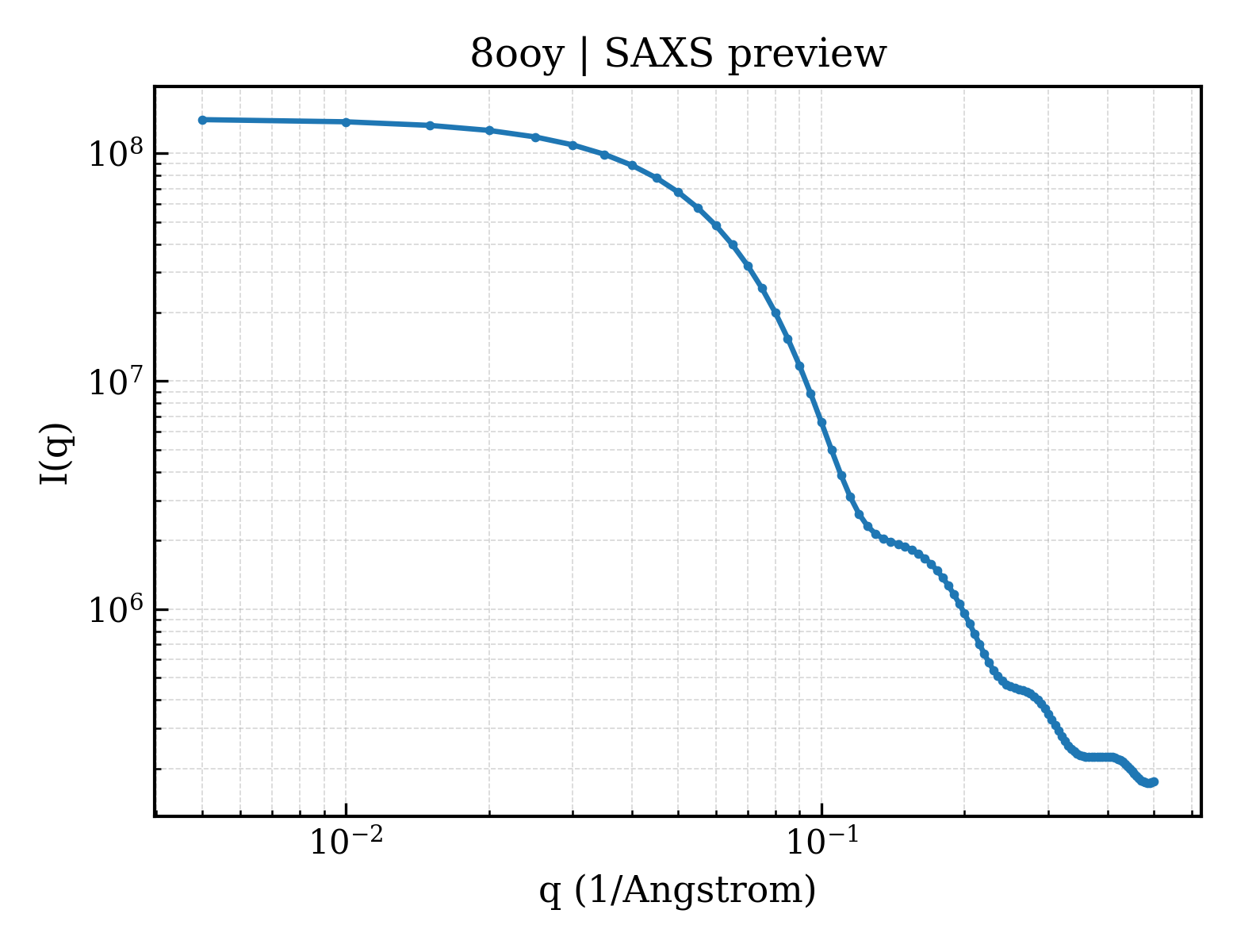
SAXS scattering curve SAXS Profile

SAXS profile for 8ooy

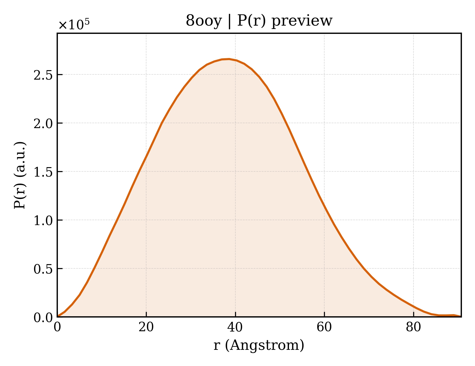
P(r) Distance Distribution P(r) Distribution

P(r) distribution for 8ooy
Download Download

2. Structure Basics 2. Structure Basics

Entry ID entry_id8ooy
Deposition date deposition_date2023-04-06
Structure title titlePol I bound to extended and displaced DNA section - open conformation
Keywords keywordsDNA Polymerase I, Okazaki fragment maturation, DNA BINDING PROTEIN; DNA BINDING PROTEIN
Experimental Method methodELECTRON MICROSCOPY

3. SAXS Parameters (CRYSOL theoretical calculation) 3. SAXS Parameters (CRYSOL)

Radius of gyration Rg (Guinier) rg_guinier29.55
Radius of gyration Rg (electron density) rg_electron28.63
Forward intensity I(0) i0141457000.00
Molecular weight molecular_weight83663.0 kDa
Excluded volume excluded_volume100270 ų
Envelope volume envelope_volume132680 ų
Hydration-shell volume shell_volume38363 ų
Envelope diameter envelope_diameter96.8
Shell Rg shell_rg36.21
Envelope Rg envelope_rg28.42
Shape Rg shape_rg28.61
Total Rg total_rg29.32
Total atoms total_atoms5828
Residues n_residues655
Spherical-harmonic order n_harmonics20
q range q_range— – 0.5000 −1
Data points n_points101
Shell type shell_typedirectional
Solvent electron density solvent_density0.3340 e/ų
Shell contrast contrast_shell0.0300 e/ų
CRYSOL version crysol_version4.1.3

4. P(r) Distance Distribution (GNOM inversion) 4. P(r) Analysis (GNOM)

Maximum dimension Dmax dmax90.8
Rg (real space) rg_real29.42
Rg uncertainty (real space) rg_real_error0.65
I(0) (real space) i0_real1.4150e+08
I(0) uncertainty (real space) i0_real_error2.0370e+06
Rg (reciprocal space) rg_reciprocal29.48
I(0) (reciprocal space) i0_reciprocal141500000.0000
Solution quality estimate total_estimate0.9025
Solution quality rating solution_quality EXCELLENT a EXCELLENT solution
P(r) peaks n_peaks2
Primary peak position r_peak_primary38.7
Skewness Skewness skewness0.176
Kurtosis Kurtosis kurtosis-0.420
Angular range angular_range— – 0.2700 −1
Current regularization parameter α current_alpha0.0000
Highest regularization parameter α highest_alpha12870000.0000
Real-space data points n_real_points55
GNOM version gnom_version4.1.3
Quality Criteria quality_criteria AN1: 0.000; Oscil: 0.941; Stabil: 1.000; Sysdev: 1.000; Positv: 1.000; Valcen: 0.988; Smooth: 0.919

5. Crystallography and Experiment 5. Crystallography & Experiment

6. Entities and Polymers Entities & Polymers (5)

7. Fold Classification (SCOP + CATH) 3 domains

CATH v4.4 (3 domains)

Domain ID domain_id8ooyA01
Class class3 — Alpha Beta
Architecture architecture30 — 2-Layer Sandwich
Topology topology420 — Nucleotidyltransferase; domain 5
Homologous superfamily homologous superfamily10 — Ribonuclease H-like superfamily/Ribonuclease H
Domain ID domain_id8ooyA02
Class class1 — Mainly Alpha
Architecture architecture20 — Up-down Bundle
Topology topology1060 — Taq DNA Polymerase; Chain T, domain 4
Homologous superfamily homologous superfamily10 — Taq DNA Polymerase; Chain T, domain 4
Domain ID domain_id8ooyA03
Class class1 — Mainly Alpha
Architecture architecture10 — Orthogonal Bundle
Topology topology150 — DNA polymerase; domain 1
Homologous superfamily homologous superfamily20 — 5' to 3' exonuclease, C-terminal subdomain

8. Citations (1)

9. Files and Curves (10)