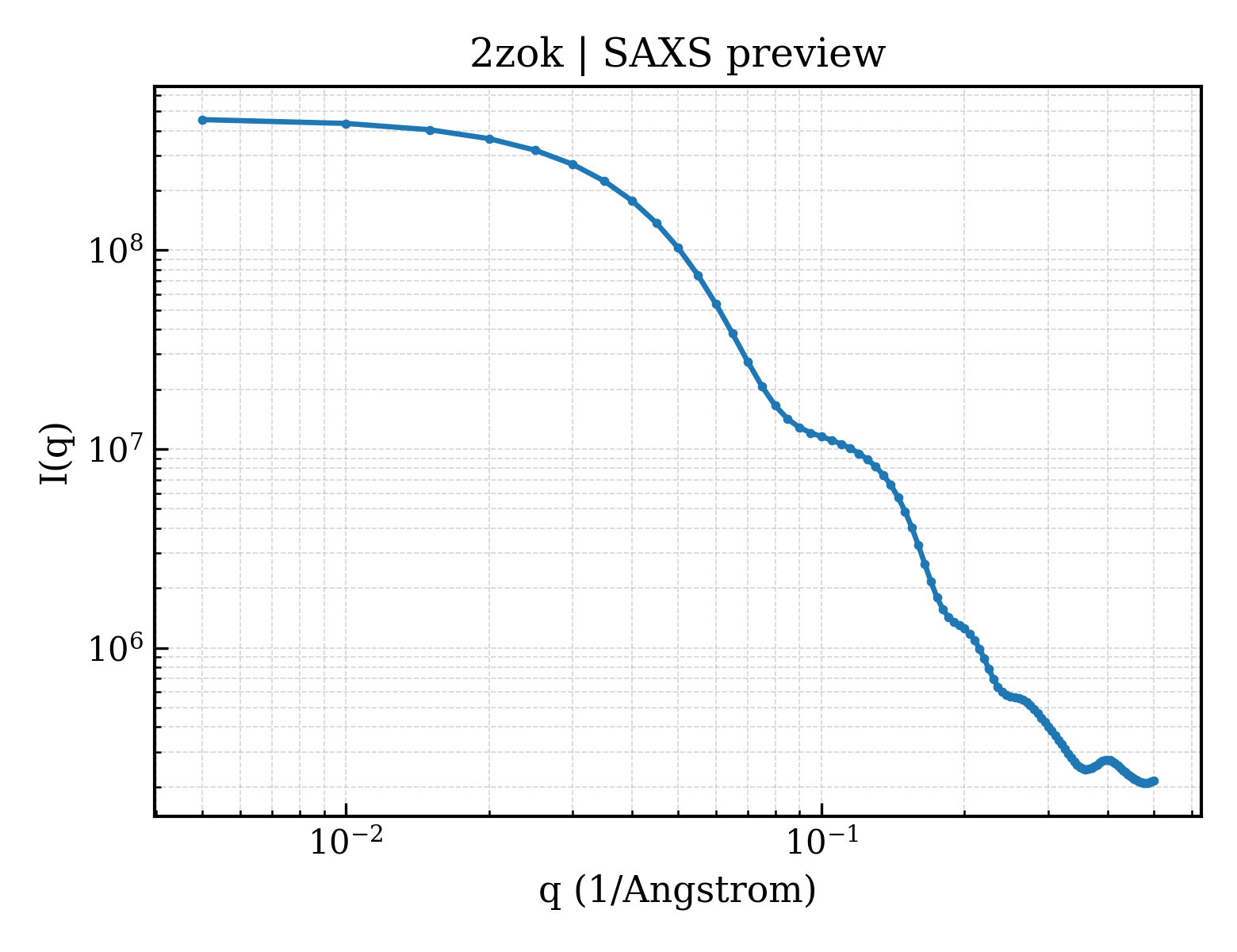
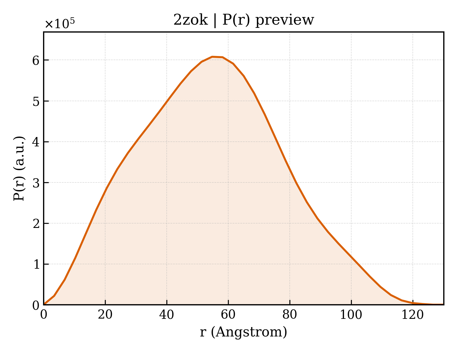
H-2 class I histocompatibility antigen, D-B alpha chain
Mus musculus
State in the Current Structure
| Assembly | Oligomeric State | Construct | Mutations and Modifications | Ligands, Ions and Associated Components | Method and Experimental Conditions | Structure Quality |
|---|---|---|---|---|---|---|
| 1 | Protein heterocomplex Heteromer Protein × 3 PDB declaration: trimeric(3) Consistent with protein copy count | Chain A; UniProt 25–299 | Fragment:extracellular domain, UNP residues 25-299 | Beta-2-microglobulin × 1 (P01887) 9-meric peptide from Spike glycoprotein × 1 (Q02385) SO4 SULFATE ION × 2 GOL GLYCEROL × 1 | X-RAY DIFFRACTION X-ray crystallization conditions:VAPOR DIFFUSION, HANGING DROP;pH 7.5;294 K;0.1M sodium citrate, 28% PEG 3350, 0.2M lithium sulfate, pH 7.5, VAPOR DIFFUSION, HANGING DROP, temperature 294K | Resolution 2.10 Å R-free 0.266 |
| 2 | Protein heterocomplex Heteromer Protein × 3 PDB declaration: trimeric(3) Consistent with protein copy count | Chain C; UniProt 25–299 | Fragment:extracellular domain, UNP residues 25-299 | Beta-2-microglobulin × 1 (P01887) 9-meric peptide from Spike glycoprotein × 1 (Q02385) GOL GLYCEROL × 1 | X-RAY DIFFRACTION X-ray crystallization conditions:VAPOR DIFFUSION, HANGING DROP;pH 7.5;294 K;0.1M sodium citrate, 28% PEG 3350, 0.2M lithium sulfate, pH 7.5, VAPOR DIFFUSION, HANGING DROP, temperature 294K | Resolution 2.10 Å R-free 0.266 |
| 3 | Protein heterocomplex Heteromer Protein × 3 PDB declaration: trimeric(3) Consistent with protein copy count | Chain E; UniProt 25–299 | Fragment:extracellular domain, UNP residues 25-299 | Beta-2-microglobulin × 1 (P01887) 9-meric peptide from Spike glycoprotein × 1 (Q02385) SO4 SULFATE ION × 1 GOL GLYCEROL × 1 | X-RAY DIFFRACTION X-ray crystallization conditions:VAPOR DIFFUSION, HANGING DROP;pH 7.5;294 K;0.1M sodium citrate, 28% PEG 3350, 0.2M lithium sulfate, pH 7.5, VAPOR DIFFUSION, HANGING DROP, temperature 294K | Resolution 2.10 Å R-free 0.266 |
| 4 | Protein heterocomplex Heteromer Protein × 3 PDB declaration: trimeric(3) Consistent with protein copy count | Chain G; UniProt 25–299 | Fragment:extracellular domain, UNP residues 25-299 | Beta-2-microglobulin × 1 (P01887) 9-meric peptide from Spike glycoprotein × 1 (Q02385) SO4 SULFATE ION × 1 GOL GLYCEROL × 1 | X-RAY DIFFRACTION X-ray crystallization conditions:VAPOR DIFFUSION, HANGING DROP;pH 7.5;294 K;0.1M sodium citrate, 28% PEG 3350, 0.2M lithium sulfate, pH 7.5, VAPOR DIFFUSION, HANGING DROP, temperature 294K | Resolution 2.10 Å R-free 0.266 |
| 5 | Protein heterocomplex Heteromer Protein × 6 PDB declaration: hexameric(6) Consistent with protein copy count | Chain A; UniProt 25–299 Chain C; UniProt 25–299 | Fragment:extracellular domain, UNP residues 25-299 | Beta-2-microglobulin × 2 (P01887) 9-meric peptide from Spike glycoprotein × 2 (Q02385) SO4 SULFATE ION × 2 GOL GLYCEROL × 2 | X-RAY DIFFRACTION X-ray crystallization conditions:VAPOR DIFFUSION, HANGING DROP;pH 7.5;294 K;0.1M sodium citrate, 28% PEG 3350, 0.2M lithium sulfate, pH 7.5, VAPOR DIFFUSION, HANGING DROP, temperature 294K | Resolution 2.10 Å R-free 0.266 |
| 6 | Protein heterocomplex Heteromer Protein × 6 PDB declaration: hexameric(6) Consistent with protein copy count | Chain E; UniProt 25–299 Chain G; UniProt 25–299 | Fragment:extracellular domain, UNP residues 25-299 | Beta-2-microglobulin × 2 (P01887) 9-meric peptide from Spike glycoprotein × 2 (Q02385) SO4 SULFATE ION × 2 GOL GLYCEROL × 2 | X-RAY DIFFRACTION X-ray crystallization conditions:VAPOR DIFFUSION, HANGING DROP;pH 7.5;294 K;0.1M sodium citrate, 28% PEG 3350, 0.2M lithium sulfate, pH 7.5, VAPOR DIFFUSION, HANGING DROP, temperature 294K | Resolution 2.10 Å R-free 0.266 |
Other States of the Same Protein in the Database
Each row is a biological assembly of the same UniProt protein in another PDB entry. The “Difference from current entry” column identifies evidence-level differences; no tag means the currently parsed fields agree.
| Other PDB | Difference from Current Entry 2ZOK | Assembly / Oligomeric State | Construct | Mutations and Modifications | Ligands, Ions and Non-polymers | Method and Experimental Conditions | Structure Quality |
|---|---|---|---|---|---|---|---|
| 1BZ9 CRYSTAL STRUCTURE OF MURINE CLASS I MHC H2-DB COMPLEXED WITH A SYNTHETIC PEPTIDE P1027 Deposited 1998-11-06 | Different construct Different ligand/ion Different experimental conditions Different structure-quality metrics | Assembly 1 Protein heterocomplex Heteromer;Protein × 3 PDB declaration: trimeric |
Chain A
26–299(274 aa)
|
Not recorded | No recorded non-water small molecule |
X-RAY DIFFRACTION
X-ray crystallization conditions
pH 6.5;pH 6.5
|
Resolution 2.80 Å R-free 0.319 |
| 1BZ9 CRYSTAL STRUCTURE OF MURINE CLASS I MHC H2-DB COMPLEXED WITH A SYNTHETIC PEPTIDE P1027 Deposited 1998-11-06 | Different construct Different ligand/ion Different experimental conditions Different structure-quality metrics | Assembly 2 Protein heterocomplex Heteromer;Protein × 6 PDB declaration: hexameric |
Chain A
26–299(274 aa)
|
Not recorded | No recorded non-water small molecule |
X-RAY DIFFRACTION
X-ray crystallization conditions
pH 6.5;pH 6.5
|
Resolution 2.80 Å R-free 0.319 |
| 1CE6 MHC CLASS I H-2DB COMPLEXED WITH A SENDAI VIRUS NUCLEOPROTEIN PEPTIDE Deposited 1999-03-17 | Different construct Different ligand/ion Different experimental conditions Different structure-quality metrics | Assembly 1 Protein heterocomplex Heteromer;Protein × 3 PDB declaration: trimeric |
Chain A
25–298(274 aa)
Fragment:FRAGMENT: EXTRACELLULAR DOMAINS
|
Not recorded | SO4 SULFATE ION × 1 |
X-RAY DIFFRACTION
X-ray crystallization conditions
pH 5;pH 5.0
|
Resolution 2.90 Å R-free 0.274 |
| 1FFN CRYSTAL STRUCTURE OF MURINE CLASS I H-2DB COMPLEXED WITH PEPTIDE GP33(C9M) Deposited 2000-07-25 | Different construct Different ligand/ion Different experimental conditions Different structure-quality metrics | Assembly 1 Protein heterocomplex Heteromer;Protein × 3 PDB declaration: trimeric |
Chain A
26–298(273 aa)
Fragment:EXTRACELLULAR PORTION
|
Not recorded | No recorded non-water small molecule |
X-RAY DIFFRACTION
X-ray crystallization conditions
VAPOR DIFFUSION, HANGING DROP;pH 6.5;298 K;14% PEG8000, 1% Dioxane, 25mM MES, pH 6.5, VAPOR DIFFUSION, HANGING DROP, temperature 298K
|
Resolution 2.70 Å R-free 0.286 |
| 1FFN CRYSTAL STRUCTURE OF MURINE CLASS I H-2DB COMPLEXED WITH PEPTIDE GP33(C9M) Deposited 2000-07-25 | Different construct Different ligand/ion Different experimental conditions Different structure-quality metrics | Assembly 2 Protein heterocomplex Heteromer;Protein × 3 PDB declaration: trimeric |
Chain D
26–298(273 aa)
Fragment:EXTRACELLULAR PORTION
|
Not recorded | No recorded non-water small molecule |
X-RAY DIFFRACTION
X-ray crystallization conditions
VAPOR DIFFUSION, HANGING DROP;pH 6.5;298 K;14% PEG8000, 1% Dioxane, 25mM MES, pH 6.5, VAPOR DIFFUSION, HANGING DROP, temperature 298K
|
Resolution 2.70 Å R-free 0.286 |
| 1FFO CRYSTAL STRUCTURE OF MURINE CLASS I H-2DB COMPLEXED WITH SYNTHETIC PEPTIDE GP33 (C9M/K1A) Deposited 2000-07-25 | Different construct Different ligand/ion Different experimental conditions Different structure-quality metrics | Assembly 1 Protein heterocomplex Heteromer;Protein × 3 PDB declaration: trimeric |
Chain A
26–298(273 aa)
Fragment:EXTRACELLULAR PORTION
|
Not recorded | No recorded non-water small molecule |
X-RAY DIFFRACTION
X-ray crystallization conditions
VAPOR DIFFUSION, HANGING DROP;pH 6.5;298 K;23% PEG8000, 6% DMSO, 0.8 M Glycine.
150 mM NaCl, 25mM MES buffer, pH 6.5, VAPOR DIFFUSION, HANGING DROP, temperature 298K
|
Resolution 2.65 Å R-free 0.292 |
| 1FFO CRYSTAL STRUCTURE OF MURINE CLASS I H-2DB COMPLEXED WITH SYNTHETIC PEPTIDE GP33 (C9M/K1A) Deposited 2000-07-25 | Different construct Different ligand/ion Different experimental conditions Different structure-quality metrics | Assembly 2 Protein heterocomplex Heteromer;Protein × 3 PDB declaration: trimeric |
Chain D
26–298(273 aa)
Fragment:EXTRACELLULAR PORTION
|
Not recorded | No recorded non-water small molecule |
X-RAY DIFFRACTION
X-ray crystallization conditions
VAPOR DIFFUSION, HANGING DROP;pH 6.5;298 K;23% PEG8000, 6% DMSO, 0.8 M Glycine.
150 mM NaCl, 25mM MES buffer, pH 6.5, VAPOR DIFFUSION, HANGING DROP, temperature 298K
|
Resolution 2.65 Å R-free 0.292 |
| 1FFP CRYSTAL STRUCTURE OF MURINE CLASS I H-2DB COMPLEXED WITH PEPTIDE GP33 (C9M/K1S) Deposited 2000-07-25 | Different construct Different ligand/ion Different experimental conditions Different structure-quality metrics | Assembly 1 Protein heterocomplex Heteromer;Protein × 3 PDB declaration: trimeric |
Chain A
26–298(273 aa)
Fragment:EXTRACELLULAR PORTION
|
Not recorded | No recorded non-water small molecule |
X-RAY DIFFRACTION
X-ray crystallization conditions
VAPOR DIFFUSION, HANGING DROP;pH 6.5;298 K;25% PEG 8000, 6% DMSO, 0.8M Glycine, 150mM NaCl
25mM MES buffer, pH 6.5, VAPOR DIFFUSION, HANGING DROP, temperature 298K
|
Resolution 2.60 Å R-free 0.303 |
| 1FFP CRYSTAL STRUCTURE OF MURINE CLASS I H-2DB COMPLEXED WITH PEPTIDE GP33 (C9M/K1S) Deposited 2000-07-25 | Different construct Different ligand/ion Different experimental conditions Different structure-quality metrics | Assembly 2 Protein heterocomplex Heteromer;Protein × 3 PDB declaration: trimeric |
Chain D
26–298(273 aa)
Fragment:EXTRACELLULAR PORTION
|
Not recorded | No recorded non-water small molecule |
X-RAY DIFFRACTION
X-ray crystallization conditions
VAPOR DIFFUSION, HANGING DROP;pH 6.5;298 K;25% PEG 8000, 6% DMSO, 0.8M Glycine, 150mM NaCl
25mM MES buffer, pH 6.5, VAPOR DIFFUSION, HANGING DROP, temperature 298K
|
Resolution 2.60 Å R-free 0.303 |
| 1FG2 CRYSTAL STRUCTURE OF THE LCMV PEPTIDIC EPITOPE GP33 IN COMPLEX WITH THE MURINE CLASS I MHC MOLECULE H-2DB Deposited 2000-07-27 | Different construct Different ligand/ion Different experimental conditions Different structure-quality metrics | Assembly 1 Protein heterocomplex Heteromer;Protein × 3 PDB declaration: trimeric |
Chain A
25–304(280 aa)
Fragment:EXTRACELLULAR ALPHA CHAIN (1,2,3)
|
Not recorded | No recorded non-water small molecule |
X-RAY DIFFRACTION
X-ray crystallization conditions
VAPOR DIFFUSION, HANGING DROP;pH 7.4;277 K;ammonium sulfate,
PEG 4000,
hepes, pH 7.4, VAPOR DIFFUSION, HANGING DROP, temperature 277K
|
Resolution 2.75 Å R-free 0.276 |
| 1FG2 CRYSTAL STRUCTURE OF THE LCMV PEPTIDIC EPITOPE GP33 IN COMPLEX WITH THE MURINE CLASS I MHC MOLECULE H-2DB Deposited 2000-07-27 | Different construct Different ligand/ion Different experimental conditions Different structure-quality metrics | Assembly 2 Protein heterocomplex Heteromer;Protein × 3 PDB declaration: trimeric |
Chain D
25–304(280 aa)
Fragment:EXTRACELLULAR ALPHA CHAIN (1,2,3)
|
Not recorded | No recorded non-water small molecule |
X-RAY DIFFRACTION
X-ray crystallization conditions
VAPOR DIFFUSION, HANGING DROP;pH 7.4;277 K;ammonium sulfate,
PEG 4000,
hepes, pH 7.4, VAPOR DIFFUSION, HANGING DROP, temperature 277K
|
Resolution 2.75 Å R-free 0.276 |
| 1FG2 CRYSTAL STRUCTURE OF THE LCMV PEPTIDIC EPITOPE GP33 IN COMPLEX WITH THE MURINE CLASS I MHC MOLECULE H-2DB Deposited 2000-07-27 | Different construct Different ligand/ion Different experimental conditions Different structure-quality metrics | Assembly 3 Protein heterocomplex Heteromer;Protein × 3 PDB declaration: trimeric |
Chain G
25–304(280 aa)
Fragment:EXTRACELLULAR ALPHA CHAIN (1,2,3)
|
Not recorded | No recorded non-water small molecule |
X-RAY DIFFRACTION
X-ray crystallization conditions
VAPOR DIFFUSION, HANGING DROP;pH 7.4;277 K;ammonium sulfate,
PEG 4000,
hepes, pH 7.4, VAPOR DIFFUSION, HANGING DROP, temperature 277K
|
Resolution 2.75 Å R-free 0.276 |
| 1FG2 CRYSTAL STRUCTURE OF THE LCMV PEPTIDIC EPITOPE GP33 IN COMPLEX WITH THE MURINE CLASS I MHC MOLECULE H-2DB Deposited 2000-07-27 | Different construct Different ligand/ion Different experimental conditions Different structure-quality metrics | Assembly 4 Protein heterocomplex Heteromer;Protein × 3 PDB declaration: trimeric |
Chain J
25–304(280 aa)
Fragment:EXTRACELLULAR ALPHA CHAIN (1,2,3)
|
Not recorded | No recorded non-water small molecule |
X-RAY DIFFRACTION
X-ray crystallization conditions
VAPOR DIFFUSION, HANGING DROP;pH 7.4;277 K;ammonium sulfate,
PEG 4000,
hepes, pH 7.4, VAPOR DIFFUSION, HANGING DROP, temperature 277K
|
Resolution 2.75 Å R-free 0.276 |
| 1HOC THE THREE-DIMENSIONAL STRUCTURE OF H-2DB AT 2.4 ANGSTROMS RESOLUTION: IMPLICATIONS FOR ANTIGEN-DETERMINANT SELECTION Deposited 1994-01-02 | Different construct Different ligand/ion Different experimental conditions Different structure-quality metrics | Assembly 1 Protein heterocomplex Heteromer;Protein × 3 PDB declaration: trimeric |
Chain A
25–296(272 aa)
|
Not recorded | No recorded non-water small molecule | X-RAY DIFFRACTION mmCIF provides none of the parsed conditions | Resolution 2.40 Å |
| 1INQ Structure of Minor Histocompatibility Antigen peptide, H13a, complexed to H2-Db Deposited 2001-05-14 | Different construct Different ligand/ion Different experimental conditions Different structure-quality metrics | Assembly 1 Protein heterocomplex Heteromer;Protein × 3 PDB declaration: trimeric |
Chain A
25–299(275 aa)
|
Not recorded | DMS DIMETHYL SULFOXIDE × 1 |
X-RAY DIFFRACTION
X-ray crystallization conditions
VAPOR DIFFUSION, HANGING DROP;pH 7;290 K;12% PEG 4000, 0.1 M Hepes, pH 7.0, VAPOR DIFFUSION, HANGING DROP, temperature 290K
|
Resolution 2.20 Å R-free 0.258 |
| 1JPF Crystal Structure Of The LCMV Peptidic Epitope Gp276 In Complex With The Murine Class I Mhc Molecule H-2Db Deposited 2001-08-02 | Different construct Different ligand/ion Different experimental conditions Different structure-quality metrics | Assembly 1 Protein heterocomplex Heteromer;Protein × 3 PDB declaration: trimeric |
Chain A
25–304(280 aa)
Fragment:extracellular domains
|
Not recorded | No recorded non-water small molecule |
X-RAY DIFFRACTION
X-ray crystallization conditions
VAPOR DIFFUSION, HANGING DROP;pH 6.4;277 K;Ammonium phosphate, PEG8000, Tris-HCl, pH 6.4, VAPOR DIFFUSION, HANGING DROP, temperature 277K
|
Resolution 2.18 Å R-free 0.262 |
| 1JPG Crystal Structure Of The LCMV Peptidic Epitope Np396 In Complex With The Murine Class I Mhc Molecule H-2Db Deposited 2001-08-02 | Different construct Different ligand/ion Different experimental conditions Different structure-quality metrics | Assembly 1 Protein heterocomplex Heteromer;Protein × 3 PDB declaration: trimeric |
Chain A
25–304(280 aa)
Fragment:extracellular domains
|
Not recorded | No recorded non-water small molecule |
X-RAY DIFFRACTION
X-ray crystallization conditions
VAPOR DIFFUSION, HANGING DROP;pH 6.4;277 K;Ammonium phosphate, PEG8000, Tris-HCl, pH 6.4, VAPOR DIFFUSION, HANGING DROP, temperature 277K
|
Resolution 2.20 Å R-free 0.288 |
| 1JUF Structure of Minor Histocompatibility Antigen peptide, H13b, complexed to H2-Db Deposited 2001-08-24 | Different construct Different ligand/ion Different experimental conditions Different structure-quality metrics | Assembly 1 Protein heterocomplex Heteromer;Protein × 3 PDB declaration: trimeric |
Chain A
25–300(276 aa)
Fragment:extracellular domain
|
Not recorded | No recorded non-water small molecule |
X-RAY DIFFRACTION
X-ray crystallization conditions
VAPOR DIFFUSION, HANGING DROP;pH 7.5;291 K;PEG 4000, Sodium Acetates, Hepes, pH 7.5, VAPOR DIFFUSION, HANGING DROP, temperature 291K
|
Resolution 2.00 Å R-free 0.242 |
| 1N3N Crystal structure of a mycobacterial hsp60 epitope with the murine class I MHC molecule H-2Db Deposited 2002-10-29 | Different construct Different ligand/ion Different experimental conditions Different structure-quality metrics | Assembly 1 Protein heterocomplex Heteromer;Protein × 3 PDB declaration: trimeric |
Chain A
25–304(280 aa)
Fragment:EXTRACELLULAR DOMAINS
|
Not recorded | No recorded non-water small molecule |
X-RAY DIFFRACTION
X-ray crystallization conditions
VAPOR DIFFUSION, HANGING DROP;pH 8;277 K;ammonim sulfate, glycerol, tris, pH 8.0, VAPOR DIFFUSION, HANGING DROP, temperature 277K
|
Resolution 3.00 Å R-free 0.307 |
| 1N3N Crystal structure of a mycobacterial hsp60 epitope with the murine class I MHC molecule H-2Db Deposited 2002-10-29 | Different construct Different ligand/ion Different experimental conditions Different structure-quality metrics | Assembly 2 Protein heterocomplex Heteromer;Protein × 3 PDB declaration: trimeric |
Chain G
25–304(280 aa)
Fragment:EXTRACELLULAR DOMAINS
|
Not recorded | SO4 SULFATE ION × 1 |
X-RAY DIFFRACTION
X-ray crystallization conditions
VAPOR DIFFUSION, HANGING DROP;pH 8;277 K;ammonim sulfate, glycerol, tris, pH 8.0, VAPOR DIFFUSION, HANGING DROP, temperature 277K
|
Resolution 3.00 Å R-free 0.307 |
| 1N3N Crystal structure of a mycobacterial hsp60 epitope with the murine class I MHC molecule H-2Db Deposited 2002-10-29 | Different construct Different ligand/ion Different experimental conditions Different structure-quality metrics | Assembly 3 Protein heterocomplex Heteromer;Protein × 3 PDB declaration: trimeric |
Chain E
25–304(280 aa)
Fragment:EXTRACELLULAR DOMAINS
|
Not recorded | SO4 SULFATE ION × 1 |
X-RAY DIFFRACTION
X-ray crystallization conditions
VAPOR DIFFUSION, HANGING DROP;pH 8;277 K;ammonim sulfate, glycerol, tris, pH 8.0, VAPOR DIFFUSION, HANGING DROP, temperature 277K
|
Resolution 3.00 Å R-free 0.307 |
| 1N3N Crystal structure of a mycobacterial hsp60 epitope with the murine class I MHC molecule H-2Db Deposited 2002-10-29 | Different construct Different ligand/ion Different experimental conditions Different structure-quality metrics | Assembly 4 Protein heterocomplex Heteromer;Protein × 3 PDB declaration: trimeric |
Chain C
25–304(280 aa)
Fragment:EXTRACELLULAR DOMAINS
|
Not recorded | No recorded non-water small molecule |
X-RAY DIFFRACTION
X-ray crystallization conditions
VAPOR DIFFUSION, HANGING DROP;pH 8;277 K;ammonim sulfate, glycerol, tris, pH 8.0, VAPOR DIFFUSION, HANGING DROP, temperature 277K
|
Resolution 3.00 Å R-free 0.307 |
| 1N3N Crystal structure of a mycobacterial hsp60 epitope with the murine class I MHC molecule H-2Db Deposited 2002-10-29 | Different construct Different ligand/ion Different experimental conditions Different structure-quality metrics | Assembly 5 Protein heterocomplex Heteromer;Protein × 6 PDB declaration: hexameric |
Chain C
25–304(280 aa)
Fragment:EXTRACELLULAR DOMAINS
Chain G
25–304(280 aa)
Fragment:EXTRACELLULAR DOMAINS
|
Not recorded | SO4 SULFATE ION × 1 |
X-RAY DIFFRACTION
X-ray crystallization conditions
VAPOR DIFFUSION, HANGING DROP;pH 8;277 K;ammonim sulfate, glycerol, tris, pH 8.0, VAPOR DIFFUSION, HANGING DROP, temperature 277K
|
Resolution 3.00 Å R-free 0.307 |
| 1N3N Crystal structure of a mycobacterial hsp60 epitope with the murine class I MHC molecule H-2Db Deposited 2002-10-29 | Different construct Different ligand/ion Different experimental conditions Different structure-quality metrics | Assembly 6 Protein heterocomplex Heteromer;Protein × 6 PDB declaration: hexameric |
Chain A
25–304(280 aa)
Fragment:EXTRACELLULAR DOMAINS
Chain E
25–304(280 aa)
Fragment:EXTRACELLULAR DOMAINS
|
Not recorded | SO4 SULFATE ION × 1 |
X-RAY DIFFRACTION
X-ray crystallization conditions
VAPOR DIFFUSION, HANGING DROP;pH 8;277 K;ammonim sulfate, glycerol, tris, pH 8.0, VAPOR DIFFUSION, HANGING DROP, temperature 277K
|
Resolution 3.00 Å R-free 0.307 |
| 1N5A Crystal structure of the Murine class I Major Histocompatibility Complex of H-2DB, B2-Microglobulin, and A 9-Residue immunodominant peptide epitope gp33 derived from LCMV Deposited 2002-11-05 | Different construct Different ligand/ion Different experimental conditions Different structure-quality metrics | Assembly 1 Protein heterocomplex Heteromer;Protein × 3 PDB declaration: trimeric |
Chain A
25–300(276 aa)
Fragment:Extracellular part
|
Not recorded | No recorded non-water small molecule |
X-RAY DIFFRACTION
X-ray crystallization conditions
VAPOR DIFFUSION, HANGING DROP;pH 8.5;298 K;Ammonium sulfate, ethylene glycol, pH 8.5, VAPOR DIFFUSION, HANGING DROP, temperature 298.0K
|
Resolution 2.85 Å R-free 0.279 |
| 1N5A Crystal structure of the Murine class I Major Histocompatibility Complex of H-2DB, B2-Microglobulin, and A 9-Residue immunodominant peptide epitope gp33 derived from LCMV Deposited 2002-11-05 | Different construct Different ligand/ion Different experimental conditions Different structure-quality metrics | Assembly 2 Protein heterocomplex Heteromer;Protein × 3 PDB declaration: trimeric |
Chain D
25–300(276 aa)
Fragment:Extracellular part
|
Not recorded | No recorded non-water small molecule |
X-RAY DIFFRACTION
X-ray crystallization conditions
VAPOR DIFFUSION, HANGING DROP;pH 8.5;298 K;Ammonium sulfate, ethylene glycol, pH 8.5, VAPOR DIFFUSION, HANGING DROP, temperature 298.0K
|
Resolution 2.85 Å R-free 0.279 |
| 1N5A Crystal structure of the Murine class I Major Histocompatibility Complex of H-2DB, B2-Microglobulin, and A 9-Residue immunodominant peptide epitope gp33 derived from LCMV Deposited 2002-11-05 | Different construct Different ligand/ion Different experimental conditions Different structure-quality metrics | Assembly 3 Protein heterocomplex Heteromer;Protein × 3 PDB declaration: trimeric |
Chain G
25–300(276 aa)
Fragment:Extracellular part
|
Not recorded | No recorded non-water small molecule |
X-RAY DIFFRACTION
X-ray crystallization conditions
VAPOR DIFFUSION, HANGING DROP;pH 8.5;298 K;Ammonium sulfate, ethylene glycol, pH 8.5, VAPOR DIFFUSION, HANGING DROP, temperature 298.0K
|
Resolution 2.85 Å R-free 0.279 |
| 1N5A Crystal structure of the Murine class I Major Histocompatibility Complex of H-2DB, B2-Microglobulin, and A 9-Residue immunodominant peptide epitope gp33 derived from LCMV Deposited 2002-11-05 | Different construct Different ligand/ion Different experimental conditions Different structure-quality metrics | Assembly 4 Protein heterocomplex Heteromer;Protein × 3 PDB declaration: trimeric |
Chain J
25–300(276 aa)
Fragment:Extracellular part
|
Not recorded | No recorded non-water small molecule |
X-RAY DIFFRACTION
X-ray crystallization conditions
VAPOR DIFFUSION, HANGING DROP;pH 8.5;298 K;Ammonium sulfate, ethylene glycol, pH 8.5, VAPOR DIFFUSION, HANGING DROP, temperature 298.0K
|
Resolution 2.85 Å R-free 0.279 |
| 1QLF MHC CLASS I H-2DB COMPLEXED WITH GLYCOPEPTIDE K3G Deposited 1999-08-30 | Different construct Different ligand/ion Different experimental conditions Different structure-quality metrics | Assembly 1 Protein heterocomplex Heteromer;Protein × 3 PDB declaration: trimeric |
Chain A
25–300(276 aa)
Fragment:EXTRACELLULAR DOMAINS
|
Not recorded | GOL GLYCEROL × 2 SO4 SULFATE ION × 1 NAG 2-acetamido-2-deoxy-beta-D-glucopyranose × 1 |
X-RAY DIFFRACTION
X-ray crystallization conditions
pH 5;pH 5.00
|
Resolution 2.65 Å R-free 0.267 |
| 1S7U Crystal structures of the murine class I major histocompatibility complex H-2Db in complex with LCMV-derived gp33 index peptide and three of its escape variants Deposited 2004-01-30 | Different construct Different ligand/ion Different experimental conditions Different structure-quality metrics | Assembly 1 Protein heterocomplex Heteromer;Protein × 3 PDB declaration: trimeric |
Chain A
25–362(338 aa)
|
Not recorded | No recorded non-water small molecule |
X-RAY DIFFRACTION
X-ray crystallization conditions
VAPOR DIFFUSION, HANGING DROP;pH 7.5;293 K;Ammonium sulfate, Tris , pH 7.5, VAPOR DIFFUSION, HANGING DROP, temperature 293K
|
Resolution 2.20 Å R-free 0.262 |
| 1S7U Crystal structures of the murine class I major histocompatibility complex H-2Db in complex with LCMV-derived gp33 index peptide and three of its escape variants Deposited 2004-01-30 | Different construct Different ligand/ion Different experimental conditions Different structure-quality metrics | Assembly 2 Protein heterocomplex Heteromer;Protein × 3 PDB declaration: trimeric |
Chain D
25–362(338 aa)
|
Not recorded | No recorded non-water small molecule |
X-RAY DIFFRACTION
X-ray crystallization conditions
VAPOR DIFFUSION, HANGING DROP;pH 7.5;293 K;Ammonium sulfate, Tris , pH 7.5, VAPOR DIFFUSION, HANGING DROP, temperature 293K
|
Resolution 2.20 Å R-free 0.262 |
| 1S7U Crystal structures of the murine class I major histocompatibility complex H-2Db in complex with LCMV-derived gp33 index peptide and three of its escape variants Deposited 2004-01-30 | Different construct Different ligand/ion Different experimental conditions Different structure-quality metrics | Assembly 3 Protein heterocomplex Heteromer;Protein × 3 PDB declaration: trimeric |
Chain G
25–362(338 aa)
|
Not recorded | No recorded non-water small molecule |
X-RAY DIFFRACTION
X-ray crystallization conditions
VAPOR DIFFUSION, HANGING DROP;pH 7.5;293 K;Ammonium sulfate, Tris , pH 7.5, VAPOR DIFFUSION, HANGING DROP, temperature 293K
|
Resolution 2.20 Å R-free 0.262 |
| 1S7U Crystal structures of the murine class I major histocompatibility complex H-2Db in complex with LCMV-derived gp33 index peptide and three of its escape variants Deposited 2004-01-30 | Different construct Different ligand/ion Different experimental conditions Different structure-quality metrics | Assembly 4 Protein heterocomplex Heteromer;Protein × 3 PDB declaration: trimeric |
Chain J
25–362(338 aa)
|
Not recorded | No recorded non-water small molecule |
X-RAY DIFFRACTION
X-ray crystallization conditions
VAPOR DIFFUSION, HANGING DROP;pH 7.5;293 K;Ammonium sulfate, Tris , pH 7.5, VAPOR DIFFUSION, HANGING DROP, temperature 293K
|
Resolution 2.20 Å R-free 0.262 |
| 1S7V Crystal structures of the murine class I major histocompatibility complex H-2Db in complex with LCMV-derived gp33 index peptide and three of its escape variants Deposited 2004-01-30 | Different construct Different ligand/ion Different experimental conditions Different structure-quality metrics | Assembly 1 Protein heterocomplex Heteromer;Protein × 3 PDB declaration: trimeric |
Chain A
25–362(338 aa)
|
Not recorded | No recorded non-water small molecule |
X-RAY DIFFRACTION
X-ray crystallization conditions
VAPOR DIFFUSION, HANGING DROP;pH 7.5;293 K;Ammonium sulfate, Tris, pH 7.5, VAPOR DIFFUSION, HANGING DROP, temperature 293K
|
Resolution 2.20 Å R-free 0.267 |
| 1S7V Crystal structures of the murine class I major histocompatibility complex H-2Db in complex with LCMV-derived gp33 index peptide and three of its escape variants Deposited 2004-01-30 | Different construct Different ligand/ion Different experimental conditions Different structure-quality metrics | Assembly 2 Protein heterocomplex Heteromer;Protein × 3 PDB declaration: trimeric |
Chain D
25–362(338 aa)
|
Not recorded | No recorded non-water small molecule |
X-RAY DIFFRACTION
X-ray crystallization conditions
VAPOR DIFFUSION, HANGING DROP;pH 7.5;293 K;Ammonium sulfate, Tris, pH 7.5, VAPOR DIFFUSION, HANGING DROP, temperature 293K
|
Resolution 2.20 Å R-free 0.267 |
| 1S7W Crystal structures of the murine class I major histocompatibility complex H-2Db in complex with LCMV-derived gp33 index peptide and three of its escape variants Deposited 2004-01-30 | Different construct Different ligand/ion Different experimental conditions Different structure-quality metrics | Assembly 1 Protein heterocomplex Heteromer;Protein × 3 PDB declaration: trimeric |
Chain A
25–362(338 aa)
|
Not recorded | No recorded non-water small molecule |
X-RAY DIFFRACTION
X-ray crystallization conditions
VAPOR DIFFUSION, HANGING DROP;pH 7.5;293 K;Ammonium sulfate, Tris, pH 7.5, VAPOR DIFFUSION, HANGING DROP, temperature 293K
|
Resolution 2.40 Å R-free 0.246 |
| 1S7W Crystal structures of the murine class I major histocompatibility complex H-2Db in complex with LCMV-derived gp33 index peptide and three of its escape variants Deposited 2004-01-30 | Different construct Different ligand/ion Different experimental conditions Different structure-quality metrics | Assembly 2 Protein heterocomplex Heteromer;Protein × 3 PDB declaration: trimeric |
Chain D
25–362(338 aa)
|
Not recorded | No recorded non-water small molecule |
X-RAY DIFFRACTION
X-ray crystallization conditions
VAPOR DIFFUSION, HANGING DROP;pH 7.5;293 K;Ammonium sulfate, Tris, pH 7.5, VAPOR DIFFUSION, HANGING DROP, temperature 293K
|
Resolution 2.40 Å R-free 0.246 |
| 1S7W Crystal structures of the murine class I major histocompatibility complex H-2Db in complex with LCMV-derived gp33 index peptide and three of its escape variants Deposited 2004-01-30 | Different construct Different ligand/ion Different experimental conditions Different structure-quality metrics | Assembly 3 Protein heterocomplex Heteromer;Protein × 3 PDB declaration: trimeric |
Chain G
25–362(338 aa)
|
Not recorded | No recorded non-water small molecule |
X-RAY DIFFRACTION
X-ray crystallization conditions
VAPOR DIFFUSION, HANGING DROP;pH 7.5;293 K;Ammonium sulfate, Tris, pH 7.5, VAPOR DIFFUSION, HANGING DROP, temperature 293K
|
Resolution 2.40 Å R-free 0.246 |
| 1S7W Crystal structures of the murine class I major histocompatibility complex H-2Db in complex with LCMV-derived gp33 index peptide and three of its escape variants Deposited 2004-01-30 | Different construct Different ligand/ion Different experimental conditions Different structure-quality metrics | Assembly 4 Protein heterocomplex Heteromer;Protein × 3 PDB declaration: trimeric |
Chain J
25–362(338 aa)
|
Not recorded | No recorded non-water small molecule |
X-RAY DIFFRACTION
X-ray crystallization conditions
VAPOR DIFFUSION, HANGING DROP;pH 7.5;293 K;Ammonium sulfate, Tris, pH 7.5, VAPOR DIFFUSION, HANGING DROP, temperature 293K
|
Resolution 2.40 Å R-free 0.246 |
| 1S7X Crystal structures of the murine class I major histocompatibility complex H-2Db in complex with LCMV-derived gp33 index peptide and three of its escape variants Deposited 2004-01-30 | Different construct Different ligand/ion Different experimental conditions Different structure-quality metrics | Assembly 1 Protein heterocomplex Heteromer;Protein × 3 PDB declaration: trimeric |
Chain A
25–362(338 aa)
|
Not recorded | No recorded non-water small molecule |
X-RAY DIFFRACTION
X-ray crystallization conditions
VAPOR DIFFUSION, HANGING DROP;pH 7.5;293 K;Ammonium sulfate, Tris , pH 7.5, VAPOR DIFFUSION, HANGING DROP, temperature 293K
|
Resolution 2.41 Å R-free 0.254 |
| 1S7X Crystal structures of the murine class I major histocompatibility complex H-2Db in complex with LCMV-derived gp33 index peptide and three of its escape variants Deposited 2004-01-30 | Different construct Different ligand/ion Different experimental conditions Different structure-quality metrics | Assembly 2 Protein heterocomplex Heteromer;Protein × 3 PDB declaration: trimeric |
Chain D
25–362(338 aa)
|
Not recorded | No recorded non-water small molecule |
X-RAY DIFFRACTION
X-ray crystallization conditions
VAPOR DIFFUSION, HANGING DROP;pH 7.5;293 K;Ammonium sulfate, Tris , pH 7.5, VAPOR DIFFUSION, HANGING DROP, temperature 293K
|
Resolution 2.41 Å R-free 0.254 |
| 1S7X Crystal structures of the murine class I major histocompatibility complex H-2Db in complex with LCMV-derived gp33 index peptide and three of its escape variants Deposited 2004-01-30 | Different construct Different ligand/ion Different experimental conditions Different structure-quality metrics | Assembly 3 Protein heterocomplex Heteromer;Protein × 3 PDB declaration: trimeric |
Chain G
25–362(338 aa)
|
Not recorded | No recorded non-water small molecule |
X-RAY DIFFRACTION
X-ray crystallization conditions
VAPOR DIFFUSION, HANGING DROP;pH 7.5;293 K;Ammonium sulfate, Tris , pH 7.5, VAPOR DIFFUSION, HANGING DROP, temperature 293K
|
Resolution 2.41 Å R-free 0.254 |
| 1S7X Crystal structures of the murine class I major histocompatibility complex H-2Db in complex with LCMV-derived gp33 index peptide and three of its escape variants Deposited 2004-01-30 | Different construct Different ligand/ion Different experimental conditions Different structure-quality metrics | Assembly 4 Protein heterocomplex Heteromer;Protein × 3 PDB declaration: trimeric |
Chain J
25–362(338 aa)
|
Not recorded | No recorded non-water small molecule |
X-RAY DIFFRACTION
X-ray crystallization conditions
VAPOR DIFFUSION, HANGING DROP;pH 7.5;293 K;Ammonium sulfate, Tris , pH 7.5, VAPOR DIFFUSION, HANGING DROP, temperature 293K
|
Resolution 2.41 Å R-free 0.254 |
| 1WBX CRYSTAL STRUCTURES OF MURINE MHC CLASS I H-2 Db AND Kb MOLECULES IN COMPLEX WITH CTL EPITOPES FROM INFLUENZA A VIRUS: IMPLICATIONS FOR TCR REPERTOIRE SELECTION AND IMMUNODOMINANCE Deposited 2004-11-05 | Different construct Different ligand/ion Different experimental conditions Different structure-quality metrics | Assembly 1 Protein heterocomplex Heteromer;Protein × 3 PDB declaration: trimeric |
Chain A
25–300(276 aa)
Fragment:EXTRACELLULAR DOMAIN, RESIDUES 25-300
|
Not recorded | No recorded non-water small molecule | X-RAY DIFFRACTION mmCIF provides none of the parsed conditions | Resolution 1.90 Å R-free 0.255 |
| 1WBY CRYSTAL STRUCTURES OF MURINE MHC CLASS I H-2 Db AND Kb MOLECULES IN COMPLEX WITH CTL EPITOPES FROM INFLUENZA A VIRUS: IMPLICATIONS FOR TCR REPERTOIRE SELECTION AND IMMUNODOMINANCE Deposited 2004-11-05 | Different construct Different ligand/ion Different experimental conditions Different structure-quality metrics | Assembly 1 Protein heterocomplex Heteromer;Protein × 3 PDB declaration: trimeric |
Chain A
25–300(276 aa)
Fragment:EXTRACELLULAR DOMAIN, RESIDUES 25-300
|
Not recorded | No recorded non-water small molecule | X-RAY DIFFRACTION mmCIF provides none of the parsed conditions | Resolution 2.30 Å R-free 0.304 |
| 1YN6 Crystal structure of a mouse MHC class I protein, H2-Db, in complex with a peptide from the influenza A acid polymerase Deposited 2005-01-23 | Different construct Different ligand/ion Different experimental conditions Different structure-quality metrics | Assembly 1 Protein heterocomplex Heteromer;Protein × 3 PDB declaration: trimeric |
Chain A
26–298(273 aa)
|
Not recorded | No recorded non-water small molecule |
X-RAY DIFFRACTION
X-ray crystallization conditions
VAPOR DIFFUSION, HANGING DROP;pH 5.6;293 K;sodium citrate, ammonium acetate, PEG 4000, pH 5.6, VAPOR DIFFUSION, HANGING DROP, temperature 293K
|
Resolution 2.20 Å R-free 0.254 |
| 1YN7 Crystal structure of a mouse MHC class I protein, H2-Db, in complex with a mutated peptide (R7A) of the influenza A acid polymerase Deposited 2005-01-24 | Different construct Different ligand/ion Different experimental conditions Different structure-quality metrics | Assembly 1 Protein heterocomplex Heteromer;Protein × 3 PDB declaration: trimeric |
Chain A
26–298(273 aa)
|
Not recorded | No recorded non-water small molecule |
X-RAY DIFFRACTION
X-ray crystallization conditions
VAPOR DIFFUSION, HANGING DROP;pH 5.6;293 K;sodium citrate, ammonium acetate, PEG 4000, pH 5.6, VAPOR DIFFUSION, HANGING DROP, temperature 293K
|
Resolution 2.20 Å R-free 0.265 |
| 1ZHB Crystal Structure Of The Murine Class I Major Histocompatibility Complex Of H-2Db, B2-Microglobulin, and a 9-Residue Peptide Derived from rat dopamine beta-monooxigenase Deposited 2005-04-25 | Different construct Different ligand/ion Different experimental conditions Different structure-quality metrics | Assembly 1 Protein heterocomplex Heteromer;Protein × 3 PDB declaration: trimeric |
Chain A
25–300(276 aa)
Fragment:EXTRAcellular part
|
Not recorded | No recorded non-water small molecule |
X-RAY DIFFRACTION
X-ray crystallization conditions
VAPOR DIFFUSION, HANGING DROP;pH 9;298 K;ammonium sulphate, Tris HCl, pH 9.0, VAPOR DIFFUSION, HANGING DROP, temperature 298.0K
|
Resolution 2.70 Å R-free 0.262 |
| 1ZHB Crystal Structure Of The Murine Class I Major Histocompatibility Complex Of H-2Db, B2-Microglobulin, and a 9-Residue Peptide Derived from rat dopamine beta-monooxigenase Deposited 2005-04-25 | Different construct Different ligand/ion Different experimental conditions Different structure-quality metrics | Assembly 2 Protein heterocomplex Heteromer;Protein × 3 PDB declaration: trimeric |
Chain D
25–300(276 aa)
Fragment:EXTRAcellular part
|
Not recorded | No recorded non-water small molecule |
X-RAY DIFFRACTION
X-ray crystallization conditions
VAPOR DIFFUSION, HANGING DROP;pH 9;298 K;ammonium sulphate, Tris HCl, pH 9.0, VAPOR DIFFUSION, HANGING DROP, temperature 298.0K
|
Resolution 2.70 Å R-free 0.262 |
| 1ZHB Crystal Structure Of The Murine Class I Major Histocompatibility Complex Of H-2Db, B2-Microglobulin, and a 9-Residue Peptide Derived from rat dopamine beta-monooxigenase Deposited 2005-04-25 | Different construct Different ligand/ion Different experimental conditions Different structure-quality metrics | Assembly 3 Protein heterocomplex Heteromer;Protein × 3 PDB declaration: trimeric |
Chain G
25–300(276 aa)
Fragment:EXTRAcellular part
|
Not recorded | No recorded non-water small molecule |
X-RAY DIFFRACTION
X-ray crystallization conditions
VAPOR DIFFUSION, HANGING DROP;pH 9;298 K;ammonium sulphate, Tris HCl, pH 9.0, VAPOR DIFFUSION, HANGING DROP, temperature 298.0K
|
Resolution 2.70 Å R-free 0.262 |
| 1ZHB Crystal Structure Of The Murine Class I Major Histocompatibility Complex Of H-2Db, B2-Microglobulin, and a 9-Residue Peptide Derived from rat dopamine beta-monooxigenase Deposited 2005-04-25 | Different construct Different ligand/ion Different experimental conditions Different structure-quality metrics | Assembly 4 Protein heterocomplex Heteromer;Protein × 3 PDB declaration: trimeric |
Chain J
25–300(276 aa)
Fragment:EXTRAcellular part
|
Not recorded | No recorded non-water small molecule |
X-RAY DIFFRACTION
X-ray crystallization conditions
VAPOR DIFFUSION, HANGING DROP;pH 9;298 K;ammonium sulphate, Tris HCl, pH 9.0, VAPOR DIFFUSION, HANGING DROP, temperature 298.0K
|
Resolution 2.70 Å R-free 0.262 |
| 2CII The crystal structure of H-2Db complexed with a partial peptide epitope suggests an MHC Class I assembly-intermediate Deposited 2006-03-21 | Different construct Different ligand/ion Different experimental conditions Different structure-quality metrics | Assembly 1 Protein heterocomplex Heteromer;Protein × 3 PDB declaration: trimeric |
Chain A
25–299(275 aa)
Fragment:RESIDUES 25-299
|
Not recorded | GOL GLYCEROL × 2 |
X-RAY DIFFRACTION
X-ray crystallization conditions
pH 5;25% PEG 6000, 100MM AMMONIUM SULFATE, 100 MM SODIUM CHLORIDE, 50 MM MES PH 5.0
|
Resolution 2.55 Å R-free 0.297 |
| 2F74 Murine MHC class I H-2Db in complex with human b2-microglobulin and LCMV-derived immunodminant peptide gp33 Deposited 2005-11-30 | Different construct Different ligand/ion Different experimental conditions Different structure-quality metrics | Assembly 1 Protein heterocomplex Heteromer;Protein × 3 PDB declaration: trimeric |
Chain A
25–300(276 aa)
|
Not recorded | No recorded non-water small molecule |
X-RAY DIFFRACTION
X-ray crystallization conditions
VAPOR DIFFUSION;pH 8;290 K;20% PEG 6K, pH 8, VAPOR DIFFUSION, temperature 290K
|
Resolution 2.70 Å R-free 0.298 |
| 2F74 Murine MHC class I H-2Db in complex with human b2-microglobulin and LCMV-derived immunodminant peptide gp33 Deposited 2005-11-30 | Different construct Different ligand/ion Different experimental conditions Different structure-quality metrics | Assembly 2 Protein heterocomplex Heteromer;Protein × 3 PDB declaration: trimeric |
Chain D
25–300(276 aa)
|
Not recorded | No recorded non-water small molecule |
X-RAY DIFFRACTION
X-ray crystallization conditions
VAPOR DIFFUSION;pH 8;290 K;20% PEG 6K, pH 8, VAPOR DIFFUSION, temperature 290K
|
Resolution 2.70 Å R-free 0.298 |
| 2VE6 Crystal structure of a Murine MHC class I H2-Db molecule in complex with a photocleavable peptide Deposited 2007-10-17 | Different construct Different ligand/ion Different experimental conditions Different structure-quality metrics | Assembly 1 Protein heterocomplex Heteromer;Protein × 3 PDB declaration: trimeric |
Chain A
25–301(277 aa)
Fragment:RESIDUES 25-301
|
Not recorded | No recorded non-water small molecule |
X-RAY DIFFRACTION
X-ray crystallization conditions
VAPOR DIFFUSION, HANGING DROP;CRYSTALS WERE GROWN AT ROOM TEMPERATURE USING THE HANGING-DROP, VAPOR-DIFFUSION METHOD WITH A WELL SOLUTION OF 15% (W/V) PEG 8000, 0.05 M K/NA PHOSPHATE, 50-100 MM BETA-OCTYL-GLUCOPYRANOSIDE AND 0.1 M CACODYLATE AT PH 6.4.
|
Resolution 2.65 Å R-free 0.291 |
| 2VE6 Crystal structure of a Murine MHC class I H2-Db molecule in complex with a photocleavable peptide Deposited 2007-10-17 | Different construct Different ligand/ion Different experimental conditions Different structure-quality metrics | Assembly 2 Protein heterocomplex Heteromer;Protein × 3 PDB declaration: trimeric |
Chain D
25–301(277 aa)
Fragment:RESIDUES 25-301
|
Not recorded | No recorded non-water small molecule |
X-RAY DIFFRACTION
X-ray crystallization conditions
VAPOR DIFFUSION, HANGING DROP;CRYSTALS WERE GROWN AT ROOM TEMPERATURE USING THE HANGING-DROP, VAPOR-DIFFUSION METHOD WITH A WELL SOLUTION OF 15% (W/V) PEG 8000, 0.05 M K/NA PHOSPHATE, 50-100 MM BETA-OCTYL-GLUCOPYRANOSIDE AND 0.1 M CACODYLATE AT PH 6.4.
|
Resolution 2.65 Å R-free 0.291 |
| 2VE6 Crystal structure of a Murine MHC class I H2-Db molecule in complex with a photocleavable peptide Deposited 2007-10-17 | Different construct Different ligand/ion Different experimental conditions Different structure-quality metrics | Assembly 3 Protein heterocomplex Heteromer;Protein × 3 PDB declaration: trimeric |
Chain G
25–301(277 aa)
Fragment:RESIDUES 25-301
|
Not recorded | No recorded non-water small molecule |
X-RAY DIFFRACTION
X-ray crystallization conditions
VAPOR DIFFUSION, HANGING DROP;CRYSTALS WERE GROWN AT ROOM TEMPERATURE USING THE HANGING-DROP, VAPOR-DIFFUSION METHOD WITH A WELL SOLUTION OF 15% (W/V) PEG 8000, 0.05 M K/NA PHOSPHATE, 50-100 MM BETA-OCTYL-GLUCOPYRANOSIDE AND 0.1 M CACODYLATE AT PH 6.4.
|
Resolution 2.65 Å R-free 0.291 |
| 2VE6 Crystal structure of a Murine MHC class I H2-Db molecule in complex with a photocleavable peptide Deposited 2007-10-17 | Different construct Different ligand/ion Different experimental conditions Different structure-quality metrics | Assembly 4 Protein heterocomplex Heteromer;Protein × 3 PDB declaration: trimeric |
Chain J
25–301(277 aa)
Fragment:RESIDUES 25-301
|
Not recorded | No recorded non-water small molecule |
X-RAY DIFFRACTION
X-ray crystallization conditions
VAPOR DIFFUSION, HANGING DROP;CRYSTALS WERE GROWN AT ROOM TEMPERATURE USING THE HANGING-DROP, VAPOR-DIFFUSION METHOD WITH A WELL SOLUTION OF 15% (W/V) PEG 8000, 0.05 M K/NA PHOSPHATE, 50-100 MM BETA-OCTYL-GLUCOPYRANOSIDE AND 0.1 M CACODYLATE AT PH 6.4.
|
Resolution 2.65 Å R-free 0.291 |
| 2ZOL Crystal structure of H-2Db in complex with the W513S variant of JHMV epitope S510 Deposited 2008-05-22 | Different ligand/ion Different experimental conditions Different structure-quality metrics | Assembly 1 Protein heterocomplex Heteromer;Protein × 3 PDB declaration: trimeric |
Chain A
25–299(275 aa)
Fragment:extracellular domain, UNP residues 25-299
|
Not recorded | No recorded non-water small molecule |
X-RAY DIFFRACTION
X-ray crystallization conditions
VAPOR DIFFUSION, HANGING DROP;pH 6.4;294 K;0.1M sodium citrate, 28% PEG 3350, 0.15M lithium sulfate, pH 6.4, VAPOR DIFFUSION, HANGING DROP, temperature 294K
|
Resolution 2.70 Å R-free 0.307 |
| 2ZOL Crystal structure of H-2Db in complex with the W513S variant of JHMV epitope S510 Deposited 2008-05-22 | Different ligand/ion Different experimental conditions Different structure-quality metrics | Assembly 2 Protein heterocomplex Heteromer;Protein × 3 PDB declaration: trimeric |
Chain C
25–299(275 aa)
Fragment:extracellular domain, UNP residues 25-299
|
Not recorded | SO4 SULFATE ION × 1 |
X-RAY DIFFRACTION
X-ray crystallization conditions
VAPOR DIFFUSION, HANGING DROP;pH 6.4;294 K;0.1M sodium citrate, 28% PEG 3350, 0.15M lithium sulfate, pH 6.4, VAPOR DIFFUSION, HANGING DROP, temperature 294K
|
Resolution 2.70 Å R-free 0.307 |
| 2ZOL Crystal structure of H-2Db in complex with the W513S variant of JHMV epitope S510 Deposited 2008-05-22 | Different ligand/ion Different experimental conditions Different structure-quality metrics | Assembly 3 Protein heterocomplex Heteromer;Protein × 6 PDB declaration: hexameric |
Chain A
25–299(275 aa)
Fragment:extracellular domain, UNP residues 25-299
Chain C
25–299(275 aa)
Fragment:extracellular domain, UNP residues 25-299
|
Not recorded | SO4 SULFATE ION × 1 |
X-RAY DIFFRACTION
X-ray crystallization conditions
VAPOR DIFFUSION, HANGING DROP;pH 6.4;294 K;0.1M sodium citrate, 28% PEG 3350, 0.15M lithium sulfate, pH 6.4, VAPOR DIFFUSION, HANGING DROP, temperature 294K
|
Resolution 2.70 Å R-free 0.307 |
| 3BUY MHC-I in complex with peptide Deposited 2008-01-03 | Different construct Different ligand/ion Different experimental conditions Different structure-quality metrics | Assembly 1 Protein heterocomplex Heteromer;Protein × 3 PDB declaration: trimeric |
Chain A
26–300(275 aa)
Fragment:residues 2-276
|
Not recorded | No recorded non-water small molecule | X-RAY DIFFRACTION mmCIF provides none of the parsed conditions | Resolution 2.60 Å R-free 0.306 |
| 3CC5 H-2Db complex with human gp100 Deposited 2008-02-24 | Different construct Different ligand/ion Different experimental conditions Different structure-quality metrics | Assembly 1 Protein heterocomplex Heteromer;Protein × 3 PDB declaration: trimeric |
Chain A
25–300(276 aa)
Fragment:Extracellular part, UNP residues 25-300
|
Not recorded | SO4 SULFATE ION × 2 GOL GLYCEROL × 3 |
X-RAY DIFFRACTION
X-ray crystallization conditions
VAPOR DIFFUSION, HANGING DROP;pH 7;293 K;10% PEG 6000, 0.1M Ammonium sulfate, 0.1M Tris, pH 7.0, VAPOR DIFFUSION, HANGING DROP, temperature 293K
|
Resolution 1.91 Å R-free 0.268 |
| 3CC5 H-2Db complex with human gp100 Deposited 2008-02-24 | Different construct Different ligand/ion Different experimental conditions Different structure-quality metrics | Assembly 2 Protein heterocomplex Heteromer;Protein × 3 PDB declaration: trimeric |
Chain D
25–300(276 aa)
Fragment:Extracellular part, UNP residues 25-300
|
Not recorded | SO4 SULFATE ION × 1 GOL GLYCEROL × 3 |
X-RAY DIFFRACTION
X-ray crystallization conditions
VAPOR DIFFUSION, HANGING DROP;pH 7;293 K;10% PEG 6000, 0.1M Ammonium sulfate, 0.1M Tris, pH 7.0, VAPOR DIFFUSION, HANGING DROP, temperature 293K
|
Resolution 1.91 Å R-free 0.268 |
| 3CCH H-2Db complex with murine gp100 Deposited 2008-02-25 | Different construct Different ligand/ion Different experimental conditions Different structure-quality metrics | Assembly 1 Protein heterocomplex Heteromer;Protein × 3 PDB declaration: trimeric |
Chain A
25–300(276 aa)
Fragment:UNP residues 25-300
|
Not recorded | GOL GLYCEROL × 1 SO4 SULFATE ION × 4 |
X-RAY DIFFRACTION
X-ray crystallization conditions
VAPOR DIFFUSION, HANGING DROP;pH 9;277 K;1.8M Ammonium Sulfate, 0.1M Tris, pH 9.0,
VAPOR DIFFUSION, HANGING DROP, temperature 277K
|
Resolution 2.60 Å R-free 0.279 |
| 3CCH H-2Db complex with murine gp100 Deposited 2008-02-25 | Different construct Different ligand/ion Different experimental conditions Different structure-quality metrics | Assembly 2 Protein heterocomplex Heteromer;Protein × 3 PDB declaration: trimeric |
Chain D
25–300(276 aa)
Fragment:UNP residues 25-300
|
Not recorded | GOL GLYCEROL × 1 SO4 SULFATE ION × 7 |
X-RAY DIFFRACTION
X-ray crystallization conditions
VAPOR DIFFUSION, HANGING DROP;pH 9;277 K;1.8M Ammonium Sulfate, 0.1M Tris, pH 9.0,
VAPOR DIFFUSION, HANGING DROP, temperature 277K
|
Resolution 2.60 Å R-free 0.279 |
| 3CCH H-2Db complex with murine gp100 Deposited 2008-02-25 | Different construct Different ligand/ion Different experimental conditions Different structure-quality metrics | Assembly 3 Protein heterocomplex Heteromer;Protein × 3 PDB declaration: trimeric |
Chain G
25–300(276 aa)
Fragment:UNP residues 25-300
|
Not recorded | GOL GLYCEROL × 1 SO4 SULFATE ION × 5 |
X-RAY DIFFRACTION
X-ray crystallization conditions
VAPOR DIFFUSION, HANGING DROP;pH 9;277 K;1.8M Ammonium Sulfate, 0.1M Tris, pH 9.0,
VAPOR DIFFUSION, HANGING DROP, temperature 277K
|
Resolution 2.60 Å R-free 0.279 |
| 3CCH H-2Db complex with murine gp100 Deposited 2008-02-25 | Different construct Different ligand/ion Different experimental conditions Different structure-quality metrics | Assembly 4 Protein heterocomplex Heteromer;Protein × 3 PDB declaration: trimeric |
Chain J
25–300(276 aa)
Fragment:UNP residues 25-300
|
Not recorded | GOL GLYCEROL × 1 SO4 SULFATE ION × 5 |
X-RAY DIFFRACTION
X-ray crystallization conditions
VAPOR DIFFUSION, HANGING DROP;pH 9;277 K;1.8M Ammonium Sulfate, 0.1M Tris, pH 9.0,
VAPOR DIFFUSION, HANGING DROP, temperature 277K
|
Resolution 2.60 Å R-free 0.279 |
| 3CCH H-2Db complex with murine gp100 Deposited 2008-02-25 | Different construct Different ligand/ion Different experimental conditions Different structure-quality metrics | Assembly 5 Protein heterocomplex Heteromer;Protein × 6 PDB declaration: hexameric |
Chain D
25–300(276 aa)
Fragment:UNP residues 25-300
Chain J
25–300(276 aa)
Fragment:UNP residues 25-300
|
Not recorded | GOL GLYCEROL × 2 SO4 SULFATE ION × 12 |
X-RAY DIFFRACTION
X-ray crystallization conditions
VAPOR DIFFUSION, HANGING DROP;pH 9;277 K;1.8M Ammonium Sulfate, 0.1M Tris, pH 9.0,
VAPOR DIFFUSION, HANGING DROP, temperature 277K
|
Resolution 2.60 Å R-free 0.279 |
| 3CCH H-2Db complex with murine gp100 Deposited 2008-02-25 | Different construct Different ligand/ion Different experimental conditions Different structure-quality metrics | Assembly 6 Protein heterocomplex Heteromer;Protein × 6 PDB declaration: hexameric |
Chain A
25–300(276 aa)
Fragment:UNP residues 25-300
Chain G
25–300(276 aa)
Fragment:UNP residues 25-300
|
Not recorded | GOL GLYCEROL × 2 SO4 SULFATE ION × 9 |
X-RAY DIFFRACTION
X-ray crystallization conditions
VAPOR DIFFUSION, HANGING DROP;pH 9;277 K;1.8M Ammonium Sulfate, 0.1M Tris, pH 9.0,
VAPOR DIFFUSION, HANGING DROP, temperature 277K
|
Resolution 2.60 Å R-free 0.279 |
| 3CH1 Crystal structure of H-2Db in complex with chimeric gp100 Deposited 2008-03-06 | Different construct Different ligand/ion Different experimental conditions Different structure-quality metrics | Assembly 1 Protein heterocomplex Heteromer;Protein × 3 PDB declaration: trimeric |
Chain D
25–300(276 aa)
Fragment:UNP residues 25-300
|
Not recorded | GOL GLYCEROL × 7 SO4 SULFATE ION × 1 |
X-RAY DIFFRACTION
X-ray crystallization conditions
VAPOR DIFFUSION, HANGING DROP;pH 9;277 K;1.8M ammonium sulfate, 0.1M Tris Cl, pH 9.0, VAPOR DIFFUSION, HANGING DROP, temperature 277K
|
Resolution 2.30 Å R-free 0.264 |
| 3CH1 Crystal structure of H-2Db in complex with chimeric gp100 Deposited 2008-03-06 | Different construct Different ligand/ion Different experimental conditions Different structure-quality metrics | Assembly 2 Protein heterocomplex Heteromer;Protein × 3 PDB declaration: trimeric |
Chain A
25–300(276 aa)
Fragment:UNP residues 25-300
|
Not recorded | GOL GLYCEROL × 9 SO4 SULFATE ION × 1 |
X-RAY DIFFRACTION
X-ray crystallization conditions
VAPOR DIFFUSION, HANGING DROP;pH 9;277 K;1.8M ammonium sulfate, 0.1M Tris Cl, pH 9.0, VAPOR DIFFUSION, HANGING DROP, temperature 277K
|
Resolution 2.30 Å R-free 0.264 |
| 3CH1 Crystal structure of H-2Db in complex with chimeric gp100 Deposited 2008-03-06 | Different construct Different ligand/ion Different experimental conditions Different structure-quality metrics | Assembly 3 Protein heterocomplex Heteromer;Protein × 3 PDB declaration: trimeric |
Chain G
25–300(276 aa)
Fragment:UNP residues 25-300
|
Not recorded | GOL GLYCEROL × 4 |
X-RAY DIFFRACTION
X-ray crystallization conditions
VAPOR DIFFUSION, HANGING DROP;pH 9;277 K;1.8M ammonium sulfate, 0.1M Tris Cl, pH 9.0, VAPOR DIFFUSION, HANGING DROP, temperature 277K
|
Resolution 2.30 Å R-free 0.264 |
| 3CH1 Crystal structure of H-2Db in complex with chimeric gp100 Deposited 2008-03-06 | Different construct Different ligand/ion Different experimental conditions Different structure-quality metrics | Assembly 4 Protein heterocomplex Heteromer;Protein × 3 PDB declaration: trimeric |
Chain J
25–300(276 aa)
Fragment:UNP residues 25-300
|
Not recorded | GOL GLYCEROL × 6 SO4 SULFATE ION × 3 |
X-RAY DIFFRACTION
X-ray crystallization conditions
VAPOR DIFFUSION, HANGING DROP;pH 9;277 K;1.8M ammonium sulfate, 0.1M Tris Cl, pH 9.0, VAPOR DIFFUSION, HANGING DROP, temperature 277K
|
Resolution 2.30 Å R-free 0.264 |
| 3CPL Crystal Structure of H-2Db in complex with a variant M6A of the NP366 peptide from influenza A virus Deposited 2008-03-31 | Different construct Different ligand/ion Different experimental conditions Different structure-quality metrics | Assembly 1 Protein heterocomplex Heteromer;Protein × 3 PDB declaration: trimeric |
Chain A
25–299(275 aa)
Fragment:residues 1-275 (UNIPROT 25-299)
|
Not recorded | No recorded non-water small molecule |
X-RAY DIFFRACTION
X-ray crystallization conditions
VAPOR DIFFUSION, HANGING DROP;293 K;0.1M potassium thiocianate, 30% PEG 2000, vapor diffusion, hanging drop, temperature 293K
|
Resolution 2.50 Å R-free 0.286 |
| 3CPL Crystal Structure of H-2Db in complex with a variant M6A of the NP366 peptide from influenza A virus Deposited 2008-03-31 | Different construct Different ligand/ion Different experimental conditions Different structure-quality metrics | Assembly 2 Protein heterocomplex Heteromer;Protein × 3 PDB declaration: trimeric |
Chain C
25–299(275 aa)
Fragment:residues 1-275 (UNIPROT 25-299)
|
Not recorded | No recorded non-water small molecule |
X-RAY DIFFRACTION
X-ray crystallization conditions
VAPOR DIFFUSION, HANGING DROP;293 K;0.1M potassium thiocianate, 30% PEG 2000, vapor diffusion, hanging drop, temperature 293K
|
Resolution 2.50 Å R-free 0.286 |
| 3FTG Crystal Structure of H2Db in complex with NP366-N3A variant peptide from influenza Deposited 2009-01-12 | Different construct Different ligand/ion Different experimental conditions Different structure-quality metrics | Assembly 1 Protein heterocomplex Heteromer;Protein × 3 PDB declaration: trimeric |
Chain A
25–304(280 aa)
|
Not recorded | No recorded non-water small molecule |
X-RAY DIFFRACTION
X-ray crystallization conditions
VAPOR DIFFUSION, HANGING DROP;pH 5.6;293 K;0.2M lithium sulfate, 28% PEG 3350, 0.1M sodium citrate, pH 5.6, vapor diffusion, hanging drop, temperature 293K
|
Resolution 2.60 Å R-free 0.304 |
| 3L3H X-ray crystal structure of the F6A mutant of influenza A acid polymerase epitope PA224 bound to murine H2-Db MHC Deposited 2009-12-17 | Different construct Different ligand/ion Different experimental conditions Different structure-quality metrics | Assembly 1 Protein heterocomplex Heteromer;Protein × 3 PDB declaration: trimeric |
Chain A
26–300(275 aa)
|
Not recorded | No recorded non-water small molecule |
X-RAY DIFFRACTION
X-ray crystallization conditions
VAPOR DIFFUSION, HANGING DROP;pH 5.6;293 K;0.1M citrate, 0.2M ammonium acetate, 25% PEG4000, pH 5.6, VAPOR DIFFUSION, HANGING DROP, temperature 293K
|
Resolution 2.70 Å R-free 0.284 |
| 3PQY Crystal Structure of 6218 TCR in complex with the H2Db-PA224 Deposited 2010-11-28 | Different construct Different oligomeric state Different ligand/ion Different experimental conditions Different structure-quality metrics | Assembly 1 Insufficient information Heteromer;Protein × 5 PDB declaration: pentameric |
Chain A
26–300(275 aa)
Fragment:UNP residues 26-300
|
Not recorded | No recorded non-water small molecule |
X-RAY DIFFRACTION
X-ray crystallization conditions
VAPOR DIFFUSION, HANGING DROP;pH 6.8;277 K;10-14% PEG 3350, 0.2M KCN, 0.1M Bis Tris Propane pH 6.8, 5% glycerol, vapor diffusion, hanging drop, temperature 277K
|
Resolution 3.19 Å R-free 0.316 |
| 3PQY Crystal Structure of 6218 TCR in complex with the H2Db-PA224 Deposited 2010-11-28 | Different construct Different oligomeric state Different ligand/ion Different experimental conditions Different structure-quality metrics | Assembly 2 Insufficient information Heteromer;Protein × 5 PDB declaration: pentameric |
Chain F
26–300(275 aa)
Fragment:UNP residues 26-300
|
Not recorded | No recorded non-water small molecule |
X-RAY DIFFRACTION
X-ray crystallization conditions
VAPOR DIFFUSION, HANGING DROP;pH 6.8;277 K;10-14% PEG 3350, 0.2M KCN, 0.1M Bis Tris Propane pH 6.8, 5% glycerol, vapor diffusion, hanging drop, temperature 277K
|
Resolution 3.19 Å R-free 0.316 |
| 3PQY Crystal Structure of 6218 TCR in complex with the H2Db-PA224 Deposited 2010-11-28 | Different construct Different oligomeric state Different ligand/ion Different experimental conditions Different structure-quality metrics | Assembly 3 Insufficient information Heteromer;Protein × 5 PDB declaration: pentameric |
Chain K
26–300(275 aa)
Fragment:UNP residues 26-300
|
Not recorded | No recorded non-water small molecule |
X-RAY DIFFRACTION
X-ray crystallization conditions
VAPOR DIFFUSION, HANGING DROP;pH 6.8;277 K;10-14% PEG 3350, 0.2M KCN, 0.1M Bis Tris Propane pH 6.8, 5% glycerol, vapor diffusion, hanging drop, temperature 277K
|
Resolution 3.19 Å R-free 0.316 |
| 3PQY Crystal Structure of 6218 TCR in complex with the H2Db-PA224 Deposited 2010-11-28 | Different construct Different oligomeric state Different ligand/ion Different experimental conditions Different structure-quality metrics | Assembly 4 Insufficient information Heteromer;Protein × 5 PDB declaration: pentameric |
Chain P
26–300(275 aa)
Fragment:UNP residues 26-300
|
Not recorded | No recorded non-water small molecule |
X-RAY DIFFRACTION
X-ray crystallization conditions
VAPOR DIFFUSION, HANGING DROP;pH 6.8;277 K;10-14% PEG 3350, 0.2M KCN, 0.1M Bis Tris Propane pH 6.8, 5% glycerol, vapor diffusion, hanging drop, temperature 277K
|
Resolution 3.19 Å R-free 0.316 |
| 3QUK Crystal structures of the murine class I major histocompatibility complex H-2Db in complex with LCMV-derived gp33 altered peptide ligand (Y4A) Deposited 2011-02-24 | Different construct Different ligand/ion Different experimental conditions Different structure-quality metrics | Assembly 1 Protein heterocomplex Heteromer;Protein × 3 PDB declaration: trimeric |
Chain A
25–300(276 aa)
Fragment:UNP Residues 25-362
|
Not recorded | No recorded non-water small molecule |
X-RAY DIFFRACTION
X-ray crystallization conditions
VAPOR DIFFUSION, HANGING DROP;pH 9;293 K;1.8 M ammonium sulfate, 0.1 M Tris HCl pH 9.0., VAPOR DIFFUSION, HANGING DROP, temperature 293K
|
Resolution 2.41 Å R-free 0.285 |
| 3QUK Crystal structures of the murine class I major histocompatibility complex H-2Db in complex with LCMV-derived gp33 altered peptide ligand (Y4A) Deposited 2011-02-24 | Different construct Different ligand/ion Different experimental conditions Different structure-quality metrics | Assembly 2 Protein heterocomplex Heteromer;Protein × 3 PDB declaration: trimeric |
Chain D
25–300(276 aa)
Fragment:UNP Residues 25-362
|
Not recorded | No recorded non-water small molecule |
X-RAY DIFFRACTION
X-ray crystallization conditions
VAPOR DIFFUSION, HANGING DROP;pH 9;293 K;1.8 M ammonium sulfate, 0.1 M Tris HCl pH 9.0., VAPOR DIFFUSION, HANGING DROP, temperature 293K
|
Resolution 2.41 Å R-free 0.285 |
| 3QUL Crystal structures of the murine class I major histocompatibility complex H-2Db in complex with LCMV-derived gp33 altered peptide ligand (Y4S) Deposited 2011-02-24 | Different construct Different ligand/ion Different experimental conditions Different structure-quality metrics | Assembly 1 Protein heterocomplex Heteromer;Protein × 3 PDB declaration: trimeric |
Chain A
25–300(276 aa)
Fragment:UNP Residues 25-362
|
Not recorded | No recorded non-water small molecule |
X-RAY DIFFRACTION
X-ray crystallization conditions
VAPOR DIFFUSION, HANGING DROP;pH 9;293 K;1.8 M ammonium sulfate, 0.1 M Tris HCl pH 9.0., VAPOR DIFFUSION, HANGING DROP, temperature 293K
|
Resolution 2.00 Å R-free 0.234 |
| 3QUL Crystal structures of the murine class I major histocompatibility complex H-2Db in complex with LCMV-derived gp33 altered peptide ligand (Y4S) Deposited 2011-02-24 | Different construct Different ligand/ion Different experimental conditions Different structure-quality metrics | Assembly 2 Protein heterocomplex Heteromer;Protein × 3 PDB declaration: trimeric |
Chain D
25–300(276 aa)
Fragment:UNP Residues 25-362
|
Not recorded | No recorded non-water small molecule |
X-RAY DIFFRACTION
X-ray crystallization conditions
VAPOR DIFFUSION, HANGING DROP;pH 9;293 K;1.8 M ammonium sulfate, 0.1 M Tris HCl pH 9.0., VAPOR DIFFUSION, HANGING DROP, temperature 293K
|
Resolution 2.00 Å R-free 0.234 |
| 3QUL Crystal structures of the murine class I major histocompatibility complex H-2Db in complex with LCMV-derived gp33 altered peptide ligand (Y4S) Deposited 2011-02-24 | Different construct Different ligand/ion Different experimental conditions Different structure-quality metrics | Assembly 3 Protein heterocomplex Heteromer;Protein × 3 PDB declaration: trimeric |
Chain G
25–300(276 aa)
Fragment:UNP Residues 25-362
|
Not recorded | No recorded non-water small molecule |
X-RAY DIFFRACTION
X-ray crystallization conditions
VAPOR DIFFUSION, HANGING DROP;pH 9;293 K;1.8 M ammonium sulfate, 0.1 M Tris HCl pH 9.0., VAPOR DIFFUSION, HANGING DROP, temperature 293K
|
Resolution 2.00 Å R-free 0.234 |
| 3QUL Crystal structures of the murine class I major histocompatibility complex H-2Db in complex with LCMV-derived gp33 altered peptide ligand (Y4S) Deposited 2011-02-24 | Different construct Different ligand/ion Different experimental conditions Different structure-quality metrics | Assembly 4 Protein heterocomplex Heteromer;Protein × 3 PDB declaration: trimeric |
Chain J
25–300(276 aa)
Fragment:UNP Residues 25-362
|
Not recorded | No recorded non-water small molecule |
X-RAY DIFFRACTION
X-ray crystallization conditions
VAPOR DIFFUSION, HANGING DROP;pH 9;293 K;1.8 M ammonium sulfate, 0.1 M Tris HCl pH 9.0., VAPOR DIFFUSION, HANGING DROP, temperature 293K
|
Resolution 2.00 Å R-free 0.234 |
| 3TBS CRYSTAL STRUCTURE OF THE MURINE CLASS I MAJOR HISTOCOMPATIBILITY COMPLEX H-2DB IN COMPLEX THE WITH LCMV-DERIVED GP33 ALTERED PEPTIDE ligand (V3P,Y4A) Deposited 2011-08-08 | Different construct Different experimental conditions Different structure-quality metrics | Assembly 1 Protein heterocomplex Heteromer;Protein × 3 PDB declaration: trimeric |
Chain A
25–301(277 aa)
Fragment:UNP RESIDUES 25-362
|
Not recorded | SO4 SULFATE ION × 1 GOL GLYCEROL × 1 |
X-RAY DIFFRACTION
X-ray crystallization conditions
VAPOR DIFFUSION, HANGING DROP;pH 7;293 K;Crystals were obtained in 1.6-1.8 M ammonium sulfate, 0.1 M Tris HCl pH 7.0-9.0 screening conditions. 4 ul of a 5mg/ml protein solution were mixed in a 4:2 ratio with the crystallization reservoir, VAPOR DIFFUSION, HANGING DROP, temperature 293K
|
Resolution 2.49 Å R-free 0.270 |
| 3TBS CRYSTAL STRUCTURE OF THE MURINE CLASS I MAJOR HISTOCOMPATIBILITY COMPLEX H-2DB IN COMPLEX THE WITH LCMV-DERIVED GP33 ALTERED PEPTIDE ligand (V3P,Y4A) Deposited 2011-08-08 | Different construct Different experimental conditions Different structure-quality metrics | Assembly 2 Protein heterocomplex Heteromer;Protein × 3 PDB declaration: trimeric |
Chain D
25–301(277 aa)
Fragment:UNP RESIDUES 25-362
|
Not recorded | SO4 SULFATE ION × 1 GOL GLYCEROL × 1 |
X-RAY DIFFRACTION
X-ray crystallization conditions
VAPOR DIFFUSION, HANGING DROP;pH 7;293 K;Crystals were obtained in 1.6-1.8 M ammonium sulfate, 0.1 M Tris HCl pH 7.0-9.0 screening conditions. 4 ul of a 5mg/ml protein solution were mixed in a 4:2 ratio with the crystallization reservoir, VAPOR DIFFUSION, HANGING DROP, temperature 293K
|
Resolution 2.49 Å R-free 0.270 |
| 3TBT CRYSTAL STRUCTURE OF THE MURINE CLASS I MAJOR HISTOCOMPATIBILITY COMPLEX H-2DB IN COMPLEX WITH THE LCMV-DERIVED GP33 ALTERED PEPTIDE ligand (V3P, Y4S) Deposited 2011-08-08 | Different construct Different ligand/ion Different experimental conditions Different structure-quality metrics | Assembly 1 Protein heterocomplex Heteromer;Protein × 3 PDB declaration: trimeric |
Chain A
25–301(277 aa)
Fragment:RESIDUES 25-362
|
Not recorded | No recorded non-water small molecule |
X-RAY DIFFRACTION
X-ray crystallization conditions
VAPOR DIFFUSION, HANGING DROP;pH 7;293 K;Crystals were obtained in 1.6-1.8 M ammonium sulfate, 0.1 M Tris HCl pH 7.0-9.0 screening conditions. 4 ul of a 5mg/ml protein solution were mixed in a 4:2 ratio with the crystallization reservoir , VAPOR DIFFUSION, HANGING DROP, temperature 293K
|
Resolution 2.30 Å R-free 0.295 |
| 3TBT CRYSTAL STRUCTURE OF THE MURINE CLASS I MAJOR HISTOCOMPATIBILITY COMPLEX H-2DB IN COMPLEX WITH THE LCMV-DERIVED GP33 ALTERED PEPTIDE ligand (V3P, Y4S) Deposited 2011-08-08 | Different construct Different ligand/ion Different experimental conditions Different structure-quality metrics | Assembly 2 Protein heterocomplex Heteromer;Protein × 3 PDB declaration: trimeric |
Chain D
25–301(277 aa)
Fragment:RESIDUES 25-362
|
Not recorded | No recorded non-water small molecule |
X-RAY DIFFRACTION
X-ray crystallization conditions
VAPOR DIFFUSION, HANGING DROP;pH 7;293 K;Crystals were obtained in 1.6-1.8 M ammonium sulfate, 0.1 M Tris HCl pH 7.0-9.0 screening conditions. 4 ul of a 5mg/ml protein solution were mixed in a 4:2 ratio with the crystallization reservoir , VAPOR DIFFUSION, HANGING DROP, temperature 293K
|
Resolution 2.30 Å R-free 0.295 |
| 3TBT CRYSTAL STRUCTURE OF THE MURINE CLASS I MAJOR HISTOCOMPATIBILITY COMPLEX H-2DB IN COMPLEX WITH THE LCMV-DERIVED GP33 ALTERED PEPTIDE ligand (V3P, Y4S) Deposited 2011-08-08 | Different construct Different ligand/ion Different experimental conditions Different structure-quality metrics | Assembly 3 Protein heterocomplex Heteromer;Protein × 3 PDB declaration: trimeric |
Chain G
25–301(277 aa)
Fragment:RESIDUES 25-362
|
Not recorded | No recorded non-water small molecule |
X-RAY DIFFRACTION
X-ray crystallization conditions
VAPOR DIFFUSION, HANGING DROP;pH 7;293 K;Crystals were obtained in 1.6-1.8 M ammonium sulfate, 0.1 M Tris HCl pH 7.0-9.0 screening conditions. 4 ul of a 5mg/ml protein solution were mixed in a 4:2 ratio with the crystallization reservoir , VAPOR DIFFUSION, HANGING DROP, temperature 293K
|
Resolution 2.30 Å R-free 0.295 |
| 3TBT CRYSTAL STRUCTURE OF THE MURINE CLASS I MAJOR HISTOCOMPATIBILITY COMPLEX H-2DB IN COMPLEX WITH THE LCMV-DERIVED GP33 ALTERED PEPTIDE ligand (V3P, Y4S) Deposited 2011-08-08 | Different construct Different ligand/ion Different experimental conditions Different structure-quality metrics | Assembly 4 Protein heterocomplex Heteromer;Protein × 3 PDB declaration: trimeric |
Chain J
25–301(277 aa)
Fragment:RESIDUES 25-362
|
Not recorded | No recorded non-water small molecule |
X-RAY DIFFRACTION
X-ray crystallization conditions
VAPOR DIFFUSION, HANGING DROP;pH 7;293 K;Crystals were obtained in 1.6-1.8 M ammonium sulfate, 0.1 M Tris HCl pH 7.0-9.0 screening conditions. 4 ul of a 5mg/ml protein solution were mixed in a 4:2 ratio with the crystallization reservoir , VAPOR DIFFUSION, HANGING DROP, temperature 293K
|
Resolution 2.30 Å R-free 0.295 |
| 3TBV CRYSTAL STRUCTURE OF THE MURINE CLASS I MAJOR HISTOCOMPATIBILITY COMPLEX H-2DB IN COMPLEX WITH THE LCMV-DERIVED GP33 ALTERED PEPTIDE ligand (A2G,V3P,Y4A) Deposited 2011-08-08 | Different construct Different ligand/ion Different experimental conditions Different structure-quality metrics | Assembly 1 Protein heterocomplex Heteromer;Protein × 3 PDB declaration: trimeric |
Chain A
25–301(277 aa)
Fragment:RESIDUES 25-362
|
Not recorded | SO4 SULFATE ION × 1 GOL GLYCEROL × 3 |
X-RAY DIFFRACTION
X-ray crystallization conditions
VAPOR DIFFUSION, HANGING DROP;pH 7;293 K;Crystals were obtained in 1.6-1.8 M ammonium sulfate, 0.1 M Tris HCl pH 7.0-9.0 screening conditions. 4 ul of a 5mg/ml protein solution were mixed in a 4:2 ratio with the crystallization reservoir, VAPOR DIFFUSION, HANGING DROP, temperature 293K
|
Resolution 2.10 Å R-free 0.278 |
| 3TBV CRYSTAL STRUCTURE OF THE MURINE CLASS I MAJOR HISTOCOMPATIBILITY COMPLEX H-2DB IN COMPLEX WITH THE LCMV-DERIVED GP33 ALTERED PEPTIDE ligand (A2G,V3P,Y4A) Deposited 2011-08-08 | Different construct Different ligand/ion Different experimental conditions Different structure-quality metrics | Assembly 2 Protein heterocomplex Heteromer;Protein × 3 PDB declaration: trimeric |
Chain C
25–301(277 aa)
Fragment:RESIDUES 25-362
|
Not recorded | SO4 SULFATE ION × 1 |
X-RAY DIFFRACTION
X-ray crystallization conditions
VAPOR DIFFUSION, HANGING DROP;pH 7;293 K;Crystals were obtained in 1.6-1.8 M ammonium sulfate, 0.1 M Tris HCl pH 7.0-9.0 screening conditions. 4 ul of a 5mg/ml protein solution were mixed in a 4:2 ratio with the crystallization reservoir, VAPOR DIFFUSION, HANGING DROP, temperature 293K
|
Resolution 2.10 Å R-free 0.278 |
| 3TBV CRYSTAL STRUCTURE OF THE MURINE CLASS I MAJOR HISTOCOMPATIBILITY COMPLEX H-2DB IN COMPLEX WITH THE LCMV-DERIVED GP33 ALTERED PEPTIDE ligand (A2G,V3P,Y4A) Deposited 2011-08-08 | Different construct Different experimental conditions Different structure-quality metrics | Assembly 3 Protein heterocomplex Heteromer;Protein × 3 PDB declaration: trimeric |
Chain E
25–301(277 aa)
Fragment:RESIDUES 25-362
|
Not recorded | SO4 SULFATE ION × 2 GOL GLYCEROL × 2 |
X-RAY DIFFRACTION
X-ray crystallization conditions
VAPOR DIFFUSION, HANGING DROP;pH 7;293 K;Crystals were obtained in 1.6-1.8 M ammonium sulfate, 0.1 M Tris HCl pH 7.0-9.0 screening conditions. 4 ul of a 5mg/ml protein solution were mixed in a 4:2 ratio with the crystallization reservoir, VAPOR DIFFUSION, HANGING DROP, temperature 293K
|
Resolution 2.10 Å R-free 0.278 |
| 3TBV CRYSTAL STRUCTURE OF THE MURINE CLASS I MAJOR HISTOCOMPATIBILITY COMPLEX H-2DB IN COMPLEX WITH THE LCMV-DERIVED GP33 ALTERED PEPTIDE ligand (A2G,V3P,Y4A) Deposited 2011-08-08 | Different construct Different ligand/ion Different experimental conditions Different structure-quality metrics | Assembly 4 Protein heterocomplex Heteromer;Protein × 3 PDB declaration: trimeric |
Chain G
25–301(277 aa)
Fragment:RESIDUES 25-362
|
Not recorded | GOL GLYCEROL × 2 |
X-RAY DIFFRACTION
X-ray crystallization conditions
VAPOR DIFFUSION, HANGING DROP;pH 7;293 K;Crystals were obtained in 1.6-1.8 M ammonium sulfate, 0.1 M Tris HCl pH 7.0-9.0 screening conditions. 4 ul of a 5mg/ml protein solution were mixed in a 4:2 ratio with the crystallization reservoir, VAPOR DIFFUSION, HANGING DROP, temperature 293K
|
Resolution 2.10 Å R-free 0.278 |
| 3TBW CRYSTAL STRUCTURE OF THE MURINE CLASS I MAJOR HISTOCOMPATIBILITY COMPLEX H-2DB IN COMPLEX WITH THE LCMV-DERIVED GP33 ALTERED PEPTIDE ligand (A2G, V3P, Y4S) Deposited 2011-08-08 | Different construct Different ligand/ion Different experimental conditions Different structure-quality metrics | Assembly 1 Protein heterocomplex Heteromer;Protein × 3 PDB declaration: trimeric |
Chain A
25–301(277 aa)
Fragment:RESIDUES 25-362
|
Not recorded | No recorded non-water small molecule |
X-RAY DIFFRACTION
X-ray crystallization conditions
VAPOR DIFFUSION, HANGING DROP;pH 7;293 K;Crystals were obtained in 1.6-1.8 M ammonium sulfate, 0.1 M Tris HCl pH 7.0-9.0 screening conditions. 4 ul of a 5mg/ml protein solution were mixed in a 4:2 ratio with the crystallization reservoir, VAPOR DIFFUSION, HANGING DROP, temperature 293K
|
Resolution 2.15 Å R-free 0.283 |
| 3TBW CRYSTAL STRUCTURE OF THE MURINE CLASS I MAJOR HISTOCOMPATIBILITY COMPLEX H-2DB IN COMPLEX WITH THE LCMV-DERIVED GP33 ALTERED PEPTIDE ligand (A2G, V3P, Y4S) Deposited 2011-08-08 | Different construct Different ligand/ion Different experimental conditions Different structure-quality metrics | Assembly 2 Protein heterocomplex Heteromer;Protein × 3 PDB declaration: trimeric |
Chain C
25–301(277 aa)
Fragment:RESIDUES 25-362
|
Not recorded | No recorded non-water small molecule |
X-RAY DIFFRACTION
X-ray crystallization conditions
VAPOR DIFFUSION, HANGING DROP;pH 7;293 K;Crystals were obtained in 1.6-1.8 M ammonium sulfate, 0.1 M Tris HCl pH 7.0-9.0 screening conditions. 4 ul of a 5mg/ml protein solution were mixed in a 4:2 ratio with the crystallization reservoir, VAPOR DIFFUSION, HANGING DROP, temperature 293K
|
Resolution 2.15 Å R-free 0.283 |
| 3TBW CRYSTAL STRUCTURE OF THE MURINE CLASS I MAJOR HISTOCOMPATIBILITY COMPLEX H-2DB IN COMPLEX WITH THE LCMV-DERIVED GP33 ALTERED PEPTIDE ligand (A2G, V3P, Y4S) Deposited 2011-08-08 | Different construct Different ligand/ion Different experimental conditions Different structure-quality metrics | Assembly 3 Protein heterocomplex Heteromer;Protein × 3 PDB declaration: trimeric |
Chain E
25–301(277 aa)
Fragment:RESIDUES 25-362
|
Not recorded | No recorded non-water small molecule |
X-RAY DIFFRACTION
X-ray crystallization conditions
VAPOR DIFFUSION, HANGING DROP;pH 7;293 K;Crystals were obtained in 1.6-1.8 M ammonium sulfate, 0.1 M Tris HCl pH 7.0-9.0 screening conditions. 4 ul of a 5mg/ml protein solution were mixed in a 4:2 ratio with the crystallization reservoir, VAPOR DIFFUSION, HANGING DROP, temperature 293K
|
Resolution 2.15 Å R-free 0.283 |
| 3TBW CRYSTAL STRUCTURE OF THE MURINE CLASS I MAJOR HISTOCOMPATIBILITY COMPLEX H-2DB IN COMPLEX WITH THE LCMV-DERIVED GP33 ALTERED PEPTIDE ligand (A2G, V3P, Y4S) Deposited 2011-08-08 | Different construct Different ligand/ion Different experimental conditions Different structure-quality metrics | Assembly 4 Protein heterocomplex Heteromer;Protein × 3 PDB declaration: trimeric |
Chain G
25–301(277 aa)
Fragment:RESIDUES 25-362
|
Not recorded | No recorded non-water small molecule |
X-RAY DIFFRACTION
X-ray crystallization conditions
VAPOR DIFFUSION, HANGING DROP;pH 7;293 K;Crystals were obtained in 1.6-1.8 M ammonium sulfate, 0.1 M Tris HCl pH 7.0-9.0 screening conditions. 4 ul of a 5mg/ml protein solution were mixed in a 4:2 ratio with the crystallization reservoir, VAPOR DIFFUSION, HANGING DROP, temperature 293K
|
Resolution 2.15 Å R-free 0.283 |
| 3TBY CRYSTAL STRUCTURE OF THE MURINE CLASS I MAJOR HISTOCOMPATIBILITY COMPLEX H-2DB IN COMPLEX WITH THE LCMV-DERIVED GP33 ALTERED PEPTIDE ligand (V3P, Y4F) Deposited 2011-08-08 | Different construct Different ligand/ion Different experimental conditions Different structure-quality metrics | Assembly 1 Protein heterocomplex Heteromer;Protein × 3 PDB declaration: trimeric |
Chain A
25–301(277 aa)
Fragment:RESIDUES 25-362
|
Not recorded | No recorded non-water small molecule |
X-RAY DIFFRACTION
X-ray crystallization conditions
VAPOR DIFFUSION, HANGING DROP;pH 7;293 K;Crystals were obtained in 1.6-1.8 M ammonium sulfate, 0.1 M Tris HCl pH 7.0-9.0 screening conditions. 4 ul of a 5mg/ml protein solution were mixed in a 4:2 ratio with the crystallization reservoir , VAPOR DIFFUSION, HANGING DROP, temperature 293K
|
Resolution 2.50 Å R-free 0.315 |
| 3TBY CRYSTAL STRUCTURE OF THE MURINE CLASS I MAJOR HISTOCOMPATIBILITY COMPLEX H-2DB IN COMPLEX WITH THE LCMV-DERIVED GP33 ALTERED PEPTIDE ligand (V3P, Y4F) Deposited 2011-08-08 | Different construct Different ligand/ion Different experimental conditions Different structure-quality metrics | Assembly 2 Protein heterocomplex Heteromer;Protein × 3 PDB declaration: trimeric |
Chain D
25–301(277 aa)
Fragment:RESIDUES 25-362
|
Not recorded | No recorded non-water small molecule |
X-RAY DIFFRACTION
X-ray crystallization conditions
VAPOR DIFFUSION, HANGING DROP;pH 7;293 K;Crystals were obtained in 1.6-1.8 M ammonium sulfate, 0.1 M Tris HCl pH 7.0-9.0 screening conditions. 4 ul of a 5mg/ml protein solution were mixed in a 4:2 ratio with the crystallization reservoir , VAPOR DIFFUSION, HANGING DROP, temperature 293K
|
Resolution 2.50 Å R-free 0.315 |
| 3TBY CRYSTAL STRUCTURE OF THE MURINE CLASS I MAJOR HISTOCOMPATIBILITY COMPLEX H-2DB IN COMPLEX WITH THE LCMV-DERIVED GP33 ALTERED PEPTIDE ligand (V3P, Y4F) Deposited 2011-08-08 | Different construct Different ligand/ion Different experimental conditions Different structure-quality metrics | Assembly 3 Protein heterocomplex Heteromer;Protein × 3 PDB declaration: trimeric |
Chain G
25–301(277 aa)
Fragment:RESIDUES 25-362
|
Not recorded | No recorded non-water small molecule |
X-RAY DIFFRACTION
X-ray crystallization conditions
VAPOR DIFFUSION, HANGING DROP;pH 7;293 K;Crystals were obtained in 1.6-1.8 M ammonium sulfate, 0.1 M Tris HCl pH 7.0-9.0 screening conditions. 4 ul of a 5mg/ml protein solution were mixed in a 4:2 ratio with the crystallization reservoir , VAPOR DIFFUSION, HANGING DROP, temperature 293K
|
Resolution 2.50 Å R-free 0.315 |
| 3TBY CRYSTAL STRUCTURE OF THE MURINE CLASS I MAJOR HISTOCOMPATIBILITY COMPLEX H-2DB IN COMPLEX WITH THE LCMV-DERIVED GP33 ALTERED PEPTIDE ligand (V3P, Y4F) Deposited 2011-08-08 | Different construct Different ligand/ion Different experimental conditions Different structure-quality metrics | Assembly 4 Protein heterocomplex Heteromer;Protein × 3 PDB declaration: trimeric |
Chain J
25–301(277 aa)
Fragment:RESIDUES 25-362
|
Not recorded | No recorded non-water small molecule |
X-RAY DIFFRACTION
X-ray crystallization conditions
VAPOR DIFFUSION, HANGING DROP;pH 7;293 K;Crystals were obtained in 1.6-1.8 M ammonium sulfate, 0.1 M Tris HCl pH 7.0-9.0 screening conditions. 4 ul of a 5mg/ml protein solution were mixed in a 4:2 ratio with the crystallization reservoir , VAPOR DIFFUSION, HANGING DROP, temperature 293K
|
Resolution 2.50 Å R-free 0.315 |
| 3WS3 Crystal Structure of H-2D in complex with an insulin derived peptide Deposited 2014-02-28 | Different construct Different ligand/ion Different experimental conditions Different structure-quality metrics | Assembly 1 Protein heterocomplex Heteromer;Protein × 3 PDB declaration: trimeric |
Chain A
26–298(273 aa)
Fragment:EXTRACELLULAR DOMAIN
|
Not recorded | No recorded non-water small molecule |
X-RAY DIFFRACTION
X-ray crystallization conditions
VAPOR DIFFUSION, SITTING DROP;pH 7.5;298 K;0.2M Ammonium Sulfate, 25% PEG 3350, 0.1M HEPES, 30% Ethylene Glycol, pH 7.5, VAPOR DIFFUSION, SITTING DROP, temperature 298K
|
Resolution 2.33 Å R-free 0.294 |
| 3WS3 Crystal Structure of H-2D in complex with an insulin derived peptide Deposited 2014-02-28 | Different construct Different ligand/ion Different experimental conditions Different structure-quality metrics | Assembly 2 Protein heterocomplex Heteromer;Protein × 3 PDB declaration: trimeric |
Chain C
26–298(273 aa)
Fragment:EXTRACELLULAR DOMAIN
|
Not recorded | No recorded non-water small molecule |
X-RAY DIFFRACTION
X-ray crystallization conditions
VAPOR DIFFUSION, SITTING DROP;pH 7.5;298 K;0.2M Ammonium Sulfate, 25% PEG 3350, 0.1M HEPES, 30% Ethylene Glycol, pH 7.5, VAPOR DIFFUSION, SITTING DROP, temperature 298K
|
Resolution 2.33 Å R-free 0.294 |
| 3WS6 Crystal Structure of H-2D in complex with a mimotopic peptide Deposited 2014-02-28 | Different construct Different ligand/ion Different experimental conditions Different structure-quality metrics | Assembly 1 Protein heterocomplex Heteromer;Protein × 3 PDB declaration: trimeric |
Chain A
26–300(275 aa)
Fragment:EXTRACELLULAR DOMAIN
|
Not recorded | ZN ZINC ION × 2 IMD IMIDAZOLE × 3 GOL GLYCEROL × 1 SO4 SULFATE ION × 1 |
X-RAY DIFFRACTION
X-ray crystallization conditions
VAPOR DIFFUSION, SITTING DROP;pH 4.2;298 K;0.2M Sodium Chloride, 10% PEG 3000, 0.1M Phosphate/Citrate buffer, 20% Glycerol, pH 4.2, VAPOR DIFFUSION, SITTING DROP, temperature 298K
|
Resolution 1.98 Å R-free 0.224 |
| 3WS6 Crystal Structure of H-2D in complex with a mimotopic peptide Deposited 2014-02-28 | Different construct Different ligand/ion Different experimental conditions Different structure-quality metrics | Assembly 2 Protein heterocomplex Heteromer;Protein × 3 PDB declaration: trimeric |
Chain B
26–300(275 aa)
Fragment:EXTRACELLULAR DOMAIN
|
Not recorded | ZN ZINC ION × 2 IMD IMIDAZOLE × 3 GOL GLYCEROL × 3 SO4 SULFATE ION × 2 |
X-RAY DIFFRACTION
X-ray crystallization conditions
VAPOR DIFFUSION, SITTING DROP;pH 4.2;298 K;0.2M Sodium Chloride, 10% PEG 3000, 0.1M Phosphate/Citrate buffer, 20% Glycerol, pH 4.2, VAPOR DIFFUSION, SITTING DROP, temperature 298K
|
Resolution 1.98 Å R-free 0.224 |
| 4HUU Crystal Structure of H2Db-NPM6I Deposited 2012-11-04 | Different construct Different ligand/ion Different experimental conditions Different structure-quality metrics | Assembly 1 Protein heterocomplex Heteromer;Protein × 6 PDB declaration: hexameric |
Chain A
25–304(280 aa)
|
Not recorded | ACT ACETATE ION × 4 |
X-RAY DIFFRACTION
X-ray crystallization conditions
VAPOR DIFFUSION, HANGING DROP;pH 8.5;298 K;0.1M Tris HCl, 0.2M LiSO4, 25-30% PEG 8000, pH 8.5, vapor diffusion, hanging drop, temperature 298K
|
Resolution 2.00 Å R-free 0.231 |
| 4HUU Crystal Structure of H2Db-NPM6I Deposited 2012-11-04 | Different construct Different ligand/ion Different experimental conditions Different structure-quality metrics | Assembly 2 Protein heterocomplex Heteromer;Protein × 6 PDB declaration: hexameric |
Chain D
25–304(280 aa)
|
Not recorded | No recorded non-water small molecule |
X-RAY DIFFRACTION
X-ray crystallization conditions
VAPOR DIFFUSION, HANGING DROP;pH 8.5;298 K;0.1M Tris HCl, 0.2M LiSO4, 25-30% PEG 8000, pH 8.5, vapor diffusion, hanging drop, temperature 298K
|
Resolution 2.00 Å R-free 0.231 |
| 4HUV Crystal Structure of H2Db-NPM6W Deposited 2012-11-04 | Different construct Different ligand/ion Different experimental conditions Different structure-quality metrics | Assembly 1 Protein heterocomplex Heteromer;Protein × 3 PDB declaration: trimeric |
Chain A
25–304(280 aa)
|
Not recorded | SO4 SULFATE ION × 1 |
X-RAY DIFFRACTION
X-ray crystallization conditions
VAPOR DIFFUSION, HANGING DROP;pH 8.5;298 K;0.1M Tris HCl, 0.2M LiSO4, 25-30% PEG 8000, pH 8.5, vapor diffusion, hanging drop, temperature 298K
|
Resolution 2.50 Å R-free 0.245 |
| 4HUV Crystal Structure of H2Db-NPM6W Deposited 2012-11-04 | Different construct Different ligand/ion Different experimental conditions Different structure-quality metrics | Assembly 2 Protein heterocomplex Heteromer;Protein × 3 PDB declaration: trimeric |
Chain D
25–304(280 aa)
|
Not recorded | SO4 SULFATE ION × 1 |
X-RAY DIFFRACTION
X-ray crystallization conditions
VAPOR DIFFUSION, HANGING DROP;pH 8.5;298 K;0.1M Tris HCl, 0.2M LiSO4, 25-30% PEG 8000, pH 8.5, vapor diffusion, hanging drop, temperature 298K
|
Resolution 2.50 Å R-free 0.245 |
| 4HUW Crystal Structure of H2Db-NPM6T Deposited 2012-11-04 | Different construct Different ligand/ion Different experimental conditions Different structure-quality metrics | Assembly 1 Protein heterocomplex Heteromer;Protein × 6 PDB declaration: hexameric |
Chain A
25–304(280 aa)
Chain C
25–304(280 aa)
|
Not recorded | SO4 SULFATE ION × 3 |
X-RAY DIFFRACTION
X-ray crystallization conditions
VAPOR DIFFUSION, HANGING DROP;pH 8.5;298 K;0.1M Tris HCl, 0.2M LiSO4, 25-30% PEG 8000, pH 8.5, vapor diffusion, hanging drop, temperature 298K
|
Resolution 3.16 Å R-free 0.247 |
| 4HUW Crystal Structure of H2Db-NPM6T Deposited 2012-11-04 | Different construct Different ligand/ion Different experimental conditions Different structure-quality metrics | Assembly 2 Protein heterocomplex Heteromer;Protein × 6 PDB declaration: hexameric |
Chain E
25–304(280 aa)
Chain G
25–304(280 aa)
|
Not recorded | SO4 SULFATE ION × 2 |
X-RAY DIFFRACTION
X-ray crystallization conditions
VAPOR DIFFUSION, HANGING DROP;pH 8.5;298 K;0.1M Tris HCl, 0.2M LiSO4, 25-30% PEG 8000, pH 8.5, vapor diffusion, hanging drop, temperature 298K
|
Resolution 3.16 Å R-free 0.247 |
| 4HUX Crystal Structure of H2Db-H155A-NP Deposited 2012-11-05 | Different construct Different mutation/modification Different ligand/ion Different experimental conditions Different structure-quality metrics | Assembly 1 Protein heterocomplex Heteromer;Protein × 3 PDB declaration: trimeric |
Chain A
25–304(280 aa)
|
Mutation:H155A | GOL GLYCEROL × 2 ACT ACETATE ION × 1 |
X-RAY DIFFRACTION
X-ray crystallization conditions
VAPOR DIFFUSION, HANGING DROP;pH 8.5;298 K;0.1M Tris HCl, 0.2M LiSO4, 25-30% PEG 8000, pH 8.5, vapor diffusion, hanging drop, temperature 298K
|
Resolution 2.20 Å R-free 0.237 |
| 4HV8 Crystal Structure of H2Db-H155A-NPM6I Deposited 2012-11-05 | Different construct Different mutation/modification Different ligand/ion Different experimental conditions Different structure-quality metrics | Assembly 1 Protein heterocomplex Heteromer;Protein × 6 PDB declaration: hexameric |
Chain A
25–304(280 aa)
|
Mutation:H155A | SO4 SULFATE ION × 8 |
X-RAY DIFFRACTION
X-ray crystallization conditions
VAPOR DIFFUSION, HANGING DROP;pH 8.5;298 K;0.1M Tris HCl, 0.2M LiSO4, 25-30% PEG 8000, pH 8.5, vapor diffusion, hanging drop, temperature 298K
|
Resolution 2.00 Å R-free 0.236 |
| 4HV8 Crystal Structure of H2Db-H155A-NPM6I Deposited 2012-11-05 | Different construct Different mutation/modification Different ligand/ion Different experimental conditions Different structure-quality metrics | Assembly 2 Protein heterocomplex Heteromer;Protein × 6 PDB declaration: hexameric |
Chain C
25–304(280 aa)
|
Mutation:H155A | SO4 SULFATE ION × 4 |
X-RAY DIFFRACTION
X-ray crystallization conditions
VAPOR DIFFUSION, HANGING DROP;pH 8.5;298 K;0.1M Tris HCl, 0.2M LiSO4, 25-30% PEG 8000, pH 8.5, vapor diffusion, hanging drop, temperature 298K
|
Resolution 2.00 Å R-free 0.236 |
| 4IHO Crystal structure of H-2Db Y159F in complex with chimeric gp100 Deposited 2012-12-19 | Different construct Different mutation/modification Different ligand/ion Different experimental conditions Different structure-quality metrics | Assembly 1 Protein heterocomplex Heteromer;Protein × 3 PDB declaration: trimeric |
Chain A
25–300(276 aa)
Fragment:UNP RESIDUES 25-300
|
Mutation:Y159F | GOL GLYCEROL × 3 SO4 SULFATE ION × 1 |
X-RAY DIFFRACTION
X-ray crystallization conditions
VAPOR DIFFUSION, HANGING DROP;pH 7.5;298 K;1.6-1.8M NH4SO4, 100mM Tris HCl, pH 7.5, VAPOR DIFFUSION, HANGING DROP, temperature 298K
|
Resolution 2.80 Å R-free 0.305 |
| 4IHO Crystal structure of H-2Db Y159F in complex with chimeric gp100 Deposited 2012-12-19 | Different construct Different mutation/modification Different ligand/ion Different experimental conditions Different structure-quality metrics | Assembly 2 Protein heterocomplex Heteromer;Protein × 3 PDB declaration: trimeric |
Chain D
25–300(276 aa)
Fragment:UNP RESIDUES 25-300
|
Mutation:Y159F | No recorded non-water small molecule |
X-RAY DIFFRACTION
X-ray crystallization conditions
VAPOR DIFFUSION, HANGING DROP;pH 7.5;298 K;1.6-1.8M NH4SO4, 100mM Tris HCl, pH 7.5, VAPOR DIFFUSION, HANGING DROP, temperature 298K
|
Resolution 2.80 Å R-free 0.305 |
| 4L8B Crystal structure of the H2Db in complex with the NP-N5H peptide Deposited 2013-06-16 | Different construct Different ligand/ion Different experimental conditions Different structure-quality metrics | Assembly 1 Protein heterocomplex Heteromer;Protein × 3 PDB declaration: trimeric |
Chain A
25–304(280 aa)
Fragment:UNP residues 25-304
|
Not recorded | SO4 SULFATE ION × 2 PEG DI(HYDROXYETHYL)ETHER × 1 NA SODIUM ION × 1 |
X-RAY DIFFRACTION
X-ray crystallization conditions
VAPOR DIFFUSION, HANGING DROP;pH 8.5;298 K;0.1 M Tris-HCl, pH 8.5, 0.2 M lithium sulfate, 25-30% PEG8000, VAPOR DIFFUSION, HANGING DROP, temperature 298K
|
Resolution 2.20 Å R-free 0.232 |
| 4L8C Crystal structure of the H2Db in complex with the NP-N3D peptide Deposited 2013-06-16 | Different construct Different ligand/ion Different experimental conditions Different structure-quality metrics | Assembly 1 Protein heterocomplex Heteromer;Protein × 3 PDB declaration: trimeric |
Chain A
25–304(280 aa)
Fragment:UNP residues 25-304
|
Not recorded | SO4 SULFATE ION × 1 |
X-RAY DIFFRACTION
X-ray crystallization conditions
VAPOR DIFFUSION, HANGING DROP;pH 8.5;298 K;0.1 M Tris-HCl, pH 8.5, 0.2 M lithium sulfate, 25-30% PEG8000, VAPOR DIFFUSION, HANGING DROP, temperature 298K
|
Resolution 2.80 Å R-free 0.258 |
| 4L8C Crystal structure of the H2Db in complex with the NP-N3D peptide Deposited 2013-06-16 | Different construct Different ligand/ion Different experimental conditions Different structure-quality metrics | Assembly 2 Protein heterocomplex Heteromer;Protein × 3 PDB declaration: trimeric |
Chain C
25–304(280 aa)
Fragment:UNP residues 25-304
|
Not recorded | SO4 SULFATE ION × 2 |
X-RAY DIFFRACTION
X-ray crystallization conditions
VAPOR DIFFUSION, HANGING DROP;pH 8.5;298 K;0.1 M Tris-HCl, pH 8.5, 0.2 M lithium sulfate, 25-30% PEG8000, VAPOR DIFFUSION, HANGING DROP, temperature 298K
|
Resolution 2.80 Å R-free 0.258 |
| 4L8C Crystal structure of the H2Db in complex with the NP-N3D peptide Deposited 2013-06-16 | Different construct Different ligand/ion Different experimental conditions Different structure-quality metrics | Assembly 3 Protein heterocomplex Heteromer;Protein × 3 PDB declaration: trimeric |
Chain E
25–304(280 aa)
Fragment:UNP residues 25-304
|
Not recorded | SO4 SULFATE ION × 2 |
X-RAY DIFFRACTION
X-ray crystallization conditions
VAPOR DIFFUSION, HANGING DROP;pH 8.5;298 K;0.1 M Tris-HCl, pH 8.5, 0.2 M lithium sulfate, 25-30% PEG8000, VAPOR DIFFUSION, HANGING DROP, temperature 298K
|
Resolution 2.80 Å R-free 0.258 |
| 4L8C Crystal structure of the H2Db in complex with the NP-N3D peptide Deposited 2013-06-16 | Different construct Different ligand/ion Different experimental conditions Different structure-quality metrics | Assembly 4 Protein heterocomplex Heteromer;Protein × 3 PDB declaration: trimeric |
Chain G
25–304(280 aa)
Fragment:UNP residues 25-304
|
Not recorded | No recorded non-water small molecule |
X-RAY DIFFRACTION
X-ray crystallization conditions
VAPOR DIFFUSION, HANGING DROP;pH 8.5;298 K;0.1 M Tris-HCl, pH 8.5, 0.2 M lithium sulfate, 25-30% PEG8000, VAPOR DIFFUSION, HANGING DROP, temperature 298K
|
Resolution 2.80 Å R-free 0.258 |
| 4L8D Crystal structure of the H2Db in complex with the NP-N5D peptide Deposited 2013-06-16 | Different construct Different ligand/ion Different experimental conditions Different structure-quality metrics | Assembly 1 Protein heterocomplex Heteromer;Protein × 3 PDB declaration: trimeric |
Chain A
25–304(280 aa)
Fragment:UNP residues 25-304
|
Not recorded | SO4 SULFATE ION × 2 |
X-RAY DIFFRACTION
X-ray crystallization conditions
VAPOR DIFFUSION, HANGING DROP;pH 8.5;298 K;0.1 M Tris-HCl, pH 8.5, 0.2 M lithium sulfate, 25-30% PEG8000, VAPOR DIFFUSION, HANGING DROP, temperature 298K
|
Resolution 1.90 Å R-free 0.226 |
| 4L8D Crystal structure of the H2Db in complex with the NP-N5D peptide Deposited 2013-06-16 | Different construct Different ligand/ion Different experimental conditions Different structure-quality metrics | Assembly 2 Protein heterocomplex Heteromer;Protein × 3 PDB declaration: trimeric |
Chain C
25–304(280 aa)
Fragment:UNP residues 25-304
|
Not recorded | SO4 SULFATE ION × 3 |
X-RAY DIFFRACTION
X-ray crystallization conditions
VAPOR DIFFUSION, HANGING DROP;pH 8.5;298 K;0.1 M Tris-HCl, pH 8.5, 0.2 M lithium sulfate, 25-30% PEG8000, VAPOR DIFFUSION, HANGING DROP, temperature 298K
|
Resolution 1.90 Å R-free 0.226 |
| 4NSK CRYSTAL STRUCTURE OF THE MURINE CLASS I MAJOR HISTOCOMPATIBILITY COMPLEX H-2DB IN COMPLEX WITH LCMV-DERIVED GP33 ALTERED PEPTIDE ligand V3P Deposited 2013-11-28 | Different construct Different ligand/ion Different experimental conditions Different structure-quality metrics | Assembly 1 Protein heterocomplex Heteromer;Protein × 3 PDB declaration: trimeric |
Chain A
25–300(276 aa)
|
Not recorded | No recorded non-water small molecule |
X-RAY DIFFRACTION
X-ray crystallization conditions
VAPOR DIFFUSION, HANGING DROP;pH 7;293 K;1.6-1.8 M AMMONIUM SULFATE, 0.1 M TRIS HCL PH 7.0-9.0, VAPOR DIFFUSION, HANGING DROP, temperature 293K
|
Resolution 2.60 Å R-free 0.283 |
| 4PG2 The crystal structure of H-2Db with a S-glutathionylated peptide Deposited 2014-05-01 | Different construct Different ligand/ion Different experimental conditions Different structure-quality metrics | Assembly 1 Protein heterocomplex Heteromer;Protein × 3 PDB declaration: trimeric |
Chain A
25–299(275 aa)
Fragment:UNP residues 25-299
|
Not recorded | SO4 SULFATE ION × 4 GSH Glutathione × 1 |
X-RAY DIFFRACTION
X-ray crystallization conditions
VAPOR DIFFUSION, HANGING DROP;pH 7.5;292 K;0.1M citrate pH7.5, 28% PEG 3350, 0.2M LiSO4
|
Resolution 2.80 Å R-free 0.267 |
| 5E8N The structure of the TEIPP associated Trh4 peptide in complex with H-2D(b) Deposited 2015-10-14 | Different construct Different experimental conditions Different structure-quality metrics | Assembly 1 Protein heterocomplex Heteromer;Protein × 3 PDB declaration: trimeric |
Chain A
25–300(276 aa)
|
Not recorded | GOL GLYCEROL × 1 |
X-RAY DIFFRACTION
X-ray crystallization conditions
VAPOR DIFFUSION, HANGING DROP;pH 7.5;293 K;1.9 M Ammonium sulphate, 0.1 M Tris-HCl
|
Resolution 2.25 Å R-free 0.286 |
| 5E8N The structure of the TEIPP associated Trh4 peptide in complex with H-2D(b) Deposited 2015-10-14 | Different construct Different ligand/ion Different experimental conditions Different structure-quality metrics | Assembly 2 Protein heterocomplex Heteromer;Protein × 3 PDB declaration: trimeric |
Chain D
25–300(276 aa)
|
Not recorded | No recorded non-water small molecule |
X-RAY DIFFRACTION
X-ray crystallization conditions
VAPOR DIFFUSION, HANGING DROP;pH 7.5;293 K;1.9 M Ammonium sulphate, 0.1 M Tris-HCl
|
Resolution 2.25 Å R-free 0.286 |
| 5E8N The structure of the TEIPP associated Trh4 peptide in complex with H-2D(b) Deposited 2015-10-14 | Different construct Different ligand/ion Different experimental conditions Different structure-quality metrics | Assembly 3 Protein heterocomplex Heteromer;Protein × 3 PDB declaration: trimeric |
Chain G
25–300(276 aa)
|
Not recorded | SO4 SULFATE ION × 1 |
X-RAY DIFFRACTION
X-ray crystallization conditions
VAPOR DIFFUSION, HANGING DROP;pH 7.5;293 K;1.9 M Ammonium sulphate, 0.1 M Tris-HCl
|
Resolution 2.25 Å R-free 0.286 |
| 5E8N The structure of the TEIPP associated Trh4 peptide in complex with H-2D(b) Deposited 2015-10-14 | Different construct Different ligand/ion Different experimental conditions Different structure-quality metrics | Assembly 4 Protein heterocomplex Heteromer;Protein × 3 PDB declaration: trimeric |
Chain J
25–300(276 aa)
|
Not recorded | No recorded non-water small molecule |
X-RAY DIFFRACTION
X-ray crystallization conditions
VAPOR DIFFUSION, HANGING DROP;pH 7.5;293 K;1.9 M Ammonium sulphate, 0.1 M Tris-HCl
|
Resolution 2.25 Å R-free 0.286 |
| 5E8O The structure of the TEIPP associated altered peptide ligand Trh4-p2ABU in complex with H-2D(b) Deposited 2015-10-14 | Different construct Different ligand/ion Different experimental conditions Different structure-quality metrics | Assembly 1 Protein heterocomplex Heteromer;Protein × 3 PDB declaration: trimeric |
Chain A
25–300(276 aa)
|
Not recorded | No recorded non-water small molecule |
X-RAY DIFFRACTION
X-ray crystallization conditions
VAPOR DIFFUSION, HANGING DROP;pH 7.5;277 K;1.5 M ammonium sulphate, 0.1 M Tris-HCl, 0.5 M NaCl
|
Resolution 1.98 Å R-free 0.254 |
| 5E8O The structure of the TEIPP associated altered peptide ligand Trh4-p2ABU in complex with H-2D(b) Deposited 2015-10-14 | Different construct Different ligand/ion Different experimental conditions Different structure-quality metrics | Assembly 2 Protein heterocomplex Heteromer;Protein × 3 PDB declaration: trimeric |
Chain D
25–300(276 aa)
|
Not recorded | No recorded non-water small molecule |
X-RAY DIFFRACTION
X-ray crystallization conditions
VAPOR DIFFUSION, HANGING DROP;pH 7.5;277 K;1.5 M ammonium sulphate, 0.1 M Tris-HCl, 0.5 M NaCl
|
Resolution 1.98 Å R-free 0.254 |
| 5E8P The structure of the TEIPP associated altered peptide ligand Trh4-p5NLE in complex with H-2D(b) Deposited 2015-10-14 | Different construct Different ligand/ion Different experimental conditions Different structure-quality metrics | Assembly 1 Protein heterocomplex Heteromer;Protein × 3 PDB declaration: trimeric |
Chain A
25–300(276 aa)
|
Not recorded | SO4 SULFATE ION × 1 |
X-RAY DIFFRACTION
X-ray crystallization conditions
VAPOR DIFFUSION, HANGING DROP;pH 8;277 K;1.6 M ammonium sulphate, 0.1 M Tris-HCl, 0.5 M NaCl
|
Resolution 2.00 Å R-free 0.258 |
| 5E8P The structure of the TEIPP associated altered peptide ligand Trh4-p5NLE in complex with H-2D(b) Deposited 2015-10-14 | Different construct Different ligand/ion Different experimental conditions Different structure-quality metrics | Assembly 2 Protein heterocomplex Heteromer;Protein × 3 PDB declaration: trimeric |
Chain D
25–300(276 aa)
|
Not recorded | No recorded non-water small molecule |
X-RAY DIFFRACTION
X-ray crystallization conditions
VAPOR DIFFUSION, HANGING DROP;pH 8;277 K;1.6 M ammonium sulphate, 0.1 M Tris-HCl, 0.5 M NaCl
|
Resolution 2.00 Å R-free 0.258 |
| 5JWD Crystal structure of H-2Db in complex with the LCMV-derived GP392-401 peptide Deposited 2016-05-12 | Different construct Different ligand/ion Different experimental conditions Different structure-quality metrics | Assembly 1 Protein heterocomplex Heteromer;Protein × 3 PDB declaration: trimeric |
Chain A
25–298(274 aa)
|
Not recorded | GOL GLYCEROL × 3 SO4 SULFATE ION × 1 |
X-RAY DIFFRACTION
X-ray crystallization conditions
VAPOR DIFFUSION, HANGING DROP;pH 8;277 K;1.8 M ammonium sulfate, 100 mM Tris-HCl
|
Resolution 2.50 Å R-free 0.229 |
| 5JWE Crystal structure of H-2Db in complex with the LCMV-derived GP92-101 peptide Deposited 2016-05-12 | Different construct Different ligand/ion Different experimental conditions Different structure-quality metrics | Assembly 1 Protein heterocomplex Heteromer;Protein × 3 PDB declaration: trimeric |
Chain A
25–300(276 aa)
|
Not recorded | SO4 SULFATE ION × 3 GOL GLYCEROL × 4 |
X-RAY DIFFRACTION
X-ray crystallization conditions
VAPOR DIFFUSION, HANGING DROP;pH 7.5;277 K;100 mM Ammonium Sulfate, 100 mM Tris-HCl, 25% PEG 6000
|
Resolution 2.40 Å R-free 0.259 |
| 5JWE Crystal structure of H-2Db in complex with the LCMV-derived GP92-101 peptide Deposited 2016-05-12 | Different construct Different ligand/ion Different experimental conditions Different structure-quality metrics | Assembly 2 Protein heterocomplex Heteromer;Protein × 3 PDB declaration: trimeric |
Chain C
25–300(276 aa)
|
Not recorded | SO4 SULFATE ION × 2 GOL GLYCEROL × 5 |
X-RAY DIFFRACTION
X-ray crystallization conditions
VAPOR DIFFUSION, HANGING DROP;pH 7.5;277 K;100 mM Ammonium Sulfate, 100 mM Tris-HCl, 25% PEG 6000
|
Resolution 2.40 Å R-free 0.259 |
| 5JWE Crystal structure of H-2Db in complex with the LCMV-derived GP92-101 peptide Deposited 2016-05-12 | Different construct Different ligand/ion Different experimental conditions Different structure-quality metrics | Assembly 3 Protein heterocomplex Heteromer;Protein × 3 PDB declaration: trimeric |
Chain E
25–300(276 aa)
|
Not recorded | SO4 SULFATE ION × 3 GOL GLYCEROL × 2 |
X-RAY DIFFRACTION
X-ray crystallization conditions
VAPOR DIFFUSION, HANGING DROP;pH 7.5;277 K;100 mM Ammonium Sulfate, 100 mM Tris-HCl, 25% PEG 6000
|
Resolution 2.40 Å R-free 0.259 |
| 5JWE Crystal structure of H-2Db in complex with the LCMV-derived GP92-101 peptide Deposited 2016-05-12 | Different construct Different ligand/ion Different experimental conditions Different structure-quality metrics | Assembly 4 Protein heterocomplex Heteromer;Protein × 3 PDB declaration: trimeric |
Chain G
25–300(276 aa)
|
Not recorded | SO4 SULFATE ION × 5 GOL GLYCEROL × 4 |
X-RAY DIFFRACTION
X-ray crystallization conditions
VAPOR DIFFUSION, HANGING DROP;pH 7.5;277 K;100 mM Ammonium Sulfate, 100 mM Tris-HCl, 25% PEG 6000
|
Resolution 2.40 Å R-free 0.259 |
| 5M00 Crystal structure of murine P14 TCR complex with H-2Db and Y4A, modified gp33 peptide from LCMV Deposited 2016-10-03 | Different construct Different oligomeric state Different ligand/ion Different experimental conditions Different structure-quality metrics | Assembly 1 Insufficient information Heteromer;Protein × 5 PDB declaration: pentameric |
Chain A
25–300(276 aa)
|
Not recorded | No recorded non-water small molecule |
X-RAY DIFFRACTION
X-ray crystallization conditions
VAPOR DIFFUSION, HANGING DROP;pH 8;293 K;19% PEG 6000, 0.1M Tris HCl pH 8.0
|
Resolution 1.95 Å R-free 0.237 |
| 5M01 Crystal structure of murine P14 TCR/ H-2Db complex with PA, modified gp33 peptide from LCMV Deposited 2016-10-03 | Different construct Different oligomeric state Different ligand/ion Different experimental conditions Different structure-quality metrics | Assembly 1 Insufficient information Heteromer;Protein × 5 PDB declaration: pentameric |
Chain A
25–300(276 aa)
|
Not recorded | GOL GLYCEROL × 2 |
X-RAY DIFFRACTION
X-ray crystallization conditions
VAPOR DIFFUSION, HANGING DROP;280 K;19% PEG 6000, 0.1M Tris HCl pH 8.0
|
Resolution 1.95 Å R-free 0.229 |
| 5M02 Crystal structure of murine P14 TCR / H-2Db with PF, modified gp33 peptide from LCMV Deposited 2016-10-03 | Different construct Different oligomeric state Different ligand/ion Different experimental conditions Different structure-quality metrics | Assembly 1 Insufficient information Heteromer;Protein × 5 PDB declaration: pentameric |
Chain A
25–300(276 aa)
|
Not recorded | GOL GLYCEROL × 3 |
X-RAY DIFFRACTION
X-ray crystallization conditions
VAPOR DIFFUSION, HANGING DROP;293 K;19% PEG 6000, 0.1M Tris HCl pH 8.0
|
Resolution 1.75 Å R-free 0.214 |
| 5MZM Structure of H-2Db in complex with TEIPP APL Trh4 p3P Deposited 2017-02-01 | Different construct Different experimental conditions Different structure-quality metrics | Assembly 1 Protein heterocomplex Heteromer;Protein × 3 PDB declaration: trimeric |
Chain A
25–300(276 aa)
|
Not recorded | GOL GLYCEROL × 1 SO4 SULFATE ION × 2 |
X-RAY DIFFRACTION
X-ray crystallization conditions
VAPOR DIFFUSION, HANGING DROP;pH 8;277.15 K;2 M Ammonium sulphate, 0.1 M Tris-HCl, 0.5 M NaCl
|
Resolution 2.40 Å R-free 0.268 |
| 5MZM Structure of H-2Db in complex with TEIPP APL Trh4 p3P Deposited 2017-02-01 | Different construct Different ligand/ion Different experimental conditions Different structure-quality metrics | Assembly 2 Protein heterocomplex Heteromer;Protein × 3 PDB declaration: trimeric |
Chain D
25–300(276 aa)
|
Not recorded | No recorded non-water small molecule |
X-RAY DIFFRACTION
X-ray crystallization conditions
VAPOR DIFFUSION, HANGING DROP;pH 8;277.15 K;2 M Ammonium sulphate, 0.1 M Tris-HCl, 0.5 M NaCl
|
Resolution 2.40 Å R-free 0.268 |
| 5OPI Crystal structure of the TAPBPR-MHC I peptide editing complex Deposited 2017-08-09 | Different construct Different ligand/ion Different experimental conditions Different structure-quality metrics | Assembly 1 Protein heterocomplex Heteromer;Protein × 3 PDB declaration: trimeric |
Chain A
25–300(276 aa)
|
Not recorded | No recorded non-water small molecule |
X-RAY DIFFRACTION
X-ray crystallization conditions
VAPOR DIFFUSION, HANGING DROP;291.15 K;19% PEG400, 5% poly-gamma-glutamic acid, and 450 mM sodium cacodylate pH 6.5
|
Resolution 3.30 Å R-free 0.303 |
| 5SWS Crystal Structure of NP2-B17 TCR-H2Db-NP complex Deposited 2016-08-08 | Different construct Different oligomeric state Different ligand/ion Different experimental conditions Different structure-quality metrics | Assembly 1 Protein heterocomplex Heteromer;Protein × 5 PDB declaration: pentameric |
Chain A
25–304(280 aa)
|
Not recorded | No recorded non-water small molecule |
X-RAY DIFFRACTION
X-ray crystallization conditions
VAPOR DIFFUSION, HANGING DROP;pH 7.5;277 K;17% 3350, 0.2 KNaTartrate, 0.1 BTP 7.5
|
Resolution 2.86 Å R-free 0.274 |
| 5SWZ Crystal Structure of NP1-B17 TCR-H2Db-NP complex Deposited 2016-08-09 | Different construct Different oligomeric state Different ligand/ion Different experimental conditions Different structure-quality metrics | Assembly 1 Protein heterocomplex Heteromer;Protein × 5 PDB declaration: pentameric |
Chain A
25–304(280 aa)
|
Not recorded | SO4 SULFATE ION × 2 NA SODIUM ION × 1 |
X-RAY DIFFRACTION
X-ray crystallization conditions
VAPOR DIFFUSION, HANGING DROP;pH 7.5;277 K;17% 3350, 0.2 KNaTartrate, 0.1 BTP 7.5
|
Resolution 2.65 Å R-free 0.249 |
| 5SWZ Crystal Structure of NP1-B17 TCR-H2Db-NP complex Deposited 2016-08-09 | Different construct Different oligomeric state Different ligand/ion Different experimental conditions Different structure-quality metrics | Assembly 2 Protein heterocomplex Heteromer;Protein × 5 PDB declaration: pentameric |
Chain F
25–304(280 aa)
|
Not recorded | NA SODIUM ION × 2 |
X-RAY DIFFRACTION
X-ray crystallization conditions
VAPOR DIFFUSION, HANGING DROP;pH 7.5;277 K;17% 3350, 0.2 KNaTartrate, 0.1 BTP 7.5
|
Resolution 2.65 Å R-free 0.249 |
| 5SWZ Crystal Structure of NP1-B17 TCR-H2Db-NP complex Deposited 2016-08-09 | Different construct Different oligomeric state Different ligand/ion Different experimental conditions Different structure-quality metrics | Assembly 3 Protein heterocomplex Heteromer;Protein × 5 PDB declaration: pentameric |
Chain K
25–304(280 aa)
|
Not recorded | SO4 SULFATE ION × 2 NA SODIUM ION × 1 |
X-RAY DIFFRACTION
X-ray crystallization conditions
VAPOR DIFFUSION, HANGING DROP;pH 7.5;277 K;17% 3350, 0.2 KNaTartrate, 0.1 BTP 7.5
|
Resolution 2.65 Å R-free 0.249 |
| 5SWZ Crystal Structure of NP1-B17 TCR-H2Db-NP complex Deposited 2016-08-09 | Different construct Different oligomeric state Different ligand/ion Different experimental conditions Different structure-quality metrics | Assembly 4 Protein heterocomplex Heteromer;Protein × 5 PDB declaration: pentameric |
Chain P
25–304(280 aa)
|
Not recorded | SO4 SULFATE ION × 1 NA SODIUM ION × 2 |
X-RAY DIFFRACTION
X-ray crystallization conditions
VAPOR DIFFUSION, HANGING DROP;pH 7.5;277 K;17% 3350, 0.2 KNaTartrate, 0.1 BTP 7.5
|
Resolution 2.65 Å R-free 0.249 |
| 5TIL Murine class I major histocompatibility complex H-2 Db in complex with LCMV-derived GP33 altered peptide V3P and T-cell receptor P14 Deposited 2016-10-03 | Different construct Different oligomeric state Different ligand/ion Different experimental conditions Different structure-quality metrics | Assembly 1 Protein heterocomplex Heteromer;Protein × 5 PDB declaration: pentameric |
Chain A
25–300(276 aa)
|
Not recorded | No recorded non-water small molecule |
X-RAY DIFFRACTION
X-ray crystallization conditions
VAPOR DIFFUSION, HANGING DROP;pH 8;293 K;PEG 6000, Tris HCL
|
Resolution 2.83 Å R-free 0.275 |
| 5TIL Murine class I major histocompatibility complex H-2 Db in complex with LCMV-derived GP33 altered peptide V3P and T-cell receptor P14 Deposited 2016-10-03 | Different construct Different oligomeric state Different ligand/ion Different experimental conditions Different structure-quality metrics | Assembly 2 Protein heterocomplex Heteromer;Protein × 5 PDB declaration: pentameric |
Chain D
25–300(276 aa)
|
Not recorded | No recorded non-water small molecule |
X-RAY DIFFRACTION
X-ray crystallization conditions
VAPOR DIFFUSION, HANGING DROP;pH 8;293 K;PEG 6000, Tris HCL
|
Resolution 2.83 Å R-free 0.275 |
| 5TJE Murine class I major histocompatibility complex H-2Db in complex with LCMV-derived gp33 and T cell receptor P14 Deposited 2016-10-04 | Different construct Different oligomeric state Different ligand/ion Different experimental conditions Different structure-quality metrics | Assembly 1 Protein heterocomplex Heteromer;Protein × 5 PDB declaration: pentameric |
Chain A
25–300(276 aa)
|
Not recorded | No recorded non-water small molecule |
X-RAY DIFFRACTION
X-ray crystallization conditions
VAPOR DIFFUSION, HANGING DROP;pH 8;293 K;0.1M TRIS HCL
PEG 6000
|
Resolution 3.20 Å R-free 0.314 |
| 5TJE Murine class I major histocompatibility complex H-2Db in complex with LCMV-derived gp33 and T cell receptor P14 Deposited 2016-10-04 | Different construct Different oligomeric state Different ligand/ion Different experimental conditions Different structure-quality metrics | Assembly 2 Protein heterocomplex Heteromer;Protein × 5 PDB declaration: pentameric |
Chain C
25–300(276 aa)
|
Not recorded | No recorded non-water small molecule |
X-RAY DIFFRACTION
X-ray crystallization conditions
VAPOR DIFFUSION, HANGING DROP;pH 8;293 K;0.1M TRIS HCL
PEG 6000
|
Resolution 3.20 Å R-free 0.314 |
| 5WLG Crystal Structure of H-2Db with the GAP501 peptide (SQL) Deposited 2017-07-26 | Different construct Different oligomeric state Different ligand/ion Different experimental conditions Different structure-quality metrics | Assembly 1 Insufficient information Heteromer;Protein × 5 PDB declaration: pentameric |
Chain A
25–302(278 aa)
|
Not recorded | NA SODIUM ION × 1 CL CHLORIDE ION × 1 |
X-RAY DIFFRACTION
X-ray crystallization conditions
VAPOR DIFFUSION;pH 7.2;277 K;18%PEG3350, 2% ethylen glycol, 0.2M CaCl2, 0.1M HEPES pH 7.2
|
Resolution 2.10 Å R-free 0.249 |
| 5WLG Crystal Structure of H-2Db with the GAP501 peptide (SQL) Deposited 2017-07-26 | Different construct Different oligomeric state Different ligand/ion Different experimental conditions Different structure-quality metrics | Assembly 2 Insufficient information Heteromer;Protein × 5 PDB declaration: pentameric |
Chain F
25–302(278 aa)
|
Not recorded | NA SODIUM ION × 2 |
X-RAY DIFFRACTION
X-ray crystallization conditions
VAPOR DIFFUSION;pH 7.2;277 K;18%PEG3350, 2% ethylen glycol, 0.2M CaCl2, 0.1M HEPES pH 7.2
|
Resolution 2.10 Å R-free 0.249 |
| 5WLI Crystal Structure of H-2Db with the GAP501 peptide (SQL) Deposited 2017-07-27 | Different construct Different ligand/ion Different experimental conditions Different structure-quality metrics | Assembly 1 Protein heterocomplex Heteromer;Protein × 3 PDB declaration: trimeric |
Chain A
25–302(278 aa)
|
Not recorded | PO4 PHOSPHATE ION × 1 |
X-RAY DIFFRACTION
X-ray crystallization conditions
VAPOR DIFFUSION;pH 8.5;277 K;25-30%PEG8000, 0.1M Tris-HCl pH8.5, 0.2M LiSO4
|
Resolution 2.20 Å R-free 0.247 |
| 5WLI Crystal Structure of H-2Db with the GAP501 peptide (SQL) Deposited 2017-07-27 | Different construct Different ligand/ion Different experimental conditions Different structure-quality metrics | Assembly 2 Protein heterocomplex Heteromer;Protein × 3 PDB declaration: trimeric |
Chain D
25–302(278 aa)
|
Not recorded | No recorded non-water small molecule |
X-RAY DIFFRACTION
X-ray crystallization conditions
VAPOR DIFFUSION;pH 8.5;277 K;25-30%PEG8000, 0.1M Tris-HCl pH8.5, 0.2M LiSO4
|
Resolution 2.20 Å R-free 0.247 |
| 5WLI Crystal Structure of H-2Db with the GAP501 peptide (SQL) Deposited 2017-07-27 | Different construct Different ligand/ion Different experimental conditions Different structure-quality metrics | Assembly 3 Protein heterocomplex Heteromer;Protein × 3 PDB declaration: trimeric |
Chain G
25–302(278 aa)
|
Not recorded | PO4 PHOSPHATE ION × 1 |
X-RAY DIFFRACTION
X-ray crystallization conditions
VAPOR DIFFUSION;pH 8.5;277 K;25-30%PEG8000, 0.1M Tris-HCl pH8.5, 0.2M LiSO4
|
Resolution 2.20 Å R-free 0.247 |
| 5WLI Crystal Structure of H-2Db with the GAP501 peptide (SQL) Deposited 2017-07-27 | Different construct Different ligand/ion Different experimental conditions Different structure-quality metrics | Assembly 4 Protein heterocomplex Heteromer;Protein × 3 PDB declaration: trimeric |
Chain J
25–302(278 aa)
|
Not recorded | PO4 PHOSPHATE ION × 1 |
X-RAY DIFFRACTION
X-ray crystallization conditions
VAPOR DIFFUSION;pH 8.5;277 K;25-30%PEG8000, 0.1M Tris-HCl pH8.5, 0.2M LiSO4
|
Resolution 2.20 Å R-free 0.247 |
| 6G9Q Ternary complex of P14 TCR with murine MHC class I H-2 Db in complex with self-antigen derived from dopamine monooxygenase. Deposited 2018-04-11 | Different construct Different oligomeric state Different experimental conditions Different structure-quality metrics | Assembly 1 Insufficient information Heteromer;Protein × 5 PDB declaration: pentameric |
Chain A
25–300(276 aa)
Fragment:UNP residues 25-300
|
Not recorded | GOL GLYCEROL × 1 |
X-RAY DIFFRACTION
X-ray crystallization conditions
VAPOR DIFFUSION, HANGING DROP;pH 8;293 K;Tris HCl pH 8.0 PEG 6000
|
Resolution 1.89 Å R-free 0.248 |
| 6G9R Murine class I major histocompatibility complex H-2 Db in complex with self-antigen derived from dopamine monooxygenase. Deposited 2018-04-11 | Different construct Different ligand/ion Different experimental conditions Different structure-quality metrics | Assembly 1 Protein heterocomplex Heteromer;Protein × 3 PDB declaration: trimeric |
Chain A
25–300(276 aa)
Fragment:UNP residues 25-300
|
Not recorded | No recorded non-water small molecule |
X-RAY DIFFRACTION
X-ray crystallization conditions
VAPOR DIFFUSION, HANGING DROP;pH 8;293 K;1.5-2.0M ammonium sulfate
0.1M TRIS HCL, pH 8.0
|
Resolution 2.70 Å R-free 0.264 |
| 6G9R Murine class I major histocompatibility complex H-2 Db in complex with self-antigen derived from dopamine monooxygenase. Deposited 2018-04-11 | Different construct Different ligand/ion Different experimental conditions Different structure-quality metrics | Assembly 2 Protein heterocomplex Heteromer;Protein × 3 PDB declaration: trimeric |
Chain C
25–300(276 aa)
Fragment:UNP residues 25-300
|
Not recorded | No recorded non-water small molecule |
X-RAY DIFFRACTION
X-ray crystallization conditions
VAPOR DIFFUSION, HANGING DROP;pH 8;293 K;1.5-2.0M ammonium sulfate
0.1M TRIS HCL, pH 8.0
|
Resolution 2.70 Å R-free 0.264 |
| 6G9R Murine class I major histocompatibility complex H-2 Db in complex with self-antigen derived from dopamine monooxygenase. Deposited 2018-04-11 | Different construct Different ligand/ion Different experimental conditions Different structure-quality metrics | Assembly 3 Protein heterocomplex Heteromer;Protein × 3 PDB declaration: trimeric |
Chain E
25–300(276 aa)
Fragment:UNP residues 25-300
|
Not recorded | No recorded non-water small molecule |
X-RAY DIFFRACTION
X-ray crystallization conditions
VAPOR DIFFUSION, HANGING DROP;pH 8;293 K;1.5-2.0M ammonium sulfate
0.1M TRIS HCL, pH 8.0
|
Resolution 2.70 Å R-free 0.264 |
| 6G9R Murine class I major histocompatibility complex H-2 Db in complex with self-antigen derived from dopamine monooxygenase. Deposited 2018-04-11 | Different construct Different ligand/ion Different experimental conditions Different structure-quality metrics | Assembly 4 Protein heterocomplex Heteromer;Protein × 3 PDB declaration: trimeric |
Chain G
25–300(276 aa)
Fragment:UNP residues 25-300
|
Not recorded | No recorded non-water small molecule |
X-RAY DIFFRACTION
X-ray crystallization conditions
VAPOR DIFFUSION, HANGING DROP;pH 8;293 K;1.5-2.0M ammonium sulfate
0.1M TRIS HCL, pH 8.0
|
Resolution 2.70 Å R-free 0.264 |
| 6GB5 Structure of H-2Db with truncated SEV peptide and GL Deposited 2018-04-13 | Different construct Different oligomeric state Different experimental conditions Different structure-quality metrics | Assembly 1 Protein heterocomplex Heteromer;Protein × 4 PDB declaration: tetrameric |
Chain A
25–362(338 aa)
|
Not recorded | SO4 SULFATE ION × 2 GOL GLYCEROL × 1 |
X-RAY DIFFRACTION
X-ray crystallization conditions
VAPOR DIFFUSION, HANGING DROP;pH 7.5;277 K;2M ammonium sulfate, 0.1M Tris-HCl (pH 7.5) and 0.5M NaCl
|
Resolution 2.30 Å R-free 0.240 |
| 6GB5 Structure of H-2Db with truncated SEV peptide and GL Deposited 2018-04-13 | Different construct Different oligomeric state Different ligand/ion Different experimental conditions Different structure-quality metrics | Assembly 2 Protein heterocomplex Heteromer;Protein × 4 PDB declaration: tetrameric |
Chain C
25–362(338 aa)
|
Not recorded | SO4 SULFATE ION × 4 GOL GLYCEROL × 2 |
X-RAY DIFFRACTION
X-ray crystallization conditions
VAPOR DIFFUSION, HANGING DROP;pH 7.5;277 K;2M ammonium sulfate, 0.1M Tris-HCl (pH 7.5) and 0.5M NaCl
|
Resolution 2.30 Å R-free 0.240 |
| 6GB7 Structure of H-2Db with scoop loop from tapasin Deposited 2018-04-13 | Different construct Different oligomeric state Different ligand/ion Different experimental conditions Different structure-quality metrics | Assembly 1 Protein heterocomplex Heteromer;Protein × 4 PDB declaration: tetrameric |
Chain A
25–362(338 aa)
|
Not recorded | SO4 SULFATE ION × 1 GOL GLYCEROL × 7 |
X-RAY DIFFRACTION
X-ray crystallization conditions
VAPOR DIFFUSION, HANGING DROP;pH 8;277 K;1.8M ammonium sulfate, 0.1M Tris-HCl (pH 8) and 0.5M NaCl
|
Resolution 2.15 Å R-free 0.266 |
| 6GB7 Structure of H-2Db with scoop loop from tapasin Deposited 2018-04-13 | Different construct Different oligomeric state Different ligand/ion Different experimental conditions Different structure-quality metrics | Assembly 2 Protein heterocomplex Heteromer;Protein × 4 PDB declaration: tetrameric |
Chain C
25–362(338 aa)
|
Not recorded | SO4 SULFATE ION × 1 GOL GLYCEROL × 2 |
X-RAY DIFFRACTION
X-ray crystallization conditions
VAPOR DIFFUSION, HANGING DROP;pH 8;277 K;1.8M ammonium sulfate, 0.1M Tris-HCl (pH 8) and 0.5M NaCl
|
Resolution 2.15 Å R-free 0.266 |
| 6GB7 Structure of H-2Db with scoop loop from tapasin Deposited 2018-04-13 | Different construct Different ligand/ion Different experimental conditions Different structure-quality metrics | Assembly 3 Protein heterocomplex Heteromer;Protein × 3 PDB declaration: trimeric |
Chain E
25–362(338 aa)
|
Not recorded | SO4 SULFATE ION × 1 |
X-RAY DIFFRACTION
X-ray crystallization conditions
VAPOR DIFFUSION, HANGING DROP;pH 8;277 K;1.8M ammonium sulfate, 0.1M Tris-HCl (pH 8) and 0.5M NaCl
|
Resolution 2.15 Å R-free 0.266 |
| 6GB7 Structure of H-2Db with scoop loop from tapasin Deposited 2018-04-13 | Different construct Different ligand/ion Different experimental conditions Different structure-quality metrics | Assembly 4 Protein heterocomplex Heteromer;Protein × 3 PDB declaration: trimeric |
Chain G
25–362(338 aa)
|
Not recorded | SO4 SULFATE ION × 1 GOL GLYCEROL × 2 |
X-RAY DIFFRACTION
X-ray crystallization conditions
VAPOR DIFFUSION, HANGING DROP;pH 8;277 K;1.8M ammonium sulfate, 0.1M Tris-HCl (pH 8) and 0.5M NaCl
|
Resolution 2.15 Å R-free 0.266 |
| 6H6D Crystal structures of the murine class I major histocompatibility complex H-2Db in complex with adenovirus-derived peptide Ad10 Deposited 2018-07-27 | Different construct Different ligand/ion Different experimental conditions Different structure-quality metrics | Assembly 1 Protein heterocomplex Heteromer;Protein × 3 PDB declaration: trimeric |
Chain A
25–362(338 aa)
|
Not recorded | SO4 SULFATE ION × 3 GOL GLYCEROL × 5 CL CHLORIDE ION × 7 |
X-RAY DIFFRACTION
X-ray crystallization conditions
VAPOR DIFFUSION, HANGING DROP;pH 9;277 K;1.8M ammonium sulphate, 0.1M Tris
|
Resolution 2.40 Å R-free 0.233 |
| 6H6D Crystal structures of the murine class I major histocompatibility complex H-2Db in complex with adenovirus-derived peptide Ad10 Deposited 2018-07-27 | Different construct Different ligand/ion Different experimental conditions Different structure-quality metrics | Assembly 2 Protein heterocomplex Heteromer;Protein × 3 PDB declaration: trimeric |
Chain D
25–362(338 aa)
|
Not recorded | SO4 SULFATE ION × 1 GOL GLYCEROL × 3 CL CHLORIDE ION × 4 |
X-RAY DIFFRACTION
X-ray crystallization conditions
VAPOR DIFFUSION, HANGING DROP;pH 9;277 K;1.8M ammonium sulphate, 0.1M Tris
|
Resolution 2.40 Å R-free 0.233 |
| 6MP0 Crystal structures of the murine class I major histocompatibility complex H-2Db in complex with the TRP1-M9 peptide Deposited 2018-10-05 | Different construct Different mutation/modification Different oligomeric state Different ligand/ion Different experimental conditions Different structure-quality metrics | Assembly 1 Insufficient information Monomer;Protein × 1 PDB declaration: monomeric |
Chain A
25–300(276 aa)
|
Mutation:Y84A, A9M | NAG 2-acetamido-2-deoxy-beta-D-glucopyranose × 2 |
X-RAY DIFFRACTION
X-ray crystallization conditions
VAPOR DIFFUSION, SITTING DROP;295 K;16% PEG-6000, 200 mM LiCl, and 100 mM Tris-HCl (pH 8.0)
|
Resolution 2.00 Å R-free 0.228 |
| 6MP1 Crystal structures of the murine class I major histocompatibility complex H-2Db in complex with the mutant TRP1-K8 peptide Deposited 2018-10-05 | Different construct Different mutation/modification Different oligomeric state Different ligand/ion Different experimental conditions Different structure-quality metrics | Assembly 1 Insufficient information Monomer;Protein × 1 PDB declaration: monomeric |
Chain A
25–300(276 aa)
|
Mutation:Y84A, Y8K, A9M | NAG 2-acetamido-2-deoxy-beta-D-glucopyranose × 2 |
X-RAY DIFFRACTION
X-ray crystallization conditions
VAPOR DIFFUSION, SITTING DROP;295 K;30% PEG-1500, 100 mM MIB buffer, pH 7.0
|
Resolution 2.21 Å R-free 0.244 |
| 6WZY Structure of DbNA(10) peptides bound to H-2Db MHC-I Deposited 2020-05-14 | Different construct Different ligand/ion Different experimental conditions Different structure-quality metrics | Assembly 1 Protein heterocomplex Heteromer;Protein × 3 PDB declaration: trimeric |
Chain A
25–302(278 aa)
|
Not recorded | EDO 1,2-ETHANEDIOL × 2 PEG DI(HYDROXYETHYL)ETHER × 2 PO4 PHOSPHATE ION × 1 |
X-RAY DIFFRACTION
X-ray crystallization conditions
VAPOR DIFFUSION, HANGING DROP;pH 5.7;298 K;0.1M sodium citrate pH 5.7, 0.2M LiSO4 and 24-28% (w/v) PEG3350
|
Resolution 1.50 Å R-free 0.204 |
| 6X00 Structure of DbNA(11) peptides bound to H-2Db MHC-I Deposited 2020-05-14 | Different construct Different ligand/ion Different experimental conditions Different structure-quality metrics | Assembly 1 Protein heterocomplex Heteromer;Protein × 3 PDB declaration: trimeric |
Chain A
25–304(280 aa)
|
Not recorded | SO4 SULFATE ION × 1 |
X-RAY DIFFRACTION
X-ray crystallization conditions
VAPOR DIFFUSION, HANGING DROP;pH 5.7;298 K;0.1M sodium citrate pH 5.7, 0.2M Li2SO4 and 24-28% (w/v) PEG3350
|
Resolution 1.55 Å R-free 0.215 |
| 7JWI Crystal structure of B17.R2 TCR in complex with H2D-b-NP366 Deposited 2020-08-25 | Different construct Different oligomeric state Different ligand/ion Different experimental conditions Different structure-quality metrics | Assembly 1 Protein heterocomplex Heteromer;Protein × 5 PDB declaration: pentameric |
Chain A
1–362(362 aa)
|
Not recorded | EDO 1,2-ETHANEDIOL × 1 |
X-RAY DIFFRACTION
X-ray crystallization conditions
VAPOR DIFFUSION, HANGING DROP;pH 6.5;298 K;20% PEG3350, 0.2 M potassium/sodium tartrate, 0.1 M Bis-tris propane
|
Resolution 3.02 Å R-free 0.281 |
| 7JWJ Crystal Structure of B17-C1 TCR-H2Db Deposited 2020-08-25 | Different construct Different oligomeric state Different ligand/ion Different experimental conditions Different structure-quality metrics | Assembly 1 Protein heterocomplex Heteromer;Protein × 5 PDB declaration: pentameric |
Chain A
1–362(362 aa)
|
Not recorded | No recorded non-water small molecule |
X-RAY DIFFRACTION
X-ray crystallization conditions
VAPOR DIFFUSION, HANGING DROP;pH 7.6;298 K;16% PEG3350, 0.2 M potassium thiocyanide, 4% ethylene glycol, 0.1 M Bis-tris propane
|
Resolution 3.25 Å R-free 0.306 |
| 7N4K 6218 TCR in complex with H2-Db PA 224 Deposited 2021-06-04 | Different construct Different oligomeric state Different ligand/ion Different experimental conditions Different structure-quality metrics | Assembly 1 Insufficient information Heteromer;Protein × 5 PDB declaration: pentameric |
Chain A
25–301(277 aa)
|
Not recorded | NA SODIUM ION × 4 |
X-RAY DIFFRACTION
X-ray crystallization conditions
VAPOR DIFFUSION;293 K;20% PEG3350, 0.2 M NaF, 0.05 M NaForm, 1% 1,5-Diaminopentane dihydrochloride
|
Resolution 1.85 Å R-free 0.214 |
| 7N5C 6218 TCR in complex with H2Db PA with an engineered TCR-pMHC disulfide bond Deposited 2021-06-05 | Different construct Different oligomeric state Different ligand/ion Different experimental conditions Different structure-quality metrics | Assembly 1 Insufficient information Heteromer;Protein × 5 PDB declaration: pentameric |
Chain A
25–300(276 aa)
|
Not recorded | NA SODIUM ION × 1 |
X-RAY DIFFRACTION
X-ray crystallization conditions
VAPOR DIFFUSION;293 K;20% PEG3350, 0.2M NaF, 0.05M NaForm, 1% 1,5-Diaminopentane dihydrochloride
|
Resolution 1.87 Å R-free 0.233 |
| 7N5P 6218 TCR in complex with H2-Db PA224-233 with a cysteine mutant Deposited 2021-06-06 | Different construct Different oligomeric state Different ligand/ion Different experimental conditions Different structure-quality metrics | Assembly 1 Insufficient information Heteromer;Protein × 5 PDB declaration: pentameric |
Chain A
25–301(277 aa)
|
Not recorded | NA SODIUM ION × 4 |
X-RAY DIFFRACTION
X-ray crystallization conditions
VAPOR DIFFUSION;293 K;20% PEG3350, 0.2M NaF, 0.05M NaForm, 1% 1,5-Diaminopentane dihydrochloride
|
Resolution 2.09 Å R-free 0.236 |
| 7N5Q Peptide-MHC complex of mouse H2-Db presenting PA224 with E4C mutation Deposited 2021-06-06 | Different construct Different ligand/ion Different experimental conditions Different structure-quality metrics | Assembly 1 Protein heterocomplex Heteromer;Protein × 3 PDB declaration: trimeric |
Chain A
25–305(281 aa)
|
Not recorded | NA SODIUM ION × 8 ACT ACETATE ION × 2 |
X-RAY DIFFRACTION
X-ray crystallization conditions
VAPOR DIFFUSION;293 K;20% PEG2000, 0.1M KSCN, 2% MPD
|
Resolution 1.76 Å R-free 0.218 |
| 7N5Q Peptide-MHC complex of mouse H2-Db presenting PA224 with E4C mutation Deposited 2021-06-06 | Different construct Different ligand/ion Different experimental conditions Different structure-quality metrics | Assembly 2 Protein heterocomplex Heteromer;Protein × 3 PDB declaration: trimeric |
Chain F
25–305(281 aa)
|
Not recorded | NA SODIUM ION × 3 ACT ACETATE ION × 1 |
X-RAY DIFFRACTION
X-ray crystallization conditions
VAPOR DIFFUSION;293 K;20% PEG2000, 0.1M KSCN, 2% MPD
|
Resolution 1.76 Å R-free 0.218 |
| 7N9J Crystal structure of H2DB in complex with HSF2 melanoma neoantigen Deposited 2021-06-18 | Different construct Different ligand/ion Different experimental conditions Different structure-quality metrics | Assembly 1 Protein heterocomplex Heteromer;Protein × 3 PDB declaration: trimeric |
Chain A
25–304(280 aa)
Fragment:UNP residues 25-304
|
Not recorded | No recorded non-water small molecule |
X-RAY DIFFRACTION
X-ray crystallization conditions
VAPOR DIFFUSION, SITTING DROP;pH 8.5;290 K;25% PEG3350, 0.1 M Tris-HCl, pH 8.5, 0.2 M sodium acetate, 30% PEG4000
|
Resolution 1.74 Å R-free 0.213 |
| 7NA5 Structure of the H2DB-TCR ternary complex with HSF2 melanoma neoantigen Deposited 2021-06-19 | Different construct Different oligomeric state Different ligand/ion Different experimental conditions Different structure-quality metrics | Assembly 1 Protein heterocomplex Heteromer;Protein × 5 PDB declaration: pentameric |
Chain A
25–304(280 aa)
Fragment:UNP residues 25-304
|
Not recorded | GOL GLYCEROL × 13 SO4 SULFATE ION × 4 |
X-RAY DIFFRACTION
X-ray crystallization conditions
VAPOR DIFFUSION, SITTING DROP;pH 5.5;290 K;25% PEG3350, 0.1 M Bis-Tris, pH 5.5, 0.2 M lithium sulfate, 15% glycerol
|
Resolution 2.50 Å R-free 0.249 |
| 7P0A CRYSTAL STRUCTURE OF THE MURINE CLASS I MAJOR HISTOCOMPATIBILITY COMPLEX H-2DB IN COMPLEX WITH LCMV-DERIVED GP33 PEPTIDE with D-AMINOACID (p3P6f) Deposited 2021-06-29 | Different construct Different ligand/ion Different experimental conditions Different structure-quality metrics | Assembly 1 Protein heterocomplex Heteromer;Protein × 3 PDB declaration: trimeric |
Chain A
25–300(276 aa)
Fragment:UNP residues 25-300
|
Not recorded | SO4 SULFATE ION × 3 |
X-RAY DIFFRACTION
X-ray crystallization conditions
VAPOR DIFFUSION, HANGING DROP;295 K;1.7 M Ammonium Sulphate, 100 mM Tris-HCl, pH 8.3
|
Resolution 2.43 Å R-free 0.256 |
| 7P0A CRYSTAL STRUCTURE OF THE MURINE CLASS I MAJOR HISTOCOMPATIBILITY COMPLEX H-2DB IN COMPLEX WITH LCMV-DERIVED GP33 PEPTIDE with D-AMINOACID (p3P6f) Deposited 2021-06-29 | Different construct Different ligand/ion Different experimental conditions Different structure-quality metrics | Assembly 2 Protein heterocomplex Heteromer;Protein × 3 PDB declaration: trimeric |
Chain D
25–300(276 aa)
Fragment:UNP residues 25-300
|
Not recorded | SO4 SULFATE ION × 3 CL CHLORIDE ION × 2 |
X-RAY DIFFRACTION
X-ray crystallization conditions
VAPOR DIFFUSION, HANGING DROP;295 K;1.7 M Ammonium Sulphate, 100 mM Tris-HCl, pH 8.3
|
Resolution 2.43 Å R-free 0.256 |
| 7P0T CRYSTAL STRUCTURE OF THE MURINE CLASS I MAJOR HISTOCOMPATIBILITY COMPLEX H-2DB IN COMPLEX WITH LCMV-DERIVED GP33 PEPTIDE with D-AMINOACID Deposited 2021-06-30 | Different construct Different ligand/ion Different experimental conditions Different structure-quality metrics | Assembly 1 Protein heterocomplex Heteromer;Protein × 3 PDB declaration: trimeric |
Chain A
25–300(276 aa)
Fragment:UNP residues 25-300
|
Not recorded | SO4 SULFATE ION × 5 CL CHLORIDE ION × 1 |
X-RAY DIFFRACTION
X-ray crystallization conditions
VAPOR DIFFUSION, HANGING DROP;pH 8.3;295 K;1.7 M Ammonium Sulphate, 100 mM Tris-HCl, pH 8.3
|
Resolution 2.60 Å R-free 0.244 |
| 7P0T CRYSTAL STRUCTURE OF THE MURINE CLASS I MAJOR HISTOCOMPATIBILITY COMPLEX H-2DB IN COMPLEX WITH LCMV-DERIVED GP33 PEPTIDE with D-AMINOACID Deposited 2021-06-30 | Different construct Different ligand/ion Different experimental conditions Different structure-quality metrics | Assembly 2 Protein heterocomplex Heteromer;Protein × 3 PDB declaration: trimeric |
Chain D
25–300(276 aa)
Fragment:UNP residues 25-300
|
Not recorded | SO4 SULFATE ION × 3 |
X-RAY DIFFRACTION
X-ray crystallization conditions
VAPOR DIFFUSION, HANGING DROP;pH 8.3;295 K;1.7 M Ammonium Sulphate, 100 mM Tris-HCl, pH 8.3
|
Resolution 2.60 Å R-free 0.244 |
| 7QNG Structure of a MHC I-Tapasin-ERp57 complex Deposited 2021-12-20 | Different construct Different oligomeric state Different ligand/ion Different experimental conditions Different structure-quality metrics | Assembly 1 Other combination Heteromer;Protein × 4 PDB declaration: tetrameric |
Chain C
25–300(276 aa)
|
Not recorded | No recorded non-water small molecule |
X-RAY DIFFRACTION
X-ray crystallization conditions
VAPOR DIFFUSION, SITTING DROP;291.15 K;100 mM Gly-Gly, AMPD, pH 8.5, 300 mM lithium sulfate, 300 mM sodium sulfate, 300 mM potassium sulfate, 20% (v/v) PEG8000, 40% (v/v) 1,5-pentanediol
|
Resolution 2.70 Å R-free 0.229 |
| 8TQA Crystal structure of Fab.28.14.8 in complex with MHC-I (H2-Db) Deposited 2023-08-06 | Different construct Different oligomeric state Different ligand/ion Different experimental conditions Different structure-quality metrics | Assembly 1 Protein heterocomplex Heteromer;Protein × 5 PDB declaration: pentameric |
Chain A
26–298(273 aa)
|
Not recorded | GOL GLYCEROL × 2 EDO 1,2-ETHANEDIOL × 9 NAG 2-acetamido-2-deoxy-beta-D-glucopyranose × 1 |
X-RAY DIFFRACTION
X-ray crystallization conditions
VAPOR DIFFUSION, HANGING DROP;pH 7.5;291 K;16% PEG4000, 0.1 M HEPES, pH 7.5, 0.2 M magnesium chloride
|
Resolution 2.60 Å R-free 0.253 |
| 9HY4 Solubly expressed miniaturized SMART H2-Db Deposited 2025-01-09 | Different construct Different oligomeric state Different ligand/ion Different experimental conditions Different structure-quality metrics | Assembly 1 Protein heterocomplex Heteromer;Protein × 4 PDB declaration: tetrameric |
Chain A
24–203(180 aa)
Chain C
24–203(180 aa)
|
Not recorded | No recorded non-water small molecule |
X-RAY DIFFRACTION
X-ray crystallization conditions
VAPOR DIFFUSION, SITTING DROP;pH 8.5;293.15 K;0.2 M sodium acetate trihydrate, 0.1 M TRIS hydrochloride pH 8.5, and 30% w/v polyethylene glycol, PEG 4,000
|
Resolution 2.00 Å R-free 0.281 |
93 other PDB entries and 197 assemblies. Open the comparison page and filter oligomeric states
View Construct and Data Evidence
| UniProt name | HA11_MOUSE |
| Isoform | — |
| PDB entities | 1 |
| Chains and sequence ranges | Author chain A; PDBConstruct 1–275; UniProt 25–299 Author chain C; PDBConstruct 1–275; UniProt 25–299 Author chain E; PDBConstruct 1–275; UniProt 25–299 Author chain G; PDBConstruct 1–275; UniProt 25–299 |