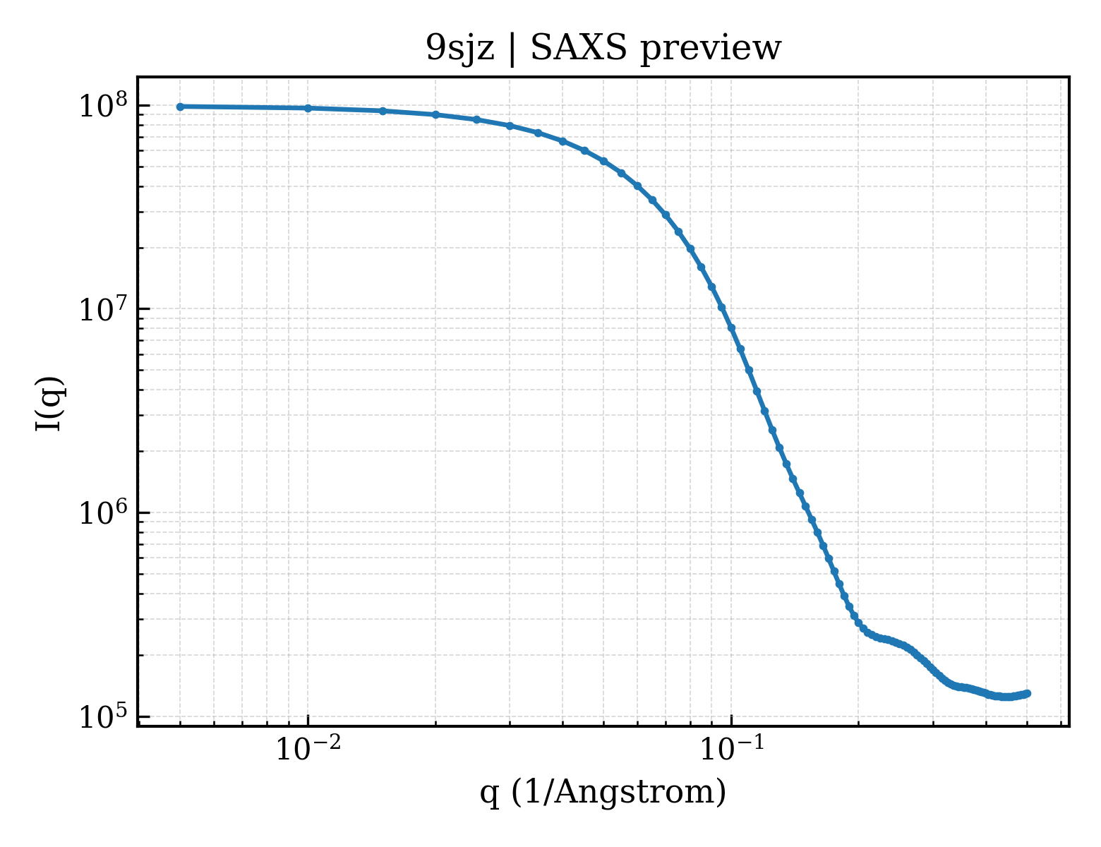
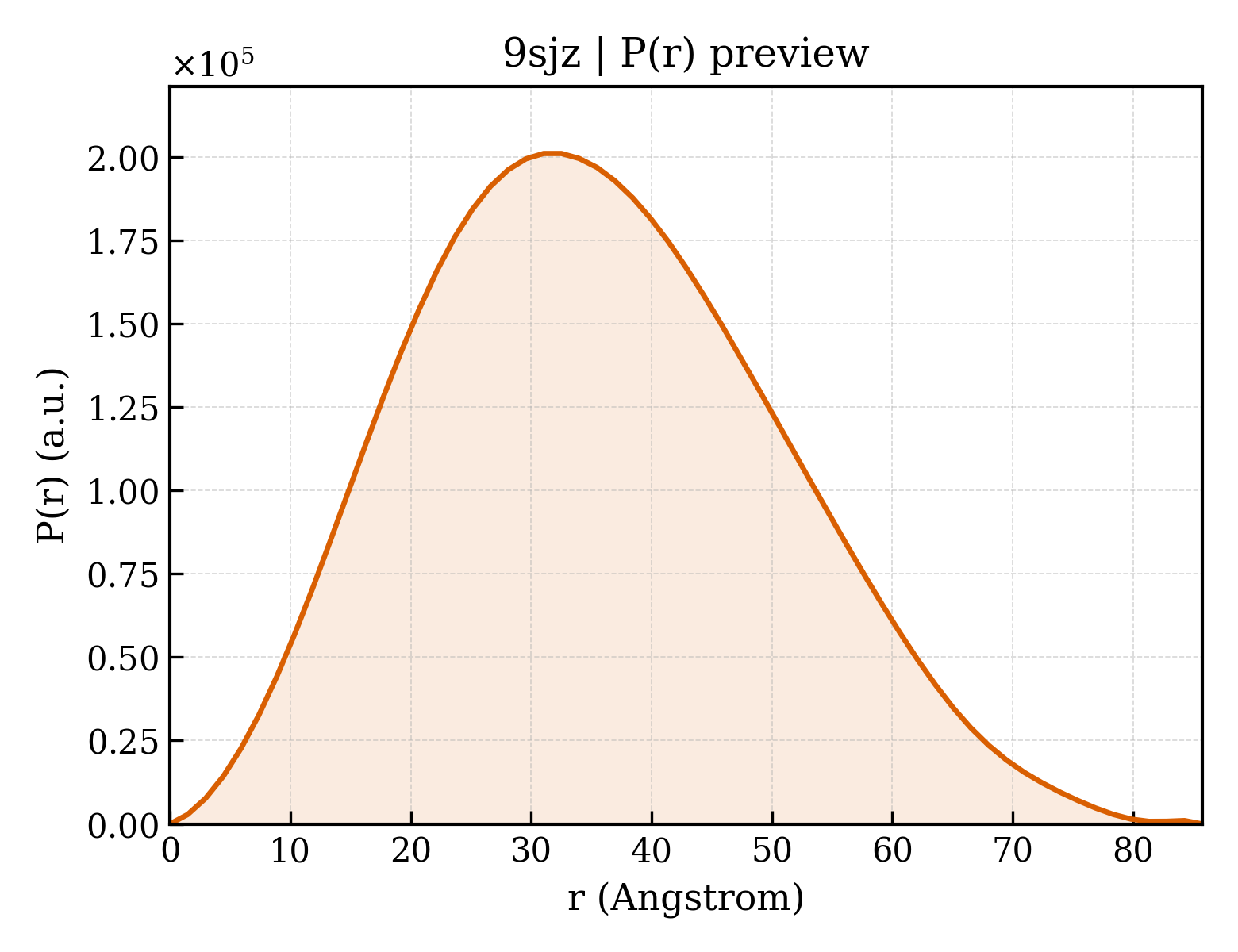
Ribonucleoside-diphosphate reductase 1 subunit beta
Escherichia coli
State in the Current Structure
| Assembly | Oligomeric State | Construct | Mutations and Modifications | Ligands, Ions and Associated Components | Method and Experimental Conditions | Structure Quality |
|---|---|---|---|---|---|---|
| 1 | Protein homooligomer Homooligomer Protein × 2 PDB declaration: dimeric(2) Consistent with protein copy count | Chain A; UniProt 2–376 Chain B; UniProt 2–376 | Not recorded | FE FE (III) ION × 4 | ELECTRON CRYSTALLOGRAPHY cryo-EM buffer:pH 5.5;Crystallization was performed using 23.5 uL protein solution - 25 mM HEPES-Na pH 7.0, 50 mM NaCl and 50 mM sodium dithionite - and 20 uL crystallization buffer containing seeds, 25 percent PEG 3350, 0.1 M Bis-Tris pH 5.5 and 2 mM sodium dithioninte. The crystals were then oxidized through repeated wash with oxygen containing buffer of 12% PEG, 0.05 M Bis-Tris at pH 5.5. cryo-EM vitrification conditions:Cryogen ETHANE | Resolution 1.70 Å R-free 0.232 |
Other States of the Same Protein in the Database
Each row is a biological assembly of the same UniProt protein in another PDB entry. The “Difference from current entry” column identifies evidence-level differences; no tag means the currently parsed fields agree.
| Other PDB | Difference from Current Entry 9SJZ | Assembly / Oligomeric State | Construct | Mutations and Modifications | Ligands, Ions and Non-polymers | Method and Experimental Conditions | Structure Quality |
|---|---|---|---|---|---|---|---|
| 1AV8 RIBONUCLEOTIDE REDUCTASE R2 SUBUNIT FROM E. COLI Deposited 1997-09-30 | Different construct Different ligand/ion Different experimental method Different experimental conditions Different structure-quality metrics | Assembly 1 Protein homooligomer Homooligomer;Protein × 2 PDB declaration: dimeric |
Chain A
1–340(340 aa)
Chain B
1–340(340 aa)
|
Not recorded | FEO MU-OXO-DIIRON × 2 |
X-RAY DIFFRACTION
X-ray crystallization conditions
pH 7.6;PROTEIN WAS CRYSTALLIZED FROM 80% SATURATED NACL, NEAR PHYSIOLOGICAL PH (PH 7.6)
|
Resolution 2.80 Å R-free 0.192 |
| 1BIQ RIBONUCLEOSIDE-DIPHOSPHATE REDUCTASE 1 BETA CHAIN MUTANT E238A Deposited 1998-06-18 | Different construct Different mutation/modification Different ligand/ion Different experimental method Different experimental conditions Different structure-quality metrics | Assembly 1 Protein homooligomer Homooligomer;Protein × 2 PDB declaration: dimeric |
Chain A
1–375(375 aa)
Fragment:BETA CHAIN
Chain B
1–375(375 aa)
Fragment:BETA CHAIN
|
Mutation:Y122F, E238A Non-standard monomer:Yes (specific site not provided by mmCIF) Mutation:Y122F, E238A | FE2 FE (II) ION × 3 FE FE (III) ION × 1 OH HYDROXIDE ION × 3 HG MERCURY (II) ION × 16 |
X-RAY DIFFRACTION
X-ray crystallization conditions
pH 6;pH 6.0
|
Resolution 2.05 Å R-free 0.260 |
| 1JPR Mn substituted Ribonucleotide reductase R2 from E. coli oxidized by nitric oxide Deposited 2001-08-03 | Different construct Different ligand/ion Different experimental method Different experimental conditions Different structure-quality metrics | Assembly 1 Protein homooligomer Homooligomer;Protein × 2 PDB declaration: dimeric |
Chain A
1–375(375 aa)
Chain B
1–375(375 aa)
|
Not recorded | MN MANGANESE (II) ION × 4 HG MERCURY (II) ION × 14 |
X-RAY DIFFRACTION
X-ray crystallization conditions
VAPOR DIFFUSION, HANGING DROP;pH 6;298 K;PEG 4000, sodium chloride, EMTS, MES, pH 6.0, VAPOR DIFFUSION, HANGING DROP, temperature 298K
|
Resolution 1.88 Å R-free 0.213 |
| 1JQC Mn substituted Ribonucleotide reductase R2 from E. Coli oxidized by hydrogen peroxide and hydroxylamine Deposited 2001-08-06 | Different construct Different ligand/ion Different experimental method Different experimental conditions Different structure-quality metrics | Assembly 1 Protein homooligomer Homooligomer;Protein × 2 PDB declaration: dimeric |
Chain A
1–375(375 aa)
Chain B
1–375(375 aa)
|
Not recorded | MN MANGANESE (II) ION × 4 HG MERCURY (II) ION × 13 |
X-RAY DIFFRACTION
X-ray crystallization conditions
VAPOR DIFFUSION, HANGING DROP;pH 6;298 K;peg 4000, MES, EMTS, sodium chloride, pH 6.0, VAPOR DIFFUSION, HANGING DROP, temperature 298K
|
Resolution 1.61 Å R-free 0.219 |
| 1MRR SUBSTITUTION OF MANGANESE FOR IRON IN RIBONUCLEOTIDE REDUCTASE FROM ESCHERICHIA COLI. SPECTROSCOPIC AND CRYSTALLOGRAPHIC CHARACTERIZATION Deposited 1992-07-28 | Different construct Different ligand/ion Different experimental method Different experimental conditions Different structure-quality metrics | Assembly 1 Protein homooligomer Homooligomer;Protein × 2 PDB declaration: dimeric |
Chain A
1–375(375 aa)
Chain B
1–375(375 aa)
|
Not recorded | MN MANGANESE (II) ION × 4 HG MERCURY (II) ION × 13 | X-RAY DIFFRACTION mmCIF provides none of the parsed conditions | Resolution 2.50 Å |
| 1MXR High resolution structure of Ribonucleotide reductase R2 from E. coli in its oxidised (Met) form Deposited 2002-10-03 | Different construct Different ligand/ion Different experimental method Different experimental conditions Different structure-quality metrics | Assembly 1 Protein homooligomer Homooligomer;Protein × 2 PDB declaration: dimeric |
Chain A
1–375(375 aa)
Chain B
1–375(375 aa)
|
Not recorded | FE FE (III) ION × 4 HG MERCURY (II) ION × 11 GOL GLYCEROL × 1 |
X-RAY DIFFRACTION
X-ray crystallization conditions
VAPOR DIFFUSION, HANGING DROP;pH 6;298 K;PEG 4000, MES, NaCl, EMTS, pH 6.0, VAPOR DIFFUSION, HANGING DROP, temperature 298K
|
Resolution 1.42 Å R-free 0.185 |
| 1PFR RIBONUCLEOSIDE-DIPHOSPHATE REDUCTASE 1 BETA CHAIN Deposited 1996-12-03 | Different construct Different mutation/modification Different ligand/ion Different experimental method Different experimental conditions Different structure-quality metrics | Assembly 1 Protein homooligomer Homooligomer;Protein × 2 PDB declaration: dimeric |
Chain A
1–340(340 aa)
Chain B
1–340(340 aa)
|
Mutation:S211A Mutation:S211A | FE FE (III) ION × 4 HG MERCURY (II) ION × 8 |
X-RAY DIFFRACTION
X-ray crystallization conditions
VAPOR DIFFUSION, HANGING DROP;pH 6;HANGING DROP VAPOR DIFFUSION. CRYSTALLISED FROM 20% PEG 4000, 0.2M NACL, 0.1M MES PH 6.0, 1MM ETHYLMERCURY SALICYLATE., vapor diffusion - hanging drop
|
Resolution 2.20 Å |
| 1PIM DITHIONITE REDUCED E. COLI RIBONUCLEOTIDE REDUCTASE R2 SUBUNIT, D84E MUTANT Deposited 2003-05-30 | Different construct Different mutation/modification Different ligand/ion Different experimental method Different experimental conditions Different structure-quality metrics | Assembly 1 Protein homooligomer Homooligomer;Protein × 2 PDB declaration: dimeric |
Chain A
1–375(375 aa)
Chain B
1–375(375 aa)
|
Mutation:D84E Mutation:D84E | FE FE (III) ION × 4 HG MERCURY (II) ION × 7 |
X-RAY DIFFRACTION
X-ray crystallization conditions
VAPOR DIFFUSION, HANGING DROP;pH 6;310 K;20% PEG 4000, 200mM NaCl, 50mM MES, 0.3% dioxane, pH 6.0, VAPOR DIFFUSION, HANGING DROP, temperature 310K
|
Resolution 2.00 Å R-free 0.246 |
| 1PIU OXIDIZED RIBONUCLEOTIDE REDUCTASE R2-D84E MUTANT CONTAINING OXO-BRIDGED DIFERRIC CLUSTER Deposited 2003-05-30 | Different construct Different mutation/modification Different ligand/ion Different experimental method Different experimental conditions Different structure-quality metrics | Assembly 1 Protein homooligomer Homooligomer;Protein × 2 PDB declaration: dimeric |
Chain A
1–375(375 aa)
Chain B
1–375(375 aa)
|
Mutation:D84E Mutation:D84E | FE FE (III) ION × 4 HG MERCURY (II) ION × 10 O OXYGEN ATOM × 2 |
X-RAY DIFFRACTION
X-ray crystallization conditions
VAPOR DIFFUSION, HANGING DROP;pH 6;310 K;20% PEG 4000, 200mM NaCl, 50mM MES, 0.3% dioxane, pH 6.0, VAPOR DIFFUSION, HANGING DROP, temperature 310K
|
Resolution 2.20 Å R-free 0.232 |
| 1PIY RIBONUCLEOTIDE REDUCTASE R2 SOAKED WITH FERROUS ION AT NEUTRAL PH Deposited 2003-05-30 | Different construct Different ligand/ion Different experimental method Different experimental conditions Different structure-quality metrics | Assembly 1 Protein homooligomer Homooligomer;Protein × 2 PDB declaration: dimeric |
Chain A
1–375(375 aa)
Chain B
1–375(375 aa)
|
Not recorded | FE FE (III) ION × 4 HG MERCURY (II) ION × 14 |
X-RAY DIFFRACTION
X-ray crystallization conditions
VAPOR DIFFUSION, HANGING DROP;pH 6;310 K;20% PEG 4000, 200mM NaCl, 50mM MES, 0.3% dioxane, pH 6.0, VAPOR DIFFUSION, HANGING DROP, temperature 310K
|
Resolution 1.68 Å R-free 0.246 |
| 1PIZ RIBONUCLEOTIDE REDUCTASE R2 D84E MUTANT SOAKED WITH FERROUS IONS AT NEUTRAL PH Deposited 2003-05-30 | Different construct Different mutation/modification Different ligand/ion Different experimental method Different experimental conditions Different structure-quality metrics | Assembly 1 Protein homooligomer Homooligomer;Protein × 2 PDB declaration: dimeric |
Chain A
1–375(375 aa)
Chain B
1–375(375 aa)
|
Mutation:D84E Mutation:D84E | FE FE (III) ION × 4 HG MERCURY (II) ION × 11 |
X-RAY DIFFRACTION
X-ray crystallization conditions
VAPOR DIFFUSION, HANGING DROP;pH 6;310 K;20% PEG 4000, 200mM NaCl, 50mM MES, 0.3% dioxane, pH 6.0, VAPOR DIFFUSION, HANGING DROP, temperature 310K
|
Resolution 1.90 Å R-free 0.242 |
| 1PJ0 RIBONUCLEOTIDE REDUCTASE R2-D84E/W48F MUTANT SOAKED WITH FERROUS IONS AT NEUTRAL PH Deposited 2003-05-30 | Different construct Different mutation/modification Different ligand/ion Different experimental method Different experimental conditions Different structure-quality metrics | Assembly 1 Protein homooligomer Homooligomer;Protein × 2 PDB declaration: dimeric |
Chain A
1–375(375 aa)
Chain B
1–375(375 aa)
|
Mutation:D84E, W48F Mutation:D84E, W48F | FE FE (III) ION × 4 HG MERCURY (II) ION × 9 |
X-RAY DIFFRACTION
X-ray crystallization conditions
VAPOR DIFFUSION, HANGING DROP;pH 6;310 K;20% PEG 4000, 200mM NaCl, 50mM MES, 0.3% dioxane, pH 6.0, VAPOR DIFFUSION, HANGING DROP, temperature 310K
|
Resolution 1.90 Å R-free 0.236 |
| 1PJ1 RIBONUCLEOTIDE REDUCTASE R2-D84E/W48F SOAKED WITH FERROUS IONS AT PH 5 Deposited 2003-05-30 | Different construct Different mutation/modification Different ligand/ion Different experimental method Different experimental conditions Different structure-quality metrics | Assembly 1 Protein homooligomer Homooligomer;Protein × 2 PDB declaration: dimeric |
Chain A
1–375(375 aa)
Chain B
1–375(375 aa)
|
Mutation:D84E, W48F Mutation:D84E, W48F | FE FE (III) ION × 4 HG MERCURY (II) ION × 15 |
X-RAY DIFFRACTION
X-ray crystallization conditions
VAPOR DIFFUSION, HANGING DROP;pH 6;310 K;20% PEG 4000, 200mM NaCl, 50mM MES, 0.3% dioxane, pH 6.0, VAPOR DIFFUSION, HANGING DROP, temperature 310K
|
Resolution 1.95 Å R-free 0.221 |
| 1PM2 CRYSTAL STRUCTURE OF MANGANESE SUBSTITUTED R2-D84E (D84E MUTANT OF THE R2 SUBUNIT OF E. COLI RIBONUCLEOTIDE REDUCTASE) Deposited 2003-06-09 | Different construct Different mutation/modification Different ligand/ion Different experimental method Different experimental conditions Different structure-quality metrics | Assembly 1 Protein homooligomer Homooligomer;Protein × 2 PDB declaration: dimeric |
Chain A
1–339(339 aa)
Chain B
1–339(339 aa)
|
Mutation:D84E Mutation:D84E | MN MANGANESE (II) ION × 4 HG MERCURY (II) ION × 14 |
X-RAY DIFFRACTION
X-ray crystallization conditions
VAPOR DIFFUSION, HANGING DROP;pH 7.3;310 K;PEG 4000, MES, sodium chloride, sodium ethylmercurithiosalicylicate , pH 7.3, VAPOR DIFFUSION, HANGING DROP, temperature 310K
|
Resolution 1.80 Å R-free 0.215 |
| 1R1R RIBONUCLEOTIDE REDUCTASE R1 PROTEIN MUTANT Y730F WITH A REDUCED ACTIVE SITE FROM ESCHERICHIA COLI Deposited 1997-07-15 | Different construct Different oligomeric state Different ligand/ion Different experimental method Different experimental conditions Different structure-quality metrics | Assembly 1 Protein heterocomplex Heteromer;Protein × 3 PDB declaration: trimeric |
Chain D
356–375(20 aa)
Fragment:C-TERMINAL PORTION, 20 RESIDUES
Chain P
356–375(20 aa)
Fragment:C-TERMINAL PORTION, 20 RESIDUES
|
Not recorded | No recorded non-water small molecule |
X-RAY DIFFRACTION
X-ray crystallization conditions
pH 6;PROTEIN WAS CRYSTALLIZED FROM 1.7 M LITHIUM SULFATE, AND 10 MM MAGNESIUM SULFATE IN 25 MM CITRATE BUFFER AT PH 6.0 THE PROTEIN SOLUTION CONTAINED 17 MG/ML R1 PROTEIN, 20-FOLD EXCESS OF A 20-RESIDUE PEPTIDE CORRESPONDING TO THE C-TERMINUS OF THE R2 SUBUNIT AND IS ESSENTIAL FOR CRYSTALLIZATION
|
Resolution 2.90 Å R-free 0.245 |
| 1R1R RIBONUCLEOTIDE REDUCTASE R1 PROTEIN MUTANT Y730F WITH A REDUCED ACTIVE SITE FROM ESCHERICHIA COLI Deposited 1997-07-15 | Different construct Different mutation/modification Different oligomeric state Different ligand/ion Different experimental method Different experimental conditions Different structure-quality metrics | Assembly 2 Protein heterocomplex Heteromer;Protein × 2 PDB declaration: dimeric |
Chain E
356–375(20 aa)
Fragment:C-TERMINAL PORTION, 20 RESIDUES
|
Not recorded | No recorded non-water small molecule |
X-RAY DIFFRACTION
X-ray crystallization conditions
pH 6;PROTEIN WAS CRYSTALLIZED FROM 1.7 M LITHIUM SULFATE, AND 10 MM MAGNESIUM SULFATE IN 25 MM CITRATE BUFFER AT PH 6.0 THE PROTEIN SOLUTION CONTAINED 17 MG/ML R1 PROTEIN, 20-FOLD EXCESS OF A 20-RESIDUE PEPTIDE CORRESPONDING TO THE C-TERMINUS OF THE R2 SUBUNIT AND IS ESSENTIAL FOR CRYSTALLIZATION
|
Resolution 2.90 Å R-free 0.245 |
| 1R1R RIBONUCLEOTIDE REDUCTASE R1 PROTEIN MUTANT Y730F WITH A REDUCED ACTIVE SITE FROM ESCHERICHIA COLI Deposited 1997-07-15 | Different construct Different mutation/modification Different oligomeric state Different ligand/ion Different experimental method Different experimental conditions Different structure-quality metrics | Assembly 3 Protein heterocomplex Heteromer;Protein × 2 PDB declaration: dimeric |
Chain F
356–375(20 aa)
Fragment:C-TERMINAL PORTION, 20 RESIDUES
|
Not recorded | No recorded non-water small molecule |
X-RAY DIFFRACTION
X-ray crystallization conditions
pH 6;PROTEIN WAS CRYSTALLIZED FROM 1.7 M LITHIUM SULFATE, AND 10 MM MAGNESIUM SULFATE IN 25 MM CITRATE BUFFER AT PH 6.0 THE PROTEIN SOLUTION CONTAINED 17 MG/ML R1 PROTEIN, 20-FOLD EXCESS OF A 20-RESIDUE PEPTIDE CORRESPONDING TO THE C-TERMINUS OF THE R2 SUBUNIT AND IS ESSENTIAL FOR CRYSTALLIZATION
|
Resolution 2.90 Å R-free 0.245 |
| 1R1R RIBONUCLEOTIDE REDUCTASE R1 PROTEIN MUTANT Y730F WITH A REDUCED ACTIVE SITE FROM ESCHERICHIA COLI Deposited 1997-07-15 | Different construct Different oligomeric state Different ligand/ion Different experimental method Different experimental conditions Different structure-quality metrics | Assembly 4 Protein heterocomplex Heteromer;Protein × 12 PDB declaration: dodecameric |
Chain D
356–375(20 aa)
Fragment:C-TERMINAL PORTION, 20 RESIDUES
Chain E
356–375(20 aa)
Fragment:C-TERMINAL PORTION, 20 RESIDUES
|
Not recorded | No recorded non-water small molecule |
X-RAY DIFFRACTION
X-ray crystallization conditions
pH 6;PROTEIN WAS CRYSTALLIZED FROM 1.7 M LITHIUM SULFATE, AND 10 MM MAGNESIUM SULFATE IN 25 MM CITRATE BUFFER AT PH 6.0 THE PROTEIN SOLUTION CONTAINED 17 MG/ML R1 PROTEIN, 20-FOLD EXCESS OF A 20-RESIDUE PEPTIDE CORRESPONDING TO THE C-TERMINUS OF THE R2 SUBUNIT AND IS ESSENTIAL FOR CRYSTALLIZATION
|
Resolution 2.90 Å R-free 0.245 |
| 1R1R RIBONUCLEOTIDE REDUCTASE R1 PROTEIN MUTANT Y730F WITH A REDUCED ACTIVE SITE FROM ESCHERICHIA COLI Deposited 1997-07-15 | Different construct Different mutation/modification Different oligomeric state Different ligand/ion Different experimental method Different experimental conditions Different structure-quality metrics | Assembly 5 Protein heterocomplex Heteromer;Protein × 12 PDB declaration: dodecameric |
Chain F
356–375(20 aa)
Fragment:C-TERMINAL PORTION, 20 RESIDUES
|
Not recorded | No recorded non-water small molecule |
X-RAY DIFFRACTION
X-ray crystallization conditions
pH 6;PROTEIN WAS CRYSTALLIZED FROM 1.7 M LITHIUM SULFATE, AND 10 MM MAGNESIUM SULFATE IN 25 MM CITRATE BUFFER AT PH 6.0 THE PROTEIN SOLUTION CONTAINED 17 MG/ML R1 PROTEIN, 20-FOLD EXCESS OF A 20-RESIDUE PEPTIDE CORRESPONDING TO THE C-TERMINUS OF THE R2 SUBUNIT AND IS ESSENTIAL FOR CRYSTALLIZATION
|
Resolution 2.90 Å R-free 0.245 |
| 1R65 Crystal structure of ferrous soaked Ribonucleotide Reductase R2 subunit (wildtype) at pH 5 from E. coli Deposited 2003-10-14 | Different construct Different ligand/ion Different experimental method Different experimental conditions Different structure-quality metrics | Assembly 1 Protein homooligomer Homooligomer;Protein × 2 PDB declaration: dimeric |
Chain A
1–375(375 aa)
Fragment:Ribonucleotide Reductase R2
Chain B
1–375(375 aa)
Fragment:Ribonucleotide Reductase R2
|
Not recorded | FE2 FE (II) ION × 4 HG MERCURY (II) ION × 13 |
X-RAY DIFFRACTION
X-ray crystallization conditions
VAPOR DIFFUSION, HANGING DROP;pH 6;310 K;PEG4000, sodium chloride, EMTS, MES, pH 6, VAPOR DIFFUSION, HANGING DROP, temperature 310K
|
Resolution 1.95 Å R-free 0.236 |
| 1RIB STRUCTURE AND FUNCTION OF THE ESCHERICHIA COLI RIBONUCLEOTIDE REDUCTASE PROTEIN R2 Deposited 1993-01-19 | Different construct Different ligand/ion Different experimental method Different experimental conditions Different structure-quality metrics | Assembly 1 Protein homooligomer Homooligomer;Protein × 2 PDB declaration: dimeric |
Chain A
1–375(375 aa)
Chain B
1–375(375 aa)
|
Not recorded | FEO MU-OXO-DIIRON × 2 | X-RAY DIFFRACTION mmCIF provides none of the parsed conditions | Resolution 2.20 Å |
| 1RNR AUTOCATALYTIC GENERATION OF DOPA IN THE ENGINEERED PROTEIN R2 F208Y FROM ESCHERICHIA COLI RIBONUCLEOTIDE REDUCTASE AND CRYSTAL STRUCTURE OF THE DOPA-208 PROTEIN Deposited 1993-04-26 | Different mutation/modification Different ligand/ion Different experimental method Different experimental conditions Different structure-quality metrics | Assembly 1 Protein homooligomer Homooligomer;Protein × 2 PDB declaration: dimeric |
Chain A
2–376(375 aa)
Chain B
2–376(375 aa)
|
Non-standard monomer:Yes (specific site not provided by mmCIF) Non-standard monomer:Yes (specific site not provided by mmCIF) | FE FE (III) ION × 4 HG MERCURY (II) ION × 14 | X-RAY DIFFRACTION mmCIF provides none of the parsed conditions | Resolution 2.50 Å |
| 1RSR azide complex of the diferrous F208A mutant R2 subunit of ribonucleotide reductase Deposited 2003-12-10 | Different construct Different mutation/modification Different ligand/ion Different experimental method Different experimental conditions Different structure-quality metrics | Assembly 1 Protein homooligomer Homooligomer;Protein × 2 PDB declaration: dimeric |
Chain A
1–375(375 aa)
Chain B
1–375(375 aa)
|
Mutation:Y122F, F208A Mutation:Y122F, F208A | FE2 FE (II) ION × 4 HG MERCURY (II) ION × 12 AZI AZIDE ION × 1 |
X-RAY DIFFRACTION
X-ray crystallization conditions
VAPOR DIFFUSION, HANGING DROP;pH 6;298 K;100mM MES, 200mM NaCl, 1mM EMTS (Thimerosal), 16-24% PEG 4000, pH 6.0, VAPOR DIFFUSION, HANGING DROP, temperature 298K
|
Resolution 2.00 Å R-free 0.289 |
| 1RSV azide complex of the diferrous E238A mutant R2 subunit of ribonucleotide reductase Deposited 2003-12-10 | Different construct Different mutation/modification Different ligand/ion Different experimental method Different experimental conditions Different structure-quality metrics | Assembly 1 Protein homooligomer Homooligomer;Protein × 2 PDB declaration: dimeric |
Chain A
1–375(375 aa)
Chain B
1–375(375 aa)
|
Mutation:Y122F, E238A Mutation:Y122F, E238A | FE FE (III) ION × 4 HG MERCURY (II) ION × 12 AZI AZIDE ION × 1 |
X-RAY DIFFRACTION
X-ray crystallization conditions
VAPOR DIFFUSION, HANGING DROP;pH 6;298 K;100mM MES, 200mM NaCl, 1mM EMTS (Thimerosal), 16-24% PEG 4000, pH 6.0, VAPOR DIFFUSION, HANGING DROP, temperature 298K
|
Resolution 2.20 Å R-free 0.256 |
| 1XIK RIBONUCLEOSIDE-DIPHOSPHATE REDUCTASE 1 BETA CHAIN Deposited 1996-08-06 | Different construct Different ligand/ion Different experimental method Different experimental conditions Different structure-quality metrics | Assembly 1 Protein homooligomer Homooligomer;Protein × 2 PDB declaration: dimeric |
Chain A
1–375(375 aa)
Fragment:BETA CHAIN
Chain B
1–375(375 aa)
Fragment:BETA CHAIN
|
Not recorded | FE2 FE (II) ION × 4 HG MERCURY (II) ION × 10 |
X-RAY DIFFRACTION
X-ray crystallization conditions
VAPOR DIFFUSION, HANGING DROP;pH 6;HANGING DROP VAPOR DIFFUSION METHOD, 20% PEG 4000, 0.2M NACL, 50MM MES BUFFER PH 6.0, 1MM ETHYLMERCURY SALICYLATE, vapor diffusion - hanging drop
|
Resolution 1.70 Å R-free 0.280 |
| 1YFD Crystal structure of the Y122H mutant of ribonucleotide reductase R2 protein from E. coli Deposited 2004-12-31 | Different construct Different mutation/modification Different ligand/ion Different experimental method Different experimental conditions Different structure-quality metrics | Assembly 1 Protein homooligomer Homooligomer;Protein × 2 PDB declaration: dimeric |
Chain A
1–375(375 aa)
Chain B
1–375(375 aa)
|
Mutation:Y122H Mutation:Y122H | HG MERCURY (II) ION × 13 FEO MU-OXO-DIIRON × 2 |
X-RAY DIFFRACTION
X-ray crystallization conditions
VAPOR DIFFUSION, HANGING DROP;pH 6;295 K;PEG 4000, 0.1M MES, 0.4M NACL, 1mM EMTS, pH 6.0, VAPOR DIFFUSION, HANGING DROP, temperature 295K
|
Resolution 1.90 Å R-free 0.206 |
| 2ALX Ribonucleotide Reductase R2 from Escherichia coli in space group P6(1)22 Deposited 2005-08-08 | Different construct Different mutation/modification Different ligand/ion Different experimental method Different experimental conditions Different structure-quality metrics | Assembly 1 Protein homooligomer Homooligomer;Protein × 2 PDB declaration: dimeric |
Chain A
0–339(340 aa)
Fragment:residues 0-339
|
Not recorded | MN MANGANESE (II) ION × 4 HG MERCURY (II) ION × 8 |
X-RAY DIFFRACTION
X-ray crystallization conditions
VAPOR DIFFUSION, HANGING DROP;pH 6.5;298 K;ammonium sulfate, MES, manganese chloride, magnesium chloride, 4-(2-Hydroxyethyl)-1-piperazinepropanesulfonic acid, ethylmercurithiosalicylate, pH 6.5, VAPOR DIFFUSION, HANGING DROP, temperature 298K
|
Resolution 2.60 Å R-free 0.276 |
| 2AV8 Y122F MUTANT OF RIBONUCLEOTIDE REDUCTASE FROM ESCHERICHIA COLI Deposited 1997-09-30 | Different construct Different mutation/modification Different ligand/ion Different experimental method Different experimental conditions Different structure-quality metrics | Assembly 1 Protein homooligomer Homooligomer;Protein × 2 PDB declaration: dimeric |
Chain A
1–340(340 aa)
Chain B
1–340(340 aa)
|
Mutation:Y122F Mutation:Y122F | FE2 FE (II) ION × 1 FEO MU-OXO-DIIRON × 2 |
X-RAY DIFFRACTION
X-ray crystallization conditions
pH 7.6;PROTEIN WAS CRYSTALLIZED FROM 80% SATURATED NACL, NEAR PHYSIOLOGICAL PH (PH 7.6)
|
Resolution 2.46 Å R-free 0.237 |
| 2R1R RIBONUCLEOTIDE REDUCTASE R1 PROTEIN WITH DTTP OCCUPYING THE SPECIFICITY SITE FROM ESCHERICHIA COLI Deposited 1997-07-21 | Different construct Different oligomeric state Different ligand/ion Different experimental method Different experimental conditions Different structure-quality metrics | Assembly 1 Protein heterocomplex Heteromer;Protein × 3 PDB declaration: trimeric |
Chain D
356–375(20 aa)
Fragment:C-TERMINAL PORTION, 20 RESIDUES
Chain P
356–375(20 aa)
Fragment:C-TERMINAL PORTION, 20 RESIDUES
|
Not recorded | TTP THYMIDINE-5'-TRIPHOSPHATE × 1 |
X-RAY DIFFRACTION
X-ray crystallization conditions
pH 6;PROTEIN WAS CRYSTALLIZED FROM 1.7 M LITHIUM SULFATE, AND 10 MM MAGNESIUM SULFATE IN 25 MM CITRATE BUFFER AT PH 6.0 THE PROTEIN SOLUTION CONTAINED 17 MG/ML R1 PROTEIN, 20-FOLD EXCESS FO A 20-RESIDUE PEPTIDE CORRESPONDING TO THE C-TERMINUS OF THE R2 SUBUNIT AND IS ESSENTIAL FOR CRYSTALLIZATION. 10 MM DTTP AND 10 MM CDP WAS ALSO INCLUDED IN THE PROTEIN SOLUTION
|
Resolution 3.00 Å R-free 0.280 |
| 2R1R RIBONUCLEOTIDE REDUCTASE R1 PROTEIN WITH DTTP OCCUPYING THE SPECIFICITY SITE FROM ESCHERICHIA COLI Deposited 1997-07-21 | Different construct Different mutation/modification Different oligomeric state Different ligand/ion Different experimental method Different experimental conditions Different structure-quality metrics | Assembly 2 Protein heterocomplex Heteromer;Protein × 2 PDB declaration: dimeric |
Chain E
356–375(20 aa)
Fragment:C-TERMINAL PORTION, 20 RESIDUES
|
Not recorded | TTP THYMIDINE-5'-TRIPHOSPHATE × 1 |
X-RAY DIFFRACTION
X-ray crystallization conditions
pH 6;PROTEIN WAS CRYSTALLIZED FROM 1.7 M LITHIUM SULFATE, AND 10 MM MAGNESIUM SULFATE IN 25 MM CITRATE BUFFER AT PH 6.0 THE PROTEIN SOLUTION CONTAINED 17 MG/ML R1 PROTEIN, 20-FOLD EXCESS FO A 20-RESIDUE PEPTIDE CORRESPONDING TO THE C-TERMINUS OF THE R2 SUBUNIT AND IS ESSENTIAL FOR CRYSTALLIZATION. 10 MM DTTP AND 10 MM CDP WAS ALSO INCLUDED IN THE PROTEIN SOLUTION
|
Resolution 3.00 Å R-free 0.280 |
| 2R1R RIBONUCLEOTIDE REDUCTASE R1 PROTEIN WITH DTTP OCCUPYING THE SPECIFICITY SITE FROM ESCHERICHIA COLI Deposited 1997-07-21 | Different construct Different mutation/modification Different oligomeric state Different ligand/ion Different experimental method Different experimental conditions Different structure-quality metrics | Assembly 3 Protein heterocomplex Heteromer;Protein × 2 PDB declaration: dimeric |
Chain F
356–375(20 aa)
Fragment:C-TERMINAL PORTION, 20 RESIDUES
|
Not recorded | TTP THYMIDINE-5'-TRIPHOSPHATE × 1 |
X-RAY DIFFRACTION
X-ray crystallization conditions
pH 6;PROTEIN WAS CRYSTALLIZED FROM 1.7 M LITHIUM SULFATE, AND 10 MM MAGNESIUM SULFATE IN 25 MM CITRATE BUFFER AT PH 6.0 THE PROTEIN SOLUTION CONTAINED 17 MG/ML R1 PROTEIN, 20-FOLD EXCESS FO A 20-RESIDUE PEPTIDE CORRESPONDING TO THE C-TERMINUS OF THE R2 SUBUNIT AND IS ESSENTIAL FOR CRYSTALLIZATION. 10 MM DTTP AND 10 MM CDP WAS ALSO INCLUDED IN THE PROTEIN SOLUTION
|
Resolution 3.00 Å R-free 0.280 |
| 2R1R RIBONUCLEOTIDE REDUCTASE R1 PROTEIN WITH DTTP OCCUPYING THE SPECIFICITY SITE FROM ESCHERICHIA COLI Deposited 1997-07-21 | Different construct Different oligomeric state Different ligand/ion Different experimental method Different experimental conditions Different structure-quality metrics | Assembly 4 Protein heterocomplex Heteromer;Protein × 12 PDB declaration: dodecameric |
Chain D
356–375(20 aa)
Fragment:C-TERMINAL PORTION, 20 RESIDUES
Chain E
356–375(20 aa)
Fragment:C-TERMINAL PORTION, 20 RESIDUES
|
Not recorded | TTP THYMIDINE-5'-TRIPHOSPHATE × 6 |
X-RAY DIFFRACTION
X-ray crystallization conditions
pH 6;PROTEIN WAS CRYSTALLIZED FROM 1.7 M LITHIUM SULFATE, AND 10 MM MAGNESIUM SULFATE IN 25 MM CITRATE BUFFER AT PH 6.0 THE PROTEIN SOLUTION CONTAINED 17 MG/ML R1 PROTEIN, 20-FOLD EXCESS FO A 20-RESIDUE PEPTIDE CORRESPONDING TO THE C-TERMINUS OF THE R2 SUBUNIT AND IS ESSENTIAL FOR CRYSTALLIZATION. 10 MM DTTP AND 10 MM CDP WAS ALSO INCLUDED IN THE PROTEIN SOLUTION
|
Resolution 3.00 Å R-free 0.280 |
| 2R1R RIBONUCLEOTIDE REDUCTASE R1 PROTEIN WITH DTTP OCCUPYING THE SPECIFICITY SITE FROM ESCHERICHIA COLI Deposited 1997-07-21 | Different construct Different mutation/modification Different oligomeric state Different ligand/ion Different experimental method Different experimental conditions Different structure-quality metrics | Assembly 5 Protein heterocomplex Heteromer;Protein × 12 PDB declaration: dodecameric |
Chain F
356–375(20 aa)
Fragment:C-TERMINAL PORTION, 20 RESIDUES
|
Not recorded | TTP THYMIDINE-5'-TRIPHOSPHATE × 6 |
X-RAY DIFFRACTION
X-ray crystallization conditions
pH 6;PROTEIN WAS CRYSTALLIZED FROM 1.7 M LITHIUM SULFATE, AND 10 MM MAGNESIUM SULFATE IN 25 MM CITRATE BUFFER AT PH 6.0 THE PROTEIN SOLUTION CONTAINED 17 MG/ML R1 PROTEIN, 20-FOLD EXCESS FO A 20-RESIDUE PEPTIDE CORRESPONDING TO THE C-TERMINUS OF THE R2 SUBUNIT AND IS ESSENTIAL FOR CRYSTALLIZATION. 10 MM DTTP AND 10 MM CDP WAS ALSO INCLUDED IN THE PROTEIN SOLUTION
|
Resolution 3.00 Å R-free 0.280 |
| 2R1R RIBONUCLEOTIDE REDUCTASE R1 PROTEIN WITH DTTP OCCUPYING THE SPECIFICITY SITE FROM ESCHERICHIA COLI Deposited 1997-07-21 | Different construct Different oligomeric state Different ligand/ion Different experimental method Different experimental conditions Different structure-quality metrics | Assembly 6 Protein heterocomplex Heteromer;Protein × 4 PDB declaration: tetrameric |
Chain D
356–375(20 aa)
Fragment:C-TERMINAL PORTION, 20 RESIDUES
Chain E
356–375(20 aa)
Fragment:C-TERMINAL PORTION, 20 RESIDUES
|
Not recorded | TTP THYMIDINE-5'-TRIPHOSPHATE × 2 |
X-RAY DIFFRACTION
X-ray crystallization conditions
pH 6;PROTEIN WAS CRYSTALLIZED FROM 1.7 M LITHIUM SULFATE, AND 10 MM MAGNESIUM SULFATE IN 25 MM CITRATE BUFFER AT PH 6.0 THE PROTEIN SOLUTION CONTAINED 17 MG/ML R1 PROTEIN, 20-FOLD EXCESS FO A 20-RESIDUE PEPTIDE CORRESPONDING TO THE C-TERMINUS OF THE R2 SUBUNIT AND IS ESSENTIAL FOR CRYSTALLIZATION. 10 MM DTTP AND 10 MM CDP WAS ALSO INCLUDED IN THE PROTEIN SOLUTION
|
Resolution 3.00 Å R-free 0.280 |
| 2R1R RIBONUCLEOTIDE REDUCTASE R1 PROTEIN WITH DTTP OCCUPYING THE SPECIFICITY SITE FROM ESCHERICHIA COLI Deposited 1997-07-21 | Different construct Different mutation/modification Different oligomeric state Different ligand/ion Different experimental method Different experimental conditions Different structure-quality metrics | Assembly 7 Protein heterocomplex Heteromer;Protein × 4 PDB declaration: tetrameric |
Chain F
356–375(20 aa)
Fragment:C-TERMINAL PORTION, 20 RESIDUES
|
Not recorded | TTP THYMIDINE-5'-TRIPHOSPHATE × 2 |
X-RAY DIFFRACTION
X-ray crystallization conditions
pH 6;PROTEIN WAS CRYSTALLIZED FROM 1.7 M LITHIUM SULFATE, AND 10 MM MAGNESIUM SULFATE IN 25 MM CITRATE BUFFER AT PH 6.0 THE PROTEIN SOLUTION CONTAINED 17 MG/ML R1 PROTEIN, 20-FOLD EXCESS FO A 20-RESIDUE PEPTIDE CORRESPONDING TO THE C-TERMINUS OF THE R2 SUBUNIT AND IS ESSENTIAL FOR CRYSTALLIZATION. 10 MM DTTP AND 10 MM CDP WAS ALSO INCLUDED IN THE PROTEIN SOLUTION
|
Resolution 3.00 Å R-free 0.280 |
| 2R1R RIBONUCLEOTIDE REDUCTASE R1 PROTEIN WITH DTTP OCCUPYING THE SPECIFICITY SITE FROM ESCHERICHIA COLI Deposited 1997-07-21 | Different construct Different mutation/modification Different oligomeric state Different ligand/ion Different experimental method Different experimental conditions Different structure-quality metrics | Assembly 8 Protein heterocomplex Heteromer;Protein × 2 PDB declaration: dimeric |
Chain D
356–375(20 aa)
Fragment:C-TERMINAL PORTION, 20 RESIDUES
|
Not recorded | TTP THYMIDINE-5'-TRIPHOSPHATE × 1 |
X-RAY DIFFRACTION
X-ray crystallization conditions
pH 6;PROTEIN WAS CRYSTALLIZED FROM 1.7 M LITHIUM SULFATE, AND 10 MM MAGNESIUM SULFATE IN 25 MM CITRATE BUFFER AT PH 6.0 THE PROTEIN SOLUTION CONTAINED 17 MG/ML R1 PROTEIN, 20-FOLD EXCESS FO A 20-RESIDUE PEPTIDE CORRESPONDING TO THE C-TERMINUS OF THE R2 SUBUNIT AND IS ESSENTIAL FOR CRYSTALLIZATION. 10 MM DTTP AND 10 MM CDP WAS ALSO INCLUDED IN THE PROTEIN SOLUTION
|
Resolution 3.00 Å R-free 0.280 |
| 2X0X Ribonucleotide reductase R1 subunit of E. coli to 2.3 A resolution Deposited 2009-12-18 | Different construct Different mutation/modification Different oligomeric state Different ligand/ion Different experimental method Different experimental conditions Different structure-quality metrics | Assembly 1 Protein heterocomplex Heteromer;Protein × 12 PDB declaration: dodecameric |
Chain F
357–376(20 aa)
Fragment:RIBONUCLEOTIDE REDUCTASE R2-PEPTIDE, RESIDUES 357-376
|
Not recorded | SO4 SULFATE ION × 6 |
X-RAY DIFFRACTION
X-ray crystallization conditions
pH 6;LITHIUM SULPHATE APP 1.5M, SODIUM CITRATE BUFFER PH APP 6.
|
Resolution 2.30 Å R-free 0.224 |
| 2X0X Ribonucleotide reductase R1 subunit of E. coli to 2.3 A resolution Deposited 2009-12-18 | Different construct Different oligomeric state Different ligand/ion Different experimental method Different experimental conditions Different structure-quality metrics | Assembly 2 Protein heterocomplex Heteromer;Protein × 12 PDB declaration: dodecameric |
Chain D
357–376(20 aa)
Fragment:RIBONUCLEOTIDE REDUCTASE R2-PEPTIDE, RESIDUES 357-376
Chain E
357–376(20 aa)
Fragment:RIBONUCLEOTIDE REDUCTASE R2-PEPTIDE, RESIDUES 357-376
|
Not recorded | SO4 SULFATE ION × 6 |
X-RAY DIFFRACTION
X-ray crystallization conditions
pH 6;LITHIUM SULPHATE APP 1.5M, SODIUM CITRATE BUFFER PH APP 6.
|
Resolution 2.30 Å R-free 0.224 |
| 2X0X Ribonucleotide reductase R1 subunit of E. coli to 2.3 A resolution Deposited 2009-12-18 | Different construct Different mutation/modification Different oligomeric state Different ligand/ion Different experimental method Different experimental conditions Different structure-quality metrics | Assembly 3 Protein monomer Monomer;Protein × 1 PDB declaration: monomeric |
Chain P
357–376(20 aa)
Fragment:RIBONUCLEOTIDE REDUCTASE R2-PEPTIDE, RESIDUES 357-376
|
Not recorded | No recorded non-water small molecule |
X-RAY DIFFRACTION
X-ray crystallization conditions
pH 6;LITHIUM SULPHATE APP 1.5M, SODIUM CITRATE BUFFER PH APP 6.
|
Resolution 2.30 Å R-free 0.224 |
| 2XAK Ribonucleotide reductase Y730NO2Y modified R1 subunit of E. coli Deposited 2010-03-31 | Different construct Different oligomeric state Different ligand/ion Different experimental method Different experimental conditions Different structure-quality metrics | Assembly 1 Protein heterocomplex Heteromer;Protein × 12 PDB declaration: dodecameric |
Chain D
357–376(20 aa)
Fragment:RIBONUCLEOTIDE REDUCTASE R2-PEPTIDE, RESIDUES 357-376
Chain E
357–376(20 aa)
Fragment:RIBONUCLEOTIDE REDUCTASE R2-PEPTIDE, RESIDUES 357-376
|
Not recorded | No recorded non-water small molecule |
X-RAY DIFFRACTION
X-ray crystallization conditions
pH 6;LITHIUM SULPHATE 1.5M, SODIUM CHLORIDE BUFFER PH 6.
|
Resolution 2.80 Å R-free 0.241 |
| 2XAK Ribonucleotide reductase Y730NO2Y modified R1 subunit of E. coli Deposited 2010-03-31 | Different construct Different mutation/modification Different oligomeric state Different ligand/ion Different experimental method Different experimental conditions Different structure-quality metrics | Assembly 2 Protein heterocomplex Heteromer;Protein × 12 PDB declaration: dodecameric |
Chain F
357–376(20 aa)
Fragment:RIBONUCLEOTIDE REDUCTASE R2-PEPTIDE, RESIDUES 357-376
|
Not recorded | No recorded non-water small molecule |
X-RAY DIFFRACTION
X-ray crystallization conditions
pH 6;LITHIUM SULPHATE 1.5M, SODIUM CHLORIDE BUFFER PH 6.
|
Resolution 2.80 Å R-free 0.241 |
| 2XAK Ribonucleotide reductase Y730NO2Y modified R1 subunit of E. coli Deposited 2010-03-31 | Different construct Different mutation/modification Different oligomeric state Different ligand/ion Different experimental method Different experimental conditions Different structure-quality metrics | Assembly 3 Protein monomer Monomer;Protein × 1 PDB declaration: monomeric |
Chain P
357–376(20 aa)
Fragment:RIBONUCLEOTIDE REDUCTASE R2-PEPTIDE, RESIDUES 357-376
|
Not recorded | No recorded non-water small molecule |
X-RAY DIFFRACTION
X-ray crystallization conditions
pH 6;LITHIUM SULPHATE 1.5M, SODIUM CHLORIDE BUFFER PH 6.
|
Resolution 2.80 Å R-free 0.241 |
| 2XAP Ribonucleotide reductase Y731NO2Y modified R1 subunit of E. coli to 2. 1 A resolution Deposited 2010-03-31 | Different construct Different oligomeric state Different ligand/ion Different experimental method Different experimental conditions Different structure-quality metrics | Assembly 1 Protein heterocomplex Heteromer;Protein × 12 PDB declaration: dodecameric |
Chain D
357–376(20 aa)
Fragment:RIBONUCLEOTIDE REDUCTASE R2-PEPTIDE, RESIDUES 357-376
Chain E
357–376(20 aa)
Fragment:RIBONUCLEOTIDE REDUCTASE R2-PEPTIDE, RESIDUES 357-376
|
Not recorded | No recorded non-water small molecule |
X-RAY DIFFRACTION
X-ray crystallization conditions
pH 6;LITHIUM SULPHATE 1.5M, SODIUM CHLORIDE BUFFER PH 6.
|
Resolution 2.10 Å R-free 0.230 |
| 2XAP Ribonucleotide reductase Y731NO2Y modified R1 subunit of E. coli to 2. 1 A resolution Deposited 2010-03-31 | Different construct Different mutation/modification Different oligomeric state Different ligand/ion Different experimental method Different experimental conditions Different structure-quality metrics | Assembly 2 Protein heterocomplex Heteromer;Protein × 12 PDB declaration: dodecameric |
Chain F
357–376(20 aa)
Fragment:RIBONUCLEOTIDE REDUCTASE R2-PEPTIDE, RESIDUES 357-376
|
Not recorded | No recorded non-water small molecule |
X-RAY DIFFRACTION
X-ray crystallization conditions
pH 6;LITHIUM SULPHATE 1.5M, SODIUM CHLORIDE BUFFER PH 6.
|
Resolution 2.10 Å R-free 0.230 |
| 2XAP Ribonucleotide reductase Y731NO2Y modified R1 subunit of E. coli to 2. 1 A resolution Deposited 2010-03-31 | Different construct Different mutation/modification Different oligomeric state Different ligand/ion Different experimental method Different experimental conditions Different structure-quality metrics | Assembly 3 Protein monomer Monomer;Protein × 1 PDB declaration: monomeric |
Chain P
357–376(20 aa)
Fragment:RIBONUCLEOTIDE REDUCTASE R2-PEPTIDE, RESIDUES 357-376
|
Not recorded | No recorded non-water small molecule |
X-RAY DIFFRACTION
X-ray crystallization conditions
pH 6;LITHIUM SULPHATE 1.5M, SODIUM CHLORIDE BUFFER PH 6.
|
Resolution 2.10 Å R-free 0.230 |
| 2XAV Ribonucleotide reductase Y731NO2Y and Y730F modified R1 subunit of E. coli Deposited 2010-03-31 | Different construct Different oligomeric state Different ligand/ion Different experimental method Different experimental conditions Different structure-quality metrics | Assembly 1 Protein heterocomplex Heteromer;Protein × 12 PDB declaration: dodecameric |
Chain D
357–376(20 aa)
Fragment:RIBONUCLEOTIDE REDUCTASE R2-PEPTIDE, RESIDUES 357-376
Chain E
357–376(20 aa)
Fragment:RIBONUCLEOTIDE REDUCTASE R2-PEPTIDE, RESIDUES 357-376
|
Not recorded | No recorded non-water small molecule |
X-RAY DIFFRACTION
X-ray crystallization conditions
pH 6;LITHIUM SULPHATE 1.5M, SODIUM CHLORIDE BUFFER PH 6.
|
Resolution 2.80 Å R-free 0.247 |
| 2XAV Ribonucleotide reductase Y731NO2Y and Y730F modified R1 subunit of E. coli Deposited 2010-03-31 | Different construct Different mutation/modification Different oligomeric state Different ligand/ion Different experimental method Different experimental conditions Different structure-quality metrics | Assembly 2 Protein heterocomplex Heteromer;Protein × 12 PDB declaration: dodecameric |
Chain F
357–376(20 aa)
Fragment:RIBONUCLEOTIDE REDUCTASE R2-PEPTIDE, RESIDUES 357-376
|
Not recorded | No recorded non-water small molecule |
X-RAY DIFFRACTION
X-ray crystallization conditions
pH 6;LITHIUM SULPHATE 1.5M, SODIUM CHLORIDE BUFFER PH 6.
|
Resolution 2.80 Å R-free 0.247 |
| 2XAV Ribonucleotide reductase Y731NO2Y and Y730F modified R1 subunit of E. coli Deposited 2010-03-31 | Different construct Different mutation/modification Different oligomeric state Different ligand/ion Different experimental method Different experimental conditions Different structure-quality metrics | Assembly 3 Protein monomer Monomer;Protein × 1 PDB declaration: monomeric |
Chain P
357–376(20 aa)
Fragment:RIBONUCLEOTIDE REDUCTASE R2-PEPTIDE, RESIDUES 357-376
|
Not recorded | No recorded non-water small molecule |
X-RAY DIFFRACTION
X-ray crystallization conditions
pH 6;LITHIUM SULPHATE 1.5M, SODIUM CHLORIDE BUFFER PH 6.
|
Resolution 2.80 Å R-free 0.247 |
| 2XAW Ribonucleotide reductase Y730NO2Y and Y731F modified R1 subunit of E. coli Deposited 2010-03-31 | Different construct Different mutation/modification Different oligomeric state Different ligand/ion Different experimental method Different experimental conditions Different structure-quality metrics | Assembly 1 Protein heterocomplex Heteromer;Protein × 12 PDB declaration: dodecameric |
Chain F
357–376(20 aa)
Fragment:RIBONUCLEOTIDE REDUCTASE R2-PEPTIDE, RESIDUES 357-376
|
Not recorded | No recorded non-water small molecule |
X-RAY DIFFRACTION
X-ray crystallization conditions
pH 6;LITHIUM SULPHATE 1.5M, SODIUM CHLORIDE BUFFER PH 6.
|
Resolution 3.10 Å R-free 0.234 |
| 2XAW Ribonucleotide reductase Y730NO2Y and Y731F modified R1 subunit of E. coli Deposited 2010-03-31 | Different construct Different oligomeric state Different ligand/ion Different experimental method Different experimental conditions Different structure-quality metrics | Assembly 2 Protein heterocomplex Heteromer;Protein × 12 PDB declaration: dodecameric |
Chain D
357–376(20 aa)
Fragment:RIBONUCLEOTIDE REDUCTASE R2-PEPTIDE, RESIDUES 357-376
Chain E
357–376(20 aa)
Fragment:RIBONUCLEOTIDE REDUCTASE R2-PEPTIDE, RESIDUES 357-376
|
Not recorded | No recorded non-water small molecule |
X-RAY DIFFRACTION
X-ray crystallization conditions
pH 6;LITHIUM SULPHATE 1.5M, SODIUM CHLORIDE BUFFER PH 6.
|
Resolution 3.10 Å R-free 0.234 |
| 2XAW Ribonucleotide reductase Y730NO2Y and Y731F modified R1 subunit of E. coli Deposited 2010-03-31 | Different construct Different mutation/modification Different oligomeric state Different ligand/ion Different experimental method Different experimental conditions Different structure-quality metrics | Assembly 3 Protein monomer Monomer;Protein × 1 PDB declaration: monomeric |
Chain P
357–376(20 aa)
Fragment:RIBONUCLEOTIDE REDUCTASE R2-PEPTIDE, RESIDUES 357-376
|
Not recorded | No recorded non-water small molecule |
X-RAY DIFFRACTION
X-ray crystallization conditions
pH 6;LITHIUM SULPHATE 1.5M, SODIUM CHLORIDE BUFFER PH 6.
|
Resolution 3.10 Å R-free 0.234 |
| 2XAX Ribonucleotide reductase Y730NO2Y and Y731A modified R1 subunit of E. coli Deposited 2010-04-01 | Different construct Different mutation/modification Different oligomeric state Different ligand/ion Different experimental method Different experimental conditions Different structure-quality metrics | Assembly 1 Protein monomer Monomer;Protein × 1 PDB declaration: monomeric |
Chain P
357–376(20 aa)
Fragment:RIBONUCLEOTIDE REDUCTASE R2-PEPTIDE, RESIDUES 357-376
|
Not recorded | No recorded non-water small molecule |
X-RAY DIFFRACTION
X-ray crystallization conditions
pH 6;LITHIUM SULPHATE 1.5M, SODIUM CHLORIDE BUFFER PH 6.
|
Resolution 2.75 Å R-free 0.261 |
| 2XAX Ribonucleotide reductase Y730NO2Y and Y731A modified R1 subunit of E. coli Deposited 2010-04-01 | Different construct Different oligomeric state Different ligand/ion Different experimental method Different experimental conditions Different structure-quality metrics | Assembly 2 Protein heterocomplex Heteromer;Protein × 12 PDB declaration: dodecameric |
Chain D
357–376(20 aa)
Fragment:RIBONUCLEOTIDE REDUCTASE R2-PEPTIDE, RESIDUES 357-376
Chain E
357–376(20 aa)
Fragment:RIBONUCLEOTIDE REDUCTASE R2-PEPTIDE, RESIDUES 357-376
|
Not recorded | No recorded non-water small molecule |
X-RAY DIFFRACTION
X-ray crystallization conditions
pH 6;LITHIUM SULPHATE 1.5M, SODIUM CHLORIDE BUFFER PH 6.
|
Resolution 2.75 Å R-free 0.261 |
| 2XAX Ribonucleotide reductase Y730NO2Y and Y731A modified R1 subunit of E. coli Deposited 2010-04-01 | Different construct Different mutation/modification Different oligomeric state Different ligand/ion Different experimental method Different experimental conditions Different structure-quality metrics | Assembly 3 Protein heterocomplex Heteromer;Protein × 12 PDB declaration: dodecameric |
Chain F
357–376(20 aa)
Fragment:RIBONUCLEOTIDE REDUCTASE R2-PEPTIDE, RESIDUES 357-376
|
Not recorded | No recorded non-water small molecule |
X-RAY DIFFRACTION
X-ray crystallization conditions
pH 6;LITHIUM SULPHATE 1.5M, SODIUM CHLORIDE BUFFER PH 6.
|
Resolution 2.75 Å R-free 0.261 |
| 2XAY Ribonucleotide reductase Y730NO2Y and C439A modified R1 subunit of E. coli Deposited 2010-04-01 | Different construct Different oligomeric state Different ligand/ion Different experimental method Different experimental conditions Different structure-quality metrics | Assembly 1 Protein heterocomplex Heteromer;Protein × 12 PDB declaration: dodecameric |
Chain D
357–376(20 aa)
Fragment:RIBONUCLEOTIDE REDUCTASE R2-PEPTIDE, RESIDUES 357-376
Chain E
357–376(20 aa)
Fragment:RIBONUCLEOTIDE REDUCTASE R2-PEPTIDE, RESIDUES 357-376
|
Not recorded | No recorded non-water small molecule |
X-RAY DIFFRACTION
X-ray crystallization conditions
pH 6;LITHIUM SULPHATE 1.5M, SODIUM CHLORIDE BUFFER PH 6.
|
Resolution 2.65 Å R-free 0.235 |
| 2XAY Ribonucleotide reductase Y730NO2Y and C439A modified R1 subunit of E. coli Deposited 2010-04-01 | Different construct Different mutation/modification Different oligomeric state Different ligand/ion Different experimental method Different experimental conditions Different structure-quality metrics | Assembly 2 Protein heterocomplex Heteromer;Protein × 12 PDB declaration: dodecameric |
Chain F
357–376(20 aa)
Fragment:RIBONUCLEOTIDE REDUCTASE R2-PEPTIDE, RESIDUES 357-376
|
Not recorded | No recorded non-water small molecule |
X-RAY DIFFRACTION
X-ray crystallization conditions
pH 6;LITHIUM SULPHATE 1.5M, SODIUM CHLORIDE BUFFER PH 6.
|
Resolution 2.65 Å R-free 0.235 |
| 2XAY Ribonucleotide reductase Y730NO2Y and C439A modified R1 subunit of E. coli Deposited 2010-04-01 | Different construct Different mutation/modification Different oligomeric state Different ligand/ion Different experimental method Different experimental conditions Different structure-quality metrics | Assembly 3 Protein monomer Monomer;Protein × 1 PDB declaration: monomeric |
Chain P
357–376(20 aa)
Fragment:RIBONUCLEOTIDE REDUCTASE R2-PEPTIDE, RESIDUES 357-376
|
Not recorded | No recorded non-water small molecule |
X-RAY DIFFRACTION
X-ray crystallization conditions
pH 6;LITHIUM SULPHATE 1.5M, SODIUM CHLORIDE BUFFER PH 6.
|
Resolution 2.65 Å R-free 0.235 |
| 2XAZ Ribonucleotide reductase Y730NO2Y and C439S modified R1 subunit of E. coli Deposited 2010-04-01 | Different construct Different oligomeric state Different ligand/ion Different experimental method Different experimental conditions Different structure-quality metrics | Assembly 1 Protein heterocomplex Heteromer;Protein × 12 PDB declaration: dodecameric |
Chain D
357–376(20 aa)
Fragment:RIBONUCLEOTIDE REDUCTASE R2-PEPTIDE, RESIDUES 357-376
Chain E
357–376(20 aa)
Fragment:RIBONUCLEOTIDE REDUCTASE R2-PEPTIDE, RESIDUES 357-376
|
Not recorded | No recorded non-water small molecule |
X-RAY DIFFRACTION
X-ray crystallization conditions
pH 6;LITHIUM SULPHATE 1.5M, SODIUM CHLORIDE BUFFER PH 6.
|
Resolution 2.60 Å R-free 0.246 |
| 2XAZ Ribonucleotide reductase Y730NO2Y and C439S modified R1 subunit of E. coli Deposited 2010-04-01 | Different construct Different mutation/modification Different oligomeric state Different ligand/ion Different experimental method Different experimental conditions Different structure-quality metrics | Assembly 2 Protein heterocomplex Heteromer;Protein × 12 PDB declaration: dodecameric |
Chain F
357–376(20 aa)
Fragment:RIBONUCLEOTIDE REDUCTASE R2-PEPTIDE, RESIDUES 357-376
|
Not recorded | No recorded non-water small molecule |
X-RAY DIFFRACTION
X-ray crystallization conditions
pH 6;LITHIUM SULPHATE 1.5M, SODIUM CHLORIDE BUFFER PH 6.
|
Resolution 2.60 Å R-free 0.246 |
| 2XAZ Ribonucleotide reductase Y730NO2Y and C439S modified R1 subunit of E. coli Deposited 2010-04-01 | Different construct Different mutation/modification Different oligomeric state Different ligand/ion Different experimental method Different experimental conditions Different structure-quality metrics | Assembly 3 Protein monomer Monomer;Protein × 1 PDB declaration: monomeric |
Chain P
357–376(20 aa)
Fragment:RIBONUCLEOTIDE REDUCTASE R2-PEPTIDE, RESIDUES 357-376
|
Not recorded | No recorded non-water small molecule |
X-RAY DIFFRACTION
X-ray crystallization conditions
pH 6;LITHIUM SULPHATE 1.5M, SODIUM CHLORIDE BUFFER PH 6.
|
Resolution 2.60 Å R-free 0.246 |
| 2XO4 RIBONUCLEOTIDE REDUCTASE Y730NH2Y MODIFIED R1 SUBUNIT OF E. COLI Deposited 2010-08-09 | Different construct Different mutation/modification Different oligomeric state Different ligand/ion Different experimental method Different experimental conditions Different structure-quality metrics | Assembly 1 Protein heterocomplex Heteromer;Protein × 12 PDB declaration: dodecameric |
Chain F
357–376(20 aa)
Fragment:RIBONUCLEOTIDE REDUCTASE R2-PEPTIDE, RESIDUES 357-376
|
Not recorded | No recorded non-water small molecule |
X-RAY DIFFRACTION
X-ray crystallization conditions
pH 6;LITHIUM SULPHATE 1.5M, SODIUM CHLORIDE BUFFER PH 6.
|
Resolution 2.50 Å R-free 0.233 |
| 2XO4 RIBONUCLEOTIDE REDUCTASE Y730NH2Y MODIFIED R1 SUBUNIT OF E. COLI Deposited 2010-08-09 | Different construct Different oligomeric state Different ligand/ion Different experimental method Different experimental conditions Different structure-quality metrics | Assembly 2 Protein heterocomplex Heteromer;Protein × 12 PDB declaration: dodecameric |
Chain D
357–376(20 aa)
Fragment:RIBONUCLEOTIDE REDUCTASE R2-PEPTIDE, RESIDUES 357-376
Chain E
357–376(20 aa)
Fragment:RIBONUCLEOTIDE REDUCTASE R2-PEPTIDE, RESIDUES 357-376
|
Not recorded | No recorded non-water small molecule |
X-RAY DIFFRACTION
X-ray crystallization conditions
pH 6;LITHIUM SULPHATE 1.5M, SODIUM CHLORIDE BUFFER PH 6.
|
Resolution 2.50 Å R-free 0.233 |
| 2XO4 RIBONUCLEOTIDE REDUCTASE Y730NH2Y MODIFIED R1 SUBUNIT OF E. COLI Deposited 2010-08-09 | Different construct Different mutation/modification Different oligomeric state Different ligand/ion Different experimental method Different experimental conditions Different structure-quality metrics | Assembly 3 Protein monomer Monomer;Protein × 1 PDB declaration: monomeric |
Chain P
357–376(20 aa)
Fragment:RIBONUCLEOTIDE REDUCTASE R2-PEPTIDE, RESIDUES 357-376
|
Not recorded | No recorded non-water small molecule |
X-RAY DIFFRACTION
X-ray crystallization conditions
pH 6;LITHIUM SULPHATE 1.5M, SODIUM CHLORIDE BUFFER PH 6.
|
Resolution 2.50 Å R-free 0.233 |
| 2XO5 RIBONUCLEOTIDE REDUCTASE Y731NH2Y MODIFIED R1 SUBUNIT OF E. COLI Deposited 2010-08-09 | Different construct Different mutation/modification Different oligomeric state Different ligand/ion Different experimental method Different experimental conditions Different structure-quality metrics | Assembly 1 Protein heterocomplex Heteromer;Protein × 12 PDB declaration: dodecameric |
Chain F
357–376(20 aa)
Fragment:R1 BINDING PEPTIDE
|
Not recorded | No recorded non-water small molecule |
X-RAY DIFFRACTION
X-ray crystallization conditions
pH 6;LITHIUM SULPHATE 1.5M, SODIUM CHLORIDE BUFFER PH 6.
|
Resolution 2.70 Å R-free 0.228 |
| 2XO5 RIBONUCLEOTIDE REDUCTASE Y731NH2Y MODIFIED R1 SUBUNIT OF E. COLI Deposited 2010-08-09 | Different construct Different oligomeric state Different ligand/ion Different experimental method Different experimental conditions Different structure-quality metrics | Assembly 2 Protein heterocomplex Heteromer;Protein × 12 PDB declaration: dodecameric |
Chain D
357–376(20 aa)
Fragment:R1 BINDING PEPTIDE
Chain E
357–376(20 aa)
Fragment:R1 BINDING PEPTIDE
|
Not recorded | No recorded non-water small molecule |
X-RAY DIFFRACTION
X-ray crystallization conditions
pH 6;LITHIUM SULPHATE 1.5M, SODIUM CHLORIDE BUFFER PH 6.
|
Resolution 2.70 Å R-free 0.228 |
| 2XO5 RIBONUCLEOTIDE REDUCTASE Y731NH2Y MODIFIED R1 SUBUNIT OF E. COLI Deposited 2010-08-09 | Different construct Different mutation/modification Different oligomeric state Different ligand/ion Different experimental method Different experimental conditions Different structure-quality metrics | Assembly 3 Protein monomer Monomer;Protein × 1 PDB declaration: monomeric |
Chain P
357–376(20 aa)
Fragment:R1 BINDING PEPTIDE
|
Not recorded | No recorded non-water small molecule |
X-RAY DIFFRACTION
X-ray crystallization conditions
pH 6;LITHIUM SULPHATE 1.5M, SODIUM CHLORIDE BUFFER PH 6.
|
Resolution 2.70 Å R-free 0.228 |
| 2XOF Ribonucleotide reductase Y122NO2Y modified R2 subunit of E. coli Deposited 2010-08-15 | Different mutation/modification Different ligand/ion Different experimental method Different experimental conditions Different structure-quality metrics | Assembly 1 Protein homooligomer Homooligomer;Protein × 2 PDB declaration: dimeric |
Chain A
2–376(375 aa)
Chain B
2–376(375 aa)
|
Non-standard monomer:Yes (specific site not provided by mmCIF) Non-standard monomer:Yes (specific site not provided by mmCIF) | FEO MU-OXO-DIIRON × 2 |
X-RAY DIFFRACTION
X-ray crystallization conditions
VAPOR DIFFUSION;pH 7.9;VAPOR DIFFUSION USING 90% OF SATURATED NACL, PH 7.9
|
Resolution 2.20 Å R-free 0.224 |
| 3UUS Crystal structure of the dATP inhibited E. coli class Ia ribonucleotide reductase complex Deposited 2011-11-28 | Different construct Different mutation/modification Different oligomeric state Different ligand/ion Different experimental method Different experimental conditions Different structure-quality metrics | Assembly 1 Protein heterocomplex Heteromer;Protein × 8 PDB declaration: octameric |
Chain E
2–376(375 aa)
Chain F
2–376(375 aa)
Chain G
2–376(375 aa)
Chain H
2–376(375 aa)
|
Not recorded | DTP 2'-DEOXYADENOSINE 5'-TRIPHOSPHATE × 8 FE FE (III) ION × 8 |
X-RAY DIFFRACTION
X-ray crystallization conditions
VAPOR DIFFUSION, HANGING DROP;pH 7.5;298 K;10.8% PEG 3350, 0.18M magnesium acetate, 0.09M MOPS pH 7.5, 0.01M iron (III) chloride, 4.5% glycerol , VAPOR DIFFUSION, HANGING DROP, temperature 298K
|
Resolution 5.65 Å R-free 0.303 |
| 4ERM Crystal structure of the dATP inhibited E. coli class Ia ribonucleotide reductase complex at 4 Angstroms resolution Deposited 2012-04-20 | Different construct Different mutation/modification Different oligomeric state Different ligand/ion Different experimental method Different experimental conditions Different structure-quality metrics | Assembly 1 Protein heterocomplex Heteromer;Protein × 8 PDB declaration: octameric |
Chain E
2–376(375 aa)
Chain F
2–376(375 aa)
Chain G
2–376(375 aa)
Chain H
2–376(375 aa)
|
Not recorded | DTP 2'-DEOXYADENOSINE 5'-TRIPHOSPHATE × 8 DAT 2'-DEOXYADENOSINE-5'-DIPHOSPHATE × 4 MG MAGNESIUM ION × 4 FEO MU-OXO-DIIRON × 4 |
X-RAY DIFFRACTION
X-ray crystallization conditions
VAPOR DIFFUSION, HANGING DROP;pH 7.5;298 K;Precipitant conditions: 10% PEG 3350, 0.1 M MOPS pH 7.5, 0.2 M Magnesium Acetate, 0.025 M Magnesium Chloride, 0.006 M n-Nonyl-beta-D-maltopyranoside, and 5% glycerol. Mixed 1:1 with protein., VAPOR DIFFUSION, HANGING DROP, temperature 298K
|
Resolution 3.95 Å R-free 0.284 |
| 4ERP Crystal structure of a gemcitabine-diphosphate inhibited E. coli class Ia ribonucleotide reductase complex Deposited 2012-04-20 | Different oligomeric state Different ligand/ion Different experimental method Different experimental conditions Different structure-quality metrics | Assembly 1 Protein heterocomplex Heteromer;Protein × 8 PDB declaration: octameric |
Chain F
2–376(375 aa)
Chain H
2–376(375 aa)
|
Not recorded | ATP ADENOSINE-5'-TRIPHOSPHATE × 4 FEO MU-OXO-DIIRON × 4 |
X-RAY DIFFRACTION
X-ray crystallization conditions
VAPOR DIFFUSION, HANGING DROP;pH 7.5;298 K;Precipitant solution: 25% PEG 3350, 0.1 M HEPES pH 7.5, 0.2 M ammonium acetate, 5% glycerol mixed 2:1 with protein and streak seeded from crystals grown under similar conditions. , VAPOR DIFFUSION, HANGING DROP, temperature 298K
|
Resolution 4.45 Å R-free 0.267 |
| 4ERP Crystal structure of a gemcitabine-diphosphate inhibited E. coli class Ia ribonucleotide reductase complex Deposited 2012-04-20 | Different oligomeric state Different ligand/ion Different experimental method Different experimental conditions Different structure-quality metrics | Assembly 2 Protein heterocomplex Heteromer;Protein × 8 PDB declaration: octameric |
Chain E
2–376(375 aa)
Chain G
2–376(375 aa)
|
Not recorded | ATP ADENOSINE-5'-TRIPHOSPHATE × 4 FEO MU-OXO-DIIRON × 4 |
X-RAY DIFFRACTION
X-ray crystallization conditions
VAPOR DIFFUSION, HANGING DROP;pH 7.5;298 K;Precipitant solution: 25% PEG 3350, 0.1 M HEPES pH 7.5, 0.2 M ammonium acetate, 5% glycerol mixed 2:1 with protein and streak seeded from crystals grown under similar conditions. , VAPOR DIFFUSION, HANGING DROP, temperature 298K
|
Resolution 4.45 Å R-free 0.267 |
| 4R1R RIBONUCLEOTIDE REDUCTASE R1 PROTEIN WITH SUBSTRATE, GDP AND EFFECTOR DTTP FROM ESCHERICHIA COLI Deposited 1997-07-22 | Different construct Different oligomeric state Different ligand/ion Different experimental method Different experimental conditions Different structure-quality metrics | Assembly 1 Protein heterocomplex Heteromer;Protein × 3 PDB declaration: trimeric |
Chain D
356–375(20 aa)
Fragment:C-TERMINAL PORTION, 20 RESIDUES
Chain P
356–375(20 aa)
Fragment:C-TERMINAL PORTION, 20 RESIDUES
|
Not recorded | TTP THYMIDINE-5'-TRIPHOSPHATE × 1 GDP GUANOSINE-5'-DIPHOSPHATE × 1 |
X-RAY DIFFRACTION
X-ray crystallization conditions
pH 6;PROTEIN WAS CRYSTALLIZED FROM 1.7 M LITHIUM SULFATE, AND 10 MM MAGNESIUM SULFATE IN 25 MM CITRATE BUFFER AT PH 6.0. THE PROTEIN SOLUTION CONTAINED 17 MG/ML R1 PROTEIN, 20-FOLD EXCESS FO A 20-RESIDUE PEPTIDE CORRESPONDING TO THE C-TERMINUS OF THE R2 SUBUNIT AND IS ESSENTIAL FOR CRYSTALLIZATION. 10 MM DTTP AND 10 MM GDP WAS ADDED TO THE PROTEIN SOLUTION 48 H BEFORE FREEZING THE PROTEIN CRYSTALS
|
Resolution 3.20 Å R-free 0.308 |
| 4R1R RIBONUCLEOTIDE REDUCTASE R1 PROTEIN WITH SUBSTRATE, GDP AND EFFECTOR DTTP FROM ESCHERICHIA COLI Deposited 1997-07-22 | Different construct Different mutation/modification Different oligomeric state Different ligand/ion Different experimental method Different experimental conditions Different structure-quality metrics | Assembly 10 Protein heterocomplex Heteromer;Protein × 2 PDB declaration: dimeric |
Chain D
356–375(20 aa)
Fragment:C-TERMINAL PORTION, 20 RESIDUES
|
Not recorded | TTP THYMIDINE-5'-TRIPHOSPHATE × 1 GDP GUANOSINE-5'-DIPHOSPHATE × 1 |
X-RAY DIFFRACTION
X-ray crystallization conditions
pH 6;PROTEIN WAS CRYSTALLIZED FROM 1.7 M LITHIUM SULFATE, AND 10 MM MAGNESIUM SULFATE IN 25 MM CITRATE BUFFER AT PH 6.0. THE PROTEIN SOLUTION CONTAINED 17 MG/ML R1 PROTEIN, 20-FOLD EXCESS FO A 20-RESIDUE PEPTIDE CORRESPONDING TO THE C-TERMINUS OF THE R2 SUBUNIT AND IS ESSENTIAL FOR CRYSTALLIZATION. 10 MM DTTP AND 10 MM GDP WAS ADDED TO THE PROTEIN SOLUTION 48 H BEFORE FREEZING THE PROTEIN CRYSTALS
|
Resolution 3.20 Å R-free 0.308 |
| 4R1R RIBONUCLEOTIDE REDUCTASE R1 PROTEIN WITH SUBSTRATE, GDP AND EFFECTOR DTTP FROM ESCHERICHIA COLI Deposited 1997-07-22 | Different construct Different mutation/modification Different oligomeric state Different ligand/ion Different experimental method Different experimental conditions Different structure-quality metrics | Assembly 2 Protein heterocomplex Heteromer;Protein × 2 PDB declaration: dimeric |
Chain E
356–375(20 aa)
Fragment:C-TERMINAL PORTION, 20 RESIDUES
|
Not recorded | TTP THYMIDINE-5'-TRIPHOSPHATE × 1 GDP GUANOSINE-5'-DIPHOSPHATE × 1 |
X-RAY DIFFRACTION
X-ray crystallization conditions
pH 6;PROTEIN WAS CRYSTALLIZED FROM 1.7 M LITHIUM SULFATE, AND 10 MM MAGNESIUM SULFATE IN 25 MM CITRATE BUFFER AT PH 6.0. THE PROTEIN SOLUTION CONTAINED 17 MG/ML R1 PROTEIN, 20-FOLD EXCESS FO A 20-RESIDUE PEPTIDE CORRESPONDING TO THE C-TERMINUS OF THE R2 SUBUNIT AND IS ESSENTIAL FOR CRYSTALLIZATION. 10 MM DTTP AND 10 MM GDP WAS ADDED TO THE PROTEIN SOLUTION 48 H BEFORE FREEZING THE PROTEIN CRYSTALS
|
Resolution 3.20 Å R-free 0.308 |
| 4R1R RIBONUCLEOTIDE REDUCTASE R1 PROTEIN WITH SUBSTRATE, GDP AND EFFECTOR DTTP FROM ESCHERICHIA COLI Deposited 1997-07-22 | Different construct Different mutation/modification Different oligomeric state Different ligand/ion Different experimental method Different experimental conditions Different structure-quality metrics | Assembly 3 Protein heterocomplex Heteromer;Protein × 2 PDB declaration: dimeric |
Chain F
356–375(20 aa)
Fragment:C-TERMINAL PORTION, 20 RESIDUES
|
Not recorded | TTP THYMIDINE-5'-TRIPHOSPHATE × 1 GDP GUANOSINE-5'-DIPHOSPHATE × 1 |
X-RAY DIFFRACTION
X-ray crystallization conditions
pH 6;PROTEIN WAS CRYSTALLIZED FROM 1.7 M LITHIUM SULFATE, AND 10 MM MAGNESIUM SULFATE IN 25 MM CITRATE BUFFER AT PH 6.0. THE PROTEIN SOLUTION CONTAINED 17 MG/ML R1 PROTEIN, 20-FOLD EXCESS FO A 20-RESIDUE PEPTIDE CORRESPONDING TO THE C-TERMINUS OF THE R2 SUBUNIT AND IS ESSENTIAL FOR CRYSTALLIZATION. 10 MM DTTP AND 10 MM GDP WAS ADDED TO THE PROTEIN SOLUTION 48 H BEFORE FREEZING THE PROTEIN CRYSTALS
|
Resolution 3.20 Å R-free 0.308 |
| 4R1R RIBONUCLEOTIDE REDUCTASE R1 PROTEIN WITH SUBSTRATE, GDP AND EFFECTOR DTTP FROM ESCHERICHIA COLI Deposited 1997-07-22 | Different construct Different mutation/modification Different oligomeric state Different ligand/ion Different experimental method Different experimental conditions Different structure-quality metrics | Assembly 4 Protein heterocomplex Heteromer;Protein × 15 PDB declaration: pentadecameric |
Chain D
356–375(20 aa)
Fragment:C-TERMINAL PORTION, 20 RESIDUES
Chain E
356–375(20 aa)
Fragment:C-TERMINAL PORTION, 20 RESIDUES
Chain P
356–375(20 aa)
Fragment:C-TERMINAL PORTION, 20 RESIDUES
|
Not recorded | TTP THYMIDINE-5'-TRIPHOSPHATE × 6 GDP GUANOSINE-5'-DIPHOSPHATE × 6 |
X-RAY DIFFRACTION
X-ray crystallization conditions
pH 6;PROTEIN WAS CRYSTALLIZED FROM 1.7 M LITHIUM SULFATE, AND 10 MM MAGNESIUM SULFATE IN 25 MM CITRATE BUFFER AT PH 6.0. THE PROTEIN SOLUTION CONTAINED 17 MG/ML R1 PROTEIN, 20-FOLD EXCESS FO A 20-RESIDUE PEPTIDE CORRESPONDING TO THE C-TERMINUS OF THE R2 SUBUNIT AND IS ESSENTIAL FOR CRYSTALLIZATION. 10 MM DTTP AND 10 MM GDP WAS ADDED TO THE PROTEIN SOLUTION 48 H BEFORE FREEZING THE PROTEIN CRYSTALS
|
Resolution 3.20 Å R-free 0.308 |
| 4R1R RIBONUCLEOTIDE REDUCTASE R1 PROTEIN WITH SUBSTRATE, GDP AND EFFECTOR DTTP FROM ESCHERICHIA COLI Deposited 1997-07-22 | Different construct Different mutation/modification Different oligomeric state Different ligand/ion Different experimental method Different experimental conditions Different structure-quality metrics | Assembly 5 Protein heterocomplex Heteromer;Protein × 12 PDB declaration: dodecameric |
Chain F
356–375(20 aa)
Fragment:C-TERMINAL PORTION, 20 RESIDUES
|
Not recorded | TTP THYMIDINE-5'-TRIPHOSPHATE × 6 GDP GUANOSINE-5'-DIPHOSPHATE × 6 |
X-RAY DIFFRACTION
X-ray crystallization conditions
pH 6;PROTEIN WAS CRYSTALLIZED FROM 1.7 M LITHIUM SULFATE, AND 10 MM MAGNESIUM SULFATE IN 25 MM CITRATE BUFFER AT PH 6.0. THE PROTEIN SOLUTION CONTAINED 17 MG/ML R1 PROTEIN, 20-FOLD EXCESS FO A 20-RESIDUE PEPTIDE CORRESPONDING TO THE C-TERMINUS OF THE R2 SUBUNIT AND IS ESSENTIAL FOR CRYSTALLIZATION. 10 MM DTTP AND 10 MM GDP WAS ADDED TO THE PROTEIN SOLUTION 48 H BEFORE FREEZING THE PROTEIN CRYSTALS
|
Resolution 3.20 Å R-free 0.308 |
| 4R1R RIBONUCLEOTIDE REDUCTASE R1 PROTEIN WITH SUBSTRATE, GDP AND EFFECTOR DTTP FROM ESCHERICHIA COLI Deposited 1997-07-22 | Different construct Different mutation/modification Different oligomeric state Different ligand/ion Different experimental method Different experimental conditions Different structure-quality metrics | Assembly 6 Protein heterocomplex Heteromer;Protein × 5 PDB declaration: pentameric |
Chain D
356–375(20 aa)
Fragment:C-TERMINAL PORTION, 20 RESIDUES
Chain E
356–375(20 aa)
Fragment:C-TERMINAL PORTION, 20 RESIDUES
Chain P
356–375(20 aa)
Fragment:C-TERMINAL PORTION, 20 RESIDUES
|
Not recorded | TTP THYMIDINE-5'-TRIPHOSPHATE × 2 GDP GUANOSINE-5'-DIPHOSPHATE × 2 |
X-RAY DIFFRACTION
X-ray crystallization conditions
pH 6;PROTEIN WAS CRYSTALLIZED FROM 1.7 M LITHIUM SULFATE, AND 10 MM MAGNESIUM SULFATE IN 25 MM CITRATE BUFFER AT PH 6.0. THE PROTEIN SOLUTION CONTAINED 17 MG/ML R1 PROTEIN, 20-FOLD EXCESS FO A 20-RESIDUE PEPTIDE CORRESPONDING TO THE C-TERMINUS OF THE R2 SUBUNIT AND IS ESSENTIAL FOR CRYSTALLIZATION. 10 MM DTTP AND 10 MM GDP WAS ADDED TO THE PROTEIN SOLUTION 48 H BEFORE FREEZING THE PROTEIN CRYSTALS
|
Resolution 3.20 Å R-free 0.308 |
| 4R1R RIBONUCLEOTIDE REDUCTASE R1 PROTEIN WITH SUBSTRATE, GDP AND EFFECTOR DTTP FROM ESCHERICHIA COLI Deposited 1997-07-22 | Different construct Different mutation/modification Different oligomeric state Different ligand/ion Different experimental method Different experimental conditions Different structure-quality metrics | Assembly 7 Protein heterocomplex Heteromer;Protein × 4 PDB declaration: tetrameric |
Chain F
356–375(20 aa)
Fragment:C-TERMINAL PORTION, 20 RESIDUES
|
Not recorded | TTP THYMIDINE-5'-TRIPHOSPHATE × 2 GDP GUANOSINE-5'-DIPHOSPHATE × 2 |
X-RAY DIFFRACTION
X-ray crystallization conditions
pH 6;PROTEIN WAS CRYSTALLIZED FROM 1.7 M LITHIUM SULFATE, AND 10 MM MAGNESIUM SULFATE IN 25 MM CITRATE BUFFER AT PH 6.0. THE PROTEIN SOLUTION CONTAINED 17 MG/ML R1 PROTEIN, 20-FOLD EXCESS FO A 20-RESIDUE PEPTIDE CORRESPONDING TO THE C-TERMINUS OF THE R2 SUBUNIT AND IS ESSENTIAL FOR CRYSTALLIZATION. 10 MM DTTP AND 10 MM GDP WAS ADDED TO THE PROTEIN SOLUTION 48 H BEFORE FREEZING THE PROTEIN CRYSTALS
|
Resolution 3.20 Å R-free 0.308 |
| 4R1R RIBONUCLEOTIDE REDUCTASE R1 PROTEIN WITH SUBSTRATE, GDP AND EFFECTOR DTTP FROM ESCHERICHIA COLI Deposited 1997-07-22 | Different construct Different oligomeric state Different ligand/ion Different experimental method Different experimental conditions Different structure-quality metrics | Assembly 8 Protein heterocomplex Heteromer;Protein × 12 PDB declaration: dodecameric |
Chain D
356–375(20 aa)
Fragment:C-TERMINAL PORTION, 20 RESIDUES
Chain E
356–375(20 aa)
Fragment:C-TERMINAL PORTION, 20 RESIDUES
|
Not recorded | TTP THYMIDINE-5'-TRIPHOSPHATE × 6 GDP GUANOSINE-5'-DIPHOSPHATE × 6 |
X-RAY DIFFRACTION
X-ray crystallization conditions
pH 6;PROTEIN WAS CRYSTALLIZED FROM 1.7 M LITHIUM SULFATE, AND 10 MM MAGNESIUM SULFATE IN 25 MM CITRATE BUFFER AT PH 6.0. THE PROTEIN SOLUTION CONTAINED 17 MG/ML R1 PROTEIN, 20-FOLD EXCESS FO A 20-RESIDUE PEPTIDE CORRESPONDING TO THE C-TERMINUS OF THE R2 SUBUNIT AND IS ESSENTIAL FOR CRYSTALLIZATION. 10 MM DTTP AND 10 MM GDP WAS ADDED TO THE PROTEIN SOLUTION 48 H BEFORE FREEZING THE PROTEIN CRYSTALS
|
Resolution 3.20 Å R-free 0.308 |
| 4R1R RIBONUCLEOTIDE REDUCTASE R1 PROTEIN WITH SUBSTRATE, GDP AND EFFECTOR DTTP FROM ESCHERICHIA COLI Deposited 1997-07-22 | Different construct Different oligomeric state Different ligand/ion Different experimental method Different experimental conditions Different structure-quality metrics | Assembly 9 Protein heterocomplex Heteromer;Protein × 4 PDB declaration: tetrameric |
Chain D
356–375(20 aa)
Fragment:C-TERMINAL PORTION, 20 RESIDUES
Chain E
356–375(20 aa)
Fragment:C-TERMINAL PORTION, 20 RESIDUES
|
Not recorded | TTP THYMIDINE-5'-TRIPHOSPHATE × 2 GDP GUANOSINE-5'-DIPHOSPHATE × 2 |
X-RAY DIFFRACTION
X-ray crystallization conditions
pH 6;PROTEIN WAS CRYSTALLIZED FROM 1.7 M LITHIUM SULFATE, AND 10 MM MAGNESIUM SULFATE IN 25 MM CITRATE BUFFER AT PH 6.0. THE PROTEIN SOLUTION CONTAINED 17 MG/ML R1 PROTEIN, 20-FOLD EXCESS FO A 20-RESIDUE PEPTIDE CORRESPONDING TO THE C-TERMINUS OF THE R2 SUBUNIT AND IS ESSENTIAL FOR CRYSTALLIZATION. 10 MM DTTP AND 10 MM GDP WAS ADDED TO THE PROTEIN SOLUTION 48 H BEFORE FREEZING THE PROTEIN CRYSTALS
|
Resolution 3.20 Å R-free 0.308 |
| 5CNS Crystal structure of the dATP inhibited E. coli class Ia ribonucleotide reductase complex bound to CDP and dATP at 2.97 Angstroms resolution Deposited 2015-07-18 | Different construct Different mutation/modification Different oligomeric state Different ligand/ion Different experimental method Different experimental conditions Different structure-quality metrics | Assembly 1 Protein heterocomplex Heteromer;Protein × 8 PDB declaration: octameric |
Chain E
2–376(375 aa)
Chain F
2–376(375 aa)
Chain G
2–376(375 aa)
Chain H
2–376(375 aa)
|
Not recorded | CDP CYTIDINE-5'-DIPHOSPHATE × 4 DAT 2'-DEOXYADENOSINE-5'-DIPHOSPHATE × 4 MG MAGNESIUM ION × 8 DTP 2'-DEOXYADENOSINE 5'-TRIPHOSPHATE × 4 FEO MU-OXO-DIIRON × 4 |
X-RAY DIFFRACTION
X-ray crystallization conditions
VAPOR DIFFUSION, HANGING DROP;pH 7.5;298 K;9.5% (w/v) PEG 3350, 100 mM MOPS, 250 mM Mg(CH3COO)2, 25 mM
394 MgCl2, 5% (v/v) glycerol
|
Resolution 2.98 Å R-free 0.214 |
| 5CNT Crystal structure of the dATP inhibited E. coli class Ia ribonucleotide reductase complex bound to UDP and dATP at 3.25 Angstroms resolution Deposited 2015-07-18 | Different construct Different mutation/modification Different oligomeric state Different ligand/ion Different experimental method Different experimental conditions Different structure-quality metrics | Assembly 1 Protein heterocomplex Heteromer;Protein × 8 PDB declaration: octameric |
Chain E
2–376(375 aa)
Chain F
2–376(375 aa)
Chain G
2–376(375 aa)
Chain H
2–376(375 aa)
|
Not recorded | UDP URIDINE-5'-DIPHOSPHATE × 4 DTP 2'-DEOXYADENOSINE 5'-TRIPHOSPHATE × 12 MG MAGNESIUM ION × 8 FEO MU-OXO-DIIRON × 4 |
X-RAY DIFFRACTION
X-ray crystallization conditions
VAPOR DIFFUSION, HANGING DROP;pH 7.5;298 K;2% (w/v) PEG 3350, 100 mM MOPS pH 7.5, 300 mM Mg(CH3COO)2, 30 mM MgCl2, and 5% (v/v) glycerol
|
Resolution 3.25 Å R-free 0.221 |
| 5CNU Crystal structure of the dATP inhibited E. coli class Ia ribonucleotide reductase complex bound to ADP and dGTP at 3.40 Angstroms resolution Deposited 2015-07-18 | Different construct Different mutation/modification Different oligomeric state Different ligand/ion Different experimental method Different experimental conditions Different structure-quality metrics | Assembly 1 Protein heterocomplex Heteromer;Protein × 8 PDB declaration: octameric |
Chain E
2–376(375 aa)
Chain F
2–376(375 aa)
Chain G
2–376(375 aa)
Chain H
2–376(375 aa)
|
Not recorded | DGT 2'-DEOXYGUANOSINE-5'-TRIPHOSPHATE × 4 MG MAGNESIUM ION × 8 ADP ADENOSINE-5'-DIPHOSPHATE × 4 DAT 2'-DEOXYADENOSINE-5'-DIPHOSPHATE × 4 FEO MU-OXO-DIIRON × 4 |
X-RAY DIFFRACTION
X-ray crystallization conditions
VAPOR DIFFUSION, HANGING DROP;pH 7.5;298 K;2% (w/v) PEG 3350, 100 mM MOPS pH 7.5, 300 mM Mg(CH3COO)2, 30 mM MgCl2, and 5% (v/v) glycerol
|
Resolution 3.40 Å R-free 0.221 |
| 5CNV Crystal structure of the dATP inhibited E. coli class Ia ribonucleotide reductase complex bound to GDP and TTP at 3.20 Angstroms resolution Deposited 2015-07-18 | Different construct Different mutation/modification Different oligomeric state Different ligand/ion Different experimental method Different experimental conditions Different structure-quality metrics | Assembly 1 Protein heterocomplex Heteromer;Protein × 8 PDB declaration: octameric |
Chain E
2–376(375 aa)
Chain F
2–376(375 aa)
Chain G
2–376(375 aa)
Chain H
2–376(375 aa)
|
Not recorded | GDP GUANOSINE-5'-DIPHOSPHATE × 4 DAT 2'-DEOXYADENOSINE-5'-DIPHOSPHATE × 4 MG MAGNESIUM ION × 8 TTP THYMIDINE-5'-TRIPHOSPHATE × 8 FEO MU-OXO-DIIRON × 4 |
X-RAY DIFFRACTION
X-ray crystallization conditions
VAPOR DIFFUSION, HANGING DROP;pH 7.5;298 K;2% (w/v) PEG 3350, 100 mM MOPS pH 7.5, 300 mM Mg(CH3COO)2, 30 mM MgCl2, and 5% (v/v) glycerol
|
Resolution 3.20 Å R-free 0.219 |
| 5R1R RIBONUCLEOTIDE REDUCTASE E441A MUTANT R1 PROTEIN FROM ESCHERICHIA COLI Deposited 1997-09-17 | Different construct Different oligomeric state Different ligand/ion Different experimental method Different experimental conditions Different structure-quality metrics | Assembly 1 Protein heterocomplex Heteromer;Protein × 3 PDB declaration: trimeric |
Chain D
356–375(20 aa)
Fragment:C-TERMINAL PORTION, 20 RESIDUES
Chain P
356–375(20 aa)
Fragment:C-TERMINAL PORTION, 20 RESIDUES
|
Not recorded | No recorded non-water small molecule |
X-RAY DIFFRACTION
X-ray crystallization conditions
pH 6;PROTEIN WAS CRYSTALLIZED FROM 1.7 M LITHIUM SULFATE, AND 10 MM MAGNESIUM SULFATE IN 25 MM CITRATE BUFFER AT PH 6.0 THE PROTEIN SOLUTION CONTAINED 17 MG/ML R1 PROTEIN, 20 FOLD EXCESS OF A 20-RESIDUE PEPTIDE CORRESPONDING TO THE C-TERMINUS OF THE R2 SUBUNIT AND IS ESSENTIAL FOR CRYSTALLIZATION
|
Resolution 3.10 Å R-free 0.227 |
| 5R1R RIBONUCLEOTIDE REDUCTASE E441A MUTANT R1 PROTEIN FROM ESCHERICHIA COLI Deposited 1997-09-17 | Different construct Different oligomeric state Different ligand/ion Different experimental method Different experimental conditions Different structure-quality metrics | Assembly 10 Protein heterocomplex Heteromer;Protein × 4 PDB declaration: tetrameric |
Chain D
356–375(20 aa)
Fragment:C-TERMINAL PORTION, 20 RESIDUES
Chain E
356–375(20 aa)
Fragment:C-TERMINAL PORTION, 20 RESIDUES
|
Not recorded | No recorded non-water small molecule |
X-RAY DIFFRACTION
X-ray crystallization conditions
pH 6;PROTEIN WAS CRYSTALLIZED FROM 1.7 M LITHIUM SULFATE, AND 10 MM MAGNESIUM SULFATE IN 25 MM CITRATE BUFFER AT PH 6.0 THE PROTEIN SOLUTION CONTAINED 17 MG/ML R1 PROTEIN, 20 FOLD EXCESS OF A 20-RESIDUE PEPTIDE CORRESPONDING TO THE C-TERMINUS OF THE R2 SUBUNIT AND IS ESSENTIAL FOR CRYSTALLIZATION
|
Resolution 3.10 Å R-free 0.227 |
| 5R1R RIBONUCLEOTIDE REDUCTASE E441A MUTANT R1 PROTEIN FROM ESCHERICHIA COLI Deposited 1997-09-17 | Different construct Different mutation/modification Different oligomeric state Different ligand/ion Different experimental method Different experimental conditions Different structure-quality metrics | Assembly 11 Protein heterocomplex Heteromer;Protein × 4 PDB declaration: tetrameric |
Chain F
356–375(20 aa)
Fragment:C-TERMINAL PORTION, 20 RESIDUES
|
Not recorded | No recorded non-water small molecule |
X-RAY DIFFRACTION
X-ray crystallization conditions
pH 6;PROTEIN WAS CRYSTALLIZED FROM 1.7 M LITHIUM SULFATE, AND 10 MM MAGNESIUM SULFATE IN 25 MM CITRATE BUFFER AT PH 6.0 THE PROTEIN SOLUTION CONTAINED 17 MG/ML R1 PROTEIN, 20 FOLD EXCESS OF A 20-RESIDUE PEPTIDE CORRESPONDING TO THE C-TERMINUS OF THE R2 SUBUNIT AND IS ESSENTIAL FOR CRYSTALLIZATION
|
Resolution 3.10 Å R-free 0.227 |
| 5R1R RIBONUCLEOTIDE REDUCTASE E441A MUTANT R1 PROTEIN FROM ESCHERICHIA COLI Deposited 1997-09-17 | Different construct Different mutation/modification Different oligomeric state Different ligand/ion Different experimental method Different experimental conditions Different structure-quality metrics | Assembly 12 Protein heterocomplex Heteromer;Protein × 2 PDB declaration: dimeric |
Chain D
356–375(20 aa)
Fragment:C-TERMINAL PORTION, 20 RESIDUES
|
Not recorded | No recorded non-water small molecule |
X-RAY DIFFRACTION
X-ray crystallization conditions
pH 6;PROTEIN WAS CRYSTALLIZED FROM 1.7 M LITHIUM SULFATE, AND 10 MM MAGNESIUM SULFATE IN 25 MM CITRATE BUFFER AT PH 6.0 THE PROTEIN SOLUTION CONTAINED 17 MG/ML R1 PROTEIN, 20 FOLD EXCESS OF A 20-RESIDUE PEPTIDE CORRESPONDING TO THE C-TERMINUS OF THE R2 SUBUNIT AND IS ESSENTIAL FOR CRYSTALLIZATION
|
Resolution 3.10 Å R-free 0.227 |
| 5R1R RIBONUCLEOTIDE REDUCTASE E441A MUTANT R1 PROTEIN FROM ESCHERICHIA COLI Deposited 1997-09-17 | Different construct Different mutation/modification Different oligomeric state Different ligand/ion Different experimental method Different experimental conditions Different structure-quality metrics | Assembly 2 Protein heterocomplex Heteromer;Protein × 2 PDB declaration: dimeric |
Chain E
356–375(20 aa)
Fragment:C-TERMINAL PORTION, 20 RESIDUES
|
Not recorded | No recorded non-water small molecule |
X-RAY DIFFRACTION
X-ray crystallization conditions
pH 6;PROTEIN WAS CRYSTALLIZED FROM 1.7 M LITHIUM SULFATE, AND 10 MM MAGNESIUM SULFATE IN 25 MM CITRATE BUFFER AT PH 6.0 THE PROTEIN SOLUTION CONTAINED 17 MG/ML R1 PROTEIN, 20 FOLD EXCESS OF A 20-RESIDUE PEPTIDE CORRESPONDING TO THE C-TERMINUS OF THE R2 SUBUNIT AND IS ESSENTIAL FOR CRYSTALLIZATION
|
Resolution 3.10 Å R-free 0.227 |
| 5R1R RIBONUCLEOTIDE REDUCTASE E441A MUTANT R1 PROTEIN FROM ESCHERICHIA COLI Deposited 1997-09-17 | Different construct Different mutation/modification Different oligomeric state Different ligand/ion Different experimental method Different experimental conditions Different structure-quality metrics | Assembly 3 Protein heterocomplex Heteromer;Protein × 2 PDB declaration: dimeric |
Chain F
356–375(20 aa)
Fragment:C-TERMINAL PORTION, 20 RESIDUES
|
Not recorded | No recorded non-water small molecule |
X-RAY DIFFRACTION
X-ray crystallization conditions
pH 6;PROTEIN WAS CRYSTALLIZED FROM 1.7 M LITHIUM SULFATE, AND 10 MM MAGNESIUM SULFATE IN 25 MM CITRATE BUFFER AT PH 6.0 THE PROTEIN SOLUTION CONTAINED 17 MG/ML R1 PROTEIN, 20 FOLD EXCESS OF A 20-RESIDUE PEPTIDE CORRESPONDING TO THE C-TERMINUS OF THE R2 SUBUNIT AND IS ESSENTIAL FOR CRYSTALLIZATION
|
Resolution 3.10 Å R-free 0.227 |
| 5R1R RIBONUCLEOTIDE REDUCTASE E441A MUTANT R1 PROTEIN FROM ESCHERICHIA COLI Deposited 1997-09-17 | Different construct Different oligomeric state Different ligand/ion Different experimental method Different experimental conditions Different structure-quality metrics | Assembly 4 Protein heterocomplex Heteromer;Protein × 12 PDB declaration: dodecameric |
Chain D
356–375(20 aa)
Fragment:C-TERMINAL PORTION, 20 RESIDUES
Chain E
356–375(20 aa)
Fragment:C-TERMINAL PORTION, 20 RESIDUES
|
Not recorded | No recorded non-water small molecule |
X-RAY DIFFRACTION
X-ray crystallization conditions
pH 6;PROTEIN WAS CRYSTALLIZED FROM 1.7 M LITHIUM SULFATE, AND 10 MM MAGNESIUM SULFATE IN 25 MM CITRATE BUFFER AT PH 6.0 THE PROTEIN SOLUTION CONTAINED 17 MG/ML R1 PROTEIN, 20 FOLD EXCESS OF A 20-RESIDUE PEPTIDE CORRESPONDING TO THE C-TERMINUS OF THE R2 SUBUNIT AND IS ESSENTIAL FOR CRYSTALLIZATION
|
Resolution 3.10 Å R-free 0.227 |
| 5R1R RIBONUCLEOTIDE REDUCTASE E441A MUTANT R1 PROTEIN FROM ESCHERICHIA COLI Deposited 1997-09-17 | Different construct Different mutation/modification Different oligomeric state Different ligand/ion Different experimental method Different experimental conditions Different structure-quality metrics | Assembly 5 Protein heterocomplex Heteromer;Protein × 12 PDB declaration: dodecameric |
Chain F
356–375(20 aa)
Fragment:C-TERMINAL PORTION, 20 RESIDUES
|
Not recorded | No recorded non-water small molecule |
X-RAY DIFFRACTION
X-ray crystallization conditions
pH 6;PROTEIN WAS CRYSTALLIZED FROM 1.7 M LITHIUM SULFATE, AND 10 MM MAGNESIUM SULFATE IN 25 MM CITRATE BUFFER AT PH 6.0 THE PROTEIN SOLUTION CONTAINED 17 MG/ML R1 PROTEIN, 20 FOLD EXCESS OF A 20-RESIDUE PEPTIDE CORRESPONDING TO THE C-TERMINUS OF THE R2 SUBUNIT AND IS ESSENTIAL FOR CRYSTALLIZATION
|
Resolution 3.10 Å R-free 0.227 |
| 5R1R RIBONUCLEOTIDE REDUCTASE E441A MUTANT R1 PROTEIN FROM ESCHERICHIA COLI Deposited 1997-09-17 | Different construct Different mutation/modification Different oligomeric state Different ligand/ion Different experimental method Different experimental conditions Different structure-quality metrics | Assembly 6 Protein heterocomplex Heteromer;Protein × 6 PDB declaration: hexameric |
Chain E
356–375(20 aa)
Fragment:C-TERMINAL PORTION, 20 RESIDUES
|
Not recorded | No recorded non-water small molecule |
X-RAY DIFFRACTION
X-ray crystallization conditions
pH 6;PROTEIN WAS CRYSTALLIZED FROM 1.7 M LITHIUM SULFATE, AND 10 MM MAGNESIUM SULFATE IN 25 MM CITRATE BUFFER AT PH 6.0 THE PROTEIN SOLUTION CONTAINED 17 MG/ML R1 PROTEIN, 20 FOLD EXCESS OF A 20-RESIDUE PEPTIDE CORRESPONDING TO THE C-TERMINUS OF THE R2 SUBUNIT AND IS ESSENTIAL FOR CRYSTALLIZATION
|
Resolution 3.10 Å R-free 0.227 |
| 5R1R RIBONUCLEOTIDE REDUCTASE E441A MUTANT R1 PROTEIN FROM ESCHERICHIA COLI Deposited 1997-09-17 | Different construct Different mutation/modification Different oligomeric state Different ligand/ion Different experimental method Different experimental conditions Different structure-quality metrics | Assembly 7 Protein heterocomplex Heteromer;Protein × 6 PDB declaration: hexameric |
Chain D
356–375(20 aa)
Fragment:C-TERMINAL PORTION, 20 RESIDUES
|
Not recorded | No recorded non-water small molecule |
X-RAY DIFFRACTION
X-ray crystallization conditions
pH 6;PROTEIN WAS CRYSTALLIZED FROM 1.7 M LITHIUM SULFATE, AND 10 MM MAGNESIUM SULFATE IN 25 MM CITRATE BUFFER AT PH 6.0 THE PROTEIN SOLUTION CONTAINED 17 MG/ML R1 PROTEIN, 20 FOLD EXCESS OF A 20-RESIDUE PEPTIDE CORRESPONDING TO THE C-TERMINUS OF THE R2 SUBUNIT AND IS ESSENTIAL FOR CRYSTALLIZATION
|
Resolution 3.10 Å R-free 0.227 |
| 5R1R RIBONUCLEOTIDE REDUCTASE E441A MUTANT R1 PROTEIN FROM ESCHERICHIA COLI Deposited 1997-09-17 | Different construct Different mutation/modification Different oligomeric state Different ligand/ion Different experimental method Different experimental conditions Different structure-quality metrics | Assembly 8 Protein heterocomplex Heteromer;Protein × 6 PDB declaration: hexameric |
Chain F
356–375(20 aa)
Fragment:C-TERMINAL PORTION, 20 RESIDUES
|
Not recorded | No recorded non-water small molecule |
X-RAY DIFFRACTION
X-ray crystallization conditions
pH 6;PROTEIN WAS CRYSTALLIZED FROM 1.7 M LITHIUM SULFATE, AND 10 MM MAGNESIUM SULFATE IN 25 MM CITRATE BUFFER AT PH 6.0 THE PROTEIN SOLUTION CONTAINED 17 MG/ML R1 PROTEIN, 20 FOLD EXCESS OF A 20-RESIDUE PEPTIDE CORRESPONDING TO THE C-TERMINUS OF THE R2 SUBUNIT AND IS ESSENTIAL FOR CRYSTALLIZATION
|
Resolution 3.10 Å R-free 0.227 |
| 6R1R RIBONUCLEOTIDE REDUCTASE E441D MUTANT R1 PROTEIN FROM ESCHERICHIA COLI Deposited 1997-09-17 | Different construct Different oligomeric state Different ligand/ion Different experimental method Different experimental conditions Different structure-quality metrics | Assembly 1 Protein heterocomplex Heteromer;Protein × 3 PDB declaration: trimeric |
Chain D
356–375(20 aa)
Fragment:C-TERMINAL PORTION, 20 RESIDUES
Chain P
356–375(20 aa)
Fragment:C-TERMINAL PORTION, 20 RESIDUES
|
Not recorded | No recorded non-water small molecule |
X-RAY DIFFRACTION
X-ray crystallization conditions
pH 6;PROTEIN WAS CRYSTALLIZED FROM 1.7 M LITHIUM SULFATE, AND 10 MM MAGNESIUM SULFATE IN 25 MM CITRATE BUFFER AT PH 6.0 THE PROTEIN SOLUTION CONTAINED 17 MG/ML R1 PROTEIN, 20 FOLD EXCESS OF A 20-RESIDUE PEPTIDE CORRESPONDING TO THE C-TERMINUS OF THE R2 SUBUNIT AND IS ESSENTIAL FOR CRYSTALLIZATION
|
Resolution 3.10 Å R-free 0.240 |
| 6R1R RIBONUCLEOTIDE REDUCTASE E441D MUTANT R1 PROTEIN FROM ESCHERICHIA COLI Deposited 1997-09-17 | Different construct Different mutation/modification Different oligomeric state Different ligand/ion Different experimental method Different experimental conditions Different structure-quality metrics | Assembly 2 Protein heterocomplex Heteromer;Protein × 2 PDB declaration: dimeric |
Chain E
356–375(20 aa)
Fragment:C-TERMINAL PORTION, 20 RESIDUES
|
Not recorded | No recorded non-water small molecule |
X-RAY DIFFRACTION
X-ray crystallization conditions
pH 6;PROTEIN WAS CRYSTALLIZED FROM 1.7 M LITHIUM SULFATE, AND 10 MM MAGNESIUM SULFATE IN 25 MM CITRATE BUFFER AT PH 6.0 THE PROTEIN SOLUTION CONTAINED 17 MG/ML R1 PROTEIN, 20 FOLD EXCESS OF A 20-RESIDUE PEPTIDE CORRESPONDING TO THE C-TERMINUS OF THE R2 SUBUNIT AND IS ESSENTIAL FOR CRYSTALLIZATION
|
Resolution 3.10 Å R-free 0.240 |
| 6R1R RIBONUCLEOTIDE REDUCTASE E441D MUTANT R1 PROTEIN FROM ESCHERICHIA COLI Deposited 1997-09-17 | Different construct Different mutation/modification Different oligomeric state Different ligand/ion Different experimental method Different experimental conditions Different structure-quality metrics | Assembly 3 Protein heterocomplex Heteromer;Protein × 2 PDB declaration: dimeric |
Chain F
356–375(20 aa)
Fragment:C-TERMINAL PORTION, 20 RESIDUES
|
Not recorded | No recorded non-water small molecule |
X-RAY DIFFRACTION
X-ray crystallization conditions
pH 6;PROTEIN WAS CRYSTALLIZED FROM 1.7 M LITHIUM SULFATE, AND 10 MM MAGNESIUM SULFATE IN 25 MM CITRATE BUFFER AT PH 6.0 THE PROTEIN SOLUTION CONTAINED 17 MG/ML R1 PROTEIN, 20 FOLD EXCESS OF A 20-RESIDUE PEPTIDE CORRESPONDING TO THE C-TERMINUS OF THE R2 SUBUNIT AND IS ESSENTIAL FOR CRYSTALLIZATION
|
Resolution 3.10 Å R-free 0.240 |
| 6R1R RIBONUCLEOTIDE REDUCTASE E441D MUTANT R1 PROTEIN FROM ESCHERICHIA COLI Deposited 1997-09-17 | Different construct Different oligomeric state Different ligand/ion Different experimental method Different experimental conditions Different structure-quality metrics | Assembly 4 Protein heterocomplex Heteromer;Protein × 12 PDB declaration: dodecameric |
Chain D
356–375(20 aa)
Fragment:C-TERMINAL PORTION, 20 RESIDUES
Chain E
356–375(20 aa)
Fragment:C-TERMINAL PORTION, 20 RESIDUES
|
Not recorded | No recorded non-water small molecule |
X-RAY DIFFRACTION
X-ray crystallization conditions
pH 6;PROTEIN WAS CRYSTALLIZED FROM 1.7 M LITHIUM SULFATE, AND 10 MM MAGNESIUM SULFATE IN 25 MM CITRATE BUFFER AT PH 6.0 THE PROTEIN SOLUTION CONTAINED 17 MG/ML R1 PROTEIN, 20 FOLD EXCESS OF A 20-RESIDUE PEPTIDE CORRESPONDING TO THE C-TERMINUS OF THE R2 SUBUNIT AND IS ESSENTIAL FOR CRYSTALLIZATION
|
Resolution 3.10 Å R-free 0.240 |
| 6R1R RIBONUCLEOTIDE REDUCTASE E441D MUTANT R1 PROTEIN FROM ESCHERICHIA COLI Deposited 1997-09-17 | Different construct Different mutation/modification Different oligomeric state Different ligand/ion Different experimental method Different experimental conditions Different structure-quality metrics | Assembly 5 Protein heterocomplex Heteromer;Protein × 12 PDB declaration: dodecameric |
Chain F
356–375(20 aa)
Fragment:C-TERMINAL PORTION, 20 RESIDUES
|
Not recorded | No recorded non-water small molecule |
X-RAY DIFFRACTION
X-ray crystallization conditions
pH 6;PROTEIN WAS CRYSTALLIZED FROM 1.7 M LITHIUM SULFATE, AND 10 MM MAGNESIUM SULFATE IN 25 MM CITRATE BUFFER AT PH 6.0 THE PROTEIN SOLUTION CONTAINED 17 MG/ML R1 PROTEIN, 20 FOLD EXCESS OF A 20-RESIDUE PEPTIDE CORRESPONDING TO THE C-TERMINUS OF THE R2 SUBUNIT AND IS ESSENTIAL FOR CRYSTALLIZATION
|
Resolution 3.10 Å R-free 0.240 |
| 6R1R RIBONUCLEOTIDE REDUCTASE E441D MUTANT R1 PROTEIN FROM ESCHERICHIA COLI Deposited 1997-09-17 | Different construct Different oligomeric state Different ligand/ion Different experimental method Different experimental conditions Different structure-quality metrics | Assembly 7 Protein heterocomplex Heteromer;Protein × 4 PDB declaration: tetrameric |
Chain D
356–375(20 aa)
Fragment:C-TERMINAL PORTION, 20 RESIDUES
Chain E
356–375(20 aa)
Fragment:C-TERMINAL PORTION, 20 RESIDUES
|
Not recorded | No recorded non-water small molecule |
X-RAY DIFFRACTION
X-ray crystallization conditions
pH 6;PROTEIN WAS CRYSTALLIZED FROM 1.7 M LITHIUM SULFATE, AND 10 MM MAGNESIUM SULFATE IN 25 MM CITRATE BUFFER AT PH 6.0 THE PROTEIN SOLUTION CONTAINED 17 MG/ML R1 PROTEIN, 20 FOLD EXCESS OF A 20-RESIDUE PEPTIDE CORRESPONDING TO THE C-TERMINUS OF THE R2 SUBUNIT AND IS ESSENTIAL FOR CRYSTALLIZATION
|
Resolution 3.10 Å R-free 0.240 |
| 6R1R RIBONUCLEOTIDE REDUCTASE E441D MUTANT R1 PROTEIN FROM ESCHERICHIA COLI Deposited 1997-09-17 | Different construct Different mutation/modification Different oligomeric state Different ligand/ion Different experimental method Different experimental conditions Different structure-quality metrics | Assembly 8 Protein heterocomplex Heteromer;Protein × 4 PDB declaration: tetrameric |
Chain F
356–375(20 aa)
Fragment:C-TERMINAL PORTION, 20 RESIDUES
|
Not recorded | No recorded non-water small molecule |
X-RAY DIFFRACTION
X-ray crystallization conditions
pH 6;PROTEIN WAS CRYSTALLIZED FROM 1.7 M LITHIUM SULFATE, AND 10 MM MAGNESIUM SULFATE IN 25 MM CITRATE BUFFER AT PH 6.0 THE PROTEIN SOLUTION CONTAINED 17 MG/ML R1 PROTEIN, 20 FOLD EXCESS OF A 20-RESIDUE PEPTIDE CORRESPONDING TO THE C-TERMINUS OF THE R2 SUBUNIT AND IS ESSENTIAL FOR CRYSTALLIZATION
|
Resolution 3.10 Å R-free 0.240 |
| 6R1R RIBONUCLEOTIDE REDUCTASE E441D MUTANT R1 PROTEIN FROM ESCHERICHIA COLI Deposited 1997-09-17 | Different construct Different mutation/modification Different oligomeric state Different ligand/ion Different experimental method Different experimental conditions Different structure-quality metrics | Assembly 9 Protein heterocomplex Heteromer;Protein × 2 PDB declaration: dimeric |
Chain D
356–375(20 aa)
Fragment:C-TERMINAL PORTION, 20 RESIDUES
|
Not recorded | No recorded non-water small molecule |
X-RAY DIFFRACTION
X-ray crystallization conditions
pH 6;PROTEIN WAS CRYSTALLIZED FROM 1.7 M LITHIUM SULFATE, AND 10 MM MAGNESIUM SULFATE IN 25 MM CITRATE BUFFER AT PH 6.0 THE PROTEIN SOLUTION CONTAINED 17 MG/ML R1 PROTEIN, 20 FOLD EXCESS OF A 20-RESIDUE PEPTIDE CORRESPONDING TO THE C-TERMINUS OF THE R2 SUBUNIT AND IS ESSENTIAL FOR CRYSTALLIZATION
|
Resolution 3.10 Å R-free 0.240 |
| 6W4X Holocomplex of E. coli class Ia ribonucleotide reductase with GDP and TTP Deposited 2020-03-11 | Different construct Different mutation/modification Different oligomeric state Different ligand/ion Different experimental method Different experimental conditions Different structure-quality metrics | Assembly 1 Protein heterocomplex Heteromer;Protein × 4 PDB declaration: tetrameric |
Chain C
1–376(376 aa)
Chain D
1–376(376 aa)
|
Non-standard monomer:Yes (specific site not provided by mmCIF) Non-standard monomer:Yes (specific site not provided by mmCIF) | TTP THYMIDINE-5'-TRIPHOSPHATE × 2 MG MAGNESIUM ION × 2 GDP GUANOSINE-5'-DIPHOSPHATE × 1 FEO MU-OXO-DIIRON × 2 |
ELECTRON MICROSCOPY
cryo-EM buffer
pH 7.6
cryo-EM vitrification conditions
Cryogen ETHANE;blot for 4.5 seconds before plunging
|
Resolution 3.60 Å |
| 7AI8 Structure of Ribonucleotide reductase R2 from Escherichia coli collected by still serial crystallography on a COC membrane at a synchrotron source Deposited 2020-09-26 | Different construct Different mutation/modification Different experimental method Different experimental conditions Different structure-quality metrics | Assembly 1 Protein homooligomer Homooligomer;Protein × 2 PDB declaration: dimeric |
Chain A
1–376(376 aa)
|
Not recorded | FE FE (III) ION × 4 |
X-RAY DIFFRACTION
X-ray crystallization conditions
VAPOR DIFFUSION, SITTING DROP;pH 7;294 K;34-37 % PAA 2100, 100 mM HEPES 7.0, 250-450 mM NaCl, 200 mM ammonium sulfate
[and]
26% PAA 2100, 100 mM HEPES 7.0, 150 NaCl, 100 Malonate
|
Resolution 2.10 Å R-free 0.219 |
| 7AI9 Structure of Ribonucleotide reductase R2 from Escherichia coli collected by rotation serial crystallography on a COC membrane at a synchrotron source Deposited 2020-09-26 | Different construct Different mutation/modification Different experimental method Different experimental conditions Different structure-quality metrics | Assembly 1 Protein homooligomer Homooligomer;Protein × 2 PDB declaration: dimeric |
Chain A
1–376(376 aa)
|
Not recorded | FE FE (III) ION × 4 |
X-RAY DIFFRACTION
X-ray crystallization conditions
VAPOR DIFFUSION, SITTING DROP;pH 7;294 K;34-37 % PAA 2100, 100 mM HEPES 7.0, 250-450 mM NaCl, 200 mM ammonium sulfate
[and]
26% PAA 2100, 100 mM HEPES 7.0, 150 NaCl, 100 Malonate
|
Resolution 2.00 Å R-free 0.191 |
| 7BET Structure of Ribonucleotide reductase R2 from Escherichia coli collected by femtosecond serial crystallography on a COC membrane Deposited 2020-12-24 | Different construct Different mutation/modification Different experimental method Different experimental conditions Different structure-quality metrics | Assembly 1 Protein homooligomer Homooligomer;Protein × 2 PDB declaration: dimeric |
Chain A
1–376(376 aa)
|
Not recorded | FE FE (III) ION × 4 |
X-RAY DIFFRACTION
X-ray crystallization conditions
VAPOR DIFFUSION, SITTING DROP;pH 7;294 K;34-37 % PAA 2100, 100 mM HEPES 7.0, 250-450 mM NaCl, 200 mM ammonium sulfate
[and]
26% PAA 2100, 100 mM HEPES 7.0, 150 NaCl, 100 Malonate
|
Resolution 2.30 Å R-free 0.245 |
| 7R1R RIBONUCLEOTIDE REDUCTASE E441Q MUTANT R1 PROTEIN FROM ESCHERICHIA COLI Deposited 1997-09-17 | Different construct Different oligomeric state Different ligand/ion Different experimental method Different experimental conditions Different structure-quality metrics | Assembly 1 Protein heterocomplex Heteromer;Protein × 3 PDB declaration: trimeric |
Chain D
356–375(20 aa)
Fragment:C-TERMINAL PORTION, 20 RESIDUES
Chain P
356–375(20 aa)
Fragment:C-TERMINAL PORTION, 20 RESIDUES
|
Not recorded | No recorded non-water small molecule |
X-RAY DIFFRACTION
X-ray crystallization conditions
pH 6;PROTEIN WAS CRYSTALLIZED FROM 1.7 M LITHIUM SULFATE, AND 10 MM MAGNESIUM SULFATE IN 25 MM CITRATE BUFFER AT PH 6.0 THE PROTEIN SOLUTION CONTAINED 17 MG/ML R1 PROTEIN, 20 FOLD EXCESS OF A 20-RESIDUE PEPTIDE CORRESPONDING TO THE C-TERMINUS OF THE R2 SUBUNIT AND IS ESSENTIAL FOR CRYSTALLIZATION
|
Resolution 3.10 Å R-free 0.231 |
| 7R1R RIBONUCLEOTIDE REDUCTASE E441Q MUTANT R1 PROTEIN FROM ESCHERICHIA COLI Deposited 1997-09-17 | Different construct Different mutation/modification Different oligomeric state Different ligand/ion Different experimental method Different experimental conditions Different structure-quality metrics | Assembly 2 Protein heterocomplex Heteromer;Protein × 2 PDB declaration: dimeric |
Chain E
356–375(20 aa)
Fragment:C-TERMINAL PORTION, 20 RESIDUES
|
Not recorded | No recorded non-water small molecule |
X-RAY DIFFRACTION
X-ray crystallization conditions
pH 6;PROTEIN WAS CRYSTALLIZED FROM 1.7 M LITHIUM SULFATE, AND 10 MM MAGNESIUM SULFATE IN 25 MM CITRATE BUFFER AT PH 6.0 THE PROTEIN SOLUTION CONTAINED 17 MG/ML R1 PROTEIN, 20 FOLD EXCESS OF A 20-RESIDUE PEPTIDE CORRESPONDING TO THE C-TERMINUS OF THE R2 SUBUNIT AND IS ESSENTIAL FOR CRYSTALLIZATION
|
Resolution 3.10 Å R-free 0.231 |
| 7R1R RIBONUCLEOTIDE REDUCTASE E441Q MUTANT R1 PROTEIN FROM ESCHERICHIA COLI Deposited 1997-09-17 | Different construct Different mutation/modification Different oligomeric state Different ligand/ion Different experimental method Different experimental conditions Different structure-quality metrics | Assembly 3 Protein heterocomplex Heteromer;Protein × 2 PDB declaration: dimeric |
Chain F
356–375(20 aa)
Fragment:C-TERMINAL PORTION, 20 RESIDUES
|
Not recorded | No recorded non-water small molecule |
X-RAY DIFFRACTION
X-ray crystallization conditions
pH 6;PROTEIN WAS CRYSTALLIZED FROM 1.7 M LITHIUM SULFATE, AND 10 MM MAGNESIUM SULFATE IN 25 MM CITRATE BUFFER AT PH 6.0 THE PROTEIN SOLUTION CONTAINED 17 MG/ML R1 PROTEIN, 20 FOLD EXCESS OF A 20-RESIDUE PEPTIDE CORRESPONDING TO THE C-TERMINUS OF THE R2 SUBUNIT AND IS ESSENTIAL FOR CRYSTALLIZATION
|
Resolution 3.10 Å R-free 0.231 |
| 7R1R RIBONUCLEOTIDE REDUCTASE E441Q MUTANT R1 PROTEIN FROM ESCHERICHIA COLI Deposited 1997-09-17 | Different construct Different mutation/modification Different oligomeric state Different ligand/ion Different experimental method Different experimental conditions Different structure-quality metrics | Assembly 4 Protein heterocomplex Heteromer;Protein × 12 PDB declaration: dodecameric |
Chain F
356–375(20 aa)
Fragment:C-TERMINAL PORTION, 20 RESIDUES
|
Not recorded | No recorded non-water small molecule |
X-RAY DIFFRACTION
X-ray crystallization conditions
pH 6;PROTEIN WAS CRYSTALLIZED FROM 1.7 M LITHIUM SULFATE, AND 10 MM MAGNESIUM SULFATE IN 25 MM CITRATE BUFFER AT PH 6.0 THE PROTEIN SOLUTION CONTAINED 17 MG/ML R1 PROTEIN, 20 FOLD EXCESS OF A 20-RESIDUE PEPTIDE CORRESPONDING TO THE C-TERMINUS OF THE R2 SUBUNIT AND IS ESSENTIAL FOR CRYSTALLIZATION
|
Resolution 3.10 Å R-free 0.231 |
| 7R1R RIBONUCLEOTIDE REDUCTASE E441Q MUTANT R1 PROTEIN FROM ESCHERICHIA COLI Deposited 1997-09-17 | Different construct Different oligomeric state Different ligand/ion Different experimental method Different experimental conditions Different structure-quality metrics | Assembly 5 Protein heterocomplex Heteromer;Protein × 12 PDB declaration: dodecameric |
Chain D
356–375(20 aa)
Fragment:C-TERMINAL PORTION, 20 RESIDUES
Chain E
356–375(20 aa)
Fragment:C-TERMINAL PORTION, 20 RESIDUES
|
Not recorded | No recorded non-water small molecule |
X-RAY DIFFRACTION
X-ray crystallization conditions
pH 6;PROTEIN WAS CRYSTALLIZED FROM 1.7 M LITHIUM SULFATE, AND 10 MM MAGNESIUM SULFATE IN 25 MM CITRATE BUFFER AT PH 6.0 THE PROTEIN SOLUTION CONTAINED 17 MG/ML R1 PROTEIN, 20 FOLD EXCESS OF A 20-RESIDUE PEPTIDE CORRESPONDING TO THE C-TERMINUS OF THE R2 SUBUNIT AND IS ESSENTIAL FOR CRYSTALLIZATION
|
Resolution 3.10 Å R-free 0.231 |
| 7R1R RIBONUCLEOTIDE REDUCTASE E441Q MUTANT R1 PROTEIN FROM ESCHERICHIA COLI Deposited 1997-09-17 | Different construct Different mutation/modification Different oligomeric state Different ligand/ion Different experimental method Different experimental conditions Different structure-quality metrics | Assembly 7 Protein heterocomplex Heteromer;Protein × 4 PDB declaration: tetrameric |
Chain F
356–375(20 aa)
Fragment:C-TERMINAL PORTION, 20 RESIDUES
|
Not recorded | No recorded non-water small molecule |
X-RAY DIFFRACTION
X-ray crystallization conditions
pH 6;PROTEIN WAS CRYSTALLIZED FROM 1.7 M LITHIUM SULFATE, AND 10 MM MAGNESIUM SULFATE IN 25 MM CITRATE BUFFER AT PH 6.0 THE PROTEIN SOLUTION CONTAINED 17 MG/ML R1 PROTEIN, 20 FOLD EXCESS OF A 20-RESIDUE PEPTIDE CORRESPONDING TO THE C-TERMINUS OF THE R2 SUBUNIT AND IS ESSENTIAL FOR CRYSTALLIZATION
|
Resolution 3.10 Å R-free 0.231 |
| 7R1R RIBONUCLEOTIDE REDUCTASE E441Q MUTANT R1 PROTEIN FROM ESCHERICHIA COLI Deposited 1997-09-17 | Different construct Different oligomeric state Different ligand/ion Different experimental method Different experimental conditions Different structure-quality metrics | Assembly 8 Protein heterocomplex Heteromer;Protein × 4 PDB declaration: tetrameric |
Chain D
356–375(20 aa)
Fragment:C-TERMINAL PORTION, 20 RESIDUES
Chain E
356–375(20 aa)
Fragment:C-TERMINAL PORTION, 20 RESIDUES
|
Not recorded | No recorded non-water small molecule |
X-RAY DIFFRACTION
X-ray crystallization conditions
pH 6;PROTEIN WAS CRYSTALLIZED FROM 1.7 M LITHIUM SULFATE, AND 10 MM MAGNESIUM SULFATE IN 25 MM CITRATE BUFFER AT PH 6.0 THE PROTEIN SOLUTION CONTAINED 17 MG/ML R1 PROTEIN, 20 FOLD EXCESS OF A 20-RESIDUE PEPTIDE CORRESPONDING TO THE C-TERMINUS OF THE R2 SUBUNIT AND IS ESSENTIAL FOR CRYSTALLIZATION
|
Resolution 3.10 Å R-free 0.231 |
| 8VHU Crystal structure of dATP bound E. coli class Ia ribonucleotide reductase alpha construct fused with the C-terminal tail of E. coli class Ia beta subunit Deposited 2024-01-02 | Different construct Different mutation/modification Different oligomeric state Different ligand/ion Different experimental method Different experimental conditions Different structure-quality metrics | Assembly 1 Insufficient information Homooligomer;Protein × 2 PDB declaration: dimeric |
Chain A
342–376(35 aa)
Chain B
342–376(35 aa)
|
Mutation:Truncated final 8 residues of alpha,fused C-terminal 35 residues of beta Mutation:Truncated final 8 residues of alpha,fused C-terminal 35 residues of beta | DTP 2'-DEOXYADENOSINE 5'-TRIPHOSPHATE × 3 MG MAGNESIUM ION × 3 NA SODIUM ION × 1 CL CHLORIDE ION × 6 GOL GLYCEROL × 1 |
X-RAY DIFFRACTION
X-ray crystallization conditions
VAPOR DIFFUSION, SITTING DROP;277.15 K;1.4M NaCl, 8% (w/vol) PEG 6000
|
Resolution 2.10 Å R-free 0.192 |
| 9DB2 Class Ia ribonucleotide reductase with mechanism-based inhibitor N3CDP Deposited 2024-08-23 | Different construct Different oligomeric state Different ligand/ion Different experimental method Different experimental conditions Different structure-quality metrics | Assembly 1 Protein heterocomplex Heteromer;Protein × 4 PDB declaration: tetrameric |
Chain C
1–376(376 aa)
Chain D
1–376(376 aa)
|
Not recorded | DTP 2'-DEOXYADENOSINE 5'-TRIPHOSPHATE × 2 MG MAGNESIUM ION × 4 ATP ADENOSINE-5'-TRIPHOSPHATE × 4 UNL UNKNOWN LIGAND × 1 A1A3L 4-amino-1-{(3xi)-3-C-amino-2-deoxy-5-O-[(S)-hydroxy(phosphonooxy)phosphoryl]-beta-D-threo-pentofuranosyl}pyrimidin-2(1H)-one × 1 FEO MU-OXO-DIIRON × 2 |
ELECTRON MICROSCOPY
cryo-EM buffer
pH 7.6
cryo-EM vitrification conditions
Cryogen ETHANE
|
Resolution 2.60 Å |
| 9SIG XFEL structure of oxidised Ribonucleotide reductase R2a Y122F mutant from E. coli Deposited 2025-08-28 | Different mutation/modification Different experimental method Different experimental conditions Different structure-quality metrics | Assembly 1 Protein homooligomer Homooligomer;Protein × 2 PDB declaration: dimeric |
Chain A
2–376(375 aa)
Chain B
2–376(375 aa)
|
Mutation:Y122F Mutation:Y122F | FE FE (III) ION × 4 |
X-RAY DIFFRACTION
X-ray crystallization conditions
BATCH MODE;pH 5.5;297 K;0.1M Bis-Tris pH 5.5, PEG 3350 25%
|
Resolution 1.90 Å R-free 0.185 |
| 9SIH XFEL structure of Ribonucleotide reductase R2a Y122F mutant from E. coli,reduced form Deposited 2025-08-28 | Different mutation/modification Different ligand/ion Different experimental method Different experimental conditions Different structure-quality metrics | Assembly 1 Protein homooligomer Homooligomer;Protein × 2 PDB declaration: dimeric |
Chain A
2–376(375 aa)
Chain B
2–376(375 aa)
|
Mutation:Y122F Mutation:Y122F | FE2 FE (II) ION × 4 |
X-RAY DIFFRACTION
X-ray crystallization conditions
BATCH MODE;pH 5.5;297 K;0.1M Bis-Tris pH 5.5, PEG 3350 25%
|
Resolution 1.70 Å R-free 0.191 |
| 9SJW Serial electron diffraction (SerialED) structure of Y122F mutant Ribonucleotide reductase R2 from E. coli in its oxidised (met) form Deposited 2025-09-01 | Different experimental conditions Different structure-quality metrics | Assembly 1 Protein homooligomer Homooligomer;Protein × 2 PDB declaration: dimeric |
Chain A
2–376(375 aa)
Chain B
2–376(375 aa)
|
Not recorded | FE FE (III) ION × 4 |
ELECTRON CRYSTALLOGRAPHY
cryo-EM buffer
pH 5.5;Crystallization was performed using 23.5 uL protein solution, 25 mM HEPES-Na pH 7.0, 50 mM NaCl, and 20 uL crystallization buffer with seeds, 25 percent PEG 3350, 0.1 M Bis-Tris, pH 5.5.
cryo-EM vitrification conditions
Cryogen ETHANE;Excess liquid was manually blotted from the backside for approximately 10 seconds, followed by plunge freezing in liquid ethane.
|
Resolution 1.80 Å R-free 0.234 |
| 9SJX Serial electron diffraction (SerialED) structure of Y122F mutant Ribonucleotide reductase R2 from E. coli in its oxidised (met) form (re-oxidised) Deposited 2025-09-01 | Different structure-quality metrics | Assembly 1 Protein homooligomer Homooligomer;Protein × 2 PDB declaration: dimeric |
Chain A
2–376(375 aa)
Chain B
2–376(375 aa)
|
Not recorded | FE FE (III) ION × 4 |
ELECTRON CRYSTALLOGRAPHY
cryo-EM buffer
pH 5.5;Crystallization was performed using 23.5 uL protein solution - 25 mM HEPES-Na pH 7.0, 50 mM NaCl and 50 mM sodium dithionite - and 20 uL crystallization buffer containing seeds, 25 percent PEG 3350, 0.1 M Bis-Tris pH 5.5 and 2 mM sodium dithioninte.
The crystals were then oxidized through repeated wash with oxygen containing buffer of 12% PEG, 0.05 M Bis-Tris at pH 5.5.
cryo-EM vitrification conditions
Cryogen ETHANE
|
Resolution 2.00 Å R-free 0.285 |
| 9SJY Serial electron diffraction (SerialED) structure of Ribonucleotide reductase R2 from E. coli in its oxidised (met) form Deposited 2025-09-01 | Different experimental conditions Different structure-quality metrics | Assembly 1 Protein homooligomer Homooligomer;Protein × 2 PDB declaration: dimeric |
Chain A
2–376(375 aa)
Chain B
2–376(375 aa)
|
Not recorded | FE FE (III) ION × 4 |
ELECTRON CRYSTALLOGRAPHY
cryo-EM buffer
pH 5.5;Crystallization was performed using 23.5 uL protein solution, 25 mM HEPES-Na pH 7.0, 50 mM NaCl, and 20 uL crystallization buffer with seeds, 25 percent PEG 3350, 0.1 M Bis-Tris, pH 5.5.
cryo-EM vitrification conditions
Cryogen ETHANE;Excess liquid was manually blotted from the backside for approximately 10 seconds, followed by plunge freezing in liquid ethane.
|
Resolution 1.80 Å R-free 0.253 |
| 9SK1 Serial electron diffraction (SerialED) structure of Y122F mutant Ribonucleotide reductase R2 from E. coli in its reduced (red) form Deposited 2025-09-01 | Different ligand/ion Different experimental conditions Different structure-quality metrics | Assembly 1 Protein homooligomer Homooligomer;Protein × 2 PDB declaration: dimeric |
Chain A
2–376(375 aa)
Chain B
2–376(375 aa)
|
Not recorded | FE2 FE (II) ION × 4 |
ELECTRON CRYSTALLOGRAPHY
cryo-EM buffer
pH 5.5;Crystallization was performed using 23.5 uL protein solution - 25 mM HEPES-Na pH 7.0, 50 mM NaCl and 50 mM sodium dithionite - and 20 uL crystallization buffer containing seeds, 25 percent PEG 3350, 0.1 M Bis-Tris pH 5.5 and 2 mM sodium dithioninte.
cryo-EM vitrification conditions
Cryogen ETHANE;Excess liquid was manually blotted from the backside for approximately 10 seconds, followed by plunge freezing in liquid ethane. Sample was prepared in an inert environment within a glove box.
|
Resolution 1.80 Å R-free 0.222 |
| 9SKG Serial electron diffraction (SerialED) structure of Ribonucleotide reductase R2 from E. coli in its reduced (red) form Deposited 2025-09-02 | Different ligand/ion Different experimental conditions Different structure-quality metrics | Assembly 1 Protein homooligomer Homooligomer;Protein × 2 PDB declaration: dimeric |
Chain A
2–376(375 aa)
Chain B
2–376(375 aa)
|
Not recorded | FE2 FE (II) ION × 4 |
ELECTRON CRYSTALLOGRAPHY
cryo-EM buffer
pH 5.5;Crystallization was performed using 23.5 uL protein solution - 25 mM HEPES-Na pH 7.0, 50 mM NaCl and 50 mM sodium dithionite - and 20 uL crystallization buffer containing seeds, 25 percent PEG 3350, 0.1 M Bis-Tris pH 5.5 and 2 mM sodium dithioninte.
cryo-EM vitrification conditions
Cryogen ETHANE;Excess liquid was manually blotted from the backside for approximately 10 seconds, followed by plunge freezing in liquid ethane. Sample was prepared in an inert environment within a glove box.
|
Resolution 1.80 Å R-free 0.247 |
| 9TCD XFEL structure of oxidised Ribonucleotide reductase R2a Y122F mutant from E. coli, hexagonal P6122 form Deposited 2025-11-21 | Different construct Different mutation/modification Different experimental method Different experimental conditions Different structure-quality metrics | Assembly 1 Protein homooligomer Homooligomer;Protein × 2 PDB declaration: dimeric |
Chain A
2–376(375 aa)
|
Mutation:Y122F | FE FE (III) ION × 4 |
X-RAY DIFFRACTION
X-ray crystallization conditions
BATCH MODE;297 K;4M Sodium Formate
|
Resolution 2.70 Å R-free 0.254 |
| 9TCE XFEL structure of Ribonucleotide reductase R2a Y122F mutant from E. coli,reduced form, hexagonal P6122 Deposited 2025-11-21 | Different construct Different mutation/modification Different ligand/ion Different experimental method Different experimental conditions Different structure-quality metrics | Assembly 1 Protein homooligomer Homooligomer;Protein × 2 PDB declaration: dimeric |
Chain A
2–376(375 aa)
|
Mutation:Y122F | FE2 FE (II) ION × 4 |
X-RAY DIFFRACTION
X-ray crystallization conditions
BATCH MODE;297 K;4M Sodium Formate
|
Resolution 2.70 Å R-free 0.281 |
62 other PDB entries and 126 assemblies. Open the comparison page and filter oligomeric states
View Construct and Data Evidence
| UniProt name | RIR2_ECOLI |
| Isoform | — |
| PDB entities | 1 |
| Chains and sequence ranges | Author chain A; PDBConstruct 1–375; UniProt 2–376 Author chain B; PDBConstruct 1–375; UniProt 2–376 |