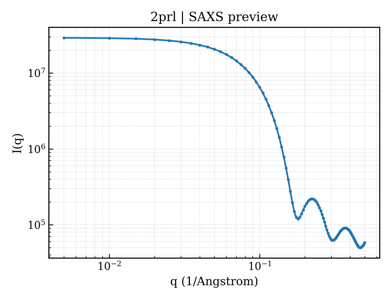
Dihydroorotate dehydrogenase, mitochondrial
Homo sapiens
State in the Current Structure
| Assembly | Oligomeric State | Construct | Mutations and Modifications | Ligands, Ions and Associated Components | Method and Experimental Conditions | Structure Quality |
|---|---|---|---|---|---|---|
| 1 | Protein monomer Monomer Protein × 1 PDB declaration: monomeric(1) Consistent with protein copy count | Chain A; UniProt 29–395 | Mutation:N-terminus truncated | SO4 SULFATE ION × 1 ACT ACETATE ION × 2 FMN FLAVIN MONONUCLEOTIDE × 1 ORO OROTIC ACID × 1 R2C 5-METHOXY-2-[(4-PHENOXYPHENYL)AMINO]BENZOIC ACID × 1 DDQ DECYLAMINE-N,N-DIMETHYL-N-OXIDE × 1 | X-RAY DIFFRACTION X-ray crystallization conditions:VAPOR DIFFUSION, HANGING DROP;pH 4.8;293 K;DROPS WERE FORMED BY MIXING EQUAL AMOUNTS OF 18-24 MG/ML PROTEIN IN 100 MM HEPES PH 7.0, 400 MM NACL, 30% GLYCEROL, 1 MM EDTA AND 10 MM N,N- DIMETHYLUNDECYLAMIN-N-OXIDE (C11DAO) WITH A PRECIPITANT SOLUTION OF 0.1 M ACETATE PH 4.8 40 MM C11DAO, 20.8 MM N,-DIMETHYLDECYLAMINE-N-OXIDE (DDAO), 2 MM DIHYDROOROTATE (DHO) THE HANGING DROPS WERE INCUBATED AGAINST 0.5 ML RESERVOIR OF 0.1 M ACETATE PH 4.8, 1.6-2.2 M AMMONIUM SULFATE AND 30% GLYCEROL., VAPOR DIFFUSION, HANGING DROP, temperature 293K | Resolution 2.10 Å R-free 0.194 |
Other States of the Same Protein in the Database
Each row is a biological assembly of the same UniProt protein in another PDB entry. The “Difference from current entry” column identifies evidence-level differences; no tag means the currently parsed fields agree.
| Other PDB | Difference from Current Entry 2PRL | Assembly / Oligomeric State | Construct | Mutations and Modifications | Ligands, Ions and Non-polymers | Method and Experimental Conditions | Structure Quality |
|---|---|---|---|---|---|---|---|
| 1D3G HUMAN DIHYDROOROTATE DEHYDROGENASE COMPLEXED WITH BREQUINAR ANALOG Deposited 1999-09-29 | Different construct Different mutation/modification Different ligand/ion Different experimental conditions Different structure-quality metrics | Assembly 1 Protein monomer Monomer;Protein × 1 PDB declaration: monomeric |
Chain A
30–396(367 aa)
|
Not recorded | SO4 SULFATE ION × 1 ACT ACETATE ION × 1 BRE 2-BIPHENYL-4-YL-6-FLUORO-3-METHYL-QUINOLINE-4-CARBOXYLIC ACID × 1 FMN FLAVIN MONONUCLEOTIDE × 1 ORO OROTIC ACID × 1 DDQ DECYLAMINE-N,N-DIMETHYL-N-OXIDE × 1 |
X-RAY DIFFRACTION
X-ray crystallization conditions
VAPOR DIFFUSION;293 K;AMMONIUM SULFATE, ACETATE, GLYCEROL, DDAO, C11DAO, BREQUINAR, OROTATE, VAPOR DIFFUSION, temperature 293K
|
Resolution 1.60 Å R-free 0.188 |
| 1D3H HUMAN DIHYDROOROTATE DEHYDROGENASE COMPLEXED WITH ANTIPROLIFERATIVE AGENT A771726 Deposited 1999-09-29 | Different construct Different mutation/modification Different ligand/ion Different experimental conditions Different structure-quality metrics | Assembly 1 Protein monomer Monomer;Protein × 1 PDB declaration: monomeric |
Chain A
30–396(367 aa)
|
Not recorded | SO4 SULFATE ION × 1 ACT ACETATE ION × 1 A26 (2Z)-2-cyano-3-hydroxy-N-[4-(trifluoromethyl)phenyl]but-2-enamide × 1 FMN FLAVIN MONONUCLEOTIDE × 1 ORO OROTIC ACID × 1 |
X-RAY DIFFRACTION
X-ray crystallization conditions
VAPOR DIFFUSION;293 K;ammonium sulphate, acetate buffer, glycerol, DDAO, C11DAO, A771726, orotate, VAPOR DIFFUSION, temperature 293K
|
Resolution 1.80 Å R-free 0.185 |
| 2B0M Human dihydroorotate dehydrogenase bound to a novel inhibitor Deposited 2005-09-14 | Different mutation/modification Different ligand/ion Different experimental conditions Different structure-quality metrics | Assembly 1 Protein monomer Monomer;Protein × 1 PDB declaration: monomeric |
Chain A
29–395(367 aa)
|
Not recorded | 201 3-AMIDO-5-BIPHENYL-BENZOIC ACID × 1 FMN FLAVIN MONONUCLEOTIDE × 1 ORO OROTIC ACID × 1 |
X-RAY DIFFRACTION
X-ray crystallization conditions
VAPOR DIFFUSION, SITTING DROP;pH 4.6;277 K;ammonium sulfate, sodium acetate, C11DAO, C10DAO, pH 4.6, VAPOR DIFFUSION, SITTING DROP, temperature 277K
|
Resolution 2.00 Å R-free 0.249 |
| 2BXV Dual binding mode of a novel series of DHODH inhibitors Deposited 2005-07-27 | Different mutation/modification Different ligand/ion Different experimental conditions Different structure-quality metrics | Assembly 1 Protein monomer Monomer;Protein × 1 PDB declaration: monomeric |
Chain A
29–395(367 aa)
|
Not recorded | SO4 SULFATE ION × 1 ACT ACETATE ION × 1 FMN FLAVIN MONONUCLEOTIDE × 1 ORO OROTIC ACID × 1 3FT 2-({[3-FLUORO-3'-(TRIFLUOROMETHOXY)BIPHENYL-4-YL]AMINO}CARBONYL)CYCLOPENT-1-ENE-1-CARBOXYLIC ACID × 1 |
X-RAY DIFFRACTION
X-ray crystallization conditions
VAPOR DIFFUSION;pH 4.6;293 K;AMMONIUM SULPHATE, ACETATE BUFFER, GLYCEROL, DDAO, C11DAO, A771726, OROTATE, VAPOR DIFFUSION, TEMPERATURE 293K, pH 4.60
|
Resolution 2.15 Å R-free 0.223 |
| 2FPT Dual Binding Mode of a Novel Series of DHODH inhibitors Deposited 2006-01-17 | Different mutation/modification Different ligand/ion Different experimental conditions Different structure-quality metrics | Assembly 1 Protein monomer Monomer;Protein × 1 PDB declaration: monomeric |
Chain A
29–395(367 aa)
|
Not recorded | SO4 SULFATE ION × 4 ACT ACETATE ION × 1 FMN FLAVIN MONONUCLEOTIDE × 1 ORO OROTIC ACID × 1 ILB 2-({[3,5-DIFLUORO-3'-(TRIFLUOROMETHOXY)BIPHENYL-4-YL]AMINO}CARBONYL)CYCLOPENT-1-ENE-1-CARBOXYLIC ACID × 1 |
X-RAY DIFFRACTION
X-ray crystallization conditions
VAPOR DIFFUSION, HANGING DROP;pH 7.7;293 K;Drops were formed by mixing equal amounts of 20 mg/ml protein in 50 mM HEPES pH 7.7, 400 mM NaCl, 30% glycerol, 1 mM EDTA and 10 mM N,N-dimethylundecylamin-N-oxide (C11DAO) with a precipitant solution of 0.1 M acetate pH 4.6 5.0, 40 mM C11DAO, 20.8 mM N,N-dimethyldecylamine-N-oxide (DDAO), 2 mM dihydroorotate (DHO), 1.8 2.4 M ammonium sulfate. The hanging drops were incubated against 0.5 mL reservoir of 0.1 M acetate pH 4.8, 2.4 2.6 M ammonium sulfate and 30% glycerol., VAPOR DIFFUSION, HANGING DROP, temperature 293K
|
Resolution 2.40 Å R-free 0.211 |
| 2FPV Dual binding mode of a novel series of DHODH inhibitors Deposited 2006-01-17 | Different construct Different mutation/modification Different ligand/ion Different experimental conditions Different structure-quality metrics | Assembly 1 Protein monomer Monomer;Protein × 1 PDB declaration: monomeric |
Chain A
32–398(367 aa)
|
Not recorded | ACT ACETATE ION × 2 SO4 SULFATE ION × 4 FMN FLAVIN MONONUCLEOTIDE × 1 ORO OROTIC ACID × 1 ILC 3-{[(3-FLUORO-3'-METHOXYBIPHENYL-4-YL)AMINO]CARBONYL}THIOPHENE-2-CARBOXYLIC ACID × 1 |
X-RAY DIFFRACTION
X-ray crystallization conditions
VAPOR DIFFUSION, HANGING DROP;pH 7.7;293 K;Drops were formed by mixing equal amounts of 20 mg/ml protein in 50 mM HEPES pH 7.7, 400 mM NaCl, 30% glycerol, 1 mM EDTA and 10 mM N,N-dimethylundecylamin-N-oxide (C11DAO) with a precipitant solution of 0.1 M acetate pH 4.6 5.0, 40 mM C11DAO, 20.8 mM N,N-dimethyldecylamine-N-oxide (DDAO), 2 mM dihydroorotate (DHO), 1.8 2.4 M ammonium sulfate, 1 mM compound. The hanging drops were incubated against 0.5 mL reservoir of 0.1 M acetate pH 4.8, 2.4 2.6 M ammonium sulfate and 30% glycerol. , VAPOR DIFFUSION, HANGING DROP, temperature 293K
|
Resolution 1.80 Å R-free 0.205 |
| 2FPY Dual binding mode of a novel series of DHODH inhibitors Deposited 2006-01-17 | Different mutation/modification Different ligand/ion Different experimental conditions Different structure-quality metrics | Assembly 1 Protein monomer Monomer;Protein × 1 PDB declaration: monomeric |
Chain A
29–395(367 aa)
|
Not recorded | ACT ACETATE ION × 1 SO4 SULFATE ION × 5 FMN FLAVIN MONONUCLEOTIDE × 1 ORO OROTIC ACID × 1 ILF 3-({[3,5-DIFLUORO-3'-(TRIFLUOROMETHOXY)BIPHENYL-4-YL]AMINO}CARBONYL)THIOPHENE-2-CARBOXYLIC ACID × 1 |
X-RAY DIFFRACTION
X-ray crystallization conditions
VAPOR DIFFUSION, HANGING DROP;pH 7.7;293 K;Drops were formed by mixing equal amounts of 20 mg/ml protein in 50 mM HEPES pH 7.7, 400 mM NaCl, 30% glycerol, 1 mM EDTA and 10 mM N,N-dimethylundecylamin-N-oxide (C11DAO) with a precipitant solution of 0.1 M acetate pH 4.6 5.0, 40 mM C11DAO, 20.8 mM N,N-dimethyldecylamine-N-oxide (DDAO), 2 mM dihydroorotate (DHO), 1.8 2.4 M ammonium sulfate, 1 mM compound. The hanging drops were incubated against 0.5 mL reservoir of 0.1 M acetate pH 4.8, 2.4 2.6 M ammonium sulfate and 30% glycerol, VAPOR DIFFUSION, HANGING DROP, temperature 293K
|
Resolution 2.00 Å R-free 0.200 |
| 2FQI dual binding modes of a novel series of DHODH inhibitors Deposited 2006-01-18 | Different mutation/modification Different ligand/ion Different experimental conditions Different structure-quality metrics | Assembly 1 Protein monomer Monomer;Protein × 1 PDB declaration: monomeric |
Chain A
29–395(367 aa)
|
Not recorded | SO4 SULFATE ION × 2 ACT ACETATE ION × 3 FMN FLAVIN MONONUCLEOTIDE × 1 ORO OROTIC ACID × 1 ILH 2-({[2,3,5,6-TETRAFLUORO-3'-(TRIFLUOROMETHOXY)BIPHENYL-4-YL]AMINO}CARBONYL)CYCLOPENTA-1,3-DIENE-1-CARBOXYLIC ACID × 1 |
X-RAY DIFFRACTION
X-ray crystallization conditions
VAPOR DIFFUSION, HANGING DROP;pH 7.7;293 K;Drops were formed by mixing equal amounts of 20 mg/ml protein in 50 mM HEPES pH 7.7, 400 mM NaCl, 30% glycerol, 1 mM EDTA and 10 mM N,N-dimethylundecylamin-N-oxide (C11DAO) with a precipitant solution of 0.1 M acetate pH 4.6 5.0, 40 mM C11DAO, 20.8 mM N,N-dimethyldecylamine-N-oxide (DDAO), 2 mM dihydroorotate (DHO), 1.8 2.4 M ammonium sulfate, 1 mM compound. The hanging drops were incubated against 0.5 mL reservoir of 0.1 M acetate pH 4.8, 2.4 2.6 M ammonium sulfate and 30% glycerol, VAPOR DIFFUSION, HANGING DROP, temperature 293K
|
Resolution 1.95 Å R-free 0.202 |
| 2PRH The structures of apo- and inhibitor bound human dihydroorotate dehydrogenase reveal conformational flexibility within the inhibitor binding site Deposited 2007-05-04 | Different ligand/ion Different experimental conditions Different structure-quality metrics | Assembly 1 Protein monomer Monomer;Protein × 1 PDB declaration: monomeric |
Chain A
29–395(367 aa)
|
Mutation:N-terminus truncated | FMN FLAVIN MONONUCLEOTIDE × 1 ORO OROTIC ACID × 1 238 6-CHLORO-2-(2'-FLUOROBIPHENYL-4-YL)-3-METHYLQUINOLINE-4-CARBOXYLIC ACID × 1 DDQ DECYLAMINE-N,N-DIMETHYL-N-OXIDE × 1 |
X-RAY DIFFRACTION
X-ray crystallization conditions
VAPOR DIFFUSION, HANGING DROP;pH 4.8;298 K;0.1M acetate, 1.6-2.4M ammonium sulphate, 30% glycerol, pH 4.8, VAPOR DIFFUSION, HANGING DROP, temperature 298K
|
Resolution 2.40 Å R-free 0.217 |
| 2PRM The structures of apo- and inhibitor bound human dihydroorotate dehydrogenase reveal conformational flexibility within the inhibitor binding site Deposited 2007-05-04 | Different ligand/ion Different experimental conditions Different structure-quality metrics | Assembly 1 Protein monomer Monomer;Protein × 1 PDB declaration: monomeric |
Chain A
29–395(367 aa)
|
Mutation:N-terminus truncated | FMN FLAVIN MONONUCLEOTIDE × 1 ORO OROTIC ACID × 1 |
X-RAY DIFFRACTION
X-ray crystallization conditions
VAPOR DIFFUSION, HANGING DROP;pH 4.8;293 K;DROPS WERE FORMED BY MIXING EQUAL AMOUNTS OF 18-24 MG/ML PROTEIN IN
100 MM HEPES PH 7.0, 400 MM NACL, 30% GLYCEROL, 1 MM EDTA AND 10 MM
N,N- DIMETHYLUNDECYLAMIN-N-OXIDE (C11DAO) WITH A PRECIPITANT SOLUTION
OF 0.1 M ACETATE PH 4.8 40 MM C11DAO, 20.8 MM N,
N-DIMETHYLDECYLAMINE-N-OXIDE (DDAO), 2 MM DIHYDROOROTATE (DHO) THE
HANGING DROPS WERE INCUBATED AGAINST 0.5 ML RESERVOIR OF 0.1 M ACETATE
PH 4.8, 1.6-2.2 M AMMONIUM SULFATE AND 30% GLYCEROL., VAPOR DIFFUSION, HANGING DROP, temperature 293K
|
Resolution 3.00 Å R-free 0.253 |
| 2WV8 Complex of human dihydroorotate dehydrogenase with the inhibitor 221290 Deposited 2009-10-15 | Different construct Different mutation/modification Different ligand/ion Different experimental conditions Different structure-quality metrics | Assembly 1 Protein monomer Monomer;Protein × 1 PDB declaration: monomeric |
Chain A
31–395(365 aa)
Fragment:RESIDUES 31-395
|
Not recorded | FMN FLAVIN MONONUCLEOTIDE × 1 ORO OROTIC ACID × 1 VGN 2-ACETAMIDO-5-(4-PHENYLPHENYL)BENZOIC ACID × 1 DDQ DECYLAMINE-N,N-DIMETHYL-N-OXIDE × 1 ACT ACETATE ION × 1 SO4 SULFATE ION × 1 | X-RAY DIFFRACTION mmCIF provides none of the parsed conditions | Resolution 1.90 Å R-free 0.201 |
| 3F1Q Human dihydroorotate dehydrogenase in complex with a leflunomide derivative inhibitor 1 Deposited 2008-10-28 | Different construct Different mutation/modification Different ligand/ion Different experimental conditions Different structure-quality metrics | Assembly 1 Protein monomer Monomer;Protein × 1 PDB declaration: monomeric |
Chain A
29–395(367 aa)
Fragment:residues 30-396
|
Not recorded | FMN FLAVIN MONONUCLEOTIDE × 1 ORO OROTIC ACID × 1 BCE (2Z)-N-biphenyl-4-yl-2-cyano-3-hydroxybut-2-enamide × 1 ACY ACETIC ACID × 2 SO4 SULFATE ION × 1 |
X-RAY DIFFRACTION
X-ray crystallization conditions
VAPOR DIFFUSION, HANGING DROP;pH 4.8;293 K;drops: 50mM HEPES, pH 7.7, 400mM NaCl, 30% glycerol, 1mM EDTA, 10mM N,N-dimethylundecylamin-N-oxide (C11DAO), precipitant solution: 0.1M acetate pH 4.6-5.0, 40mM C11DAO, 20.8mM N,N-dimethyldecylamine-N-oxide (DDAO), 2mM dihydroorotate (DHO), 1.8-2.4M ammonium sulfate, 1mM inhibitors, reservoir: 0.1M acetate pH 4.8, 2.4-2.6M ammonium sulfate, 30% glycerol , VAPOR DIFFUSION, HANGING DROP, temperature 293K
|
Resolution 2.00 Å R-free 0.199 |
| 3FJ6 Human dihydroorotate dehydrogenase in complex with a leflunomide derivative inhibitor 2 Deposited 2008-12-14 | Different construct Different mutation/modification Different ligand/ion Different experimental conditions Different structure-quality metrics | Assembly 1 Protein monomer Monomer;Protein × 1 PDB declaration: monomeric |
Chain A
29–395(367 aa)
Fragment:residues 30-396
|
Not recorded | ORO OROTIC ACID × 1 FMN FLAVIN MONONUCLEOTIDE × 1 SO4 SULFATE ION × 1 ACT ACETATE ION × 2 CIH (2Z)-2-cyano-N-(2,2'-dichlorobiphenyl-4-yl)-3-hydroxybut-2-enamide × 1 |
X-RAY DIFFRACTION
X-ray crystallization conditions
VAPOR DIFFUSION, HANGING DROP;pH 4.8;291 K;drops: 50mM HEPES, pH 7.7, 400mM NaCl, 30% glycerol, 1mM EDTA, 10mM N,N-dimethylundecylamin-N-oxide (C11DAO),precipitant solution: 0.1M acetate pH 4.6-5.0, 40mM C11DAO, 20.8mM N,N-dimethyldecylamine-N-oxide (DDAO), 2mM dihydroorotate (DHO), 1.8-2.4M ammonium sulfate, 1mM inhibitors, reservoir: 0.1M acetate pH 4.8, 2.4-2.6M ammonium sulfate, 30% glycerol , VAPOR DIFFUSION, HANGING DROP, temperature 291K
|
Resolution 1.80 Å R-free 0.204 |
| 3FJL Human dihydroorotate dehydrogenase in complex with a leflunomide derivative inhibitor 3 Deposited 2008-12-14 | Different construct Different mutation/modification Different ligand/ion Different experimental conditions Different structure-quality metrics | Assembly 1 Protein monomer Monomer;Protein × 1 PDB declaration: monomeric |
Chain A
29–395(367 aa)
Fragment:residues 30-396
|
Not recorded | FMN FLAVIN MONONUCLEOTIDE × 1 ORO OROTIC ACID × 1 SO4 SULFATE ION × 1 ACY ACETIC ACID × 2 CJH (2Z)-2-cyano-N-(3'-ethoxybiphenyl-4-yl)-3-hydroxybut-2-enamide × 1 |
X-RAY DIFFRACTION
X-ray crystallization conditions
VAPOR DIFFUSION, HANGING DROP;pH 4.8;291 K;drops: 50mM HEPES, pH 7.7, 400mM NaCl, 30% glycerol, 1mM EDTA, 10mM N,N-dimethylundecylamin-N-oxide (C11DAO), precipitant solution: 0.1M acetate pH 4.6-5.0, 40mM C11DAO, 20.8mM N,N-dimethyldecylamine-N-oxide (DDAO), 2mM dihydroorotate (DHO), 1.8-2.4M ammonium sulfate, 1mM inhibitors, reservoir: 0.1M acetate pH 4.8, 2.4-2.6M ammonium sulfate, 30% glycerol, VAPOR DIFFUSION, HANGING DROP, temperature 291K
|
Resolution 1.90 Å R-free 0.209 |
| 3G0U Human dihydroorotate dehydrogenase in complex with a leflunomide derivative inhibitor 4 Deposited 2009-01-28 | Different construct Different mutation/modification Different ligand/ion Different experimental conditions Different structure-quality metrics | Assembly 1 Protein monomer Monomer;Protein × 1 PDB declaration: monomeric |
Chain A
29–395(367 aa)
Fragment:residues 30-396
|
Not recorded | ORO OROTIC ACID × 1 FMN FLAVIN MONONUCLEOTIDE × 1 MDY (2Z)-N-(3-chloro-2'-methoxybiphenyl-4-yl)-2-cyano-3-hydroxybut-2-enamide × 1 |
X-RAY DIFFRACTION
X-ray crystallization conditions
VAPOR DIFFUSION, HANGING DROP;pH 4.8;291 K;Drops: 20mg/mL protein in 50mM HEPES, pH 7.7, 400mM NaCl, 30% glycerol, 1mM EDTA, 10mM N,N-dimethylundecylamin-N-oxide (C11DAO)
precipitant solution: 0.1M acetate pH 4.6-5.0, 40mM C11DAO, 20.8mM N,N-dimethyldecylamine-N-oxide (DDAO), 2mM dihydroorotate (DHO), 1.8-2.4M ammonium sulfate, 1mM inhibitors
reservoir: 0.1M acetate pH 4.8, 2.4-2.6M ammonium sulfate, 30% glycerol , VAPOR DIFFUSION, HANGING DROP, temperature 291K
|
Resolution 2.00 Å R-free 0.203 |
| 3G0X Human dihydroorotate dehydrogenase in complex with a leflunomide derivative inhibitor 5 Deposited 2009-01-29 | Different construct Different mutation/modification Different ligand/ion Different experimental conditions Different structure-quality metrics | Assembly 1 Protein monomer Monomer;Protein × 1 PDB declaration: monomeric |
Chain A
29–395(367 aa)
Fragment:residues 30-396
|
Not recorded | FMN FLAVIN MONONUCLEOTIDE × 1 ORO OROTIC ACID × 1 MD7 (2Z)-N-biphenyl-4-yl-2-cyano-3-cyclopropyl-3-hydroxyprop-2-enamide × 1 SO4 SULFATE ION × 1 |
X-RAY DIFFRACTION
X-ray crystallization conditions
VAPOR DIFFUSION, HANGING DROP;pH 4.8;291 K;Drops: 20mg/mL protein in 50mM HEPES, pH 7.7, 400mM NaCl, 30% glycerol, 1mM EDTA, 10mM N,N-dimethylundecylamin-N-oxide (C11DAO),
precipitant solution: 0.1M acetate pH 4.6-5.0, 40mM C11DAO, 20.8mM N,N-dimethyldecylamine-N-oxide (DDAO), 2mM dihydroorotate (DHO), 1.8-2.4M ammonium sulfate, 1mM inhibitors,
reservoir: 0.1M acetate pH 4.8, 2.4-2.6M ammonium sulfate, 30% glycerol, VAPOR DIFFUSION, HANGING DROP, temperature 291K
|
Resolution 1.80 Å R-free 0.211 |
| 3KVJ Crystal Structure of Human Dihydroorotate Dehydrogenase (DHODH) with Amino-Benzoic Acid Inhibitor 105 at 1.94A Resolution Deposited 2009-11-30 | Different mutation/modification Different ligand/ion Different experimental conditions Different structure-quality metrics | Assembly 1 Protein monomer Monomer;Protein × 1 PDB declaration: monomeric |
Chain A
29–395(367 aa)
|
Not recorded | SO4 SULFATE ION × 1 FMN FLAVIN MONONUCLEOTIDE × 1 DOR (4S)-2,6-DIOXOHEXAHYDROPYRIMIDINE-4-CARBOXYLIC ACID × 1 DET UNDECYLAMINE-N,N-DIMETHYL-N-OXIDE × 1 ACT ACETATE ION × 1 1X5 2-[(cyclopropylcarbonyl)amino]-5-[methyl(pyridin-3-ylmethyl)amino]benzoic acid × 1 |
X-RAY DIFFRACTION
X-ray crystallization conditions
VAPOR DIFFUSION, HANGING DROP;pH 5;298 K;2.4 M AMMONIUM SULFATE, 15% GLYCEROL, 0.1 M NA-ACETATE, pH 5.0, VAPOR DIFFUSION, HANGING DROP, temperature 298K
|
Resolution 1.94 Å R-free 0.195 |
| 3KVK Crystal structure of human dihydroorotate dehydrogenase (DHODH) with amino-benzoic acid inhibitor 641 at 2.05A resolution Deposited 2009-11-30 | Different mutation/modification Different ligand/ion Different experimental conditions Different structure-quality metrics | Assembly 1 Protein monomer Monomer;Protein × 1 PDB declaration: monomeric |
Chain A
29–395(367 aa)
|
Not recorded | SO4 SULFATE ION × 1 FMN FLAVIN MONONUCLEOTIDE × 1 DOR (4S)-2,6-DIOXOHEXAHYDROPYRIMIDINE-4-CARBOXYLIC ACID × 1 DET UNDECYLAMINE-N,N-DIMETHYL-N-OXIDE × 1 6X1 2-{[(3,5-dichlorophenyl)carbamoyl]amino}benzoic acid × 1 |
X-RAY DIFFRACTION
X-ray crystallization conditions
VAPOR DIFFUSION, HANGING DROP;pH 5;298 K;2.4 M AMMONIUM SULFATE, 15% GLYCEROL, 0.1 M NA-ACETATE, pH 5.0, VAPOR DIFFUSION, HANGING DROP, temperature 298K
|
Resolution 2.05 Å R-free 0.205 |
| 3KVL Crystal structure of human dihydroorotate dehydrogenase (DHODH) with amino-benzoic acid inhibitor 715 at 1.85A resolution Deposited 2009-11-30 | Different mutation/modification Different ligand/ion Different experimental conditions Different structure-quality metrics | Assembly 1 Protein monomer Monomer;Protein × 1 PDB declaration: monomeric |
Chain A
29–395(367 aa)
|
Not recorded | SO4 SULFATE ION × 1 FMN FLAVIN MONONUCLEOTIDE × 1 DOR (4S)-2,6-DIOXOHEXAHYDROPYRIMIDINE-4-CARBOXYLIC ACID × 1 DET UNDECYLAMINE-N,N-DIMETHYL-N-OXIDE × 1 ACT ACETATE ION × 1 7Z5 5-[methyl(pyridin-3-ylmethyl)amino]-2-(propanoylamino)benzoic acid × 1 |
X-RAY DIFFRACTION
X-ray crystallization conditions
VAPOR DIFFUSION, HANGING DROP;pH 5;298 K;2.4 M AMMONIUM SULFATE, 15% GLYCEROL, 0.1 M NA-ACETATE, pH 5.0, VAPOR DIFFUSION, HANGING DROP, temperature 298K
|
Resolution 1.85 Å R-free 0.191 |
| 3KVM Crystal structure of human dihydroorotate dehydrogenase (DHODH) with amino-benzoic acid inhibitor 951 at 2.00A resolution Deposited 2009-11-30 | Different mutation/modification Different ligand/ion Different experimental conditions Different structure-quality metrics | Assembly 1 Protein monomer Monomer;Protein × 1 PDB declaration: monomeric |
Chain A
29–395(367 aa)
|
Not recorded | SO4 SULFATE ION × 1 FMN FLAVIN MONONUCLEOTIDE × 1 DOR (4S)-2,6-DIOXOHEXAHYDROPYRIMIDINE-4-CARBOXYLIC ACID × 1 DET UNDECYLAMINE-N,N-DIMETHYL-N-OXIDE × 1 ACT ACETATE ION × 1 951 2-[(2E)-2-{[5-(2-chlorophenyl)furan-2-yl]methylidene}hydrazino]benzoic acid × 1 |
X-RAY DIFFRACTION
X-ray crystallization conditions
VAPOR DIFFUSION, HANGING DROP;pH 5.2;298 K;2.4 M AMMONIUM SULFATE, 15% GLYCEROL, 0.1 M NA-ACETATE, pH 5.2, VAPOR DIFFUSION, HANGING DROP, temperature 298K
|
Resolution 2.00 Å R-free 0.215 |
| 3U2O Dihydroorotate Dehydrogenase (DHODH) crystal structure in complex with small molecule inhibitor Deposited 2011-10-04 | Different construct Different mutation/modification Different ligand/ion Different experimental conditions Different structure-quality metrics | Assembly 1 Protein monomer Monomer;Protein × 1 PDB declaration: monomeric |
Chain A
1–395(395 aa)
|
Not recorded | FMN FLAVIN MONONUCLEOTIDE × 1 ORO OROTIC ACID × 1 03U methyl 4-{[(2Z)-2-cyano-3-hydroxypent-2-enoyl]amino}-4'-fluorobiphenyl-2-carboxylate × 1 ACT ACETATE ION × 3 SO4 SULFATE ION × 2 |
X-RAY DIFFRACTION
X-ray crystallization conditions
VAPOR DIFFUSION;pH 4.8;293 K;ammonium sulfate, pH 4.8, vapor diffusion, temperature 293K
|
Resolution 2.18 Å R-free 0.205 |
| 3W7R Structure of Human dihydroorotate dehydrogenase in complex with mii-4-097 Deposited 2013-03-06 | Different construct Different mutation/modification Different ligand/ion Different experimental conditions Different structure-quality metrics | Assembly 1 Protein monomer Monomer;Protein × 1 PDB declaration: monomeric |
Chain A
29–395(367 aa)
Fragment:UNP residues 29-395
|
Not recorded | CL CHLORIDE ION × 1 DDQ DECYLAMINE-N,N-DIMETHYL-N-OXIDE × 6 W7A 2,6-dioxo-5-[2-(4-phenylphenyl)ethyl]-1,2,3,6- tetrahydropyrimidine-4-carboxylic acid × 1 FMN FLAVIN MONONUCLEOTIDE × 1 GOL GLYCEROL × 22 ORO OROTIC ACID × 1 SO4 SULFATE ION × 4 ACT ACETATE ION × 7 |
X-RAY DIFFRACTION
X-ray crystallization conditions
VAPOR DIFFUSION, HANGING DROP;pH 4.9;293 K;100mM SODIUM ACETATE, 1.8-1.9M AMMONIUM SULPHATE, 40mM C11DAO, 20.8mM DDAO, 2mM DIHYDROOROTATE, pH 4.9, VAPOR DIFFUSION, HANGING DROP, temperature 293K
|
Resolution 1.68 Å R-free 0.172 |
| 3ZWS Structure of Human Dihydroorotate Dehydrogenase with a Bound Inhibitor Deposited 2011-08-02 | Different construct Different mutation/modification Different ligand/ion Different experimental conditions Different structure-quality metrics | Assembly 1 Protein monomer Monomer;Protein × 1 PDB declaration: monomeric |
Chain A
29–395(367 aa)
Fragment:RESIDUES 29-395
|
Not recorded | FMN FLAVIN MONONUCLEOTIDE × 1 ORO OROTIC ACID × 1 SO4 SULFATE ION × 2 CL CHLORIDE ION × 1 AVQ 2-[(2,5-DICHLOROBENZYL)SULFANYL]-5-METHYL[1,2,4]TRIAZOLO[1,5-A]PYRIMIDIN-7-OL × 1 ACT ACETATE ION × 2 |
X-RAY DIFFRACTION
X-ray crystallization conditions
0.1 M SODIUM ACETATE (PH 4.8), 2.4-2.6 M AMMONIUM SULPHATE, 40 MM C11DAO, 20 MM DDAO.
|
Resolution 1.60 Å R-free 0.176 |
| 3ZWT Structure of Human Dihydroorotate Dehydrogenase with a Bound Inhibitor Deposited 2011-08-02 | Different construct Different mutation/modification Different ligand/ion Different experimental conditions Different structure-quality metrics | Assembly 1 Protein monomer Monomer;Protein × 1 PDB declaration: monomeric |
Chain A
29–395(367 aa)
Fragment:RESIDUES 29-395
|
Not recorded | FMN FLAVIN MONONUCLEOTIDE × 1 ORO OROTIC ACID × 1 SO4 SULFATE ION × 2 KFZ 2-{[(2,5-DICHLOROPHENYL)METHYL]SULFANYL}-5-ETHYL-[1,2,4]TRIAZOLO[1,5-A]PYRIMIDIN-7-OL × 1 ACT ACETATE ION × 2 CL CHLORIDE ION × 1 |
X-RAY DIFFRACTION
X-ray crystallization conditions
0.1 M SODIUM ACETATE (PH 4.8), 2.4-2.6 M AMMONIUM SULPHATE, 40 MM C11DAO, 20 MM DDAO.
|
Resolution 1.55 Å R-free 0.177 |
| 4IGH High resolution crystal structure of human dihydroorotate dehydrogenase bound with 4-quinoline carboxylic acid analog Deposited 2012-12-17 | Different construct Different mutation/modification Different ligand/ion Different experimental conditions Different structure-quality metrics | Assembly 1 Protein monomer Monomer;Protein × 1 PDB declaration: monomeric |
Chain A
32–395(364 aa)
Fragment:UNP residues 32-395
|
Not recorded | FMN FLAVIN MONONUCLEOTIDE × 1 ORO OROTIC ACID × 1 1EA 6-fluoro-2-[2-methyl-4-phenoxy-5-(propan-2-yl)phenyl]quinoline-4-carboxylic acid × 1 ZWI 3-[decyl(dimethyl)ammonio]propane-1-sulfonate × 1 GOL GLYCEROL × 2 |
X-RAY DIFFRACTION
X-ray crystallization conditions
VAPOR DIFFUSION, HANGING DROP;pH 5.4;296 K;1.72 M ammonium sulfate, 0.1 M sodium acetate, pH 5.4, 1.5 M sodium chloride, 10 mM DTT, VAPOR DIFFUSION, HANGING DROP, temperature 296K
|
Resolution 1.30 Å R-free 0.148 |
| 4JGD Crystal structure of human dihydroorotate dehydrogenase (DHODH) with DH03A016 Deposited 2013-03-01 | Different mutation/modification Different ligand/ion Different experimental conditions Different structure-quality metrics | Assembly 1 Protein monomer Monomer;Protein × 1 PDB declaration: monomeric |
Chain A
29–395(367 aa)
|
Not recorded | FMN FLAVIN MONONUCLEOTIDE × 1 ORO OROTIC ACID × 1 DET UNDECYLAMINE-N,N-DIMETHYL-N-OXIDE × 1 SO4 SULFATE ION × 9 4JG 1-[4-methyl-2-(naphthalen-2-ylamino)-1,3-thiazol-5-yl]ethanone × 1 |
X-RAY DIFFRACTION
X-ray crystallization conditions
VAPOR DIFFUSION, SITTING DROP;pH 4.8;298 K;0.1M acetate, 40mM UDAO, 20.8mM N,N-dimethyldecylamine-N-oxide(DDAO), 2mM DHO, 2.4-2.6M ammonium sulfate., pH 4.8, VAPOR DIFFUSION, SITTING DROP, temperature 298K
|
Resolution 2.05 Å R-free 0.175 |
| 4JS3 Crystal structure of human dihydroorotate dehydrogenase (DHODH) with 057 Deposited 2013-03-22 | Different construct Different mutation/modification Different ligand/ion Different experimental conditions Different structure-quality metrics | Assembly 1 Protein monomer Monomer;Protein × 1 PDB declaration: monomeric |
Chain A
29–395(367 aa)
Fragment:UNP residues 29-395
|
Not recorded | FMN FLAVIN MONONUCLEOTIDE × 1 ORO OROTIC ACID × 1 JS3 2-chloro-4-methyl-N-(naphthalen-2-yl)-1,3-thiazole-5-carboxamide × 1 SO4 SULFATE ION × 7 ACY ACETIC ACID × 5 |
X-RAY DIFFRACTION
X-ray crystallization conditions
VAPOR DIFFUSION, HANGING DROP;pH 4.8;293 K;0.1M acetate pH 4.8, 40mM UDAO, 20.8mM
N,N-dimethyldecylamine-N-oxide(DDAO), 2mM DHO, 2.4-2.6M ammonium sulfate, VAPOR DIFFUSION, HANGING DROP, temperature 293K
|
Resolution 2.00 Å R-free 0.179 |
| 4JTS Crystal structure of human dihydroorotate dehydrogenase (DHODH) with 072 Deposited 2013-03-24 | Different construct Different mutation/modification Different ligand/ion Different experimental conditions Different structure-quality metrics | Assembly 1 Protein monomer Monomer;Protein × 1 PDB declaration: monomeric |
Chain A
29–395(367 aa)
Fragment:UNP residues 29-395
|
Not recorded | FMN FLAVIN MONONUCLEOTIDE × 1 ORO OROTIC ACID × 1 SO4 SULFATE ION × 7 ACT ACETATE ION × 2 JTS 4-methyl-N-(naphthalen-2-yl)-1,3-thiazole-5-carboxamide × 1 |
X-RAY DIFFRACTION
X-ray crystallization conditions
VAPOR DIFFUSION, HANGING DROP;pH 4.8;293 K;0.1M acetate pH 4.8, 40mM
C11DAO, 20.8mM N,N-dimethyldecylamine-N-oxide (DDAO), 2mM DHO 1.6-1.8 M ammonium sulfate, 2mM 072, VAPOR DIFFUSION, HANGING DROP, temperature 293K
|
Resolution 2.21 Å R-free 0.187 |
| 4JTT Crystal structure of human dihydroorotate dehydrogenase (DHODH) with 066 Deposited 2013-03-24 | Different construct Different mutation/modification Different ligand/ion Different experimental conditions Different structure-quality metrics | Assembly 1 Protein monomer Monomer;Protein × 1 PDB declaration: monomeric |
Chain A
29–395(367 aa)
Fragment:UNP residues 29-395
|
Not recorded | FMN FLAVIN MONONUCLEOTIDE × 1 ORO OROTIC ACID × 1 SO4 SULFATE ION × 6 JTT 2,4-dimethyl-N-(naphthalen-2-yl)-1,3-thiazole-5-carboxamide × 1 ACT ACETATE ION × 3 |
X-RAY DIFFRACTION
X-ray crystallization conditions
VAPOR DIFFUSION, HANGING DROP;pH 4.8;293 K;0.1M acetate pH 4.8, 40mM
C11DAO, 20.8 mM N,N-dimethyldecylamine-N-oxide(DDAO), 2mM DHO 1.6-1.8M ammonium sulfate, 2mM 072, VAPOR DIFFUSION, HANGING DROP, temperature 293K
|
Resolution 2.10 Å R-free 0.192 |
| 4JTU Crystal structure of human dihydroorotate dehydrogenase (DHODH) with brequinar analogue Deposited 2013-03-24 | Different construct Different mutation/modification Different oligomeric state Different ligand/ion Different experimental conditions Different structure-quality metrics | Assembly 1 Protein homooligomer Homooligomer;Protein × 2 PDB declaration: dimeric |
Chain A
29–395(367 aa)
Fragment:UNP residues 29-395
|
Not recorded | FMN FLAVIN MONONUCLEOTIDE × 2 ORO OROTIC ACID × 2 SO4 SULFATE ION × 6 DET UNDECYLAMINE-N,N-DIMETHYL-N-OXIDE × 2 JTU 6-bromo-2-{4-[(2R)-butan-2-yl]phenyl}-3-methylquinoline-4-carboxylic acid × 2 |
X-RAY DIFFRACTION
X-ray crystallization conditions
VAPOR DIFFUSION, HANGING DROP;pH 4.8;293 K;0.1M acetate pH 4.8, 40mM UDAO, 20.8mM N,N-dimethyldecylamine-N-oxide(DDAO), 2mM DHO, 1.6-1.8M ammonium sulfate, 1mM inhibitor., VAPOR DIFFUSION, HANGING DROP, temperature 293K
|
Resolution 1.90 Å R-free 0.199 |
| 4JTU Crystal structure of human dihydroorotate dehydrogenase (DHODH) with brequinar analogue Deposited 2013-03-24 | Different construct Different mutation/modification Different ligand/ion Different experimental conditions Different structure-quality metrics | Assembly 2 Protein monomer Monomer;Protein × 1 PDB declaration: monomeric |
Chain A
29–395(367 aa)
Fragment:UNP residues 29-395
|
Not recorded | FMN FLAVIN MONONUCLEOTIDE × 1 ORO OROTIC ACID × 1 SO4 SULFATE ION × 3 DET UNDECYLAMINE-N,N-DIMETHYL-N-OXIDE × 1 JTU 6-bromo-2-{4-[(2R)-butan-2-yl]phenyl}-3-methylquinoline-4-carboxylic acid × 1 |
X-RAY DIFFRACTION
X-ray crystallization conditions
VAPOR DIFFUSION, HANGING DROP;pH 4.8;293 K;0.1M acetate pH 4.8, 40mM UDAO, 20.8mM N,N-dimethyldecylamine-N-oxide(DDAO), 2mM DHO, 1.6-1.8M ammonium sulfate, 1mM inhibitor., VAPOR DIFFUSION, HANGING DROP, temperature 293K
|
Resolution 1.90 Å R-free 0.199 |
| 4LS0 Crystal structure of human dihydroorotate dehydrogenase (DHODH) with DH01B0033 Deposited 2013-07-21 | Different construct Different mutation/modification Different ligand/ion Different experimental conditions Different structure-quality metrics | Assembly 1 Protein monomer Monomer;Protein × 1 PDB declaration: monomeric |
Chain A
29–395(367 aa)
Fragment:UNP residues 29-395
|
Not recorded | FMN FLAVIN MONONUCLEOTIDE × 1 ORO OROTIC ACID × 1 SO4 SULFATE ION × 10 B3B 2-{(E)-[2-(4-phenyl-1,3-thiazol-2-yl)hydrazinylidene]methyl}benzaldehyde × 1 |
X-RAY DIFFRACTION
X-ray crystallization conditions
VAPOR DIFFUSION, HANGING DROP;pH 4.8;293 K;0.1M acetate, 40mM UDAO, 20.8mM N,N-dimethyldecylamine-N-oxide (DDAO), 2mM DHO, 1.6-1.8M ammonium sulfate, pH 4.8, VAPOR DIFFUSION, HANGING DROP, temperature 293K
|
Resolution 2.07 Å R-free 0.172 |
| 4LS1 Crystal structure of human dihydroorotate dehydrogenase (DHODH) with DH03A312 Deposited 2013-07-21 | Different construct Different mutation/modification Different ligand/ion Different experimental conditions Different structure-quality metrics | Assembly 1 Protein monomer Monomer;Protein × 1 PDB declaration: monomeric |
Chain A
29–395(367 aa)
Fragment:UNP residues 29-395
|
Not recorded | FMN FLAVIN MONONUCLEOTIDE × 1 ORO OROTIC ACID × 1 3X2 2-[(E)-{2-[4-(2-chlorophenyl)-1,3-thiazol-2-yl]hydrazinylidene}methyl]benzoic acid × 1 SO4 SULFATE ION × 1 ACT ACETATE ION × 4 |
X-RAY DIFFRACTION
X-ray crystallization conditions
VAPOR DIFFUSION, HANGING DROP;pH 4.8;293 K;0.1M acetate, 40mM UDAO, 20.8mM N,N-dimethyldecylamine-N-oxide (DDAO), 2mM DHO, 1.6-1.8M ammonium sulfate, pH 4.8, VAPOR DIFFUSION, HANGING DROP, temperature 293K
|
Resolution 2.20 Å R-free 0.185 |
| 4LS2 Crystal structure of human dihydroorotate dehydrogenase (DHODH) with DH03A313 Deposited 2013-07-21 | Different construct Different mutation/modification Different ligand/ion Different experimental conditions Different structure-quality metrics | Assembly 1 Protein monomer Monomer;Protein × 1 PDB declaration: monomeric |
Chain A
29–395(367 aa)
Fragment:UNP residues 29-395
|
Not recorded | FMN FLAVIN MONONUCLEOTIDE × 1 ORO OROTIC ACID × 1 SO4 SULFATE ION × 5 ACT ACETATE ION × 1 3XS 2-[(E)-{2-[4-(3-methoxyphenyl)-1,3-thiazol-2-yl]hydrazinylidene}methyl]benzoic acid × 1 |
X-RAY DIFFRACTION
X-ray crystallization conditions
VAPOR DIFFUSION, HANGING DROP;pH 4.8;293 K;0.1M acetate, 40mM UDAO, 20.8mM N,N-dimethyldecylamine-N-oxide (DDAO), 2mM DHO, 1.6-1.8M ammonium sulfate, pH 4.8, VAPOR DIFFUSION, HANGING DROP, temperature 293K
|
Resolution 2.27 Å R-free 0.190 |
| 4OQV High resolution crystal structure of human dihydroorotate dehydrogenase bound with DSM338 (N-[3,5-difluoro-4-(trifluoromethyl)phenyl]-5-methyl-2-(trifluoromethyl)[1,2,4]triazolo[1,5-a]pyrimidin-7-amine) Deposited 2014-02-10 | Different construct Different mutation/modification Different ligand/ion Different experimental conditions Different structure-quality metrics | Assembly 1 Protein monomer Monomer;Protein × 1 PDB declaration: monomeric |
Chain A
32–395(364 aa)
Fragment:UNP residues 32-395
|
Not recorded | FMN FLAVIN MONONUCLEOTIDE × 1 ORO OROTIC ACID × 1 2V6 N-[3,5-difluoro-4-(trifluoromethyl)phenyl]-5-methyl-2-(trifluoromethyl)[1,2,4]triazolo[1,5-a]pyrimidin-7-amine × 1 ACY ACETIC ACID × 2 GOL GLYCEROL × 1 NA SODIUM ION × 2 SO4 SULFATE ION × 1 |
X-RAY DIFFRACTION
X-ray crystallization conditions
VAPOR DIFFUSION, HANGING DROP;pH 5.4;293 K;1.76 M Ammonium sulfate, 0.1 M Sodium Acetate, pH 5.4, 1.9 M NaCl, VAPOR DIFFUSION, HANGING DROP, temperature 293K
|
Resolution 1.23 Å R-free 0.157 |
| 4RK8 Crystal structure of human dihydroorotate dehydrogenase (DHODH) with DH03A356 Deposited 2014-10-12 | Different mutation/modification Different ligand/ion Different experimental conditions Different structure-quality metrics | Assembly 1 Protein monomer Monomer;Protein × 1 PDB declaration: monomeric |
Chain A
29–395(367 aa)
|
Not recorded | FMN FLAVIN MONONUCLEOTIDE × 1 ORO OROTIC ACID × 1 3RV 5-fluoro-2-{[(5Z)-5-(naphthalen-1-ylmethylidene)-4-oxo-4,5-dihydro-1,3-thiazol-2-yl]amino}benzoic acid × 1 SO4 SULFATE ION × 11 |
X-RAY DIFFRACTION
X-ray crystallization conditions
VAPOR DIFFUSION, HANGING DROP;pH 4.8;293 K;0.1M acetate pH 4.8, 40 mM UDAO, 20.8 mM, N,N-dimethyldecylamine-N-oxide (DDAO), 2mM DHO, 1.6-1.8M ammonium sulfate, 1 mM inhibitor, VAPOR DIFFUSION, HANGING DROP, temperature 293K
|
Resolution 2.22 Å R-free 0.177 |
| 4RKA Crystal structure of human dihydroorotate dehydrogenase (DHODH) with DH03A347 Deposited 2014-10-12 | Different mutation/modification Different ligand/ion Different experimental conditions Different structure-quality metrics | Assembly 1 Protein monomer Monomer;Protein × 1 PDB declaration: monomeric |
Chain A
29–395(367 aa)
|
Not recorded | FMN FLAVIN MONONUCLEOTIDE × 1 ORO OROTIC ACID × 1 SO4 SULFATE ION × 6 3RY 2-{[5-(naphthalen-1-ylmethyl)-4-oxo-4H-1lambda~4~,3-thiazol-2-yl]amino}benzoic acid × 1 |
X-RAY DIFFRACTION
X-ray crystallization conditions
VAPOR DIFFUSION, HANGING DROP;pH 4.8;293 K;0.1M acetate pH 4.8, 40 mM UDAO, 20.8 mM N,N-dimethyldecylamine-N-oxide (DDAO), 2mM DHO, 1.6-1.8M ammonium sulfate, 1 mM inhibitor, VAPOR DIFFUSION, HANGING DROP, temperature 293K
|
Resolution 2.71 Å R-free 0.228 |
| 4RLI Crystal structure of human dihydroorotate dehydrogenase (DHODH) with DH03A048 Deposited 2014-10-17 | Different mutation/modification Different ligand/ion Different experimental conditions Different structure-quality metrics | Assembly 1 Protein monomer Monomer;Protein × 1 PDB declaration: monomeric |
Chain A
29–395(367 aa)
|
Not recorded | FMN FLAVIN MONONUCLEOTIDE × 1 ORO OROTIC ACID × 1 3SH ethyl 2-[(3-chloro-4-methylphenyl)amino]-4-phenyl-1,3-thiazole-5-carboxylate × 1 |
X-RAY DIFFRACTION
X-ray crystallization conditions
VAPOR DIFFUSION, HANGING DROP;pH 4.8;293 K;0.1M acetate pH 4.8, 40 mM UDAO, 20.8 mM N,N-dimethyldecylamine-N-oxide (DDAO), 2mM DHO, 1.6-1.8M ammonium sulfate, 1 mM inhibitor, VAPOR DIFFUSION, HANGING DROP, temperature 293K
|
Resolution 2.50 Å R-free 0.206 |
| 4RR4 Crystal structure of human dihydroorotate dehydrogenase (DHODH) with DH03A367 Deposited 2014-11-06 | Different mutation/modification Different ligand/ion Different experimental conditions Different structure-quality metrics | Assembly 1 Protein monomer Monomer;Protein × 1 PDB declaration: monomeric |
Chain A
29–395(367 aa)
|
Not recorded | FMN FLAVIN MONONUCLEOTIDE × 1 ORO OROTIC ACID × 1 3V1 2-chloro-N-[3-(4-{[(2Z)-2-cyano-3-cyclopropyl-3-hydroxyprop-2-enoyl]amino}phenoxy)phenyl]-4-methyl-1,3-thiazole-5-carboxamide × 1 SO4 SULFATE ION × 2 |
X-RAY DIFFRACTION
X-ray crystallization conditions
VAPOR DIFFUSION, HANGING DROP;pH 4.8;293 K;0.1M acetate pH 4.8, 40 mM UDAO, 20.8 mM N,N-dimethyldecylamine-N-oxide (DDAO), 2mM DHO, 1.6-1.8M ammonium sulfate, 1 mM inhibitor, VAPOR DIFFUSION, HANGING DROP, temperature 293K
|
Resolution 2.38 Å R-free 0.207 |
| 4YLW Crystal structure of human dihydroorotate dehydrogenase (DHODH) with No.33 compound Deposited 2015-03-06 | Different construct Different mutation/modification Different ligand/ion Different experimental conditions Different structure-quality metrics | Assembly 1 Protein monomer Monomer;Protein × 1 PDB declaration: monomeric |
Chain A
29–395(367 aa)
Fragment:UNP residues 29-395
|
Not recorded | 52Y methyl (2Z)-cyano[3-(3-fluoro-4'-methoxybiphenyl-4-yl)-4-oxo-1,3-thiazolidin-2-ylidene]acetate × 1 FMN FLAVIN MONONUCLEOTIDE × 1 ORO OROTIC ACID × 1 SO4 SULFATE ION × 10 |
X-RAY DIFFRACTION
X-ray crystallization conditions
VAPOR DIFFUSION, HANGING DROP;pH 4.8;293 K;0.1M acetate, 40mM UDAO, 20.8mM N,N-dimethyldecylamine-N-oxide (DDAO), 2mM DHO, 1.6-1.8M ammonium sulfate
|
Resolution 1.79 Å R-free 0.178 |
| 4ZL1 Crystal structure of human dihydroorotate dehydrogenase (DHODH) with 18X at 1.86 A resolution Deposited 2015-05-01 | Different mutation/modification Different ligand/ion Different experimental conditions Different structure-quality metrics | Assembly 1 Protein monomer Monomer;Protein × 1 PDB declaration: monomeric |
Chain A
29–395(367 aa)
|
Not recorded | ORO OROTIC ACID × 1 FMN FLAVIN MONONUCLEOTIDE × 1 18X methyl (2Z)-cyano[3-(3-fluorobiphenyl-4-yl)-4-oxo-1,3-thiazolidin-2-ylidene]acetate × 1 SO4 SULFATE ION × 4 |
X-RAY DIFFRACTION
X-ray crystallization conditions
VAPOR DIFFUSION, HANGING DROP;pH 4.8;293 K;0.1M acetate, 40mM UDAO, 20.8mM N,N-dimethyldecylamine-N-oxide(DDAO), 2mM DHO, 1.6-1.8M ammonium sulfate, 1mM inhibitor
|
Resolution 1.86 Å R-free 0.179 |
| 4ZMG Crystal structure of Human Dihydroorotate Dehydrogenase (DHODH) with DH03A338 Deposited 2015-05-04 | Different mutation/modification Different ligand/ion Different experimental conditions Different structure-quality metrics | Assembly 1 Protein monomer Monomer;Protein × 1 PDB declaration: monomeric |
Chain A
29–395(367 aa)
|
Not recorded | FMN FLAVIN MONONUCLEOTIDE × 1 ORO OROTIC ACID × 1 4R5 1-(3,5-difluoro-3'-methoxybiphenyl-4-yl)-3-(1,3-thiazol-5-yl)urea × 1 SO4 SULFATE ION × 2 ACT ACETATE ION × 1 |
X-RAY DIFFRACTION
X-ray crystallization conditions
VAPOR DIFFUSION, HANGING DROP;pH 4.8;293 K;0.1M acetate, 40 mM UDAO, 20.8 mM N,N-dimethyldecylamine-N-oxide (DDAO), 2mM DHO, 1.6-1.8M ammonium sulfate, 1 mM inhibitor
|
Resolution 1.90 Å R-free 0.198 |
| 5H2Z Crystal structure of Human Dihydroorotate Dehydrogenase (DHODH) with 7GF Deposited 2016-10-19 | Different construct Different mutation/modification Different ligand/ion Different experimental conditions Different structure-quality metrics | Assembly 1 Protein monomer Monomer;Protein × 1 PDB declaration: monomeric |
Chain A
29–395(367 aa)
Fragment:UNP residues 29-395
|
Not recorded | FMN FLAVIN MONONUCLEOTIDE × 1 ORO OROTIC ACID × 1 SO4 SULFATE ION × 2 ACT ACETATE ION × 2 7GF methyl (2Z)-3-azanyl-2-[3-(4-bromophenyl)-4-oxidanylidene-1,3-thiazolidin-2-ylidene]propanoate × 1 |
X-RAY DIFFRACTION
X-ray crystallization conditions
VAPOR DIFFUSION, HANGING DROP;pH 4.8;293 K;0.1M acetate, 40mM C11DAO, 20.8mM N,N-dimethyldecylamine-N-oxide (DDAO), 2mM DHO, 1.6-1.8 M ammonium sulfate
|
Resolution 1.58 Å R-free 0.186 |
| 5H73 Crystal structure of human DHODH with 18F Deposited 2016-11-16 | Different construct Different mutation/modification Different ligand/ion Different experimental conditions Different structure-quality metrics | Assembly 1 Protein monomer Monomer;Protein × 1 PDB declaration: monomeric |
Chain A
29–395(367 aa)
Fragment:UNP residues 30-395
|
Not recorded | 7L7 methyl (2~{Z})-2-cyano-2-[3-(2-fluorophenyl)-4-oxidanylidene-1,3-thiazolidin-2-ylidene]ethanoate × 1 FMN FLAVIN MONONUCLEOTIDE × 1 ORO OROTIC ACID × 1 SO4 SULFATE ION × 2 ACT ACETATE ION × 4 |
X-RAY DIFFRACTION
X-ray crystallization conditions
VAPOR DIFFUSION, HANGING DROP;pH 4.8;293 K;30% glycerol, 2.4M Ammonium sulfate, PH4.8, 0.1M Acetate, 40mM UDAO, 20.8mM DDAO
|
Resolution 1.58 Å R-free 0.186 |
| 5HIN Crystal structure of human dihydroorotate dehydrogenase (DHODH) with 18L compound Deposited 2016-01-12 | Different construct Different mutation/modification Different ligand/ion Different experimental conditions Different structure-quality metrics | Assembly 1 Protein monomer Monomer;Protein × 1 PDB declaration: monomeric |
Chain A
29–395(367 aa)
Fragment:UNP residues 29-395
|
Not recorded | FMN FLAVIN MONONUCLEOTIDE × 1 ORO OROTIC ACID × 1 SO4 SULFATE ION × 5 1KL methyl (2Z)-(3-{4-[(4-tert-butylphenyl)carbamoyl]phenyl}-4-oxo-1,3-thiazolidin-2-ylidene)(cyano)acetate × 1 ACT ACETATE ION × 2 |
X-RAY DIFFRACTION
X-ray crystallization conditions
VAPOR DIFFUSION, HANGING DROP;pH 4.8;293 K;0.1M acetate, 40mM C11DAO, 20.8mM N,N-dimethyldecylamine-N-oxide (DDAO), 2mM DHO, 1.6-1.8 M ammonium sulfate
|
Resolution 1.60 Å R-free 0.182 |
| 5HQE Crystal structure of human dihydroorotate dehydrogenase (DHODH) with compound 18T Deposited 2016-01-21 | Different construct Different mutation/modification Different ligand/ion Different experimental conditions Different structure-quality metrics | Assembly 1 Protein monomer Monomer;Protein × 1 PDB declaration: monomeric |
Chain A
29–395(367 aa)
Fragment:UNP residues 29-395
|
Not recorded | 64B methyl (2Z)-cyano[3-(2-fluoro-4-methoxyphenyl)-4-oxo-1,3-thiazolidin-2-ylidene]acetate × 1 FMN FLAVIN MONONUCLEOTIDE × 1 ORO OROTIC ACID × 1 SO4 SULFATE ION × 3 ACT ACETATE ION × 3 |
X-RAY DIFFRACTION
X-ray crystallization conditions
VAPOR DIFFUSION, HANGING DROP;pH 4.8;293 K;0.1M acetate, 40mM C11DAO, 20.8mM N,N-dimethyldecylamine-N-oxide (DDAO), 2mM DHO, 1.6-1.8 M ammonium sulfate
|
Resolution 1.62 Å R-free 0.182 |
| 5K9C Crystal structure of human dihydroorotate dehydrogenase with ML390 Deposited 2016-05-31 | Different construct Different mutation/modification Different ligand/ion Different experimental conditions Different structure-quality metrics | Assembly 1 Protein monomer Monomer;Protein × 1 PDB declaration: monomeric |
Chain A
29–395(367 aa)
Fragment:residues 29-395
|
Not recorded | FNR 1-DEOXY-1-(7,8-DIMETHYL-2,4-DIOXO-3,4-DIHYDRO-2H-BENZO[G]PTERIDIN-1-ID-10(5H)-YL)-5-O-PHOSPHONATO-D-RIBITOL × 1 ORO OROTIC ACID × 1 MLJ ~{N}-[3-oxidanylidene-3-[[(1~{R})-1,2,3,4-tetrahydronaphthalen-1-yl]amino]propyl]-4-(trifluoromethyloxy)benzamide × 1 SO4 SULFATE ION × 2 ACT ACETATE ION × 3 CL CHLORIDE ION × 2 GOL GLYCEROL × 1 |
X-RAY DIFFRACTION
X-ray crystallization conditions
VAPOR DIFFUSION, SITTING DROP;pH 4.8;298 K;0.1 M sodium acetate trihydrate pH 4.8
1.9 M ammonium sulfate
30% (v/v) glycerol
|
Resolution 1.66 Å R-free 0.150 |
| 5K9D Crystal structure of human dihydroorotate dehydrogenase at 1.7 A resolution Deposited 2016-05-31 | Different construct Different mutation/modification Different ligand/ion Different experimental conditions Different structure-quality metrics | Assembly 1 Protein monomer Monomer;Protein × 1 PDB declaration: monomeric |
Chain A
29–395(367 aa)
Fragment:residues 29-395
|
Not recorded | FNR 1-DEOXY-1-(7,8-DIMETHYL-2,4-DIOXO-3,4-DIHYDRO-2H-BENZO[G]PTERIDIN-1-ID-10(5H)-YL)-5-O-PHOSPHONATO-D-RIBITOL × 1 ORO OROTIC ACID × 1 CE9 DODECYL NONA ETHYLENE GLYCOL ETHER × 1 SO4 SULFATE ION × 2 ACT ACETATE ION × 3 CL CHLORIDE ION × 3 GOL GLYCEROL × 1 |
X-RAY DIFFRACTION
X-ray crystallization conditions
VAPOR DIFFUSION, SITTING DROP;pH 4.8;298 K;0.1 M sodium acetate trihydrate pH 4.8
1.8 M ammonium sulfate
30% (v/v) glycerol
|
Resolution 1.70 Å R-free 0.160 |
| 5MUT Crystal structure of potent human Dihydroorotate Dehydrogenase inhibitors based on hydroxylated azole scaffolds Deposited 2017-01-14 | Different construct Different mutation/modification Different ligand/ion Different experimental conditions Different structure-quality metrics | Assembly 1 Protein monomer Monomer;Protein × 1 PDB declaration: monomeric |
Chain A
31–395(365 aa)
|
Not recorded | FMN FLAVIN MONONUCLEOTIDE × 1 ORO OROTIC ACID × 1 HYT 2-methyl-5-oxidanyl-~{N}-[2,3,5,6-tetrakis(fluoranyl)-4-phenyl-phenyl]-1,2,3-triazole-4-carboxamide × 1 ACT ACETATE ION × 2 CL CHLORIDE ION × 3 |
X-RAY DIFFRACTION
X-ray crystallization conditions
VAPOR DIFFUSION, SITTING DROP;pH 5;293 K;0.2M KBr, 0.2M KSCN, 0.1M NaAc pH 5.2, 25% PEG 400, 2% PGA-LM
|
Resolution 1.75 Å R-free 0.198 |
| 5MVC Crystal structure of potent human Dihydroorotate Dehydrogenase inhibitors based on hydroxylated azole scaffolds Deposited 2017-01-16 | Different mutation/modification Different ligand/ion Different experimental conditions Different structure-quality metrics | Assembly 1 Protein monomer Monomer;Protein × 1 PDB declaration: monomeric |
Chain A
29–395(367 aa)
|
Not recorded | FMN FLAVIN MONONUCLEOTIDE × 1 ORO OROTIC ACID × 1 Y9B 4-oxidanyl-~{N}-[2,3,5,6-tetrakis(fluoranyl)-4-phenyl-phenyl]-1,2,5-thiadiazole-3-carboxamide × 1 ACT ACETATE ION × 2 CL CHLORIDE ION × 5 |
X-RAY DIFFRACTION
X-ray crystallization conditions
VAPOR DIFFUSION, SITTING DROP;pH 5;293 K;0.2M KBr, 0.2M KSCN, 0.1M NaAc pH 5.2, 35% PEG 400, 5% PGA-LM
|
Resolution 1.85 Å R-free 0.189 |
| 5MVD Crystal structure of potent human Dihydroorotate Dehydrogenase inhibitors based on hydroxylated azole scaffolds Deposited 2017-01-16 | Different construct Different mutation/modification Different ligand/ion Different experimental conditions Different structure-quality metrics | Assembly 1 Protein monomer Monomer;Protein × 1 PDB declaration: monomeric |
Chain A
33–395(363 aa)
|
Not recorded | FMN FLAVIN MONONUCLEOTIDE × 1 ORO OROTIC ACID × 1 U91 1,5-dimethyl-3-oxidanyl-~{N}-[2,3,5,6-tetrakis(fluoranyl)-4-phenyl-phenyl]pyrazole-4-carboxamide × 1 ACT ACETATE ION × 2 CL CHLORIDE ION × 3 |
X-RAY DIFFRACTION
X-ray crystallization conditions
VAPOR DIFFUSION, SITTING DROP;pH 5;293 K;0.2M KBr, 0.2M KSCN, 0.1M NaAc pH 5.2, 35% PEG 400, 5% PGA-LM
|
Resolution 1.95 Å R-free 0.202 |
| 5TCE N-terminal microdomain of 34-mers from HsDHODH - N-t(DH) Deposited 2016-09-14 | Different construct Different mutation/modification Different ligand/ion Different experimental method Different experimental conditions Different structure-quality metrics | Assembly 1 Protein monomer Monomer;Protein × 1 PDB declaration: monomeric |
Chain A
32–65(34 aa)
Fragment:residues 32-65
|
Non-standard monomer:Yes (specific site not provided by mmCIF) | No recorded non-water small molecule |
SOLUTION NMR
NMR measurement conditions
pH 7.4;310 K;Ionic strength (raw mmCIF value) 0;Pressure ambient
NMR sample composition
700 uM peptide, 90% H2O/10% D2O | 90% H2O/10% D2O
|
Resolution not provided |
| 5ZF4 Structure of human dihydroorotate dehydrogenase in complex with 275-10-COOMe Deposited 2018-03-02 | Different mutation/modification Different ligand/ion Different experimental conditions Different structure-quality metrics | Assembly 1 Protein monomer Monomer;Protein × 1 PDB declaration: monomeric |
Chain A
29–395(367 aa)
|
Not recorded | FMN FLAVIN MONONUCLEOTIDE × 1 ORO OROTIC ACID × 1 SO4 SULFATE ION × 4 ACT ACETATE ION × 5 9BL methyl (2E,6E)-8-(3-chloro-5-formyl-2,6-dihydroxy-4-methylphenyl)-2,6-dimethylocta-2,6-dienoate × 1 |
X-RAY DIFFRACTION
X-ray crystallization conditions
VAPOR DIFFUSION, HANGING DROP;pH 4.9;293 K;100MM SODIUM ACETATE, 1.8-1.9M AMMONIUM SULPHATE, 40MM C11DAO, 20.8MM DDAO, 2MM DIHYDROOROTATE
|
Resolution 1.66 Å R-free 0.181 |
| 5ZF7 Structure of human dihydroorotate dehydrogenase in complex with 277-9-OH Deposited 2018-03-02 | Different mutation/modification Different ligand/ion Different experimental conditions Different structure-quality metrics | Assembly 1 Protein monomer Monomer;Protein × 1 PDB declaration: monomeric |
Chain A
29–395(367 aa)
|
Not recorded | FMN FLAVIN MONONUCLEOTIDE × 1 ORO OROTIC ACID × 1 ACT ACETATE ION × 2 SO4 SULFATE ION × 3 CHW 3-chloro-4,6-dihydroxy-5-[(2E,6E,8S)-8-hydroxy-3,7-dimethylnona-2,6-dien-1-yl]-2-methylbenzaldehyde × 1 |
X-RAY DIFFRACTION
X-ray crystallization conditions
VAPOR DIFFUSION, HANGING DROP;pH 4.9;293 K;100MM SODIUM ACETATE, 1.8-1.9M AMMONIUM SULPHATE, 40MM C11DAO, 20.8MM DDAO, 2MM DIHYDROOROTATE
|
Resolution 1.79 Å R-free 0.180 |
| 5ZF8 Structure of human dihydroorotate dehydrogenase in complex with 277-11-OAc Deposited 2018-03-02 | Different mutation/modification Different ligand/ion Different experimental conditions Different structure-quality metrics | Assembly 1 Protein monomer Monomer;Protein × 1 PDB declaration: monomeric |
Chain A
29–395(367 aa)
|
Not recorded | FMN FLAVIN MONONUCLEOTIDE × 1 ORO OROTIC ACID × 1 ACT ACETATE ION × 4 SO4 SULFATE ION × 3 9BO (2S,3E,7E)-9-(3-chloro-5-formyl-2,6-dihydroxy-4-methylphenyl)-3,7-dimethylnona-3,7-dien-2-yl acetate × 1 |
X-RAY DIFFRACTION
X-ray crystallization conditions
VAPOR DIFFUSION, HANGING DROP;pH 4.9;293 K;100MM SODIUM ACETATE, 1.8-1.9M AMMONIUM SULPHATE, 40MM C11DAO, 20.8MM DDAO, 2MM DIHYDROOROTATE
|
Resolution 1.70 Å R-free 0.182 |
| 5ZF9 Structure of human dihydroorotate dehydrogenase in complex with 280-12 Deposited 2018-03-02 | Different mutation/modification Different ligand/ion Different experimental conditions Different structure-quality metrics | Assembly 1 Protein monomer Monomer;Protein × 1 PDB declaration: monomeric |
Chain A
29–395(367 aa)
|
Not recorded | FMN FLAVIN MONONUCLEOTIDE × 1 ORO OROTIC ACID × 1 ACT ACETATE ION × 4 SO4 SULFATE ION × 2 9BR 3-chloro-4,6-dihydroxy-2-methyl-5-[(2E,6E)-3,7,11-trimethyldodeca-2,6,10-trien-1-yl]benzaldehyde × 1 |
X-RAY DIFFRACTION
X-ray crystallization conditions
VAPOR DIFFUSION, HANGING DROP;pH 4.9;293 K;100MM SODIUM ACETATE, 1.8-1.9M AMMONIUM SULPHATE, 40MM C11DAO, 20.8MM DDAO, 2MM DIHYDROOROTATE
|
Resolution 1.77 Å R-free 0.181 |
| 5ZFA Structure of human dihydroorotate dehydrogenase in complex with 287-12-OCOiPr Deposited 2018-03-02 | Different mutation/modification Different ligand/ion Different experimental conditions Different structure-quality metrics | Assembly 1 Protein monomer Monomer;Protein × 1 PDB declaration: monomeric |
Chain A
29–395(367 aa)
|
Not recorded | FMN FLAVIN MONONUCLEOTIDE × 1 ORO OROTIC ACID × 1 ACT ACETATE ION × 3 SO4 SULFATE ION × 3 9BU (2E,6E)-8-(3-chloro-5-formyl-2,6-dihydroxy-4-methylphenyl)-3,6-dimethylocta-2,6-dien-1-yl 2-methylpropanoate × 1 |
X-RAY DIFFRACTION
X-ray crystallization conditions
VAPOR DIFFUSION, HANGING DROP;pH 4.9;293 K;100MM SODIUM ACETATE, 1.8-1.9M AMMONIUM SULPHATE, 40MM C11DAO, 20.8MM DDAO, 2MM DIHYDROOROTATE
|
Resolution 1.75 Å R-free 0.183 |
| 5ZFB Structure of human dihydroorotate dehydrogenase in complex with ascofuranone (open-form) Deposited 2018-03-02 | Different mutation/modification Different ligand/ion Different experimental conditions Different structure-quality metrics | Assembly 1 Protein monomer Monomer;Protein × 1 PDB declaration: monomeric |
Chain A
29–395(367 aa)
|
Not recorded | FMN FLAVIN MONONUCLEOTIDE × 1 ORO OROTIC ACID × 1 ACT ACETATE ION × 1 SO4 SULFATE ION × 6 9BX 3-chloro-4,6-dihydroxy-5-[(2Z,6Z,8E)-11-hydroxy-3,7,11-trimethyl-10-oxododeca-2,6,8-trien-1-yl]-2-methylbenzaldehyde × 1 |
X-RAY DIFFRACTION
X-ray crystallization conditions
VAPOR DIFFUSION, HANGING DROP;pH 4.9;293 K;100MM SODIUM ACETATE, 1.8-1.9M AMMONIUM SULPHATE, 40MM C11DAO, 20.8MM DDAO, 2MM DIHYDROOROTATE
|
Resolution 2.00 Å R-free 0.189 |
| 6CJF Human dihydroorotate dehydrogenase bound to 4-quinoline carboxylic acid inhibitor 43 Deposited 2018-02-26 | Different construct Different mutation/modification Different ligand/ion Different experimental conditions Different structure-quality metrics | Assembly 1 Protein monomer Monomer;Protein × 1 PDB declaration: monomeric |
Chain A
32–395(364 aa)
|
Not recorded | FMN FLAVIN MONONUCLEOTIDE × 1 ORO OROTIC ACID × 1 F54 2-[4-(2-chloro-6-methylpyridin-3-yl)phenyl]-6-fluoro-3-methylquinoline-4-carboxylic acid × 1 ZWI 3-[decyl(dimethyl)ammonio]propane-1-sulfonate × 1 |
X-RAY DIFFRACTION
X-ray crystallization conditions
VAPOR DIFFUSION, SITTING DROP;293 K;1.72 M ammonium sulfate, 1.4 M sodium chloride, 0.1 M sodium acetate pH 5.3, 10 mM DTT
|
Resolution 1.63 Å R-free 0.189 |
| 6CJF Human dihydroorotate dehydrogenase bound to 4-quinoline carboxylic acid inhibitor 43 Deposited 2018-02-26 | Different construct Different mutation/modification Different ligand/ion Different experimental conditions Different structure-quality metrics | Assembly 2 Protein monomer Monomer;Protein × 1 PDB declaration: monomeric |
Chain B
32–395(364 aa)
|
Not recorded | FMN FLAVIN MONONUCLEOTIDE × 1 ORO OROTIC ACID × 1 F54 2-[4-(2-chloro-6-methylpyridin-3-yl)phenyl]-6-fluoro-3-methylquinoline-4-carboxylic acid × 1 ZWI 3-[decyl(dimethyl)ammonio]propane-1-sulfonate × 2 |
X-RAY DIFFRACTION
X-ray crystallization conditions
VAPOR DIFFUSION, SITTING DROP;293 K;1.72 M ammonium sulfate, 1.4 M sodium chloride, 0.1 M sodium acetate pH 5.3, 10 mM DTT
|
Resolution 1.63 Å R-free 0.189 |
| 6CJG Human dihydroorotate dehydrogenase bound to napthyridine inhibitor 46 Deposited 2018-02-26 | Different construct Different mutation/modification Different ligand/ion Different experimental conditions Different structure-quality metrics | Assembly 1 Protein monomer Monomer;Protein × 1 PDB declaration: monomeric |
Chain A
32–395(364 aa)
|
Not recorded | FMN FLAVIN MONONUCLEOTIDE × 1 ORO OROTIC ACID × 1 F51 2-([1,1'-biphenyl]-4-yl)-3-methyl-1,7-naphthyridine-4-carboxylic acid × 1 GOL GLYCEROL × 1 SO4 SULFATE ION × 1 ZWI 3-[decyl(dimethyl)ammonio]propane-1-sulfonate × 1 |
X-RAY DIFFRACTION
X-ray crystallization conditions
VAPOR DIFFUSION, SITTING DROP;293 K;2.2 M Ammonium Acetate, 1.5 M sodium chloride, 0.1 M sodium cacodylate pH 5.3, 10 mM DTT
|
Resolution 2.85 Å R-free 0.206 |
| 6ET4 HUMAN DIHYDROOROTATE DEHYDROGENASE IN COMPLEX WITH NOVEL INHIBITOR Deposited 2017-10-25 | Different construct Different mutation/modification Different ligand/ion Different experimental conditions Different structure-quality metrics | Assembly 1 Protein monomer Monomer;Protein × 1 PDB declaration: monomeric |
Chain A
29–395(367 aa)
Fragment:N-TERMINALLY TRUNCATED HUMAN DHODH, RESIDUES 29-395
|
Not recorded | FMN FLAVIN MONONUCLEOTIDE × 1 DOR (4S)-2,6-DIOXOHEXAHYDROPYRIMIDINE-4-CARBOXYLIC ACID × 1 SDV N-[(4R)-1-(2-fluorophenyl)-4,5,6,7-tetrahydroindazol-4-yl]-2,1-benzoxazole-3-carboxamide × 1 DDQ DECYLAMINE-N,N-DIMETHYL-N-OXIDE × 1 ACY ACETIC ACID × 4 GOL GLYCEROL × 3 SO4 SULFATE ION × 2 CL CHLORIDE ION × 3 |
X-RAY DIFFRACTION
X-ray crystallization conditions
VAPOR DIFFUSION, HANGING DROP;pH 4.8;293 K;30% glycerol, 100 mM Sodium acetate pH 4.8, 2 M Ammoniumsulphate
|
Resolution 1.70 Å R-free 0.138 |
| 6FMD Targeting myeloid differentiation using potent human dihydroorotate dehydrogenase (hDHODH) inhibitors based on 2-hydroxypyrazolo[1,5-a]pyridine scaffold Deposited 2018-01-30 | Different construct Different mutation/modification Different ligand/ion Different experimental conditions Different structure-quality metrics | Assembly 1 Protein monomer Monomer;Protein × 1 PDB declaration: monomeric |
Chain A
1–395(395 aa)
|
Not recorded | DUH 2-oxidanyl-~{N}-[2,3,5,6-tetrakis(fluoranyl)-4-phenyl-phenyl]pyrazolo[1,5-a]pyridine-3-carboxamide × 1 CL CHLORIDE ION × 2 FMN FLAVIN MONONUCLEOTIDE × 1 ACT ACETATE ION × 6 ORO OROTIC ACID × 1 GOL GLYCEROL × 4 PGE TRIETHYLENE GLYCOL × 6 |
X-RAY DIFFRACTION
X-ray crystallization conditions
VAPOR DIFFUSION, HANGING DROP;pH 5.2;293 K;0.2M KBr, 0.2M KSCN, 0.1M NaAc pH 5.2, 25% PEG 400, 2% PGA-LM
|
Resolution 1.58 Å R-free 0.160 |
| 6GK0 HUMAN DIHYDROOROTATE DEHYDROGENASE IN COMPLEX WITH CLASS III HISTONE DEACETYLASE INHIBITOR Deposited 2018-05-17 | Different mutation/modification Different ligand/ion Different experimental conditions Different structure-quality metrics | Assembly 1 Protein monomer Monomer;Protein × 1 PDB declaration: monomeric |
Chain A
29–395(367 aa)
|
Not recorded | FMN FLAVIN MONONUCLEOTIDE × 1 DOR (4S)-2,6-DIOXOHEXAHYDROPYRIMIDINE-4-CARBOXYLIC ACID × 1 ACY ACETIC ACID × 4 GOL GLYCEROL × 1 SO4 SULFATE ION × 5 CL CHLORIDE ION × 1 DDQ DECYLAMINE-N,N-DIMETHYL-N-OXIDE × 1 F1W 4-~{tert}-butyl-~{N}-[[4-[5-(dimethylamino)pentanoylamino]phenyl]carbamothioyl]benzamide × 1 |
X-RAY DIFFRACTION
X-ray crystallization conditions
VAPOR DIFFUSION, SITTING DROP;pH 4.8;293 K;30% glycerol, 100 mM sodium acetate pH 4.8, 1.8 M Ammoniumsulphate, 40 mM DDAO, 6.4 mM C11DAO, 2 mM L-DHO, 1 mM Tenovin
|
Resolution 1.85 Å R-free 0.193 |
| 6IDJ Crystal structure of human DHODH in complex with ferulenol Deposited 2018-09-10 | Different mutation/modification Different ligand/ion Different experimental conditions Different structure-quality metrics | Assembly 1 Protein monomer Monomer;Protein × 1 PDB declaration: monomeric |
Chain A
29–395(367 aa)
|
Not recorded | SO4 SULFATE ION × 3 ACT ACETATE ION × 3 FMN FLAVIN MONONUCLEOTIDE × 1 ORO OROTIC ACID × 1 9AU 4-oxidanyl-3-[(2~{E},6~{E})-3,7,11-trimethyldodeca-2,6,10-trienyl]chromen-2-one × 1 |
X-RAY DIFFRACTION
X-ray crystallization conditions
VAPOR DIFFUSION, HANGING DROP;pH 4.9;293 K;100 MM SODIUM ACETATE, 1.8-1.9 M AMMONIUM SULPHATE, 40 MM C11DAO, 20.8 MM DDAO, 2 MM DIHYDROOROTATE,
|
Resolution 1.90 Å R-free 0.178 |
| 6J3B Crystal structure of human DHODH in complex with inhibitor 1289 Deposited 2019-01-04 | Different construct Different mutation/modification Different ligand/ion Different experimental conditions Different structure-quality metrics | Assembly 1 Protein monomer Monomer;Protein × 1 PDB declaration: monomeric |
Chain A
30–395(366 aa)
|
Not recorded | SO4 SULFATE ION × 6 FMN FLAVIN MONONUCLEOTIDE × 1 ORO OROTIC ACID × 1 B5O (6R)-1-[3,5-bis(fluoranyl)-4-[2-fluoranyl-5-(hydroxymethyl)phenyl]phenyl]-6-propan-2-yl-6,7-dihydro-5H-benzotriazol-4-one × 1 LDA LAURYL DIMETHYLAMINE-N-OXIDE × 1 DMS DIMETHYL SULFOXIDE × 2 ACT ACETATE ION × 6 CL CHLORIDE ION × 3 NA SODIUM ION × 2 |
X-RAY DIFFRACTION
X-ray crystallization conditions
VAPOR DIFFUSION, HANGING DROP;293 K;HEPES pH4.6, 2M AMMONIUM SULPHATE, 30% Glycerol
|
Resolution 1.60 Å R-free 0.176 |
| 6J3C Crystal structure of human DHODH in complex with inhibitor 1291 Deposited 2019-01-04 | Different construct Different mutation/modification Different ligand/ion Different experimental conditions Different structure-quality metrics | Assembly 1 Protein monomer Monomer;Protein × 1 PDB declaration: monomeric |
Chain A
30–395(366 aa)
|
Not recorded | SO4 SULFATE ION × 4 FMN FLAVIN MONONUCLEOTIDE × 1 ORO OROTIC ACID × 1 B5X (6R)-1-[4-[3-(dimethylamino)phenyl]-3,5-bis(fluoranyl)phenyl]-6-propan-2-yl-6,7-dihydro-5H-benzotriazol-4-one × 1 LDA LAURYL DIMETHYLAMINE-N-OXIDE × 1 |
X-RAY DIFFRACTION
X-ray crystallization conditions
VAPOR DIFFUSION, HANGING DROP;293 K;hepes pH4.6, 2M AMMONIUM SULPHATE, 30% Glycerol
|
Resolution 1.85 Å R-free 0.195 |
| 6JMD Crystal structure of human DHODH in complex with inhibitor 1223 Deposited 2019-03-08 | Different construct Different mutation/modification Different ligand/ion Different experimental conditions Different structure-quality metrics | Assembly 1 Protein monomer Monomer;Protein × 1 PDB declaration: monomeric |
Chain A
30–395(366 aa)
|
Not recorded | FMN FLAVIN MONONUCLEOTIDE × 1 ORO OROTIC ACID × 1 SO4 SULFATE ION × 2 ACT ACETATE ION × 2 LDA LAURYL DIMETHYLAMINE-N-OXIDE × 1 BVO 3-[3,5-bis(fluoranyl)-4-[3-(hydroxymethyl)phenyl]phenyl]benzo[f]benzotriazole-4,9-dione × 1 |
X-RAY DIFFRACTION
X-ray crystallization conditions
VAPOR DIFFUSION, HANGING DROP;293 K;hepes pH4.6, 2M AMMONIUM SULPHATE, 30% Glycerol
|
Resolution 1.78 Å R-free 0.177 |
| 6JME Crystal structure of human DHODH in complex with inhibitor 0946 Deposited 2019-03-08 | Different construct Different mutation/modification Different ligand/ion Different experimental conditions Different structure-quality metrics | Assembly 1 Protein monomer Monomer;Protein × 1 PDB declaration: monomeric |
Chain A
30–395(366 aa)
|
Not recorded | FMN FLAVIN MONONUCLEOTIDE × 1 ORO OROTIC ACID × 1 SO4 SULFATE ION × 3 ACT ACETATE ION × 3 LDA LAURYL DIMETHYLAMINE-N-OXIDE × 1 BVU 3-[3,5-bis(fluoranyl)-4-(2-fluorophenyl)phenyl]benzo[f]benzotriazole-4,9-dione × 1 NA SODIUM ION × 3 |
X-RAY DIFFRACTION
X-ray crystallization conditions
VAPOR DIFFUSION, HANGING DROP;293 K;hepes pH4.6, 2M AMMONIUM SULPHATE, 30% Glycerol
|
Resolution 1.80 Å R-free 0.170 |
| 6LP6 Crystal structure of human DHODH in complex with inhibitor 1214 Deposited 2020-01-09 | Different construct Different mutation/modification Different ligand/ion Different experimental conditions Different structure-quality metrics | Assembly 1 Protein monomer Monomer;Protein × 1 PDB declaration: monomeric |
Chain A
30–395(366 aa)
|
Not recorded | FMN FLAVIN MONONUCLEOTIDE × 1 ORO OROTIC ACID × 1 SO4 SULFATE ION × 2 ACT ACETATE ION × 5 LDA LAURYL DIMETHYLAMINE-N-OXIDE × 1 B6U 3-[3,5-bis(fluoranyl)-4-[2-fluoranyl-5-(hydroxymethyl)phenyl]phenyl]benzo[f]benzotriazole-4,9-dione × 1 NA SODIUM ION × 3 |
X-RAY DIFFRACTION
X-ray crystallization conditions
VAPOR DIFFUSION, HANGING DROP;293 K;hepes pH4.6
2M AMMONIUM SULPHATE
30%Glycerol
|
Resolution 1.79 Å R-free 0.178 |
| 6LP7 Crystal structure of human DHODH in complex with inhibitor 0944 Deposited 2020-01-09 | Different construct Different mutation/modification Different ligand/ion Different experimental conditions Different structure-quality metrics | Assembly 1 Protein monomer Monomer;Protein × 1 PDB declaration: monomeric |
Chain A
30–395(366 aa)
|
Not recorded | FMN FLAVIN MONONUCLEOTIDE × 1 ORO OROTIC ACID × 1 SO4 SULFATE ION × 2 ACT ACETATE ION × 2 B6R 3-[3,5-bis(fluoranyl)-4-(3-methoxyphenyl)phenyl]benzo[f]benzotriazole-4,9-dione × 1 LDA LAURYL DIMETHYLAMINE-N-OXIDE × 1 |
X-RAY DIFFRACTION
X-ray crystallization conditions
VAPOR DIFFUSION, HANGING DROP;293 K;hepes pH4.6
2M AMMONIUM SULPHATE
30%Glycerol
|
Resolution 1.80 Å R-free 0.171 |
| 6LP8 Crystal structure of human DHODH in complex with inhibitor 1243 Deposited 2020-01-09 | Different construct Different mutation/modification Different ligand/ion Different experimental conditions Different structure-quality metrics | Assembly 1 Protein monomer Monomer;Protein × 1 PDB declaration: monomeric |
Chain A
30–395(366 aa)
|
Not recorded | FMN FLAVIN MONONUCLEOTIDE × 1 ORO OROTIC ACID × 1 SO4 SULFATE ION × 2 ACT ACETATE ION × 5 LDA LAURYL DIMETHYLAMINE-N-OXIDE × 1 B6O 3-[4-[3-(dimethylamino)phenyl]-3,5-bis(fluoranyl)phenyl]benzo[f]benzotriazole-4,9-dione × 1 NA SODIUM ION × 3 |
X-RAY DIFFRACTION
X-ray crystallization conditions
VAPOR DIFFUSION, HANGING DROP;293 K;hepes pH4.6
2M AMMONIUM SULPHATE
30%Glycerol
|
Resolution 1.79 Å R-free 0.174 |
| 6LZL Crystal structure of human dihydroorotate dehydrogenase (DHODH) with Piperine Deposited 2020-02-19 | Different construct Different mutation/modification Different ligand/ion Different experimental conditions Different structure-quality metrics | Assembly 1 Protein monomer Monomer;Protein × 1 PDB declaration: monomeric |
Chain A
31–395(365 aa)
|
Not recorded | ORO OROTIC ACID × 1 FMN FLAVIN MONONUCLEOTIDE × 1 AYR (2E,4E)-5-(2H-1,3-benzodioxol-5-yl)-1-(piperidin-1-yl)penta-2,4-dien-1-one × 1 |
X-RAY DIFFRACTION
X-ray crystallization conditions
VAPOR DIFFUSION, HANGING DROP;pH 4.8;293 K;HEPES pH 7.7, NaCl, 20% Glycerol, EDTA, UDAO, DDAO
|
Resolution 1.98 Å R-free 0.209 |
| 6M2B Crystal structure of human dihydroorotate dehydrogenase (DHODH) with S416 Deposited 2020-02-27 | Different mutation/modification Different ligand/ion Different experimental conditions Different structure-quality metrics | Assembly 1 Protein monomer Monomer;Protein × 1 PDB declaration: monomeric |
Chain A
29–395(367 aa)
|
Not recorded | FMN FLAVIN MONONUCLEOTIDE × 1 ORO OROTIC ACID × 1 EZO 2-[(E)-[[4-(2-chlorophenyl)-1,3-thiazol-2-yl]-methyl-hydrazinylidene]methyl]benzoic acid × 1 |
X-RAY DIFFRACTION
X-ray crystallization conditions
VAPOR DIFFUSION, HANGING DROP;pH 4.8;293 K;0.1M acetate, 40mM UDAO, 20.8mM N,N-dimethyldecylamine-N-oxide (DDAO), 2mM DHO, 1.6-1.8M ammonium sulfate, pH 4.8, VAPOR DIFFUSION, HANGING DROP, temperature 293K
|
Resolution 1.76 Å R-free 0.193 |
| 6OC0 Crystal structure of human DHODH with OSU-03012 Deposited 2019-03-21 | Different construct Different mutation/modification Different ligand/ion Different experimental conditions Different structure-quality metrics | Assembly 1 Protein monomer Monomer;Protein × 1 PDB declaration: monomeric |
Chain A
29–395(367 aa)
Fragment:residues 29-395
|
Not recorded | ORO OROTIC ACID × 1 FMN FLAVIN MONONUCLEOTIDE × 1 SO4 SULFATE ION × 3 M4J N-{4-[5-(phenanthren-2-yl)-3-(trifluoromethyl)-1H-pyrazol-1-yl]phenyl}glycinamide × 1 |
X-RAY DIFFRACTION
X-ray crystallization conditions
VAPOR DIFFUSION, HANGING DROP;pH 4.5;293 K;Ammonium sulfate, glycerol, DHO, DDAO, OSU-03012
|
Resolution 1.40 Å R-free 0.184 |
| 6OC1 Crystal structure of human DHODH with TAK-632 Deposited 2019-03-21 | Different construct Different mutation/modification Different ligand/ion Different experimental conditions Different structure-quality metrics | Assembly 1 Protein monomer Monomer;Protein × 1 PDB declaration: monomeric |
Chain A
29–395(367 aa)
Fragment:residues 29-395
|
Not recorded | ORO OROTIC ACID × 1 FMN FLAVIN MONONUCLEOTIDE × 1 PO4 PHOSPHATE ION × 1 1SU N-{7-cyano-6-[4-fluoro-3-({[3-(trifluoromethyl)phenyl]acetyl}amino)phenoxy]-1,3-benzothiazol-2-yl}cyclopropanecarboxamide × 1 |
X-RAY DIFFRACTION
X-ray crystallization conditions
VAPOR DIFFUSION, HANGING DROP;pH 8.2;293 K;Sodium Phosphate
|
Resolution 2.70 Å R-free 0.294 |
| 6QU7 Crystal structure of human DHODH in complex with BAY 2402234 Deposited 2019-02-26 | Different construct Different mutation/modification Different ligand/ion Different experimental conditions Different structure-quality metrics | Assembly 1 Protein monomer Monomer;Protein × 1 PDB declaration: monomeric |
Chain A
30–395(366 aa)
|
Not recorded | ORO OROTIC ACID × 1 FMN FLAVIN MONONUCLEOTIDE × 1 JJE ~{N}-(2-chloranyl-6-fluoranyl-phenyl)-4-[4-ethyl-3-(hydroxymethyl)-5-oxidanylidene-1,2,4-triazol-1-yl]-5-fluoranyl-2-[(2~{S})-1,1,1-tris(fluoranyl)propan-2-yl]oxy-benzamide × 1 SO4 SULFATE ION × 2 ACT ACETATE ION × 4 |
X-RAY DIFFRACTION
X-ray crystallization conditions
VAPOR DIFFUSION, HANGING DROP;293 K;2.3 M NH4SO4, 0.1 M Na-acetate, 30% Glycerol
|
Resolution 1.52 Å R-free 0.173 |
| 6SYP Human DHODH bound to inhibitor IPP/CNRS-A017 Deposited 2019-09-30 | Different construct Different mutation/modification Different ligand/ion Different experimental conditions Different structure-quality metrics | Assembly 1 Protein monomer Monomer;Protein × 1 PDB declaration: monomeric |
Chain AAA
1–395(395 aa)
|
Not recorded | FMN FLAVIN MONONUCLEOTIDE × 1 ORO OROTIC ACID × 1 SO4 SULFATE ION × 1 M0B 2-[4-[2,6-bis(fluoranyl)phenoxy]-5-methyl-3-propan-2-yloxy-pyrazol-1-yl]-5-cyclopropyl-3-fluoranyl-pyridine × 1 |
X-RAY DIFFRACTION
X-ray crystallization conditions
VAPOR DIFFUSION, SITTING DROP;pH 4.8;293 K;32% PEG 400
0.2M KSCN
0.2M NaBr
0.1M acetate 4.8
|
Resolution 1.80 Å R-free 0.203 |
| 6VND Quaternary Complex of human dihydroorotate dehydrogenase (DHODH) with flavin mononucleotide (FMN), orotic acid and AG-636 Deposited 2020-01-29 | Different mutation/modification Different ligand/ion Different experimental conditions Different structure-quality metrics | Assembly 1 Protein monomer Monomer;Protein × 1 PDB declaration: monomeric |
Chain A
29–395(367 aa)
|
Not recorded | R4P 1-methyl-5-(2'-methyl[1,1'-biphenyl]-4-yl)-1H-benzotriazole-7-carboxylic acid × 1 DET UNDECYLAMINE-N,N-DIMETHYL-N-OXIDE × 2 DDQ DECYLAMINE-N,N-DIMETHYL-N-OXIDE × 2 CL CHLORIDE ION × 1 NA SODIUM ION × 3 FMN FLAVIN MONONUCLEOTIDE × 1 ORO OROTIC ACID × 1 ACT ACETATE ION × 3 GOL GLYCEROL × 6 |
X-RAY DIFFRACTION
X-ray crystallization conditions
VAPOR DIFFUSION, SITTING DROP;pH 4.8;293 K;2.5 M ammonium sulfate, 30% glycerol, 0.1 M sodium acetate, pH 4.8
|
Resolution 1.97 Å R-free 0.190 |
| 7K2U DHODH IN COMPLEX WITH LIGAND 13 Deposited 2020-09-09 | Different construct Different mutation/modification Different ligand/ion Different experimental conditions Different structure-quality metrics | Assembly 1 Protein monomer Monomer;Protein × 1 PDB declaration: monomeric |
Chain A
29–395(367 aa)
Fragment:TRUNCATED
|
Not recorded | FMN FLAVIN MONONUCLEOTIDE × 1 ORO OROTIC ACID × 1 VU7 6-fluoro-2-(2'-fluoro[1,1'-biphenyl]-4-yl)-N-methoxy-3-methylquinoline-4-carboxamide × 1 ACT ACETATE ION × 4 SO4 SULFATE ION × 4 GOL GLYCEROL × 2 PGE TRIETHYLENE GLYCOL × 1 |
X-RAY DIFFRACTION
X-ray crystallization conditions
VAPOR DIFFUSION;293 K;0.10 M NaAcetate pH 4.8, 2.30 M (NH4)2SO4, 30 % Glycerol, 18% (w/v) PEG 4000, 0.1M Na3Citrate pH5.75
|
Resolution 1.73 Å R-free 0.179 |
| 7Z6C Crystal structure of human Dihydroorotate Dehydrogenase in complex with the inhibitor 2-Hydroxy-N-(2-ispropyl-5-methyl-4-phenoxyphenyl)pyrazolo[1,5-a]pyridine-3-carboxamide. Deposited 2022-03-11 | Different construct Different mutation/modification Different ligand/ion Different experimental conditions Different structure-quality metrics | Assembly 1 Protein monomer Monomer;Protein × 1 PDB declaration: monomeric |
Chain A
30–395(366 aa)
|
Not recorded | FMN FLAVIN MONONUCLEOTIDE × 1 ORO OROTIC ACID × 1 IEQ ~{N}-(5-methyl-4-phenoxy-2-propan-2-yl-phenyl)-2-oxidanyl-pyrazolo[1,5-a]pyridine-3-carboxamide × 1 P6G HEXAETHYLENE GLYCOL × 1 ACT ACETATE ION × 1 |
X-RAY DIFFRACTION
X-ray crystallization conditions
VAPOR DIFFUSION, HANGING DROP;293 K;Ammonium sulfate 2 M, 100 mM sodium acetate pH 4.8, 30% glycerol.
|
Resolution 1.85 Å R-free 0.190 |
| 8DHF DHODH IN COMPLEX WITH LIGAND 11 Deposited 2022-06-27 | Different construct Different mutation/modification Different ligand/ion Different experimental conditions Different structure-quality metrics | Assembly 1 Protein monomer Monomer;Protein × 1 PDB declaration: monomeric |
Chain A
29–395(367 aa)
Fragment:TRUNCATED
|
Not recorded | FMN FLAVIN MONONUCLEOTIDE × 1 ORO OROTIC ACID × 1 T6L (6M)-N-(2-chloro-6-fluorophenyl)-6-[4-ethyl-3-(hydroxymethyl)-5-oxo-4,5-dihydro-1H-1,2,4-triazol-1-yl]-5-fluoro-2-{[(2S)-1,1,1-trifluoropropan-2-yl]oxy}pyridine-3-carboxamide × 1 ACT ACETATE ION × 4 GOL GLYCEROL × 8 SO4 SULFATE ION × 5 |
X-RAY DIFFRACTION
X-ray crystallization conditions
VAPOR DIFFUSION;pH 4.8;293 K;0.10 M Sodium Acetate pH 4.8, 2.30 M (NH4)2SO4, 30% Glycerol, 18% (w/v) PEG 4000, 0.1M Trisodium Citrate pH 5.75
|
Resolution 1.78 Å R-free 0.181 |
| 8DHG DHODH IN COMPLEX WITH LIGAND 19 Deposited 2022-06-27 | Different construct Different mutation/modification Different ligand/ion Different experimental conditions Different structure-quality metrics | Assembly 1 Protein monomer Monomer;Protein × 1 PDB declaration: monomeric |
Chain A
29–395(367 aa)
Fragment:TRUNCATED
|
Not recorded | FMN FLAVIN MONONUCLEOTIDE × 1 ORO OROTIC ACID × 1 T78 (6M)-N-(2-chloro-4-methylpyridin-3-yl)-6-[4-ethyl-3-(hydroxymethyl)-5-oxo-4,5-dihydro-1H-1,2,4-triazol-1-yl]-5-fluoro-2-{[(2S)-1,1,1-trifluoropropan-2-yl]oxy}pyridine-3-carboxamide × 1 SO4 SULFATE ION × 2 ACT ACETATE ION × 2 GOL GLYCEROL × 1 |
X-RAY DIFFRACTION
X-ray crystallization conditions
VAPOR DIFFUSION;pH 4.8;293 K;0.10 M Sodium Acetate pH 4.8, 2.30 M (NH4)2SO4, 30% Glycerol
|
Resolution 1.85 Å R-free 0.186 |
| 8DHH DHODH IN COMPLEX WITH LIGAND 29 Deposited 2022-06-27 | Different construct Different mutation/modification Different ligand/ion Different experimental conditions Different structure-quality metrics | Assembly 1 Protein monomer Monomer;Protein × 1 PDB declaration: monomeric |
Chain A
29–395(367 aa)
Fragment:TRUNCATED
|
Not recorded | FMN FLAVIN MONONUCLEOTIDE × 1 ORO OROTIC ACID × 1 TMK (6M)-N-(3-chloro-2-methoxy-5-methylpyridin-4-yl)-6-[4-ethyl-3-(hydroxymethyl)-5-oxo-4,5-dihydro-1H-1,2,4-triazol-1-yl]-5-fluoro-2-{[(2S)-1,1,1-trifluoropropan-2-yl]oxy}pyridine-3-carboxamide × 1 ACT ACETATE ION × 3 GOL GLYCEROL × 1 SO4 SULFATE ION × 1 |
X-RAY DIFFRACTION
X-ray crystallization conditions
VAPOR DIFFUSION;pH 4.8;293 K;0.10 M Sodium Acetate pH4.8, 2.30 M (NH4)2SO4, 30% Glycerol
|
Resolution 2.02 Å R-free 0.216 |
| 8K4F DHODH in complex with compound A0 Deposited 2023-07-18 | Different construct Different mutation/modification Different oligomeric state Different ligand/ion Different experimental conditions Different structure-quality metrics | Assembly 1 Protein homooligomer Homooligomer;Protein × 2 PDB declaration: dimeric |
Chain A
30–395(366 aa)
|
Not recorded | SO4 SULFATE ION × 6 PG4 TETRAETHYLENE GLYCOL × 4 FMN FLAVIN MONONUCLEOTIDE × 2 OG6 6-[bis(oxidanyl)methyl]-5~{H}-pyrimidine-2,4-dione × 2 ACT ACETATE ION × 6 CL CHLORIDE ION × 4 FJW 5-cyclopropyl-2-[1-[(2-fluorophenyl)methyl]pyrazolo[3,4-b]pyridin-3-yl]pyrimidin-4-amine × 2 |
X-RAY DIFFRACTION
X-ray crystallization conditions
VAPOR DIFFUSION, HANGING DROP;293 K;0.1 M PH 4.6 CH3COONa, 1.6-2.0 M (NH4)2SO4, 30-36% glycerol
|
Resolution 2.48 Å R-free 0.204 |
| 8RAK Crystal structure of human Dihydroorotate Dehydrogenase in complex with the inhibitor 2-Hydroxy-N-(2-isopropyl-5-methyl-4-(pyridin-4-yloxy)phenyl)pyrazolo[1,5-a]pyridine-3-carboxamide Deposited 2023-12-01 | Different mutation/modification Different ligand/ion Different experimental conditions Different structure-quality metrics | Assembly 1 Protein monomer Monomer;Protein × 1 PDB declaration: monomeric |
Chain A
29–395(367 aa)
|
Not recorded | FMN FLAVIN MONONUCLEOTIDE × 1 ORO OROTIC ACID × 1 YMH ~{N}-(5-methyl-2-propan-2-yl-4-pyridin-4-yloxy-phenyl)-2-oxidanyl-pyrazolo[1,5-a]pyridine-3-carboxamide × 1 ACT ACETATE ION × 5 SO4 SULFATE ION × 2 |
X-RAY DIFFRACTION
X-ray crystallization conditions
VAPOR DIFFUSION, HANGING DROP;293.15 K;2M ammonium sulphate, 100 mM sodium acetate pH 4.8, and 30 % v/v glycerol
|
Resolution 1.85 Å R-free 0.186 |
| 8VHL Structure of DHODH in Complex with Ligand 17 Deposited 2024-01-02 | Different construct Different mutation/modification Different ligand/ion Different experimental conditions Different structure-quality metrics | Assembly 1 Protein monomer Monomer;Protein × 1 PDB declaration: monomeric |
Chain A
29–395(367 aa)
Fragment:residues 29-395
|
Not recorded | FMN FLAVIN MONONUCLEOTIDE × 1 ORO OROTIC ACID × 1 A1AA2 N-(2-chloro-6-fluorophenyl)-4-[ethyl(2-hydroxyethyl)carbamamido]-5-fluoro-2-{[(2S)-1,1,1-trifluoropropan-2-yl]oxy}benzamide × 1 ACT ACETATE ION × 3 SO4 SULFATE ION × 3 GOL GLYCEROL × 4 |
X-RAY DIFFRACTION
X-ray crystallization conditions
VAPOR DIFFUSION;pH 4.8;293 K;0.10 M NaAcetat_pH4,8 2.30 M (NH4)2SO4 30 % Glycerol, 18% (w/v) PEG 4000, 0,1M Na3Citrate pH=5.75
|
Resolution 1.93 Å R-free 0.199 |
| 8VHM Structure of DHODH in Complex with Fragment 2 Deposited 2024-01-02 | Different construct Different mutation/modification Different ligand/ion Different experimental conditions Different structure-quality metrics | Assembly 1 Protein monomer Monomer;Protein × 1 PDB declaration: monomeric |
Chain A
29–395(367 aa)
Fragment:residues 29-395
|
Not recorded | FMN FLAVIN MONONUCLEOTIDE × 1 ORO OROTIC ACID × 1 A1AA1 N-{4-[(R)-cyclohexyl(hydroxy)methyl]phenyl}acetamide × 1 SO4 SULFATE ION × 2 ACT ACETATE ION × 4 CL CHLORIDE ION × 2 |
X-RAY DIFFRACTION
X-ray crystallization conditions
VAPOR DIFFUSION;pH 4.8;293 K;0.10 M NaAcetate pH 4.8, 2.30 M (NH4)2SO4, 30% Glycerol
|
Resolution 2.26 Å R-free 0.228 |
| 8YHR DHODH in complex with furocoumavirin Deposited 2024-02-28 | Different mutation/modification Different ligand/ion Different experimental conditions Different structure-quality metrics | Assembly 1 Protein monomer Monomer;Protein × 1 PDB declaration: monomeric |
Chain A
29–395(367 aa)
|
Not recorded | ORO OROTIC ACID × 1 FMN FLAVIN MONONUCLEOTIDE × 1 GOL GLYCEROL × 3 ACT ACETATE ION × 5 SO4 SULFATE ION × 1 A1LYU 4-methyl-8-[(S)-oxidanyl(phenyl)methyl]-9-phenyl-furo[2,3-h]chromen-2-one × 1 |
X-RAY DIFFRACTION
X-ray crystallization conditions
VAPOR DIFFUSION, HANGING DROP;293 K;Sodium acetate (pH 4.8), Ammonium sulfate, Glycerol
|
Resolution 1.70 Å R-free 0.165 |
| 9BKM DHODH in complex with Ligand 10 Deposited 2024-04-29 | Different construct Different mutation/modification Different ligand/ion Different experimental conditions Different structure-quality metrics | Assembly 1 Protein monomer Monomer;Protein × 1 PDB declaration: monomeric |
Chain A
29–395(367 aa)
Fragment:residues 29-395
|
Not recorded | FMN FLAVIN MONONUCLEOTIDE × 1 ORO OROTIC ACID × 1 GOL GLYCEROL × 1 ACT ACETATE ION × 4 A1AQM (2M,6P)-2-(2-chloro-6-fluorophenyl)-6-[4-ethyl-3-(hydroxymethyl)-5-oxo-4,5-dihydro-1H-1,2,4-triazol-1-yl]-7-fluoro-4-(propan-2-yl)isoquinolin-1(2H)-one × 1 SO4 SULFATE ION × 1 |
X-RAY DIFFRACTION
X-ray crystallization conditions
VAPOR DIFFUSION;293 K;0.10 M NaAcetat_pH4.8, 2.30 M (NH4)2SO4, 30 % Glycerol, 18% (w/v) PEG 4000, 0.1M Na3Citrate pH=5.75
|
Resolution 2.08 Å R-free 0.228 |
| 9BKN DHODH in complex with Ligand 16 Deposited 2024-04-29 | Different construct Different mutation/modification Different ligand/ion Different experimental conditions Different structure-quality metrics | Assembly 1 Protein monomer Monomer;Protein × 1 PDB declaration: monomeric |
Chain A
30–395(366 aa)
|
Not recorded | FMN FLAVIN MONONUCLEOTIDE × 1 ORO OROTIC ACID × 1 A1AQK (2P,6P)-6-[4-ethyl-3-(hydroxymethyl)-5-oxo-4,5-dihydro-1H-1,2,4-triazol-1-yl]-7-fluoro-2-(2-methylphenyl)-4-(propan-2-yl)isoquinolin-1(2H)-one × 1 ACT ACETATE ION × 3 SO4 SULFATE ION × 3 CL CHLORIDE ION × 1 GOL GLYCEROL × 1 |
X-RAY DIFFRACTION
X-ray crystallization conditions
VAPOR DIFFUSION;293 K;0.10 M NaAcetate pH=4.8, 2.0 M (NH4)2SO4, 30 % Glycerol
|
Resolution 1.30 Å R-free 0.152 |
| 9BKO DHODH in complex with Ligand 26 Deposited 2024-04-29 | Different construct Different mutation/modification Different ligand/ion Different experimental conditions Different structure-quality metrics | Assembly 1 Protein monomer Monomer;Protein × 1 PDB declaration: monomeric |
Chain A
30–395(366 aa)
|
Not recorded | FMN FLAVIN MONONUCLEOTIDE × 1 ORO OROTIC ACID × 1 A1AQL (2P,6P)-6-[4-ethyl-3-(hydroxymethyl)-5-oxo-4,5-dihydro-1H-1,2,4-triazol-1-yl]-7-fluoro-2-(2-methylphenyl)-4-[(2R)-1,1,1-trifluoropropan-2-yl]isoquinolin-1(2H)-one × 1 SO4 SULFATE ION × 3 CL CHLORIDE ION × 3 GOL GLYCEROL × 2 ACT ACETATE ION × 6 |
X-RAY DIFFRACTION
X-ray crystallization conditions
VAPOR DIFFUSION;293 K;0.10 M NaAcetate pH 4.8, 2.0 M (NH4)2SO4, 30 % Glycerol
|
Resolution 1.44 Å R-free 0.170 |
| 9CCC Crystal structure of human dihydroorotate dehydrogenase with new alkyne ligand Deposited 2024-06-21 | Different mutation/modification Different ligand/ion Different experimental conditions Different structure-quality metrics | Assembly 1 Protein monomer Monomer;Protein × 1 PDB declaration: monomeric |
Chain A
29–395(367 aa)
|
Not recorded | ORO OROTIC ACID × 1 A1AVV N-{4-[(3-aminophenyl)ethynyl]phenyl}acetamide × 1 SO4 SULFATE ION × 2 GOL GLYCEROL × 3 CL CHLORIDE ION × 1 FMN FLAVIN MONONUCLEOTIDE × 1 |
X-RAY DIFFRACTION
X-ray crystallization conditions
VAPOR DIFFUSION, SITTING DROP;pH 4.8;294.15 K;0.1 M sodium acetate trihydrate pH 4.8 2 M ammonium sulfate 30% (v/v) glycerol
|
Resolution 2.00 Å R-free 0.273 |
| 9CZE High-Resolution Structure of Human DHODH for Molecular Replacement in Fragment Screening Campaign Deposited 2024-08-05 | Different construct Different mutation/modification Different ligand/ion Different experimental conditions Different structure-quality metrics | Assembly 1 Protein monomer Monomer;Protein × 1 PDB declaration: monomeric |
Chain A
29–395(367 aa)
Fragment:residues 29-395
|
Not recorded | SO4 SULFATE ION × 1 DMS DIMETHYL SULFOXIDE × 2 ACY ACETIC ACID × 6 CL CHLORIDE ION × 1 ORO OROTIC ACID × 1 FMN FLAVIN MONONUCLEOTIDE × 1 |
X-RAY DIFFRACTION
X-ray crystallization conditions
VAPOR DIFFUSION, SITTING DROP;pH 4.8;293.15 K;0.1 M sodium acetate trihydrate pH 4.8, 1.8 M ammonium sulfate and 30% (v/v) glycerol
|
Resolution 1.60 Å R-free 0.197 |
| 9DHG DHODH in complex with Compound 5 Deposited 2024-09-03 | Different construct Different mutation/modification Different ligand/ion Different experimental conditions Different structure-quality metrics | Assembly 1 Protein monomer Monomer;Protein × 1 PDB declaration: monomeric |
Chain A
29–395(367 aa)
Fragment:residues 29-295
|
Not recorded | FMN FLAVIN MONONUCLEOTIDE × 1 ORO OROTIC ACID × 1 A1A4P (3P,7P)-7-[4-ethyl-3-(hydroxymethyl)-5-oxo-4,5-dihydro-1H-1,2,4-triazol-1-yl]-6-fluoro-3-(2-methylphenyl)-1-(propan-2-yl)quinolin-4(1H)-one × 1 SO4 SULFATE ION × 5 ACT ACETATE ION × 2 CL CHLORIDE ION × 10 EDO 1,2-ETHANEDIOL × 6 GOL GLYCEROL × 2 PGE TRIETHYLENE GLYCOL × 1 |
X-RAY DIFFRACTION
X-ray crystallization conditions
VAPOR DIFFUSION;293 K;0.10 M NaAc pH4.8, 2.0 M (NH4)2SO4, 30 % (v/v) Glycerol
|
Resolution 1.39 Å R-free 0.139 |
| 9DHH DHODH in complex with Compound 8 Deposited 2024-09-03 | Different construct Different mutation/modification Different ligand/ion Different experimental conditions Different structure-quality metrics | Assembly 1 Protein monomer Monomer;Protein × 1 PDB declaration: monomeric |
Chain A
29–395(367 aa)
Fragment:TRUNCATED
|
Not recorded | FMN FLAVIN MONONUCLEOTIDE × 1 ORO OROTIC ACID × 1 ACT ACETATE ION × 8 A1A4O (3S,7P)-7-[4-ethyl-3-(hydroxymethyl)-5-oxo-4,5-dihydro-1H-1,2,4-triazol-1-yl]-6-fluoro-3-(2-methylphenyl)-1-(propan-2-yl)-2,3-dihydroquinolin-4(1H)-one × 1 SO4 SULFATE ION × 4 CL CHLORIDE ION × 2 GOL GLYCEROL × 1 EDO 1,2-ETHANEDIOL × 1 LDA LAURYL DIMETHYLAMINE-N-OXIDE × 1 |
X-RAY DIFFRACTION
X-ray crystallization conditions
VAPOR DIFFUSION;293 K;0.10 M NaAcetate pH4.8, 2.0 M (NH4)2SO4 30 % Glycerol
|
Resolution 1.49 Å R-free 0.156 |
| 9DHI DHODH in complex with Compound 7 Deposited 2024-09-03 | Different construct Different mutation/modification Different ligand/ion Different experimental conditions Different structure-quality metrics | Assembly 1 Protein monomer Monomer;Protein × 1 PDB declaration: monomeric |
Chain A
29–395(367 aa)
Fragment:residues 29-295
|
Not recorded | FMN FLAVIN MONONUCLEOTIDE × 1 ORO OROTIC ACID × 1 A1A4N (2M,6P)-6-[4-ethyl-3-(hydroxymethyl)-5-oxo-4,5-dihydro-1H-1,2,4-triazol-1-yl]-7-fluoro-2-(2-methylphenyl)-4-(propan-2-yl)phthalazin-1(2H)-one × 1 SO4 SULFATE ION × 3 ACT ACETATE ION × 4 CL CHLORIDE ION × 8 EDO 1,2-ETHANEDIOL × 10 PGE TRIETHYLENE GLYCOL × 1 |
X-RAY DIFFRACTION
X-ray crystallization conditions
VAPOR DIFFUSION;293 K;0.10 M NaAc pH4.8, 2.0 M (NH4)2SO4, 30 % (v/v) Glycerol
|
Resolution 1.38 Å R-free 0.139 |
| 9DHJ DHODH in complex with Compound 15 Deposited 2024-09-03 | Different construct Different mutation/modification Different ligand/ion Different experimental conditions Different structure-quality metrics | Assembly 1 Protein monomer Monomer;Protein × 1 PDB declaration: monomeric |
Chain A
29–395(367 aa)
Fragment:residues 29-295
|
Not recorded | FMN FLAVIN MONONUCLEOTIDE × 1 ORO OROTIC ACID × 1 A1A4M (2M,6P)-6-[4-ethyl-3-(hydroxymethyl)-5-oxo-4,5-dihydro-1H-1,2,4-triazol-1-yl]-7-fluoro-2-(2-methylphenyl)-4-[(2R)-1,1,1-trifluoropropan-2-yl]phthalazin-1(2H)-one × 1 SO4 SULFATE ION × 5 ACT ACETATE ION × 5 GOL GLYCEROL × 1 CL CHLORIDE ION × 7 PGE TRIETHYLENE GLYCOL × 1 EDO 1,2-ETHANEDIOL × 5 |
X-RAY DIFFRACTION
X-ray crystallization conditions
VAPOR DIFFUSION;293 K;0.10 M NaAc pH4.8, 2.0 M (NH4)2SO4, 30 % (v/v) Glycerol
|
Resolution 1.40 Å R-free 0.144 |
| 9EG9 Crystal structure of human dihydroorotate dehydrogenase in complex with lapachol Deposited 2024-11-21 | Different mutation/modification Different ligand/ion Different experimental conditions Different structure-quality metrics | Assembly 1 Protein monomer Monomer;Protein × 1 PDB declaration: monomeric |
Chain A
29–395(367 aa)
|
Not recorded | FMN FLAVIN MONONUCLEOTIDE × 1 ORO OROTIC ACID × 1 SO4 SULFATE ION × 3 ACT ACETATE ION × 1 GOL GLYCEROL × 3 A1BIJ 2-hydroxy-3-(3-methylbut-2-en-1-yl)naphthalene-1,4-dione × 1 |
X-RAY DIFFRACTION
X-ray crystallization conditions
VAPOR DIFFUSION, SITTING DROP;pH 4.8;293.15 K;0.1 M sodium acetate trihydrate pH 4.8, 1.8 M ammonium sulfate and 30% (v/v) glycerol
|
Resolution 1.31 Å R-free 0.180 |
| 9EZ9 Crystal structure of human Dihydroorotate Dehydrogenase in complex with the inhibitor N-(3'-(2,2-difluoropropoxy)-2,3,5,6-tetrafluoro-[1,1'-biphenyl]-4-yl)-2-hydroxypyrazolo[1,5-a]pyridine-3-carboxamide Deposited 2024-04-11 | Different mutation/modification Different ligand/ion Different experimental conditions Different structure-quality metrics | Assembly 1 Protein monomer Monomer;Protein × 1 PDB declaration: monomeric |
Chain A
29–395(367 aa)
|
Not recorded | ORO OROTIC ACID × 1 FMN FLAVIN MONONUCLEOTIDE × 1 P6G HEXAETHYLENE GLYCOL × 1 A1H8I N-[4-[3-[2,2-bis(fluoranyl)propoxy]phenyl]-2,3,5,6-tetrakis(fluoranyl)phenyl]-2-oxidanyl-pyrazolo[1,5-a]pyridine-3-carboxamide × 1 SO4 SULFATE ION × 1 |
X-RAY DIFFRACTION
X-ray crystallization conditions
VAPOR DIFFUSION, HANGING DROP;293.15 K;2 M ammonium sulphate, 100 mM sodium acetate pH 4.8 and 20 % v/v glycerol
|
Resolution 2.60 Å R-free 0.233 |
| 9I2Y Human DHODH in Complex with QC6352 Deposited 2025-01-22 | Different construct Different mutation/modification Different ligand/ion Different experimental conditions Different structure-quality metrics | Assembly 1 Protein monomer Monomer;Protein × 1 PDB declaration: monomeric |
Chain A
31–395(365 aa)
|
Not recorded | SO4 SULFATE ION × 3 9DJ 3-[({(1R)-6-[methyl(phenyl)amino]-1,2,3,4-tetrahydronaphthalen-1-yl}methyl)amino]pyridine-4-carboxylic acid × 1 ACT ACETATE ION × 1 ORO OROTIC ACID × 1 FMN FLAVIN MONONUCLEOTIDE × 1 |
X-RAY DIFFRACTION
X-ray crystallization conditions
VAPOR DIFFUSION, SITTING DROP;293 K;34% PEG 400
0.2 M KSCN
0.2 M NaBr
0.1M acetate pH 4.8
|
Resolution 1.95 Å R-free 0.198 |
| 9M4M Crystal structure of human DHODH in complex with Lapachol derivatives, 511-12 Deposited 2025-03-04 | Different mutation/modification Different ligand/ion Different experimental conditions Different structure-quality metrics | Assembly 1 Protein monomer Monomer;Protein × 1 PDB declaration: monomeric |
Chain A
29–395(367 aa)
|
Not recorded | SO4 SULFATE ION × 2 ACT ACETATE ION × 3 FMN FLAVIN MONONUCLEOTIDE × 1 ORO OROTIC ACID × 1 A1L8J 2-HYDROXY-3-FARNESYL-1,4-NAPHTHOQUINONE × 1 |
X-RAY DIFFRACTION
X-ray crystallization conditions
VAPOR DIFFUSION, HANGING DROP;pH 4.9;293 K;100 mM SODIUM ACETATE, 1.8-1.9 M, AMMONIUM SULPHATE, 40 mM C11DAO,
20.8 mM DDAO, 2 mM DIHYDROOROTATE, PH 4.9
|
Resolution 1.75 Å R-free 0.176 |
| 9M4O Crystal structure of human DHODH in complex with Lapachol Deposited 2025-03-04 | Different construct Different mutation/modification Different ligand/ion Different experimental conditions Different structure-quality metrics | Assembly 1 Protein monomer Monomer;Protein × 1 PDB declaration: monomeric |
Chain A
30–395(366 aa)
|
Not recorded | FMN FLAVIN MONONUCLEOTIDE × 1 ORO OROTIC ACID × 1 A1BIJ 2-hydroxy-3-(3-methylbut-2-en-1-yl)naphthalene-1,4-dione × 1 SO4 SULFATE ION × 1 ACT ACETATE ION × 3 |
X-RAY DIFFRACTION
X-ray crystallization conditions
VAPOR DIFFUSION, HANGING DROP;pH 4.9;293 K;100 mM SODIUM ACETATE, 1.8-1.9 M, AMMONIUM SULPHATE, 40 mM C11DAO,
20.8 mM DDAO, 2 mM DIHYDROOROTATE, PH 4.9
|
Resolution 1.75 Å R-free 0.171 |
| 9S1I Crystal structure of human Dihydroorotate Dehydrogenase in complex with arzanol Deposited 2025-07-18 | Different mutation/modification Different ligand/ion Different experimental conditions Different structure-quality metrics | Assembly 1 Protein monomer Monomer;Protein × 1 PDB declaration: monomeric |
Chain A
29–395(367 aa)
|
Not recorded | FMN FLAVIN MONONUCLEOTIDE × 1 ORO OROTIC ACID × 1 A1JKW 3-[[3-ethanoyl-5-(3-methylbut-2-enyl)-2,4,6-tris(oxidanyl)phenyl]methyl]-6-ethyl-5-methyl-4-oxidanyl-pyran-2-one × 1 ACT ACETATE ION × 1 SO4 SULFATE ION × 2 |
X-RAY DIFFRACTION
X-ray crystallization conditions
VAPOR DIFFUSION, HANGING DROP;pH 4.8;293 K;2M Ammonium Sulphate, 100 mM Sodium Acetate pH 4.8, 20 % glycerol
|
Resolution 1.61 Å R-free 0.175 |
| 9UB2 Crystal structure of human DHODH in complex with inhibitor 006 Deposited 2025-04-02 | Different construct Different mutation/modification Different ligand/ion Different experimental conditions Different structure-quality metrics | Assembly 1 Protein monomer Monomer;Protein × 1 PDB declaration: monomeric |
Chain A
30–395(366 aa)
|
Not recorded | ORO OROTIC ACID × 1 FMN FLAVIN MONONUCLEOTIDE × 1 A1EOU (4R)-2-[(3-cyclobutyloxy-4-phenyl-pyridin-2-yl)amino]-4,5,6,7-tetrahydro-1,3-benzothiazol-4-ol × 1 SO4 SULFATE ION × 2 |
X-RAY DIFFRACTION
X-ray crystallization conditions
VAPOR DIFFUSION, HANGING DROP;pH 7.7;293.15 K;acetate, ammonium sulfate, glycerol, sodium chloride, 4-(2-hydroxyethyl)-1-piperazineethanesulfonic acid, ethylenediaminetetraacetic acid
|
Resolution 1.90 Å R-free 0.222 |
| 9V34 Crystal structure of DHODH inhibitor HL6 in complex with DHODH Deposited 2025-05-21 | Different mutation/modification Different ligand/ion Different experimental conditions Different structure-quality metrics | Assembly 1 Protein monomer Monomer;Protein × 1 PDB declaration: monomeric |
Chain A
29–395(367 aa)
|
Not recorded | ORO OROTIC ACID × 1 FMN FLAVIN MONONUCLEOTIDE × 1 A1EZL (4R)-2-[[2-(cyclobutylmethoxy)-3-phenyl-phenyl]amino]-4,5,6,7-tetrahydro-1,3-benzothiazol-4-ol × 1 SO4 SULFATE ION × 1 |
X-RAY DIFFRACTION
X-ray crystallization conditions
VAPOR DIFFUSION, HANGING DROP;293.15 K;Sodium chloride, 4-(2-Hydroxyethyl)piperazine-1-ethanesulfonic acid, Ethylene Diamine Tetraacetic Acid, Glycerol
|
Resolution 1.60 Å R-free 0.203 |
104 other PDB entries and 106 assemblies. Open the comparison page and filter oligomeric states
View Construct and Data Evidence
| UniProt name | PYRD_HUMAN |
| Isoform | — |
| PDB entities | 1 |
| Chains and sequence ranges | Author chain A; PDBConstruct 1–367; UniProt 29–395 |