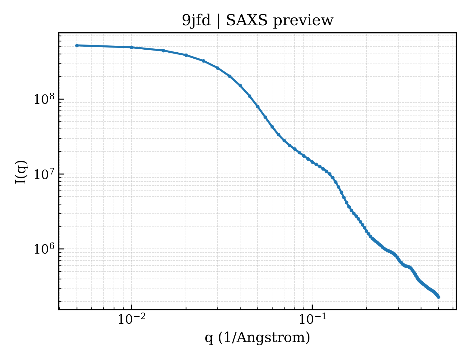
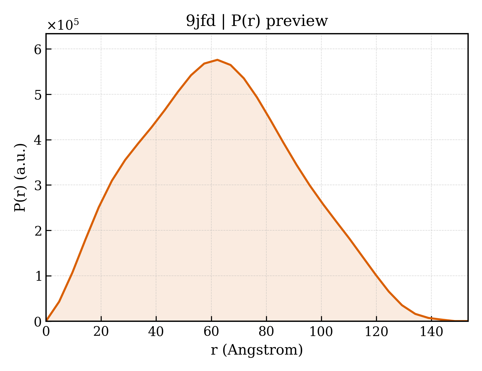
Insulin receptor
Homo sapiens
State in the Current Structure
| Assembly | Oligomeric State | Construct | Mutations and Modifications | Ligands, Ions and Associated Components | Method and Experimental Conditions | Structure Quality |
|---|---|---|---|---|---|---|
| 1 | Protein–DNA Homooligomer Protein × 2 DNA 1 PDB declaration: trimeric(3) Consistent with all polymer counts | Chain A; UniProt 28–946 Chain B; UniProt 28–946 | Not recorded | A62 × 1 | ELECTRON MICROSCOPY cryo-EM buffer:pH 7.5 cryo-EM vitrification conditions:Cryogen ETHANE | Resolution 9.35 Å |
Other States of the Same Protein in the Database
Each row is a biological assembly of the same UniProt protein in another PDB entry. The “Difference from current entry” column identifies evidence-level differences; no tag means the currently parsed fields agree.
| Other PDB | Difference from Current Entry 9JFD | Assembly / Oligomeric State | Construct | Mutations and Modifications | Ligands, Ions and Non-polymers | Method and Experimental Conditions | Structure Quality |
|---|---|---|---|---|---|---|---|
| 1GAG CRYSTAL STRUCTURE OF THE INSULIN RECEPTOR KINASE IN COMPLEX WITH A BISUBSTRATE INHIBITOR Deposited 2000-11-29 | Different construct Different mutation/modification Different oligomeric state Different ligand/ion Different experimental method Different experimental conditions Different structure-quality metrics | Assembly 1 Protein heterocomplex Heteromer;Protein × 2 PDB declaration: dimeric |
Chain A
1005–1310(306 aa)
Fragment:TYROSINE KINASE DOMAIN
|
Mutation:C981S, Y984F Non-standard monomer:Yes (specific site not provided by mmCIF) | MG MAGNESIUM ION × 2 112 THIOPHOSPHORIC ACID O-((ADENOSYL-PHOSPHO)PHOSPHO)-S-ACETAMIDYL-DIESTER × 1 |
X-RAY DIFFRACTION
X-ray crystallization conditions
VAPOR DIFFUSION, HANGING DROP;pH 7.5;277 K;PEG 8000, Tris-HCl, pH 7.5, VAPOR DIFFUSION, HANGING DROP, temperature 277K
|
Resolution 2.70 Å R-free 0.269 |
| 1GAG CRYSTAL STRUCTURE OF THE INSULIN RECEPTOR KINASE IN COMPLEX WITH A BISUBSTRATE INHIBITOR Deposited 2000-11-29 | Different construct Different mutation/modification Different oligomeric state Different ligand/ion Different experimental method Different experimental conditions Different structure-quality metrics | Assembly 2 Protein heterocomplex Heteromer;Protein × 4 PDB declaration: tetrameric |
Chain A
1005–1310(306 aa)
Fragment:TYROSINE KINASE DOMAIN
|
Mutation:C981S, Y984F Non-standard monomer:Yes (specific site not provided by mmCIF) | MG MAGNESIUM ION × 4 112 THIOPHOSPHORIC ACID O-((ADENOSYL-PHOSPHO)PHOSPHO)-S-ACETAMIDYL-DIESTER × 2 |
X-RAY DIFFRACTION
X-ray crystallization conditions
VAPOR DIFFUSION, HANGING DROP;pH 7.5;277 K;PEG 8000, Tris-HCl, pH 7.5, VAPOR DIFFUSION, HANGING DROP, temperature 277K
|
Resolution 2.70 Å R-free 0.269 |
| 1I44 CRYSTALLOGRAPHIC STUDIES OF AN ACTIVATION LOOP MUTANT OF THE INSULIN RECEPTOR TYROSINE KINASE Deposited 2001-02-19 | Different construct Different mutation/modification Different oligomeric state Different ligand/ion Different experimental method Different experimental conditions Different structure-quality metrics | Assembly 1 Protein monomer Monomer;Protein × 1 PDB declaration: monomeric |
Chain A
1005–1310(306 aa)
Fragment:TYROSINE KINASE DOMAIN
|
Mutation:C981S, Y984PHE, D1161A | MG MAGNESIUM ION × 1 ACP PHOSPHOMETHYLPHOSPHONIC ACID ADENYLATE ESTER × 1 |
X-RAY DIFFRACTION
X-ray crystallization conditions
VAPOR DIFFUSION, HANGING DROP;pH 7.5;293 K;PEG 8000, Tris-HCl, NaCl, dithiothreitol, pH 7.5, VAPOR DIFFUSION, HANGING DROP, temperature 293K
|
Resolution 2.40 Å R-free 0.268 |
| 1IR3 PHOSPHORYLATED INSULIN RECEPTOR TYROSINE KINASE IN COMPLEX WITH PEPTIDE SUBSTRATE AND ATP ANALOG Deposited 1997-09-22 | Different construct Different mutation/modification Different oligomeric state Different ligand/ion Different experimental method Different experimental conditions Different structure-quality metrics | Assembly 1 Protein heterocomplex Heteromer;Protein × 4 PDB declaration: tetrameric |
Chain A
1005–1310(306 aa)
Fragment:TYROSINE KINASE DOMAIN
|
Mutation:C981S, Y984F Non-standard monomer:Yes (specific site not provided by mmCIF) | MG MAGNESIUM ION × 4 ANP PHOSPHOAMINOPHOSPHONIC ACID-ADENYLATE ESTER × 2 |
X-RAY DIFFRACTION
X-ray crystallization conditions
pH 7.5;22% PEG 8000, 100 MM TRIS-HCL, PH 7.5, 2% ETHYLENE GLYCOL
|
Resolution 1.90 Å R-free 0.226 |
| 1IRK CRYSTAL STRUCTURE OF THE TYROSINE KINASE DOMAIN OF THE HUMAN INSULIN RECEPTOR Deposited 1995-01-02 | Different construct Different mutation/modification Different oligomeric state Different ligand/ion Different experimental method Different experimental conditions Different structure-quality metrics | Assembly 1 Protein monomer Monomer;Protein × 1 PDB declaration: monomeric |
Chain A
1005–1310(306 aa)
|
Not recorded | EMC ETHYL MERCURY ION × 2 | X-RAY DIFFRACTION mmCIF provides none of the parsed conditions | Resolution 2.10 Å R-free 0.232 |
| 1P14 Crystal structure of a catalytic-loop mutant of the insulin receptor tyrosine kinase Deposited 2003-04-11 | Different construct Different mutation/modification Different oligomeric state Different experimental method Different experimental conditions Different structure-quality metrics | Assembly 1 Protein monomer Monomer;Protein × 1 PDB declaration: monomeric |
Chain A
993–1298(306 aa)
Fragment:tyrosine kinase domain, residues 978-1283
|
Mutation:C981S, D1132N | No recorded non-water small molecule |
X-RAY DIFFRACTION
X-ray crystallization conditions
pH 7.5;pH 7.50
|
Resolution 1.90 Å R-free 0.240 |
| 1RQQ Crystal Structure of the Insulin Receptor Kinase in Complex with the SH2 Domain of APS Deposited 2003-12-06 | Different construct Different mutation/modification Different oligomeric state Different ligand/ion Different experimental method Different experimental conditions Different structure-quality metrics | Assembly 1 Protein heterocomplex Heteromer;Protein × 6 PDB declaration: hexameric |
Chain A
1005–1310(306 aa)
Fragment:Kinase domain
Chain B
1005–1310(306 aa)
Fragment:Kinase domain
|
Mutation:C980S, K1251N Non-standard monomer:Yes (specific site not provided by mmCIF) Mutation:C980S, K1251N Non-standard monomer:Yes (specific site not provided by mmCIF) | MN MANGANESE (II) ION × 2 112 THIOPHOSPHORIC ACID O-((ADENOSYL-PHOSPHO)PHOSPHO)-S-ACETAMIDYL-DIESTER × 2 |
X-RAY DIFFRACTION
X-ray crystallization conditions
pH 8;pH 8.0
|
Resolution 2.60 Å R-free 0.277 |
| 2AUH Crystal structure of the Grb14 BPS region in complex with the insulin receptor tyrosine kinase Deposited 2005-08-27 | Different construct Different mutation/modification Different oligomeric state Different ligand/ion Different experimental method Different experimental conditions Different structure-quality metrics | Assembly 1 Protein heterocomplex Heteromer;Protein × 2 PDB declaration: dimeric |
Chain A
1005–1310(306 aa)
Fragment:tyrosine kinase domain
|
Non-standard monomer:Yes (specific site not provided by mmCIF) | CA CALCIUM ION × 2 | X-RAY DIFFRACTION mmCIF provides none of the parsed conditions | Resolution 3.20 Å R-free 0.254 |
| 2B4S Crystal structure of a complex between PTP1B and the insulin receptor tyrosine kinase Deposited 2005-09-26 | Different construct Different mutation/modification Different oligomeric state Different ligand/ion Different experimental method Different experimental conditions Different structure-quality metrics | Assembly 1 Protein heterocomplex Heteromer;Protein × 4 PDB declaration: tetrameric |
Chain B
1005–1310(306 aa)
Fragment:Protein kinase
Chain D
1005–1310(306 aa)
Fragment:Protein kinase
|
Non-standard monomer:Yes (specific site not provided by mmCIF) Non-standard monomer:Yes (specific site not provided by mmCIF) | SO4 SULFATE ION × 10 |
X-RAY DIFFRACTION
X-ray crystallization conditions
VAPOR DIFFUSION, HANGING DROP;pH 7.5;277 K;100 mM Tris-HCl, 1.9 M ammonium sulfate, 2% PEG 400, pH 7.5, temperature 277K, VAPOR DIFFUSION, HANGING DROP
|
Resolution 2.30 Å R-free 0.239 |
| 2HR7 Insulin receptor (domains 1-3) Deposited 2006-07-19 | Different construct Different oligomeric state Different ligand/ion Different experimental method Different experimental conditions Different structure-quality metrics | Assembly 1 Other combination Homooligomer;Protein × 2 PDB declaration: dimeric |
Chain A
28–512(485 aa)
Fragment:Domains 1-3 (residues 28-512)
Chain B
28–512(485 aa)
Fragment:Domains 1-3 (residues 28-512)
|
Not recorded | NAG 2-acetamido-2-deoxy-beta-D-glucopyranose × 4 SO4 SULFATE ION × 8 P33 3,6,9,12,15,18-HEXAOXAICOSANE-1,20-DIOL × 1 GOL GLYCEROL × 19 |
X-RAY DIFFRACTION
X-ray crystallization conditions
pH 8.5;291 K;1.5-1.65 M ammonium sulfate, 2 % PEG 400, pH 8.5, VAPOR DIFFUSION, HANGING DROP, temperature 291K, pH 8.50
|
Resolution 2.32 Å R-free 0.231 |
| 2MFR Solution structure of the transmembrane domain of the insulin receptor in micelles Deposited 2013-10-20 | Different construct Different mutation/modification Different oligomeric state Different experimental method Different experimental conditions Different structure-quality metrics | Assembly 1 Protein monomer Monomer;Protein × 1 PDB declaration: monomeric |
Chain A
940–988(49 aa)
Fragment:UNP residues 940-988
|
Not recorded | No recorded non-water small molecule |
SOLUTION NMR
NMR measurement conditions
pH 6.5;313 K;Ionic strength (raw mmCIF value) 20;Pressure ambient
NMR sample composition
0.2-0.4 mM [U-100% 15N] protein-1, 90% H2O/10% D2O | 90% H2O/10% D2O
NMR sample composition
0.2-0.4 mM [U-100% 13C; U-100% 15N] protein-2, 90% H2O/10% D2O | 90% H2O/10% D2O
|
Resolution not provided |
| 2Z8C Phosphorylated insulin receptor tyrosine kinase in complex with (4-{[5-carbamoyl-4-(3-methylanilino)pyrimidin-2-yl]amino}phenyl)acetic acid Deposited 2007-09-05 | Different construct Different mutation/modification Different oligomeric state Different ligand/ion Different experimental method Different experimental conditions Different structure-quality metrics | Assembly 1 Protein heterocomplex Heteromer;Protein × 2 PDB declaration: dimeric |
Chain A
1008–1310(303 aa)
Fragment:TYROSINE KINASE DOMAIN
|
Mutation:C981S, Y984F Non-standard monomer:Yes (specific site not provided by mmCIF) | S91 [4-({5-(AMINOCARBONYL)-4-[(3-METHYLPHENYL)AMINO]PYRIMIDIN-2-YL}AMINO)PHENYL]ACETIC ACID × 1 |
X-RAY DIFFRACTION
X-ray crystallization conditions
VAPOR DIFFUSION, HANGING DROP;PEG 3350, VAPOR DIFFUSION, HANGING DROP
|
Resolution 3.25 Å R-free 0.292 |
| 3BU3 Crystal structure of the insulin receptor kinase in complex with IRS2 KRLB peptide Deposited 2007-12-31 | Different construct Different mutation/modification Different oligomeric state Different experimental method Different experimental conditions Different structure-quality metrics | Assembly 1 Protein heterocomplex Heteromer;Protein × 2 PDB declaration: dimeric |
Chain A
1005–1310(306 aa)
Fragment:protein kinase
|
Non-standard monomer:Yes (specific site not provided by mmCIF) | No recorded non-water small molecule |
X-RAY DIFFRACTION
X-ray crystallization conditions
VAPOR DIFFUSION, HANGING DROP;pH 7.5;277 K;28% PEG8000
0.1 M HEPES, pH 7.5, VAPOR DIFFUSION, HANGING DROP, temperature 277K
|
Resolution 1.65 Å R-free 0.220 |
| 3BU5 Crystal structure of the insulin receptor kinase in complex with IRS2 KRLB peptide and ATP Deposited 2007-12-31 | Different construct Different mutation/modification Different oligomeric state Different ligand/ion Different experimental method Different experimental conditions Different structure-quality metrics | Assembly 1 Protein heterocomplex Heteromer;Protein × 2 PDB declaration: dimeric |
Chain A
1005–1310(306 aa)
Fragment:protein kinase
|
Non-standard monomer:Yes (specific site not provided by mmCIF) | MG MAGNESIUM ION × 1 ATP ADENOSINE-5'-TRIPHOSPHATE × 1 |
X-RAY DIFFRACTION
X-ray crystallization conditions
VAPOR DIFFUSION, HANGING DROP;pH 7.5;277 K;28% PEG8000
0.1 M HEPES, pH 7.5, VAPOR DIFFUSION, HANGING DROP, temperature 277K
|
Resolution 2.10 Å R-free 0.239 |
| 3BU6 Crystal structure of the insulin receptor kinase in complex with IRS2 KRLB phosphopeptide Deposited 2007-12-31 | Different construct Different mutation/modification Different oligomeric state Different experimental method Different experimental conditions Different structure-quality metrics | Assembly 1 Protein heterocomplex Heteromer;Protein × 2 PDB declaration: dimeric |
Chain A
1005–1310(306 aa)
Fragment:protein kinase
|
Non-standard monomer:Yes (specific site not provided by mmCIF) | No recorded non-water small molecule |
X-RAY DIFFRACTION
X-ray crystallization conditions
VAPOR DIFFUSION, HANGING DROP;pH 7.5;277 K;28% PEG8000
0.1 M HEPES, pH 7.5, VAPOR DIFFUSION, HANGING DROP, temperature 277K
|
Resolution 1.95 Å R-free 0.239 |
| 3EKK Insulin receptor kinase complexed with an inhibitor Deposited 2008-09-19 | Different construct Different mutation/modification Different oligomeric state Different ligand/ion Different experimental method Different experimental conditions Different structure-quality metrics | Assembly 1 Protein monomer Monomer;Protein × 1 PDB declaration: monomeric |
Chain A
1005–1310(306 aa)
Fragment:kinase domain
|
Mutation:C1008S, D1159N | GS2 2-[(2-{[1-(N,N-dimethylglycyl)-5-methoxy-1H-indol-6-yl]amino}-7H-pyrrolo[2,3-d]pyrimidin-4-yl)amino]-6-fluoro-N-methylbenzamide × 1 |
X-RAY DIFFRACTION
X-ray crystallization conditions
VAPOR DIFFUSION, HANGING DROP;pH 7;295 K;0.1 M MOPS, 1.0 M Na3citrate, pH 7.0, VAPOR DIFFUSION, HANGING DROP, temperature 295K
|
Resolution 2.10 Å R-free 0.260 |
| 3EKN Insulin receptor kinase complexed with an inhibitor Deposited 2008-09-19 | Different construct Different mutation/modification Different oligomeric state Different ligand/ion Different experimental method Different experimental conditions Different structure-quality metrics | Assembly 1 Protein monomer Monomer;Protein × 1 PDB declaration: monomeric |
Chain A
1005–1310(306 aa)
Fragment:kinase domain
|
Mutation:C1008S, D1159N | GS3 2-fluoro-6-{[2-({2-methoxy-4-[4-(1-methylethyl)piperazin-1-yl]phenyl}amino)-7H-pyrrolo[2,3-d]pyrimidin-4-yl]amino}benzamide × 1 |
X-RAY DIFFRACTION
X-ray crystallization conditions
VAPOR DIFFUSION, HANGING DROP;pH 7;295 K;0.1 M MOPS, 1.0 M Na3citrate, pH 7.0, VAPOR DIFFUSION, HANGING DROP, temperature 295K
|
Resolution 2.20 Å R-free 0.274 |
| 3ETA Kinase domain of insulin receptor complexed with a pyrrolo pyridine inhibitor Deposited 2008-10-07 | Different construct Different mutation/modification Different oligomeric state Different ligand/ion Different experimental method Different experimental conditions Different structure-quality metrics | Assembly 1 Protein monomer Monomer;Protein × 1 PDB declaration: monomeric |
Chain A
1017–1322(306 aa)
Fragment:kinase domain
|
Not recorded | 351 1-(3-{5-[4-(aminomethyl)phenyl]-1H-pyrrolo[2,3-b]pyridin-3-yl}phenyl)-3-(2-phenoxyphenyl)urea × 1 |
X-RAY DIFFRACTION
X-ray crystallization conditions
VAPOR DIFFUSION, HANGING DROP;pH 7;295 K;0.1 M MOPS pH 7.0, 1.1 M sodium citrate, 1% Jeffamine M89, VAPOR DIFFUSION, HANGING DROP, temperature 295K
|
Resolution 2.60 Å R-free 0.232 |
| 3ETA Kinase domain of insulin receptor complexed with a pyrrolo pyridine inhibitor Deposited 2008-10-07 | Different construct Different mutation/modification Different oligomeric state Different ligand/ion Different experimental method Different experimental conditions Different structure-quality metrics | Assembly 2 Protein monomer Monomer;Protein × 1 PDB declaration: monomeric |
Chain B
1017–1322(306 aa)
Fragment:kinase domain
|
Not recorded | 351 1-(3-{5-[4-(aminomethyl)phenyl]-1H-pyrrolo[2,3-b]pyridin-3-yl}phenyl)-3-(2-phenoxyphenyl)urea × 1 |
X-RAY DIFFRACTION
X-ray crystallization conditions
VAPOR DIFFUSION, HANGING DROP;pH 7;295 K;0.1 M MOPS pH 7.0, 1.1 M sodium citrate, 1% Jeffamine M89, VAPOR DIFFUSION, HANGING DROP, temperature 295K
|
Resolution 2.60 Å R-free 0.232 |
| 3ETA Kinase domain of insulin receptor complexed with a pyrrolo pyridine inhibitor Deposited 2008-10-07 | Different construct Different oligomeric state Different ligand/ion Different experimental method Different experimental conditions Different structure-quality metrics | Assembly 3 Protein homooligomer Homooligomer;Protein × 2 PDB declaration: dimeric |
Chain A
1017–1322(306 aa)
Fragment:kinase domain
Chain B
1017–1322(306 aa)
Fragment:kinase domain
|
Not recorded | 351 1-(3-{5-[4-(aminomethyl)phenyl]-1H-pyrrolo[2,3-b]pyridin-3-yl}phenyl)-3-(2-phenoxyphenyl)urea × 2 |
X-RAY DIFFRACTION
X-ray crystallization conditions
VAPOR DIFFUSION, HANGING DROP;pH 7;295 K;0.1 M MOPS pH 7.0, 1.1 M sodium citrate, 1% Jeffamine M89, VAPOR DIFFUSION, HANGING DROP, temperature 295K
|
Resolution 2.60 Å R-free 0.232 |
| 3W11 Insulin receptor ectodomain construct comprising domains L1-CR in complex with human insulin, Alpha-CT peptide(704-719) and FAB 83-7 Deposited 2012-11-06 | Different construct Different oligomeric state Different ligand/ion Different experimental method Different experimental conditions Different structure-quality metrics | Assembly 1 Other combination Heteromer;Protein × 6 PDB declaration: hexameric |
Chain E
28–337(310 aa)
Fragment:UNP residues 28-337
Chain F
731–746(16 aa)
Fragment:UNP residues 731-746
|
Not recorded | NAG 2-acetamido-2-deoxy-beta-D-glucopyranose × 3 |
X-RAY DIFFRACTION
X-ray crystallization conditions
pH 8;0.9-1.1M TRI-SODIUM CITRATE, 0.1M IMIDAZOLE-HCL, 0.02% SODIUM AZIDE, PH 8.0
|
Resolution 3.90 Å R-free 0.292 |
| 3W12 Insulin receptor ectodomain construct comprising domains L1-CR in complex with high-affinity insulin analogue [D-PRO-B26]-DTI-NH2, alpha-CT peptide(704-719) and FAB 83-7 Deposited 2012-11-06 | Different construct Different oligomeric state Different ligand/ion Different experimental method Different experimental conditions Different structure-quality metrics | Assembly 1 Other combination Heteromer;Protein × 6 PDB declaration: hexameric |
Chain E
28–337(310 aa)
Fragment:UNP residues 28-337
Chain F
731–746(16 aa)
Fragment:UNP residues 731-746
|
Not recorded | NAG 2-acetamido-2-deoxy-beta-D-glucopyranose × 2 |
X-RAY DIFFRACTION
X-ray crystallization conditions
pH 8;0.9-1.1M TRI-SODIUM CITRATE, 0.1M IMIDAZOLE-HCL, 0.02% SODIUM AZIDE, PH 8.0
|
Resolution 4.30 Å R-free 0.349 |
| 3W13 Insulin receptor ectodomain construct comprising domains L1-CR in complex with high-affinity insulin analogue [D-PRO-B26]-DTI-NH2, alphact peptide(693-719) and FAB 83-7 Deposited 2012-11-06 | Different construct Different oligomeric state Different ligand/ion Different experimental method Different experimental conditions Different structure-quality metrics | Assembly 1 Other combination Heteromer;Protein × 6 PDB declaration: hexameric |
Chain E
28–337(310 aa)
Fragment:UNP residues 28-337
Chain F
724–746(23 aa)
Fragment:UNP residues 724-746
|
Not recorded | NAG 2-acetamido-2-deoxy-beta-D-glucopyranose × 2 |
X-RAY DIFFRACTION
X-ray crystallization conditions
pH 8;0.9-1.1M TRI-SODIUM CITRATE, 0.1M IMIDAZOLE-HCL, 0.02% SOIDUM AZIDE, PH 8.0
|
Resolution 4.30 Å R-free 0.335 |
| 4IBM Crystal structure of insulin receptor kinase domain in complex with an inhibitor Irfin-1 Deposited 2012-12-08 | Different construct Different mutation/modification Different oligomeric state Different ligand/ion Different experimental method Different experimental conditions Different structure-quality metrics | Assembly 1 Protein monomer Monomer;Protein × 1 PDB declaration: monomeric |
Chain A
1005–1310(306 aa)
|
Mutation:K1278N | IR1 5-(2-phenylpyrazolo[1,5-a]pyridin-3-yl)-3H-pyrazolo[3,4-c]pyridazin-3-one × 1 |
X-RAY DIFFRACTION
X-ray crystallization conditions
VAPOR DIFFUSION;pH 6.5;293 K;0.1 M Sodium cacodylate pH 6.5, 23% PEG-4000, 10% Ethylene glycol, and 0.03 M Glycyl-glycyl-glycine, VAPOR DIFFUSION, temperature 293K
|
Resolution 1.80 Å R-free 0.218 |
| 4IBM Crystal structure of insulin receptor kinase domain in complex with an inhibitor Irfin-1 Deposited 2012-12-08 | Different construct Different mutation/modification Different oligomeric state Different ligand/ion Different experimental method Different experimental conditions Different structure-quality metrics | Assembly 2 Protein monomer Monomer;Protein × 1 PDB declaration: monomeric |
Chain B
1005–1310(306 aa)
|
Mutation:K1278N | IR1 5-(2-phenylpyrazolo[1,5-a]pyridin-3-yl)-3H-pyrazolo[3,4-c]pyridazin-3-one × 1 |
X-RAY DIFFRACTION
X-ray crystallization conditions
VAPOR DIFFUSION;pH 6.5;293 K;0.1 M Sodium cacodylate pH 6.5, 23% PEG-4000, 10% Ethylene glycol, and 0.03 M Glycyl-glycyl-glycine, VAPOR DIFFUSION, temperature 293K
|
Resolution 1.80 Å R-free 0.218 |
| 4OGA Insulin in complex with Site 1 of the human insulin receptor Deposited 2014-01-15 | Different construct Different oligomeric state Different ligand/ion Different experimental method Different experimental conditions Different structure-quality metrics | Assembly 1 Other combination Heteromer;Protein × 6 PDB declaration: hexameric |
Chain E
28–337(310 aa)
Fragment:L1-CR, UNP residues 28-377
Chain F
731–746(16 aa)
Fragment:alpha-CT peptide, UNP residues 731-746
|
Not recorded | NAG 2-acetamido-2-deoxy-beta-D-glucopyranose × 3 |
X-RAY DIFFRACTION
X-ray crystallization conditions
VAPOR DIFFUSION, HANGING DROP;pH 8;293 K;0.9-1.1M TRI-SODIUM CITRATE, 0.1M IMIDAZOLE-HCL, 0.02% SODIUM AZIDE, PH 8.0 , VAPOR DIFFUSION, HANGING DROP, temperature 293K
|
Resolution 3.50 Å R-free 0.284 |
| 4XLV Crystal structure of the activated insulin receptor tyrosine kinase dimer Deposited 2015-01-13 | Different construct Different mutation/modification Different oligomeric state Different ligand/ion Different experimental method Different experimental conditions Different structure-quality metrics | Assembly 1 Protein homooligomer Homooligomer;Protein × 2 PDB declaration: dimeric |
Chain A
983–1310(328 aa)
Fragment:UNP residues 983-1310
|
Mutation:Y965F, C981S Non-standard monomer:Yes (specific site not provided by mmCIF) | ACP PHOSPHOMETHYLPHOSPHONIC ACID ADENYLATE ESTER × 2 MG MAGNESIUM ION × 4 |
X-RAY DIFFRACTION
X-ray crystallization conditions
VAPOR DIFFUSION, HANGING DROP;pH 7.5;277 K;100 mM Tris-HCl, pH 7.5, 26% (w/v) polyethylene glycol (PEG) 4000, 200 mM MgCl2, and 5 mM DTT
|
Resolution 2.30 Å R-free 0.266 |
| 4XSS Insulin-like growth factor I in complex with site 1 of a hybrid insulin receptor / Type 1 insulin-like growth factor receptor Deposited 2015-01-22 | Different construct Different mutation/modification Different oligomeric state Different ligand/ion Different experimental method Different experimental conditions Different structure-quality metrics | Assembly 1 Other combination Heteromer;Protein × 3 PDB declaration: trimeric |
Chain E
28–337(310 aa)
Fragment:L1-CR, UNP residues 28-377
|
Not recorded | SO4 SULFATE ION × 1 NAG 2-acetamido-2-deoxy-beta-D-glucopyranose × 4 |
X-RAY DIFFRACTION
X-ray crystallization conditions
VAPOR DIFFUSION, HANGING DROP;pH 10.5;298 K;1.75 M ammonium sulfate, 0.1 M CAPS-NaOH (pH 10.5), 0.2 M LiSO4
|
Resolution 3.00 Å R-free 0.229 |
| 4XSS Insulin-like growth factor I in complex with site 1 of a hybrid insulin receptor / Type 1 insulin-like growth factor receptor Deposited 2015-01-22 | Different construct Different mutation/modification Different oligomeric state Different ligand/ion Different experimental method Different experimental conditions Different structure-quality metrics | Assembly 2 Other combination Heteromer;Protein × 18 PDB declaration: octadecameric |
Chain E
28–337(310 aa)
Fragment:L1-CR, UNP residues 28-377
|
Not recorded | SO4 SULFATE ION × 6 NAG 2-acetamido-2-deoxy-beta-D-glucopyranose × 24 |
X-RAY DIFFRACTION
X-ray crystallization conditions
VAPOR DIFFUSION, HANGING DROP;pH 10.5;298 K;1.75 M ammonium sulfate, 0.1 M CAPS-NaOH (pH 10.5), 0.2 M LiSO4
|
Resolution 3.00 Å R-free 0.229 |
| 4XST Structure of the endoglycosidase-H treated L1-CR domains of the human insulin receptor in complex with residues 697-719 of the human insulin receptor (A-isoform) Deposited 2015-01-22 | Different construct Different mutation/modification Different oligomeric state Different ligand/ion Different experimental method Different experimental conditions Different structure-quality metrics | Assembly 1 Other combination Heteromer;Protein × 2 PDB declaration: dimeric |
Chain E
28–337(310 aa)
Fragment:L1-CR, UNP residues 28-377
|
Not recorded | NAG 2-acetamido-2-deoxy-beta-D-glucopyranose × 3 SO4 SULFATE ION × 2 |
X-RAY DIFFRACTION
X-ray crystallization conditions
VAPOR DIFFUSION, HANGING DROP;298 K;2.45 M (NH4)2SO4 + 10 % glycerol
|
Resolution 3.00 Å R-free 0.262 |
| 4XST Structure of the endoglycosidase-H treated L1-CR domains of the human insulin receptor in complex with residues 697-719 of the human insulin receptor (A-isoform) Deposited 2015-01-22 | Different construct Different mutation/modification Different oligomeric state Different ligand/ion Different experimental method Different experimental conditions Different structure-quality metrics | Assembly 2 Other combination Heteromer;Protein × 12 PDB declaration: dodecameric |
Chain E
28–337(310 aa)
Fragment:L1-CR, UNP residues 28-377
|
Not recorded | NAG 2-acetamido-2-deoxy-beta-D-glucopyranose × 18 SO4 SULFATE ION × 12 |
X-RAY DIFFRACTION
X-ray crystallization conditions
VAPOR DIFFUSION, HANGING DROP;298 K;2.45 M (NH4)2SO4 + 10 % glycerol
|
Resolution 3.00 Å R-free 0.262 |
| 4ZXB Structure of the human insulin receptor ectodomain, IRDeltabeta construct, in complex with four Fab molecules Deposited 2015-05-20 | Different construct Different mutation/modification Different oligomeric state Different ligand/ion Different experimental method Different experimental conditions Different structure-quality metrics | Assembly 1 Other combination Heteromer;Protein × 10 PDB declaration: decameric |
Chain E
28–944(917 aa)
|
Mutation:Y144H, V731A, T732G, V733N, A734N, residues 735-753 deleted | NAG 2-acetamido-2-deoxy-beta-D-glucopyranose × 6 |
X-RAY DIFFRACTION
X-ray crystallization conditions
VAPOR DIFFUSION, HANGING DROP;pH 6.5;293 K;Crystals were produced by vapour diffusion using 1 microlitre of the complex (2.6 mg/ml) in 10 mM HEPES (pH 7.5), 0.02% sodium azide and 10% D-trehalose solution and 1 microlitres of well solution containing 10% PEG 8000, 0.1 M MES (pH 6.5) and 0.1 M MgAc2.
|
Resolution 3.30 Å R-free 0.237 |
| 5E1S The Crystal structure of INSR Tyrosine Kinase in complex with the Inhibitor BI 885578 Deposited 2015-09-30 | Different construct Different mutation/modification Different oligomeric state Different ligand/ion Different experimental method Different experimental conditions Different structure-quality metrics | Assembly 1 Protein monomer Monomer;Protein × 1 PDB declaration: monomeric |
Chain A
1005–1310(306 aa)
|
Not recorded | 5JA (5R)-N-(1-{2-[4-(2-methoxyethyl)piperazin-1-yl]ethyl}-1H-pyrazol-3-yl)-5,8-dimethyl-9-phenyl-6,8-dihydro-5H-pyrazolo[3,4-h]quinazolin-2-amine × 1 |
X-RAY DIFFRACTION
X-ray crystallization conditions
VAPOR DIFFUSION, HANGING DROP;pH 7.3;293 K;25% PEG 8000, 0.1M TRIS pH 7.3, 50mM NaCl, 5 mM DTT
|
Resolution 2.26 Å R-free 0.262 |
| 5HHW Crystal structure of insulin receptor kinase domain in complex with cis-(R)-7-(3-(azetidin-1-ylmethyl)cyclobutyl)-5-(3-((tetrahydro-2H-pyran-2-yl)methoxy)phenyl)-7H-pyrrolo[2,3-d]pyrimidin-4-amine. Deposited 2016-01-11 | Different construct Different mutation/modification Different oligomeric state Different ligand/ion Different experimental method Different experimental conditions Different structure-quality metrics | Assembly 1 Protein monomer Monomer;Protein × 1 PDB declaration: monomeric |
Chain A
1005–1310(306 aa)
Fragment:Kinase domain, residues 1005-1310
|
Mutation:C1008S, D1159N | 60O 7-[3-(azetidin-1-ylmethyl)cyclobutyl]-5-[3-[[(2~{R})-oxan-2-yl]methoxy]phenyl]pyrrolo[2,3-d]pyrimidin-4-amine × 1 EDO 1,2-ETHANEDIOL × 1 |
X-RAY DIFFRACTION
X-ray crystallization conditions
VAPOR DIFFUSION;pH 7.5;298 K;18% Peg3000, 0.1M Tris pH7.5
|
Resolution 1.79 Å R-free 0.198 |
| 5J3H Human insulin receptor domains L1-CR in complex with peptide S519C16 and 83-7 Fv Deposited 2016-03-30 | Different construct Different mutation/modification Different oligomeric state Different ligand/ion Different experimental method Different experimental conditions Different structure-quality metrics | Assembly 1 Other combination Heteromer;Protein × 4 PDB declaration: tetrameric |
Chain E
28–337(310 aa)
Fragment:UNP residues 28-337
|
Not recorded | NAG 2-acetamido-2-deoxy-beta-D-glucopyranose × 2 |
X-RAY DIFFRACTION
X-ray crystallization conditions
VAPOR DIFFUSION, SITTING DROP;300 K;1.75 M (NH4)2HPO4
|
Resolution 3.27 Å R-free 0.248 |
| 5KQV Insulin receptor ectodomain construct comprising domains L1,CR,L2, FnIII-1 and alphaCT peptide in complex with bovine insulin and FAB 83-14 (REVISED STRUCTURE) Deposited 2016-07-06 | Different construct Different mutation/modification Different oligomeric state Different experimental method Different experimental conditions Different structure-quality metrics | Assembly 1 Other combination Heteromer;Protein × 20 PDB declaration: eicosameric |
Chain E
28–620(593 aa)
Chain E
731–746(16 aa)
Chain F
28–620(593 aa)
Chain F
731–746(16 aa)
|
Mutation:Y144H,Y144H Mutation:Y144H,Y144H Mutation:Y144H,Y144H Mutation:Y144H,Y144H | No recorded non-water small molecule |
X-RAY DIFFRACTION
X-ray crystallization conditions
VAPOR DIFFUSION, HANGING DROP;293 K;9% POLYETHYLENE GLYCOL 3350, 200MM PROLINE, 100MM HEPES/NAOH, PH 7.5
|
Resolution 4.40 Å R-free 0.266 |
| 5U1M Structure of the IRS-1 PTB Domain Bound to the Juxtamembrane Region of the Insulin Receptor Deposited 2016-11-28 | Different construct Different mutation/modification Different oligomeric state Different experimental method Different experimental conditions Different structure-quality metrics | Assembly 1 Protein heterocomplex Heteromer;Protein × 2 PDB declaration: dimeric |
Chain B
991–999(9 aa)
Fragment:juxtamembrane region (UNP residues 991-999)
|
Non-standard monomer:Yes (specific site not provided by mmCIF) | No recorded non-water small molecule |
X-RAY DIFFRACTION
X-ray crystallization conditions
VAPOR DIFFUSION, HANGING DROP;293 K;ammonium sulfate
|
Resolution 1.80 Å |
| 6CE7 Insulin Receptor ectodomain in complex with one insulin molecule Deposited 2018-02-11 | Different construct Different mutation/modification Different oligomeric state Different experimental conditions Different structure-quality metrics | Assembly 1 Protein heterocomplex Heteromer;Protein × 5 PDB declaration: pentameric |
Chain A
28–956(929 aa)
Fragment:residues 28-956
Chain B
28–956(929 aa)
Fragment:residues 28-956
Chain P
718–747(30 aa)
Fragment:residues 718-747
|
Not recorded | No recorded non-water small molecule |
ELECTRON MICROSCOPY
cryo-EM buffer
pH 7.5;Hepes Saline (HBS)
cryo-EM vitrification conditions
Cryogen ETHANE;Grids made with SpotItOn
|
Resolution 7.40 Å |
| 6CE9 Insulin Receptor ectodomain in complex with two insulin molecules Deposited 2018-02-11 | Different construct Different mutation/modification Different oligomeric state Different ligand/ion Different experimental conditions Different structure-quality metrics | Assembly 1 Other combination Heteromer;Protein × 8 PDB declaration: octameric |
Chain A
28–944(917 aa)
Fragment:Ectodomain residues 28-944
Chain B
28–944(917 aa)
Fragment:Ectodomain residues 28-944
Chain M
718–747(30 aa)
Fragment:residues 718-747
Chain P
718–747(30 aa)
Fragment:residues 718-747
|
Not recorded | NAG 2-acetamido-2-deoxy-beta-D-glucopyranose × 4 |
ELECTRON MICROSCOPY
cryo-EM buffer
pH 7.5;Hepes Saline (HBS)
cryo-EM vitrification conditions
Cryogen ETHANE;Grids made with SpotItOn
|
Resolution 4.30 Å |
| 6CEB Insulin Receptor ectodomain in complex with two insulin molecules - C1 symmetry Deposited 2018-02-11 | Different construct Different mutation/modification Different oligomeric state Different ligand/ion Different experimental conditions Different structure-quality metrics | Assembly 1 Other combination Heteromer;Protein × 8 PDB declaration: octameric |
Chain A
28–944(917 aa)
Fragment:Ectodomain residues 28-944
Chain B
28–944(917 aa)
Fragment:Ectodomain residues 28-944
Chain M
718–747(30 aa)
Fragment:residues 718-747
Chain P
718–747(30 aa)
Fragment:residues 718-747
|
Not recorded | NAG 2-acetamido-2-deoxy-beta-D-glucopyranose × 4 |
ELECTRON MICROSCOPY
cryo-EM buffer
pH 7.5;Hepes Saline (HBS)
cryo-EM vitrification conditions
Cryogen ETHANE;Grids made with SpotItOn
|
Resolution 4.70 Å |
| 6HN4 Leucine-zippered human insulin receptor ectodomain with single bound insulin - "lower" membrane-proximal part Deposited 2018-09-14 | Different construct Different mutation/modification Different oligomeric state Different ligand/ion Different structure-quality metrics | Assembly 1 Insufficient information Homooligomer;Protein × 2 PDB declaration: dimeric |
Chain E
28–761(734 aa)
Chain E
781–943(163 aa)
Chain F
28–761(734 aa)
Chain F
781–943(163 aa)
|
Not recorded | NAG 2-acetamido-2-deoxy-beta-D-glucopyranose × 6 |
ELECTRON MICROSCOPY
cryo-EM buffer
pH 7.5
cryo-EM vitrification conditions
Cryogen ETHANE
|
Resolution 4.20 Å |
| 6HN5 Leucine-zippered human insulin receptor ectodomain with single bound insulin - "upper" membrane-distal part Deposited 2018-09-14 | Different construct Different mutation/modification Different oligomeric state Different ligand/ion Different structure-quality metrics | Assembly 1 Insufficient information Heteromer;Protein × 4 PDB declaration: tetrameric |
Chain E
28–761(734 aa)
Chain E
781–943(163 aa)
Chain F
28–761(734 aa)
Chain F
781–943(163 aa)
|
Not recorded | NAG 2-acetamido-2-deoxy-beta-D-glucopyranose × 9 |
ELECTRON MICROSCOPY
cryo-EM buffer
pH 7.5
cryo-EM vitrification conditions
Cryogen ETHANE
|
Resolution 3.20 Å |
| 6PXV Cryo-EM structure of full-length insulin receptor bound to 4 insulin. 3D refinement was focused on the extracellular region. Deposited 2019-07-27 | Different construct Different oligomeric state Different structure-quality metrics | Assembly 1 Protein heterocomplex Heteromer;Protein × 6 PDB declaration: hexameric |
Chain A
28–1370(1343 aa)
Chain C
28–1370(1343 aa)
|
Not recorded | No recorded non-water small molecule |
ELECTRON MICROSCOPY
cryo-EM buffer
pH 7.5
cryo-EM vitrification conditions
Cryogen ETHANE
|
Resolution 3.20 Å |
| 6PXW Cryo-EM structure of full-length insulin receptor bound to 4 insulin. 3D refinement was focused on the top part of the receptor complex. Deposited 2019-07-28 | Different construct Different oligomeric state Different structure-quality metrics | Assembly 1 Protein heterocomplex Heteromer;Protein × 6 PDB declaration: hexameric |
Chain A
28–1370(1343 aa)
Chain B
28–1370(1343 aa)
|
Not recorded | No recorded non-water small molecule |
ELECTRON MICROSCOPY
cryo-EM buffer
pH 7.5
cryo-EM vitrification conditions
Cryogen ETHANE
|
Resolution 3.10 Å |
| 6SOF human insulin receptor ectodomain bound by 4 insulin Deposited 2019-08-29 | Different construct Different mutation/modification Different oligomeric state Different experimental conditions Different structure-quality metrics | Assembly 1 Protein heterocomplex Heteromer;Protein × 12 PDB declaration: dodecameric |
Chain A
28–746(719 aa)
Chain B
783–944(162 aa)
Chain C
28–746(719 aa)
Chain D
783–944(162 aa)
|
Not recorded | No recorded non-water small molecule |
ELECTRON MICROSCOPY
cryo-EM buffer
pH 7.5
cryo-EM vitrification conditions
Cryogen ETHANE-PROPANE
|
Resolution 4.30 Å |
| 6VEP Human insulin in complex with the human insulin microreceptor in turn in complex with Fv 83-7 Deposited 2020-01-02 | Different construct Different oligomeric state Different ligand/ion Different experimental method Different experimental conditions Different structure-quality metrics | Assembly 1 Other combination Heteromer;Protein × 6 PDB declaration: hexameric |
Chain E
28–337(310 aa)
Fragment:residues 28-337
Chain F
731–746(16 aa)
Fragment:residues 731-746
|
Not recorded | NAG 2-acetamido-2-deoxy-beta-D-glucopyranose × 2 |
X-RAY DIFFRACTION
X-ray crystallization conditions
VAPOR DIFFUSION, HANGING DROP;300 K;Protein: 4.5 mg/ml in 10 mM HEPES pH 7.5
Well condition: 16% PEG 3350, 0.2 M sodium thiocyanate
|
Resolution 2.90 Å R-free 0.225 |
| 6VEP Human insulin in complex with the human insulin microreceptor in turn in complex with Fv 83-7 Deposited 2020-01-02 | Different construct Different oligomeric state Different ligand/ion Different experimental method Different experimental conditions Different structure-quality metrics | Assembly 2 Other combination Heteromer;Protein × 6 PDB declaration: hexameric |
Chain K
28–337(310 aa)
Fragment:residues 28-337
Chain L
731–746(16 aa)
Fragment:residues 731-746
|
Not recorded | NAG 2-acetamido-2-deoxy-beta-D-glucopyranose × 2 |
X-RAY DIFFRACTION
X-ray crystallization conditions
VAPOR DIFFUSION, HANGING DROP;300 K;Protein: 4.5 mg/ml in 10 mM HEPES pH 7.5
Well condition: 16% PEG 3350, 0.2 M sodium thiocyanate
|
Resolution 2.90 Å R-free 0.225 |
| 6VEP Human insulin in complex with the human insulin microreceptor in turn in complex with Fv 83-7 Deposited 2020-01-02 | Different construct Different oligomeric state Different ligand/ion Different experimental method Different experimental conditions Different structure-quality metrics | Assembly 3 Other combination Heteromer;Protein × 6 PDB declaration: hexameric |
Chain Q
28–337(310 aa)
Fragment:residues 28-337
Chain R
731–746(16 aa)
Fragment:residues 731-746
|
Not recorded | NAG 2-acetamido-2-deoxy-beta-D-glucopyranose × 3 |
X-RAY DIFFRACTION
X-ray crystallization conditions
VAPOR DIFFUSION, HANGING DROP;300 K;Protein: 4.5 mg/ml in 10 mM HEPES pH 7.5
Well condition: 16% PEG 3350, 0.2 M sodium thiocyanate
|
Resolution 2.90 Å R-free 0.225 |
| 6VEP Human insulin in complex with the human insulin microreceptor in turn in complex with Fv 83-7 Deposited 2020-01-02 | Different construct Different oligomeric state Different ligand/ion Different experimental method Different experimental conditions Different structure-quality metrics | Assembly 4 Other combination Heteromer;Protein × 6 PDB declaration: hexameric |
Chain W
28–337(310 aa)
Fragment:residues 28-337
Chain X
731–746(16 aa)
Fragment:residues 731-746
|
Not recorded | NAG 2-acetamido-2-deoxy-beta-D-glucopyranose × 1 |
X-RAY DIFFRACTION
X-ray crystallization conditions
VAPOR DIFFUSION, HANGING DROP;300 K;Protein: 4.5 mg/ml in 10 mM HEPES pH 7.5
Well condition: 16% PEG 3350, 0.2 M sodium thiocyanate
|
Resolution 2.90 Å R-free 0.225 |
| 6VEQ Con-Ins G1 in complex with the human insulin microreceptor in turn in complex with Fv 83-7 Deposited 2020-01-02 | Different construct Different oligomeric state Different ligand/ion Different experimental method Different experimental conditions Different structure-quality metrics | Assembly 1 Other combination Heteromer;Protein × 6 PDB declaration: hexameric |
Chain E
28–337(310 aa)
Fragment:residues 28-337
Chain F
731–746(16 aa)
Fragment:residues 731-746
|
Not recorded | NAG 2-acetamido-2-deoxy-beta-D-glucopyranose × 3 SO4 SULFATE ION × 3 |
X-RAY DIFFRACTION
X-ray crystallization conditions
VAPOR DIFFUSION, HANGING DROP;300 K;1.7 M ammonium sulphate
50 mM MOPS-NaOH (pH 7.0)
|
Resolution 3.25 Å R-free 0.277 |
| 6VEQ Con-Ins G1 in complex with the human insulin microreceptor in turn in complex with Fv 83-7 Deposited 2020-01-02 | Different construct Different oligomeric state Different ligand/ion Different experimental method Different experimental conditions Different structure-quality metrics | Assembly 2 Other combination Heteromer;Protein × 6 PDB declaration: hexameric |
Chain K
28–337(310 aa)
Fragment:residues 28-337
Chain L
731–746(16 aa)
Fragment:residues 731-746
|
Not recorded | NAG 2-acetamido-2-deoxy-beta-D-glucopyranose × 3 SO4 SULFATE ION × 1 |
X-RAY DIFFRACTION
X-ray crystallization conditions
VAPOR DIFFUSION, HANGING DROP;300 K;1.7 M ammonium sulphate
50 mM MOPS-NaOH (pH 7.0)
|
Resolution 3.25 Å R-free 0.277 |
| 7BW7 Cryo-EM Structure for the Ectodomain of the Full-length Human Insulin Receptor in Complex with 1 Insulin. Deposited 2020-04-13 | Different construct Different oligomeric state Different experimental conditions Different structure-quality metrics | Assembly 1 Insufficient information Heteromer;Protein × 3 PDB declaration: trimeric |
Chain A
28–1382(1355 aa)
Chain C
28–1382(1355 aa)
|
Not recorded | No recorded non-water small molecule |
ELECTRON MICROSCOPY
cryo-EM buffer
pH 7.4
cryo-EM vitrification conditions
Cryogen ETHANE
|
Resolution 4.10 Å |
| 7BW8 Cryo-EM Structure for the Insulin Binding Region in the Ectodomain of the Full-length Human Insulin Receptor in Complex with 1 Insulin Deposited 2020-04-14 | Different construct Different oligomeric state Different experimental conditions Different structure-quality metrics | Assembly 1 Insufficient information Heteromer;Protein × 3 PDB declaration: trimeric |
Chain A
28–1382(1355 aa)
Chain C
28–1382(1355 aa)
|
Not recorded | No recorded non-water small molecule |
ELECTRON MICROSCOPY
cryo-EM buffer
pH 7.4
cryo-EM vitrification conditions
Cryogen ETHANE
|
Resolution 3.80 Å |
| 7BWA Cryo-EM Structure for the Ectodomain of the Full-length Human Insulin Receptor in Complex with 2 Insulin Deposited 2020-04-14 | Different construct Different mutation/modification Different oligomeric state Different experimental conditions Different structure-quality metrics | Assembly 1 Insufficient information Heteromer;Protein × 2 PDB declaration: dimeric |
Chain A
28–1382(1355 aa)
Fragment:UNP residues 28-1382
|
Not recorded | No recorded non-water small molecule |
ELECTRON MICROSCOPY
cryo-EM buffer
pH 7.4
cryo-EM vitrification conditions
Cryogen ETHANE
|
Resolution 4.90 Å |
| 7BWA Cryo-EM Structure for the Ectodomain of the Full-length Human Insulin Receptor in Complex with 2 Insulin Deposited 2020-04-14 | Different construct Different mutation/modification Different oligomeric state Different experimental conditions Different structure-quality metrics | Assembly 2 Insufficient information Heteromer;Protein × 2 PDB declaration: dimeric |
Chain C
28–1382(1355 aa)
Fragment:UNP residues 28-1382
|
Not recorded | No recorded non-water small molecule |
ELECTRON MICROSCOPY
cryo-EM buffer
pH 7.4
cryo-EM vitrification conditions
Cryogen ETHANE
|
Resolution 4.90 Å |
| 7KD6 Insulin Receptor L1-CR plus alphaCT fragment in co-complex with Fv 83-7 and single-chain insulin SCI-b Deposited 2020-10-08 | Different construct Different mutation/modification Different oligomeric state Different ligand/ion Different experimental method Different experimental conditions Different structure-quality metrics | Assembly 1 Other combination Heteromer;Protein × 5 PDB declaration: pentameric |
Chain E
28–337(310 aa)
|
Mutation:Y171H | SO4 SULFATE ION × 1 GOL GLYCEROL × 1 NAG 2-acetamido-2-deoxy-beta-D-glucopyranose × 1 |
X-RAY DIFFRACTION
X-ray crystallization conditions
VAPOR DIFFUSION, HANGING DROP;pH 7.5;296 K;16% PEG 3350, 0.2 M sodium thiocyanate (NaSCN), 0.02% sodium azide (NaN3)
|
Resolution 2.60 Å R-free 0.221 |
| 7KD6 Insulin Receptor L1-CR plus alphaCT fragment in co-complex with Fv 83-7 and single-chain insulin SCI-b Deposited 2020-10-08 | Different construct Different mutation/modification Different oligomeric state Different ligand/ion Different experimental method Different experimental conditions Different structure-quality metrics | Assembly 2 Other combination Heteromer;Protein × 5 PDB declaration: pentameric |
Chain K
28–337(310 aa)
|
Mutation:Y171H | SO4 SULFATE ION × 1 NAG 2-acetamido-2-deoxy-beta-D-glucopyranose × 3 |
X-RAY DIFFRACTION
X-ray crystallization conditions
VAPOR DIFFUSION, HANGING DROP;pH 7.5;296 K;16% PEG 3350, 0.2 M sodium thiocyanate (NaSCN), 0.02% sodium azide (NaN3)
|
Resolution 2.60 Å R-free 0.221 |
| 7KD6 Insulin Receptor L1-CR plus alphaCT fragment in co-complex with Fv 83-7 and single-chain insulin SCI-b Deposited 2020-10-08 | Different construct Different mutation/modification Different oligomeric state Different ligand/ion Different experimental method Different experimental conditions Different structure-quality metrics | Assembly 3 Other combination Heteromer;Protein × 5 PDB declaration: pentameric |
Chain Q
28–337(310 aa)
|
Mutation:Y171H | SO4 SULFATE ION × 1 NAG 2-acetamido-2-deoxy-beta-D-glucopyranose × 3 |
X-RAY DIFFRACTION
X-ray crystallization conditions
VAPOR DIFFUSION, HANGING DROP;pH 7.5;296 K;16% PEG 3350, 0.2 M sodium thiocyanate (NaSCN), 0.02% sodium azide (NaN3)
|
Resolution 2.60 Å R-free 0.221 |
| 7KD6 Insulin Receptor L1-CR plus alphaCT fragment in co-complex with Fv 83-7 and single-chain insulin SCI-b Deposited 2020-10-08 | Different construct Different mutation/modification Different oligomeric state Different ligand/ion Different experimental method Different experimental conditions Different structure-quality metrics | Assembly 4 Other combination Heteromer;Protein × 5 PDB declaration: pentameric |
Chain W
28–337(310 aa)
|
Mutation:Y171H | SO4 SULFATE ION × 1 NAG 2-acetamido-2-deoxy-beta-D-glucopyranose × 3 |
X-RAY DIFFRACTION
X-ray crystallization conditions
VAPOR DIFFUSION, HANGING DROP;pH 7.5;296 K;16% PEG 3350, 0.2 M sodium thiocyanate (NaSCN), 0.02% sodium azide (NaN3)
|
Resolution 2.60 Å R-free 0.221 |
| 7MD4 Insulin receptor ectodomain dimer complexed with two IRPA-3 partial agonists Deposited 2021-04-03 | Different construct Different mutation/modification Different oligomeric state Different experimental conditions Different structure-quality metrics | Assembly 1 Protein heterocomplex Heteromer;Protein × 12 PDB declaration: dodecameric |
Chain A
28–944(917 aa)
Fragment:extracellular domain
Chain B
28–944(917 aa)
Fragment:extracellular domain
Chain M
721–747(27 aa)
Fragment:C-terminal helix
Chain N
721–747(27 aa)
Fragment:C-terminal helix
|
Not recorded | No recorded non-water small molecule |
ELECTRON MICROSCOPY
cryo-EM buffer
pH 7.4
cryo-EM vitrification conditions
Cryogen ETHANE
|
Resolution 4.50 Å |
| 7MD5 Insulin receptor ectodomain dimer complexed with two IRPA-9 partial agonists Deposited 2021-04-03 | Different construct Different mutation/modification Different oligomeric state Different ligand/ion Different experimental conditions Different structure-quality metrics | Assembly 1 Other combination Heteromer;Protein × 12 PDB declaration: dodecameric |
Chain A
28–944(917 aa)
Fragment:extracellular domain
Chain B
28–944(917 aa)
Fragment:extracellular domain
Chain M
721–747(27 aa)
Fragment:C-terminal helix
Chain N
721–747(27 aa)
Fragment:C-terminal helix
|
Not recorded | NAG 2-acetamido-2-deoxy-beta-D-glucopyranose × 4 |
ELECTRON MICROSCOPY
cryo-EM buffer
pH 7.4
cryo-EM vitrification conditions
Cryogen ETHANE
|
Resolution 5.20 Å |
| 7MQO The insulin receptor ectodomain in complex with a venom hybrid insulin analog - "head" region Deposited 2021-05-06 | Different construct Different oligomeric state Different ligand/ion Different experimental conditions Different structure-quality metrics | Assembly 1 Other combination Heteromer;Protein × 6 PDB declaration: hexameric |
Chain E
28–944(917 aa)
Fragment:Ectodomain, UNP residues 28-944
Chain F
28–944(917 aa)
Fragment:Ectodomain, UNP residues 28-944
|
Not recorded | NAG 2-acetamido-2-deoxy-beta-D-glucopyranose × 12 |
ELECTRON MICROSCOPY
cryo-EM buffer
pH 8;Equal parts HBS( 50 mM HEPES pH 7.5, 150 mM NaCl ) and TBS (25 mM Tris pH 8.5, 150 mM NaCl)
cryo-EM vitrification conditions
Cryogen ETHANE
|
Resolution 3.40 Å |
| 7MQR The insulin receptor ectodomain in complex with four venom hybrid insulins - symmetric conformation Deposited 2021-05-06 | Different construct Different oligomeric state Different ligand/ion Different experimental conditions Different structure-quality metrics | Assembly 1 Protein heterocomplex Heteromer;Protein × 10 PDB declaration: decameric |
Chain E
28–943(916 aa)
Fragment:Ectodomain, UNP residues 28-943
Chain F
28–943(916 aa)
Fragment:Ectodomain, UNP residues 28-943
|
Not recorded | NAG 2-acetamido-2-deoxy-beta-D-glucopyranose × 20 |
ELECTRON MICROSCOPY
cryo-EM buffer
pH 8;Equal parts HBS(50 mM HEPES pH 7.5, 150 mM NaCl ) and TBS (25 mM Tris pH 8.5, 150 mM NaCl)
cryo-EM vitrification conditions
Cryogen ETHANE
|
Resolution 4.10 Å |
| 7PG0 Low resolution Cryo-EM structure of full-length insulin receptor bound to 3 insulin with visible ddm micelle, conf 1 Deposited 2021-08-12 | Different construct Different oligomeric state Different experimental conditions Different structure-quality metrics | Assembly 1 Protein heterocomplex Heteromer;Protein × 8 PDB declaration: octameric |
Chain A
1–1370(1370 aa)
Chain B
1–1370(1370 aa)
|
Not recorded | No recorded non-water small molecule |
ELECTRON MICROSCOPY
cryo-EM buffer
pH 7.8
cryo-EM vitrification conditions
Cryogen ETHANE;Blotted for 3s prior to plunging
|
Resolution 7.60 Å |
| 7PG2 Low resolution Cryo-EM structure of full-length insulin receptor bound to 3 insulin, conf 1 Deposited 2021-08-12 | Different construct Different oligomeric state Different experimental conditions Different structure-quality metrics | Assembly 1 Protein heterocomplex Heteromer;Protein × 8 PDB declaration: octameric |
Chain A
1–1370(1370 aa)
Chain B
1–1370(1370 aa)
|
Not recorded | No recorded non-water small molecule |
ELECTRON MICROSCOPY
cryo-EM buffer
pH 7.8
cryo-EM vitrification conditions
Cryogen ETHANE;Blotted for 3s prior to plunging
|
Resolution 6.70 Å |
| 7PG3 Low resolution Cryo-EM structure of the full-length insulin receptor bound to 3 insulin, conf 2 Deposited 2021-08-12 | Different construct Different oligomeric state Different experimental conditions Different structure-quality metrics | Assembly 1 Protein heterocomplex Heteromer;Protein × 8 PDB declaration: octameric |
Chain A
1–1370(1370 aa)
Chain B
1–1370(1370 aa)
|
Not recorded | No recorded non-water small molecule |
ELECTRON MICROSCOPY
cryo-EM buffer
pH 7.8
cryo-EM vitrification conditions
Cryogen ETHANE;Blotted for 3s prior to plunging
|
Resolution 7.30 Å |
| 7PG4 Low resolution Cryo-EM structure of the full-length insulin receptor bound to 2 insulin, conf 3 Deposited 2021-08-12 | Different construct Different oligomeric state Different experimental conditions Different structure-quality metrics | Assembly 1 Protein heterocomplex Heteromer;Protein × 6 PDB declaration: hexameric |
Chain A
1–1370(1370 aa)
Chain B
1–1370(1370 aa)
|
Not recorded | No recorded non-water small molecule |
ELECTRON MICROSCOPY
cryo-EM buffer
pH 7.8
cryo-EM vitrification conditions
Cryogen ETHANE;Blotted for 3s prior to plunging
|
Resolution 9.10 Å |
| 7QID tentative model of the human insulin receptor ectodomain bound by three insulin Deposited 2021-12-14 | Different construct Different oligomeric state Different experimental conditions Different structure-quality metrics | Assembly 1 Protein heterocomplex Heteromer;Protein × 10 PDB declaration: decameric |
Chain A
28–746(719 aa)
Chain C
28–746(719 aa)
|
Not recorded | No recorded non-water small molecule |
ELECTRON MICROSCOPY
cryo-EM buffer
pH 7.5
cryo-EM vitrification conditions
Cryogen ETHANE-PROPANE
|
Resolution 5.00 Å |
| 7S0Q Head region of a complex of IGF-I with the ectodomain of a hybrid insulin receptor / type 1 insulin-like growth factor receptor Deposited 2021-08-30 | Different construct Different mutation/modification Different oligomeric state Different ligand/ion Different experimental conditions Different structure-quality metrics | Assembly 1 Other combination Heteromer;Protein × 3 PDB declaration: trimeric |
Chain B
28–955(928 aa)
|
Not recorded | BMA beta-D-mannopyranose × 1 NAG 2-acetamido-2-deoxy-beta-D-glucopyranose × 7 |
ELECTRON MICROSCOPY
cryo-EM buffer
pH 8;20mM Tris pH 8.0
160mM NaCl
cryo-EM vitrification conditions
Cryogen ETHANE
|
Resolution 3.70 Å |
| 7S8V Leg region of a complex of IGF-I with the ectodomain of a hybrid insulin receptor / type 1 insulin-like growth factor receptor Deposited 2021-09-20 | Different construct Different mutation/modification Different oligomeric state Different ligand/ion Different experimental conditions Different structure-quality metrics | Assembly 1 Other combination Heteromer;Protein × 2 PDB declaration: dimeric |
Chain B
28–955(928 aa)
|
Not recorded | NAG 2-acetamido-2-deoxy-beta-D-glucopyranose × 5 |
ELECTRON MICROSCOPY
cryo-EM buffer
pH 8
cryo-EM vitrification conditions
Cryogen ETHANE
|
Resolution 3.73 Å |
| 7U6D Head region of insulin receptor ectodomain (A-isoform) bound to the non-insulin agonist IM459 Deposited 2022-03-03 | Different construct Different oligomeric state Different experimental conditions Different structure-quality metrics | Assembly 1 Protein heterocomplex Heteromer;Protein × 3 PDB declaration: trimeric |
Chain B
28–943(916 aa)
Fragment:ectodomain (UNP residues 28-943)
Chain C
28–943(916 aa)
Fragment:ectodomain (UNP residues 28-943)
|
Not recorded | No recorded non-water small molecule |
ELECTRON MICROSCOPY
cryo-EM buffer
pH 8
cryo-EM vitrification conditions
Cryogen ETHANE
|
Resolution 5.03 Å |
| 7YQ3 human insulin receptor bound with A43 DNA aptamer and insulin Deposited 2022-08-05 | Different construct Different oligomeric state Different structure-quality metrics | Assembly 1 Protein–DNA Heteromer;Protein × 4 PDB declaration: pentameric |
Chain E
28–934(907 aa)
Chain F
28–934(907 aa)
|
Not recorded | No recorded non-water small molecule |
ELECTRON MICROSCOPY
cryo-EM buffer
pH 7.5
cryo-EM vitrification conditions
Cryogen ETHANE
|
Resolution 3.60 Å |
| 7YQ4 human insulin receptor bound with A62 DNA aptamer and insulin - locally refined Deposited 2022-08-05 | Different construct Different oligomeric state Different structure-quality metrics | Assembly 1 Protein–DNA Heteromer;Protein × 4 PDB declaration: pentameric |
Chain E
28–934(907 aa)
Chain F
28–934(907 aa)
|
Not recorded | No recorded non-water small molecule |
ELECTRON MICROSCOPY
cryo-EM buffer
pH 7.5
cryo-EM vitrification conditions
Cryogen ETHANE
|
Resolution 3.95 Å |
| 7YQ6 human insulin receptor bound with A62 DNA aptamer Deposited 2022-08-05 | Different construct Different oligomeric state Different structure-quality metrics | Assembly 1 Protein–DNA Homooligomer;Protein × 2 PDB declaration: tetrameric |
Chain E
28–934(907 aa)
Chain F
28–934(907 aa)
|
Not recorded | No recorded non-water small molecule |
ELECTRON MICROSCOPY
cryo-EM buffer
pH 7.5
cryo-EM vitrification conditions
Cryogen ETHANE
|
Resolution 4.18 Å |
| 8DWN Crystal structure of bis-phosphorylated insulin receptor kinase domain Deposited 2022-08-01 | Different construct Different mutation/modification Different oligomeric state Different experimental method Different experimental conditions Different structure-quality metrics | Assembly 1 Protein monomer Monomer;Protein × 1 PDB declaration: monomeric |
Chain A
1005–1310(306 aa)
|
Mutation:C981S, Y984F Non-standard monomer:Yes (specific site not provided by mmCIF) | No recorded non-water small molecule |
X-RAY DIFFRACTION
X-ray crystallization conditions
VAPOR DIFFUSION, HANGING DROP;pH 7.5;294 K;25% PEG 6000, 0.2 M malate-imidazole
|
Resolution 2.15 Å R-free 0.247 |
| 8GUY human insulin receptor bound with two insulin molecules Deposited 2022-09-14 | Different construct Different oligomeric state Different structure-quality metrics | Assembly 1 Protein heterocomplex Heteromer;Protein × 6 PDB declaration: hexameric |
Chain E
28–934(907 aa)
Chain F
28–934(907 aa)
|
Not recorded | No recorded non-water small molecule |
ELECTRON MICROSCOPY
cryo-EM buffer
pH 7.5
cryo-EM vitrification conditions
Cryogen ETHANE
|
Resolution 4.18 Å |
| 8U4B Cryo-EM structure of long form insulin receptor (IR-B) in the apo state Deposited 2023-09-10 | Different construct Different oligomeric state Different structure-quality metrics | Assembly 1 Protein homooligomer Homooligomer;Protein × 2 PDB declaration: dimeric |
Chain A
1–1382(1382 aa)
Chain B
1–1382(1382 aa)
|
Not recorded | No recorded non-water small molecule |
ELECTRON MICROSCOPY
cryo-EM buffer
pH 7.5
cryo-EM vitrification conditions
Cryogen ETHANE
|
Resolution 3.90 Å |
| 8U4C Cryo-EM structure of long form insulin receptor (IR-B) with four IGF2 bound, symmetric conformation. Deposited 2023-09-10 | Different construct Different oligomeric state Different structure-quality metrics | Assembly 1 Protein heterocomplex Heteromer;Protein × 6 PDB declaration: hexameric |
Chain A
1–1382(1382 aa)
Chain B
1–1382(1382 aa)
|
Not recorded | No recorded non-water small molecule |
ELECTRON MICROSCOPY
cryo-EM buffer
pH 7.5
cryo-EM vitrification conditions
Cryogen ETHANE
|
Resolution 3.60 Å |
| 8U4E Cryo-EM structure of long form insulin receptor (IR-B) with three IGF2 bound, asymmetric conformation. Deposited 2023-09-10 | Different construct Different oligomeric state Different structure-quality metrics | Assembly 1 Protein heterocomplex Heteromer;Protein × 5 PDB declaration: pentameric |
Chain A
1–1382(1382 aa)
Chain B
1–1382(1382 aa)
|
Not recorded | No recorded non-water small molecule |
ELECTRON MICROSCOPY
cryo-EM buffer
pH 7.5
cryo-EM vitrification conditions
Cryogen ETHANE
|
Resolution 4.20 Å |
| 8VJB Cryo-EM structure of short form insulin receptor (IR-A) with four IGF2 bound, symmetric conformation. Deposited 2024-01-06 | Different construct Different oligomeric state Different structure-quality metrics | Assembly 1 Protein heterocomplex Heteromer;Protein × 6 PDB declaration: hexameric |
Chain A
1–1370(1370 aa)
Chain B
1–1370(1370 aa)
|
Not recorded | No recorded non-water small molecule |
ELECTRON MICROSCOPY
cryo-EM buffer
pH 7.5
cryo-EM vitrification conditions
Cryogen ETHANE
|
Resolution 3.60 Å |
| 8VJC Cryo-EM structure of short form insulin receptor (IR-A) with three IGF2 bound, asymmetric conformation. Deposited 2024-01-06 | Different construct Different oligomeric state Different structure-quality metrics | Assembly 1 Protein heterocomplex Heteromer;Protein × 5 PDB declaration: pentameric |
Chain A
1–1370(1370 aa)
Chain B
1–1370(1370 aa)
|
Not recorded | No recorded non-water small molecule |
ELECTRON MICROSCOPY
cryo-EM buffer
pH 7.5
cryo-EM vitrification conditions
Cryogen ETHANE
|
Resolution 3.80 Å |
| 8X06 Cryo-EM structure of the IR/IGF-I complex, conformation 1 Deposited 2023-11-03 | Different construct Different oligomeric state Different experimental conditions Different structure-quality metrics | Assembly 1 Protein heterocomplex Heteromer;Protein × 4 PDB declaration: tetrameric |
Chain A
1–1370(1370 aa)
Chain B
1–1370(1370 aa)
|
Not recorded | No recorded non-water small molecule |
ELECTRON MICROSCOPY
cryo-EM buffer
pH 7.4
cryo-EM vitrification conditions
Cryogen ETHANE
|
Resolution 3.24 Å |
| 8X2M Cryo-EM structure of the IR/IGF-I complex, conformation 2 Deposited 2023-11-09 | Different construct Different oligomeric state Different experimental conditions Different structure-quality metrics | Assembly 1 Protein heterocomplex Heteromer;Protein × 6 PDB declaration: hexameric |
Chain A
1–1370(1370 aa)
Chain B
1–1370(1370 aa)
|
Not recorded | No recorded non-water small molecule |
ELECTRON MICROSCOPY
cryo-EM buffer
pH 7.4
cryo-EM vitrification conditions
Cryogen ETHANE
|
Resolution 3.31 Å |
| 8XJS Cryo-EM structure of human insulin receptor bound to 3 IGF-I Deposited 2023-12-22 | Different construct Different oligomeric state Different experimental conditions Different structure-quality metrics | Assembly 1 Protein heterocomplex Heteromer;Protein × 5 PDB declaration: pentameric |
Chain A
1–1370(1370 aa)
Chain B
1–1370(1370 aa)
|
Not recorded | No recorded non-water small molecule |
ELECTRON MICROSCOPY
cryo-EM buffer
pH 7.4
cryo-EM vitrification conditions
Cryogen ETHANE
|
Resolution 3.24 Å |
| 8XK1 Cryo-EM structure of human insulin receptor bound to 4 IGF-I Deposited 2023-12-22 | Different construct Different oligomeric state Different experimental conditions Different structure-quality metrics | Assembly 1 Protein heterocomplex Heteromer;Protein × 6 PDB declaration: hexameric |
Chain A
1–1370(1370 aa)
Chain B
1–1370(1370 aa)
|
Not recorded | No recorded non-water small molecule |
ELECTRON MICROSCOPY
cryo-EM buffer
pH 7.4
cryo-EM vitrification conditions
Cryogen ETHANE
|
Resolution 3.31 Å |
| 8XKM Cryo-EM structure of human insulin receptor bound to 4 IGF-I, conformation 3 Deposited 2023-12-23 | Different construct Different oligomeric state Different experimental conditions Different structure-quality metrics | Assembly 1 Protein heterocomplex Heteromer;Protein × 6 PDB declaration: hexameric |
Chain A
1–1370(1370 aa)
Chain B
1–1370(1370 aa)
|
Not recorded | No recorded non-water small molecule |
ELECTRON MICROSCOPY
cryo-EM buffer
pH 7.4
cryo-EM vitrification conditions
Cryogen ETHANE
|
Resolution 5.00 Å |
| 8XKR Cryo-EM structure of human insulin receptor bound to 4 IGF-I, conformation 2 Deposited 2023-12-24 | Different construct Different oligomeric state Different experimental conditions Different structure-quality metrics | Assembly 1 Protein heterocomplex Heteromer;Protein × 6 PDB declaration: hexameric |
Chain A
1–1370(1370 aa)
Chain B
1–1370(1370 aa)
|
Not recorded | No recorded non-water small molecule |
ELECTRON MICROSCOPY
cryo-EM buffer
pH 7.4
cryo-EM vitrification conditions
Cryogen ETHANE
|
Resolution 3.53 Å |
| 8YSZ Cryo-EM structure of the complex IR with three IGF-II Deposited 2024-03-24 | Different construct Different oligomeric state Different experimental conditions Different structure-quality metrics | Assembly 1 Protein heterocomplex Heteromer;Protein × 4 PDB declaration: tetrameric |
Chain A
1–1370(1370 aa)
Chain B
1–1370(1370 aa)
|
Not recorded | No recorded non-water small molecule |
ELECTRON MICROSCOPY
cryo-EM buffer
pH 7.4
cryo-EM vitrification conditions
Cryogen ETHANE
|
Resolution 3.38 Å |
| 8YYL Cryo-EM structure of the complex IR with one insulin Deposited 2024-04-04 | Different construct Different oligomeric state Different experimental conditions Different structure-quality metrics | Assembly 1 Protein heterocomplex Heteromer;Protein × 3 PDB declaration: trimeric |
Chain A
1–1370(1370 aa)
Chain B
1–1370(1370 aa)
|
Not recorded | No recorded non-water small molecule |
ELECTRON MICROSCOPY
cryo-EM buffer
pH 7.4
cryo-EM vitrification conditions
Cryogen ETHANE
|
Resolution 4.01 Å |
| 8YYS Cryo-EM structure of the complex IR with two insulin Deposited 2024-04-04 | Different construct Different oligomeric state Different experimental conditions Different structure-quality metrics | Assembly 1 Protein heterocomplex Heteromer;Protein × 4 PDB declaration: tetrameric |
Chain A
1–1370(1370 aa)
Chain B
1–1370(1370 aa)
|
Not recorded | No recorded non-water small molecule |
ELECTRON MICROSCOPY
cryo-EM buffer
pH 7.4
cryo-EM vitrification conditions
Cryogen ETHANE
|
Resolution 4.14 Å |
| 8YYT Cryo-EM structure of the complex IR with four insulin Deposited 2024-04-04 | Different construct Different oligomeric state Different experimental conditions Different structure-quality metrics | Assembly 1 Protein heterocomplex Heteromer;Protein × 6 PDB declaration: hexameric |
Chain A
1–1370(1370 aa)
Chain B
1–1370(1370 aa)
|
Not recorded | No recorded non-water small molecule |
ELECTRON MICROSCOPY
cryo-EM buffer
pH 7.4
cryo-EM vitrification conditions
Cryogen ETHANE
|
Resolution 3.60 Å |
| 9DN6 Insulin receptor bound with de novo designed agonist called "RF-405". Deposited 2024-09-16 | Different construct Different oligomeric state Different experimental conditions Different structure-quality metrics | Assembly 1 Protein heterocomplex Heteromer;Protein × 4 PDB declaration: tetrameric |
Chain C
1–1370(1370 aa)
Chain D
1–1370(1370 aa)
|
Not recorded | No recorded non-water small molecule |
ELECTRON MICROSCOPY
cryo-EM buffer
pH 7.4
cryo-EM vitrification conditions
Cryogen ETHANE
|
Resolution 4.00 Å |
| 9DNI Insulin receptor bound with de novo designed agonist called "S2-F1-S1" Deposited 2024-09-17 | Different construct Different oligomeric state Different experimental conditions Different structure-quality metrics | Assembly 1 Protein heterocomplex Heteromer;Protein × 4 PDB declaration: tetrameric |
Chain C
1–1370(1370 aa)
Chain D
1–1370(1370 aa)
|
Not recorded | No recorded non-water small molecule |
ELECTRON MICROSCOPY
cryo-EM buffer
pH 7.4
cryo-EM vitrification conditions
Cryogen ETHANE
|
Resolution 8.20 Å |
| 9JF9 Human insulin receptor bound with A62-dimer, Pseudo-arrowhead conformation Deposited 2024-09-04 | Different oligomeric state Different structure-quality metrics | Assembly 1 Protein–DNA Homooligomer;Protein × 2 PDB declaration: tetrameric |
Chain A
28–946(919 aa)
Chain B
28–946(919 aa)
|
Not recorded | No recorded non-water small molecule |
ELECTRON MICROSCOPY
cryo-EM buffer
pH 7.5
cryo-EM vitrification conditions
Cryogen ETHANE
|
Resolution 6.26 Å |
| 9JHS Human insulin receptor bound with A62-dimer, arrowhead conformation Deposited 2024-09-10 | Different oligomeric state Different structure-quality metrics | Assembly 1 Protein–DNA Homooligomer;Protein × 2 PDB declaration: tetrameric |
Chain E
28–946(919 aa)
Chain F
28–946(919 aa)
|
Not recorded | No recorded non-water small molecule |
ELECTRON MICROSCOPY
cryo-EM buffer
pH 7.5
cryo-EM vitrification conditions
Cryogen ETHANE
|
Resolution 5.02 Å |
| 9PUV Insulin receptor bound to S961 Deposited 2025-07-31 | Different construct Different oligomeric state Different experimental conditions Different structure-quality metrics | Assembly 1 Protein heterocomplex Heteromer;Protein × 4 PDB declaration: tetrameric |
Chain A
28–1382(1355 aa)
Chain C
28–1382(1355 aa)
|
Not recorded | No recorded non-water small molecule | ELECTRON MICROSCOPY mmCIF provides none of the parsed conditions | Resolution 3.68 Å |
| 9PUW Insulin Receptor bound to Ins-AC-S2 Deposited 2025-07-31 | Different construct Different mutation/modification Different oligomeric state Different experimental conditions Different structure-quality metrics | Assembly 1 Protein heterocomplex Heteromer;Protein × 8 PDB declaration: octameric |
Chain A
28–956(929 aa)
Chain B
28–956(929 aa)
Chain M
28–956(929 aa)
Chain N
28–956(929 aa)
|
Not recorded | No recorded non-water small molecule | ELECTRON MICROSCOPY mmCIF provides none of the parsed conditions | Resolution 3.64 Å |
| 9PVO Novel site 1 interaction of the IR/Ins-AC-S2 complex Deposited 2025-08-03 | Different construct Different oligomeric state Different experimental conditions Different structure-quality metrics | Assembly 1 Protein heterocomplex Heteromer;Protein × 4 PDB declaration: tetrameric |
Chain A
28–956(929 aa)
Chain B
28–956(929 aa)
|
Not recorded | No recorded non-water small molecule | ELECTRON MICROSCOPY mmCIF provides none of the parsed conditions | Resolution 3.89 Å |
| 9SA8 Human insulin receptor domains FnIII-1 and L2 bound to HIR-6 DNA aptamer Deposited 2025-08-07 | Different construct Different experimental conditions Different structure-quality metrics | Assembly 1 Protein–DNA Homooligomer;Protein × 2 PDB declaration: trimeric |
Chain A
496–1382(887 aa)
Chain B
1–495(495 aa)
|
Not recorded | No recorded non-water small molecule |
ELECTRON MICROSCOPY
cryo-EM buffer
pH 7.4
cryo-EM vitrification conditions
Cryogen ETHANE
|
Resolution 2.88 Å |
85 other PDB entries and 99 assemblies. Open the comparison page and filter oligomeric states
View Construct and Data Evidence
| UniProt name | INSR_HUMAN |
| Isoform | — |
| PDB entities | 1, 2 |
| Chains and sequence ranges | Author chain A; PDBConstruct 1–919; UniProt 28–946 Author chain B; PDBConstruct 1–919; UniProt 28–946 |