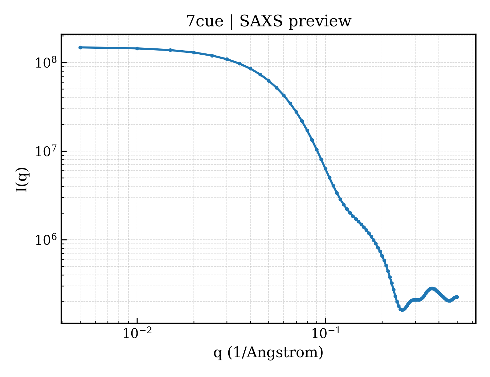
Hemoglobin subunit alpha
OrganismNot specified
State in the Current Structure
| Assembly | Oligomeric State | Construct | Mutations and Modifications | Ligands, Ions and Associated Components | Method and Experimental Conditions | Structure Quality |
|---|---|---|---|---|---|---|
| 1 | Protein heterocomplex Heteromer Protein × 7 PDB declaration: heptameric(7) Consistent with protein copy count | Chain A; UniProt 1–142 Chain C; UniProt 1–142 | Not recorded | Hemoglobin subunit beta × 2 (P68871) Amino acid ABC transporter substrate-binding protein × 3 (B0LFQ8) HEM PROTOPORPHYRIN IX CONTAINING FE × 4 SO4 SULFATE ION × 2 | X-RAY DIFFRACTION X-ray crystallization conditions:VAPOR DIFFUSION, HANGING DROP;pH 5.5;293.15 K;23% PEG 3350 (w/v), 100mM ammonium sulfate, 100mM BIS-TRIS | Resolution 2.75 Å R-free 0.259 |
Other States of the Same Protein in the Database
Each row is a biological assembly of the same UniProt protein in another PDB entry. The “Difference from current entry” column identifies evidence-level differences; no tag means the currently parsed fields agree.
| Other PDB | Difference from Current Entry 7CUE | Assembly / Oligomeric State | Construct | Mutations and Modifications | Ligands, Ions and Non-polymers | Method and Experimental Conditions | Structure Quality |
|---|---|---|---|---|---|---|---|
| 1A00 HEMOGLOBIN (VAL BETA1 MET, TRP BETA37 TYR) MUTANT Deposited 1997-12-08 | Different construct Different oligomeric state Different ligand/ion Different experimental conditions Different structure-quality metrics | Assembly 1 Protein heterocomplex Heteromer;Protein × 4 PDB declaration: tetrameric |
Chain A
1–141(141 aa)
Chain C
1–141(141 aa)
|
Not recorded | HEM PROTOPORPHYRIN IX CONTAINING FE × 4 |
X-RAY DIFFRACTION
X-ray crystallization conditions
pH 7;10.5% PEG 6000 10 MM POTASSIUM PHOSPHATE PH 7.0 100 MM POTASSIUM CHLORIDE 3 MM SODIUM DITHIONITE
|
Resolution 2.00 Å R-free 0.223 |
| 1A01 HEMOGLOBIN (VAL BETA1 MET, TRP BETA37 ALA) MUTANT Deposited 1997-12-08 | Different construct Different oligomeric state Different ligand/ion Different experimental conditions Different structure-quality metrics | Assembly 1 Protein heterocomplex Heteromer;Protein × 4 PDB declaration: tetrameric |
Chain A
1–141(141 aa)
Chain C
1–141(141 aa)
|
Not recorded | HEM PROTOPORPHYRIN IX CONTAINING FE × 4 |
X-RAY DIFFRACTION
X-ray crystallization conditions
pH 6.5;2.3 M AMMONIUM SULFATE 0.3 M AMMONIUM PHOSPHATE PH 6.5 10 MM FERROUS CITRATE
|
Resolution 1.80 Å R-free 0.223 |
| 1A0U HEMOGLOBIN (VAL BETA1 MET) MUTANT Deposited 1997-12-08 | Different construct Different oligomeric state Different ligand/ion Different experimental conditions Different structure-quality metrics | Assembly 1 Protein heterocomplex Heteromer;Protein × 4 PDB declaration: tetrameric |
Chain A
1–141(141 aa)
Chain C
1–141(141 aa)
|
Not recorded | HEM PROTOPORPHYRIN IX CONTAINING FE × 4 |
X-RAY DIFFRACTION
X-ray crystallization conditions
pH 7;10.5% PEG 6000 10 MM POTASSIUM PHOSPHATE PH 7.0 100 MM POTASSIUM CHLORIDE 3 MM SODIUM DITHIONITE
|
Resolution 2.14 Å R-free 0.239 |
| 1A0Z HEMOGLOBIN (VAL BETA1 MET) MUTANT Deposited 1997-12-08 | Different construct Different oligomeric state Different ligand/ion Different experimental conditions Different structure-quality metrics | Assembly 1 Protein heterocomplex Heteromer;Protein × 4 PDB declaration: tetrameric |
Chain A
1–141(141 aa)
Chain C
1–141(141 aa)
|
Not recorded | HEM PROTOPORPHYRIN IX CONTAINING FE × 4 |
X-RAY DIFFRACTION
X-ray crystallization conditions
pH 7;10.5% PEG 6000 10 MM POTASSIUM PHOSPHATE PH 7.0 100 MM POTASSIUM CHLORIDE 3 MM SODIUM DITHIONITE
|
Resolution 2.00 Å |
| 1A3N DEOXY HUMAN HEMOGLOBIN Deposited 1998-01-22 | Different construct Different oligomeric state Different ligand/ion Different experimental conditions Different structure-quality metrics | Assembly 1 Protein heterocomplex Heteromer;Protein × 4 PDB declaration: tetrameric |
Chain A
1–141(141 aa)
Chain C
1–141(141 aa)
|
Not recorded | HEM PROTOPORPHYRIN IX CONTAINING FE × 4 | X-RAY DIFFRACTION mmCIF provides none of the parsed conditions | Resolution 1.80 Å R-free 0.220 |
| 1A3O ARTIFICIAL MUTANT (ALPHA Y42H) OF DEOXY HEMOGLOBIN Deposited 1998-01-22 | Different construct Different mutation/modification Different oligomeric state Different ligand/ion Different experimental conditions Different structure-quality metrics | Assembly 1 Protein heterocomplex Heteromer;Protein × 4 PDB declaration: tetrameric |
Chain A
1–141(141 aa)
Chain C
1–141(141 aa)
|
Mutation:CHAIN A, C, Y42H Mutation:CHAIN A, C, Y42H | HEM PROTOPORPHYRIN IX CONTAINING FE × 4 | X-RAY DIFFRACTION mmCIF provides none of the parsed conditions | Resolution 1.80 Å R-free 0.227 |
| 1A9W HUMAN EMBRYONIC GOWER II CARBONMONOXY HEMOGLOBIN Deposited 1998-04-11 | Different construct Different oligomeric state Different ligand/ion Different experimental conditions Different structure-quality metrics | Assembly 1 Protein heterocomplex Heteromer;Protein × 4 PDB declaration: tetrameric |
Chain A
1–141(141 aa)
Chain C
1–141(141 aa)
|
Not recorded | HEM PROTOPORPHYRIN IX CONTAINING FE × 4 CMO CARBON MONOXIDE × 4 |
X-RAY DIFFRACTION
X-ray crystallization conditions
microseeding and macroseeding used to increase crystal size;pH 8.5;PROTEIN WAS CRYSTALLIZED FROM 21% MME-PEG 5000, 0.2M TAPS/ KOH, PH 8.5, 2MM DITHIONITE. THEN MICROSEEDING AND MACROSEEDING USED TO INCREASE CRYSTAL SIZE, microseeding and macroseeding used to increase crystal size
|
Resolution 2.90 Å R-free 0.232 |
| 1ABW DEOXY RHB1.1 (RECOMBINANT HEMOGLOBIN) Deposited 1997-01-29 | Different construct Different mutation/modification Different oligomeric state Different ligand/ion Different experimental conditions Different structure-quality metrics | Assembly 1 Protein heterocomplex Heteromer;Protein × 3 PDB declaration: trimeric |
Chain A
1–141(141 aa)
|
Mutation:CHAIN A IS COMPOSED OF TWO COPIES OF HEMOGLOBIN ALPHA CHAIN JOINED BY AN ADDITIONAL RESIDUE, GLY 142 | MET METHIONINE × 2 LEU LEUCINE × 2 HEM PROTOPORPHYRIN IX CONTAINING FE × 4 |
X-RAY DIFFRACTION
X-ray crystallization conditions
pH 6.2;pH 6.2
|
Resolution 2.00 Å R-free 0.232 |
| 1ABY CYANOMET RHB1.1 (RECOMBINANT HEMOGLOBIN) Deposited 1997-01-29 | Different construct Different mutation/modification Different oligomeric state Different ligand/ion Different experimental conditions Different structure-quality metrics | Assembly 1 Protein heterocomplex Heteromer;Protein × 3 PDB declaration: trimeric |
Chain A
1–141(141 aa)
|
Mutation:CHAIN A, V1M, CHAIN B, D, V1M, N108K | CYN CYANIDE ION × 4 HEM PROTOPORPHYRIN IX CONTAINING FE × 4 |
X-RAY DIFFRACTION
X-ray crystallization conditions
pH 5;pH 5.0
|
Resolution 2.60 Å R-free 0.278 |
| 1AJ9 R-STATE HUMAN CARBONMONOXYHEMOGLOBIN ALPHA-A53S Deposited 1997-05-16 | Different construct Different mutation/modification Different oligomeric state Different ligand/ion Different experimental conditions Different structure-quality metrics | Assembly 1 Protein heterocomplex Heteromer;Protein × 4 PDB declaration: tetrameric |
Chain A
1–141(141 aa)
|
Mutation:A53S | PO4 PHOSPHATE ION × 2 HEM PROTOPORPHYRIN IX CONTAINING FE × 4 CMO CARBON MONOXIDE × 4 |
X-RAY DIFFRACTION
X-ray crystallization conditions
batch method;pH 6.7;PROTEIN WAS BATCH CRYSTALLIZED IN 2.5 M SODIUM/POTASSIUM PHOSPHATE, PH 6.7, AND 1% (W/V) CARBOXYHEMOGLOBIN., batch method
|
Resolution 2.20 Å |
| 1B86 HUMAN DEOXYHAEMOGLOBIN-2,3-DIPHOSPHOGLYCERATE COMPLEX Deposited 1999-02-08 | Different construct Different oligomeric state Different ligand/ion Different experimental conditions Different structure-quality metrics | Assembly 1 Protein heterocomplex Heteromer;Protein × 4 PDB declaration: tetrameric |
Chain A
1–141(141 aa)
Chain C
1–141(141 aa)
|
Not recorded | HEM PROTOPORPHYRIN IX CONTAINING FE × 4 OXY OXYGEN MOLECULE × 2 DG2 (2R)-2,3-diphosphoglyceric acid × 1 |
X-RAY DIFFRACTION
X-ray crystallization conditions
pH 7.2;pH 7.2
|
Resolution 2.50 Å |
| 1BAB HEMOGLOBIN THIONVILLE: AN ALPHA-CHAIN VARIANT WITH A SUBSTITUTION OF A GLUTAMATE FOR VALINE AT NA-1 AND HAVING AN ACETYLATED METHIONINE NH2 TERMINUS Deposited 1992-05-06 | Different construct Different mutation/modification Different oligomeric state Different experimental conditions Different structure-quality metrics | Assembly 1 Protein heterocomplex Heteromer;Protein × 4 PDB declaration: tetrameric |
Chain A
2–141(140 aa)
Chain C
2–141(140 aa)
|
Non-standard monomer:Yes (specific site not provided by mmCIF) Non-standard monomer:Yes (specific site not provided by mmCIF) | HEM PROTOPORPHYRIN IX CONTAINING FE × 4 SO4 SULFATE ION × 2 | X-RAY DIFFRACTION mmCIF provides none of the parsed conditions | Resolution 1.50 Å |
| 1BBB A THIRD QUATERNARY STRUCTURE OF HUMAN HEMOGLOBIN A AT 1.7-ANGSTROMS RESOLUTION Deposited 1992-04-29 | Different construct Different oligomeric state Different ligand/ion Different experimental conditions Different structure-quality metrics | Assembly 1 Protein heterocomplex Heteromer;Protein × 4 PDB declaration: tetrameric |
Chain A
1–141(141 aa)
Chain C
1–141(141 aa)
|
Not recorded | HEM PROTOPORPHYRIN IX CONTAINING FE × 4 CMO CARBON MONOXIDE × 4 | X-RAY DIFFRACTION mmCIF provides none of the parsed conditions | Resolution 1.70 Å |
| 1BIJ CROSSLINKED, DEOXY HUMAN HEMOGLOBIN A Deposited 1998-06-18 | Different construct Different oligomeric state Different ligand/ion Different experimental conditions Different structure-quality metrics | Assembly 1 Protein heterocomplex Heteromer;Protein × 4 PDB declaration: tetrameric |
Chain A
1–141(141 aa)
Chain C
1–141(141 aa)
|
Not recorded | HEM PROTOPORPHYRIN IX CONTAINING FE × 4 2FU BUT-2-ENEDIAL × 1 |
X-RAY DIFFRACTION
X-ray crystallization conditions
pH 7;pH 7.0
|
Resolution 2.30 Å R-free 0.263 |
| 1BUW CRYSTAL STRUCTURE OF S-NITROSO-NITROSYL HUMAN HEMOGLOBIN A Deposited 1998-09-06 | Different construct Different oligomeric state Different ligand/ion Different experimental conditions Different structure-quality metrics | Assembly 1 Protein heterocomplex Heteromer;Protein × 4 PDB declaration: tetrameric |
Chain A
1–141(141 aa)
Chain C
1–141(141 aa)
|
Not recorded | HEM PROTOPORPHYRIN IX CONTAINING FE × 4 NO NITRIC OXIDE × 4 |
X-RAY DIFFRACTION
X-ray crystallization conditions
pH 5.8;1% PROTEIN 100 MM SODIUM CACODYLATE, PH 5.8 16% PEG 6000
|
Resolution 1.90 Å R-free 0.244 |
| 1BZ0 HEMOGLOBIN A (HUMAN, DEOXY, HIGH SALT) Deposited 1998-11-04 | Different construct Different oligomeric state Different ligand/ion Different experimental conditions Different structure-quality metrics | Assembly 1 Protein heterocomplex Heteromer;Protein × 4 PDB declaration: tetrameric |
Chain A
1–141(141 aa)
Chain C
1–141(141 aa)
|
Not recorded | HEM PROTOPORPHYRIN IX CONTAINING FE × 4 |
X-RAY DIFFRACTION
X-ray crystallization conditions
pH 6.5;2.3 M AMMONIUM SULFATE 0.3 M AMMONIUM PHOSPHATE PH 6.5 10 MM FERROUS CITRATE
|
Resolution 1.50 Å R-free 0.208 |
| 1BZ1 HEMOGLOBIN (ALPHA + MET) VARIANT Deposited 1998-11-05 | Different construct Different oligomeric state Different ligand/ion Different experimental conditions Different structure-quality metrics | Assembly 1 Protein heterocomplex Heteromer;Protein × 4 PDB declaration: tetrameric |
Chain A
1–141(141 aa)
Chain C
1–141(141 aa)
|
Not recorded | HEM PROTOPORPHYRIN IX CONTAINING FE × 4 |
X-RAY DIFFRACTION
X-ray crystallization conditions
pH 6.5;2.3 M AMMONIUM SULFATE 0.3 M AMMONIUM PHOSPHATE PH 6.5 10 MM FERROUS CITRATE
|
Resolution 1.59 Å R-free 0.224 |
| 1BZZ HEMOGLOBIN (ALPHA V1M) MUTANT Deposited 1998-11-04 | Different construct Different mutation/modification Different oligomeric state Different ligand/ion Different experimental conditions Different structure-quality metrics | Assembly 1 Protein heterocomplex Heteromer;Protein × 4 PDB declaration: tetrameric |
Chain A
1–141(141 aa)
Chain C
1–141(141 aa)
|
Mutation:V1M Mutation:V1M | HEM PROTOPORPHYRIN IX CONTAINING FE × 4 |
X-RAY DIFFRACTION
X-ray crystallization conditions
pH 7;2.3 M AMMONIUM SULFATE 0.3 M AMMONIUM PHOSPHATE PH 6.5 10 MM FERROUS CITRATE, pH 7.0
|
Resolution 1.59 Å R-free 0.214 |
| 1C7B DEOXY RHB1.0 (RECOMBINANT HEMOGLOBIN) Deposited 2000-02-09 | Different construct Different mutation/modification Different oligomeric state Different ligand/ion Different experimental conditions Different structure-quality metrics | Assembly 1 Protein heterocomplex Heteromer;Protein × 4 PDB declaration: tetrameric |
Chain A
1–141(141 aa)
Chain C
1–141(141 aa)
|
Mutation:V1M Mutation:V1M | HEM PROTOPORPHYRIN IX CONTAINING FE × 4 |
X-RAY DIFFRACTION
X-ray crystallization conditions
pH 6.5;PERUTZ, J.CRYST.GROWTH 2(1968)54-56, pH 6.5
|
Resolution 1.80 Å R-free 0.234 |
| 1C7C DEOXY RHB1.1 (RECOMBINANT HEMOGLOBIN) Deposited 2000-02-09 | Different construct Different mutation/modification Different oligomeric state Different ligand/ion Different experimental conditions Different structure-quality metrics | Assembly 1 Protein heterocomplex Heteromer;Protein × 3 PDB declaration: trimeric |
Chain A
1–141(141 aa)
|
Mutation:V1M,142G | HEM PROTOPORPHYRIN IX CONTAINING FE × 4 |
X-RAY DIFFRACTION
X-ray crystallization conditions
pH 6.5;PERUTZ, J.CRYST.GROWTH 2(1968)54-56, pH 6.5
|
Resolution 1.80 Å R-free 0.227 |
| 1C7D DEOXY RHB1.2 (RECOMBINANT HEMOGLOBIN) Deposited 2000-02-09 | Different construct Different mutation/modification Different oligomeric state Different ligand/ion Different experimental conditions Different structure-quality metrics | Assembly 1 Protein heterocomplex Heteromer;Protein × 3 PDB declaration: trimeric |
Chain A
1–141(141 aa)
|
Mutation:V1M,142G,143G | HEM PROTOPORPHYRIN IX CONTAINING FE × 4 |
X-RAY DIFFRACTION
X-ray crystallization conditions
pH 6.5;PERUTZ, J.CRYST.GROWTH 2(1968)54-56, pH 6.5
|
Resolution 1.80 Å R-free 0.240 |
| 1CLS CROSS-LINKED HUMAN HEMOGLOBIN DEOXY Deposited 1995-08-29 | Different construct Different oligomeric state Different ligand/ion Different experimental conditions Different structure-quality metrics | Assembly 1 Protein heterocomplex Heteromer;Protein × 4 PDB declaration: tetrameric |
Chain A
1–141(141 aa)
Chain C
1–141(141 aa)
|
Not recorded | HEM PROTOPORPHYRIN IX CONTAINING FE × 4 O OXYGEN ATOM × 2 SO4 SULFATE ION × 2 DEC SEBACIC ACID × 1 | X-RAY DIFFRACTION mmCIF provides none of the parsed conditions | Resolution 1.90 Å |
| 1CMY THE MUTATION BETA99 ASP-TYR STABILIZES Y-A NEW, COMPOSITE QUATERNARY STATE OF HUMAN HEMOGLOBIN Deposited 1992-09-18 | Different construct Different oligomeric state Different ligand/ion Different experimental conditions Different structure-quality metrics | Assembly 1 Protein heterocomplex Heteromer;Protein × 4 PDB declaration: tetrameric |
Chain A
1–141(141 aa)
Chain C
1–141(141 aa)
|
Not recorded | HEM PROTOPORPHYRIN IX CONTAINING FE × 4 | X-RAY DIFFRACTION mmCIF provides none of the parsed conditions | Resolution 3.00 Å |
| 1COH STRUCTURE OF HAEMOGLOBIN IN THE DEOXY QUATERNARY STATE WITH LIGAND BOUND AT THE ALPHA HAEMS Deposited 1989-01-13 | Different construct Different oligomeric state Different ligand/ion Different experimental conditions Different structure-quality metrics | Assembly 1 Protein heterocomplex Heteromer;Protein × 4 PDB declaration: tetrameric |
Chain A
1–141(141 aa)
Chain C
1–141(141 aa)
|
Not recorded | HEM PROTOPORPHYRIN IX CONTAINING FE × 2 CMO CARBON MONOXIDE × 2 COH PROTOPORPHYRIN IX CONTAINING CO × 2 | X-RAY DIFFRACTION mmCIF provides none of the parsed conditions | Resolution 2.90 Å |
| 1DKE NI BETA HEME HUMAN HEMOGLOBIN Deposited 1999-12-07 | Different construct Different oligomeric state Different ligand/ion Different experimental conditions Different structure-quality metrics | Assembly 1 Protein heterocomplex Heteromer;Protein × 4 PDB declaration: tetrameric |
Chain A
1–141(141 aa)
Chain C
1–141(141 aa)
|
Not recorded | HEM PROTOPORPHYRIN IX CONTAINING FE × 4 SO4 SULFATE ION × 1 |
X-RAY DIFFRACTION
X-ray crystallization conditions
VAPOR DIFFUSION, HANGING DROP;pH 7;294 K;24 MG/ML PROTEIN 21% PEG 1000 10MM POTASSIUM PHOSPHATE, 1MM IHP, 30MM SODIUM DITHIONITE, pH 7, VAPOR DIFFUSION,
HANGING DROP
|
Resolution 2.10 Å R-free 0.280 |
| 1DXT HIGH-RESOLUTION X-RAY STUDY OF DEOXY RECOMBINANT HUMAN HEMOGLOBINS SYNTHESIZED FROM BETA-GLOBINS HAVING MUTATED AMINO TERMINI Deposited 1992-05-06 | Different construct Different oligomeric state Different ligand/ion Different experimental conditions Different structure-quality metrics | Assembly 1 Protein heterocomplex Heteromer;Protein × 4 PDB declaration: tetrameric |
Chain A
1–141(141 aa)
Chain C
1–141(141 aa)
|
Not recorded | HEM PROTOPORPHYRIN IX CONTAINING FE × 4 | X-RAY DIFFRACTION mmCIF provides none of the parsed conditions | Resolution 1.70 Å |
| 1DXU HIGH-RESOLUTION X-RAY STUDY OF DEOXY RECOMBINANT HUMAN HEMOGLOBINS SYNTHESIZED FROM BETA-GLOBINS HAVING MUTATED AMINO TERMINI Deposited 1992-05-06 | Different construct Different oligomeric state Different experimental conditions Different structure-quality metrics | Assembly 1 Protein heterocomplex Heteromer;Protein × 4 PDB declaration: tetrameric |
Chain A
1–141(141 aa)
Chain C
1–141(141 aa)
|
Not recorded | HEM PROTOPORPHYRIN IX CONTAINING FE × 4 SO4 SULFATE ION × 2 | X-RAY DIFFRACTION mmCIF provides none of the parsed conditions | Resolution 1.70 Å |
| 1DXV HIGH-RESOLUTION X-RAY STUDY OF DEOXY RECOMBINANT HUMAN HEMOGLOBINS SYNTHESIZED FROM BETA-GLOBINS HAVING MUTATED AMINO TERMINI Deposited 1992-05-06 | Different construct Different oligomeric state Different experimental conditions Different structure-quality metrics | Assembly 1 Protein heterocomplex Heteromer;Protein × 4 PDB declaration: tetrameric |
Chain A
1–141(141 aa)
Chain C
1–141(141 aa)
|
Not recorded | HEM PROTOPORPHYRIN IX CONTAINING FE × 4 SO4 SULFATE ION × 2 | X-RAY DIFFRACTION mmCIF provides none of the parsed conditions | Resolution 1.70 Å |
| 1FDH STRUCTURE OF HUMAN FOETAL DEOXYHAEMOGLOBIN Deposited 1976-08-18 | Different construct Different oligomeric state Different ligand/ion Different experimental conditions Different structure-quality metrics | Assembly 1 Protein heterocomplex Heteromer;Protein × 4 PDB declaration: tetrameric |
Chain A
1–141(141 aa)
Chain B
1–141(141 aa)
|
Not recorded | HEM PROTOPORPHYRIN IX CONTAINING FE × 4 | X-RAY DIFFRACTION mmCIF provides none of the parsed conditions | Resolution 2.50 Å |
| 1FN3 CRYSTAL STRUCTURE OF NICKEL RECONSTITUTED HEMOGLOBIN-A CASE FOR PERMANENT, T-STATE HEMOGLOBIN Deposited 2000-08-20 | Different construct Different oligomeric state Different ligand/ion Different experimental conditions Different structure-quality metrics | Assembly 1 Protein heterocomplex Heteromer;Protein × 4 PDB declaration: tetrameric |
Chain A
1–141(141 aa)
Chain C
1–141(141 aa)
|
Not recorded | HNI PROTOPORPHYRIN IX CONTAINING NI(II) × 4 |
X-RAY DIFFRACTION
X-ray crystallization conditions
pH 7.2;pH 7.20
|
Resolution 2.48 Å R-free 0.324 |
| 1G9V HIGH RESOLUTION CRYSTAL STRUCTURE OF DEOXY HEMOGLOBIN COMPLEXED WITH A POTENT ALLOSTERIC EFFECTOR Deposited 2000-11-28 | Different construct Different oligomeric state Different ligand/ion Different experimental conditions Different structure-quality metrics | Assembly 1 Protein heterocomplex Heteromer;Protein × 4 PDB declaration: tetrameric |
Chain A
1–141(141 aa)
Chain C
1–141(141 aa)
|
Not recorded | HEM PROTOPORPHYRIN IX CONTAINING FE × 4 RQ3 2-{4-[(3,5-DIMETHYLANILINO)-CARBONYL-METHYL]-PHENOXY}-2-METHYLPROPIONIC ACID × 2 SO4 SULFATE ION × 2 |
X-RAY DIFFRACTION
X-ray crystallization conditions
LIQUID DIFFUSION;pH 6.5;298 K;Ammonium sulfate, ammonium phosphate, ferrous citrate, pH 6.5, LIQUID DIFFUSION, temperature 298K
|
Resolution 1.85 Å R-free 0.208 |
| 1GBU DEOXY (BETA-(C93A,C112G)) HUMAN HEMOGLOBIN Deposited 1996-01-04 | Different construct Different mutation/modification Different oligomeric state Different experimental conditions Different structure-quality metrics | Assembly 1 Protein heterocomplex Heteromer;Protein × 4 PDB declaration: tetrameric |
Chain A
1–141(141 aa)
Chain C
1–141(141 aa)
|
Mutation:CHAIN B, D, C93A, C112G Mutation:CHAIN B, D, C93A, C112G | HEM PROTOPORPHYRIN IX CONTAINING FE × 4 SO4 SULFATE ION × 2 | X-RAY DIFFRACTION mmCIF provides none of the parsed conditions | Resolution 1.80 Å |
| 1GBV (ALPHA-OXY, BETA-(C112G)DEOXY) T-STATE HUMAN HEMOGLOBIN Deposited 1995-12-20 | Different construct Different mutation/modification Different oligomeric state Different ligand/ion Different experimental conditions Different structure-quality metrics | Assembly 1 Protein heterocomplex Heteromer;Protein × 4 PDB declaration: tetrameric |
Chain A
1–141(141 aa)
Chain C
1–141(141 aa)
|
Mutation:CHAIN B, D, C112G Mutation:CHAIN B, D, C112G | HEM PROTOPORPHYRIN IX CONTAINING FE × 4 OXY OXYGEN MOLECULE × 2 | X-RAY DIFFRACTION mmCIF provides none of the parsed conditions | Resolution 2.00 Å |
| 1GLI DEOXYHEMOGLOBIN T38W (ALPHA CHAINS), V1G (ALPHA AND BETA CHAINS) Deposited 1996-02-22 | Different construct Different mutation/modification Different oligomeric state Different ligand/ion Different experimental conditions Different structure-quality metrics | Assembly 1 Protein heterocomplex Heteromer;Protein × 4 PDB declaration: tetrameric |
Chain A
2–141(140 aa)
Chain C
2–141(140 aa)
|
Mutation:V1M, CHAIN A, C, T38W Mutation:V1M, CHAIN A, C, T38W | HEM PROTOPORPHYRIN IX CONTAINING FE × 4 PO4 PHOSPHATE ION × 1 | X-RAY DIFFRACTION mmCIF provides none of the parsed conditions | Resolution 2.50 Å |
| 1GZX Oxy T State Haemoglobin - Oxygen bound at all four haems Deposited 2002-06-07 | Different construct Different oligomeric state Different ligand/ion Different experimental conditions Different structure-quality metrics | Assembly 1 Protein heterocomplex Heteromer;Protein × 4 PDB declaration: tetrameric |
Chain A
2–142(141 aa)
Chain C
2–142(141 aa)
|
Not recorded | HEM PROTOPORPHYRIN IX CONTAINING FE × 4 OXY OXYGEN MOLECULE × 4 |
X-RAY DIFFRACTION
X-ray crystallization conditions
pH 7.2;BATCH CRYSTALLISATION, OXYHB (75-90 MG/ML) IN 50MM POTASSIUM PHOSPHATE (PH 7.2), 22-28% PEG 8000 AND A 5 MOLAR EXCESS OF THE ALLOSTERIC EFFECTOR IHP
|
Resolution 2.10 Å R-free 0.221 |
| 1HAB CROSSLINKED HAEMOGLOBIN Deposited 1996-03-13 | Different construct Different oligomeric state Different ligand/ion Different experimental conditions Different structure-quality metrics | Assembly 1 Protein heterocomplex Heteromer;Protein × 4 PDB declaration: tetrameric |
Chain A
1–141(141 aa)
Chain C
1–141(141 aa)
|
Not recorded | HEM PROTOPORPHYRIN IX CONTAINING FE × 4 CMO CARBON MONOXIDE × 4 CIN 4-CARBOXYCINNAMIC ACID × 1 | X-RAY DIFFRACTION mmCIF provides none of the parsed conditions | Resolution 2.30 Å |
| 1HAC CROSSLINKED HAEMOGLOBIN Deposited 1996-03-13 | Different construct Different oligomeric state Different ligand/ion Different experimental conditions Different structure-quality metrics | Assembly 1 Protein heterocomplex Heteromer;Protein × 4 PDB declaration: tetrameric |
Chain A
1–141(141 aa)
Chain C
1–141(141 aa)
|
Not recorded | HEM PROTOPORPHYRIN IX CONTAINING FE × 4 CMO CARBON MONOXIDE × 4 NDD 2,6-DICARBOXYNAPHTHALENE × 1 | X-RAY DIFFRACTION mmCIF provides none of the parsed conditions | Resolution 2.60 Å |
| 1HBA HIGH-RESOLUTION X-RAY STUDY OF DEOXYHEMOGLOBIN ROTHSCHILD 37BETA TRP-> ARG: A MUTATION THAT CREATES AN INTERSUBUNIT CHLORIDE-BINDING SITE Deposited 1992-01-07 | Different construct Different oligomeric state Different ligand/ion Different experimental conditions Different structure-quality metrics | Assembly 1 Protein heterocomplex Heteromer;Protein × 4 PDB declaration: tetrameric |
Chain A
1–141(141 aa)
Chain C
1–141(141 aa)
|
Not recorded | HEM PROTOPORPHYRIN IX CONTAINING FE × 4 CL CHLORIDE ION × 2 | X-RAY DIFFRACTION mmCIF provides none of the parsed conditions | Resolution 2.10 Å |
| 1HBB HIGH-RESOLUTION X-RAY STUDY OF DEOXYHEMOGLOBIN ROTHSCHILD 37BETA TRP-> ARG: A MUTATION THAT CREATES AN INTERSUBUNIT CHLORIDE-BINDING SITE Deposited 1992-01-07 | Different construct Different oligomeric state Different ligand/ion Different experimental conditions Different structure-quality metrics | Assembly 1 Protein heterocomplex Heteromer;Protein × 4 PDB declaration: tetrameric |
Chain A
1–141(141 aa)
Chain C
1–141(141 aa)
|
Not recorded | HEM PROTOPORPHYRIN IX CONTAINING FE × 4 | X-RAY DIFFRACTION mmCIF provides none of the parsed conditions | Resolution 1.90 Å |
| 1HBS REFINED CRYSTAL STRUCTURE OF DEOXYHEMOGLOBIN S. I. RESTRAINED LEAST-SQUARES REFINEMENT AT 3.0-ANGSTROMS RESOLUTION Deposited 1982-06-02 | Different construct Different oligomeric state Different ligand/ion Different experimental conditions Different structure-quality metrics | Assembly 1 Protein heterocomplex Heteromer;Protein × 4 PDB declaration: tetrameric |
Chain A
1–141(141 aa)
Chain C
1–141(141 aa)
|
Not recorded | HEM PROTOPORPHYRIN IX CONTAINING FE × 4 | X-RAY DIFFRACTION mmCIF provides none of the parsed conditions | Resolution 3.00 Å |
| 1HBS REFINED CRYSTAL STRUCTURE OF DEOXYHEMOGLOBIN S. I. RESTRAINED LEAST-SQUARES REFINEMENT AT 3.0-ANGSTROMS RESOLUTION Deposited 1982-06-02 | Different construct Different oligomeric state Different ligand/ion Different experimental conditions Different structure-quality metrics | Assembly 2 Protein heterocomplex Heteromer;Protein × 4 PDB declaration: tetrameric |
Chain E
1–141(141 aa)
Chain G
1–141(141 aa)
|
Not recorded | HEM PROTOPORPHYRIN IX CONTAINING FE × 4 | X-RAY DIFFRACTION mmCIF provides none of the parsed conditions | Resolution 3.00 Å |
| 1HCO THE STRUCTURE OF HUMAN CARBONMONOXY HAEMOGLOBIN AT 2.7 ANGSTROMS RESOLUTION Deposited 1979-08-07 | Different construct Different mutation/modification Different oligomeric state Different ligand/ion Different experimental conditions Different structure-quality metrics | Assembly 1 Protein heterocomplex Heteromer;Protein × 4 PDB declaration: tetrameric |
Chain A
1–141(141 aa)
|
Not recorded | HEM PROTOPORPHYRIN IX CONTAINING FE × 4 CMO CARBON MONOXIDE × 4 | X-RAY DIFFRACTION mmCIF provides none of the parsed conditions | Resolution 2.70 Å |
| 1HGA HIGH RESOLUTION CRYSTAL STRUCTURES AND COMPARISONS OF T STATE DEOXYHAEMOGLOBIN AND TWO LIGANDED T-STATE HAEMOGLOBINS: T(ALPHA-OXY)HAEMOGLOBIN AND T(MET)HAEMOGLOBIN Deposited 1991-10-31 | Different construct Different oligomeric state Different ligand/ion Different experimental conditions Different structure-quality metrics | Assembly 1 Protein heterocomplex Heteromer;Protein × 4 PDB declaration: tetrameric |
Chain A
1–141(141 aa)
Chain C
1–141(141 aa)
|
Not recorded | HEM PROTOPORPHYRIN IX CONTAINING FE × 4 | X-RAY DIFFRACTION mmCIF provides none of the parsed conditions | Resolution 2.10 Å |
| 1HGB HIGH RESOLUTION CRYSTAL STRUCTURES AND COMPARISONS OF T STATE DEOXYHAEMOGLOBIN AND TWO LIGANDED T-STATE HAEMOGLOBINS: T(ALPHA-OXY)HAEMOGLOBIN AND T(MET)HAEMOGLOBIN Deposited 1991-10-31 | Different construct Different oligomeric state Different ligand/ion Different experimental conditions Different structure-quality metrics | Assembly 1 Protein heterocomplex Heteromer;Protein × 4 PDB declaration: tetrameric |
Chain A
1–141(141 aa)
Chain C
1–141(141 aa)
|
Not recorded | HEM PROTOPORPHYRIN IX CONTAINING FE × 4 | X-RAY DIFFRACTION mmCIF provides none of the parsed conditions | Resolution 2.10 Å |
| 1HGC HIGH RESOLUTION CRYSTAL STRUCTURES AND COMPARISONS OF T STATE DEOXYHAEMOGLOBIN AND TWO LIGANDED T-STATE HAEMOGLOBINS: T(ALPHA-OXY)HAEMOGLOBIN AND T(MET)HAEMOGLOBIN Deposited 1991-10-31 | Different construct Different oligomeric state Different ligand/ion Different experimental conditions Different structure-quality metrics | Assembly 1 Protein heterocomplex Heteromer;Protein × 4 PDB declaration: tetrameric |
Chain A
1–141(141 aa)
Chain C
1–141(141 aa)
|
Not recorded | HEM PROTOPORPHYRIN IX CONTAINING FE × 4 OXY OXYGEN MOLECULE × 2 | X-RAY DIFFRACTION mmCIF provides none of the parsed conditions | Resolution 2.10 Å |
| 1HHO STRUCTURE OF HUMAN OXYHAEMOGLOBIN AT 2.1 ANGSTROMS RESOLUTION Deposited 1983-06-10 | Different construct Different mutation/modification Different oligomeric state Different ligand/ion Different experimental conditions Different structure-quality metrics | Assembly 1 Protein heterocomplex Heteromer;Protein × 4 PDB declaration: tetrameric |
Chain A
1–141(141 aa)
|
Not recorded | PO4 PHOSPHATE ION × 2 HEM PROTOPORPHYRIN IX CONTAINING FE × 4 OXY OXYGEN MOLECULE × 4 | X-RAY DIFFRACTION mmCIF provides none of the parsed conditions | Resolution 2.10 Å |
| 1IRD Crystal Structure of Human Carbonmonoxy-Haemoglobin at 1.25 A Resolution Deposited 2001-09-27 | Different construct Different mutation/modification Different oligomeric state Different ligand/ion Different experimental conditions Different structure-quality metrics | Assembly 1 Protein heterocomplex Heteromer;Protein × 4 PDB declaration: tetrameric |
Chain A
1–141(141 aa)
|
Not recorded | HEM PROTOPORPHYRIN IX CONTAINING FE × 4 CMO CARBON MONOXIDE × 4 |
X-RAY DIFFRACTION
X-ray crystallization conditions
SMALL TUBES;pH 6.7;298 K;potassium phosphate, pH 6.7, SMALL TUBES, temperature 298K
|
Resolution 1.25 Å R-free 0.214 |
| 1J3Y Direct observation of photolysis-induced tertiary structural changes in human hemoglobin; Crystal structure of alpha(Fe)-beta(Ni) hemoglobin (laser photolysed) Deposited 2003-02-21 | Different construct Different oligomeric state Different ligand/ion Different experimental conditions Different structure-quality metrics | Assembly 1 Protein heterocomplex Heteromer;Protein × 4 PDB declaration: tetrameric |
Chain A
1–141(141 aa)
Chain C
1–141(141 aa)
|
Not recorded | HEM PROTOPORPHYRIN IX CONTAINING FE × 2 CMO CARBON MONOXIDE × 2 HNI PROTOPORPHYRIN IX CONTAINING NI(II) × 2 2FU BUT-2-ENEDIAL × 1 |
X-RAY DIFFRACTION
X-ray crystallization conditions
pH 6.6;293 K;PEG 4000, 50mM citrate-ammonium buffer, SMALL TUBES, temperature 293K, pH 6.60
|
Resolution 1.55 Å R-free 0.205 |
| 1J3Y Direct observation of photolysis-induced tertiary structural changes in human hemoglobin; Crystal structure of alpha(Fe)-beta(Ni) hemoglobin (laser photolysed) Deposited 2003-02-21 | Different construct Different oligomeric state Different ligand/ion Different experimental conditions Different structure-quality metrics | Assembly 2 Protein heterocomplex Heteromer;Protein × 4 PDB declaration: tetrameric |
Chain E
1–141(141 aa)
Chain G
1–141(141 aa)
|
Not recorded | HEM PROTOPORPHYRIN IX CONTAINING FE × 2 CMO CARBON MONOXIDE × 2 HNI PROTOPORPHYRIN IX CONTAINING NI(II) × 2 2FU BUT-2-ENEDIAL × 1 |
X-RAY DIFFRACTION
X-ray crystallization conditions
pH 6.6;293 K;PEG 4000, 50mM citrate-ammonium buffer, SMALL TUBES, temperature 293K, pH 6.60
|
Resolution 1.55 Å R-free 0.205 |
| 1J3Z Direct observation of photolysis-induced tertiary structural changes in human haemoglobin; Crystal structure of alpha(Fe-CO)-beta(Ni) hemoglobin (laser unphotolysed) Deposited 2003-02-21 | Different construct Different oligomeric state Different ligand/ion Different experimental conditions Different structure-quality metrics | Assembly 1 Protein heterocomplex Heteromer;Protein × 4 PDB declaration: tetrameric |
Chain A
1–141(141 aa)
Chain C
1–141(141 aa)
|
Not recorded | HEM PROTOPORPHYRIN IX CONTAINING FE × 2 CMO CARBON MONOXIDE × 2 HNI PROTOPORPHYRIN IX CONTAINING NI(II) × 2 2FU BUT-2-ENEDIAL × 1 |
X-RAY DIFFRACTION
X-ray crystallization conditions
pH 6.6;293 K;PEG 4000, 50mM citrate-ammonium buffer, SMALL TUBES, temperature 293K, pH 6.60
|
Resolution 1.60 Å R-free 0.196 |
| 1J3Z Direct observation of photolysis-induced tertiary structural changes in human haemoglobin; Crystal structure of alpha(Fe-CO)-beta(Ni) hemoglobin (laser unphotolysed) Deposited 2003-02-21 | Different construct Different oligomeric state Different ligand/ion Different experimental conditions Different structure-quality metrics | Assembly 2 Protein heterocomplex Heteromer;Protein × 4 PDB declaration: tetrameric |
Chain E
1–141(141 aa)
Chain G
1–141(141 aa)
|
Not recorded | HEM PROTOPORPHYRIN IX CONTAINING FE × 2 CMO CARBON MONOXIDE × 2 HNI PROTOPORPHYRIN IX CONTAINING NI(II) × 2 2FU BUT-2-ENEDIAL × 1 |
X-RAY DIFFRACTION
X-ray crystallization conditions
pH 6.6;293 K;PEG 4000, 50mM citrate-ammonium buffer, SMALL TUBES, temperature 293K, pH 6.60
|
Resolution 1.60 Å R-free 0.196 |
| 1J40 Direct observation of photolysis-induced tertiary structural changes in human haemoglobin; Crystal structure of alpha(Ni)-beta(Fe-CO) hemoglobin (laser unphotolysed) Deposited 2003-02-21 | Different construct Different oligomeric state Different ligand/ion Different experimental conditions Different structure-quality metrics | Assembly 1 Protein heterocomplex Heteromer;Protein × 4 PDB declaration: tetrameric |
Chain A
1–141(141 aa)
Chain C
1–141(141 aa)
|
Not recorded | HNI PROTOPORPHYRIN IX CONTAINING NI(II) × 2 HEM PROTOPORPHYRIN IX CONTAINING FE × 2 CMO CARBON MONOXIDE × 2 2FU BUT-2-ENEDIAL × 1 |
X-RAY DIFFRACTION
X-ray crystallization conditions
SMALL TUBES;pH 6.6;293 K;PEG 4000, 50mM citrate-ammonium buffer, pH 6.6, SMALL TUBES, temperature 293K
|
Resolution 1.45 Å R-free 0.197 |
| 1J40 Direct observation of photolysis-induced tertiary structural changes in human haemoglobin; Crystal structure of alpha(Ni)-beta(Fe-CO) hemoglobin (laser unphotolysed) Deposited 2003-02-21 | Different construct Different oligomeric state Different ligand/ion Different experimental conditions Different structure-quality metrics | Assembly 2 Protein heterocomplex Heteromer;Protein × 4 PDB declaration: tetrameric |
Chain E
1–141(141 aa)
Chain G
1–141(141 aa)
|
Not recorded | HNI PROTOPORPHYRIN IX CONTAINING NI(II) × 2 HEM PROTOPORPHYRIN IX CONTAINING FE × 2 CMO CARBON MONOXIDE × 2 2FU BUT-2-ENEDIAL × 1 |
X-RAY DIFFRACTION
X-ray crystallization conditions
SMALL TUBES;pH 6.6;293 K;PEG 4000, 50mM citrate-ammonium buffer, pH 6.6, SMALL TUBES, temperature 293K
|
Resolution 1.45 Å R-free 0.197 |
| 1J41 Direct observation of photolysis-induced tertiary structural changes in human haemoglobin; Crystal structure of alpha(Ni)-beta(Fe) hemoglobin (laser photolysed) Deposited 2003-02-21 | Different construct Different oligomeric state Different ligand/ion Different experimental conditions Different structure-quality metrics | Assembly 1 Protein heterocomplex Heteromer;Protein × 4 PDB declaration: tetrameric |
Chain A
1–141(141 aa)
Chain C
1–141(141 aa)
|
Not recorded | HNI PROTOPORPHYRIN IX CONTAINING NI(II) × 2 HEM PROTOPORPHYRIN IX CONTAINING FE × 2 CMO CARBON MONOXIDE × 2 2FU BUT-2-ENEDIAL × 1 |
X-RAY DIFFRACTION
X-ray crystallization conditions
SMALL TUBES;pH 6.6;293 K;PEG 4000, 50mM citrate-ammonium buffer, pH 6.6, SMALL TUBES, temperature 293K
|
Resolution 1.45 Å R-free 0.195 |
| 1J41 Direct observation of photolysis-induced tertiary structural changes in human haemoglobin; Crystal structure of alpha(Ni)-beta(Fe) hemoglobin (laser photolysed) Deposited 2003-02-21 | Different construct Different oligomeric state Different ligand/ion Different experimental conditions Different structure-quality metrics | Assembly 2 Protein heterocomplex Heteromer;Protein × 4 PDB declaration: tetrameric |
Chain E
1–141(141 aa)
Chain G
1–141(141 aa)
|
Not recorded | HNI PROTOPORPHYRIN IX CONTAINING NI(II) × 2 HEM PROTOPORPHYRIN IX CONTAINING FE × 2 CMO CARBON MONOXIDE × 3 2FU BUT-2-ENEDIAL × 1 |
X-RAY DIFFRACTION
X-ray crystallization conditions
SMALL TUBES;pH 6.6;293 K;PEG 4000, 50mM citrate-ammonium buffer, pH 6.6, SMALL TUBES, temperature 293K
|
Resolution 1.45 Å R-free 0.195 |
| 1J7S Crystal Structure of deoxy HbalphaYQ, a mutant of HbA Deposited 2001-05-18 | Different construct Different mutation/modification Different oligomeric state Different ligand/ion Different experimental conditions Different structure-quality metrics | Assembly 1 Protein heterocomplex Heteromer;Protein × 4 PDB declaration: tetrameric |
Chain A
1–141(141 aa)
Fragment:alpha chain
Chain C
1–141(141 aa)
Fragment:alpha chain
|
Mutation:V1M,L29Y,H54Q Mutation:V1M,L29Y,H54Q | HEM PROTOPORPHYRIN IX CONTAINING FE × 4 |
X-RAY DIFFRACTION
X-ray crystallization conditions
SMALL TUBES;pH 6.7;293 K;Ammonium sulphate, ammonium phosphate, pH 6.7, SMALL TUBES, temperature 293K
|
Resolution 2.20 Å R-free 0.210 |
| 1J7W Crystal structure of deoxy HbbetaYQ, a site directed mutant of HbA Deposited 2001-05-19 | Different construct Different mutation/modification Different oligomeric state Different ligand/ion Different experimental conditions Different structure-quality metrics | Assembly 1 Protein heterocomplex Heteromer;Protein × 4 PDB declaration: tetrameric |
Chain A
1–141(141 aa)
Fragment:alpha chain
Chain C
1–141(141 aa)
Fragment:alpha chain
|
Mutation:V1M Mutation:V1M | HEM PROTOPORPHYRIN IX CONTAINING FE × 4 |
X-RAY DIFFRACTION
X-ray crystallization conditions
SMALL TUBES;pH 6.7;298 K;ammonium sulphate, ammonium phosphate, pH 6.7, SMALL TUBES, temperature 298.0K
|
Resolution 2.00 Å R-free 0.223 |
| 1J7Y Crystal structure of partially ligated mutant of HbA Deposited 2001-05-19 | Different construct Different mutation/modification Different oligomeric state Different ligand/ion Different experimental conditions Different structure-quality metrics | Assembly 1 Protein heterocomplex Heteromer;Protein × 4 PDB declaration: tetrameric |
Chain A
1–141(141 aa)
Fragment:alpha chain
Chain C
1–141(141 aa)
Fragment:alpha chain
|
Mutation:V1M, L29Y, H54Q Mutation:V1M, L29Y, H54Q | HEM PROTOPORPHYRIN IX CONTAINING FE × 4 SO4 SULFATE ION × 2 CMO CARBON MONOXIDE × 2 |
X-RAY DIFFRACTION
X-ray crystallization conditions
SMALL TUBES;pH 6.7;298 K;ammonium sulphate, ammonium phosphate, pH 6.7, SMALL TUBES, temperature 298.0K
|
Resolution 1.70 Å R-free 0.201 |
| 1JY7 THE STRUCTURE OF HUMAN METHEMOGLOBIN. THE VARIATION OF A THEME Deposited 2001-09-11 | Different construct Different oligomeric state Different ligand/ion Different experimental conditions Different structure-quality metrics | Assembly 1 Protein heterocomplex Heteromer;Protein × 4 PDB declaration: tetrameric |
Chain A
1–141(141 aa)
Chain C
1–141(141 aa)
|
Not recorded | HEM PROTOPORPHYRIN IX CONTAINING FE × 4 |
X-RAY DIFFRACTION
X-ray crystallization conditions
LIQUID DIFFUSION;pH 6.7;293 K;PEG 4000, Sodium potassium phosphate, pH 6.7, LIQUID DIFFUSION at 293K
|
Resolution 3.20 Å R-free 0.245 |
| 1JY7 THE STRUCTURE OF HUMAN METHEMOGLOBIN. THE VARIATION OF A THEME Deposited 2001-09-11 | Different construct Different oligomeric state Different ligand/ion Different experimental conditions Different structure-quality metrics | Assembly 2 Protein heterocomplex Heteromer;Protein × 4 PDB declaration: tetrameric |
Chain P
1–141(141 aa)
Chain R
1–141(141 aa)
|
Not recorded | HEM PROTOPORPHYRIN IX CONTAINING FE × 4 |
X-RAY DIFFRACTION
X-ray crystallization conditions
LIQUID DIFFUSION;pH 6.7;293 K;PEG 4000, Sodium potassium phosphate, pH 6.7, LIQUID DIFFUSION at 293K
|
Resolution 3.20 Å R-free 0.245 |
| 1JY7 THE STRUCTURE OF HUMAN METHEMOGLOBIN. THE VARIATION OF A THEME Deposited 2001-09-11 | Different construct Different oligomeric state Different ligand/ion Different experimental conditions Different structure-quality metrics | Assembly 3 Protein heterocomplex Heteromer;Protein × 4 PDB declaration: tetrameric |
Chain U
1–141(141 aa)
Chain W
1–141(141 aa)
|
Not recorded | HEM PROTOPORPHYRIN IX CONTAINING FE × 4 |
X-RAY DIFFRACTION
X-ray crystallization conditions
LIQUID DIFFUSION;pH 6.7;293 K;PEG 4000, Sodium potassium phosphate, pH 6.7, LIQUID DIFFUSION at 293K
|
Resolution 3.20 Å R-free 0.245 |
| 1K0Y X-ray Crystallographic Analyses of Symmetrical Allosteric Effectors of Hemoglobin. Compounds Designed to Link Primary and Secondary Binding Sites Deposited 2001-09-21 | Different construct Different oligomeric state Different ligand/ion Different experimental conditions Different structure-quality metrics | Assembly 1 Protein heterocomplex Heteromer;Protein × 4 PDB declaration: tetrameric |
Chain A
1–141(141 aa)
Chain C
1–141(141 aa)
|
Not recorded | HEM PROTOPORPHYRIN IX CONTAINING FE × 4 SO4 SULFATE ION × 2 CNO 2-{4-[(3{2-[4-(1-CARBOXY-1-METHYL-ETHOXY)-PHENYL]-ACETYLAMINO}-PHENYLCARBAMOYL)-METHYL]-PHENOXY}-2-METHYL-PROPIONIC ACID × 2 |
X-RAY DIFFRACTION
X-ray crystallization conditions
LIQUID DIFFUSION;pH 6.5;298 K;Ammonium sulfate, ammonium phosphate, ferrous citrate, pH 6.5, LIQUID DIFFUSION, temperature 298K
|
Resolution 1.87 Å R-free 0.189 |
| 1K1K Structure of Mutant Human Carbonmonoxyhemoglobin C (beta E6K) at 2.0 Angstrom Resolution in Phosphate Buffer. Deposited 2001-09-25 | Different construct Different mutation/modification Different oligomeric state Different ligand/ion Different experimental conditions Different structure-quality metrics | Assembly 1 Protein heterocomplex Heteromer;Protein × 4 PDB declaration: tetrameric |
Chain A
1–141(141 aa)
|
Not recorded | HEM PROTOPORPHYRIN IX CONTAINING FE × 4 CMO CARBON MONOXIDE × 4 |
X-RAY DIFFRACTION
X-ray crystallization conditions
batch method;pH 7.35;310 K;potassium phosphate 1.75M, pH 7.35, batch method, temperature 310K
|
Resolution 2.00 Å R-free 0.238 |
| 1KD2 Crystal Structure of Human Deoxyhemoglobin in Absence of Any Anions Deposited 2001-11-12 | Different construct Different oligomeric state Different ligand/ion Different experimental conditions Different structure-quality metrics | Assembly 1 Protein heterocomplex Heteromer;Protein × 4 PDB declaration: tetrameric |
Chain A
1–141(141 aa)
Chain C
1–141(141 aa)
|
Not recorded | HEM PROTOPORPHYRIN IX CONTAINING FE × 4 |
X-RAY DIFFRACTION
X-ray crystallization conditions
BATCH METHOD;pH 7;298 K;DEIONIZED PEG 3350 12.5%, pH 7.0, BATCH METHOD, temperature 298K
|
Resolution 1.87 Å R-free 0.250 |
| 1LFL DEOXY HEMOGLOBIN (90% RELATIVE HUMIDITY) Deposited 2002-04-11 | Different construct Different oligomeric state Different ligand/ion Different experimental conditions Different structure-quality metrics | Assembly 1 Protein heterocomplex Heteromer;Protein × 4 PDB declaration: tetrameric |
Chain A
1–141(141 aa)
Chain C
1–141(141 aa)
|
Not recorded | HEM PROTOPORPHYRIN IX CONTAINING FE × 4 |
X-RAY DIFFRACTION
X-ray crystallization conditions
LIQUID DIFFUSION;pH 6.7;298 K;phosphate buffer, pH 6.7, LIQUID DIFFUSION at 298K
|
Resolution 2.70 Å R-free 0.263 |
| 1LFL DEOXY HEMOGLOBIN (90% RELATIVE HUMIDITY) Deposited 2002-04-11 | Different construct Different oligomeric state Different ligand/ion Different experimental conditions Different structure-quality metrics | Assembly 2 Protein heterocomplex Heteromer;Protein × 4 PDB declaration: tetrameric |
Chain P
1–141(141 aa)
Chain R
1–141(141 aa)
|
Not recorded | HEM PROTOPORPHYRIN IX CONTAINING FE × 4 |
X-RAY DIFFRACTION
X-ray crystallization conditions
LIQUID DIFFUSION;pH 6.7;298 K;phosphate buffer, pH 6.7, LIQUID DIFFUSION at 298K
|
Resolution 2.70 Å R-free 0.263 |
| 1LFQ OXY HEMOGLOBIN (93% RELATIVE HUMIDITY) Deposited 2002-04-12 | Different construct Different mutation/modification Different oligomeric state Different ligand/ion Different experimental conditions Different structure-quality metrics | Assembly 1 Protein heterocomplex Heteromer;Protein × 4 PDB declaration: tetrameric |
Chain A
1–141(141 aa)
|
Not recorded | HEM PROTOPORPHYRIN IX CONTAINING FE × 4 |
X-RAY DIFFRACTION
X-ray crystallization conditions
LIQUID DIFFUSION;pH 6.7;298 K;phosphate buffer, pH 6.7, LIQUID DIFFUSION at 298K
|
Resolution 2.60 Å R-free 0.258 |
| 1LFT OXY HEMOGLOBIN (90% RELATIVE HUMIDITY) Deposited 2002-04-12 | Different construct Different mutation/modification Different oligomeric state Different ligand/ion Different experimental conditions Different structure-quality metrics | Assembly 1 Protein heterocomplex Heteromer;Protein × 4 PDB declaration: tetrameric |
Chain A
1–141(141 aa)
|
Not recorded | HEM PROTOPORPHYRIN IX CONTAINING FE × 4 |
X-RAY DIFFRACTION
X-ray crystallization conditions
LIQUID DIFFUSION;pH 6.7;298 K;phosphate buffer, pH 6.7, LIQUID DIFFUSION at 298K
|
Resolution 2.60 Å R-free 0.265 |
| 1LFV OXY HEMOGLOBIN (88% RELATIVE HUMIDITY) Deposited 2002-04-12 | Different construct Different mutation/modification Different oligomeric state Different ligand/ion Different experimental conditions Different structure-quality metrics | Assembly 1 Protein heterocomplex Heteromer;Protein × 4 PDB declaration: tetrameric |
Chain A
1–141(141 aa)
|
Not recorded | HEM PROTOPORPHYRIN IX CONTAINING FE × 4 |
X-RAY DIFFRACTION
X-ray crystallization conditions
LIQUID DIFFUSION;pH 6.7;298 K;phosphate buffer, pH 6.7, LIQUID DIFFUSION at 298K
|
Resolution 2.80 Å R-free 0.270 |
| 1LFY OXY HEMOGLOBIN (84% RELATIVE HUMIDITY) Deposited 2002-04-12 | Different construct Different mutation/modification Different oligomeric state Different ligand/ion Different experimental conditions Different structure-quality metrics | Assembly 1 Protein heterocomplex Heteromer;Protein × 4 PDB declaration: tetrameric |
Chain A
1–141(141 aa)
|
Not recorded | HEM PROTOPORPHYRIN IX CONTAINING FE × 4 |
X-RAY DIFFRACTION
X-ray crystallization conditions
LIQUID DIFFUSION;pH 6.7;298 K;phosphate buffer, pH 6.7, LIQUID DIFFUSION at 298K
|
Resolution 3.30 Å R-free 0.261 |
| 1LFZ OXY HEMOGLOBIN (25% METHANOL) Deposited 2002-04-12 | Different construct Different mutation/modification Different oligomeric state Different ligand/ion Different experimental conditions Different structure-quality metrics | Assembly 1 Protein heterocomplex Heteromer;Protein × 4 PDB declaration: tetrameric |
Chain A
1–141(141 aa)
|
Not recorded | HEM PROTOPORPHYRIN IX CONTAINING FE × 4 |
X-RAY DIFFRACTION
X-ray crystallization conditions
LIQUID DIFFUSION;pH 6.7;298 K;phosphate buffer, pH 6.7, LIQUID DIFFUSION at 298K
|
Resolution 3.10 Å R-free 0.268 |
| 1LJW Crystal Structure of Human Carbonmonoxy Hemoglobin at 2.16 A: A Snapshot of the Allosteric Transition Deposited 2002-04-22 | Different construct Different mutation/modification Different oligomeric state Different ligand/ion Different experimental conditions Different structure-quality metrics | Assembly 1 Protein heterocomplex Heteromer;Protein × 4 PDB declaration: tetrameric |
Chain A
1–141(141 aa)
|
Not recorded | CMO CARBON MONOXIDE × 4 HEM PROTOPORPHYRIN IX CONTAINING FE × 4 PO4 PHOSPHATE ION × 2 |
X-RAY DIFFRACTION
X-ray crystallization conditions
LIQUID DIFFUSION;pH 6.4;298 K;Potassium phosphate, Sodium phosphate, toluene, hemoglobin, pH 6.4, LIQUID DIFFUSION, temperature 298K
|
Resolution 2.16 Å R-free 0.259 |
| 1M9P Crystalline Human Carbonmonoxy Hemoglobin C Exhibits The R2 Quaternary State at Neutral pH In The Presence of Polyethylene Glycol: The 2.1 Angstrom Resolution Crystal Structure Deposited 2002-07-29 | Different construct Different oligomeric state Different ligand/ion Different experimental conditions Different structure-quality metrics | Assembly 1 Protein heterocomplex Heteromer;Protein × 4 PDB declaration: tetrameric |
Chain A
1–141(141 aa)
Chain C
1–141(141 aa)
|
Not recorded | HEM PROTOPORPHYRIN IX CONTAINING FE × 4 CMO CARBON MONOXIDE × 4 |
X-RAY DIFFRACTION
X-ray crystallization conditions
VAPOR DIFFUSION, SITTING DROP;pH 7.1;298 K;21% Polyethylene glycol, 0.150M HEPES-Na, chloride anion traces, pH 7.1, VAPOR DIFFUSION, SITTING DROP, temperature 298K
|
Resolution 2.10 Å R-free 0.269 |
| 1MKO A Fourth Quaternary Structure of Human Hemoglobin A at 2.18 A Resolution Deposited 2002-08-29 | Different construct Different oligomeric state Different ligand/ion Different experimental conditions Different structure-quality metrics | Assembly 1 Protein heterocomplex Heteromer;Protein × 4 PDB declaration: tetrameric |
Chain A
1–141(141 aa)
Chain C
1–141(141 aa)
|
Not recorded | PO4 PHOSPHATE ION × 3 CMO CARBON MONOXIDE × 4 HEM PROTOPORPHYRIN IX CONTAINING FE × 4 |
X-RAY DIFFRACTION
X-ray crystallization conditions
LIQUID DIFFUSION;pH 6.4;298 K;potassium and sodium phosphate, toluene, hemoglobin, pH 6.4, LIQUID DIFFUSION at 298K
|
Resolution 2.18 Å R-free 0.284 |
| 1NEJ Crystalline Human Carbonmonoxy Hemoglobin S (Liganded Sickle Cell Hemoglobin) Exhibits The R2 Quaternary State At Neutral pH In The Presence Of Polyethylene Glycol: The 2.1 Angstrom Resolution Crystal Structure Deposited 2002-12-11 | Different construct Different oligomeric state Different ligand/ion Different experimental conditions Different structure-quality metrics | Assembly 1 Protein heterocomplex Heteromer;Protein × 4 PDB declaration: tetrameric |
Chain A
1–141(141 aa)
Fragment:alpha chain
Chain C
1–141(141 aa)
Fragment:alpha chain
|
Not recorded | HEM PROTOPORPHYRIN IX CONTAINING FE × 4 CMO CARBON MONOXIDE × 4 |
X-RAY DIFFRACTION
X-ray crystallization conditions
VAPOR DIFFUSION, SITTING DROP;pH 7.1;298 K;25% PEG 4000, 0.15M HEPES-Na,chloride anion traces; pH 7.1,
VAPOR DIFFUSION, SITTING DROP, TEMPERATURE 298K
|
Resolution 2.10 Å R-free 0.268 |
| 1NIH Structure of deoxy-quaternary haemoglobin with liganded beta subunits Deposited 1990-03-14 | Different construct Different oligomeric state Different ligand/ion Different experimental conditions Different structure-quality metrics | Assembly 1 Protein heterocomplex Heteromer;Protein × 4 PDB declaration: tetrameric |
Chain A
1–141(141 aa)
Chain C
1–141(141 aa)
|
Not recorded | HNI PROTOPORPHYRIN IX CONTAINING NI(II) × 2 HEM PROTOPORPHYRIN IX CONTAINING FE × 2 CMO CARBON MONOXIDE × 2 IHP INOSITOL HEXAKISPHOSPHATE × 1 | X-RAY DIFFRACTION mmCIF provides none of the parsed conditions | Resolution 2.60 Å |
| 1NQP Crystal structure of Human hemoglobin E at 1.73 A resolution Deposited 2003-01-22 | Different construct Different oligomeric state Different ligand/ion Different experimental conditions Different structure-quality metrics | Assembly 1 Insufficient information Heteromer;Protein × 4 PDB declaration: tetrameric |
Chain not uniquely mapped
Reference range not declared
Chain A
1–141(141 aa)
|
Not recorded | CYN CYANIDE ION × 4 HEM PROTOPORPHYRIN IX CONTAINING FE × 4 |
X-RAY DIFFRACTION
X-ray crystallization conditions
VAPOR DIFFUSION, SITTING DROP;pH 6.5;277 K;PEG and Glycerol, pH 6.5, VAPOR DIFFUSION, SITTING DROP, temperature 277K
|
Resolution 1.73 Å R-free 0.211 |
| 1O1I Cyanomet hemoglobin (A-GLY-C:V1M,L29F,H58Q; B,D:V1M,L106W) Deposited 2002-11-19 | Different construct Different mutation/modification Different oligomeric state Different ligand/ion Different experimental conditions Different structure-quality metrics | Assembly 1 Protein heterocomplex Heteromer;Protein × 4 PDB declaration: tetrameric |
Chain A
1–141(141 aa)
|
Mutation:V1M, L29F, H58Q | CYN CYANIDE ION × 4 HEM PROTOPORPHYRIN IX CONTAINING FE × 4 |
X-RAY DIFFRACTION
X-ray crystallization conditions
pH 6.7;pH 6.70
|
Resolution 2.30 Å R-free 0.267 |
| 1O1J Deoxy hemoglobin (A-GLY-C:V1M,L29F,H58Q; B,D:V1M,L106W) Deposited 2002-11-17 | Different construct Different mutation/modification Different oligomeric state Different ligand/ion Different experimental conditions Different structure-quality metrics | Assembly 1 Protein heterocomplex Heteromer;Protein × 3 PDB declaration: trimeric |
Chain A
1–141(141 aa)
|
Mutation:V1M, L29F, H58Q | HEM PROTOPORPHYRIN IX CONTAINING FE × 4 |
X-RAY DIFFRACTION
X-ray crystallization conditions
pH 6.5;pH 6.50
|
Resolution 1.90 Å R-free 0.229 |
| 1O1K Deoxy hemoglobin (A,C:V1M; B,D:V1M,V67W) Deposited 2002-11-17 | Different construct Different mutation/modification Different oligomeric state Different ligand/ion Different experimental conditions Different structure-quality metrics | Assembly 1 Protein heterocomplex Heteromer;Protein × 4 PDB declaration: tetrameric |
Chain A
1–141(141 aa)
Chain C
1–141(141 aa)
|
Mutation:V1M Mutation:V1M | HEM PROTOPORPHYRIN IX CONTAINING FE × 4 |
X-RAY DIFFRACTION
X-ray crystallization conditions
pH 6.5;pH 6.50
|
Resolution 2.00 Å R-free 0.249 |
| 1O1L Deoxy hemoglobin (A-GLY-C:V1M,L29W,H58Q; B,D:V1M) Deposited 2002-11-17 | Different construct Different mutation/modification Different oligomeric state Different ligand/ion Different experimental conditions Different structure-quality metrics | Assembly 1 Protein heterocomplex Heteromer;Protein × 3 PDB declaration: trimeric |
Chain A
1–141(141 aa)
|
Mutation:V1M, L29W, H58Q | HEM PROTOPORPHYRIN IX CONTAINING FE × 4 |
X-RAY DIFFRACTION
X-ray crystallization conditions
pH 6.5;pH 6.50
|
Resolution 1.80 Å R-free 0.254 |
| 1O1M Deoxy hemoglobin (A-GLYGLYGLY-C:V1M,L29F,H58Q B,D:V1M,V67W) Deposited 2002-11-17 | Different construct Different mutation/modification Different oligomeric state Different ligand/ion Different experimental conditions Different structure-quality metrics | Assembly 1 Protein heterocomplex Heteromer;Protein × 3 PDB declaration: trimeric |
Chain A
1–141(141 aa)
|
Mutation:V1M, L29F, H58Q | HEM PROTOPORPHYRIN IX CONTAINING FE × 4 |
X-RAY DIFFRACTION
X-ray crystallization conditions
pH 6.5;pH 6.50
|
Resolution 1.85 Å R-free 0.250 |
| 1O1N Deoxy hemoglobin (A-GLYGLYGLY-C:V1M,L29W; B,D:V1M) Deposited 2002-11-17 | Different construct Different mutation/modification Different oligomeric state Different ligand/ion Different experimental conditions Different structure-quality metrics | Assembly 1 Protein heterocomplex Heteromer;Protein × 3 PDB declaration: trimeric |
Chain A
1–141(141 aa)
|
Not recorded | HEM PROTOPORPHYRIN IX CONTAINING FE × 4 |
X-RAY DIFFRACTION
X-ray crystallization conditions
pH 6.5;pH 6.50
|
Resolution 1.80 Å R-free 0.242 |
| 1O1O Deoxy hemoglobin (A,C:V1M,V62L; B,D:V1M,V67L) Deposited 2002-11-17 | Different construct Different mutation/modification Different oligomeric state Different ligand/ion Different experimental conditions Different structure-quality metrics | Assembly 1 Protein heterocomplex Heteromer;Protein × 4 PDB declaration: tetrameric |
Chain A
1–141(141 aa)
Chain C
1–141(141 aa)
|
Mutation:V1M, V62L Mutation:V1M, V62L | HEM PROTOPORPHYRIN IX CONTAINING FE × 4 |
X-RAY DIFFRACTION
X-ray crystallization conditions
pH 6.5;pH 6.50
|
Resolution 1.80 Å R-free 0.222 |
| 1O1P Deoxy hemoglobin (A-GLY-C:V1M; B,D:V1M,C93A,N108K) Deposited 2002-11-17 | Different construct Different mutation/modification Different oligomeric state Different ligand/ion Different experimental conditions Different structure-quality metrics | Assembly 1 Protein heterocomplex Heteromer;Protein × 3 PDB declaration: trimeric |
Chain A
1–141(141 aa)
|
Mutation:V1M | HEM PROTOPORPHYRIN IX CONTAINING FE × 4 |
X-RAY DIFFRACTION
X-ray crystallization conditions
pH 6.5;pH 6.50
|
Resolution 1.80 Å R-free 0.245 |
| 1QI8 DEOXYGENATED STRUCTURE OF A DISTAL POCKET HEMOGLOBIN MUTANT Deposited 1999-06-07 | Different construct Different oligomeric state Different ligand/ion Different experimental conditions Different structure-quality metrics | Assembly 1 Protein heterocomplex Heteromer;Protein × 4 PDB declaration: tetrameric |
Chain A
1–141(141 aa)
Fragment:ALPHA CHAIN
Chain C
1–141(141 aa)
Fragment:ALPHA CHAIN
|
Not recorded | HEM PROTOPORPHYRIN IX CONTAINING FE × 4 |
X-RAY DIFFRACTION
X-ray crystallization conditions
pH 6.5;HIGH SALT (PERUTZ, 1968)., pH 6.5
|
Resolution 1.80 Å R-free 0.222 |
| 1QSH MAGNESIUM(II)-AND ZINC(II)-PROTOPORPHYRIN IX'S STABILIZE THE LOWEST OXYGEN AFFINITY STATE OF HUMAN HEMOGLOBIN EVEN MORE STRONGLY THAN DEOXYHEME Deposited 1999-06-22 | Different construct Different oligomeric state Different ligand/ion Different experimental conditions Different structure-quality metrics | Assembly 1 Protein heterocomplex Heteromer;Protein × 4 PDB declaration: tetrameric |
Chain A
1–141(141 aa)
Chain C
1–141(141 aa)
|
Not recorded | HEM PROTOPORPHYRIN IX CONTAINING FE × 2 HEG PROTOPORPHYRIN IX CONTAINING MG × 2 |
X-RAY DIFFRACTION
X-ray crystallization conditions
Batch Method;pH 6.5;298 K;Ammonium sulfate , pH 6.5, Batch Method, temperature 298K
|
Resolution 1.70 Å |
| 1QSI MAGNESIUM(II)-AND ZINC(II)-PROTOPORPHYRIN IX'S STABILIZE THE LOWEST OXYGEN AFFINITY STATE OF HUMAN HEMOGLOBIN EVEN MORE STRONGLY THAN DEOXYHEME Deposited 1999-06-22 | Different construct Different oligomeric state Different ligand/ion Different experimental conditions Different structure-quality metrics | Assembly 1 Protein heterocomplex Heteromer;Protein × 4 PDB declaration: tetrameric |
Chain A
1–141(141 aa)
Chain C
1–141(141 aa)
|
Not recorded | HEM PROTOPORPHYRIN IX CONTAINING FE × 2 CMO CARBON MONOXIDE × 2 HEG PROTOPORPHYRIN IX CONTAINING MG × 2 |
X-RAY DIFFRACTION
X-ray crystallization conditions
BATCH METHOD;pH 6.5;298 K;AMMONIUM SULFATE, pH 6.5, BATCH METHOD, temperature 298K
|
Resolution 1.70 Å |
| 1QXD Structural Basis for the Potent Antisickling Effect of a Novel Class of 5-Membered Heterocyclic Aldehydic Compounds Deposited 2003-09-05 | Different construct Different oligomeric state Different ligand/ion Different experimental conditions Different structure-quality metrics | Assembly 1 Protein heterocomplex Heteromer;Protein × 4 PDB declaration: tetrameric |
Chain A
1–141(141 aa)
Fragment:alpha chain
Chain C
1–141(141 aa)
Fragment:alpha chain
|
Not recorded | SO4 SULFATE ION × 7 HEM PROTOPORPHYRIN IX CONTAINING FE × 4 FU2 FURFURAL × 2 |
X-RAY DIFFRACTION
X-ray crystallization conditions
LIQUID DIFFUSION;pH 6.5;298 K;ammonium phosphate and sulfate, pH 6.5, LIQUID DIFFUSION, temperature 298K
|
Resolution 2.25 Å R-free 0.274 |
| 1QXD Structural Basis for the Potent Antisickling Effect of a Novel Class of 5-Membered Heterocyclic Aldehydic Compounds Deposited 2003-09-05 | Different construct Different oligomeric state Different ligand/ion Different experimental conditions Different structure-quality metrics | Assembly 2 Protein heterocomplex Heteromer;Protein × 8 PDB declaration: octameric |
Chain A
1–141(141 aa)
Fragment:alpha chain
Chain C
1–141(141 aa)
Fragment:alpha chain
|
Not recorded | SO4 SULFATE ION × 14 HEM PROTOPORPHYRIN IX CONTAINING FE × 8 FU2 FURFURAL × 4 |
X-RAY DIFFRACTION
X-ray crystallization conditions
LIQUID DIFFUSION;pH 6.5;298 K;ammonium phosphate and sulfate, pH 6.5, LIQUID DIFFUSION, temperature 298K
|
Resolution 2.25 Å R-free 0.274 |
| 1QXE Structural Basis for the Potent Antisickling Effect of a Novel Class of 5-Membered Heterocyclic Aldehydic Compounds Deposited 2003-09-05 | Different construct Different oligomeric state Different ligand/ion Different experimental conditions Different structure-quality metrics | Assembly 1 Protein heterocomplex Heteromer;Protein × 4 PDB declaration: tetrameric |
Chain A
1–141(141 aa)
Fragment:alpha chain
Chain C
1–141(141 aa)
Fragment:alpha chain
|
Not recorded | SO4 SULFATE ION × 7 OXY OXYGEN MOLECULE × 4 HEM PROTOPORPHYRIN IX CONTAINING FE × 4 FUX 5-HYDROXYMETHYL-FURFURAL × 2 |
X-RAY DIFFRACTION
X-ray crystallization conditions
LIQUID DIFFUSION;pH 6.5;298 K;ammonium phosphate and sulfate, pH 6.5, LIQUID DIFFUSION, temperature 298K
|
Resolution 1.85 Å R-free 0.223 |
| 1QXE Structural Basis for the Potent Antisickling Effect of a Novel Class of 5-Membered Heterocyclic Aldehydic Compounds Deposited 2003-09-05 | Different construct Different oligomeric state Different ligand/ion Different experimental conditions Different structure-quality metrics | Assembly 2 Protein heterocomplex Heteromer;Protein × 8 PDB declaration: octameric |
Chain A
1–141(141 aa)
Fragment:alpha chain
Chain C
1–141(141 aa)
Fragment:alpha chain
|
Not recorded | SO4 SULFATE ION × 14 OXY OXYGEN MOLECULE × 8 HEM PROTOPORPHYRIN IX CONTAINING FE × 8 FUX 5-HYDROXYMETHYL-FURFURAL × 4 |
X-RAY DIFFRACTION
X-ray crystallization conditions
LIQUID DIFFUSION;pH 6.5;298 K;ammonium phosphate and sulfate, pH 6.5, LIQUID DIFFUSION, temperature 298K
|
Resolution 1.85 Å R-free 0.223 |
| 1QXE Structural Basis for the Potent Antisickling Effect of a Novel Class of 5-Membered Heterocyclic Aldehydic Compounds Deposited 2003-09-05 | Different construct Different oligomeric state Different ligand/ion Different experimental conditions Different structure-quality metrics | Assembly 3 Protein heterocomplex Heteromer;Protein × 8 PDB declaration: octameric |
Chain A
1–141(141 aa)
Fragment:alpha chain
Chain C
1–141(141 aa)
Fragment:alpha chain
|
Not recorded | SO4 SULFATE ION × 14 OXY OXYGEN MOLECULE × 8 HEM PROTOPORPHYRIN IX CONTAINING FE × 8 FUX 5-HYDROXYMETHYL-FURFURAL × 4 |
X-RAY DIFFRACTION
X-ray crystallization conditions
LIQUID DIFFUSION;pH 6.5;298 K;ammonium phosphate and sulfate, pH 6.5, LIQUID DIFFUSION, temperature 298K
|
Resolution 1.85 Å R-free 0.223 |
| 1R1X Crystal structure of oxy-human hemoglobin Bassett at 2.15 angstrom Deposited 2003-09-25 | Different construct Different mutation/modification Different oligomeric state Different ligand/ion Different experimental conditions Different structure-quality metrics | Assembly 1 Protein heterocomplex Heteromer;Protein × 4 PDB declaration: tetrameric |
Chain A
1–141(141 aa)
|
Mutation:D94A | CMO CARBON MONOXIDE × 4 HEM PROTOPORPHYRIN IX CONTAINING FE × 4 MBN TOLUENE × 4 |
X-RAY DIFFRACTION
X-ray crystallization conditions
LIQUID DIFFUSION;pH 6.7;298 K;potassium/Sodium phosphate, toluene, hemoglobin, pH 6.7, LIQUID DIFFUSION, temperature 298K
|
Resolution 2.15 Å R-free 0.288 |
| 1R1Y Crystal structure of deoxy-human hemoglobin Bassett at 1.8 angstrom Deposited 2003-09-25 | Different construct Different mutation/modification Different oligomeric state Different experimental conditions Different structure-quality metrics | Assembly 1 Protein heterocomplex Heteromer;Protein × 4 PDB declaration: tetrameric |
Chain A
1–141(141 aa)
Chain C
1–141(141 aa)
|
Mutation:D94A Mutation:D94A | HEM PROTOPORPHYRIN IX CONTAINING FE × 4 SO4 SULFATE ION × 2 |
X-RAY DIFFRACTION
X-ray crystallization conditions
LIQUID DIFFUSION;pH 6.5;298 K;Ammonium phosphate/sulfate, iron citrate, sodium dithionite, deoxyhemoglobin, pH 6.5, LIQUID DIFFUSION, temperature 298K
|
Resolution 1.80 Å R-free 0.236 |
| 1RPS Crystallographic Analysis of the Interaction of Nitric Oxide with Quaternary-T Human Hemoglobin. Hemoglobin exposed to NO under anerobic conditions Deposited 2003-12-03 | Different construct Different oligomeric state Different ligand/ion Different experimental conditions Different structure-quality metrics | Assembly 1 Protein heterocomplex Heteromer;Protein × 4 PDB declaration: tetrameric |
Chain A
1–141(141 aa)
Chain C
1–141(141 aa)
|
Not recorded | HEM PROTOPORPHYRIN IX CONTAINING FE × 4 NO NITRIC OXIDE × 4 |
X-RAY DIFFRACTION
X-ray crystallization conditions
pH 7;298 K;10 mg/mL
human hemoglobin, 10 mM potassium phosphate, 100 mM potassium chloride, 10-12% PEG 6000, 3
mM sodium dithionite (under anaerobic conditions), BATCH METHOD, temperature 298K, pH 7
|
Resolution 2.11 Å R-free 0.232 |
| 1RQ3 Crystallographic Analysis of the Interaction of Nitric Oxide with Quaternary-T Human Deoxyhemoglobin, Deoxyhemoglobin Deposited 2003-12-04 | Different construct Different oligomeric state Different ligand/ion Different experimental conditions Different structure-quality metrics | Assembly 1 Protein heterocomplex Heteromer;Protein × 4 PDB declaration: tetrameric |
Chain A
1–141(141 aa)
Chain C
1–141(141 aa)
|
Not recorded | HEM PROTOPORPHYRIN IX CONTAINING FE × 4 |
X-RAY DIFFRACTION
X-ray crystallization conditions
pH 7;pH 7.0
|
Resolution 1.91 Å R-free 0.198 |
| 1RQ4 Crystallographic Analysis of the Interaction of Nitric Oxide with Quaternary-T Human Hemoglobin, HEMOGLOBIN EXPOSED TO NO UNDER AEROBIC CONDITIONS Deposited 2003-12-04 | Different construct Different oligomeric state Different ligand/ion Different experimental conditions Different structure-quality metrics | Assembly 1 Protein heterocomplex Heteromer;Protein × 4 PDB declaration: tetrameric |
Chain A
1–141(141 aa)
Chain C
1–141(141 aa)
|
Not recorded | HEM PROTOPORPHYRIN IX CONTAINING FE × 4 NO NITRIC OXIDE × 4 |
X-RAY DIFFRACTION
X-ray crystallization conditions
pH 7;pH 7.0
|
Resolution 2.11 Å R-free 0.241 |
| 1RQA Crystallographic Analysis of the Interaction of Nitric Oxide with Quaternary-T Human Hemoglobin. Beta W73E hemoglobin exposed to NO under anaerobic conditions Deposited 2003-12-04 | Different construct Different oligomeric state Different ligand/ion Different experimental conditions Different structure-quality metrics | Assembly 1 Protein heterocomplex Heteromer;Protein × 4 PDB declaration: tetrameric |
Chain A
1–141(141 aa)
Chain C
1–141(141 aa)
|
Not recorded | HEM PROTOPORPHYRIN IX CONTAINING FE × 4 NO NITRIC OXIDE × 4 |
X-RAY DIFFRACTION
X-ray crystallization conditions
pH 7;298 K;10 mg/mL human hemoglobin, 10 mM potassium phosphate, 100 mM potassium chloride, 10-12% PEG 6000, 3mM sodium dithionite (under anaerobic conditions), pH 7.0, BATCH METHOD, temperature 298K
|
Resolution 2.11 Å R-free 0.231 |
| 1RVW R STATE HUMAN HEMOGLOBIN [ALPHA V96W], CARBONMONOXY Deposited 1997-03-20 | Different construct Different mutation/modification Different oligomeric state Different ligand/ion Different experimental conditions Different structure-quality metrics | Assembly 1 Protein heterocomplex Heteromer;Protein × 4 PDB declaration: tetrameric |
Chain A
1–141(141 aa)
|
Mutation:CHAIN A, V96W | PO4 PHOSPHATE ION × 2 HEM PROTOPORPHYRIN IX CONTAINING FE × 4 CMO CARBON MONOXIDE × 4 | X-RAY DIFFRACTION mmCIF provides none of the parsed conditions | Resolution 2.50 Å |
| 1SDK CROSS-LINKED, CARBONMONOXY HEMOGLOBIN A Deposited 1996-02-26 | Different construct Different oligomeric state Different ligand/ion Different experimental conditions Different structure-quality metrics | Assembly 1 Protein heterocomplex Heteromer;Protein × 4 PDB declaration: tetrameric |
Chain A
1–141(141 aa)
Chain C
1–141(141 aa)
|
Not recorded | HEM PROTOPORPHYRIN IX CONTAINING FE × 4 CMO CARBON MONOXIDE × 4 TMM 1,3,5-BENZENETRICARBOXYLIC ACID × 1 | X-RAY DIFFRACTION mmCIF provides none of the parsed conditions | Resolution 1.80 Å |
| 1SDK CROSS-LINKED, CARBONMONOXY HEMOGLOBIN A Deposited 1996-02-26 | Different construct Different oligomeric state Different ligand/ion Different experimental conditions Different structure-quality metrics | Assembly 2 Protein heterocomplex Heteromer;Protein × 8 PDB declaration: octameric |
Chain A
1–141(141 aa)
Chain C
1–141(141 aa)
|
Not recorded | HEM PROTOPORPHYRIN IX CONTAINING FE × 8 CMO CARBON MONOXIDE × 8 TMM 1,3,5-BENZENETRICARBOXYLIC ACID × 2 | X-RAY DIFFRACTION mmCIF provides none of the parsed conditions | Resolution 1.80 Å |
| 1SDL CROSS-LINKED, CARBONMONOXY HEMOGLOBIN A Deposited 1996-02-26 | Different construct Different oligomeric state Different ligand/ion Different experimental conditions Different structure-quality metrics | Assembly 1 Protein heterocomplex Heteromer;Protein × 4 PDB declaration: tetrameric |
Chain A
1–141(141 aa)
Chain C
1–141(141 aa)
|
Not recorded | HEM PROTOPORPHYRIN IX CONTAINING FE × 4 CMO CARBON MONOXIDE × 4 TMM 1,3,5-BENZENETRICARBOXYLIC ACID × 1 | X-RAY DIFFRACTION mmCIF provides none of the parsed conditions | Resolution 1.80 Å |
| 1SDL CROSS-LINKED, CARBONMONOXY HEMOGLOBIN A Deposited 1996-02-26 | Different construct Different oligomeric state Different ligand/ion Different experimental conditions Different structure-quality metrics | Assembly 2 Protein heterocomplex Heteromer;Protein × 8 PDB declaration: octameric |
Chain A
1–141(141 aa)
Chain C
1–141(141 aa)
|
Not recorded | HEM PROTOPORPHYRIN IX CONTAINING FE × 8 CMO CARBON MONOXIDE × 8 TMM 1,3,5-BENZENETRICARBOXYLIC ACID × 2 | X-RAY DIFFRACTION mmCIF provides none of the parsed conditions | Resolution 1.80 Å |
| 1SHR Crystal structure of ferrocyanide bound human hemoglobin A2 at 1.88A resolution Deposited 2004-02-26 | Different construct Different oligomeric state Different ligand/ion Different experimental conditions Different structure-quality metrics | Assembly 1 Protein heterocomplex Heteromer;Protein × 4 PDB declaration: tetrameric |
Chain A
1–141(141 aa)
Chain C
1–141(141 aa)
|
Not recorded | CYN CYANIDE ION × 10 FE FE (III) ION × 3 HEM PROTOPORPHYRIN IX CONTAINING FE × 4 |
X-RAY DIFFRACTION
X-ray crystallization conditions
VAPOR DIFFUSION, HANGING DROP;pH 8.5;277 K;PEG, glycerol, pH 8.5, VAPOR DIFFUSION, HANGING DROP, temperature 277K
|
Resolution 1.88 Å R-free 0.195 |
| 1SI4 Crystal structure of Human hemoglobin A2 (in R2 state) at 2.2 A resolution Deposited 2004-02-27 | Different construct Different oligomeric state Different ligand/ion Different experimental conditions Different structure-quality metrics | Assembly 1 Protein heterocomplex Heteromer;Protein × 4 PDB declaration: tetrameric |
Chain A
1–141(141 aa)
Chain C
1–141(141 aa)
|
Not recorded | CYN CYANIDE ION × 4 HEM PROTOPORPHYRIN IX CONTAINING FE × 4 |
X-RAY DIFFRACTION
X-ray crystallization conditions
VAPOR DIFFUSION, HANGING DROP;pH 8.5;277 K;PEG, Glycerol, pH 8.5, VAPOR DIFFUSION, HANGING DROP, temperature 277K
|
Resolution 2.20 Å R-free 0.258 |
| 1THB REFINEMENT OF A PARTIALLY OXYGENATED T STATE HAEMOGLOBIN AT 1.5 ANGSTROMS RESOLUTION Deposited 1990-01-23 | Different construct Different oligomeric state Different ligand/ion Different experimental conditions Different structure-quality metrics | Assembly 1 Protein heterocomplex Heteromer;Protein × 4 PDB declaration: tetrameric |
Chain A
1–141(141 aa)
Chain C
1–141(141 aa)
|
Not recorded | HEM PROTOPORPHYRIN IX CONTAINING FE × 4 OXY OXYGEN MOLECULE × 2 IHP INOSITOL HEXAKISPHOSPHATE × 1 | X-RAY DIFFRACTION mmCIF provides none of the parsed conditions | Resolution 1.50 Å |
| 1UIW Crystal Structures of Unliganded and Half-Liganded Human Hemoglobin Derivatives Cross-Linked between Lys 82beta1 and Lys 82beta2 Deposited 2003-07-23 | Different construct Different oligomeric state Different ligand/ion Different experimental conditions Different structure-quality metrics | Assembly 1 Protein heterocomplex Heteromer;Protein × 4 PDB declaration: tetrameric |
Chain A
1–141(141 aa)
Chain C
1–141(141 aa)
|
Not recorded | HEM PROTOPORPHYRIN IX CONTAINING FE × 4 2FU BUT-2-ENEDIAL × 1 |
X-RAY DIFFRACTION
X-ray crystallization conditions
SMALL TUBES;pH 6.6;293 K;PEG6000, pH 6.60, SMALL TUBES, temperature 293K
|
Resolution 1.50 Å R-free 0.205 |
| 1UIW Crystal Structures of Unliganded and Half-Liganded Human Hemoglobin Derivatives Cross-Linked between Lys 82beta1 and Lys 82beta2 Deposited 2003-07-23 | Different construct Different oligomeric state Different ligand/ion Different experimental conditions Different structure-quality metrics | Assembly 2 Protein heterocomplex Heteromer;Protein × 4 PDB declaration: tetrameric |
Chain E
1–141(141 aa)
Chain G
1–141(141 aa)
|
Not recorded | HEM PROTOPORPHYRIN IX CONTAINING FE × 4 2FU BUT-2-ENEDIAL × 1 |
X-RAY DIFFRACTION
X-ray crystallization conditions
SMALL TUBES;pH 6.6;293 K;PEG6000, pH 6.60, SMALL TUBES, temperature 293K
|
Resolution 1.50 Å R-free 0.205 |
| 1VWT T STATE HUMAN HEMOGLOBIN [ALPHA V96W], ALPHA AQUOMET, BETA DEOXY Deposited 1997-03-20 | Different construct Different mutation/modification Different oligomeric state Different experimental conditions Different structure-quality metrics | Assembly 1 Protein heterocomplex Heteromer;Protein × 4 PDB declaration: tetrameric |
Chain A
1–141(141 aa)
Chain C
1–141(141 aa)
|
Mutation:CHAIN A, C, V96W Mutation:CHAIN A, C, V96W | HEM PROTOPORPHYRIN IX CONTAINING FE × 4 SO4 SULFATE ION × 2 | X-RAY DIFFRACTION mmCIF provides none of the parsed conditions | Resolution 1.90 Å |
| 1XXT The T-to-T High Transitions in Human Hemoglobin: wild-type deoxy Hb A (low salt, one test set) Deposited 2004-11-08 | Different construct Different oligomeric state Different ligand/ion Different experimental conditions Different structure-quality metrics | Assembly 1 Protein heterocomplex Heteromer;Protein × 4 PDB declaration: tetrameric |
Chain A
1–141(141 aa)
Chain C
1–141(141 aa)
|
Not recorded | HEM PROTOPORPHYRIN IX CONTAINING FE × 4 |
X-RAY DIFFRACTION
X-ray crystallization conditions
pH 7;298 K;10% PEG 6000, 10 mM potassium phosphate, 100 mM potassium chloride, 3 mM sodium dithionite, 10 mg/ml hemoglobin, pH 7.0, BATCH, temperature 298K
|
Resolution 1.91 Å R-free 0.239 |
| 1XY0 T-to-THigh Transitions in Human Hemoglobin: alphaK40G deoxy low-salt Deposited 2004-11-09 | Different construct Different mutation/modification Different oligomeric state Different ligand/ion Different experimental conditions Different structure-quality metrics | Assembly 1 Protein heterocomplex Heteromer;Protein × 4 PDB declaration: tetrameric |
Chain A
1–141(141 aa)
Chain C
1–141(141 aa)
|
Mutation:K40G, V1M Mutation:K40G, V1M | HEM PROTOPORPHYRIN IX CONTAINING FE × 4 |
X-RAY DIFFRACTION
X-ray crystallization conditions
pH 7;298 K;10% PEG 6000, 10 mM potassium phosphate, 100 mM potassium chloride, 3 mM sodium dithionite, 10 mg/ml Hb, pH 7.0, batch, temperature 298K
|
Resolution 1.99 Å R-free 0.260 |
| 1XZ5 T-to-THigh Quaternary Transitions in Human Hemoglobin: alphaL91A deoxy low-salt Deposited 2004-11-11 | Different construct Different mutation/modification Different oligomeric state Different ligand/ion Different experimental conditions Different structure-quality metrics | Assembly 1 Protein heterocomplex Heteromer;Protein × 4 PDB declaration: tetrameric |
Chain A
1–141(141 aa)
Chain C
1–141(141 aa)
|
Mutation:V1M, L91A Mutation:V1M, L91A | HEM PROTOPORPHYRIN IX CONTAINING FE × 4 |
X-RAY DIFFRACTION
X-ray crystallization conditions
pH 7;298 K;10% PEG 6000, 10 mM ammonium phosphate, 100 mM potassium chloride, 3 mM sodium dithionite, 10 mg/ml Hb, pH 7.0, batch, temperature 298K
|
Resolution 2.11 Å R-free 0.248 |
| 1XZ7 T-to-THigh Quaternary Transitions in Human Hemoglobin: alphaR92A deoxy low-salt Deposited 2004-11-11 | Different construct Different mutation/modification Different oligomeric state Different ligand/ion Different experimental conditions Different structure-quality metrics | Assembly 1 Protein heterocomplex Heteromer;Protein × 4 PDB declaration: tetrameric |
Chain A
1–141(141 aa)
Chain C
1–141(141 aa)
|
Mutation:V1M, R92A Mutation:V1M, R92A | HEM PROTOPORPHYRIN IX CONTAINING FE × 4 |
X-RAY DIFFRACTION
X-ray crystallization conditions
batch;pH 7;298 K;10% PEG 6000, 10 mM potassium phosphate, 100 mM potassium chloride, 3 mM sodium dithionite, 10 mg/ml Hb, pH 7.0, batch, temperature 298K
|
Resolution 1.90 Å R-free 0.203 |
| 1XZU T-to-THigh Quaternary Transitions in Human Hemoglobin: alphaD94G deoxy low-salt Deposited 2004-11-12 | Different construct Different mutation/modification Different oligomeric state Different ligand/ion Different experimental conditions Different structure-quality metrics | Assembly 1 Protein heterocomplex Heteromer;Protein × 4 PDB declaration: tetrameric |
Chain A
1–141(141 aa)
Chain C
1–141(141 aa)
|
Mutation:V1M, D94G Mutation:V1M, D94G | HEM PROTOPORPHYRIN IX CONTAINING FE × 4 |
X-RAY DIFFRACTION
X-ray crystallization conditions
pH 7;298 K;10% PEG 6000, 10 mM ammonium phosphate, 100 mM potassium chloride, 3 mM sodium dithionite, 10 mg/ml Hb, pH 7.0, batch, temperature , temperature 298K
|
Resolution 2.16 Å R-free 0.260 |
| 1XZV T-to-THigh Quaternary Transitions in Human Hemoglobin: alphaP95A deoxy low-salt Deposited 2004-11-12 | Different construct Different mutation/modification Different oligomeric state Different ligand/ion Different experimental conditions Different structure-quality metrics | Assembly 1 Protein heterocomplex Heteromer;Protein × 4 PDB declaration: tetrameric |
Chain A
1–141(141 aa)
Chain C
1–141(141 aa)
|
Mutation:V1M, P95A Mutation:V1M, P95A | HEM PROTOPORPHYRIN IX CONTAINING FE × 4 |
X-RAY DIFFRACTION
X-ray crystallization conditions
pH 7;298 K;10% PEG 6000, 10 mM potassium phosphate, 100 mM potassium chloride, 3 mM sodium dithionite, 10 mg/ml Hb, pH 7.0, batch, temperature 298K
|
Resolution 2.11 Å R-free 0.246 |
| 1Y01 Crystal structure of AHSP bound to Fe(II) alpha-hemoglobin Deposited 2004-11-14 | Different construct Different mutation/modification Different oligomeric state Different ligand/ion Different experimental conditions Different structure-quality metrics | Assembly 1 Protein heterocomplex Heteromer;Protein × 2 PDB declaration: dimeric |
Chain B
1–141(141 aa)
|
Not recorded | HEM PROTOPORPHYRIN IX CONTAINING FE × 1 OXY OXYGEN MOLECULE × 1 CHK 6-[(CYCLOHEXYLACETYL)(2-HYDROXYETHYL)AMINO]-6-DEOXY-D-XYLO-HEXITOL × 1 |
X-RAY DIFFRACTION
X-ray crystallization conditions
VAPOR DIFFUSION, HANGING DROP;pH 6.5;277 K;MES, PEG2000, pH 6.5, VAPOR DIFFUSION, HANGING DROP, temperature 277K
|
Resolution 2.80 Å R-free 0.292 |
| 1Y09 T-to-T(High) Quaternary Transitions in Human Hemoglobin: alphaN97A deoxy low-salt Deposited 2004-11-15 | Different construct Different mutation/modification Different oligomeric state Different ligand/ion Different experimental conditions Different structure-quality metrics | Assembly 1 Protein heterocomplex Heteromer;Protein × 4 PDB declaration: tetrameric |
Chain A
1–141(141 aa)
Chain C
1–141(141 aa)
|
Mutation:V1M, N97A Mutation:V1M, N97A | HEM PROTOPORPHYRIN IX CONTAINING FE × 4 |
X-RAY DIFFRACTION
X-ray crystallization conditions
batch;pH 7;298 K;10% PEG 6000, 10 mM potassium phosphate, 100 mM potassium chloride, 3 mM sodium dithionite, 10 mg/ml Hb, pH 7.0, batch, temperature 298K
|
Resolution 2.25 Å R-free 0.206 |
| 1Y0A T-to-THigh Quaternary Transitions in Human Hemoglobin: alphaY140A deoxy low-salt Deposited 2004-11-15 | Different construct Different mutation/modification Different oligomeric state Different ligand/ion Different experimental conditions Different structure-quality metrics | Assembly 1 Protein heterocomplex Heteromer;Protein × 4 PDB declaration: tetrameric |
Chain A
1–141(141 aa)
Chain C
1–141(141 aa)
|
Mutation:V1M, Y140A Mutation:V1M, Y140A | HEM PROTOPORPHYRIN IX CONTAINING FE × 4 |
X-RAY DIFFRACTION
X-ray crystallization conditions
pH 7;298 K;10% PEG 6000, 10 mM potassium phosphate, 100 mM potassium chloride, 3 mM sodium dithionite, 10 mg/ml Hb, pH 7.0, batch, temperature 298K
|
Resolution 2.22 Å R-free 0.237 |
| 1Y0C T-to-THigh Quaternary Transitions in Human Hemoglobin: alphaY140F deoxy low-salt Deposited 2004-11-15 | Different construct Different mutation/modification Different oligomeric state Different ligand/ion Different experimental conditions Different structure-quality metrics | Assembly 1 Protein heterocomplex Heteromer;Protein × 4 PDB declaration: tetrameric |
Chain A
1–141(141 aa)
Chain C
1–141(141 aa)
|
Mutation:V1M, Y140F Mutation:V1M, Y140F | HEM PROTOPORPHYRIN IX CONTAINING FE × 4 |
X-RAY DIFFRACTION
X-ray crystallization conditions
pH 7;298 K;10% PEG 6000, 10 mM potassium phosphate, 100 mM potassium chloride, 3 mM sodium dithionite, 10 mg/ml Hb, pH 7.0, batch, temperature 298K
|
Resolution 2.30 Å R-free 0.256 |
| 1Y0D T-to-THigh Quaternary Transitions in Human Hemoglobin: desArg141alpha deoxy low-salt Deposited 2004-11-15 | Different construct Different oligomeric state Different ligand/ion Different experimental conditions Different structure-quality metrics | Assembly 1 Protein heterocomplex Heteromer;Protein × 4 PDB declaration: tetrameric |
Chain A
1–140(140 aa)
Fragment:Residues 1-140
Chain C
1–140(140 aa)
Fragment:Residues 1-140
|
Not recorded | HEM PROTOPORPHYRIN IX CONTAINING FE × 4 |
X-RAY DIFFRACTION
X-ray crystallization conditions
pH 7;298 K;10% PEG 6000, 10 mM potassium phosphate, 100 mM potassium chloride, 3 mM sodium dithionite, 10 mg/ml Hb, pH 7.0, batch, temperature 298K
|
Resolution 2.10 Å R-free 0.242 |
| 1Y0T T-to-T(High) Quaternary Transitions in Human Hemoglobin: betaV1M deoxy low-salt (1 test set) Deposited 2004-11-16 | Different construct Different oligomeric state Different ligand/ion Different experimental conditions Different structure-quality metrics | Assembly 1 Protein heterocomplex Heteromer;Protein × 4 PDB declaration: tetrameric |
Chain A
1–141(141 aa)
Chain C
1–141(141 aa)
|
Not recorded | HEM PROTOPORPHYRIN IX CONTAINING FE × 4 |
X-RAY DIFFRACTION
X-ray crystallization conditions
pH 7;298 K;10% PEG 6000, 10 mM potassium phosphate, 100 mM potassium chloride, 3 mM sodium dithionite, 10 mg/ml Hb, pH 7.0, batch, temperature 298K
|
Resolution 2.14 Å R-free 0.238 |
| 1Y0W T-to-THigh quaternary Transitions in Human Hemoglobin: betaV1M deoxy low-salt (10 test sets) Deposited 2004-11-16 | Different construct Different oligomeric state Different ligand/ion Different experimental conditions Different structure-quality metrics | Assembly 1 Protein heterocomplex Heteromer;Protein × 4 PDB declaration: tetrameric |
Chain A
1–141(141 aa)
Chain C
1–141(141 aa)
|
Not recorded | HEM PROTOPORPHYRIN IX CONTAINING FE × 4 |
X-RAY DIFFRACTION
X-ray crystallization conditions
batch;pH 7;298 K;10% PEG 6000, 10 mM potassium phosphate, 100 mM potassium chloride, 3 mM sodium dithionite, 10 mg/ml Hb, pH 7.0, batch, temperature 298K
|
Resolution 2.14 Å R-free 0.192 |
| 1Y22 T-To-T(High) quaternary transitions in human hemoglobin: betaV33A deoxy low-salt (1 test set) Deposited 2004-11-19 | Different construct Different oligomeric state Different ligand/ion Different experimental conditions Different structure-quality metrics | Assembly 1 Protein heterocomplex Heteromer;Protein × 4 PDB declaration: tetrameric |
Chain A
1–141(141 aa)
Chain C
1–141(141 aa)
|
Not recorded | HEM PROTOPORPHYRIN IX CONTAINING FE × 4 |
X-RAY DIFFRACTION
X-ray crystallization conditions
pH 7;298 K;10% PEG 6000, 10 mM POTASSIUM PHOSPHATE, 100 mM POTASSIUM CHLORIDE, 3 mM SODIUM DITHIONITE, 10 mg/ml Hb, pH 7.00, temperature 298K
|
Resolution 2.16 Å R-free 0.248 |
| 1Y2Z T-To-T(High) quaternary transitions in human hemoglobin: betaV34G deoxy low-salt (1 test set) Deposited 2004-11-23 | Different construct Different oligomeric state Different ligand/ion Different experimental conditions Different structure-quality metrics | Assembly 1 Protein heterocomplex Heteromer;Protein × 4 PDB declaration: tetrameric |
Chain A
1–141(141 aa)
Chain C
1–141(141 aa)
|
Not recorded | HEM PROTOPORPHYRIN IX CONTAINING FE × 4 |
X-RAY DIFFRACTION
X-ray crystallization conditions
batch;pH 7;298 K;10% PEG 6000, 10 mM potassium phosphate, 100 mM potassium chloride, 3 mM sodium dithionite, 10 mg/ml Hb, pH 7.0, batch, temperature 298.0K
|
Resolution 2.07 Å R-free 0.240 |
| 1Y31 T-To-T(High) quaternary transitions in human hemoglobin: betaY35A deoxy low-salt (1 test set) Deposited 2004-11-23 | Different construct Different oligomeric state Different ligand/ion Different experimental conditions Different structure-quality metrics | Assembly 1 Protein heterocomplex Heteromer;Protein × 4 PDB declaration: tetrameric |
Chain A
1–141(141 aa)
Chain C
1–141(141 aa)
|
Not recorded | HEM PROTOPORPHYRIN IX CONTAINING FE × 4 |
X-RAY DIFFRACTION
X-ray crystallization conditions
batch;pH 7;298 K;10% PEG 6000, 10 mM potassium phosphate, 100 mM potassium chloride, 3 mM sodium dithionite, 10 mg/ml Hb, pH 7.0, batch, temperature 298.0K
|
Resolution 2.13 Å R-free 0.251 |
| 1Y35 T-To-T(High) quaternary transitions in human hemoglobin: betaY35F deoxy low-salt (1 test set) Deposited 2004-11-23 | Different construct Different oligomeric state Different ligand/ion Different experimental conditions Different structure-quality metrics | Assembly 1 Protein heterocomplex Heteromer;Protein × 4 PDB declaration: tetrameric |
Chain A
1–141(141 aa)
Chain C
1–141(141 aa)
|
Not recorded | HEM PROTOPORPHYRIN IX CONTAINING FE × 4 |
X-RAY DIFFRACTION
X-ray crystallization conditions
batch;pH 7;298 K;10% PEG 6000, 10 mM potassium phosphate, 100 mM potassium chloride, 3 mM sodium dithionite, 10 mg/ml Hb, pH 7.0, batch, temperature 298K
|
Resolution 2.12 Å R-free 0.244 |
| 1Y45 T-To-T(high) quaternary transitions in human hemoglobin: betaP36A deoxy low-salt (10 test sets) Deposited 2004-11-30 | Different construct Different oligomeric state Different ligand/ion Different experimental conditions Different structure-quality metrics | Assembly 1 Protein heterocomplex Heteromer;Protein × 4 PDB declaration: tetrameric |
Chain A
1–141(141 aa)
Chain C
1–141(141 aa)
|
Not recorded | HEM PROTOPORPHYRIN IX CONTAINING FE × 4 |
X-RAY DIFFRACTION
X-ray crystallization conditions
batch;pH 7;298 K;10% PEG 6000, 10 mM potassium phosphate, 100 mM potassium chloride, 3 mM sodium dithionite, 10 mg/ml Hb, pH 7.0, batch, temperature 298K
|
Resolution 2.00 Å R-free 0.209 |
| 1Y46 T-To-T(High) quaternary transitions in human hemoglobin: betaW37Y deoxy low-salt (10 test sets) Deposited 2004-11-30 | Different construct Different oligomeric state Different ligand/ion Different experimental conditions Different structure-quality metrics | Assembly 1 Protein heterocomplex Heteromer;Protein × 4 PDB declaration: tetrameric |
Chain A
1–141(141 aa)
Chain C
1–141(141 aa)
|
Not recorded | HEM PROTOPORPHYRIN IX CONTAINING FE × 4 |
X-RAY DIFFRACTION
X-ray crystallization conditions
batch;pH 7;298 K;10% PEG 6000, 10 mM potassium phosphate, 100 mM potassium chloride, 3 mM sodium dithionite, 10 mg/ml Hb, pH 7.0, batch, temperature 298K
|
Resolution 2.22 Å R-free 0.243 |
| 1Y4B T-To-T(High) quaternary transitions in human hemoglobin: betaW37H deoxy low-salt (10 test sets) Deposited 2004-11-30 | Different construct Different oligomeric state Different ligand/ion Different experimental conditions Different structure-quality metrics | Assembly 1 Protein heterocomplex Heteromer;Protein × 4 PDB declaration: tetrameric |
Chain A
1–141(141 aa)
Chain C
1–141(141 aa)
|
Not recorded | HEM PROTOPORPHYRIN IX CONTAINING FE × 4 |
X-RAY DIFFRACTION
X-ray crystallization conditions
batch;pH 7;298 K;10% PEG 6000, 10 mM potassium phosphate, 100 mM potassium chloride, 3 mM sodium dithionite, 10 mg/ml Hb, pH 7.0, batch, temperature 298K
|
Resolution 2.10 Å R-free 0.238 |
| 1Y4F T-To-T(High) quaternary transitions in human hemoglobin: betaW37A deoxy low-salt (10 test sets) Deposited 2004-11-30 | Different construct Different oligomeric state Different ligand/ion Different experimental conditions Different structure-quality metrics | Assembly 1 Protein heterocomplex Heteromer;Protein × 4 PDB declaration: tetrameric |
Chain A
1–141(141 aa)
Chain C
1–141(141 aa)
|
Not recorded | HEM PROTOPORPHYRIN IX CONTAINING FE × 4 |
X-RAY DIFFRACTION
X-ray crystallization conditions
batch;pH 7;298 K;10% PEG 6000, 10 mM potassium phosphate, 100 mM potassium chloride, 3 mM sodium dithionite, 10 mg/ml Hb, pH 7.0, batch, temperature 298K
|
Resolution 2.00 Å R-free 0.216 |
| 1Y4G T-To-T(High) quaternary transitions in human hemoglobin: betaW37G deoxy low-salt (10 test sets) Deposited 2004-11-30 | Different construct Different oligomeric state Different ligand/ion Different experimental conditions Different structure-quality metrics | Assembly 1 Protein heterocomplex Heteromer;Protein × 4 PDB declaration: tetrameric |
Chain A
1–141(141 aa)
Chain C
1–141(141 aa)
|
Not recorded | HEM PROTOPORPHYRIN IX CONTAINING FE × 4 |
X-RAY DIFFRACTION
X-ray crystallization conditions
batch;pH 7;298 K;10% PEG 6000, 10 mM potassium phosphate, 100 mM potassium chloride, 3 mM sodium dithionite, 10 mg/ml Hb, pH 7.0, batch, temperature 298K
|
Resolution 1.91 Å R-free 0.235 |
| 1Y4P T-To-T(high) quaternary transitions in human hemoglobin: betaW37E deoxy low-salt (10 test sets) Deposited 2004-12-01 | Different construct Different oligomeric state Different ligand/ion Different experimental conditions Different structure-quality metrics | Assembly 1 Protein heterocomplex Heteromer;Protein × 4 PDB declaration: tetrameric |
Chain A
1–141(141 aa)
Chain C
1–141(141 aa)
|
Not recorded | HEM PROTOPORPHYRIN IX CONTAINING FE × 4 |
X-RAY DIFFRACTION
X-ray crystallization conditions
batch;pH 7;298 K;10% PEG 6000, 10 mM potassium phosphate, 100 mM potassium chloride, 3 mM sodium dithionite, 10 mg/ml Hb, pH 7.0, batch, temperature 298K
|
Resolution 1.98 Å R-free 0.231 |
| 1Y4Q T-To-T(High) quaternary transitions in human hemoglobin: betaF42A deoxy low-salt (1 test set) Deposited 2004-12-01 | Different construct Different oligomeric state Different ligand/ion Different experimental conditions Different structure-quality metrics | Assembly 1 Protein heterocomplex Heteromer;Protein × 4 PDB declaration: tetrameric |
Chain A
1–141(141 aa)
Chain C
1–141(141 aa)
|
Not recorded | HEM PROTOPORPHYRIN IX CONTAINING FE × 4 |
X-RAY DIFFRACTION
X-ray crystallization conditions
batch;pH 7;298 K;10% PEG 6000, 10 mM potassium phosphate, 100 mM potassium chloride, 3 mM sodium dithionite, 10 mg/ml Hb, pH 7.0, batch, temperature 298K
|
Resolution 2.11 Å R-free 0.245 |
| 1Y4R T-To-T(High) quaternary transitions in human hemoglobin: betaF45A deoxy low-salt (1 test set) Deposited 2004-12-01 | Different construct Different oligomeric state Different ligand/ion Different experimental conditions Different structure-quality metrics | Assembly 1 Protein heterocomplex Heteromer;Protein × 4 PDB declaration: tetrameric |
Chain A
1–141(141 aa)
Chain C
1–141(141 aa)
|
Not recorded | HEM PROTOPORPHYRIN IX CONTAINING FE × 4 |
X-RAY DIFFRACTION
X-ray crystallization conditions
batch;pH 7;298 K;10% PEG 6000, 10 mM potassium phosphate, 100 mM potassium chloride, 3 mM sodium dithionite, 10 mg/ml Hb, pH 7.0, batch, temperature 298K
|
Resolution 2.22 Å R-free 0.246 |
| 1Y4V T-To-T(High) quaternary transitions in human hemoglobin: betaC93A deoxy low-salt (1 test set) Deposited 2004-12-01 | Different construct Different oligomeric state Different ligand/ion Different experimental conditions Different structure-quality metrics | Assembly 1 Protein heterocomplex Heteromer;Protein × 4 PDB declaration: tetrameric |
Chain A
1–141(141 aa)
Chain C
1–141(141 aa)
|
Not recorded | HEM PROTOPORPHYRIN IX CONTAINING FE × 4 |
X-RAY DIFFRACTION
X-ray crystallization conditions
batch;pH 7;298 K;10% PEG 6000, 10 mM potassium phosphate, 100 mM potassium chloride, 3 mM sodium dithionite, 10 mg/ml Hb, pH 7.0, batch, temperature 298K
|
Resolution 1.84 Å R-free 0.230 |
| 1Y5F T-To-T(High) quaternary transitions in human hemoglobin: betaL96A deoxy low-salt (1 test set) Deposited 2004-12-02 | Different construct Different oligomeric state Different ligand/ion Different experimental conditions Different structure-quality metrics | Assembly 1 Protein heterocomplex Heteromer;Protein × 4 PDB declaration: tetrameric |
Chain A
1–141(141 aa)
Chain C
1–141(141 aa)
|
Not recorded | HEM PROTOPORPHYRIN IX CONTAINING FE × 4 |
X-RAY DIFFRACTION
X-ray crystallization conditions
batch;pH 7;298 K;10% PEG 6000, 10 mM potassium phosphate, 100 mM potassium chloride, 3 mM sodium dithionite, 10 mg/ml Hb, pH 7.0, batch, temperature 298K
|
Resolution 2.14 Å R-free 0.243 |
| 1Y5J T-To-T(High) quaternary transitions in human hemoglobin: betaH97A deoxy low-salt (1 test set) Deposited 2004-12-02 | Different construct Different oligomeric state Different ligand/ion Different experimental conditions Different structure-quality metrics | Assembly 1 Protein heterocomplex Heteromer;Protein × 4 PDB declaration: tetrameric |
Chain A
1–141(141 aa)
Chain C
1–141(141 aa)
|
Not recorded | HEM PROTOPORPHYRIN IX CONTAINING FE × 4 |
X-RAY DIFFRACTION
X-ray crystallization conditions
batch;pH 7;298 K;10% PEG 6000, 10 mM potassium phosphate, 100 mM potassium chloride, 3 mM sodium dithionite, 10 mg/ml Hb, pH 7.0, batch, temperature 298K
|
Resolution 2.03 Å R-free 0.245 |
| 1Y5K T-To-T(High) quaternary transitions in human hemoglobin: betaD99A deoxy low-salt (10 test sets) Deposited 2004-12-02 | Different construct Different oligomeric state Different ligand/ion Different experimental conditions Different structure-quality metrics | Assembly 1 Protein heterocomplex Heteromer;Protein × 4 PDB declaration: tetrameric |
Chain A
1–141(141 aa)
Chain C
1–141(141 aa)
|
Not recorded | HEM PROTOPORPHYRIN IX CONTAINING FE × 4 |
X-RAY DIFFRACTION
X-ray crystallization conditions
batch;pH 7;298 K;10% PEG 6000, 10 mM potassium phosphate, 100 mM potassium chloride, 3 mM sodium dithionite, 10 mg/ml Hb, pH 7.0, batch, temperature 298K
|
Resolution 2.20 Å R-free 0.227 |
| 1Y7C T-To-T(High) quaternary transitions in human hemoglobin: betaP100A deoxy low-salt (1 test set) Deposited 2004-12-08 | Different construct Different oligomeric state Different ligand/ion Different experimental conditions Different structure-quality metrics | Assembly 1 Protein heterocomplex Heteromer;Protein × 4 PDB declaration: tetrameric |
Chain A
1–141(141 aa)
Chain C
1–141(141 aa)
|
Not recorded | HEM PROTOPORPHYRIN IX CONTAINING FE × 4 |
X-RAY DIFFRACTION
X-ray crystallization conditions
batch;pH 7;298 K;10% PEG 6000, 10 mM potassium phosphate, 100 mM potassium chloride, 3 mM sodium dithionite, 10 mg/ml Hb, pH 7.0, batch, temperature 298K
|
Resolution 2.10 Å R-free 0.284 |
| 1Y7D T-To-T(High) quaternary transitions in human hemoglobin: betaP100G deoxy low-salt (1 test set) Deposited 2004-12-08 | Different construct Different oligomeric state Different ligand/ion Different experimental conditions Different structure-quality metrics | Assembly 1 Protein heterocomplex Heteromer;Protein × 4 PDB declaration: tetrameric |
Chain A
1–141(141 aa)
Chain C
1–141(141 aa)
|
Not recorded | HEM PROTOPORPHYRIN IX CONTAINING FE × 4 |
X-RAY DIFFRACTION
X-ray crystallization conditions
batch;pH 7;298 K;10% PEG 6000, 10 mM potassium phosphate, 100 mM potassium chloride, 3 mM sodium dithionite, 10 mg/ml Hb, pH 7.0, batch, temperature 298K
|
Resolution 1.90 Å R-free 0.252 |
| 1Y7G T-To-T(high) quaternary transitions in human hemoglobin: betaN102A deoxy low-salt (1 test set) Deposited 2004-12-08 | Different construct Different oligomeric state Different ligand/ion Different experimental conditions Different structure-quality metrics | Assembly 1 Protein heterocomplex Heteromer;Protein × 4 PDB declaration: tetrameric |
Chain A
1–141(141 aa)
Chain C
1–141(141 aa)
|
Not recorded | HEM PROTOPORPHYRIN IX CONTAINING FE × 4 |
X-RAY DIFFRACTION
X-ray crystallization conditions
batch;pH 7;298 K;10% PEG 6000, 10 mM potassium phosphate, 100 mM potassium chloride, 3 mM sodium dithionite, 10 mg/ml Hb, pH 7.0, batch, temperature 298K
|
Resolution 2.10 Å R-free 0.264 |
| 1Y7Z T-To-T(High) quaternary transitions in human hemoglobin: betaN108A deoxy low-salt (1 test set) Deposited 2004-12-10 | Different construct Different oligomeric state Different ligand/ion Different experimental conditions Different structure-quality metrics | Assembly 1 Protein heterocomplex Heteromer;Protein × 4 PDB declaration: tetrameric |
Chain A
1–141(141 aa)
Chain C
1–141(141 aa)
|
Not recorded | HEM PROTOPORPHYRIN IX CONTAINING FE × 4 |
X-RAY DIFFRACTION
X-ray crystallization conditions
batch;pH 7;298 K;10% PEG 6000, 10 mM potassium phosphate, 100 mM potassium chloride, 3 mM sodium dithionite, 10 mg/ml Hb, pH 7.0, batch, temperature 298K
|
Resolution 1.98 Å R-free 0.233 |
| 1Y83 T-To-T(High) quaternary transitions in human hemoglobin: betaY145G deoxy low-salt (1 test set) Deposited 2004-12-10 | Different construct Different oligomeric state Different ligand/ion Different experimental conditions Different structure-quality metrics | Assembly 1 Protein heterocomplex Heteromer;Protein × 4 PDB declaration: tetrameric |
Chain A
1–141(141 aa)
Chain C
1–141(141 aa)
|
Not recorded | HEM PROTOPORPHYRIN IX CONTAINING FE × 4 |
X-RAY DIFFRACTION
X-ray crystallization conditions
batch;pH 7;298 K;10% PEG 6000, 10 mM potassium phosphate, 100 mM potassium chloride, 3 mM sodium dithionite, 10 mg/ml Hb, pH 7.0, batch, temperature 298K
|
Resolution 1.90 Å R-free 0.248 |
| 1Y85 T-To-T(High) quaternary transitions in human hemoglobin: desHIS146beta deoxy low-salt Deposited 2004-12-10 | Different construct Different oligomeric state Different ligand/ion Different experimental conditions Different structure-quality metrics | Assembly 1 Protein heterocomplex Heteromer;Protein × 4 PDB declaration: tetrameric |
Chain A
1–141(141 aa)
Chain C
1–141(141 aa)
|
Not recorded | HEM PROTOPORPHYRIN IX CONTAINING FE × 4 |
X-RAY DIFFRACTION
X-ray crystallization conditions
batch;pH 7;298 K;10% PEG 6000, 10 mM potassium phosphate, 100 mM potassium chloride, 3 mM sodium dithionite, 10 mg/ml Hb, pH 7.00, batch, temperature 298.0K
|
Resolution 2.13 Å R-free 0.237 |
| 1Y8W T-To-T(High) quaternary transitions in human hemoglobin: alphaR92A oxy (2mM IHP, 20% PEG) (10 test sets) Deposited 2004-12-13 | Different construct Different mutation/modification Different oligomeric state Different ligand/ion Different experimental conditions Different structure-quality metrics | Assembly 1 Protein heterocomplex Heteromer;Protein × 4 PDB declaration: tetrameric |
Chain A
1–141(141 aa)
Chain C
1–141(141 aa)
|
Mutation:V1M, R92A Mutation:V1M, R92A | HEM PROTOPORPHYRIN IX CONTAINING FE × 4 OXY OXYGEN MOLECULE × 4 |
X-RAY DIFFRACTION
X-ray crystallization conditions
10% PEG 6000, 10 MM POTASSIUM PHOSPHATE, 100 MM POTASSIUM CHLORIDE, 3 MM SODIUM DITHIONITE, 10 MG/ML DEOXYHB, PH 7.0, BATCH, TEMPERATURE 298K, 1 ATM N2.
EXPOSING DEOXY CRYSTAL TO LIGAND: A DEOXY CRYSTAL WAS SOAKED (UNDER 1 ATM NITROGEN) IN SUBSTITUTE MOTHER LIQUOR CONTAINING 20% PEG 6000, 10 MM POTASSIUM PHOSPHATE (PH 7.0), 100 MM POTASSIUM CHLORIDE, AND 2.2 MM IHP. THE IHP-STABILIZED CRYSTAL WAS THEN EXPOSED TO 1 ATM OF OXYGEN AT 177K AND DATA WAS COLLECTED AT 169K.
|
Resolution 2.90 Å R-free 0.267 |
| 1YDZ T-To-T(High) quaternary transitions in human hemoglobin: alphaY140F oxy (2MM IHP, 20% PEG) (1 test set) Deposited 2004-12-27 | Different construct Different mutation/modification Different oligomeric state Different ligand/ion Different experimental conditions Different structure-quality metrics | Assembly 1 Protein heterocomplex Heteromer;Protein × 4 PDB declaration: tetrameric |
Chain A
1–141(141 aa)
Chain C
1–141(141 aa)
|
Mutation:V1M, Y140F Mutation:V1M, Y140F | HEM PROTOPORPHYRIN IX CONTAINING FE × 4 OXY OXYGEN MOLECULE × 4 |
X-RAY DIFFRACTION
X-ray crystallization conditions
10% PEG 6000, 10 MM POTASSIUM PHOSPHATE, 100 MM POTASSIUM CHLORIDE, 3 MM SODIUM DITHIONITE, 10 MG/ML DEOXYHB, PH 7.0, BATCH, TEMPERATURE 298K, 1 ATM N2.
EXPOSING DEOXY CRYSTAL TO LIGAND: A DEOXY CRYSTAL WAS SOAKED (UNDER 1 ATM NITROGEN) IN SUBSTITUTE MOTHER LIQUOR CONTAINING 20% PEG 6000, 10 MM POTASSIUM PHOSPHATE (PH 7.0), 100 MM POTASSIUM CHLORIDE, AND 2.2 MM IHP. THE IHP-STABILIZED CRYSTAL WAS THEN EXPOSED TO 1 ATM OF OXYGEN AT 177K AND DATA WAS COLLECTED AT 169K.
|
Resolution 3.30 Å R-free 0.307 |
| 1YE0 T-To-T(High) quaternary transitions in human hemoglobin: betaV33A oxy (2MM IHP, 20% PEG) (1 test set) Deposited 2004-12-27 | Different construct Different oligomeric state Different ligand/ion Different experimental conditions Different structure-quality metrics | Assembly 1 Protein heterocomplex Heteromer;Protein × 4 PDB declaration: tetrameric |
Chain A
1–141(141 aa)
Chain C
1–141(141 aa)
|
Not recorded | HEM PROTOPORPHYRIN IX CONTAINING FE × 4 OXY OXYGEN MOLECULE × 4 |
X-RAY DIFFRACTION
X-ray crystallization conditions
10% PEG 6000, 10 MM POTASSIUM PHOSPHATE, 100 MM POTASSIUM CHLORIDE, 3 MM SODIUM DITHIONITE, 10 MG/ML DEOXYHB, PH 7.0, BATCH, TEMPERATURE 298K, 1 ATM N2.
EXPOSING DEOXY CRYSTAL TO LIGAND: A DEOXY CRYSTAL WAS SOAKED (UNDER 1 ATM NITROGEN) IN SUBSTITUTE MOTHER LIQUOR CONTAINING 20% PEG 6000, 10 MM POTASSIUM PHOSPHATE (PH 7.0), 100 MM POTASSIUM CHLORIDE, AND 2.2 MM IHP. THE IHP-STABILIZED CRYSTAL WAS THEN EXPOSED TO 1 ATM OF OXYGEN AT 177K AND DATA WAS COLLECTED AT 169K.
|
Resolution 2.50 Å R-free 0.264 |
| 1YE1 T-To-T(High) quaternary transitions in human hemoglobin: betaY35A oxy (2MM IHP, 20% PEG) (1 test set) Deposited 2004-12-27 | Different construct Different oligomeric state Different ligand/ion Different experimental conditions Different structure-quality metrics | Assembly 1 Protein heterocomplex Heteromer;Protein × 4 PDB declaration: tetrameric |
Chain A
1–141(141 aa)
Chain C
1–141(141 aa)
|
Not recorded | HEM PROTOPORPHYRIN IX CONTAINING FE × 4 |
X-RAY DIFFRACTION
X-ray crystallization conditions
10% PEG 6000, 10 MM POTASSIUM PHOSPHATE, 100 MM POTASSIUM CHLORIDE, 3 MM SODIUM DITHIONITE, 10 MG/ML DEOXYHB, PH 7.0, BATCH, TEMPERATURE 298K, 1 ATM N2. EXPOSING DEOXY CRYSTAL TO LIGAND: A DEOXY CRYSTAL WAS SOAKED (UNDER 1 ATM NITROGEN) IN SUBSTITUTE MOTHER LIQUOR CONTAINING 20% PEG 6000, 10 MM POTASSIUM PHOSPHATE (PH 7.0), 100 MM POTASSIUM CHLORIDE, AND 2.2 MM IHP. THE IHP-STABILIZED CRYSTAL WAS THEN EXPOSED TO 1 ATM OF OXYGEN AT 177K AND DATA WAS COLLECTED AT 169K.
|
Resolution 4.50 Å R-free 0.343 |
| 1YE2 T-To-T(High) quaternary transitions in human hemoglobin: betaY35F oxy (2MM IHP, 20% PEG) (1 test set) Deposited 2004-12-27 | Different construct Different oligomeric state Different ligand/ion Different experimental conditions Different structure-quality metrics | Assembly 1 Protein heterocomplex Heteromer;Protein × 4 PDB declaration: tetrameric |
Chain A
1–141(141 aa)
Chain C
1–141(141 aa)
|
Not recorded | HEM PROTOPORPHYRIN IX CONTAINING FE × 4 OXY OXYGEN MOLECULE × 4 |
X-RAY DIFFRACTION
X-ray crystallization conditions
10% PEG 6000, 10 MM POTASSIUM PHOSPHATE, 100 MM POTASSIUM CHLORIDE, 3 MM SODIUM DITHIONITE, 10 MG/ML DEOXYHB, PH 7.0, BATCH, TEMPERATURE 298K, 1 ATM N2. EXPOSING DEOXY CRYSTAL TO LIGAND: A DEOXY CRYSTAL WAS SOAKED (UNDER 1 ATM NITROGEN) IN SUBSTITUTE MOTHER LIQUOR CONTAINING 20% PEG 6000, 10 MM POTASSIUM PHOSPHATE (PH 7.0), 100 MM POTASSIUM CHLORIDE, AND 2.2 MM IHP. THE IHP-STABILIZED CRYSTAL WAS THEN EXPOSED TO 1 ATM OF OXYGEN AT 177K AND DATA WAS COLLECTED AT 169K.
|
Resolution 1.80 Å R-free 0.212 |
| 1YEN T-To-T(High) quaternary transitions in human hemoglobin: betaP36A oxy (2MM IHP, 20% PEG) (10 test sets) Deposited 2004-12-28 | Different construct Different oligomeric state Different ligand/ion Different experimental conditions Different structure-quality metrics | Assembly 1 Protein heterocomplex Heteromer;Protein × 4 PDB declaration: tetrameric |
Chain A
1–141(141 aa)
Chain C
1–141(141 aa)
|
Not recorded | HEM PROTOPORPHYRIN IX CONTAINING FE × 4 OXY OXYGEN MOLECULE × 4 |
X-RAY DIFFRACTION
X-ray crystallization conditions
10% PEG 6000, 10 MM POTASSIUM
PHOSPHATE, 100 MM POTASSIUM CHLORIDE, 3 MM SODIUM DITHIONITE,
10 MG/ML DEOXYHB, PH 7.0, BATCH, TEMPERATURE 298K, 1 ATM N2.
EXPOSING DEOXY CRYSTAL TO LIGAND: A DEOXY CRYSTAL WAS SOAKED
(UNDER 1 ATM NITROGEN) IN SUBSTITUTE MOTHER LIQUOR CONTAINING
20% PEG 6000, 10 MM POTASSIUM PHOSPHATE (PH 7.0), 100 MM
POTASSIUM CHLORIDE, AND 2.2 MM IHP. THE IHP-STABILIZED CRYSTAL
WAS THEN EXPOSED TO 1 ATM OF OXYGEN AT 177K AND DATA WAS
COLLECTED AT 169K.
|
Resolution 2.80 Å R-free 0.267 |
| 1YEO T-To-T(High) quaternary transitions in human hemoglobin: betaW37A OXY (10 test sets) Deposited 2004-12-28 | Different construct Different oligomeric state Different ligand/ion Different experimental conditions Different structure-quality metrics | Assembly 1 Protein heterocomplex Heteromer;Protein × 4 PDB declaration: tetrameric |
Chain A
1–141(141 aa)
Chain C
1–141(141 aa)
|
Not recorded | HEM PROTOPORPHYRIN IX CONTAINING FE × 4 OXY OXYGEN MOLECULE × 4 |
X-RAY DIFFRACTION
X-ray crystallization conditions
10% PEG 6000, 10 MM POTASSIUM
PHOSPHATE, 100 MM POTASSIUM CHLORIDE, 3 MM SODIUM DITHIONITE,
10 MG/ML DEOXYHB, PH 7.0, BATCH, TEMPERATURE 298K, 1 ATM N2.
EXPOSING DEOXY CRYSTAL TO LIGAND: A DEOXY CRYSTAL WAS SOAKED
(UNDER 1 ATM NITROGEN) IN SUBSTITUTE MOTHER LIQUOR CONTAINING
13% PEG 6000, 10 MM POTASSIUM PHOSPHATE (PH 7.0), 100 MM
POTASSIUM CHLORIDE. THE CRYSTAL WAS THEN EXPOSED TO AIR AT 298K
AND DATA WAS COLLECTED AT 298K.
|
Resolution 2.22 Å R-free 0.228 |
| 1YEQ T-To-T(High) quaternary transitions in human hemoglobin: betaW37Y OXY (10 test sets) Deposited 2004-12-28 | Different construct Different oligomeric state Different ligand/ion Different experimental conditions Different structure-quality metrics | Assembly 1 Protein heterocomplex Heteromer;Protein × 4 PDB declaration: tetrameric |
Chain A
1–141(141 aa)
Chain C
1–141(141 aa)
|
Not recorded | HEM PROTOPORPHYRIN IX CONTAINING FE × 4 OXY OXYGEN MOLECULE × 4 |
X-RAY DIFFRACTION
X-ray crystallization conditions
10% PEG 6000, 10 MM POTASSIUM PHOSPHATE, 100 MM POTASSIUM CHLORIDE, 3 MM SODIUM DITHIONITE, 10 MG/ML DEOXYHB, PH 7.0, BATCH, TEMPERATURE 298K, 1 ATM N2. EXPOSING DEOXY CRYSTAL TO LIGAND: A DEOXY CRYSTAL WAS SOAKED (UNDER 1 ATM NITROGEN) IN SUBSTITUTE MOTHER LIQUOR CONTAINING 13% PEG 6000, 10 MM POTASSIUM PHOSPHATE (PH 7.0), 100 MM POTASSIUM CHLORIDE. THE CRYSTAL WAS THEN EXPOSED TO AIR AT 298K AND DATA WAS COLLECTED AT 298K.
|
Resolution 2.75 Å R-free 0.274 |
| 1YEU T-To-T(High) quaternary transitions in human hemoglobin: betaW37G OXY (10 test sets) Deposited 2004-12-28 | Different construct Different oligomeric state Different ligand/ion Different experimental conditions Different structure-quality metrics | Assembly 1 Protein heterocomplex Heteromer;Protein × 4 PDB declaration: tetrameric |
Chain A
1–141(141 aa)
Chain C
1–141(141 aa)
|
Not recorded | HEM PROTOPORPHYRIN IX CONTAINING FE × 4 OXY OXYGEN MOLECULE × 4 |
X-RAY DIFFRACTION
X-ray crystallization conditions
10% PEG 6000, 10 MM POTASSIUM PHOSPHATE, 100 MM POTASSIUM CHLORIDE, 3 MM SODIUM DITHIONITE, 10 MG/ML DEOXYHB, PH 7.0, BATCH, TEMPERATURE 298K, 1 ATM N2. EXPOSING DEOXY CRYSTAL TO LIGAND: A DEOXY CRYSTAL WAS SOAKED (UNDER 1 ATM NITROGEN) IN SUBSTITUTE MOTHER LIQUOR CONTAINING 13% PEG 6000, 10 MM POTASSIUM PHOSPHATE (PH 7.0), 100 MM POTASSIUM CHLORIDE. THE CRYSTAL WAS THEN EXPOSED TO AIR AT 298K AND DATA WAS COLLECTED AT 298K.
|
Resolution 2.12 Å R-free 0.254 |
| 1YEV T-To-T(High) quaternary transitions in human hemoglobin: betaW37E OXY (10 test sets) Deposited 2004-12-28 | Different construct Different oligomeric state Different ligand/ion Different experimental conditions Different structure-quality metrics | Assembly 1 Protein heterocomplex Heteromer;Protein × 4 PDB declaration: tetrameric |
Chain A
1–141(141 aa)
Chain C
1–141(141 aa)
|
Not recorded | HEM PROTOPORPHYRIN IX CONTAINING FE × 4 OXY OXYGEN MOLECULE × 4 |
X-RAY DIFFRACTION
X-ray crystallization conditions
10% PEG 6000, 10 MM POTASSIUM PHOSPHATE, 100 MM POTASSIUM CHLORIDE, 3 MM SODIUM DITHIONITE, 10 MG/ML DEOXYHB, PH 7.0, BATCH, TEMPERATURE 298K, 1 ATM N2. EXPOSING DEOXY CRYSTAL TO LIGAND: A DEOXY CRYSTAL WAS SOAKED (UNDER 1 ATM NITROGEN) IN SUBSTITUTE MOTHER LIQUOR CONTAINING 13% PEG 6000, 10 MM POTASSIUM PHOSPHATE (PH 7.0), 100 MM POTASSIUM CHLORIDE. THE CRYSTAL WAS THEN EXPOSED TO AIR AT 298K AND DATA WAS COLLECTED AT 298K.
|
Resolution 2.11 Å R-free 0.242 |
| 1YFF STRUCTURE OF HUMAN CARBONMONOXYHEMOGLOBIN C (BETA E6K): TWO QUATERNARY STATES (R2 and R3) IN ONE CRYSTAL Deposited 2004-12-31 | Different construct Different oligomeric state Different ligand/ion Different experimental conditions Different structure-quality metrics | Assembly 1 Protein heterocomplex Heteromer;Protein × 4 PDB declaration: tetrameric |
Chain A
1–141(141 aa)
Chain C
1–141(141 aa)
|
Not recorded | HEM PROTOPORPHYRIN IX CONTAINING FE × 4 CMO CARBON MONOXIDE × 4 PO4 PHOSPHATE ION × 1 |
X-RAY DIFFRACTION
X-ray crystallization conditions
VAPOR DIFFUSION, SITTING DROP;pH 7;298 K;PEG 4000, SODIUM PHOSPHATE, pH 7.0, VAPOR DIFFUSION, SITTING DROP, temperature 298K
|
Resolution 2.40 Å R-free 0.250 |
| 1YFF STRUCTURE OF HUMAN CARBONMONOXYHEMOGLOBIN C (BETA E6K): TWO QUATERNARY STATES (R2 and R3) IN ONE CRYSTAL Deposited 2004-12-31 | Different construct Different oligomeric state Different ligand/ion Different experimental conditions Different structure-quality metrics | Assembly 2 Protein heterocomplex Heteromer;Protein × 4 PDB declaration: tetrameric |
Chain E
1–141(141 aa)
Chain G
1–141(141 aa)
|
Not recorded | HEM PROTOPORPHYRIN IX CONTAINING FE × 4 CMO CARBON MONOXIDE × 4 |
X-RAY DIFFRACTION
X-ray crystallization conditions
VAPOR DIFFUSION, SITTING DROP;pH 7;298 K;PEG 4000, SODIUM PHOSPHATE, pH 7.0, VAPOR DIFFUSION, SITTING DROP, temperature 298K
|
Resolution 2.40 Å R-free 0.250 |
| 1YG5 T-To-T(High) quaternary transitions in human hemoglobin: betaW37H OXY (2MM IHP, 20% PEG) (10 test sets) Deposited 2005-01-04 | Different construct Different oligomeric state Different ligand/ion Different experimental conditions Different structure-quality metrics | Assembly 1 Protein heterocomplex Heteromer;Protein × 4 PDB declaration: tetrameric |
Chain A
1–141(141 aa)
Chain C
1–141(141 aa)
|
Not recorded | HEM PROTOPORPHYRIN IX CONTAINING FE × 4 OXY OXYGEN MOLECULE × 4 |
X-RAY DIFFRACTION
X-ray crystallization conditions
10% PEG 6000, 10 MM POTASSIUM PHOSPHATE, 100 MM POTASSIUM CHLORIDE, 3 MM SODIUM DITHIONITE, 10 MG/ML DEOXYHB, PH 7.0, BATCH, TEMPERATURE 298K, 1 ATM N2. EXPOSING DEOXY CRYSTAL TO LIGAND: A DEOXY CRYSTAL WAS SOAKED (UNDER 1 ATM NITROGEN) IN SUBSTITUTE MOTHER LIQUOR CONTAINING 20% PEG 6000, 10 MM POTASSIUM PHOSPHATE (PH 7.0), 100 MM POTASSIUM CHLORIDE, AND 2.2 MM IHP. THE IHP-STABILIZED CRYSTAL WAS THEN EXPOSED TO 1 ATM OF OXYGEN AT 177K AND DATA WAS COLLECTED AT 169K.
|
Resolution 2.70 Å R-free 0.273 |
| 1YGD T-To-T(High) quaternary transitions in human hemoglobin: betaW37E alpha zinc beta oxy (10 TEST SETS) Deposited 2005-01-04 | Different construct Different oligomeric state Different ligand/ion Different experimental conditions Different structure-quality metrics | Assembly 1 Protein heterocomplex Heteromer;Protein × 4 PDB declaration: tetrameric |
Chain A
1–141(141 aa)
Chain C
1–141(141 aa)
|
Not recorded | ZNH PROTOPORPHYRIN IX CONTAINING ZN × 2 HEM PROTOPORPHYRIN IX CONTAINING FE × 2 OXY OXYGEN MOLECULE × 2 |
X-RAY DIFFRACTION
X-ray crystallization conditions
10% PEG 6000, 10 MM POTASSIUM PHOSPHATE, 100 MM POTASSIUM CHLORIDE, 3 MM SODIUM DITHIONITE, 10 MG/ML DEOXYHB, PH 7.0, BATCH, TEMPERATURE 298K, 1 ATM N2. EXPOSING DEOXY CRYSTAL TO LIGAND: A DEOXY CRYSTAL WAS SOAKED (UNDER 1 ATM NITROGEN) IN SUBSTITUTE MOTHER LIQUOR CONTAINING 13% PEG 6000, 10 MM POTASSIUM PHOSPHATE (PH 7.0), 100 MM POTASSIUM CHLORIDE. THE CRYSTAL WAS THEN EXPOSED TO AIR AT 298K AND DATA WAS COLLECTED AT 298K.
|
Resolution 2.73 Å R-free 0.266 |
| 1YGF T-to-T(high) quaternary transitions in human hemoglobin: betaH97A oxy (2MM IHP, 20% PEG) (1 test set) Deposited 2005-01-04 | Different construct Different oligomeric state Different ligand/ion Different experimental conditions Different structure-quality metrics | Assembly 1 Protein heterocomplex Heteromer;Protein × 4 PDB declaration: tetrameric |
Chain A
1–141(141 aa)
Chain C
1–141(141 aa)
|
Not recorded | HEM PROTOPORPHYRIN IX CONTAINING FE × 4 OXY OXYGEN MOLECULE × 4 |
X-RAY DIFFRACTION
X-ray crystallization conditions
10% PEG 6000, 10 MM POTASSIUM PHOSPHATE, 100 MM POTASSIUM CHLORIDE, 3 MM SODIUM DITHIONITE, 10 MG/ML DEOXYHB, PH 7.0, BATCH, TEMPERATURE 298K, 1 ATM N2. EXPOSING DEOXY CRYSTAL TO LIGAND: A DEOXY CRYSTAL WAS SOAKED (UNDER 1 ATM NITROGEN) IN SUBSTITUTE MOTHER LIQUOR CONTAINING 20% PEG 6000, 10 MM POTASSIUM PHOSPHATE (PH 7.0), 100 MM POTASSIUM CHLORIDE, AND 2.2 MM IHP. THE IHP-STABILIZED CRYSTAL WAS THEN EXPOSED TO 1 ATM OF OXYGEN AT 177K AND DATA WAS COLLECTED AT 169K.
|
Resolution 2.70 Å R-free 0.270 |
| 1YH9 T-to-T(High) quaternary transitions in human hemoglobin: HbA OXY (2MM IHP, 20% PEG) (10 test sets) Deposited 2005-01-07 | Different construct Different oligomeric state Different ligand/ion Different experimental conditions Different structure-quality metrics | Assembly 1 Protein heterocomplex Heteromer;Protein × 4 PDB declaration: tetrameric |
Chain A
1–141(141 aa)
Chain C
1–141(141 aa)
|
Not recorded | HEM PROTOPORPHYRIN IX CONTAINING FE × 4 OXY OXYGEN MOLECULE × 4 |
X-RAY DIFFRACTION
X-ray crystallization conditions
10% PEG 6000, 10 MM POTASSIUM PHOSPHATE, 100 MM POTASSIUM CHLORIDE, 3 MM SODIUM DITHIONITE, 10 MG/ML DEOXYHB, PH 7.0, BATCH, TEMPERATURE 298K, 1 ATM N2. EXPOSING DEOXY CRYSTAL TO LIGAND: A DEOXY CRYSTAL WAS SOAKED (UNDER 1 ATM NITROGEN) IN SUBSTITUTE MOTHER LIQUOR CONTAINING 20% PEG 6000, 10 MM POTASSIUM PHOSPHATE (PH 7.0), 100 MM POTASSIUM CHLORIDE, AND 2.2 MM IHP. THE IHP-STABILIZED CRYSTAL WAS THEN EXPOSED TO 1 ATM OF OXYGEN AT 177K AND DATA WAS COLLECTED AT 169K.
|
Resolution 2.20 Å R-free 0.236 |
| 1YHE T-To-T(High) quaternary transitions in human hemoglobin: HbA OXY (5.0MM IHP, 20% PEG) (10 test sets) Deposited 2005-01-07 | Different construct Different oligomeric state Different ligand/ion Different experimental conditions Different structure-quality metrics | Assembly 1 Protein heterocomplex Heteromer;Protein × 4 PDB declaration: tetrameric |
Chain A
1–141(141 aa)
Chain C
1–141(141 aa)
|
Not recorded | HEM PROTOPORPHYRIN IX CONTAINING FE × 4 OXY OXYGEN MOLECULE × 4 |
X-RAY DIFFRACTION
X-ray crystallization conditions
10% PEG 6000, 10 MM POTASSIUM PHOSPHATE, 100 MM POTASSIUM CHLORIDE, 3 MM SODIUM DITHIONITE, 10 MG/ML DEOXYHB, PH 7.0, BATCH, TEMPERATURE 298K, 1 ATM N2. EXPOSING DEOXY CRYSTAL TO LIGAND: A DEOXY CRYSTAL WAS SOAKED (UNDER 1 ATM NITROGEN) IN SUBSTITUTE MOTHER LIQUOR CONTAINING 20% PEG 6000, 10 MM POTASSIUM PHOSPHATE (PH 7.0), 100 MM POTASSIUM CHLORIDE, AND 5.0 MM IHP. THE IHP-STABILIZED CRYSTAL WAS THEN EXPOSED TO 1 ATM OF OXYGEN AT 177K AND DATA WAS COLLECTED AT 169K.
|
Resolution 2.10 Å R-free 0.208 |
| 1YHR T-To-T(High) quaternary transitions in human hemoglobin: HbA OXY (10.0MM IHP, 20% PEG) (10 test sets) Deposited 2005-01-10 | Different construct Different oligomeric state Different ligand/ion Different experimental conditions Different structure-quality metrics | Assembly 1 Protein heterocomplex Heteromer;Protein × 4 PDB declaration: tetrameric |
Chain A
1–141(141 aa)
Chain C
1–141(141 aa)
|
Not recorded | OXY OXYGEN MOLECULE × 4 HEM PROTOPORPHYRIN IX CONTAINING FE × 4 |
X-RAY DIFFRACTION
X-ray crystallization conditions
pH 7;298 K;10% PEG 6000, 10 MM POTASSIUM PHOSPHATE, 100 MM POTASSIUM CHLORIDE, 3 MM SODIUM DITHIONITE, 10 MG/ML DEOXYHB, 1 ATM N2, pH 7.0, batch, temperature 298.0K
|
Resolution 2.60 Å R-free 0.270 |
| 1YIE T-to-thigh quaternary transitions in human hemoglobin: betaW37A oxy (2.2MM IHP, 13% PEG) (1 test set) Deposited 2005-01-11 | Different construct Different oligomeric state Different ligand/ion Different experimental conditions Different structure-quality metrics | Assembly 1 Protein heterocomplex Heteromer;Protein × 4 PDB declaration: tetrameric |
Chain A
1–141(141 aa)
Chain C
1–141(141 aa)
|
Not recorded | HEM PROTOPORPHYRIN IX CONTAINING FE × 4 OXY OXYGEN MOLECULE × 4 |
X-RAY DIFFRACTION
X-ray crystallization conditions
pH 7;298 K;10% PEG 6000, 10 MM POTASSIUM PHOSPHATE, 100 MM POTASSIUM CHLORIDE, 3 MM SODIUM DITHIONITE, 10 MG/ML DEOXYHB, 1 ATM N2, pH 7.00, batch, temperature 298.0K
|
Resolution 2.40 Å R-free 0.273 |
| 1YIH T-to-T(High) quaternary transitions in human hemoglobin: betaP100A oxy (2.2MM IHP, 20% PEG) (1 test set) Deposited 2005-01-11 | Different construct Different oligomeric state Different ligand/ion Different experimental conditions Different structure-quality metrics | Assembly 1 Protein heterocomplex Heteromer;Protein × 4 PDB declaration: tetrameric |
Chain A
1–141(141 aa)
Chain C
1–141(141 aa)
|
Not recorded | HEM PROTOPORPHYRIN IX CONTAINING FE × 4 OXY OXYGEN MOLECULE × 4 |
X-RAY DIFFRACTION
X-ray crystallization conditions
batch;pH 7;298 K;10% PEG 6000, 10 MM POTASSIUM PHOSPHATE, 100 MM POTASSIUM CHLORIDE, 3 MM SODIUM DITHIONITE, 10 MG/ML DEOXYHB, 1 ATM N2, pH 7.0, batch, temperature 298.0K
|
Resolution 2.00 Å R-free 0.247 |
| 1YVQ The low salt (PEG) crystal structure of CO Hemoglobin E (betaE26K) approaching physiological pH (pH 7.5) Deposited 2005-02-16 | Different construct Different oligomeric state Different ligand/ion Different experimental conditions Different structure-quality metrics | Assembly 1 Protein heterocomplex Heteromer;Protein × 4 PDB declaration: tetrameric |
Chain A
1–141(141 aa)
Chain C
1–141(141 aa)
|
Not recorded | HEM PROTOPORPHYRIN IX CONTAINING FE × 4 CMO CARBON MONOXIDE × 4 |
X-RAY DIFFRACTION
X-ray crystallization conditions
VAPOR DIFFUSION, SITTING DROP;pH 7.5;290 K;0.1M HEPES, pH 7.5; 10% isopropanol, 20% PEG4000, VAPOR DIFFUSION, SITTING DROP, temperature 290K
|
Resolution 1.80 Å R-free 0.230 |
| 1YVT The high salt (phosphate) crystal structure of CO Hemoglobin E (Glu26Lys) at physiological pH (pH 7.35) Deposited 2005-02-16 | Different construct Different mutation/modification Different oligomeric state Different ligand/ion Different experimental conditions Different structure-quality metrics | Assembly 1 Protein heterocomplex Heteromer;Protein × 4 PDB declaration: tetrameric |
Chain A
1–141(141 aa)
|
Not recorded | HEM PROTOPORPHYRIN IX CONTAINING FE × 4 CMO CARBON MONOXIDE × 4 GOL GLYCEROL × 2 |
X-RAY DIFFRACTION
X-ray crystallization conditions
pH 7.35;289 K;1.65M potassium phosphate, 0.1M HEPES, ph 7.35, Batch Method, temperature 289K
|
Resolution 1.80 Å R-free 0.236 |
| 1YZI A novel quaternary structure of human carbonmonoxy hemoglobin Deposited 2005-02-28 | Different construct Different mutation/modification Different oligomeric state Different ligand/ion Different experimental conditions Different structure-quality metrics | Assembly 1 Protein heterocomplex Heteromer;Protein × 4 PDB declaration: tetrameric |
Chain A
1–141(141 aa)
|
Not recorded | CMO CARBON MONOXIDE × 4 HEM PROTOPORPHYRIN IX CONTAINING FE × 4 MBN TOLUENE × 6 |
X-RAY DIFFRACTION
X-ray crystallization conditions
SMALL TUBES;pH 7.1;298 K;sodium/potassium phosphate, toluene, pH 7.1, SMALL TUBES, temperature 298K
|
Resolution 2.07 Å R-free 0.257 |
| 1Z8U Crystal structure of oxidized alpha hemoglobin bound to AHSP Deposited 2005-03-31 | Different construct Different mutation/modification Different oligomeric state Different ligand/ion Different experimental conditions Different structure-quality metrics | Assembly 1 Protein heterocomplex Heteromer;Protein × 2 PDB declaration: dimeric |
Chain B
1–141(141 aa)
|
Not recorded | HEM PROTOPORPHYRIN IX CONTAINING FE × 1 |
X-RAY DIFFRACTION
X-ray crystallization conditions
VAPOR DIFFUSION, HANGING DROP;pH 6.5;277 K;MES, PEG-2000 monomethyl ether, pH 6.5, VAPOR DIFFUSION, HANGING DROP, temperature 277K
|
Resolution 2.40 Å R-free 0.252 |
| 1Z8U Crystal structure of oxidized alpha hemoglobin bound to AHSP Deposited 2005-03-31 | Different construct Different mutation/modification Different oligomeric state Different ligand/ion Different experimental conditions Different structure-quality metrics | Assembly 2 Protein heterocomplex Heteromer;Protein × 2 PDB declaration: dimeric |
Chain D
1–141(141 aa)
|
Not recorded | HEM PROTOPORPHYRIN IX CONTAINING FE × 1 |
X-RAY DIFFRACTION
X-ray crystallization conditions
VAPOR DIFFUSION, HANGING DROP;pH 6.5;277 K;MES, PEG-2000 monomethyl ether, pH 6.5, VAPOR DIFFUSION, HANGING DROP, temperature 277K
|
Resolution 2.40 Å R-free 0.252 |
| 1Z8U Crystal structure of oxidized alpha hemoglobin bound to AHSP Deposited 2005-03-31 | Different construct Different oligomeric state Different ligand/ion Different experimental conditions Different structure-quality metrics | Assembly 3 Protein heterocomplex Heteromer;Protein × 4 PDB declaration: tetrameric |
Chain B
1–141(141 aa)
Chain D
1–141(141 aa)
|
Not recorded | HEM PROTOPORPHYRIN IX CONTAINING FE × 2 |
X-RAY DIFFRACTION
X-ray crystallization conditions
VAPOR DIFFUSION, HANGING DROP;pH 6.5;277 K;MES, PEG-2000 monomethyl ether, pH 6.5, VAPOR DIFFUSION, HANGING DROP, temperature 277K
|
Resolution 2.40 Å R-free 0.252 |
| 28OD Crystal structure of C35 bound to Hem Deposited 2026-02-10 | Different construct Different oligomeric state Different ligand/ion Different experimental conditions Different structure-quality metrics | Assembly 1 Protein heterocomplex Heteromer;Protein × 4 PDB declaration: tetrameric |
Chain A
2–142(141 aa)
Chain C
2–142(141 aa)
|
Not recorded | HEM PROTOPORPHYRIN IX CONTAINING FE × 4 GOL GLYCEROL × 4 A1ITR 4-[2-[[5-(1H-indol-3-yl)-1,3,4-oxadiazol-2-yl]sulfanyl]ethanoylamino]benzoic acid × 2 DMS DIMETHYL SULFOXIDE × 10 CMO CARBON MONOXIDE × 4 ACT ACETATE ION × 3 SO4 SULFATE ION × 20 PGE TRIETHYLENE GLYCOL × 1 |
X-RAY DIFFRACTION
X-ray crystallization conditions
VAPOR DIFFUSION, HANGING DROP;pH 7;293 K;3.0 M Ammonium sulfate; 1% (wv) MPD
|
Resolution 1.73 Å R-free 0.200 |
| 2D5Z Crystal structure of T-state human hemoglobin complexed with three L35 molecules Deposited 2005-11-08 | Different construct Different oligomeric state Different ligand/ion Different experimental conditions Different structure-quality metrics | Assembly 1 Protein heterocomplex Heteromer;Protein × 4 PDB declaration: tetrameric |
Chain A
1–141(141 aa)
Chain C
1–141(141 aa)
|
Not recorded | HEM PROTOPORPHYRIN IX CONTAINING FE × 4 L35 2-[4-({[(3,5-DICHLOROPHENYL)AMINO]CARBONYL}AMINO)PHENOXY]-2-METHYLPROPANOIC ACID × 3 |
X-RAY DIFFRACTION
X-ray crystallization conditions
BATCH;pH 5.8;293 K;18% PEG 6000, 100mM Na-Cacodylate, 0.1mM L35, pH 5.8, BATCH, temperature 293K
|
Resolution 1.45 Å R-free 0.215 |
| 2D60 Crystal structure of deoxy human hemoglobin complexed with two L35 molecules Deposited 2005-11-08 | Different construct Different oligomeric state Different ligand/ion Different experimental conditions Different structure-quality metrics | Assembly 1 Protein heterocomplex Heteromer;Protein × 4 PDB declaration: tetrameric |
Chain A
1–141(141 aa)
Chain C
1–141(141 aa)
|
Not recorded | HEM PROTOPORPHYRIN IX CONTAINING FE × 4 L35 2-[4-({[(3,5-DICHLOROPHENYL)AMINO]CARBONYL}AMINO)PHENOXY]-2-METHYLPROPANOIC ACID × 2 |
X-RAY DIFFRACTION
X-ray crystallization conditions
BATCH;pH 6.5;293 K;2.5M Ammonium sulfate, Ammonium phosphate, 0.1mM L35, pH 6.5, BATCH, temperature 293K
|
Resolution 1.70 Å R-free 0.240 |
| 2DN1 1.25A resolution crystal structure of human hemoglobin in the oxy form Deposited 2006-04-25 | Different construct Different mutation/modification Different oligomeric state Different ligand/ion Different experimental conditions Different structure-quality metrics | Assembly 1 Protein heterocomplex Heteromer;Protein × 4 PDB declaration: tetrameric |
Chain A
1–141(141 aa)
|
Not recorded | HEM PROTOPORPHYRIN IX CONTAINING FE × 4 OXY OXYGEN MOLECULE × 4 MBN TOLUENE × 4 |
X-RAY DIFFRACTION
X-ray crystallization conditions
Batch;pH 6.7;277 K;sodium/potassium phosphate, glycerol, pH 6.7, Batch, temperature 277K
|
Resolution 1.25 Å |
| 2DN2 1.25A resolution crystal structure of human hemoglobin in the deoxy form Deposited 2006-04-25 | Different construct Different oligomeric state Different ligand/ion Different experimental conditions Different structure-quality metrics | Assembly 1 Protein heterocomplex Heteromer;Protein × 4 PDB declaration: tetrameric |
Chain A
1–141(141 aa)
Chain C
1–141(141 aa)
|
Not recorded | HEM PROTOPORPHYRIN IX CONTAINING FE × 4 |
X-RAY DIFFRACTION
X-ray crystallization conditions
Batch;pH 6.5;293 K;Ammonium sulfate, Ammonium phosphate, pH 6.5, Batch, temperature 293K
|
Resolution 1.25 Å |
| 2DN3 1.25A resolution crystal structure of human hemoglobin in the carbonmonoxy form Deposited 2006-04-25 | Different construct Different mutation/modification Different oligomeric state Different ligand/ion Different experimental conditions Different structure-quality metrics | Assembly 1 Protein heterocomplex Heteromer;Protein × 4 PDB declaration: tetrameric |
Chain A
1–141(141 aa)
|
Not recorded | HEM PROTOPORPHYRIN IX CONTAINING FE × 4 CMO CARBON MONOXIDE × 4 |
X-RAY DIFFRACTION
X-ray crystallization conditions
BATCH;pH 6.7;293 K;sodium/potassium phosphate, glycerol, pH 6.7, BATCH, temperature 293K
|
Resolution 1.25 Å |
| 2DXM Neutron Structure Analysis of Deoxy Human Hemoglobin Deposited 2006-08-28 | Different construct Different oligomeric state Different ligand/ion Different experimental method Different experimental conditions Different structure-quality metrics | Assembly 1 Protein heterocomplex Heteromer;Protein × 4 PDB declaration: tetrameric |
Chain A
1–141(141 aa)
Chain C
1–141(141 aa)
|
Not recorded | HEM PROTOPORPHYRIN IX CONTAINING FE × 4 |
NEUTRON DIFFRACTION
X-ray crystallization conditions
MICRODIALYSIS;D2O, MICRODIALYSIS
|
Resolution 2.10 Å R-free 0.260 |
| 2H35 Solution structure of Human normal adult hemoglobin Deposited 2006-05-22 | Different construct Different oligomeric state Different ligand/ion Different experimental method Different experimental conditions Different structure-quality metrics | Assembly 1 Protein heterocomplex Heteromer;Protein × 4 PDB declaration: tetrameric |
Chain A
1–141(141 aa)
Chain C
1–141(141 aa)
|
Not recorded | HEM PROTOPORPHYRIN IX CONTAINING FE × 4 |
SOLUTION NMR
NMR measurement conditions
pH 7;303 K;Ionic strength (raw mmCIF value) 100 mM;Pressure 1
NMR sample composition
alpha_subunit 1.0mM, Na phosphate 0.1M, 90% H2O, 10% D2O | 90% H2O/10% D2O
NMR sample composition
beta_subunit 1.0mM, Na phosphate 0.1M, 90% H2O, 10% D2O | 90% H2O/10% D2O
|
Resolution not provided |
| 2HBC HIGH RESOLUTION X-RAY STRUCTURES OF MYOGLOBIN-AND HEMOGLOBIN-ALKYL ISOCYANIDE COMPLEXES Deposited 1994-08-31 | Different construct Different mutation/modification Different oligomeric state Different ligand/ion Different experimental conditions Different structure-quality metrics | Assembly 1 Protein heterocomplex Heteromer;Protein × 4 PDB declaration: tetrameric |
Chain A
1–141(141 aa)
|
Not recorded | HEM PROTOPORPHYRIN IX CONTAINING FE × 4 ENC ETHYL ISOCYANIDE × 4 | X-RAY DIFFRACTION mmCIF provides none of the parsed conditions | Resolution 2.10 Å |
| 2HBD HIGH RESOLUTION X-RAY STRUCTURES OF MYOGLOBIN-AND HEMOGLOBIN-ALKYL ISOCYANIDE COMPLEXES Deposited 1994-08-31 | Different construct Different mutation/modification Different oligomeric state Different ligand/ion Different experimental conditions Different structure-quality metrics | Assembly 1 Protein heterocomplex Heteromer;Protein × 4 PDB declaration: tetrameric |
Chain A
1–141(141 aa)
|
Not recorded | HEM PROTOPORPHYRIN IX CONTAINING FE × 4 MNC METHYL ISOCYANIDE × 4 | X-RAY DIFFRACTION mmCIF provides none of the parsed conditions | Resolution 2.20 Å |
| 2HBE HIGH RESOLUTION X-RAY STRUCTURES OF MYOGLOBIN-AND HEMOGLOBIN-ALKYL ISOCYANIDE COMPLEXES Deposited 1994-08-31 | Different construct Different mutation/modification Different oligomeric state Different ligand/ion Different experimental conditions Different structure-quality metrics | Assembly 1 Protein heterocomplex Heteromer;Protein × 4 PDB declaration: tetrameric |
Chain A
1–141(141 aa)
|
Not recorded | HEM PROTOPORPHYRIN IX CONTAINING FE × 4 NBN N-BUTYL ISOCYANIDE × 4 | X-RAY DIFFRACTION mmCIF provides none of the parsed conditions | Resolution 2.00 Å |
| 2HBF HIGH RESOLUTION X-RAY STRUCTURES OF MYOGLOBIN-AND HEMOGLOBIN-ALKYL ISOCYANIDE COMPLEXES Deposited 1994-08-31 | Different construct Different mutation/modification Different oligomeric state Different ligand/ion Different experimental conditions Different structure-quality metrics | Assembly 1 Protein heterocomplex Heteromer;Protein × 4 PDB declaration: tetrameric |
Chain A
1–141(141 aa)
|
Not recorded | HEM PROTOPORPHYRIN IX CONTAINING FE × 4 NPN N-PROPYL ISOCYANIDE × 4 | X-RAY DIFFRACTION mmCIF provides none of the parsed conditions | Resolution 2.20 Å |
| 2HBS THE HIGH RESOLUTION CRYSTAL STRUCTURE OF DEOXYHEMOGLOBIN S Deposited 1997-05-06 | Different construct Different oligomeric state Different ligand/ion Different experimental conditions Different structure-quality metrics | Assembly 1 Protein heterocomplex Heteromer;Protein × 4 PDB declaration: tetrameric |
Chain A
1–141(141 aa)
Chain C
1–141(141 aa)
|
Not recorded | HEM PROTOPORPHYRIN IX CONTAINING FE × 4 |
X-RAY DIFFRACTION
X-ray crystallization conditions
pH 4;HBS WAS SUSPENDED IN 30 MM PHOSPHATE BUFFER, PH 7.0, DEOXYGENATED AND CONCENTRATED TO 120 MG/ML. PROTEIN WAS CRYSTALLIZED IN AN ANAEROBIC CHAMBER IN TUBE CONTAINING 10 UL PROTEIN SOLUTION, 5 UL CITRATE BUFFER (PH 4.0), AND 4UL OF 33% PEG 8K, crystallized in anaerobic chamber
|
Resolution 2.05 Å R-free 0.215 |
| 2HBS THE HIGH RESOLUTION CRYSTAL STRUCTURE OF DEOXYHEMOGLOBIN S Deposited 1997-05-06 | Different construct Different oligomeric state Different ligand/ion Different experimental conditions Different structure-quality metrics | Assembly 2 Protein heterocomplex Heteromer;Protein × 4 PDB declaration: tetrameric |
Chain E
1–141(141 aa)
Chain G
1–141(141 aa)
|
Not recorded | HEM PROTOPORPHYRIN IX CONTAINING FE × 4 |
X-RAY DIFFRACTION
X-ray crystallization conditions
pH 4;HBS WAS SUSPENDED IN 30 MM PHOSPHATE BUFFER, PH 7.0, DEOXYGENATED AND CONCENTRATED TO 120 MG/ML. PROTEIN WAS CRYSTALLIZED IN AN ANAEROBIC CHAMBER IN TUBE CONTAINING 10 UL PROTEIN SOLUTION, 5 UL CITRATE BUFFER (PH 4.0), AND 4UL OF 33% PEG 8K, crystallized in anaerobic chamber
|
Resolution 2.05 Å R-free 0.215 |
| 2HCO THE STRUCTURE OF HUMAN CARBONMONOXY HAEMOGLOBIN AT 2.7 ANGSTROMS RESOLUTION Deposited 1979-08-07 | Different construct Different mutation/modification Different oligomeric state Different ligand/ion Different experimental conditions Different structure-quality metrics | Assembly 1 Protein heterocomplex Heteromer;Protein × 4 PDB declaration: tetrameric |
Chain A
1–141(141 aa)
|
Not recorded | HEM PROTOPORPHYRIN IX CONTAINING FE × 4 CMO CARBON MONOXIDE × 4 | X-RAY DIFFRACTION mmCIF provides none of the parsed conditions | Resolution 2.70 Å |
| 2HHE OXYGEN AFFINITY MODULATION BY THE N-TERMINI OF THE BETA CHAINS IN HUMAN AND BOVINE HEMOGLOBIN Deposited 1994-09-29 | Different construct Different oligomeric state Different ligand/ion Different experimental conditions Different structure-quality metrics | Assembly 1 Protein heterocomplex Heteromer;Protein × 4 PDB declaration: tetrameric |
Chain A
1–141(141 aa)
Chain C
1–141(141 aa)
|
Not recorded | HEM PROTOPORPHYRIN IX CONTAINING FE × 4 | X-RAY DIFFRACTION mmCIF provides none of the parsed conditions | Resolution 2.20 Å |
| 2M6Z Refined solution structure of Human Adult Hemoglobin in the Carbonmonoxy Form Deposited 2013-04-15 | Different construct Different oligomeric state Different ligand/ion Different experimental method Different experimental conditions Different structure-quality metrics | Assembly 1 Protein heterocomplex Heteromer;Protein × 4 PDB declaration: tetrameric |
Chain A
2–142(141 aa)
Chain C
2–142(141 aa)
|
Not recorded | HEM PROTOPORPHYRIN IX CONTAINING FE × 4 |
SOLUTION NMR
NMR measurement conditions
pH 7;308 K
NMR sample composition
1.2 mM [U-13C; U-15N] HbCO A-1, 1.0 mM [U-13C; U-15N] HbCO A-2, 90% H2O/10% D2O | 90% H2O/10% D2O
|
Resolution not provided |
| 2W6V Structure of Human deoxy Hemoglobin A in complex with Xenon Deposited 2008-12-19 | Different construct Different oligomeric state Different ligand/ion Different experimental conditions Different structure-quality metrics | Assembly 1 Protein heterocomplex Heteromer;Protein × 4 PDB declaration: tetrameric |
Chain A
2–142(141 aa)
Fragment:CHAIN ALPHA, RESIDUES 2-142
Chain C
2–142(141 aa)
Fragment:CHAIN ALPHA, RESIDUES 2-142
|
Not recorded | HEM PROTOPORPHYRIN IX CONTAINING FE × 4 GOL GLYCEROL × 3 SO4 SULFATE ION × 5 XE XENON × 10 PO4 PHOSPHATE ION × 3 |
X-RAY DIFFRACTION
X-ray crystallization conditions
pH 6.5;3.2M (NH4)2SO4, 0.1M (NH4)H2PO4, 0.3M (NH4)2HPO4, PH 6.5, 0.2% DITHIONITE.
|
Resolution 1.80 Å R-free 0.216 |
| 2W72 DEOXYGENATED STRUCTURE OF A DISTAL SITE HEMOGLOBIN MUTANT PLUS XE Deposited 2008-12-19 | Different construct Different mutation/modification Different oligomeric state Different ligand/ion Different experimental conditions Different structure-quality metrics | Assembly 1 Protein heterocomplex Heteromer;Protein × 4 PDB declaration: tetrameric |
Chain A
2–142(141 aa)
Fragment:CHAIN ALPHA, RESIDUES 2-142
Chain C
2–142(141 aa)
Fragment:CHAIN ALPHA, RESIDUES 2-142
|
Mutation:YES Mutation:YES | HEM PROTOPORPHYRIN IX CONTAINING FE × 4 SO4 SULFATE ION × 8 XE XENON × 15 K POTASSIUM ION × 3 PO4 PHOSPHATE ION × 1 |
X-RAY DIFFRACTION
X-ray crystallization conditions
pH 6.5;3.2M AMMONIUM SULPHATE 0.1M AMMONIUM PHOSPHATE 0.3M K PHOSPHATE PH 6.7
|
Resolution 1.07 Å R-free 0.153 |
| 2YRS Human hemoglobin D Los Angeles: crystal structure Deposited 2007-04-03 | Different construct Different oligomeric state Different ligand/ion Different experimental conditions Different structure-quality metrics | Assembly 1 Protein heterocomplex Heteromer;Protein × 4 PDB declaration: tetrameric |
Chain A
2–142(141 aa)
Chain C
2–142(141 aa)
|
Not recorded | HEM PROTOPORPHYRIN IX CONTAINING FE × 4 |
X-RAY DIFFRACTION
X-ray crystallization conditions
VAPOR DIFFUSION, SITTING DROP;pH 7.6;293 K;pH 7.6, VAPOR DIFFUSION, SITTING DROP, temperature 293K
|
Resolution 2.30 Å R-free 0.252 |
| 2YRS Human hemoglobin D Los Angeles: crystal structure Deposited 2007-04-03 | Different construct Different oligomeric state Different ligand/ion Different experimental conditions Different structure-quality metrics | Assembly 2 Protein heterocomplex Heteromer;Protein × 4 PDB declaration: tetrameric |
Chain I
2–142(141 aa)
Chain M
2–142(141 aa)
|
Not recorded | HEM PROTOPORPHYRIN IX CONTAINING FE × 4 |
X-RAY DIFFRACTION
X-ray crystallization conditions
VAPOR DIFFUSION, SITTING DROP;pH 7.6;293 K;pH 7.6, VAPOR DIFFUSION, SITTING DROP, temperature 293K
|
Resolution 2.30 Å R-free 0.252 |
| 3B75 Crystal Structure of Glycated Human Haemoglobin Deposited 2007-10-30 | Different construct Different oligomeric state Different ligand/ion Different experimental conditions Different structure-quality metrics | Assembly 1 Protein heterocomplex Heteromer;Protein × 4 PDB declaration: tetrameric |
Chain A
2–142(141 aa)
Chain C
2–142(141 aa)
|
Not recorded | FRU beta-D-fructofuranose × 1 HEM PROTOPORPHYRIN IX CONTAINING FE × 4 OXY OXYGEN MOLECULE × 4 GLC alpha-D-glucopyranose × 2 |
X-RAY DIFFRACTION
X-ray crystallization conditions
VAPOR DIFFUSION, HANGING DROP;pH 6.7;277 K;PEG 4000, pH 6.7, VAPOR DIFFUSION, HANGING DROP, temperature 277K
|
Resolution 2.30 Å R-free 0.331 |
| 3B75 Crystal Structure of Glycated Human Haemoglobin Deposited 2007-10-30 | Different construct Different oligomeric state Different ligand/ion Different experimental conditions Different structure-quality metrics | Assembly 2 Protein heterocomplex Heteromer;Protein × 4 PDB declaration: tetrameric |
Chain E
2–142(141 aa)
Chain G
2–142(141 aa)
|
Not recorded | FRU beta-D-fructofuranose × 1 HEM PROTOPORPHYRIN IX CONTAINING FE × 4 OXY OXYGEN MOLECULE × 4 PO4 PHOSPHATE ION × 1 |
X-RAY DIFFRACTION
X-ray crystallization conditions
VAPOR DIFFUSION, HANGING DROP;pH 6.7;277 K;PEG 4000, pH 6.7, VAPOR DIFFUSION, HANGING DROP, temperature 277K
|
Resolution 2.30 Å R-free 0.331 |
| 3B75 Crystal Structure of Glycated Human Haemoglobin Deposited 2007-10-30 | Different construct Different mutation/modification Different oligomeric state Different ligand/ion Different experimental conditions Different structure-quality metrics | Assembly 3 Protein heterocomplex Heteromer;Protein × 4 PDB declaration: tetrameric |
Chain S
2–142(141 aa)
|
Not recorded | HEM PROTOPORPHYRIN IX CONTAINING FE × 4 OXY OXYGEN MOLECULE × 4 GLC alpha-D-glucopyranose × 2 |
X-RAY DIFFRACTION
X-ray crystallization conditions
VAPOR DIFFUSION, HANGING DROP;pH 6.7;277 K;PEG 4000, pH 6.7, VAPOR DIFFUSION, HANGING DROP, temperature 277K
|
Resolution 2.30 Å R-free 0.331 |
| 3D17 A triply ligated crystal structure of relaxed state human hemoglobin Deposited 2008-05-05 | Different construct Different oligomeric state Different ligand/ion Different experimental conditions Different structure-quality metrics | Assembly 1 Protein heterocomplex Heteromer;Protein × 4 PDB declaration: tetrameric |
Chain A
2–142(141 aa)
Chain C
2–142(141 aa)
|
Not recorded | HEM PROTOPORPHYRIN IX CONTAINING FE × 4 MBN TOLUENE × 1 CMO CARBON MONOXIDE × 3 PO4 PHOSPHATE ION × 3 |
X-RAY DIFFRACTION
X-ray crystallization conditions
LIQUID DIFFUSION;pH 6.4;298 K;NaPhosph, KPhosph, Toluene, pH 6.4, liquid diffusion, temperature 298K
|
Resolution 2.80 Å R-free 0.297 |
| 3D7O Human hemoglobin, nitrogen dioxide anion modified Deposited 2008-05-21 | Different construct Different mutation/modification Different oligomeric state Different ligand/ion Different experimental conditions Different structure-quality metrics | Assembly 1 Protein heterocomplex Heteromer;Protein × 4 PDB declaration: tetrameric |
Chain A
2–142(141 aa)
|
Not recorded | NO2 NITRITE ION × 4 HEM PROTOPORPHYRIN IX CONTAINING FE × 4 MBN TOLUENE × 2 |
X-RAY DIFFRACTION
X-ray crystallization conditions
LIQUID DIFFUSION;pH 6.8;295 K;Sodium phosphate, Potassium phosphate, Toluene, Hemoglobin, pH 6.8, LIQUID DIFFUSION, temperature 295K
|
Resolution 1.80 Å R-free 0.237 |
| 3DUT The high salt (phosphate) crystal structure of deoxy hemoglobin E (GLU26LYS) at physiological pH (pH 7.35) Deposited 2008-07-17 | Different construct Different oligomeric state Different ligand/ion Different experimental conditions Different structure-quality metrics | Assembly 1 Protein heterocomplex Heteromer;Protein × 4 PDB declaration: tetrameric |
Chain A
2–142(141 aa)
Chain C
2–142(141 aa)
|
Not recorded | HEM PROTOPORPHYRIN IX CONTAINING FE × 4 PO4 PHOSPHATE ION × 2 |
X-RAY DIFFRACTION
X-ray crystallization conditions
VAPOR DIFFUSION, SITTING DROP;pH 7.35;294 K;1.65 M potassium phosphate, 0.05 M Bis-Tris, pH 7.35, VAPOR DIFFUSION, SITTING DROP, temperature 294K
|
Resolution 1.55 Å R-free 0.277 |
| 3HXN The structure of human carbonmonoxyhemoglobin complex to IHP at 2.0 angstrons resolution. Deposited 2009-06-21 | Different construct Different oligomeric state Different ligand/ion Different experimental conditions Different structure-quality metrics | Assembly 1 Protein heterocomplex Heteromer;Protein × 4 PDB declaration: tetrameric |
Chain A
2–142(141 aa)
Chain C
2–142(141 aa)
|
Not recorded | HEM PROTOPORPHYRIN IX CONTAINING FE × 4 CMO CARBON MONOXIDE × 4 IHP INOSITOL HEXAKISPHOSPHATE × 1 |
X-RAY DIFFRACTION
X-ray crystallization conditions
VAPOR DIFFUSION, HANGING DROP;pH 7;295 K;CARBONMONOXYHEMOGLOBIN:IHP 1:1, CRYSTALLIZED IN 27% PEG 3350 BUBBLED IN CO- , pH 7.0, VAPOR DIFFUSION, HANGING DROP, temperature 295K
|
Resolution 2.00 Å R-free 0.283 |
| 3IA3 A cis-proline in alpha-hemoglobin stabilizing Protein directs the structural reorganization of alpha-hemoglobin Deposited 2009-07-13 | Different construct Different mutation/modification Different oligomeric state Different ligand/ion Different experimental conditions Different structure-quality metrics | Assembly 1 Protein heterocomplex Heteromer;Protein × 2 PDB declaration: dimeric |
Chain D
1–142(142 aa)
|
Not recorded | HEM PROTOPORPHYRIN IX CONTAINING FE × 1 |
X-RAY DIFFRACTION
X-ray crystallization conditions
VAPOR DIFFUSION, HANGING DROP;pH 6.5;277 K;0.1 M MES, pH 6.5, 4% acetonitrile and 14.5% (w/v) PEG3000, VAPOR DIFFUSION, HANGING DROP, temperature 277K
|
Resolution 3.20 Å R-free 0.293 |
| 3IA3 A cis-proline in alpha-hemoglobin stabilizing Protein directs the structural reorganization of alpha-hemoglobin Deposited 2009-07-13 | Different construct Different mutation/modification Different oligomeric state Different ligand/ion Different experimental conditions Different structure-quality metrics | Assembly 2 Protein heterocomplex Heteromer;Protein × 2 PDB declaration: dimeric |
Chain B
1–142(142 aa)
|
Not recorded | HEM PROTOPORPHYRIN IX CONTAINING FE × 1 |
X-RAY DIFFRACTION
X-ray crystallization conditions
VAPOR DIFFUSION, HANGING DROP;pH 6.5;277 K;0.1 M MES, pH 6.5, 4% acetonitrile and 14.5% (w/v) PEG3000, VAPOR DIFFUSION, HANGING DROP, temperature 277K
|
Resolution 3.20 Å R-free 0.293 |
| 3IC0 Crystal Structure of liganded hemoglobin in complex with a potent antisickling agent, INN-298 Deposited 2009-07-17 | Different construct Different oligomeric state Different ligand/ion Different experimental conditions Different structure-quality metrics | Assembly 1 Protein heterocomplex Heteromer;Protein × 4 PDB declaration: tetrameric |
Chain A
1–141(141 aa)
Chain C
1–141(141 aa)
|
Not recorded | OXY OXYGEN MOLECULE × 4 HEM PROTOPORPHYRIN IX CONTAINING FE × 4 B77 2-[(2-methoxy-5-methylphenoxy)methyl]pyridine × 4 SO4 SULFATE ION × 2 |
X-RAY DIFFRACTION
X-ray crystallization conditions
LIQUID DIFFUSION;pH 6.5;298 K;3.2 - 3.6 M Sulfate/phosphate precipitant, pH 6.5, LIQUID DIFFUSION, temperature 298K
|
Resolution 1.80 Å R-free 0.230 |
| 3IC2 Crystal Structure of liganded hemoglobin in complex with a potent antisickling agent, INN-266 Deposited 2009-07-17 | Different construct Different oligomeric state Different ligand/ion Different experimental conditions Different structure-quality metrics | Assembly 1 Protein heterocomplex Heteromer;Protein × 4 PDB declaration: tetrameric |
Chain A
2–142(141 aa)
Chain C
2–142(141 aa)
|
Not recorded | OXY OXYGEN MOLECULE × 4 HEM PROTOPORPHYRIN IX CONTAINING FE × 4 B78 4-[(5-methoxy-2-methylphenoxy)methyl]pyridine × 2 SO4 SULFATE ION × 1 |
X-RAY DIFFRACTION
X-ray crystallization conditions
LIQUID DIFFUSION;pH 6.5;298 K;3.2 - 3.6 M Sulfate/phosphate precipitant, pH 6.5, LIQUID DIFFUSION, temperature 298K
|
Resolution 2.40 Å R-free 0.246 |
| 3KMF Room Temperature Time-of-Flight Neutron Diffraction Study of Deoxy Human Normal Adult Hemoglobin Deposited 2009-11-10 | Different construct Different oligomeric state Different ligand/ion Different experimental method Different experimental conditions Different structure-quality metrics | Assembly 1 Protein heterocomplex Heteromer;Protein × 4 PDB declaration: tetrameric |
Chain A
2–142(141 aa)
Chain E
2–142(141 aa)
|
Not recorded | HEM PROTOPORPHYRIN IX CONTAINING FE × 4 |
NEUTRON DIFFRACTION
X-ray crystallization conditions
batch;pH 6.7;293 K;D2O, BATCH, ammonium sulfate, pH 6.7, batch, temperature 293K
|
Resolution 2.00 Å R-free 0.300 |
| 3NL7 Human Hemoglobin A mutant beta H63W carbonmonoxy-form Deposited 2010-06-21 | Different construct Different mutation/modification Different oligomeric state Different ligand/ion Different experimental conditions Different structure-quality metrics | Assembly 1 Protein heterocomplex Heteromer;Protein × 4 PDB declaration: tetrameric |
Chain A
2–142(141 aa)
|
Not recorded | HEM PROTOPORPHYRIN IX CONTAINING FE × 4 CMO CARBON MONOXIDE × 4 |
X-RAY DIFFRACTION
X-ray crystallization conditions
SMALL TUBES;pH 6.7;293 K;Protein Solution (20mg/ml protein in 0.01 M phosphate pH 7.0) mixed with mother liquor (3.4 M Na/K phosphate, pH6.7) for final concentrations of 2.3 M Na/K phosphate, SMALL TUBES, temperature 293K
|
Resolution 1.80 Å R-free 0.233 |
| 3NMM Human Hemoglobin A mutant alpha H58W deoxy-form Deposited 2010-06-22 | Different construct Different mutation/modification Different oligomeric state Different experimental conditions Different structure-quality metrics | Assembly 1 Protein heterocomplex Heteromer;Protein × 4 PDB declaration: tetrameric |
Chain A
2–142(141 aa)
Chain C
2–142(141 aa)
|
Mutation:H59W Mutation:H59W | HEM PROTOPORPHYRIN IX CONTAINING FE × 4 SO4 SULFATE ION × 2 |
X-RAY DIFFRACTION
X-ray crystallization conditions
Batch Method;pH 6.5;293 K;20 ul Protein Solution (60mg/ml protein in 0.01 M ammonium phosphate pH 7.0) mixed with
100 ul mother liquor (0.5 ml 2M monobasic ammonium phosphate + 1.5 ml 2 M dibasic ammonium phosphate + 8 ml 4 M ammonium sulfate) for final concentrations of 2.4-2.6 M Ammonium phosphate/sulfate, Batch Method, temperature 293K
|
Resolution 1.60 Å R-free 0.198 |
| 3ODQ Structure of a Crystal Form of Human Methemoglobin Indicative of Fiber Formation Deposited 2010-08-11 | Different construct Different oligomeric state Different ligand/ion Different experimental conditions Different structure-quality metrics | Assembly 1 Protein heterocomplex Heteromer;Protein × 4 PDB declaration: tetrameric |
Chain A
2–142(141 aa)
Chain C
2–142(141 aa)
|
Not recorded | HEM PROTOPORPHYRIN IX CONTAINING FE × 4 |
X-RAY DIFFRACTION
X-ray crystallization conditions
VAPOR DIFFUSION, SITTING DROP;pH 5.5;295 K;15-30% PEG, pH 4.5-5.5, 0.2 M salt of several varieties, VAPOR DIFFUSION, SITTING DROP, temperature 295K
|
Resolution 3.10 Å R-free 0.325 |
| 3ONZ Human tetrameric hemoglobin: proximal nitrite ligand at beta Deposited 2010-08-30 | Different construct Different mutation/modification Different oligomeric state Different ligand/ion Different experimental conditions Different structure-quality metrics | Assembly 1 Protein heterocomplex Heteromer;Protein × 4 PDB declaration: tetrameric |
Chain A
2–142(141 aa)
Fragment:Hemoglobin Chain A
|
Not recorded | HEM PROTOPORPHYRIN IX CONTAINING FE × 4 NO2 NITRITE ION × 4 MBN TOLUENE × 8 |
X-RAY DIFFRACTION
X-ray crystallization conditions
LIQUID DIFFUSION;pH 7.1;295 K;sodium phospahte, potassium phosphate, toluene, nitrite, hemoglobin, pH 7.1, LIQUID DIFFUSION, temperature 295K
|
Resolution 2.09 Å R-free 0.280 |
| 3OO4 R-state human hemoglobin: nitriheme modified at alpha Deposited 2010-08-30 | Different construct Different mutation/modification Different oligomeric state Different ligand/ion Different experimental conditions Different structure-quality metrics | Assembly 1 Protein heterocomplex Heteromer;Protein × 4 PDB declaration: tetrameric |
Chain A
2–142(141 aa)
|
Not recorded | NTE [3,3'-{7-ethenyl-3,8,13,17-tetramethyl-12-[(E)-2-nitroethenyl]porphyrin-2,18-diyl-kappa~4~N~21~,N~22~,N~23~,N~24~}dipro panoato(2-)]iron × 2 NO2 NITRITE ION × 4 HEM PROTOPORPHYRIN IX CONTAINING FE × 2 |
X-RAY DIFFRACTION
X-ray crystallization conditions
LIQUID DIFFUSION;pH 6.8;295 K;sodium phosphate, potassium phosphate, nitrite, toluene, hemoglobin, pH 6.8, LIQUID DIFFUSION, temperature 295K
|
Resolution 1.90 Å R-free 0.263 |
| 3OO5 R-state human hemoglobin: nitriheme modified Deposited 2010-08-30 | Different construct Different mutation/modification Different oligomeric state Different ligand/ion Different experimental conditions Different structure-quality metrics | Assembly 1 Protein heterocomplex Heteromer;Protein × 4 PDB declaration: tetrameric |
Chain A
2–142(141 aa)
|
Not recorded | NTE [3,3'-{7-ethenyl-3,8,13,17-tetramethyl-12-[(E)-2-nitroethenyl]porphyrin-2,18-diyl-kappa~4~N~21~,N~22~,N~23~,N~24~}dipro panoato(2-)]iron × 4 NO2 NITRITE ION × 2 |
X-RAY DIFFRACTION
X-ray crystallization conditions
LIQUID DIFFUSION;295 K;sodium phosphate, potassium phosphate, nitrite, toluene, human hemoglobin, LIQUID DIFFUSION, temperature 295K
|
Resolution 2.10 Å R-free 0.287 |
| 3OVU Crystal Structure of Human Alpha-Haemoglobin Complexed with AHSP and the First NEAT Domain of IsdH from Staphylococcus aureus Deposited 2010-09-17 | Different construct Different mutation/modification Different oligomeric state Different ligand/ion Different experimental conditions Different structure-quality metrics | Assembly 1 Protein heterocomplex Heteromer;Protein × 3 PDB declaration: trimeric |
Chain C
2–142(141 aa)
|
Not recorded | HEM PROTOPORPHYRIN IX CONTAINING FE × 1 |
X-RAY DIFFRACTION
X-ray crystallization conditions
VAPOR DIFFUSION, HANGING DROP;pH 7.5;293 K;0.2M sodium acetate, 20%(w/v) PEG 3350, pH 7.5, VAPOR DIFFUSION, HANGING DROP, temperature 293K
|
Resolution 2.83 Å R-free 0.280 |
| 3P5Q Ferric R-state human aquomethemoglobin Deposited 2010-10-10 | Different construct Different mutation/modification Different oligomeric state Different ligand/ion Different experimental conditions Different structure-quality metrics | Assembly 1 Protein heterocomplex Heteromer;Protein × 4 PDB declaration: tetrameric |
Chain A
2–142(141 aa)
|
Not recorded | HEM PROTOPORPHYRIN IX CONTAINING FE × 4 MBN TOLUENE × 6 |
X-RAY DIFFRACTION
X-ray crystallization conditions
LIQUID DIFFUSION;pH 6.8;295 K;sodium phosphate, potassium phosphate, toluene, hemoglobin, pH 6.8, LIQUID DIFFUSION, temperature 295K
|
Resolution 2.00 Å R-free 0.267 |
| 3QJB Human Hemoglobin A Mutant Alpha H58L Carbonmonoxy-Form Deposited 2011-01-28 | Different construct Different mutation/modification Different oligomeric state Different ligand/ion Different experimental conditions Different structure-quality metrics | Assembly 1 Protein heterocomplex Heteromer;Protein × 4 PDB declaration: tetrameric |
Chain A
2–142(141 aa)
|
Mutation:H58L | HEM PROTOPORPHYRIN IX CONTAINING FE × 4 CMO CARBON MONOXIDE × 4 |
X-RAY DIFFRACTION
X-ray crystallization conditions
SMALL TUBES;pH 6.7;293 K;2.3 M Na/K phosphate, pH 6.7, SMALL TUBES, temperature 293K
|
Resolution 1.80 Å R-free 0.205 |
| 3QJC Human Hemoglobin A Mutant Beta H63L Carbonmonoxy-Form Deposited 2011-01-28 | Different construct Different mutation/modification Different oligomeric state Different ligand/ion Different experimental conditions Different structure-quality metrics | Assembly 1 Protein heterocomplex Heteromer;Protein × 4 PDB declaration: tetrameric |
Chain A
2–142(141 aa)
|
Not recorded | HEM PROTOPORPHYRIN IX CONTAINING FE × 4 CMO CARBON MONOXIDE × 4 |
X-RAY DIFFRACTION
X-ray crystallization conditions
SMALL TUBES;pH 6.7;293 K;2.3 M Na/K phosphate, pH 6.7, SMALL TUBES, temperature 293K
|
Resolution 2.00 Å R-free 0.250 |
| 3QJD Human Hemoglobin A Mutant Alpha H58L Deoxy-Form Deposited 2011-01-28 | Different construct Different mutation/modification Different oligomeric state Different ligand/ion Different experimental conditions Different structure-quality metrics | Assembly 1 Protein heterocomplex Heteromer;Protein × 4 PDB declaration: tetrameric |
Chain A
2–142(141 aa)
Chain C
2–142(141 aa)
|
Mutation:H58L Mutation:H58L | HEM PROTOPORPHYRIN IX CONTAINING FE × 4 SO4 SULFATE ION × 4 |
X-RAY DIFFRACTION
X-ray crystallization conditions
SMALL TUBES;pH 6.5;293 K;2.4-2.6 M ammonium phosphate/sulfate, pH 6.5, SMALL TUBES, temperature 293K
|
Resolution 1.56 Å R-free 0.191 |
| 3QJE Human Hemoglobin A Mutant Beta H63L Deoxy-Form Deposited 2011-01-28 | Different construct Different oligomeric state Different ligand/ion Different experimental conditions Different structure-quality metrics | Assembly 1 Protein heterocomplex Heteromer;Protein × 4 PDB declaration: tetrameric |
Chain A
2–142(141 aa)
Chain C
2–142(141 aa)
|
Not recorded | HEM PROTOPORPHYRIN IX CONTAINING FE × 4 SO4 SULFATE ION × 3 |
X-RAY DIFFRACTION
X-ray crystallization conditions
SMALL TUBES;pH 6.5;293 K;2.4-2.6 M ammonium phosphate/sulfate, pH 6.5, SMALL TUBES, temperature 293K
|
Resolution 1.80 Å R-free 0.243 |
| 3R5I Crystal structure of liganded Hemoglobin complexed with a potent Antisickling agent, INN-312 Deposited 2011-03-18 | Different construct Different oligomeric state Different ligand/ion Different experimental conditions Different structure-quality metrics | Assembly 1 Protein heterocomplex Heteromer;Protein × 4 PDB declaration: tetrameric |
Chain A
2–142(141 aa)
Chain C
2–142(141 aa)
|
Not recorded | OXY OXYGEN MOLECULE × 4 HEM PROTOPORPHYRIN IX CONTAINING FE × 4 3R5 5-methoxy-2-(pyridin-3-ylmethoxy)benzaldehyde × 3 SO4 SULFATE ION × 3 |
X-RAY DIFFRACTION
X-ray crystallization conditions
LIQUID DIFFUSION;pH 6.4;298 K;3.2 M Na+/K+ phosphate, pH 6.4, LIQUID DIFFUSION, temperature 298K
|
Resolution 2.20 Å R-free 0.268 |
| 3S48 Human Alpha-Haemoglobin Complexed with the First NEAT Domain of IsdH from Staphylococcus aureus Deposited 2011-05-19 | Different construct Different mutation/modification Different oligomeric state Different ligand/ion Different experimental conditions Different structure-quality metrics | Assembly 1 Protein heterocomplex Heteromer;Protein × 2 PDB declaration: dimeric |
Chain D
2–142(141 aa)
|
Not recorded | HEM PROTOPORPHYRIN IX CONTAINING FE × 1 |
X-RAY DIFFRACTION
X-ray crystallization conditions
VAPOR DIFFUSION, HANGING DROP;pH 6.5;293 K;0.2M sodium sulphate, 0.1M Bis Tris propane, 16%(w/v) PEG 3350, pH 6.5, VAPOR DIFFUSION, HANGING DROP, temperature 293K
|
Resolution 3.05 Å R-free 0.268 |
| 3S48 Human Alpha-Haemoglobin Complexed with the First NEAT Domain of IsdH from Staphylococcus aureus Deposited 2011-05-19 | Different construct Different mutation/modification Different oligomeric state Different ligand/ion Different experimental conditions Different structure-quality metrics | Assembly 2 Protein heterocomplex Heteromer;Protein × 2 PDB declaration: dimeric |
Chain C
2–142(141 aa)
|
Not recorded | HEM PROTOPORPHYRIN IX CONTAINING FE × 1 |
X-RAY DIFFRACTION
X-ray crystallization conditions
VAPOR DIFFUSION, HANGING DROP;pH 6.5;293 K;0.2M sodium sulphate, 0.1M Bis Tris propane, 16%(w/v) PEG 3350, pH 6.5, VAPOR DIFFUSION, HANGING DROP, temperature 293K
|
Resolution 3.05 Å R-free 0.268 |
| 3S65 Structures and oxygen affinities of crystalline human hemoglobin C (beta6 Lys) in the R2 quaternary structures Deposited 2011-05-25 | Different construct Different oligomeric state Different ligand/ion Different experimental conditions Different structure-quality metrics | Assembly 1 Protein heterocomplex Heteromer;Protein × 4 PDB declaration: tetrameric |
Chain A
2–142(141 aa)
Chain C
2–142(141 aa)
|
Not recorded | HEM PROTOPORPHYRIN IX CONTAINING FE × 4 CMO CARBON MONOXIDE × 4 |
X-RAY DIFFRACTION
X-ray crystallization conditions
pH 7.6;293 K;11% (w/v) polyethylene glycol 3350, 50mM HEPES buffer, 10% (v/v) glycerol, pH 7.6, Batch, temperature 293K
|
Resolution 1.80 Å R-free 0.305 |
| 3S66 Structures and oxygen affinities of crystalline human hemoglobin C (beta6 Lys) in the R quaternary structures Deposited 2011-05-25 | Different construct Different mutation/modification Different oligomeric state Different ligand/ion Different experimental conditions Different structure-quality metrics | Assembly 1 Protein heterocomplex Heteromer;Protein × 4 PDB declaration: tetrameric |
Chain A
2–142(141 aa)
|
Not recorded | HEM PROTOPORPHYRIN IX CONTAINING FE × 4 CMO CARBON MONOXIDE × 4 |
X-RAY DIFFRACTION
X-ray crystallization conditions
pH 7.2;293 K;1.65M phosphate buffer (0.528M NaH2PO4, 1.122M K2HPO4, pH 7.2), 10% (v/v) glycerol, 0.1% (w/v) D,L-homocysteine, Batch, temperature 293K
|
Resolution 1.40 Å R-free 0.233 |
| 3SZK Crystal Structure of Human metHaemoglobin Complexed with the First NEAT Domain of IsdH from Staphylococcus aureus Deposited 2011-07-19 | Different construct Different mutation/modification Different oligomeric state Different ligand/ion Different experimental conditions Different structure-quality metrics | Assembly 1 Protein heterocomplex Heteromer;Protein × 3 PDB declaration: trimeric |
Chain D
2–142(141 aa)
|
Not recorded | HEM PROTOPORPHYRIN IX CONTAINING FE × 2 |
X-RAY DIFFRACTION
X-ray crystallization conditions
VAPOR DIFFUSION, HANGING DROP;pH 7.5;293 K;0.2M potassium thiocyanate, 0.1M Tris-bis propane, 20% PEG 3350 , pH 7.5, VAPOR DIFFUSION, HANGING DROP, temperature 293K
|
Resolution 3.01 Å R-free 0.275 |
| 3SZK Crystal Structure of Human metHaemoglobin Complexed with the First NEAT Domain of IsdH from Staphylococcus aureus Deposited 2011-07-19 | Different construct Different mutation/modification Different oligomeric state Different ligand/ion Different experimental conditions Different structure-quality metrics | Assembly 2 Protein heterocomplex Heteromer;Protein × 3 PDB declaration: trimeric |
Chain A
2–142(141 aa)
|
Not recorded | HEM PROTOPORPHYRIN IX CONTAINING FE × 2 |
X-RAY DIFFRACTION
X-ray crystallization conditions
VAPOR DIFFUSION, HANGING DROP;pH 7.5;293 K;0.2M potassium thiocyanate, 0.1M Tris-bis propane, 20% PEG 3350 , pH 7.5, VAPOR DIFFUSION, HANGING DROP, temperature 293K
|
Resolution 3.01 Å R-free 0.275 |
| 3WCP Deoxyhemoglobin SH-drug complex Deposited 2013-05-30 | Different construct Different oligomeric state Different ligand/ion Different experimental conditions Different structure-quality metrics | Assembly 1 Protein heterocomplex Heteromer;Protein × 4 PDB declaration: tetrameric |
Chain A
2–142(141 aa)
Chain C
2–142(141 aa)
|
Not recorded | HEM PROTOPORPHYRIN IX CONTAINING FE × 4 DG2 (2R)-2,3-diphosphoglyceric acid × 2 |
X-RAY DIFFRACTION
X-ray crystallization conditions
batch;pH 6.6;298 K;0.2M sodium acetate trihydrate, 0.1M sodium cacodylate trihydrate, 30% PEG8000, pH 6.6, batch, temperature 298K
|
Resolution 1.94 Å R-free 0.263 |
| 3WHM Structure of Hemoglobin Complex with 18-crown-6 Deposited 2013-08-27 | Different construct Different oligomeric state Different ligand/ion Different experimental conditions Different structure-quality metrics | Assembly 1 Protein heterocomplex Heteromer;Protein × 4 PDB declaration: tetrameric |
Chain A
2–142(141 aa)
Chain E
2–142(141 aa)
|
Not recorded | HEM PROTOPORPHYRIN IX CONTAINING FE × 4 O4B 1,4,7,10,13,16-HEXAOXACYCLOOCTADECANE × 5 OXY OXYGEN MOLECULE × 3 |
X-RAY DIFFRACTION
X-ray crystallization conditions
VAPOR DIFFUSION, SITTING DROP;pH 8;277 K;30% (w/v) PEG 3350, 0.2M Lithium sulfate, 0.1M Tris pH 8.0, 25mM 18-crown-6, VAPOR DIFFUSION, SITTING DROP, temperature 277K
|
Resolution 1.85 Å R-free 0.253 |
| 4FC3 Crystal Structure of Human Methaemoglobin Complexed with the Second NEAT Domain of IsdH from Staphylococcus aureus Deposited 2012-05-24 | Different construct Different mutation/modification Different oligomeric state Different ligand/ion Different experimental conditions Different structure-quality metrics | Assembly 1 Protein heterocomplex Heteromer;Protein × 6 PDB declaration: hexameric |
Chain A
2–142(141 aa)
|
Not recorded | HEM PROTOPORPHYRIN IX CONTAINING FE × 4 |
X-RAY DIFFRACTION
X-ray crystallization conditions
VAPOR DIFFUSION, HANGING DROP;pH 7.5;293 K;0.2M Sodium formate, 0.1M bis-tris propane, 20% PEG3350, pH 7.5, VAPOR DIFFUSION, HANGING DROP, temperature 293K
|
Resolution 2.26 Å R-free 0.256 |
| 4HHB THE CRYSTAL STRUCTURE OF HUMAN DEOXYHAEMOGLOBIN AT 1.74 ANGSTROMS RESOLUTION Deposited 1984-03-07 | Different construct Different oligomeric state Different ligand/ion Different experimental conditions Different structure-quality metrics | Assembly 1 Protein heterocomplex Heteromer;Protein × 4 PDB declaration: tetrameric |
Chain A
2–142(141 aa)
Chain C
2–142(141 aa)
|
Not recorded | HEM PROTOPORPHYRIN IX CONTAINING FE × 4 PO4 PHOSPHATE ION × 2 | X-RAY DIFFRACTION mmCIF provides none of the parsed conditions | Resolution 1.74 Å |
| 4IJ2 Human methemoglobin in complex with the second and third NEAT domains of IsdH from Staphylococcus aureus Deposited 2012-12-21 | Different construct Different oligomeric state Different ligand/ion Different experimental conditions Different structure-quality metrics | Assembly 1 Protein heterocomplex Heteromer;Protein × 8 PDB declaration: octameric |
Chain A
2–142(141 aa)
Chain C
2–142(141 aa)
|
Not recorded | HEM PROTOPORPHYRIN IX CONTAINING FE × 4 |
X-RAY DIFFRACTION
X-ray crystallization conditions
VAPOR DIFFUSION, HANGING DROP;pH 4.6;294 K;0.2M diammonium citrate, 13%(w/v) PEG3350, 0.7% 1-butanol, pH 4.6, VAPOR DIFFUSION, HANGING DROP, temperature 294K
|
Resolution 4.24 Å R-free 0.310 |
| 4L7Y Deoxygenated Hb in complex with the allosteric effectors, IRL2500 and 2,3-DPG Deposited 2013-06-14 | Different construct Different oligomeric state Different ligand/ion Different experimental conditions Different structure-quality metrics | Assembly 1 Protein heterocomplex Heteromer;Protein × 4 PDB declaration: tetrameric |
Chain A
2–142(141 aa)
Chain C
2–142(141 aa)
|
Not recorded | MH0 Mesoheme × 4 IRL (2R)-2-({(2S)-3-(biphenyl-4-yl)-2-[(3,5-dimethylbenzoyl)(methyl)amino]propanoyl}amino)-3-(1H-indol-3-yl)propanoic acid (non-preferred name) × 1 DG2 (2R)-2,3-diphosphoglyceric acid × 2 |
X-RAY DIFFRACTION
X-ray crystallization conditions
LIQUID DIFFUSION;pH 6.6;297 K;0.2M sodium acetate trihydrate, 0.1M sodium cacodylate trihydrate,30% PEG8000, pH 6.6, LIQUID DIFFUSION, temperature 297K
|
Resolution 1.80 Å R-free 0.228 |
| 4M4A Human Hemoglobin Nitromethane Modified Deposited 2013-08-06 | Different construct Different mutation/modification Different oligomeric state Different ligand/ion Different experimental conditions Different structure-quality metrics | Assembly 1 Protein heterocomplex Heteromer;Protein × 4 PDB declaration: tetrameric |
Chain A
2–142(141 aa)
|
Not recorded | HEM PROTOPORPHYRIN IX CONTAINING FE × 4 NSM NITROSOMETHANE × 4 GOL GLYCEROL × 6 |
X-RAY DIFFRACTION
X-ray crystallization conditions
LIQUID DIFFUSION;pH 6.8;295 K;sodium phosphate, potassium phosphate, toluene, pH 6.8, LIQUID DIFFUSION, temperature 295K
|
Resolution 2.05 Å R-free 0.280 |
| 4M4B Human Hemoglobin Nitroethane Modified Deposited 2013-08-06 | Different construct Different mutation/modification Different oligomeric state Different ligand/ion Different experimental conditions Different structure-quality metrics | Assembly 1 Protein heterocomplex Heteromer;Protein × 4 PDB declaration: tetrameric |
Chain A
2–142(141 aa)
|
Not recorded | HEM PROTOPORPHYRIN IX CONTAINING FE × 4 GOL GLYCEROL × 2 NOE NITROSOETHANE × 2 |
X-RAY DIFFRACTION
X-ray crystallization conditions
LIQUID DIFFUSION;pH 6.8;295 K;sodium phosphate, potassium phosphate, toluene, pH 6.8, LIQUID DIFFUSION, temperature 295K
|
Resolution 2.00 Å R-free 0.270 |
| 4MQC Carbonmonoxy Structure of Hemoglobin Evans alphaV62Mbetawt Deposited 2013-09-16 | Different construct Different mutation/modification Different oligomeric state Different ligand/ion Different experimental conditions Different structure-quality metrics | Assembly 1 Protein heterocomplex Heteromer;Protein × 4 PDB declaration: tetrameric |
Chain A
2–142(141 aa)
|
Mutation:V67M | HEM PROTOPORPHYRIN IX CONTAINING FE × 4 CMO CARBON MONOXIDE × 4 |
X-RAY DIFFRACTION
X-ray crystallization conditions
LIQUID DIFFUSION;pH 6.7;298 K;Protein crystallized from 2.2M Sodium/Potassium Phosphate, batch method, pH 6.7, LIQUID DIFFUSION, temperature 298K
|
Resolution 2.20 Å R-free 0.250 |
| 4MQG Structure of Carbonmonoxy Adult Hemoglobin Bristol-Alesha alphawtbetaV67M Deposited 2013-09-16 | Different construct Different mutation/modification Different oligomeric state Different ligand/ion Different experimental conditions Different structure-quality metrics | Assembly 1 Protein heterocomplex Heteromer;Protein × 4 PDB declaration: tetrameric |
Chain A
2–142(141 aa)
|
Mutation:V67M | HEM PROTOPORPHYRIN IX CONTAINING FE × 4 CMO CARBON MONOXIDE × 4 |
X-RAY DIFFRACTION
X-ray crystallization conditions
pH 7;298 K;Protein crystallized from 1.9M Sodium/Potassium Phosphate, Batch method, pH 7.0, LIQUID DIFFUSION, temperature 298K
|
Resolution 1.68 Å R-free 0.243 |
| 4MQH Structure of Aquomet Hemoglobin Evans alphaV62Mbetawt Deposited 2013-09-16 | Different construct Different mutation/modification Different oligomeric state Different ligand/ion Different experimental conditions Different structure-quality metrics | Assembly 1 Protein heterocomplex Heteromer;Protein × 4 PDB declaration: tetrameric |
Chain A
2–140(139 aa)
|
Mutation:V67M | HEM PROTOPORPHYRIN IX CONTAINING FE × 4 |
X-RAY DIFFRACTION
X-ray crystallization conditions
LIQUID DIFFUSION;pH 6.7;298 K;Protein crystallized from 2.2M Sodium/Potassium Phosphate, pH 6.7, Batch method, temperature 298K, LIQUID DIFFUSION
|
Resolution 2.50 Å R-free 0.250 |
| 4MQI Structure of Aquomet Hemoglobin Bristol-Alesha alphawtbetaV67M Deposited 2013-09-16 | Different construct Different mutation/modification Different oligomeric state Different ligand/ion Different experimental conditions Different structure-quality metrics | Assembly 1 Protein heterocomplex Heteromer;Protein × 4 PDB declaration: tetrameric |
Chain A
2–141(140 aa)
|
Mutation:V67M | HEM PROTOPORPHYRIN IX CONTAINING FE × 4 |
X-RAY DIFFRACTION
X-ray crystallization conditions
LIQUID DIFFUSION;pH 6.7;298 K;Protein crystallized from 2.2M Sodium/Potassium Phosphate, pH 6.7, Batch method, temperature 298K, LIQUID DIFFUSION
|
Resolution 1.92 Å R-free 0.239 |
| 4MQJ Structure of Wild-type Fetal Human Hemoglobin HbF Deposited 2013-09-16 | Different construct Different oligomeric state Different ligand/ion Different experimental conditions Different structure-quality metrics | Assembly 1 Protein heterocomplex Heteromer;Protein × 4 PDB declaration: tetrameric |
Chain A
2–142(141 aa)
Chain G
2–142(141 aa)
|
Not recorded | HEM PROTOPORPHYRIN IX CONTAINING FE × 4 CMO CARBON MONOXIDE × 2 |
X-RAY DIFFRACTION
X-ray crystallization conditions
LIQUID DIFFUSION;pH 7;298 K;Protein crystallized from 1.9M Sodium/Potassium Phosphate, pH 7.0, Batch method, temperature 298K, LIQUID DIFFUSION
|
Resolution 1.80 Å R-free 0.215 |
| 4MQJ Structure of Wild-type Fetal Human Hemoglobin HbF Deposited 2013-09-16 | Different construct Different oligomeric state Different ligand/ion Different experimental conditions Different structure-quality metrics | Assembly 2 Protein heterocomplex Heteromer;Protein × 4 PDB declaration: tetrameric |
Chain C
2–142(141 aa)
Chain E
2–142(141 aa)
|
Not recorded | HEM PROTOPORPHYRIN IX CONTAINING FE × 4 CMO CARBON MONOXIDE × 2 OXY OXYGEN MOLECULE × 1 |
X-RAY DIFFRACTION
X-ray crystallization conditions
LIQUID DIFFUSION;pH 7;298 K;Protein crystallized from 1.9M Sodium/Potassium Phosphate, pH 7.0, Batch method, temperature 298K, LIQUID DIFFUSION
|
Resolution 1.80 Å R-free 0.215 |
| 4MQK Carbonmonoxy Structure of the Human Fetal Hemoglobin Mutant HbF Toms River alphawtgammaV67M Deposited 2013-09-16 | Different construct Different mutation/modification Different oligomeric state Different ligand/ion Different experimental conditions Different structure-quality metrics | Assembly 1 Protein heterocomplex Heteromer;Protein × 4 PDB declaration: tetrameric |
Chain A
2–142(141 aa)
Chain E
2–142(141 aa)
|
Mutation:V67M Mutation:V67M | HEM PROTOPORPHYRIN IX CONTAINING FE × 4 CMO CARBON MONOXIDE × 3 |
X-RAY DIFFRACTION
X-ray crystallization conditions
EVAPORATION;pH 7;298 K;1.9M Sodium/Potassium Phosphate, Batch method, pH 7, EVAPORATION, temperature 298K
|
Resolution 2.24 Å R-free 0.232 |
| 4MQK Carbonmonoxy Structure of the Human Fetal Hemoglobin Mutant HbF Toms River alphawtgammaV67M Deposited 2013-09-16 | Different construct Different mutation/modification Different oligomeric state Different ligand/ion Different experimental conditions Different structure-quality metrics | Assembly 2 Protein heterocomplex Heteromer;Protein × 4 PDB declaration: tetrameric |
Chain C
2–142(141 aa)
Chain G
2–142(141 aa)
|
Mutation:V67M Mutation:V67M | HEM PROTOPORPHYRIN IX CONTAINING FE × 4 CMO CARBON MONOXIDE × 3 |
X-RAY DIFFRACTION
X-ray crystallization conditions
EVAPORATION;pH 7;298 K;1.9M Sodium/Potassium Phosphate, Batch method, pH 7, EVAPORATION, temperature 298K
|
Resolution 2.24 Å R-free 0.232 |
| 4N7N Capturing the haemoglobin allosteric transition in a single crystal form; Crystal structure of full-liganded human haemoglobin with phosphate at 2.75 A resolution. Deposited 2013-10-16 | Different construct Different oligomeric state Different ligand/ion Different experimental conditions Different structure-quality metrics | Assembly 1 Protein heterocomplex Heteromer;Protein × 4 PDB declaration: tetrameric |
Chain A
2–142(141 aa)
Fragment:UNP residues 2-142
Chain C
2–142(141 aa)
Fragment:UNP residues 2-142
|
Not recorded | HEM PROTOPORPHYRIN IX CONTAINING FE × 4 |
X-RAY DIFFRACTION
X-ray crystallization conditions
microbatch under paraffin oil;pH 6.8;298 K;18% PEG3350, 5% glycerol , pH 6.8, microbatch under paraffin oil, temperature 298K
|
Resolution 2.75 Å R-free 0.286 |
| 4N7N Capturing the haemoglobin allosteric transition in a single crystal form; Crystal structure of full-liganded human haemoglobin with phosphate at 2.75 A resolution. Deposited 2013-10-16 | Different construct Different oligomeric state Different ligand/ion Different experimental conditions Different structure-quality metrics | Assembly 2 Protein heterocomplex Heteromer;Protein × 4 PDB declaration: tetrameric |
Chain E
2–142(141 aa)
Fragment:UNP residues 2-142
Chain G
2–142(141 aa)
Fragment:UNP residues 2-142
|
Not recorded | HEM PROTOPORPHYRIN IX CONTAINING FE × 4 |
X-RAY DIFFRACTION
X-ray crystallization conditions
microbatch under paraffin oil;pH 6.8;298 K;18% PEG3350, 5% glycerol , pH 6.8, microbatch under paraffin oil, temperature 298K
|
Resolution 2.75 Å R-free 0.286 |
| 4N7N Capturing the haemoglobin allosteric transition in a single crystal form; Crystal structure of full-liganded human haemoglobin with phosphate at 2.75 A resolution. Deposited 2013-10-16 | Different construct Different oligomeric state Different ligand/ion Different experimental conditions Different structure-quality metrics | Assembly 3 Protein heterocomplex Heteromer;Protein × 4 PDB declaration: tetrameric |
Chain I
2–142(141 aa)
Fragment:UNP residues 2-142
Chain K
2–142(141 aa)
Fragment:UNP residues 2-142
|
Not recorded | HEM PROTOPORPHYRIN IX CONTAINING FE × 4 |
X-RAY DIFFRACTION
X-ray crystallization conditions
microbatch under paraffin oil;pH 6.8;298 K;18% PEG3350, 5% glycerol , pH 6.8, microbatch under paraffin oil, temperature 298K
|
Resolution 2.75 Å R-free 0.286 |
| 4N7O Capturing the haemoglobin allosteric transition in a single crystal form; Crystal structure of half-liganded human haemoglobin with phosphate at 2.5 A resolution. Deposited 2013-10-16 | Different construct Different oligomeric state Different ligand/ion Different experimental conditions Different structure-quality metrics | Assembly 1 Protein heterocomplex Heteromer;Protein × 4 PDB declaration: tetrameric |
Chain A
2–142(141 aa)
Fragment:UNP residues 2-142
Chain C
2–142(141 aa)
Fragment:UNP residues 2-142
|
Not recorded | HNI PROTOPORPHYRIN IX CONTAINING NI(II) × 2 HEM PROTOPORPHYRIN IX CONTAINING FE × 2 |
X-RAY DIFFRACTION
X-ray crystallization conditions
microbatch under paraffin oil;pH 6.8;298 K;CO-saturated 1.0% XL[alpha(Fe-Co)beta(Ni)][alpha(Ni)beta(Fe-Co)],
17% PEG3350, 10mM Potassium phosphate pH6.8 , microbatch under paraffin oil, temperature 298K
|
Resolution 2.50 Å R-free 0.292 |
| 4N7O Capturing the haemoglobin allosteric transition in a single crystal form; Crystal structure of half-liganded human haemoglobin with phosphate at 2.5 A resolution. Deposited 2013-10-16 | Different construct Different oligomeric state Different ligand/ion Different experimental conditions Different structure-quality metrics | Assembly 2 Protein heterocomplex Heteromer;Protein × 4 PDB declaration: tetrameric |
Chain E
2–142(141 aa)
Fragment:UNP residues 2-142
Chain G
2–142(141 aa)
Fragment:UNP residues 2-142
|
Not recorded | HNI PROTOPORPHYRIN IX CONTAINING NI(II) × 2 HEM PROTOPORPHYRIN IX CONTAINING FE × 2 |
X-RAY DIFFRACTION
X-ray crystallization conditions
microbatch under paraffin oil;pH 6.8;298 K;CO-saturated 1.0% XL[alpha(Fe-Co)beta(Ni)][alpha(Ni)beta(Fe-Co)],
17% PEG3350, 10mM Potassium phosphate pH6.8 , microbatch under paraffin oil, temperature 298K
|
Resolution 2.50 Å R-free 0.292 |
| 4N7O Capturing the haemoglobin allosteric transition in a single crystal form; Crystal structure of half-liganded human haemoglobin with phosphate at 2.5 A resolution. Deposited 2013-10-16 | Different construct Different oligomeric state Different ligand/ion Different experimental conditions Different structure-quality metrics | Assembly 3 Protein heterocomplex Heteromer;Protein × 4 PDB declaration: tetrameric |
Chain I
2–142(141 aa)
Fragment:UNP residues 2-142
Chain K
2–142(141 aa)
Fragment:UNP residues 2-142
|
Not recorded | HNI PROTOPORPHYRIN IX CONTAINING NI(II) × 2 HEM PROTOPORPHYRIN IX CONTAINING FE × 2 |
X-RAY DIFFRACTION
X-ray crystallization conditions
microbatch under paraffin oil;pH 6.8;298 K;CO-saturated 1.0% XL[alpha(Fe-Co)beta(Ni)][alpha(Ni)beta(Fe-Co)],
17% PEG3350, 10mM Potassium phosphate pH6.8 , microbatch under paraffin oil, temperature 298K
|
Resolution 2.50 Å R-free 0.292 |
| 4N7P Capturing the haemoglobin allosteric transition in a single crystal form; Crystal structure of half-liganded human haemoglobin without phosphate at 2.8 A resolution. Deposited 2013-10-16 | Different construct Different oligomeric state Different ligand/ion Different experimental conditions Different structure-quality metrics | Assembly 1 Protein heterocomplex Heteromer;Protein × 4 PDB declaration: tetrameric |
Chain A
2–142(141 aa)
Fragment:UNP residues 2-142
Chain C
2–142(141 aa)
Fragment:UNP residues 2-142
|
Not recorded | HNI PROTOPORPHYRIN IX CONTAINING NI(II) × 2 HEM PROTOPORPHYRIN IX CONTAINING FE × 2 |
X-RAY DIFFRACTION
X-ray crystallization conditions
microbatch under paraffin oil;pH 7.6;298 K;pH 7.6, microbatch under paraffin oil, temperature 298K
|
Resolution 2.81 Å R-free 0.287 |
| 4N7P Capturing the haemoglobin allosteric transition in a single crystal form; Crystal structure of half-liganded human haemoglobin without phosphate at 2.8 A resolution. Deposited 2013-10-16 | Different construct Different oligomeric state Different ligand/ion Different experimental conditions Different structure-quality metrics | Assembly 2 Protein heterocomplex Heteromer;Protein × 4 PDB declaration: tetrameric |
Chain E
2–142(141 aa)
Fragment:UNP residues 2-142
Chain G
2–142(141 aa)
Fragment:UNP residues 2-142
|
Not recorded | HNI PROTOPORPHYRIN IX CONTAINING NI(II) × 2 HEM PROTOPORPHYRIN IX CONTAINING FE × 2 |
X-RAY DIFFRACTION
X-ray crystallization conditions
microbatch under paraffin oil;pH 7.6;298 K;pH 7.6, microbatch under paraffin oil, temperature 298K
|
Resolution 2.81 Å R-free 0.287 |
| 4N7P Capturing the haemoglobin allosteric transition in a single crystal form; Crystal structure of half-liganded human haemoglobin without phosphate at 2.8 A resolution. Deposited 2013-10-16 | Different construct Different oligomeric state Different ligand/ion Different experimental conditions Different structure-quality metrics | Assembly 3 Protein heterocomplex Heteromer;Protein × 4 PDB declaration: tetrameric |
Chain I
2–142(141 aa)
Fragment:UNP residues 2-142
Chain K
2–142(141 aa)
Fragment:UNP residues 2-142
|
Not recorded | HNI PROTOPORPHYRIN IX CONTAINING NI(II) × 2 HEM PROTOPORPHYRIN IX CONTAINING FE × 2 |
X-RAY DIFFRACTION
X-ray crystallization conditions
microbatch under paraffin oil;pH 7.6;298 K;pH 7.6, microbatch under paraffin oil, temperature 298K
|
Resolution 2.81 Å R-free 0.287 |
| 4N8T Human hemoglobin nitric oxide adduct Deposited 2013-10-18 | Different construct Different mutation/modification Different oligomeric state Different ligand/ion Different experimental conditions Different structure-quality metrics | Assembly 1 Protein heterocomplex Heteromer;Protein × 4 PDB declaration: tetrameric |
Chain A
2–142(141 aa)
|
Not recorded | HEM PROTOPORPHYRIN IX CONTAINING FE × 4 MBN TOLUENE × 6 NO2 NITRITE ION × 2 NO NITRIC OXIDE × 4 |
X-RAY DIFFRACTION
X-ray crystallization conditions
LIQUID DIFFUSION;pH 6.8;295 K;SOLDIUM PHOSPHATE, POTASSIUM PHOSPHATE, TOLUENE, HUMAN HEMOGLOBIN, pH 6.8, LIQUID DIFFUSION, temperature 295K
|
Resolution 1.90 Å R-free 0.252 |
| 4NI0 Quaternary R3 CO-liganded hemoglobin structure in complex with a thiol containing compound Deposited 2013-11-05 | Different construct Different mutation/modification Different oligomeric state Different ligand/ion Different experimental conditions Different structure-quality metrics | Assembly 1 Protein heterocomplex Heteromer;Protein × 4 PDB declaration: tetrameric |
Chain A
2–142(141 aa)
|
Not recorded | CMO CARBON MONOXIDE × 4 HEM PROTOPORPHYRIN IX CONTAINING FE × 4 2P3 5-[(2S)-2,3-dihydro-1,4-benzodioxin-2-yl]-2,4-dihydro-3H-1,2,4-triazole-3-thione × 8 MBN TOLUENE × 6 PO4 PHOSPHATE ION × 6 |
X-RAY DIFFRACTION
X-ray crystallization conditions
LIQUID DIFFUSION;pH 6.8;298 K;20-30mg/mL protein, 3.0-3.4 M Sodium/potassium phosphate, pH 6.8, LIQUID DIFFUSION, temperature 298K
|
Resolution 2.15 Å R-free 0.259 |
| 4NI1 Quaternary R CO-liganded hemoglobin structure in complex with a thiol containing compound Deposited 2013-11-05 | Different construct Different mutation/modification Different oligomeric state Different ligand/ion Different experimental conditions Different structure-quality metrics | Assembly 1 Protein heterocomplex Heteromer;Protein × 4 PDB declaration: tetrameric |
Chain A
2–142(141 aa)
|
Not recorded | CMO CARBON MONOXIDE × 4 HEM PROTOPORPHYRIN IX CONTAINING FE × 4 2JX 5-[(2R)-2,3-dihydro-1,4-benzodioxin-2-yl]-2,4-dihydro-3H-1,2,4-triazole-3-thione × 8 MBN TOLUENE × 4 |
X-RAY DIFFRACTION
X-ray crystallization conditions
LIQUID DIFFUSION;pH 6.4;298 K;20-30mg/mL protein, 3.0-3.4 M sodium/potassium phosphate, pH 6.4, LIQUID DIFFUSION, temperature 298K
|
Resolution 1.90 Å R-free 0.259 |
| 4ROL Deoxyhemoglobin in complex with imidazolylacryloyl derivatives Deposited 2014-10-28 | Different construct Different oligomeric state Different ligand/ion Different experimental conditions Different structure-quality metrics | Assembly 1 Protein heterocomplex Heteromer;Protein × 4 PDB declaration: tetrameric |
Chain A
2–142(141 aa)
Chain C
2–142(141 aa)
|
Not recorded | HEM PROTOPORPHYRIN IX CONTAINING FE × 4 3U7 4-{2,3-dichloro-4-[3-(1H-imidazol-2-yl)propanoyl]phenoxy}butanoic acid × 2 SO4 SULFATE ION × 3 |
X-RAY DIFFRACTION
X-ray crystallization conditions
LIQUID DIFFUSION;pH 6.8;298 K;3.2 M sulfate/phosphate precipitant, pH 6.8, LIQUID DIFFUSION, temperature 298K
|
Resolution 1.70 Å R-free 0.205 |
| 4ROM Deoxyhemoglobin in complex with imidazolylacryloyl derivatives Deposited 2014-10-28 | Different construct Different oligomeric state Different ligand/ion Different experimental conditions Different structure-quality metrics | Assembly 1 Protein heterocomplex Heteromer;Protein × 4 PDB declaration: tetrameric |
Chain A
2–142(141 aa)
Chain C
2–142(141 aa)
|
Not recorded | HEM PROTOPORPHYRIN IX CONTAINING FE × 4 3U8 4-{2-chloro-4-[3-(1H-imidazol-2-yl)propanoyl]phenoxy}butanoic acid × 2 SO4 SULFATE ION × 2 |
X-RAY DIFFRACTION
X-ray crystallization conditions
LIQUID DIFFUSION;pH 6.8;298 K;3.2 M sulfate/phosphate precipitant, pH 6.8, LIQUID DIFFUSION, temperature 298K
|
Resolution 1.90 Å R-free 0.248 |
| 4WJG Structure of T. brucei haptoglobin-hemoglobin receptor binding to human haptoglobin-hemoglobin Deposited 2014-09-30 | Different construct Different oligomeric state Different ligand/ion Different experimental conditions Different structure-quality metrics | Assembly 1 Other combination Heteromer;Protein × 10 PDB declaration: Decameric |
Chain A
2–142(141 aa)
Chain F
2–142(141 aa)
|
Not recorded | HEM PROTOPORPHYRIN IX CONTAINING FE × 4 OXY OXYGEN MOLECULE × 4 NAG 2-acetamido-2-deoxy-beta-D-glucopyranose × 8 |
X-RAY DIFFRACTION
X-ray crystallization conditions
VAPOR DIFFUSION, SITTING DROP;pH 6;277 K;15% Polyethylene glycol (PEG) 1500, 0.1M Bis-Tris
|
Resolution 3.10 Å R-free 0.271 |
| 4WJG Structure of T. brucei haptoglobin-hemoglobin receptor binding to human haptoglobin-hemoglobin Deposited 2014-09-30 | Different construct Different oligomeric state Different ligand/ion Different experimental conditions Different structure-quality metrics | Assembly 2 Other combination Heteromer;Protein × 10 PDB declaration: Decameric |
Chain K
2–142(141 aa)
Chain P
2–142(141 aa)
|
Not recorded | HEM PROTOPORPHYRIN IX CONTAINING FE × 4 OXY OXYGEN MOLECULE × 4 NAG 2-acetamido-2-deoxy-beta-D-glucopyranose × 8 |
X-RAY DIFFRACTION
X-ray crystallization conditions
VAPOR DIFFUSION, SITTING DROP;pH 6;277 K;15% Polyethylene glycol (PEG) 1500, 0.1M Bis-Tris
|
Resolution 3.10 Å R-free 0.271 |
| 4WJG Structure of T. brucei haptoglobin-hemoglobin receptor binding to human haptoglobin-hemoglobin Deposited 2014-09-30 | Different construct Different oligomeric state Different ligand/ion Different experimental conditions Different structure-quality metrics | Assembly 3 Other combination Heteromer;Protein × 10 PDB declaration: Decameric |
Chain U
2–142(141 aa)
Chain Z
2–142(141 aa)
|
Not recorded | HEM PROTOPORPHYRIN IX CONTAINING FE × 4 OXY OXYGEN MOLECULE × 4 NAG 2-acetamido-2-deoxy-beta-D-glucopyranose × 7 |
X-RAY DIFFRACTION
X-ray crystallization conditions
VAPOR DIFFUSION, SITTING DROP;pH 6;277 K;15% Polyethylene glycol (PEG) 1500, 0.1M Bis-Tris
|
Resolution 3.10 Å R-free 0.271 |
| 4X0L Human haptoglobin-haemoglobin complex Deposited 2014-11-21 | Different construct Different mutation/modification Different oligomeric state Different ligand/ion Different experimental conditions Different structure-quality metrics | Assembly 1 Other combination Heteromer;Protein × 3 PDB declaration: trimeric |
Chain A
2–142(141 aa)
|
Not recorded | HEM PROTOPORPHYRIN IX CONTAINING FE × 2 OXY OXYGEN MOLECULE × 2 GOL GLYCEROL × 2 SO4 SULFATE ION × 1 CAC CACODYLATE ION × 1 |
X-RAY DIFFRACTION
X-ray crystallization conditions
VAPOR DIFFUSION, SITTING DROP;277 K;0.2 M NaCl, 0.1 M sodium cacodylate pH 6.5 and 2 M ammonium sulphate
|
Resolution 2.05 Å R-free 0.224 |
| 4XS0 Human methemoglobin in complex with the second and third NEAT domains of IsdH(F365Y/A369F/Y642A) from Staphylococcus aureus Deposited 2015-01-21 | Different construct Different mutation/modification Different oligomeric state Different ligand/ion Different experimental conditions Different structure-quality metrics | Assembly 1 Protein heterocomplex Heteromer;Protein × 3 PDB declaration: trimeric |
Chain A
2–142(141 aa)
|
Not recorded | HEM PROTOPORPHYRIN IX CONTAINING FE × 2 CL CHLORIDE ION × 3 SO4 SULFATE ION × 1 |
X-RAY DIFFRACTION
X-ray crystallization conditions
VAPOR DIFFUSION, HANGING DROP;294 K;0.2 M K/Na tartrate, 0.1 M tri-sodium citrate pH 5.6, 2 M ammonium sulfate. Cryoprotected in 20% glycerol.
|
Resolution 2.55 Å R-free 0.287 |
| 5E29 Crystal Structure of Deoxygenated Hemoglobin in Complex with an Allosteric Effector and Nitric Oxide Deposited 2015-09-30 | Different construct Different oligomeric state Different ligand/ion Different experimental conditions Different structure-quality metrics | Assembly 1 Protein heterocomplex Heteromer;Protein × 4 PDB declaration: tetrameric |
Chain A
2–142(141 aa)
Chain C
2–142(141 aa)
|
Not recorded | NO NITRIC OXIDE × 2 HEM PROTOPORPHYRIN IX CONTAINING FE × 4 5JN 2-(nitrooxy)ethyl 2-(4-{2-[(3,5-dimethylphenyl)amino]-2-oxoethyl}phenoxy)-2-methylpropanoate × 1 RQ3 2-{4-[(3,5-DIMETHYLANILINO)-CARBONYL-METHYL]-PHENOXY}-2-METHYLPROPIONIC ACID × 2 NO3 NITRATE ION × 2 CAC CACODYLATE ION × 1 |
X-RAY DIFFRACTION
X-ray crystallization conditions
LIQUID DIFFUSION;pH 6.5;295 K;0.2 M sodium acetate trihydrate, 0.1 M sodium cacodylate trihydrate, pH 6.5, 30% w/v PEG8000, anaerobic conditions
|
Resolution 1.85 Å R-free 0.253 |
| 5E6E Crystal Structure of Carbonmonoxy Sickle Hemoglobin in R-State Conformation Deposited 2015-10-09 | Different construct Different mutation/modification Different oligomeric state Different ligand/ion Different experimental conditions Different structure-quality metrics | Assembly 1 Protein heterocomplex Heteromer;Protein × 4 PDB declaration: tetrameric |
Chain A
2–142(141 aa)
|
Not recorded | CMO CARBON MONOXIDE × 4 HEM PROTOPORPHYRIN IX CONTAINING FE × 4 MBN TOLUENE × 4 PO4 PHOSPHATE ION × 2 |
X-RAY DIFFRACTION
X-ray crystallization conditions
LIQUID DIFFUSION;pH 6.6;293 K;3.2 M phosphate
|
Resolution 1.76 Å R-free 0.243 |
| 5E83 CRYSTAL STRUCTURE OF CARBONMONOXY HEMOGLOBIN S (LIGANDED SICKLE CELL HEMOGLOBIN) COMPLEXED WITH GBT440, CO-CRYSTALLIZATION EXPERIMENT Deposited 2015-10-13 | Different construct Different oligomeric state Different ligand/ion Different experimental conditions Different structure-quality metrics | Assembly 1 Protein heterocomplex Heteromer;Protein × 4 PDB declaration: tetrameric |
Chain A
2–142(141 aa)
Chain C
2–142(141 aa)
|
Not recorded | HEM PROTOPORPHYRIN IX CONTAINING FE × 4 CMO CARBON MONOXIDE × 4 5L7 2-methyl-3-({2-[1-(propan-2-yl)-1H-pyrazol-5-yl]pyridin-3-yl}methoxy)phenol × 1 GOL GLYCEROL × 3 UNL UNKNOWN LIGAND × 1 |
X-RAY DIFFRACTION
X-ray crystallization conditions
VAPOR DIFFUSION, SITTING DROP;pH 7.4;294 K;0.1M HEPES, 25% PEG4000, PH 7.4, 40 MM NACL
|
Resolution 1.80 Å R-free 0.207 |
| 5EE4 The crystal structure of HpuA from Kingella denitrificans in complex with human haemoglobin Deposited 2015-10-22 | Different construct Different mutation/modification Different oligomeric state Different ligand/ion Different experimental conditions Different structure-quality metrics | Assembly 1 Protein heterocomplex Heteromer;Protein × 3 PDB declaration: trimeric |
Chain C
2–142(141 aa)
|
Not recorded | GOL GLYCEROL × 2 HEM PROTOPORPHYRIN IX CONTAINING FE × 2 OXY OXYGEN MOLECULE × 2 |
X-RAY DIFFRACTION
X-ray crystallization conditions
VAPOR DIFFUSION, HANGING DROP;pH 7.8;277 K;100 mM Hepes, 18 % Peg 4,000, 10 % Isopropanol
|
Resolution 2.30 Å R-free 0.244 |
| 5EE4 The crystal structure of HpuA from Kingella denitrificans in complex with human haemoglobin Deposited 2015-10-22 | Different construct Different mutation/modification Different oligomeric state Different ligand/ion Different experimental conditions Different structure-quality metrics | Assembly 2 Protein heterocomplex Heteromer;Protein × 3 PDB declaration: trimeric |
Chain E
2–142(141 aa)
|
Not recorded | HEM PROTOPORPHYRIN IX CONTAINING FE × 2 OXY OXYGEN MOLECULE × 2 |
X-RAY DIFFRACTION
X-ray crystallization conditions
VAPOR DIFFUSION, HANGING DROP;pH 7.8;277 K;100 mM Hepes, 18 % Peg 4,000, 10 % Isopropanol
|
Resolution 2.30 Å R-free 0.244 |
| 5HU6 Structure of the T. brucei haptoglobin-haemoglobin receptor bound to human haptolgobin-haemoglobin Deposited 2016-01-27 | Different construct Different mutation/modification Different oligomeric state Different ligand/ion Different experimental conditions Different structure-quality metrics | Assembly 1 Protein heterocomplex Heteromer;Protein × 4 PDB declaration: tetrameric |
Chain A
2–142(141 aa)
|
Not recorded | HEM PROTOPORPHYRIN IX CONTAINING FE × 2 OXY OXYGEN MOLECULE × 2 NAG 2-acetamido-2-deoxy-beta-D-glucopyranose × 2 |
X-RAY DIFFRACTION
X-ray crystallization conditions
VAPOR DIFFUSION, SITTING DROP;291 K;12.5% v/v MPD, 0.03 M NaBr, 0.03M NaI, 0.03M NaF, 0.1 M MES/imidazole pH 6.5, 12.5% w/v PEG 1000, 12.5% w/v PEG 3350
|
Resolution 2.90 Å R-free 0.226 |
| 5HY8 Glycation restrains allosteric transition in hemoglobin: The molecular basis of oxidative stress under hyperglycemic conditions in diabetes Deposited 2016-02-01 | Different construct Different oligomeric state Different ligand/ion Different experimental conditions Different structure-quality metrics | Assembly 1 Protein heterocomplex Heteromer;Protein × 4 PDB declaration: tetrameric |
Chain E
2–142(141 aa)
Chain G
2–142(141 aa)
|
Not recorded | HEM PROTOPORPHYRIN IX CONTAINING FE × 4 OXY OXYGEN MOLECULE × 2 FRU beta-D-fructofuranose × 1 |
X-RAY DIFFRACTION
X-ray crystallization conditions
VAPOR DIFFUSION, HANGING DROP;277 K;PEG 4000
|
Resolution 2.30 Å R-free 0.244 |
| 5HY8 Glycation restrains allosteric transition in hemoglobin: The molecular basis of oxidative stress under hyperglycemic conditions in diabetes Deposited 2016-02-01 | Different construct Different oligomeric state Different ligand/ion Different experimental conditions Different structure-quality metrics | Assembly 2 Protein heterocomplex Heteromer;Protein × 4 PDB declaration: tetrameric |
Chain A
2–142(141 aa)
Chain C
2–142(141 aa)
|
Not recorded | HEM PROTOPORPHYRIN IX CONTAINING FE × 4 OXY OXYGEN MOLECULE × 4 FRU beta-D-fructofuranose × 2 GLC alpha-D-glucopyranose × 1 |
X-RAY DIFFRACTION
X-ray crystallization conditions
VAPOR DIFFUSION, HANGING DROP;277 K;PEG 4000
|
Resolution 2.30 Å R-free 0.244 |
| 5HY8 Glycation restrains allosteric transition in hemoglobin: The molecular basis of oxidative stress under hyperglycemic conditions in diabetes Deposited 2016-02-01 | Different construct Different mutation/modification Different oligomeric state Different ligand/ion Different experimental conditions Different structure-quality metrics | Assembly 3 Protein heterocomplex Heteromer;Protein × 4 PDB declaration: tetrameric |
Chain S
2–142(141 aa)
|
Not recorded | HEM PROTOPORPHYRIN IX CONTAINING FE × 4 OXY OXYGEN MOLECULE × 4 |
X-RAY DIFFRACTION
X-ray crystallization conditions
VAPOR DIFFUSION, HANGING DROP;277 K;PEG 4000
|
Resolution 2.30 Å R-free 0.244 |
| 5JDO T. congolense haptoglobin-haemoglobin receptor in complex with haemoglobin Deposited 2016-04-17 | Different construct Different oligomeric state Different ligand/ion Different experimental conditions Different structure-quality metrics | Assembly 1 Protein heterocomplex Heteromer;Protein × 6 PDB declaration: hexameric |
Chain C
2–142(141 aa)
Chain E
2–141(140 aa)
|
Not recorded | HEM PROTOPORPHYRIN IX CONTAINING FE × 4 OXY OXYGEN MOLECULE × 4 |
X-RAY DIFFRACTION
X-ray crystallization conditions
VAPOR DIFFUSION, SITTING DROP;291 K;0.1 M sodium citrate pH 5, 8% w/v PEG 8000
|
Resolution 3.20 Å R-free 0.235 |
| 5KDQ Deoxyhemoglobin in Complex with an Aryloxyalkanoic acid Deposited 2016-06-08 | Different construct Different oligomeric state Different ligand/ion Different experimental conditions Different structure-quality metrics | Assembly 1 Protein heterocomplex Heteromer;Protein × 4 PDB declaration: tetrameric |
Chain A
2–142(141 aa)
Chain C
2–142(141 aa)
|
Not recorded | HEM PROTOPORPHYRIN IX CONTAINING FE × 4 PO4 PHOSPHATE ION × 2 KOH 3-[2-chloranyl-4-(1~{H}-imidazol-2-yl)phenoxy]propanoic acid × 1 |
X-RAY DIFFRACTION
X-ray crystallization conditions
LIQUID DIFFUSION;pH 6.5;298 K;3.2 M Sulfate/phosphate precipitant, pH 6.8, LIQUID DIFFUSION
|
Resolution 2.15 Å R-free 0.257 |
| 5KSI Crystal structure of deoxygenated hemoglobin in complex with sphingosine phosphate and 2,3-Bisphosphoglycerate Deposited 2016-07-08 | Different construct Different oligomeric state Different ligand/ion Different experimental conditions Different structure-quality metrics | Assembly 1 Protein heterocomplex Heteromer;Protein × 4 PDB declaration: tetrameric |
Chain A
2–142(141 aa)
Chain C
2–142(141 aa)
|
Not recorded | HEM PROTOPORPHYRIN IX CONTAINING FE × 4 S1P (2S,3R,4E)-2-amino-3-hydroxyoctadec-4-en-1-yl dihydrogen phosphate × 5 DG2 (2R)-2,3-diphosphoglyceric acid × 2 |
X-RAY DIFFRACTION
X-ray crystallization conditions
LIQUID DIFFUSION;pH 6.5;298 K;0.2 M sodium acetate trihydrate, 0.1 M sodium cacodylate trihydrate, pH 6.5 and 30% PEG 8000
|
Resolution 1.80 Å R-free 0.220 |
| 5KSJ Crystal structure of deoxygenated hemoglobin in complex with Sphingosine phosphate Deposited 2016-07-08 | Different construct Different oligomeric state Different ligand/ion Different experimental conditions Different structure-quality metrics | Assembly 1 Protein heterocomplex Heteromer;Protein × 4 PDB declaration: tetrameric |
Chain A
2–142(141 aa)
Chain C
2–142(141 aa)
|
Not recorded | HEM PROTOPORPHYRIN IX CONTAINING FE × 4 S1P (2S,3R,4E)-2-amino-3-hydroxyoctadec-4-en-1-yl dihydrogen phosphate × 2 |
X-RAY DIFFRACTION
X-ray crystallization conditions
LIQUID DIFFUSION;pH 6.5;298 K;0.2 M sodium acetate trihydrate, 0.1 M sodium cacodylate trihydrate, pH 6.5 and 30% PEG 8000
|
Resolution 2.40 Å R-free 0.287 |
| 5NI1 CryoEM structure of haemoglobin at 3.2 A determined with the Volta phase plate Deposited 2017-03-22 | Different construct Different oligomeric state Different ligand/ion Different experimental method Different experimental conditions Different structure-quality metrics | Assembly 1 Protein heterocomplex Heteromer;Protein × 4 PDB declaration: tetrameric |
Chain A
2–142(141 aa)
Chain C
2–142(141 aa)
|
Not recorded | HEM PROTOPORPHYRIN IX CONTAINING FE × 4 |
ELECTRON MICROSCOPY
cryo-EM buffer
pH 7.4
cryo-EM vitrification conditions
Cryogen ETHANE-PROPANE
|
Resolution 3.20 Å |
| 5SW7 Structure of the Human Hemoglobin Mutant Hb Providence (A-Gly-C:V1M; B,D:V1M,K82D; Ferrous, carbonmonoxy bound) Deposited 2016-08-08 | Different construct Different mutation/modification Different oligomeric state Different ligand/ion Different experimental conditions Different structure-quality metrics | Assembly 1 Protein heterocomplex Heteromer;Protein × 4 PDB declaration: tetrameric |
Chain A
2–142(141 aa)
|
Mutation:V1M | HEM PROTOPORPHYRIN IX CONTAINING FE × 4 CMO CARBON MONOXIDE × 4 PO4 PHOSPHATE ION × 2 |
X-RAY DIFFRACTION
X-ray crystallization conditions
BATCH MODE;pH 6.7;296 K;2.5M sodium/potassium phosphate
|
Resolution 1.85 Å R-free 0.222 |
| 5U3I CRYSTAL STRUCTURE OF CARBONMONOXY HEMOGLOBIN S (LIGANDED SICKLE CELL HEMOGLOBIN) COMPLEXED WITH GBT compound 31 Deposited 2016-12-02 | Different construct Different oligomeric state Different ligand/ion Different experimental conditions Different structure-quality metrics | Assembly 1 Protein heterocomplex Heteromer;Protein × 4 PDB declaration: tetrameric |
Chain A
2–142(141 aa)
Chain C
2–142(141 aa)
|
Not recorded | HEM PROTOPORPHYRIN IX CONTAINING FE × 4 CMO CARBON MONOXIDE × 4 7SJ 2-methoxy-5-({2-[1-(propan-2-yl)-1H-pyrazol-5-yl]pyridin-3-yl}methoxy)pyridine-4-carbaldehyde × 1 |
X-RAY DIFFRACTION
X-ray crystallization conditions
VAPOR DIFFUSION, HANGING DROP;296 K;100 mM Hepes pH 7.4
20 mM NaCl
30 % PEG 3350
|
Resolution 1.95 Å R-free 0.253 |
| 5UCU STRUCTURAL AND MECHANISTIC INSIGHTS INTO HEMOGLOBIN-CATALYZED HYDROGEN SULFIDE OXIDATION AND THE FATE OF POLYSULFIDE PRODUCTS Deposited 2016-12-22 | Different construct Different mutation/modification Different oligomeric state Different ligand/ion Different experimental conditions Different structure-quality metrics | Assembly 1 Protein heterocomplex Heteromer;Protein × 4 PDB declaration: tetrameric |
Chain A
2–142(141 aa)
|
Not recorded | HEM PROTOPORPHYRIN IX CONTAINING FE × 4 H2S HYDROSULFURIC ACID × 6 |
X-RAY DIFFRACTION
X-ray crystallization conditions
VAPOR DIFFUSION, HANGING DROP;pH 7.2;293.15 K;10% Toluene, 2.3 M K2HPO4/NH2PO4 pH, 7.2.
|
Resolution 1.80 Å R-free 0.222 |
| 5UFJ Crystal Structure of Carbonmonoxy Hemoglobin S (Liganded Sickle Cell Hemoglobin) Complexed with GBT Compound 6 Deposited 2017-01-04 | Different construct Different oligomeric state Different ligand/ion Different experimental conditions Different structure-quality metrics | Assembly 1 Protein heterocomplex Heteromer;Protein × 4 PDB declaration: tetrameric |
Chain A
2–142(141 aa)
Chain C
2–142(141 aa)
|
Not recorded | HEM PROTOPORPHYRIN IX CONTAINING FE × 4 CMO CARBON MONOXIDE × 4 86M 5-[(imidazo[1,2-a]pyridin-8-yl)methoxy]-2-methoxypyridine-4-carbaldehyde × 2 |
X-RAY DIFFRACTION
X-ray crystallization conditions
VAPOR DIFFUSION, HANGING DROP;296 K;0.1 M Hepes pH 7.4, 40 mM NaCl, 29 % PEG3350
|
Resolution 2.05 Å R-free 0.254 |
| 5URC Design, Synthesis, Functional and Biological Evaluation of Ether and Ester Derivatives of the Antisickling Agent 5-HMF for the Treatment of Sickle Cell Disease Deposited 2017-02-10 | Different construct Different oligomeric state Different ligand/ion Different experimental conditions Different structure-quality metrics | Assembly 1 Protein heterocomplex Heteromer;Protein × 4 PDB declaration: tetrameric |
Chain A
2–142(141 aa)
Chain C
2–142(141 aa)
|
Not recorded | CMO CARBON MONOXIDE × 4 HEM PROTOPORPHYRIN IX CONTAINING FE × 4 8MV (5-formylfuran-2-yl)methyl acetate × 1 FUX 5-HYDROXYMETHYL-FURFURAL × 1 |
X-RAY DIFFRACTION
X-ray crystallization conditions
BATCH MODE;pH 7.4;298 K;25% PEG4000, 0.1 M Hepes, 40 mM Sodium Chloride
|
Resolution 1.85 Å R-free 0.223 |
| 5VMM Staphylococcus aureus IsdB bound to human hemoglobin Deposited 2017-04-27 | Different construct Different oligomeric state Different ligand/ion Different experimental conditions Different structure-quality metrics | Assembly 1 Protein heterocomplex Heteromer;Protein × 8 PDB declaration: octameric |
Chain A
2–142(141 aa)
Chain C
2–142(141 aa)
|
Not recorded | HEM PROTOPORPHYRIN IX CONTAINING FE × 2 |
X-RAY DIFFRACTION
X-ray crystallization conditions
VAPOR DIFFUSION, SITTING DROP;pH 5.5;298 K;0.1 M malonic acid pH 5.5, 2.2 M ammonium sulfate
|
Resolution 3.60 Å R-free 0.302 |
| 5WOG Human Hemoglobin immersed in Liquid Oxygen for 1 minute Deposited 2017-08-02 | Different construct Different oligomeric state Different ligand/ion Different experimental conditions Different structure-quality metrics | Assembly 1 Protein heterocomplex Heteromer;Protein × 4 PDB declaration: tetrameric |
Chain A
3–139(137 aa)
Chain B
3–139(137 aa)
|
Not recorded | HEM PROTOPORPHYRIN IX CONTAINING FE × 4 |
X-RAY DIFFRACTION
X-ray crystallization conditions
BATCH MODE;277.15 K;1 mL of 40 mg/mL hemoglobin + 2.65 M (final concentration) phosphate buffer, topped with 100 uL toluene
|
Resolution 1.54 Å R-free 0.206 |
| 5WOH Human Hemoglobin Immersed in Liquid Oxygen for 20 seconds Deposited 2017-08-02 | Different construct Different oligomeric state Different ligand/ion Different experimental conditions Different structure-quality metrics | Assembly 1 Protein heterocomplex Heteromer;Protein × 4 PDB declaration: tetrameric |
Chain A
3–139(137 aa)
Chain C
3–139(137 aa)
|
Not recorded | HEM PROTOPORPHYRIN IX CONTAINING FE × 4 |
X-RAY DIFFRACTION
X-ray crystallization conditions
BATCH MODE;277.15 K;1 mL of 40 mg/mL hemoglobin + 2.65 M (final concentration) phosphate buffer, topped with 100 uL toluene
|
Resolution 1.58 Å R-free 0.207 |
| 5X2R Direct Observation of Conformational Population Shifts in Hemoglobin: Crystal Structure of Half-Liganded Hemoglobin after Adding 10 mM phosphate pH 6.9. Deposited 2017-02-02 | Different construct Different oligomeric state Different ligand/ion Different experimental conditions Different structure-quality metrics | Assembly 1 Protein heterocomplex Heteromer;Protein × 4 PDB declaration: tetrameric |
Chain A
2–142(141 aa)
Chain C
2–142(141 aa)
|
Not recorded | HNI PROTOPORPHYRIN IX CONTAINING NI(II) × 2 HEM PROTOPORPHYRIN IX CONTAINING FE × 2 |
X-RAY DIFFRACTION
X-ray crystallization conditions
MICROBATCH;pH 7.7;293 K;17%(w/v) PEG 3350, pH 7.7
|
Resolution 2.70 Å R-free 0.306 |
| 5X2R Direct Observation of Conformational Population Shifts in Hemoglobin: Crystal Structure of Half-Liganded Hemoglobin after Adding 10 mM phosphate pH 6.9. Deposited 2017-02-02 | Different construct Different oligomeric state Different ligand/ion Different experimental conditions Different structure-quality metrics | Assembly 2 Protein heterocomplex Heteromer;Protein × 4 PDB declaration: tetrameric |
Chain E
2–142(141 aa)
Chain G
2–142(141 aa)
|
Not recorded | HNI PROTOPORPHYRIN IX CONTAINING NI(II) × 2 HEM PROTOPORPHYRIN IX CONTAINING FE × 2 |
X-RAY DIFFRACTION
X-ray crystallization conditions
MICROBATCH;pH 7.7;293 K;17%(w/v) PEG 3350, pH 7.7
|
Resolution 2.70 Å R-free 0.306 |
| 5X2R Direct Observation of Conformational Population Shifts in Hemoglobin: Crystal Structure of Half-Liganded Hemoglobin after Adding 10 mM phosphate pH 6.9. Deposited 2017-02-02 | Different construct Different oligomeric state Different ligand/ion Different experimental conditions Different structure-quality metrics | Assembly 3 Protein heterocomplex Heteromer;Protein × 4 PDB declaration: tetrameric |
Chain I
2–142(141 aa)
Chain K
2–142(141 aa)
|
Not recorded | HNI PROTOPORPHYRIN IX CONTAINING NI(II) × 2 HEM PROTOPORPHYRIN IX CONTAINING FE × 2 |
X-RAY DIFFRACTION
X-ray crystallization conditions
MICROBATCH;pH 7.7;293 K;17%(w/v) PEG 3350, pH 7.7
|
Resolution 2.70 Å R-free 0.306 |
| 5X2S Direct Observation of Conformational Population Shifts in Hemoglobin: Crystal Structure of Half-Liganded Hemoglobin after Adding 4 mM bezafibrate pH 6.5. Deposited 2017-02-02 | Different construct Different oligomeric state Different ligand/ion Different experimental conditions Different structure-quality metrics | Assembly 1 Protein heterocomplex Heteromer;Protein × 4 PDB declaration: tetrameric |
Chain A
2–142(141 aa)
Chain C
2–142(141 aa)
|
Not recorded | HNI PROTOPORPHYRIN IX CONTAINING NI(II) × 2 HEM PROTOPORPHYRIN IX CONTAINING FE × 2 |
X-RAY DIFFRACTION
X-ray crystallization conditions
MICROBATCH;pH 7.7;293 K;17%(w/v) PEG 3350, pH 7.7
|
Resolution 2.39 Å R-free 0.288 |
| 5X2S Direct Observation of Conformational Population Shifts in Hemoglobin: Crystal Structure of Half-Liganded Hemoglobin after Adding 4 mM bezafibrate pH 6.5. Deposited 2017-02-02 | Different construct Different oligomeric state Different ligand/ion Different experimental conditions Different structure-quality metrics | Assembly 2 Protein heterocomplex Heteromer;Protein × 4 PDB declaration: tetrameric |
Chain E
2–142(141 aa)
Chain G
2–142(141 aa)
|
Not recorded | HNI PROTOPORPHYRIN IX CONTAINING NI(II) × 2 HEM PROTOPORPHYRIN IX CONTAINING FE × 2 |
X-RAY DIFFRACTION
X-ray crystallization conditions
MICROBATCH;pH 7.7;293 K;17%(w/v) PEG 3350, pH 7.7
|
Resolution 2.39 Å R-free 0.288 |
| 5X2S Direct Observation of Conformational Population Shifts in Hemoglobin: Crystal Structure of Half-Liganded Hemoglobin after Adding 4 mM bezafibrate pH 6.5. Deposited 2017-02-02 | Different construct Different oligomeric state Different ligand/ion Different experimental conditions Different structure-quality metrics | Assembly 3 Protein heterocomplex Heteromer;Protein × 4 PDB declaration: tetrameric |
Chain I
2–142(141 aa)
Chain K
2–142(141 aa)
|
Not recorded | HNI PROTOPORPHYRIN IX CONTAINING NI(II) × 2 HEM PROTOPORPHYRIN IX CONTAINING FE × 2 PEM 2-[P-[2-P-CHLOROBENZAMIDO)ETHYL]PHENOXY]-2-METHYLPROPIONIC ACID × 1 |
X-RAY DIFFRACTION
X-ray crystallization conditions
MICROBATCH;pH 7.7;293 K;17%(w/v) PEG 3350, pH 7.7
|
Resolution 2.39 Å R-free 0.288 |
| 5X2T Direct Observation of Conformational Population Shifts in Hemoglobin: Crystal Structure of Half-Liganded Hemoglobin after Adding 4 mM bezafibrate pH 7.2. Deposited 2017-02-02 | Different construct Different oligomeric state Different ligand/ion Different experimental conditions Different structure-quality metrics | Assembly 1 Protein heterocomplex Heteromer;Protein × 4 PDB declaration: tetrameric |
Chain A
2–142(141 aa)
Chain C
2–142(141 aa)
|
Not recorded | HNI PROTOPORPHYRIN IX CONTAINING NI(II) × 2 HEM PROTOPORPHYRIN IX CONTAINING FE × 2 |
X-RAY DIFFRACTION
X-ray crystallization conditions
MICROBATCH;pH 7.7;293 K;17%(w/v) PEG 3350, pH 7.7
|
Resolution 2.64 Å R-free 0.277 |
| 5X2T Direct Observation of Conformational Population Shifts in Hemoglobin: Crystal Structure of Half-Liganded Hemoglobin after Adding 4 mM bezafibrate pH 7.2. Deposited 2017-02-02 | Different construct Different oligomeric state Different ligand/ion Different experimental conditions Different structure-quality metrics | Assembly 2 Protein heterocomplex Heteromer;Protein × 4 PDB declaration: tetrameric |
Chain E
2–142(141 aa)
Chain G
2–142(141 aa)
|
Not recorded | HNI PROTOPORPHYRIN IX CONTAINING NI(II) × 2 HEM PROTOPORPHYRIN IX CONTAINING FE × 2 |
X-RAY DIFFRACTION
X-ray crystallization conditions
MICROBATCH;pH 7.7;293 K;17%(w/v) PEG 3350, pH 7.7
|
Resolution 2.64 Å R-free 0.277 |
| 5X2T Direct Observation of Conformational Population Shifts in Hemoglobin: Crystal Structure of Half-Liganded Hemoglobin after Adding 4 mM bezafibrate pH 7.2. Deposited 2017-02-02 | Different construct Different oligomeric state Different ligand/ion Different experimental conditions Different structure-quality metrics | Assembly 3 Protein heterocomplex Heteromer;Protein × 4 PDB declaration: tetrameric |
Chain I
2–142(141 aa)
Chain K
2–142(141 aa)
|
Not recorded | HNI PROTOPORPHYRIN IX CONTAINING NI(II) × 2 HEM PROTOPORPHYRIN IX CONTAINING FE × 2 PEM 2-[P-[2-P-CHLOROBENZAMIDO)ETHYL]PHENOXY]-2-METHYLPROPIONIC ACID × 1 |
X-RAY DIFFRACTION
X-ray crystallization conditions
MICROBATCH;pH 7.7;293 K;17%(w/v) PEG 3350, pH 7.7
|
Resolution 2.64 Å R-free 0.277 |
| 5X2U Direct Observation of Conformational Population Shifts in Hemoglobin: Crystal Structure of Half-Liganded Hemoglobin after Adding 80 mM phosphate pH 6.7. Deposited 2017-02-02 | Different construct Different oligomeric state Different ligand/ion Different experimental conditions Different structure-quality metrics | Assembly 1 Protein heterocomplex Heteromer;Protein × 4 PDB declaration: tetrameric |
Chain A
2–142(141 aa)
Chain C
2–142(141 aa)
|
Not recorded | HNI PROTOPORPHYRIN IX CONTAINING NI(II) × 2 HEM PROTOPORPHYRIN IX CONTAINING FE × 2 |
X-RAY DIFFRACTION
X-ray crystallization conditions
MICROBATCH;pH 7.7;293 K;17%(w/v) PEG 3350, pH 7.7
|
Resolution 2.53 Å R-free 0.274 |
| 5X2U Direct Observation of Conformational Population Shifts in Hemoglobin: Crystal Structure of Half-Liganded Hemoglobin after Adding 80 mM phosphate pH 6.7. Deposited 2017-02-02 | Different construct Different oligomeric state Different ligand/ion Different experimental conditions Different structure-quality metrics | Assembly 2 Protein heterocomplex Heteromer;Protein × 4 PDB declaration: tetrameric |
Chain E
2–142(141 aa)
Chain G
2–142(141 aa)
|
Not recorded | HNI PROTOPORPHYRIN IX CONTAINING NI(II) × 2 HEM PROTOPORPHYRIN IX CONTAINING FE × 2 |
X-RAY DIFFRACTION
X-ray crystallization conditions
MICROBATCH;pH 7.7;293 K;17%(w/v) PEG 3350, pH 7.7
|
Resolution 2.53 Å R-free 0.274 |
| 5X2U Direct Observation of Conformational Population Shifts in Hemoglobin: Crystal Structure of Half-Liganded Hemoglobin after Adding 80 mM phosphate pH 6.7. Deposited 2017-02-02 | Different construct Different oligomeric state Different ligand/ion Different experimental conditions Different structure-quality metrics | Assembly 3 Protein heterocomplex Heteromer;Protein × 4 PDB declaration: tetrameric |
Chain I
2–142(141 aa)
Chain K
2–142(141 aa)
|
Not recorded | HNI PROTOPORPHYRIN IX CONTAINING NI(II) × 2 HEM PROTOPORPHYRIN IX CONTAINING FE × 2 |
X-RAY DIFFRACTION
X-ray crystallization conditions
MICROBATCH;pH 7.7;293 K;17%(w/v) PEG 3350, pH 7.7
|
Resolution 2.53 Å R-free 0.274 |
| 6BB5 Human Oxy-Hemoglobin Deposited 2017-10-16 | Different construct Different mutation/modification Different oligomeric state Different ligand/ion Different experimental conditions Different structure-quality metrics | Assembly 1 Protein heterocomplex Heteromer;Protein × 4 PDB declaration: tetrameric |
Chain A
3–141(139 aa)
|
Not recorded | HEM PROTOPORPHYRIN IX CONTAINING FE × 4 OXY OXYGEN MOLECULE × 4 |
X-RAY DIFFRACTION
X-ray crystallization conditions
BATCH MODE;277 K;1 mL 40 mg/mL hemoglobin + 2.65 M (final concentration) phosphate buffer, topped with 100 uL toluene
|
Resolution 2.28 Å R-free 0.242 |
| 6BNR Carbonmonoxy hemoglobin in complex with the antisickling agent 5-methoxy-2-(pyridin-2-ylmethoxy)benzaldehyde (INN310) Deposited 2017-11-17 | Different construct Different oligomeric state Different ligand/ion Different experimental conditions Different structure-quality metrics | Assembly 1 Protein heterocomplex Heteromer;Protein × 4 PDB declaration: tetrameric |
Chain A
2–142(141 aa)
Chain C
2–142(141 aa)
|
Not recorded | CMO CARBON MONOXIDE × 4 HEM PROTOPORPHYRIN IX CONTAINING FE × 4 E0J 2-[(4-methoxy-2-methylphenoxy)methyl]pyridine × 2 |
X-RAY DIFFRACTION
X-ray crystallization conditions
BATCH MODE;pH 7.4;298 K;25% PEG 6000, 0.1M HEPES (pH 7.4)
|
Resolution 1.95 Å R-free 0.264 |
| 6BWP Crystal structure of Deoxy Hemoglobin in complex with beta Cys93 modifying agent, TD3 Deposited 2017-12-15 | Different construct Different oligomeric state Different ligand/ion Different experimental conditions Different structure-quality metrics | Assembly 1 Protein heterocomplex Heteromer;Protein × 4 PDB declaration: tetrameric |
Chain A
2–142(141 aa)
Chain C
2–142(141 aa)
|
Not recorded | HEM PROTOPORPHYRIN IX CONTAINING FE × 4 EBJ 1H-1,2,3-triazole-5-thiol × 2 |
X-RAY DIFFRACTION
X-ray crystallization conditions
BATCH MODE;pH 6.6;293 K;3.4M Na Phosphate/NH4 Sulfate
|
Resolution 1.70 Å R-free 0.197 |
| 6BWU Crystal structure of carboxyhemoglobin in complex with beta Cys93 modifying agent, TD3 Deposited 2017-12-15 | Different construct Different mutation/modification Different oligomeric state Different ligand/ion Different experimental conditions Different structure-quality metrics | Assembly 1 Protein heterocomplex Heteromer;Protein × 4 PDB declaration: tetrameric |
Chain A
2–142(141 aa)
|
Not recorded | CMO CARBON MONOXIDE × 4 HEM PROTOPORPHYRIN IX CONTAINING FE × 4 EBJ 1H-1,2,3-triazole-5-thiol × 2 |
X-RAY DIFFRACTION
X-ray crystallization conditions
BATCH MODE;pH 6.6;293 K;3.2M Na/K Phosphate
|
Resolution 2.00 Å R-free 0.265 |
| 6DI4 Rational Modification of Vanillin Derivatives to Stereospecifically Destabilize Sickle Hemoglobin Polymer Formation Deposited 2018-05-22 | Different construct Different oligomeric state Different ligand/ion Different experimental conditions Different structure-quality metrics | Assembly 1 Protein heterocomplex Heteromer;Protein × 4 PDB declaration: tetrameric |
Chain A
2–142(141 aa)
Chain C
2–142(141 aa)
|
Not recorded | CMO CARBON MONOXIDE × 4 HEM PROTOPORPHYRIN IX CONTAINING FE × 4 GJ1 {6-[(4-methoxy-2-methylphenoxy)methyl]pyridin-2-yl}methanol × 2 |
X-RAY DIFFRACTION
X-ray crystallization conditions
BATCH MODE;pH 7.4;298 K;10-20% PEG6000, 100mM Hepes pH 7.4
|
Resolution 1.90 Å R-free 0.249 |
| 6HAL Human carbonmonoxy hemoglobin SFX dataset Deposited 2018-08-07 | Different construct Different oligomeric state Different ligand/ion Different experimental conditions Different structure-quality metrics | Assembly 1 Protein heterocomplex Heteromer;Protein × 4 PDB declaration: tetrameric |
Chain A
3–141(139 aa)
Chain C
3–141(139 aa)
|
Not recorded | HEM PROTOPORPHYRIN IX CONTAINING FE × 4 CMO CARBON MONOXIDE × 4 |
X-RAY DIFFRACTION
X-ray crystallization conditions
BATCH MODE;pH 7;293 K;batch crystallization, using 1.6 M Na/K phosphate, neutal pH as precipitant
|
Resolution 2.20 Å R-free 0.211 |
| 6HBW Crystal structure of deoxy-human hemoglobin beta6 glu->trp Deposited 1998-09-10 | Different construct Different oligomeric state Different ligand/ion Different experimental conditions Different structure-quality metrics | Assembly 1 Protein heterocomplex Heteromer;Protein × 4 PDB declaration: tetrameric |
Chain A
1–141(141 aa)
Chain C
1–141(141 aa)
|
Not recorded | HEM PROTOPORPHYRIN IX CONTAINING FE × 4 |
X-RAY DIFFRACTION
X-ray crystallization conditions
pH 7;PROTEIN WAS RESUSPENDED IN 30 MM PHOSPHATE BUFFER, PH 7.0, DEOXYGENATED AND CONCENTRATED TO 160 MG/ML. PROTEIN WAS CRYSTALLIZED IN AN ANAEROBIC CHAMBER IN TUBE CONTAINING 10 UL PROTEIN SOLUTION, 5 UL CITRATE BUFFER (PH 4.0), AND 4UL OF 33% PEG 8K.
|
Resolution 2.00 Å R-free 0.264 |
| 6HK2 Crystal structure of ferric R-state human methemoglobin bound to maleimide-deferoxamine bifunctional chelator (DFO) Deposited 2018-09-05 | Different construct Different oligomeric state Different ligand/ion Different experimental conditions Different structure-quality metrics | Assembly 1 Protein heterocomplex Heteromer;Protein × 4 PDB declaration: tetrameric |
Chain A
2–142(141 aa)
Chain C
2–142(141 aa)
|
Not recorded | HEM PROTOPORPHYRIN IX CONTAINING FE × 4 OXY OXYGEN MOLECULE × 1 SO4 SULFATE ION × 7 G7Z 3-[2,5-bis(oxidanylidene)pyrrolidin-1-yl]-~{N}-methyl-propanamide × 2 |
X-RAY DIFFRACTION
X-ray crystallization conditions
BATCH MODE;pH 5.8;293 K;2.4 M (NH4)2SO4, 0.23 M (NH4)2HPO4, 0.07 M (NH4)H2PO4
|
Resolution 1.55 Å R-free 0.212 |
| 6KA9 Crosslinked alpha(Fe-CO)-beta(Ni) human hemoglobin A in the T quaternary structure at 95 K: Dark Deposited 2019-06-21 | Different construct Different oligomeric state Different ligand/ion Different experimental conditions Different structure-quality metrics | Assembly 1 Protein heterocomplex Heteromer;Protein × 4 PDB declaration: tetrameric |
Chain A
2–142(141 aa)
Chain C
2–142(141 aa)
|
Not recorded | HEM PROTOPORPHYRIN IX CONTAINING FE × 2 CMO CARBON MONOXIDE × 2 HNI PROTOPORPHYRIN IX CONTAINING NI(II) × 2 2FU BUT-2-ENEDIAL × 1 |
X-RAY DIFFRACTION
X-ray crystallization conditions
MICROBATCH;pH 6.6;293 K;18%w/v PEG 3350, 50 mM citrate-ammonium buffer, 150 mM dipotassium sulfate
|
Resolution 1.40 Å R-free 0.191 |
| 6KA9 Crosslinked alpha(Fe-CO)-beta(Ni) human hemoglobin A in the T quaternary structure at 95 K: Dark Deposited 2019-06-21 | Different construct Different oligomeric state Different ligand/ion Different experimental conditions Different structure-quality metrics | Assembly 2 Protein heterocomplex Heteromer;Protein × 4 PDB declaration: tetrameric |
Chain E
2–142(141 aa)
Chain G
2–142(141 aa)
|
Not recorded | HEM PROTOPORPHYRIN IX CONTAINING FE × 2 CMO CARBON MONOXIDE × 2 HNI PROTOPORPHYRIN IX CONTAINING NI(II) × 2 2FU BUT-2-ENEDIAL × 1 |
X-RAY DIFFRACTION
X-ray crystallization conditions
MICROBATCH;pH 6.6;293 K;18%w/v PEG 3350, 50 mM citrate-ammonium buffer, 150 mM dipotassium sulfate
|
Resolution 1.40 Å R-free 0.191 |
| 6KAE Crosslinked alpha(Fe-CO)-beta(Ni) human hemoglobin A in the T quaternary structure at 95 K: Light Deposited 2019-06-21 | Different construct Different oligomeric state Different ligand/ion Different experimental conditions Different structure-quality metrics | Assembly 1 Protein heterocomplex Heteromer;Protein × 4 PDB declaration: tetrameric |
Chain A
2–142(141 aa)
Chain C
2–142(141 aa)
|
Not recorded | HEM PROTOPORPHYRIN IX CONTAINING FE × 2 CMO CARBON MONOXIDE × 8 HNI PROTOPORPHYRIN IX CONTAINING NI(II) × 2 2FU BUT-2-ENEDIAL × 1 |
X-RAY DIFFRACTION
X-ray crystallization conditions
MICROBATCH;pH 6.6;293 K;18%w/v PEG3350, 50 mM citrate-ammonium buffer, 150 mM dipotassium sulfate
|
Resolution 1.45 Å R-free 0.200 |
| 6KAE Crosslinked alpha(Fe-CO)-beta(Ni) human hemoglobin A in the T quaternary structure at 95 K: Light Deposited 2019-06-21 | Different construct Different oligomeric state Different ligand/ion Different experimental conditions Different structure-quality metrics | Assembly 2 Protein heterocomplex Heteromer;Protein × 4 PDB declaration: tetrameric |
Chain E
2–142(141 aa)
Chain G
2–142(141 aa)
|
Not recorded | HEM PROTOPORPHYRIN IX CONTAINING FE × 2 CMO CARBON MONOXIDE × 8 HNI PROTOPORPHYRIN IX CONTAINING NI(II) × 2 2FU BUT-2-ENEDIAL × 1 |
X-RAY DIFFRACTION
X-ray crystallization conditions
MICROBATCH;pH 6.6;293 K;18%w/v PEG3350, 50 mM citrate-ammonium buffer, 150 mM dipotassium sulfate
|
Resolution 1.45 Å R-free 0.200 |
| 6KAH Crosslinked alpha(Ni)-beta(Fe-CO) human hemoglobin A in the T quaternary structure at 95 K: Dark Deposited 2019-06-23 | Different construct Different oligomeric state Different ligand/ion Different experimental conditions Different structure-quality metrics | Assembly 1 Protein heterocomplex Heteromer;Protein × 4 PDB declaration: tetrameric |
Chain A
2–142(141 aa)
Chain C
2–142(141 aa)
|
Not recorded | HNI PROTOPORPHYRIN IX CONTAINING NI(II) × 2 HEM PROTOPORPHYRIN IX CONTAINING FE × 2 CMO CARBON MONOXIDE × 2 2FU BUT-2-ENEDIAL × 1 |
X-RAY DIFFRACTION
X-ray crystallization conditions
MICROBATCH;pH 6.6;293 K;20%w/v PEG 3350, 50 mM citrate-ammonium buffer, 150 mM dipotassium sulfate
|
Resolution 1.45 Å R-free 0.196 |
| 6KAH Crosslinked alpha(Ni)-beta(Fe-CO) human hemoglobin A in the T quaternary structure at 95 K: Dark Deposited 2019-06-23 | Different construct Different oligomeric state Different ligand/ion Different experimental conditions Different structure-quality metrics | Assembly 2 Protein heterocomplex Heteromer;Protein × 4 PDB declaration: tetrameric |
Chain E
2–142(141 aa)
Chain G
2–142(141 aa)
|
Not recorded | HNI PROTOPORPHYRIN IX CONTAINING NI(II) × 2 HEM PROTOPORPHYRIN IX CONTAINING FE × 2 CMO CARBON MONOXIDE × 2 2FU BUT-2-ENEDIAL × 1 |
X-RAY DIFFRACTION
X-ray crystallization conditions
MICROBATCH;pH 6.6;293 K;20%w/v PEG 3350, 50 mM citrate-ammonium buffer, 150 mM dipotassium sulfate
|
Resolution 1.45 Å R-free 0.196 |
| 6KAI Crosslinked alpha(Ni)-beta(Fe) human hemoglobin A in the T quaternary structure at 95 K: Light Deposited 2019-06-23 | Different construct Different oligomeric state Different ligand/ion Different experimental conditions Different structure-quality metrics | Assembly 1 Protein heterocomplex Heteromer;Protein × 4 PDB declaration: tetrameric |
Chain A
2–142(141 aa)
Chain C
2–142(141 aa)
|
Not recorded | HNI PROTOPORPHYRIN IX CONTAINING NI(II) × 2 CMO CARBON MONOXIDE × 7 HEM PROTOPORPHYRIN IX CONTAINING FE × 2 2FU BUT-2-ENEDIAL × 1 |
X-RAY DIFFRACTION
X-ray crystallization conditions
MICROBATCH;pH 6.6;293 K;20%w/v PEG 3350, 50 mM citrate-ammonium buffer, 150 mM dipotassium sulfate
|
Resolution 1.45 Å R-free 0.202 |
| 6KAI Crosslinked alpha(Ni)-beta(Fe) human hemoglobin A in the T quaternary structure at 95 K: Light Deposited 2019-06-23 | Different construct Different oligomeric state Different ligand/ion Different experimental conditions Different structure-quality metrics | Assembly 2 Protein heterocomplex Heteromer;Protein × 4 PDB declaration: tetrameric |
Chain E
2–142(141 aa)
Chain G
2–142(141 aa)
|
Not recorded | HNI PROTOPORPHYRIN IX CONTAINING NI(II) × 2 CMO CARBON MONOXIDE × 8 HEM PROTOPORPHYRIN IX CONTAINING FE × 2 2FU BUT-2-ENEDIAL × 1 |
X-RAY DIFFRACTION
X-ray crystallization conditions
MICROBATCH;pH 6.6;293 K;20%w/v PEG 3350, 50 mM citrate-ammonium buffer, 150 mM dipotassium sulfate
|
Resolution 1.45 Å R-free 0.202 |
| 6KAO Carbonmonoxy human hemoglobin C in the R quaternary structure at 95 K: Dark Deposited 2019-06-23 | Different construct Different mutation/modification Different oligomeric state Different ligand/ion Different experimental conditions Different structure-quality metrics | Assembly 1 Protein heterocomplex Heteromer;Protein × 4 PDB declaration: tetrameric |
Chain A
2–142(141 aa)
|
Not recorded | HEM PROTOPORPHYRIN IX CONTAINING FE × 4 CMO CARBON MONOXIDE × 4 |
X-RAY DIFFRACTION
X-ray crystallization conditions
BATCH MODE;pH 7.2;293 K;0.528 M sodium dihydrogen phosphate, 1.122 M dipotassium hydrogen phosphate, 10%v/v glycerol
|
Resolution 1.40 Å R-free 0.211 |
| 6KAP Carbonmonoxy human hemoglobin C in the R quaternary structure at 95 K: Light Deposited 2019-06-23 | Different construct Different mutation/modification Different oligomeric state Different ligand/ion Different experimental conditions Different structure-quality metrics | Assembly 1 Protein heterocomplex Heteromer;Protein × 4 PDB declaration: tetrameric |
Chain A
2–142(141 aa)
|
Not recorded | HEM PROTOPORPHYRIN IX CONTAINING FE × 4 CMO CARBON MONOXIDE × 4 |
X-RAY DIFFRACTION
X-ray crystallization conditions
BATCH MODE;pH 7.2;293 K;0.528 M sodium dihydrogen phosphate, 1.122 M dipotassium hydrogen phosphate, 10%v/v glycerol
|
Resolution 1.45 Å R-free 0.211 |
| 6KAQ Carbonmonoxy human hemoglobin C in the R quaternary structure at 140 K: Dark Deposited 2019-06-24 | Different construct Different mutation/modification Different oligomeric state Different ligand/ion Different experimental conditions Different structure-quality metrics | Assembly 1 Protein heterocomplex Heteromer;Protein × 4 PDB declaration: tetrameric |
Chain A
2–142(141 aa)
|
Not recorded | HEM PROTOPORPHYRIN IX CONTAINING FE × 4 CMO CARBON MONOXIDE × 4 |
X-RAY DIFFRACTION
X-ray crystallization conditions
BATCH MODE;pH 7.2;293 K;0.528 M sodium dihydrogen phosphate, 1.122 M dipotassium hydrogen phosphate, 10%v/v glycerol
|
Resolution 1.50 Å R-free 0.220 |
| 6KAR Carbonmonoxy human hemoglobin C in the R quaternary structure at 140 K: Light Deposited 2019-06-24 | Different construct Different mutation/modification Different oligomeric state Different ligand/ion Different experimental conditions Different structure-quality metrics | Assembly 1 Protein heterocomplex Heteromer;Protein × 4 PDB declaration: tetrameric |
Chain A
2–142(141 aa)
|
Not recorded | HEM PROTOPORPHYRIN IX CONTAINING FE × 4 CMO CARBON MONOXIDE × 4 |
X-RAY DIFFRACTION
X-ray crystallization conditions
BATCH MODE;pH 7.2;293 K;0.528 M sodium dihydrogen phosphate, 1.122 M dipotassium hydrogen phosphate, 10%v/v glycerol
|
Resolution 1.60 Å R-free 0.226 |
| 6KAS Carbonmonoxy human hemoglobin A in the R2 quaternary structure at 95 K: Dark Deposited 2019-06-24 | Different construct Different oligomeric state Different ligand/ion Different experimental conditions Different structure-quality metrics | Assembly 1 Protein heterocomplex Heteromer;Protein × 4 PDB declaration: tetrameric |
Chain A
2–142(141 aa)
Chain C
2–142(141 aa)
|
Not recorded | HEM PROTOPORPHYRIN IX CONTAINING FE × 4 CMO CARBON MONOXIDE × 4 |
X-RAY DIFFRACTION
X-ray crystallization conditions
MICROBATCH;pH 5.8;293 K;18%w/v PEG6000, 100 mM sodium cacodylate-hydrochloride buffer, 10%v/v glycerol
|
Resolution 1.65 Å R-free 0.202 |
| 6KAT Carbonmonoxy human hemoglobin A in the R2 quaternary structure at 95 K: Light Deposited 2019-06-24 | Different construct Different oligomeric state Different ligand/ion Different experimental conditions Different structure-quality metrics | Assembly 1 Protein heterocomplex Heteromer;Protein × 4 PDB declaration: tetrameric |
Chain A
2–142(141 aa)
Chain C
2–142(141 aa)
|
Not recorded | HEM PROTOPORPHYRIN IX CONTAINING FE × 4 CMO CARBON MONOXIDE × 4 |
X-RAY DIFFRACTION
X-ray crystallization conditions
MICROBATCH;pH 5.8;293 K;18%w/v PEG 6000, 100 mM sodium cacodylate-hydrochloride buffer, 10%v/v glycerol
|
Resolution 1.70 Å R-free 0.203 |
| 6KAU Carbonmonoxy human hemoglobin A in the R2 quaternary structure at 140 K: Dark Deposited 2019-06-24 | Different construct Different oligomeric state Different ligand/ion Different experimental conditions Different structure-quality metrics | Assembly 1 Protein heterocomplex Heteromer;Protein × 4 PDB declaration: tetrameric |
Chain A
2–142(141 aa)
Chain C
2–142(141 aa)
|
Not recorded | HEM PROTOPORPHYRIN IX CONTAINING FE × 4 CMO CARBON MONOXIDE × 4 |
X-RAY DIFFRACTION
X-ray crystallization conditions
MICROBATCH;pH 5.8;293 K;18%w/v PEG6000, 100 mM sodium cacodylate-hydrochloride buffer, 10%v/v glycerol
|
Resolution 1.60 Å R-free 0.213 |
| 6KAV Carbonmonoxy human hemoglobin A in the R2 quaternary structure at 140 K: Light Deposited 2019-06-24 | Different construct Different oligomeric state Different ligand/ion Different experimental conditions Different structure-quality metrics | Assembly 1 Protein heterocomplex Heteromer;Protein × 4 PDB declaration: tetrameric |
Chain A
2–142(141 aa)
Chain C
2–142(141 aa)
|
Not recorded | HEM PROTOPORPHYRIN IX CONTAINING FE × 4 CMO CARBON MONOXIDE × 4 |
X-RAY DIFFRACTION
X-ray crystallization conditions
MICROBATCH;pH 5.8;293 K;18%w/v PEG6000, 100 mM sodium cacodylate-hydrochloride buffer, 10%v/v glycerol
|
Resolution 1.70 Å R-free 0.205 |
| 6KYE The crystal structure of recombinant human adult hemoglobin Deposited 2019-09-18 | Different oligomeric state Different ligand/ion Different experimental conditions Different structure-quality metrics | Assembly 1 Protein heterocomplex Heteromer;Protein × 4 PDB declaration: tetrameric |
Chain A
1–142(142 aa)
Chain C
1–142(142 aa)
|
Not recorded | HEM PROTOPORPHYRIN IX CONTAINING FE × 4 CMO CARBON MONOXIDE × 4 |
X-RAY DIFFRACTION
X-ray crystallization conditions
LIQUID DIFFUSION;pH 6;293 K;NaCl, PEG3350, MES
|
Resolution 2.28 Å R-free 0.268 |
| 6KYE The crystal structure of recombinant human adult hemoglobin Deposited 2019-09-18 | Different oligomeric state Different ligand/ion Different experimental conditions Different structure-quality metrics | Assembly 2 Protein heterocomplex Heteromer;Protein × 4 PDB declaration: tetrameric |
Chain E
1–142(142 aa)
Chain G
1–142(142 aa)
|
Not recorded | HEM PROTOPORPHYRIN IX CONTAINING FE × 4 CMO CARBON MONOXIDE × 4 |
X-RAY DIFFRACTION
X-ray crystallization conditions
LIQUID DIFFUSION;pH 6;293 K;NaCl, PEG3350, MES
|
Resolution 2.28 Å R-free 0.268 |
| 6KYE The crystal structure of recombinant human adult hemoglobin Deposited 2019-09-18 | Different oligomeric state Different ligand/ion Different experimental conditions Different structure-quality metrics | Assembly 3 Protein heterocomplex Heteromer;Protein × 4 PDB declaration: tetrameric |
Chain I
1–142(142 aa)
Chain K
1–142(142 aa)
|
Not recorded | HEM PROTOPORPHYRIN IX CONTAINING FE × 4 CMO CARBON MONOXIDE × 4 |
X-RAY DIFFRACTION
X-ray crystallization conditions
LIQUID DIFFUSION;pH 6;293 K;NaCl, PEG3350, MES
|
Resolution 2.28 Å R-free 0.268 |
| 6L5V Carbonmonoxy human hemoglobin C in the R quaternary structure at 95 K: Light (2 min) Deposited 2019-10-24 | Different construct Different mutation/modification Different oligomeric state Different ligand/ion Different experimental conditions Different structure-quality metrics | Assembly 1 Protein heterocomplex Heteromer;Protein × 4 PDB declaration: tetrameric |
Chain A
2–142(141 aa)
|
Not recorded | HEM PROTOPORPHYRIN IX CONTAINING FE × 4 CMO CARBON MONOXIDE × 4 |
X-RAY DIFFRACTION
X-ray crystallization conditions
BATCH MODE;pH 7.2;293 K;0.528 M sodium dihydrogen phosphate, 1.122 M dipotassium hydrogen phosphate, 10%v/v glycerol
|
Resolution 1.45 Å R-free 0.213 |
| 6L5W Carbonmonoxy human hemoglobin C in the R quaternary structure at 140 K: Light (2 min) Deposited 2019-10-24 | Different construct Different mutation/modification Different oligomeric state Different ligand/ion Different experimental conditions Different structure-quality metrics | Assembly 1 Protein heterocomplex Heteromer;Protein × 4 PDB declaration: tetrameric |
Chain A
2–142(141 aa)
|
Not recorded | HEM PROTOPORPHYRIN IX CONTAINING FE × 4 CMO CARBON MONOXIDE × 4 |
X-RAY DIFFRACTION
X-ray crystallization conditions
BATCH MODE;pH 7.2;293 K;0.528 M sodium dihydrogen phosphate, 1.122 M dipotassium hydrogen phosphate, 10%v/v glycerol
|
Resolution 1.50 Å R-free 0.215 |
| 6L5X Carbonmonoxy human hemoglobin A in the R2 quaternary structure at 95 K: Light (2 min) Deposited 2019-10-24 | Different construct Different oligomeric state Different ligand/ion Different experimental conditions Different structure-quality metrics | Assembly 1 Protein heterocomplex Heteromer;Protein × 4 PDB declaration: tetrameric |
Chain A
2–142(141 aa)
Chain C
2–142(141 aa)
|
Not recorded | HEM PROTOPORPHYRIN IX CONTAINING FE × 4 CMO CARBON MONOXIDE × 4 |
X-RAY DIFFRACTION
X-ray crystallization conditions
MICROBATCH;pH 5.8;293 K;18%w/v PEG 6000, 100 mM sodium cacodylate-hydrochloride buffer, 10%v/v glycerol
|
Resolution 1.65 Å R-free 0.207 |
| 6L5Y Carbonmonoxy human hemoglobin A in the R2 quaternary structure at 140 K: Light (2 min) Deposited 2019-10-24 | Different construct Different oligomeric state Different ligand/ion Different experimental conditions Different structure-quality metrics | Assembly 1 Protein heterocomplex Heteromer;Protein × 4 PDB declaration: tetrameric |
Chain A
2–142(141 aa)
Chain C
2–142(141 aa)
|
Not recorded | HEM PROTOPORPHYRIN IX CONTAINING FE × 4 CMO CARBON MONOXIDE × 4 |
X-RAY DIFFRACTION
X-ray crystallization conditions
MICROBATCH;pH 5.8;293 K;18%w/v PEG 6000, 100 mM sodium cacodylate-hydrochloride buffer, 10%v/v glycerol
|
Resolution 1.65 Å R-free 0.206 |
| 6LCW Crosslinked alpha(Ni)-beta(Ni) human hemoglobin A in the T quaternary structure at 95 K: Dark Deposited 2019-11-20 | Different construct Different oligomeric state Different ligand/ion Different experimental conditions Different structure-quality metrics | Assembly 1 Protein heterocomplex Heteromer;Protein × 4 PDB declaration: tetrameric |
Chain A
2–142(141 aa)
Chain C
2–142(141 aa)
|
Not recorded | HNI PROTOPORPHYRIN IX CONTAINING NI(II) × 4 2FU BUT-2-ENEDIAL × 1 |
X-RAY DIFFRACTION
X-ray crystallization conditions
MICROBATCH;pH 6.6;293 K;1.0% (w/v) protein solution that contains 18% (w/v) PEG3350, 0.15 M dipotassium sulfate, 50 mM citrate-ammonium buffer (pH 6.6)
|
Resolution 1.40 Å R-free 0.218 |
| 6LCW Crosslinked alpha(Ni)-beta(Ni) human hemoglobin A in the T quaternary structure at 95 K: Dark Deposited 2019-11-20 | Different construct Different oligomeric state Different ligand/ion Different experimental conditions Different structure-quality metrics | Assembly 2 Protein heterocomplex Heteromer;Protein × 4 PDB declaration: tetrameric |
Chain E
2–142(141 aa)
Chain G
2–142(141 aa)
|
Not recorded | HNI PROTOPORPHYRIN IX CONTAINING NI(II) × 4 2FU BUT-2-ENEDIAL × 1 |
X-RAY DIFFRACTION
X-ray crystallization conditions
MICROBATCH;pH 6.6;293 K;1.0% (w/v) protein solution that contains 18% (w/v) PEG3350, 0.15 M dipotassium sulfate, 50 mM citrate-ammonium buffer (pH 6.6)
|
Resolution 1.40 Å R-free 0.218 |
| 6LCX Crosslinked alpha(Ni)-beta(Ni) human hemoglobin A in the T quaternary structure at 95 K: Light Deposited 2019-11-20 | Different construct Different oligomeric state Different ligand/ion Different experimental conditions Different structure-quality metrics | Assembly 1 Protein heterocomplex Heteromer;Protein × 4 PDB declaration: tetrameric |
Chain A
2–142(141 aa)
Chain C
2–142(141 aa)
|
Not recorded | HNI PROTOPORPHYRIN IX CONTAINING NI(II) × 4 2FU BUT-2-ENEDIAL × 1 |
X-RAY DIFFRACTION
X-ray crystallization conditions
MICROBATCH;pH 6.6;293 K;1.0% (w/v) protein solution that contains 18% (w/v) PEG 3350, 0.15 M dipotassium sulfate, 50 mM citrate-ammonium buffer (pH 6.6)
|
Resolution 1.40 Å R-free 0.216 |
| 6LCX Crosslinked alpha(Ni)-beta(Ni) human hemoglobin A in the T quaternary structure at 95 K: Light Deposited 2019-11-20 | Different construct Different oligomeric state Different ligand/ion Different experimental conditions Different structure-quality metrics | Assembly 2 Protein heterocomplex Heteromer;Protein × 4 PDB declaration: tetrameric |
Chain E
2–142(141 aa)
Chain G
2–142(141 aa)
|
Not recorded | HNI PROTOPORPHYRIN IX CONTAINING NI(II) × 4 2FU BUT-2-ENEDIAL × 1 |
X-RAY DIFFRACTION
X-ray crystallization conditions
MICROBATCH;pH 6.6;293 K;1.0% (w/v) protein solution that contains 18% (w/v) PEG 3350, 0.15 M dipotassium sulfate, 50 mM citrate-ammonium buffer (pH 6.6)
|
Resolution 1.40 Å R-free 0.216 |
| 6NBC human methemoglobin state 1 determined using single-particle cryo-EM at 200 keV Deposited 2018-12-06 | Different construct Different oligomeric state Different ligand/ion Different experimental method Different experimental conditions Different structure-quality metrics | Assembly 1 Protein heterocomplex Heteromer;Protein × 4 PDB declaration: tetrameric |
Chain A
2–141(140 aa)
Chain C
2–141(140 aa)
|
Not recorded | HEM PROTOPORPHYRIN IX CONTAINING FE × 4 |
ELECTRON MICROSCOPY
cryo-EM buffer
pH 7.5
cryo-EM vitrification conditions
Cryogen ETHANE;Sample was manually blotted for 4-5 seconds using Whatman No. 1 filter paper immediately prior to plunge-freezing.
|
Resolution 2.80 Å |
| 6NBD Human methemoglobin state 2 determined using single-particle cryo-EM at 200 keV Deposited 2018-12-06 | Different construct Different oligomeric state Different ligand/ion Different experimental method Different experimental conditions Different structure-quality metrics | Assembly 1 Protein heterocomplex Heteromer;Protein × 4 PDB declaration: tetrameric |
Chain A
2–141(140 aa)
Chain C
2–141(140 aa)
|
Not recorded | HEM PROTOPORPHYRIN IX CONTAINING FE × 4 |
ELECTRON MICROSCOPY
cryo-EM buffer
pH 7.4
cryo-EM vitrification conditions
Cryogen ETHANE;Sample was manually blotted for 4-5 seconds using Whatman No. 1 filter paper immediately prior to plunge-freezing.
|
Resolution 3.20 Å |
| 6NQ5 human (alpha met/beta hemichrome) hemoglobin with S-nitrosation at beta-Cys93 Deposited 2019-01-19 | Different construct Different mutation/modification Different oligomeric state Different ligand/ion Different experimental conditions Different structure-quality metrics | Assembly 1 Protein heterocomplex Heteromer;Protein × 4 PDB declaration: tetrameric |
Chain A
2–142(141 aa)
|
Not recorded | HEM PROTOPORPHYRIN IX CONTAINING FE × 4 FE FE (III) ION × 2 |
X-RAY DIFFRACTION
X-ray crystallization conditions
BATCH MODE;298 K;sodium phosphate, potassium phosphate, toluene
|
Resolution 1.85 Å R-free 0.282 |
| 6TB2 Structure of human haptoglobin-hemoglobin bound to S. aureus IsdH Deposited 2019-10-31 | Different construct Different mutation/modification Different oligomeric state Different ligand/ion Different experimental conditions Different structure-quality metrics | Assembly 1 Protein heterocomplex Heteromer;Protein × 5 PDB declaration: pentameric |
Chain A
2–142(141 aa)
|
Not recorded | HEM PROTOPORPHYRIN IX CONTAINING FE × 2 |
X-RAY DIFFRACTION
X-ray crystallization conditions
VAPOR DIFFUSION, SITTING DROP;277 K;0.2 M sodium citrate tribasic dihydrate, 20 % w/v PEG 3350
|
Resolution 2.90 Å R-free 0.270 |
| 6XD9 Carbonmonoxy hemoglobin in complex with the antisickling agent 2-hydroxy-6-((6-(hydroxymethyl)pyridin-2-yl)methoxy)benzaldehyde (VZHE039) Deposited 2020-06-10 | Different construct Different oligomeric state Different ligand/ion Different experimental conditions Different structure-quality metrics | Assembly 1 Protein heterocomplex Heteromer;Protein × 4 PDB declaration: tetrameric |
Chain A
2–142(141 aa)
Chain C
2–142(141 aa)
|
Not recorded | CMO CARBON MONOXIDE × 4 HEM PROTOPORPHYRIN IX CONTAINING FE × 4 V39 3-{[6-(hydroxymethyl)pyridin-2-yl]methoxy}-2-methylphenol × 2 |
X-RAY DIFFRACTION
X-ray crystallization conditions
BATCH MODE;pH 7.4;298 K;25% PEG 6000, 0.1M HEPES
|
Resolution 2.10 Å R-free 0.258 |
| 6XDT Carbonmonoxy hemoglobin in complex with the antisickling agent methyl 5-((2-formyl-4-methoxyphenoxy)methyl)picolinate Deposited 2020-06-11 | Different construct Different oligomeric state Different ligand/ion Different experimental conditions Different structure-quality metrics | Assembly 1 Protein heterocomplex Heteromer;Protein × 4 PDB declaration: tetrameric |
Chain A
2–142(141 aa)
Chain C
2–142(141 aa)
|
Not recorded | CMO CARBON MONOXIDE × 4 HEM PROTOPORPHYRIN IX CONTAINING FE × 4 V1M methyl 5-[(4-methoxy-2-methylphenoxy)methyl]pyridine-2-carboxylate × 2 |
X-RAY DIFFRACTION
X-ray crystallization conditions
BATCH MODE;pH 7.4;298 K;25% PEG 6000, 100 mM HEPES buffer
|
Resolution 1.90 Å R-free 0.239 |
| 6XE7 Carbonmonoxy hemoglobin in complex with the antisickling agent methyl 2-((2-formyl-3-hydroxyphenoxy)methyl)nicotinate Deposited 2020-06-12 | Different construct Different oligomeric state Different ligand/ion Different experimental conditions Different structure-quality metrics | Assembly 1 Protein heterocomplex Heteromer;Protein × 4 PDB declaration: tetrameric |
Chain A
2–142(141 aa)
Chain C
2–142(141 aa)
|
Not recorded | CMO CARBON MONOXIDE × 4 HEM PROTOPORPHYRIN IX CONTAINING FE × 4 V2D methyl 2-[(3-hydroxy-2-methylphenoxy)methyl]pyridine-3-carboxylate × 2 |
X-RAY DIFFRACTION
X-ray crystallization conditions
BATCH MODE;pH 7.4;298 K;25% PEG 6000, 10 mM HEPES
|
Resolution 2.00 Å R-free 0.251 |
| 7AET Pressure wave-exposed human hemoglobin: probe only data (3500 indexed images) Deposited 2020-09-18 | Different construct Different oligomeric state Different ligand/ion Different experimental conditions Different structure-quality metrics | Assembly 1 Protein heterocomplex Heteromer;Protein × 4 PDB declaration: tetrameric |
Chain AAA
3–141(139 aa)
Chain CCC
3–141(139 aa)
|
Not recorded | HEM PROTOPORPHYRIN IX CONTAINING FE × 4 CMO CARBON MONOXIDE × 4 |
X-RAY DIFFRACTION
X-ray crystallization conditions
BATCH MODE;293 K;3.2 M NaH2PO4/ 3.2 M K2HPO4 in a 2:1 ratio
|
Resolution 2.53 Å R-free 0.288 |
| 7AEU Pressure wave-exposed human hemoglobin: probe only data (5500 indexed images) Deposited 2020-09-18 | Different construct Different oligomeric state Different ligand/ion Different experimental conditions Different structure-quality metrics | Assembly 1 Protein heterocomplex Heteromer;Protein × 4 PDB declaration: tetrameric |
Chain AAA
3–141(139 aa)
Chain CCC
3–141(139 aa)
|
Not recorded | HEM PROTOPORPHYRIN IX CONTAINING FE × 4 CMO CARBON MONOXIDE × 4 |
X-RAY DIFFRACTION
X-ray crystallization conditions
BATCH MODE;293 K;3.2 M NaH2PO4/ 3.2 M K2HPO4 in a 2:1 ratio
|
Resolution 2.54 Å R-free 0.273 |
| 7AEV Pressure wave-exposed human hemoglobin: pump/probe data (3500 indexed images) Deposited 2020-09-18 | Different construct Different oligomeric state Different ligand/ion Different experimental conditions Different structure-quality metrics | Assembly 1 Protein heterocomplex Heteromer;Protein × 4 PDB declaration: tetrameric |
Chain AAA
3–141(139 aa)
Chain CCC
3–141(139 aa)
|
Not recorded | HEM PROTOPORPHYRIN IX CONTAINING FE × 4 CMO CARBON MONOXIDE × 4 |
X-RAY DIFFRACTION
X-ray crystallization conditions
BATCH MODE;293 K;3.2 M NaH2PO4/ 3.2 M K2HPO4 in a 2:1 ratio
|
Resolution 2.77 Å R-free 0.273 |
| 7DY3 High resolution crystal structure of hemoglobin M Iwate. Deposited 2021-01-20 | Different construct Different oligomeric state Different ligand/ion Different experimental conditions Different structure-quality metrics | Assembly 1 Protein heterocomplex Heteromer;Protein × 4 PDB declaration: tetrameric |
Chain A
2–142(141 aa)
Chain C
2–142(141 aa)
|
Not recorded | HEM PROTOPORPHYRIN IX CONTAINING FE × 4 |
X-RAY DIFFRACTION
X-ray crystallization conditions
VAPOR DIFFUSION, SITTING DROP;293 K;25%w/v PEG 3350, 0.1M HEPES-NaOH buffer, 0.2M lithium sulfate, pH 7.5
|
Resolution 1.40 Å R-free 0.193 |
| 7DY3 High resolution crystal structure of hemoglobin M Iwate. Deposited 2021-01-20 | Different construct Different oligomeric state Different ligand/ion Different experimental conditions Different structure-quality metrics | Assembly 2 Protein heterocomplex Heteromer;Protein × 4 PDB declaration: tetrameric |
Chain E
2–142(141 aa)
Chain G
2–142(141 aa)
|
Not recorded | HEM PROTOPORPHYRIN IX CONTAINING FE × 4 |
X-RAY DIFFRACTION
X-ray crystallization conditions
VAPOR DIFFUSION, SITTING DROP;293 K;25%w/v PEG 3350, 0.1M HEPES-NaOH buffer, 0.2M lithium sulfate, pH 7.5
|
Resolution 1.40 Å R-free 0.193 |
| 7DY4 High resolution crystal structure of hemoglobin M Boston. Deposited 2021-01-20 | Different construct Different oligomeric state Different ligand/ion Different experimental conditions Different structure-quality metrics | Assembly 1 Protein heterocomplex Heteromer;Protein × 4 PDB declaration: tetrameric |
Chain A
2–142(141 aa)
Chain C
2–142(141 aa)
|
Not recorded | HEM PROTOPORPHYRIN IX CONTAINING FE × 4 |
X-RAY DIFFRACTION
X-ray crystallization conditions
VAPOR DIFFUSION, SITTING DROP;293 K;20% w/v PEG 3350, 0.1M Bistris-HCl buffer, 0.2M magnesium chloride, pH 6.0
|
Resolution 1.30 Å R-free 0.186 |
| 7DY4 High resolution crystal structure of hemoglobin M Boston. Deposited 2021-01-20 | Different construct Different oligomeric state Different ligand/ion Different experimental conditions Different structure-quality metrics | Assembly 2 Protein heterocomplex Heteromer;Protein × 4 PDB declaration: tetrameric |
Chain E
2–142(141 aa)
Chain G
2–142(141 aa)
|
Not recorded | HEM PROTOPORPHYRIN IX CONTAINING FE × 4 |
X-RAY DIFFRACTION
X-ray crystallization conditions
VAPOR DIFFUSION, SITTING DROP;293 K;20% w/v PEG 3350, 0.1M Bistris-HCl buffer, 0.2M magnesium chloride, pH 6.0
|
Resolution 1.30 Å R-free 0.186 |
| 7JJQ Human Hemoglobin in Complex with Nitrosoamphetamine Deposited 2020-07-27 | Different construct Different oligomeric state Different ligand/ion Different experimental conditions Different structure-quality metrics | Assembly 1 Protein heterocomplex Heteromer;Protein × 4 PDB declaration: tetrameric |
Chain A
2–142(141 aa)
Chain C
2–142(141 aa)
|
Not recorded | HEM PROTOPORPHYRIN IX CONTAINING FE × 4 GOL GLYCEROL × 1 K7M (2R)-N-hydroxy-1-phenylpropan-2-amine × 3 |
X-RAY DIFFRACTION
X-ray crystallization conditions
BATCH MODE;pH 6.74;298 K;3.2M Na/K phosphate buffer
|
Resolution 2.15 Å R-free 0.286 |
| 7JXZ Structure of HbA with compound (S)-4 Deposited 2020-08-28 | Different construct Different oligomeric state Different ligand/ion Different experimental conditions Different structure-quality metrics | Assembly 1 Protein heterocomplex Heteromer;Protein × 4 PDB declaration: tetrameric |
Chain A
2–142(141 aa)
Chain C
2–142(141 aa)
|
Not recorded | HEM PROTOPORPHYRIN IX CONTAINING FE × 4 CMO CARBON MONOXIDE × 4 O4B 1,4,7,10,13,16-HEXAOXACYCLOOCTADECANE × 5 VOJ 3-{(1S)-1-[5-fluoro-2-(1H-pyrazol-1-yl)phenyl]ethoxy}-5-(3-methyl-1H-pyrazol-4-yl)pyridin-2-amine × 2 GOL GLYCEROL × 1 |
X-RAY DIFFRACTION
X-ray crystallization conditions
VAPOR DIFFUSION, HANGING DROP;pH 8;293 K;0.1 M Tris-HCl, pH 8.0, 0.2 M lithium sulfate, 30-32% PEG3350
|
Resolution 2.23 Å R-free 0.247 |
| 7JY0 Structure of HbA with compound 9 Deposited 2020-08-28 | Different construct Different oligomeric state Different ligand/ion Different experimental conditions Different structure-quality metrics | Assembly 1 Protein heterocomplex Heteromer;Protein × 4 PDB declaration: tetrameric |
Chain A
2–142(141 aa)
Chain C
2–142(141 aa)
|
Not recorded | HEM PROTOPORPHYRIN IX CONTAINING FE × 4 CMO CARBON MONOXIDE × 4 O4B 1,4,7,10,13,16-HEXAOXACYCLOOCTADECANE × 6 VOM 2-amino-3-{(1S)-1-[5-fluoro-2-(1H-pyrazol-1-yl)phenyl]ethoxy}quinoline-6-carboxamide × 2 |
X-RAY DIFFRACTION
X-ray crystallization conditions
VAPOR DIFFUSION, HANGING DROP;293 K;0.1 M Tris-HCl, pH 8.0, 0.2 M lithium sulfate, 30-32% PEG3350
|
Resolution 1.63 Å R-free 0.197 |
| 7JY1 Structure of HbA with compound 19 Deposited 2020-08-28 | Different construct Different oligomeric state Different ligand/ion Different experimental conditions Different structure-quality metrics | Assembly 1 Protein heterocomplex Heteromer;Protein × 4 PDB declaration: tetrameric |
Chain A
2–142(141 aa)
Chain C
2–142(141 aa)
|
Not recorded | HEM PROTOPORPHYRIN IX CONTAINING FE × 4 CMO CARBON MONOXIDE × 4 O4B 1,4,7,10,13,16-HEXAOXACYCLOOCTADECANE × 7 VOP [6-{(1S)-1-[(2-amino-6-fluoroquinolin-3-yl)oxy]ethyl}-5-(1H-pyrazol-1-yl)-1H-indazol-1-yl]acetic acid × 2 |
X-RAY DIFFRACTION
X-ray crystallization conditions
VAPOR DIFFUSION, HANGING DROP;293 K;0.1 M Tris-HCl, pH 8.0, 0.2 M lithium sulfate, 30-32% PEG3350
|
Resolution 1.59 Å R-free 0.193 |
| 7JY3 Structure of HbA with compound 23 (PF-07059013) Deposited 2020-08-28 | Different construct Different oligomeric state Different ligand/ion Different experimental conditions Different structure-quality metrics | Assembly 1 Protein heterocomplex Heteromer;Protein × 4 PDB declaration: tetrameric |
Chain A
2–142(141 aa)
Chain C
2–142(141 aa)
|
Not recorded | HEM PROTOPORPHYRIN IX CONTAINING FE × 4 OXY OXYGEN MOLECULE × 4 O4B 1,4,7,10,13,16-HEXAOXACYCLOOCTADECANE × 5 VUD 6-{(1S)-1-[(2-amino-6-fluoroquinolin-3-yl)oxy]ethyl}-5-(1H-pyrazol-1-yl)pyridin-2(1H)-one × 2 |
X-RAY DIFFRACTION
X-ray crystallization conditions
VAPOR DIFFUSION, HANGING DROP;293 K;0.1 M Tris-HCl, pH 8.0, 0.2 M lithium sulfate, 30-32% PEG3350
|
Resolution 1.48 Å R-free 0.207 |
| 7K4M Crystal structure of MetAP2 Modified Hemoglobin S Deposited 2020-09-15 | Different oligomeric state Different ligand/ion Different experimental conditions Different structure-quality metrics | Assembly 1 Protein heterocomplex Heteromer;Protein × 4 PDB declaration: tetrameric |
Chain A
1–142(142 aa)
Chain C
1–142(142 aa)
|
Not recorded | CMO CARBON MONOXIDE × 4 HEM PROTOPORPHYRIN IX CONTAINING FE × 4 |
X-RAY DIFFRACTION
X-ray crystallization conditions
BATCH MODE;pH 6.6;293 K;0.2M Na acetate/0.1M Na cacodylate, 30% PEG-8000
|
Resolution 2.50 Å R-free 0.323 |
| 7K4M Crystal structure of MetAP2 Modified Hemoglobin S Deposited 2020-09-15 | Different construct Different mutation/modification Different oligomeric state Different ligand/ion Different experimental conditions Different structure-quality metrics | Assembly 2 Protein heterocomplex Heteromer;Protein × 4 PDB declaration: tetrameric |
Chain E
1–142(142 aa)
|
Not recorded | CMO CARBON MONOXIDE × 2 HEM PROTOPORPHYRIN IX CONTAINING FE × 4 |
X-RAY DIFFRACTION
X-ray crystallization conditions
BATCH MODE;pH 6.6;293 K;0.2M Na acetate/0.1M Na cacodylate, 30% PEG-8000
|
Resolution 2.50 Å R-free 0.323 |
| 7K4M Crystal structure of MetAP2 Modified Hemoglobin S Deposited 2020-09-15 | Different oligomeric state Different ligand/ion Different experimental conditions Different structure-quality metrics | Assembly 3 Protein heterocomplex Heteromer;Protein × 4 PDB declaration: tetrameric |
Chain G
1–142(142 aa)
Chain I
1–142(142 aa)
|
Not recorded | CMO CARBON MONOXIDE × 4 HEM PROTOPORPHYRIN IX CONTAINING FE × 4 |
X-RAY DIFFRACTION
X-ray crystallization conditions
BATCH MODE;pH 6.6;293 K;0.2M Na acetate/0.1M Na cacodylate, 30% PEG-8000
|
Resolution 2.50 Å R-free 0.323 |
| 7PCF Human methemoglobin bound to Staphylococcus aureus hemophore IsdB Deposited 2021-08-03 | Different construct Different mutation/modification Different oligomeric state Different ligand/ion Different experimental method Different experimental conditions Different structure-quality metrics | Assembly 1 Protein heterocomplex Heteromer;Protein × 4 PDB declaration: tetrameric |
Chain A
2–142(141 aa)
|
Not recorded | HEM PROTOPORPHYRIN IX CONTAINING FE × 2 |
ELECTRON MICROSCOPY
cryo-EM buffer
pH 7.2;CHAPSO was added immediately before plunge freezing to overcome preferred orientation of the particles in the vitreous ice.
cryo-EM vitrification conditions
Cryogen ETHANE
|
Resolution 5.82 Å |
| 7PCH Human carboxyhemoglobin bound to Staphylococcus aureus hemophore IsdB - 1:2 complex Deposited 2021-08-03 | Different construct Different oligomeric state Different ligand/ion Different experimental method Different experimental conditions Different structure-quality metrics | Assembly 1 Protein heterocomplex Heteromer;Protein × 6 PDB declaration: hexameric |
Chain A
2–142(141 aa)
Chain C
2–142(141 aa)
|
Not recorded | HEM PROTOPORPHYRIN IX CONTAINING FE × 4 |
ELECTRON MICROSCOPY
cryo-EM buffer
pH 7.2;CHAPSO was added immediately before plunge freezing to overcome preferred orientation of the particles in the vitreous ice.
cryo-EM vitrification conditions
Cryogen ETHANE
|
Resolution 2.89 Å |
| 7PCQ Human carboxyhemoglobin bound to Staphylococcus aureus hemophore IsdB - 1:1 complex Deposited 2021-08-03 | Different construct Different oligomeric state Different ligand/ion Different experimental method Different experimental conditions Different structure-quality metrics | Assembly 1 Protein heterocomplex Heteromer;Protein × 5 PDB declaration: pentameric |
Chain A
2–142(141 aa)
Chain C
2–142(141 aa)
|
Not recorded | HEM PROTOPORPHYRIN IX CONTAINING FE × 4 |
ELECTRON MICROSCOPY
cryo-EM buffer
pH 7.2;CHAPSO was added immediately before plunge freezing to overcome preferred orientation of the particles in the vitreous ice.
cryo-EM vitrification conditions
Cryogen ETHANE
|
Resolution 3.62 Å |
| 7QU4 Recombinant Human Fetal Hemoglobin mutant - alpha subunit mutations K11E,K56E,N78D,K90E Deposited 2022-01-17 | Different construct Different mutation/modification Different oligomeric state Different ligand/ion Different experimental conditions Different structure-quality metrics | Assembly 1 Protein heterocomplex Heteromer;Protein × 2 PDB declaration: dimeric |
Chain B
1–142(142 aa)
|
Mutation:K11E,K56E,N78D,K90E | HEM PROTOPORPHYRIN IX CONTAINING FE × 2 |
X-RAY DIFFRACTION
X-ray crystallization conditions
VAPOR DIFFUSION, SITTING DROP;pH 6.5;293.15 K;0.2 M Sodium fluoride, 0.1 M Bis-Tris propane pH 6.5, 20 % (w/v) PEG 3350
|
Resolution 1.66 Å R-free 0.219 |
| 7QU4 Recombinant Human Fetal Hemoglobin mutant - alpha subunit mutations K11E,K56E,N78D,K90E Deposited 2022-01-17 | Different construct Different mutation/modification Different oligomeric state Different ligand/ion Different experimental conditions Different structure-quality metrics | Assembly 2 Protein heterocomplex Heteromer;Protein × 2 PDB declaration: dimeric |
Chain A
1–142(142 aa)
|
Mutation:K11E,K56E,N78D,K90E | HEM PROTOPORPHYRIN IX CONTAINING FE × 2 |
X-RAY DIFFRACTION
X-ray crystallization conditions
VAPOR DIFFUSION, SITTING DROP;pH 6.5;293.15 K;0.2 M Sodium fluoride, 0.1 M Bis-Tris propane pH 6.5, 20 % (w/v) PEG 3350
|
Resolution 1.66 Å R-free 0.219 |
| 7UD7 Crystal structure of deoxygenated hemoglobin in complex with 5HMF-NO at 1.8 Angstrom Deposited 2022-03-18 | Different oligomeric state Different ligand/ion Different experimental conditions Different structure-quality metrics | Assembly 1 Protein heterocomplex Heteromer;Protein × 4 PDB declaration: tetrameric |
Chain A
1–142(142 aa)
Chain C
1–142(142 aa)
|
Not recorded | HEM PROTOPORPHYRIN IX CONTAINING FE × 4 MWC dihydroxy[(5-methylfuran-2-yl)methoxy]amine × 2 |
X-RAY DIFFRACTION
X-ray crystallization conditions
VAPOR DIFFUSION, HANGING DROP;pH 6.5;293 K;3.2-3.6M Sulfate/phosphate precipitant, pH 6.5, ferrous citrate
|
Resolution 1.80 Å R-free 0.191 |
| 7UD8 Crystal structure of carbon monoxy Hemoglobin in complex with 5HMF at 1.8 Angstrom Deposited 2022-03-18 | Different oligomeric state Different ligand/ion Different experimental conditions Different structure-quality metrics | Assembly 1 Protein heterocomplex Heteromer;Protein × 4 PDB declaration: tetrameric |
Chain A
1–142(142 aa)
Chain C
1–142(142 aa)
|
Not recorded | OXY OXYGEN MOLECULE × 4 HEM PROTOPORPHYRIN IX CONTAINING FE × 4 MW0 (5-methylfuran-2-yl)methanol × 2 |
X-RAY DIFFRACTION
X-ray crystallization conditions
VAPOR DIFFUSION, HANGING DROP;pH 6.5;293 K;3.2-3.6M Sulfate/phosphate precipitant, pH 6.5, ferrous citrate
|
Resolution 1.80 Å R-free 0.228 |
| 7UF6 Crystal structure of liganded Hb with the 5-HMF analog, MMA509 Deposited 2022-03-22 | Different oligomeric state Different ligand/ion Different experimental conditions Different structure-quality metrics | Assembly 1 Protein heterocomplex Heteromer;Protein × 4 PDB declaration: tetrameric |
Chain A
1–142(142 aa)
Chain C
1–142(142 aa)
|
Not recorded | HEM PROTOPORPHYRIN IX CONTAINING FE × 4 CMO CARBON MONOXIDE × 4 N1X (2S)-2-(5-methylfuran-2-yl)oxane × 2 |
X-RAY DIFFRACTION
X-ray crystallization conditions
VAPOR DIFFUSION, HANGING DROP;pH 7.4;293 K;30 mg/dL of CO-ligated Hb crystallized using 10-20% PEG 6000, 100 mM Hepes, pH 7.4
|
Resolution 2.00 Å R-free 0.239 |
| 7UF7 Crystal structure of liganded Hb with the 5-HMF analog, MMA503 Deposited 2022-03-22 | Different oligomeric state Different ligand/ion Different experimental conditions Different structure-quality metrics | Assembly 1 Protein heterocomplex Heteromer;Protein × 4 PDB declaration: tetrameric |
Chain A
1–142(142 aa)
Chain C
1–142(142 aa)
|
Not recorded | HEM PROTOPORPHYRIN IX CONTAINING FE × 4 CMO CARBON MONOXIDE × 4 N2Q (5P)-5-(5-methylfuran-2-yl)-1H-pyrazole × 2 |
X-RAY DIFFRACTION
X-ray crystallization conditions
VAPOR DIFFUSION, HANGING DROP;pH 7.4;293 K;30 mg/dL of CO-ligated Hb crystallized using 10-20% PEG 6000, 100 mM Hepes, pH 7.4
|
Resolution 2.00 Å R-free 0.223 |
| 7UVB CRYSTAL STRUCTURE OF CARBONMONOXY HEMOGLOBIN S (LIGANDED SICKLE CELL HEMOGLOBIN) COMPLEXED WITH GBT021601 Deposited 2022-04-29 | Different construct Different oligomeric state Different ligand/ion Different experimental conditions Different structure-quality metrics | Assembly 1 Protein heterocomplex Heteromer;Protein × 4 PDB declaration: tetrameric |
Chain A
2–142(141 aa)
Chain C
2–142(141 aa)
|
Not recorded | HEM PROTOPORPHYRIN IX CONTAINING FE × 4 FOR FORMYL GROUP × 4 OHF 2-hydroxy-6-({(3S)-4-[2-(2-hydroxyethyl)pyridine-3-carbonyl]morpholin-3-yl}methoxy)benzaldehyde × 2 |
X-RAY DIFFRACTION
X-ray crystallization conditions
VAPOR DIFFUSION, HANGING DROP;pH 7.4;296 K;100 mM Hepes pH 7.4, 20 mM NaCl, 28-33% PEG 3350
|
Resolution 2.05 Å R-free 0.277 |
| 7VDE 3.6 A structure of the human hemoglobin Deposited 2021-09-06 | Different oligomeric state Different ligand/ion Different experimental method Different experimental conditions Different structure-quality metrics | Assembly 1 Protein heterocomplex Heteromer;Protein × 4 PDB declaration: tetrameric |
Chain A
1–142(142 aa)
Chain C
1–142(142 aa)
|
Not recorded | HEM PROTOPORPHYRIN IX CONTAINING FE × 4 |
ELECTRON MICROSCOPY
cryo-EM buffer
pH 7.5
cryo-EM vitrification conditions
Cryogen ETHANE
|
Resolution 3.60 Å |
| 7XGY cryo-EM structure of hemoglobin Deposited 2022-04-07 | Different oligomeric state Different ligand/ion Different experimental method Different experimental conditions Different structure-quality metrics | Assembly 1 Protein heterocomplex Heteromer;Protein × 4 PDB declaration: tetrameric |
Chain A
1–142(142 aa)
Chain C
1–142(142 aa)
|
Not recorded | HEM PROTOPORPHYRIN IX CONTAINING FE × 4 |
ELECTRON MICROSCOPY
cryo-EM buffer
pH 7.5
cryo-EM vitrification conditions
Cryogen ETHANE
|
Resolution 3.50 Å |
| 8DOV Crystal structure of the Shr Hemoglobin Interacting Domain 2 (HID2) in complex with Hemoglobin Deposited 2022-07-14 | Different construct Different oligomeric state Different ligand/ion Different experimental conditions Different structure-quality metrics | Assembly 1 Protein heterocomplex Heteromer;Protein × 6 PDB declaration: hexameric |
Chain A
2–142(141 aa)
Fragment:Shr_HID2
Chain C
2–142(141 aa)
Fragment:Shr_HID2
|
Not recorded | HEM PROTOPORPHYRIN IX CONTAINING FE × 4 GOL GLYCEROL × 3 |
X-RAY DIFFRACTION
X-ray crystallization conditions
VAPOR DIFFUSION, HANGING DROP;pH 5;295 K;11% PEG-6000, 100mM sodium acetate, 240mM lithium chloride
|
Resolution 2.10 Å R-free 0.238 |
| 8DOV Crystal structure of the Shr Hemoglobin Interacting Domain 2 (HID2) in complex with Hemoglobin Deposited 2022-07-14 | Different construct Different oligomeric state Different ligand/ion Different experimental conditions Different structure-quality metrics | Assembly 2 Protein heterocomplex Heteromer;Protein × 5 PDB declaration: pentameric |
Chain E
2–142(141 aa)
Fragment:Shr_HID2
Chain G
2–142(141 aa)
Fragment:Shr_HID2
|
Not recorded | HEM PROTOPORPHYRIN IX CONTAINING FE × 4 |
X-RAY DIFFRACTION
X-ray crystallization conditions
VAPOR DIFFUSION, HANGING DROP;pH 5;295 K;11% PEG-6000, 100mM sodium acetate, 240mM lithium chloride
|
Resolution 2.10 Å R-free 0.238 |
| 8EGI X-ray structure of carbonmonoxy hemoglobin in complex with VZHE039-NO Deposited 2022-09-12 | Different oligomeric state Different ligand/ion Different experimental conditions Different structure-quality metrics | Assembly 1 Protein heterocomplex Heteromer;Protein × 4 PDB declaration: tetrameric |
Chain A
1–142(142 aa)
Chain C
1–142(142 aa)
|
Not recorded | CMO CARBON MONOXIDE × 4 HEM PROTOPORPHYRIN IX CONTAINING FE × 4 VZN {6-[(3-hydroxy-2-methylphenoxy)methyl]pyridin-2-yl}methyl nitrate × 2 |
X-RAY DIFFRACTION
X-ray crystallization conditions
VAPOR DIFFUSION, HANGING DROP;pH 6.5;293 K;30 mg/dL of CO-ligated Hb crystallized using 10-20% PEG 6000, 100 mM Hepes, pH 7.4
|
Resolution 2.30 Å R-free 0.251 |
| 8FDK Phenylhydroxylamine in Reaction with Human Hemoglobin Deposited 2022-12-03 | Different construct Different oligomeric state Different ligand/ion Different experimental conditions Different structure-quality metrics | Assembly 1 Protein heterocomplex Heteromer;Protein × 4 PDB declaration: tetrameric |
Chain A
2–142(141 aa)
Fragment:Shr_HID2
Chain C
2–142(141 aa)
Fragment:Shr_HID2
|
Not recorded | HEM PROTOPORPHYRIN IX CONTAINING FE × 4 NBE NITROSOBENZENE × 5 GOL GLYCEROL × 1 |
X-RAY DIFFRACTION
X-ray crystallization conditions
BATCH MODE;pH 6.81;298 K;3.2 M sodium/potassium phosphate
|
Resolution 1.89 Å R-free 0.246 |
| 8FDL Human Hemoglobin with Nitrosochloramphenicol Deposited 2022-12-03 | Different construct Different oligomeric state Different ligand/ion Different experimental conditions Different structure-quality metrics | Assembly 1 Protein heterocomplex Heteromer;Protein × 4 PDB declaration: tetrameric |
Chain A
2–142(141 aa)
Fragment:Shr_HID2
Chain C
2–142(141 aa)
Fragment:Shr_HID2
|
Not recorded | HEM PROTOPORPHYRIN IX CONTAINING FE × 4 SO4 SULFATE ION × 8 GOL GLYCEROL × 4 XQU nitrosochloramphenicol × 1 |
X-RAY DIFFRACTION
X-ray crystallization conditions
BATCH MODE;pH 7.4;293 K;3.6 M ammonium phosphate/sulfate
|
Resolution 1.75 Å R-free 0.210 |
| 8FDM Human Hemoglobin in Complex with Nitrosomethane Deposited 2022-12-03 | Different construct Different oligomeric state Different ligand/ion Different experimental conditions Different structure-quality metrics | Assembly 1 Protein heterocomplex Heteromer;Protein × 4 PDB declaration: tetrameric |
Chain A
2–142(141 aa)
Fragment:Shr_HID2
Chain C
2–142(141 aa)
Fragment:Shr_HID2
|
Not recorded | HEM PROTOPORPHYRIN IX CONTAINING FE × 4 GOL GLYCEROL × 5 MBN TOLUENE × 2 NSM NITROSOMETHANE × 2 PO4 PHOSPHATE ION × 1 |
X-RAY DIFFRACTION
X-ray crystallization conditions
BATCH MODE;298 K;3.2 M sodium/potassium phosphate
|
Resolution 1.91 Å R-free 0.253 |
| 8FDN Human Hemoglobin with N-tertbutylhydroxylamine Deposited 2022-12-03 | Different construct Different oligomeric state Different ligand/ion Different experimental conditions Different structure-quality metrics | Assembly 1 Protein heterocomplex Heteromer;Protein × 4 PDB declaration: tetrameric |
Chain A
2–142(141 aa)
Chain C
2–142(141 aa)
|
Not recorded | GOL GLYCEROL × 4 HEM PROTOPORPHYRIN IX CONTAINING FE × 4 XQZ N-T-Butylhydroxylamine × 4 |
X-RAY DIFFRACTION
X-ray crystallization conditions
BATCH MODE;298 K;3.2 M sodium/potassium phosphate
|
Resolution 2.20 Å R-free 0.291 |
| 8VYL The structure of Human Hemoglobin in Complex with Nanobody BtNbE11 Deposited 2024-02-08 | Different oligomeric state Different ligand/ion Different experimental conditions Different structure-quality metrics | Assembly 1 Protein heterocomplex Heteromer;Protein × 6 PDB declaration: hexameric |
Chain A
1–142(142 aa)
Chain C
1–142(142 aa)
|
Not recorded | HEM PROTOPORPHYRIN IX CONTAINING FE × 4 ACE ACETYL GROUP × 1 |
X-RAY DIFFRACTION
X-ray crystallization conditions
VAPOR DIFFUSION, SITTING DROP;pH 5.5;293 K;0.1-0.3M NH4 Acet. pH 5.5, 0.1-0.3M Na Acet., 25% w/v PEG3350
|
Resolution 2.02 Å R-free 0.266 |
| 8WJ0 cryo-EM structure of human haemoglobin in carbonmonoxy form Deposited 2023-09-25 | Different construct Different mutation/modification Different oligomeric state Different ligand/ion Different experimental method Different experimental conditions Different structure-quality metrics | Assembly 1 Protein heterocomplex Heteromer;Protein × 4 PDB declaration: tetrameric |
Chain A
1–142(142 aa)
|
Not recorded | HEM PROTOPORPHYRIN IX CONTAINING FE × 4 CMO CARBON MONOXIDE × 4 |
ELECTRON MICROSCOPY
cryo-EM buffer
pH 7.5
cryo-EM vitrification conditions
Cryogen ETHANE
|
Resolution 2.24 Å |
| 8WJ1 cryo-EM structure of human haemoglobin in oxy form Deposited 2023-09-25 | Different construct Different mutation/modification Different oligomeric state Different ligand/ion Different experimental method Different experimental conditions Different structure-quality metrics | Assembly 1 Protein heterocomplex Heteromer;Protein × 4 PDB declaration: tetrameric |
Chain A
1–142(142 aa)
|
Not recorded | HEM PROTOPORPHYRIN IX CONTAINING FE × 4 OXY OXYGEN MOLECULE × 4 |
ELECTRON MICROSCOPY
cryo-EM buffer
pH 7.5
cryo-EM vitrification conditions
Cryogen ETHANE
|
Resolution 2.27 Å |
| 8WJ2 cryo-EM structure of human haemoglobin in deoxy form Deposited 2023-09-25 | Different construct Different mutation/modification Different oligomeric state Different ligand/ion Different experimental method Different experimental conditions Different structure-quality metrics | Assembly 1 Protein heterocomplex Heteromer;Protein × 4 PDB declaration: tetrameric |
Chain A
1–142(142 aa)
|
Not recorded | HEM PROTOPORPHYRIN IX CONTAINING FE × 4 |
ELECTRON MICROSCOPY
cryo-EM buffer
pH 7.5
cryo-EM vitrification conditions
Cryogen ETHANE
|
Resolution 2.35 Å |
| 8WXW Falcilysin in complex with hemoglobin alpha chain peptide Deposited 2023-10-30 | Different construct Different mutation/modification Different oligomeric state Different ligand/ion Different experimental conditions Different structure-quality metrics | Assembly 1 Protein heterocomplex Heteromer;Protein × 2 PDB declaration: dimeric |
Chain P
35–48(14 aa)
|
Not recorded | GOL GLYCEROL × 4 EDO 1,2-ETHANEDIOL × 1 ZN ZINC ION × 1 CL CHLORIDE ION × 6 |
X-RAY DIFFRACTION
X-ray crystallization conditions
VAPOR DIFFUSION, SITTING DROP;293.15 K;4% w/v PEG 1500, 20% v/v glycerol
|
Resolution 1.86 Å R-free 0.210 |
| 8XMP Structure of CD163 in complex with Hb-Hp Deposited 2023-12-27 | Different construct Different mutation/modification Different oligomeric state Different ligand/ion Different experimental method Different experimental conditions Different structure-quality metrics | Assembly 1 Other combination Heteromer;Protein × 6 PDB declaration: hexameric |
Chain C
1–142(142 aa)
|
Not recorded | NAG 2-acetamido-2-deoxy-beta-D-glucopyranose × 10 CA CALCIUM ION × 23 HEM PROTOPORPHYRIN IX CONTAINING FE × 2 |
ELECTRON MICROSCOPY
cryo-EM buffer
pH 7.2
cryo-EM vitrification conditions
Cryogen ETHANE
|
Resolution 3.11 Å |
| 8XMQ Structure of dimeric CD163 in complex with Hb-Hp Deposited 2023-12-27 | Different construct Different mutation/modification Different oligomeric state Different ligand/ion Different experimental method Different experimental conditions Different structure-quality metrics | Assembly 1 Other combination Heteromer;Protein × 5 PDB declaration: pentameric |
Chain C
1–142(142 aa)
|
Not recorded | NAG 2-acetamido-2-deoxy-beta-D-glucopyranose × 7 CA CALCIUM ION × 12 HEM PROTOPORPHYRIN IX CONTAINING FE × 2 |
ELECTRON MICROSCOPY
cryo-EM buffer
pH 7.2
cryo-EM vitrification conditions
Cryogen ETHANE
|
Resolution 3.21 Å |
| 8XMW Local refinement of Hb-Hp bind to CD163 Deposited 2023-12-28 | Different construct Different mutation/modification Different oligomeric state Different ligand/ion Different experimental method Different experimental conditions Different structure-quality metrics | Assembly 1 Other combination Heteromer;Protein × 5 PDB declaration: pentameric |
Chain A
1–142(142 aa)
|
Not recorded | NAG 2-acetamido-2-deoxy-beta-D-glucopyranose × 4 CA CALCIUM ION × 6 HEM PROTOPORPHYRIN IX CONTAINING FE × 2 |
ELECTRON MICROSCOPY
cryo-EM buffer
pH 7.2
cryo-EM vitrification conditions
Cryogen ETHANE
|
Resolution 2.94 Å |
| 9AV9 R2-state HbG-Makassar hemoglobin Deposited 2024-03-01 | Different construct Different oligomeric state Different ligand/ion Different experimental conditions Different structure-quality metrics | Assembly 1 Protein heterocomplex Heteromer;Protein × 4 PDB declaration: tetrameric |
Chain A
2–142(141 aa)
Chain C
2–142(141 aa)
|
Not recorded | GOL GLYCEROL × 5 HEM PROTOPORPHYRIN IX CONTAINING FE × 4 |
X-RAY DIFFRACTION
X-ray crystallization conditions
VAPOR DIFFUSION;pH 8;298 K;0.1 M TRIS pH 8.0; 26-36% (v/v) PEG 6,000
|
Resolution 1.94 Å R-free 0.222 |
| 9AYZ T-state HbG Makassar hemoglobin Deposited 2024-03-09 | Different construct Different oligomeric state Different ligand/ion Different experimental conditions Different structure-quality metrics | Assembly 1 Protein heterocomplex Heteromer;Protein × 4 PDB declaration: tetrameric |
Chain A
2–142(141 aa)
Chain C
2–142(141 aa)
|
Not recorded | HEM PROTOPORPHYRIN IX CONTAINING FE × 4 |
X-RAY DIFFRACTION
X-ray crystallization conditions
VAPOR DIFFUSION, SITTING DROP;pH 4.5;298 K;0.055 M Citric acid, 0.045 M Bis-Tris propane, pH 4.5, 22% PEG 3,350 under low oxygen conditions on an MBraun anaerobic glove box
|
Resolution 2.24 Å R-free 0.259 |
| 9AYZ T-state HbG Makassar hemoglobin Deposited 2024-03-09 | Different construct Different oligomeric state Different ligand/ion Different experimental conditions Different structure-quality metrics | Assembly 2 Protein heterocomplex Heteromer;Protein × 4 PDB declaration: tetrameric |
Chain E
2–142(141 aa)
Chain G
2–142(141 aa)
|
Not recorded | HEM PROTOPORPHYRIN IX CONTAINING FE × 4 |
X-RAY DIFFRACTION
X-ray crystallization conditions
VAPOR DIFFUSION, SITTING DROP;pH 4.5;298 K;0.055 M Citric acid, 0.045 M Bis-Tris propane, pH 4.5, 22% PEG 3,350 under low oxygen conditions on an MBraun anaerobic glove box
|
Resolution 2.24 Å R-free 0.259 |
| 9BCJ Crystal structure of human hemoglobin in complex with the HbpA receptor from Corynebacterium diphtheriae Deposited 2024-04-09 | Different construct Different mutation/modification Different oligomeric state Different ligand/ion Different experimental conditions Different structure-quality metrics | Assembly 1 Protein heterocomplex Heteromer;Protein × 6 PDB declaration: hexameric |
Chain A
2–142(141 aa)
|
Not recorded | HEM PROTOPORPHYRIN IX CONTAINING FE × 4 GOL GLYCEROL × 6 PO4 PHOSPHATE ION × 2 |
X-RAY DIFFRACTION
X-ray crystallization conditions
VAPOR DIFFUSION, HANGING DROP;pH 8.5;298 K;20% PEG-2000 MME, 100 mM Tris, 240 mM TMAO
|
Resolution 1.69 Å R-free 0.215 |
| 9CQM Human OxyHb (C1 symmetry) obtained using the SPT Labtech chameleon In the presence of 25 uM sodium dithionite under Al's oil Deposited 2024-07-19 | Different construct Different oligomeric state Different ligand/ion Different experimental method Different experimental conditions Different structure-quality metrics | Assembly 1 Protein heterocomplex Heteromer;Protein × 4 PDB declaration: tetrameric |
Chain A
3–141(139 aa)
Chain C
3–141(139 aa)
|
Not recorded | HEM PROTOPORPHYRIN IX CONTAINING FE × 4 OXY OXYGEN MOLECULE × 4 |
ELECTRON MICROSCOPY
cryo-EM buffer
pH 7.5
cryo-EM vitrification conditions
Cryogen ETHANE;Samples were frozen with the SPT Labtech chameleon
|
Resolution 2.55 Å |
| 9CQN Human OxyHb (C2 symmetry) obtained using the SPT Labtech chameleon In the presence of 25 uM sodium dithionite under Al's oil Deposited 2024-07-19 | Different construct Different oligomeric state Different ligand/ion Different experimental method Different experimental conditions Different structure-quality metrics | Assembly 1 Protein heterocomplex Heteromer;Protein × 4 PDB declaration: tetrameric |
Chain A
3–141(139 aa)
Chain C
3–141(139 aa)
|
Not recorded | HEM PROTOPORPHYRIN IX CONTAINING FE × 4 OXY OXYGEN MOLECULE × 4 |
ELECTRON MICROSCOPY
cryo-EM buffer
pH 7.5
cryo-EM vitrification conditions
Cryogen ETHANE;Samples were frozen with the SPT Labtech chameleon
|
Resolution 2.37 Å |
| 9CQO Human metHb (C1 symmetry) obtained using the SPT Labtech chameleon Deposited 2024-07-19 | Different construct Different oligomeric state Different ligand/ion Different experimental method Different experimental conditions Different structure-quality metrics | Assembly 1 Protein heterocomplex Heteromer;Protein × 4 PDB declaration: tetrameric |
Chain A
2–141(140 aa)
Chain C
2–141(140 aa)
|
Not recorded | HEM PROTOPORPHYRIN IX CONTAINING FE × 4 |
ELECTRON MICROSCOPY
cryo-EM buffer
pH 7.5
cryo-EM vitrification conditions
Cryogen ETHANE;Samples were frozen with the SPT Labtech chameleon
|
Resolution 3.01 Å |
| 9CQP Human metHb (C2 symmetry) obtained using the SPT Labtech chameleon Deposited 2024-07-19 | Different construct Different oligomeric state Different ligand/ion Different experimental method Different experimental conditions Different structure-quality metrics | Assembly 1 Protein heterocomplex Heteromer;Protein × 4 PDB declaration: tetrameric |
Chain A
2–141(140 aa)
Chain C
2–141(140 aa)
|
Not recorded | HEM PROTOPORPHYRIN IX CONTAINING FE × 4 |
ELECTRON MICROSCOPY
cryo-EM buffer
pH 7.5
cryo-EM vitrification conditions
Cryogen ETHANE;Samples were frozen with the SPT Labtech chameleon
|
Resolution 2.78 Å |
| 9CQQ Human metHb (C1 symmetry) obtained using the SPT Labtech chameleon under Al's Oil Deposited 2024-07-19 | Different construct Different oligomeric state Different ligand/ion Different experimental method Different experimental conditions Different structure-quality metrics | Assembly 1 Protein heterocomplex Heteromer;Protein × 4 PDB declaration: tetrameric |
Chain A
2–141(140 aa)
Chain C
2–141(140 aa)
|
Not recorded | HEM PROTOPORPHYRIN IX CONTAINING FE × 4 |
ELECTRON MICROSCOPY
cryo-EM buffer
pH 7.5
cryo-EM vitrification conditions
Cryogen ETHANE;Samples were frozen with the SPT Labtech chameleon
|
Resolution 2.91 Å |
| 9CQR Human metHb (C2 symmetry) obtained using the SPT Labtech chameleon under Al's Oil Deposited 2024-07-19 | Different construct Different oligomeric state Different ligand/ion Different experimental method Different experimental conditions Different structure-quality metrics | Assembly 1 Protein heterocomplex Heteromer;Protein × 4 PDB declaration: tetrameric |
Chain A
2–141(140 aa)
Chain C
2–141(140 aa)
|
Not recorded | HEM PROTOPORPHYRIN IX CONTAINING FE × 4 |
ELECTRON MICROSCOPY
cryo-EM buffer
pH 7.5
cryo-EM vitrification conditions
Cryogen ETHANE;Samples were frozen with the SPT Labtech chameleon
|
Resolution 2.70 Å |
| 9CQS Human OxyHb (C1 symmetry) obtained using the SPT Labtech chameleon In the presence of 5 mM sodium dithionite under Al's oil Deposited 2024-07-19 | Different construct Different oligomeric state Different ligand/ion Different experimental method Different experimental conditions Different structure-quality metrics | Assembly 1 Protein heterocomplex Heteromer;Protein × 4 PDB declaration: tetrameric |
Chain A
3–141(139 aa)
Chain C
3–141(139 aa)
|
Not recorded | HEM PROTOPORPHYRIN IX CONTAINING FE × 4 OXY OXYGEN MOLECULE × 4 |
ELECTRON MICROSCOPY
cryo-EM buffer
pH 7.5
cryo-EM vitrification conditions
Cryogen ETHANE;Samples were frozen with the SPT Labtech chameleon
|
Resolution 2.52 Å |
| 9CQT Human OxyHb (C2 symmetry) obtained using the SPT Labtech chameleon In the presence of 5 mM sodium dithionite under Al's oil Deposited 2024-07-19 | Different construct Different oligomeric state Different ligand/ion Different experimental method Different experimental conditions Different structure-quality metrics | Assembly 1 Protein heterocomplex Heteromer;Protein × 4 PDB declaration: tetrameric |
Chain A
3–141(139 aa)
Chain C
3–141(139 aa)
|
Not recorded | HEM PROTOPORPHYRIN IX CONTAINING FE × 4 OXY OXYGEN MOLECULE × 4 |
ELECTRON MICROSCOPY
cryo-EM buffer
pH 7.5
cryo-EM vitrification conditions
Cryogen ETHANE;Samples were frozen with the SPT Labtech chameleon
|
Resolution 2.37 Å |
| 9CQU Human OxyHb (C1 symmetry) obtained using the SPT Labtech chameleon In the presence of 20 mM sodium dithionite under Al's oil Deposited 2024-07-19 | Different construct Different oligomeric state Different ligand/ion Different experimental method Different experimental conditions Different structure-quality metrics | Assembly 1 Protein heterocomplex Heteromer;Protein × 4 PDB declaration: tetrameric |
Chain A
3–141(139 aa)
Chain C
3–141(139 aa)
|
Not recorded | HEM PROTOPORPHYRIN IX CONTAINING FE × 4 OXY OXYGEN MOLECULE × 2 |
ELECTRON MICROSCOPY
cryo-EM buffer
pH 7.5
cryo-EM vitrification conditions
Cryogen ETHANE;Samples were frozen with the SPT Labtech chameleon
|
Resolution 2.72 Å |
| 9CQV Human DeoxyHb (C1 symmetry) obtained using the SPT Labtech chameleon In the presence of 60 mM sodium dithionite under Al's oil Deposited 2024-07-19 | Different construct Different oligomeric state Different ligand/ion Different experimental method Different experimental conditions Different structure-quality metrics | Assembly 1 Protein heterocomplex Heteromer;Protein × 4 PDB declaration: tetrameric |
Chain A
2–141(140 aa)
Chain C
2–141(140 aa)
|
Not recorded | HEM PROTOPORPHYRIN IX CONTAINING FE × 4 |
ELECTRON MICROSCOPY
cryo-EM buffer
pH 7.5
cryo-EM vitrification conditions
Cryogen ETHANE;Samples were frozen with the SPT Labtech chameleon
|
Resolution 2.75 Å |
| 9CQW Human DeoxyHb (C2 symmetry) obtained using the SPT Labtech chameleon In the presence of 60 mM sodium dithionite under Al's oil Deposited 2024-07-19 | Different construct Different oligomeric state Different ligand/ion Different experimental method Different experimental conditions Different structure-quality metrics | Assembly 1 Protein heterocomplex Heteromer;Protein × 4 PDB declaration: tetrameric |
Chain A
2–141(140 aa)
Chain C
2–141(140 aa)
|
Not recorded | HEM PROTOPORPHYRIN IX CONTAINING FE × 4 |
ELECTRON MICROSCOPY
cryo-EM buffer
pH 7.5
cryo-EM vitrification conditions
Cryogen ETHANE;Samples were frozen with the SPT Labtech chameleon
|
Resolution 2.61 Å |
| 9FHB Cryo-EM structure of human CD163 SRCR2-4 in complex with haptoglobin-hemoglobin Deposited 2024-05-27 | Different construct Different mutation/modification Different oligomeric state Different ligand/ion Different experimental method Different experimental conditions Different structure-quality metrics | Assembly 1 Protein heterocomplex Heteromer;Protein × 4 PDB declaration: tetrameric |
Chain A
1–142(142 aa)
|
Not recorded | HEM PROTOPORPHYRIN IX CONTAINING FE × 2 NAG 2-acetamido-2-deoxy-beta-D-glucopyranose × 3 CA CALCIUM ION × 2 |
ELECTRON MICROSCOPY
cryo-EM buffer
pH 7.6
cryo-EM vitrification conditions
Cryogen ETHANE
|
Resolution 3.87 Å |
| 9FMU Cryo-EM structure of human CD163 SRCR1-9 in complex with haptoglobin-hemoglobin Deposited 2024-06-07 | Different construct Different mutation/modification Different oligomeric state Different ligand/ion Different experimental method Different experimental conditions Different structure-quality metrics | Assembly 1 Protein heterocomplex Heteromer;Protein × 5 PDB declaration: pentameric |
Chain A
1–142(142 aa)
|
Not recorded | HEM PROTOPORPHYRIN IX CONTAINING FE × 2 NAG 2-acetamido-2-deoxy-beta-D-glucopyranose × 5 CA CALCIUM ION × 3 |
ELECTRON MICROSCOPY
cryo-EM buffer
pH 7.6
cryo-EM vitrification conditions
Cryogen ETHANE
|
Resolution 4.46 Å |
| 9FNO Cryo-EM structure of human CD163 SRCR1-9 in complex with haptoglobin-hemoglobin Deposited 2024-06-10 | Different construct Different mutation/modification Different ligand/ion Different experimental method Different experimental conditions Different structure-quality metrics | Assembly 1 Protein heterocomplex Heteromer;Protein × 7 PDB declaration: heptameric |
Chain A
1–142(142 aa)
|
Not recorded | HEM PROTOPORPHYRIN IX CONTAINING FE × 2 NAG 2-acetamido-2-deoxy-beta-D-glucopyranose × 12 CA CALCIUM ION × 4 |
ELECTRON MICROSCOPY
cryo-EM buffer
pH 7.6
cryo-EM vitrification conditions
Cryogen ETHANE
|
Resolution 5.20 Å |
| 9HBA Crystal structure of C35 bound to Hem Deposited 2024-11-05 | Different construct Different oligomeric state Different ligand/ion Different experimental conditions Different structure-quality metrics | Assembly 1 Protein heterocomplex Heteromer;Protein × 4 PDB declaration: tetrameric |
Chain A
2–142(141 aa)
Chain C
2–142(141 aa)
|
Not recorded | HEM PROTOPORPHYRIN IX CONTAINING FE × 4 GOL GLYCEROL × 6 A1ITR 4-[2-[[5-(1H-indol-3-yl)-1,3,4-oxadiazol-2-yl]sulfanyl]ethanoylamino]benzoic acid × 3 DMS DIMETHYL SULFOXIDE × 8 CMO CARBON MONOXIDE × 4 ACY ACETIC ACID × 2 SO4 SULFATE ION × 16 PGE TRIETHYLENE GLYCOL × 1 |
X-RAY DIFFRACTION
X-ray crystallization conditions
VAPOR DIFFUSION, SITTING DROP;293 K;3.0 M Ammonium sulfate; 1% (w/v) MPD
|
Resolution 1.51 Å R-free 0.215 |
| 9HEJ Dimeric CD163 bound to human haptoglobin-haemoglobin complex Deposited 2024-11-14 | Different construct Different mutation/modification Different oligomeric state Different ligand/ion Different experimental method Different experimental conditions Different structure-quality metrics | Assembly 1 Other combination Heteromer;Protein × 6 PDB declaration: hexameric |
Chain D
1–142(142 aa)
|
Not recorded | NAG 2-acetamido-2-deoxy-beta-D-glucopyranose × 4 CA CALCIUM ION × 9 HEM PROTOPORPHYRIN IX CONTAINING FE × 2 OXY OXYGEN MOLECULE × 2 |
ELECTRON MICROSCOPY
cryo-EM buffer
pH 7.5
cryo-EM vitrification conditions
Cryogen ETHANE
|
Resolution 2.82 Å |
| 9HEK Trimeric CD163 bound to human haptoglobin-haemoglobin complex Deposited 2024-11-14 | Different construct Different mutation/modification Different oligomeric state Different ligand/ion Different experimental method Different experimental conditions Different structure-quality metrics | Assembly 1 Other combination Heteromer;Protein × 6 PDB declaration: hexameric |
Chain D
1–142(142 aa)
|
Not recorded | NAG 2-acetamido-2-deoxy-beta-D-glucopyranose × 6 CA CALCIUM ION × 19 HEM PROTOPORPHYRIN IX CONTAINING FE × 2 OXY OXYGEN MOLECULE × 2 |
ELECTRON MICROSCOPY
cryo-EM buffer
pH 7.5
cryo-EM vitrification conditions
Cryogen ETHANE
|
Resolution 3.15 Å |
| 9NB6 Cryo-EM structure of the CD163/Hp(1-1)Hb complex Deposited 2025-02-13 | Different construct Different mutation/modification Different ligand/ion Different experimental method Different experimental conditions Different structure-quality metrics | Assembly 1 Protein heterocomplex Heteromer;Protein × 7 PDB declaration: heptameric |
Chain D
1–142(142 aa)
|
Not recorded | NAG 2-acetamido-2-deoxy-beta-D-glucopyranose × 9 CA CALCIUM ION × 23 HEM PROTOPORPHYRIN IX CONTAINING FE × 2 OXY OXYGEN MOLECULE × 2 |
ELECTRON MICROSCOPY
cryo-EM buffer
pH 7.5
cryo-EM vitrification conditions
Cryogen ETHANE
|
Resolution 3.30 Å |
| 9NB8 Cryo-EM structure of the CD163/HpSPHb complex Deposited 2025-02-13 | Different construct Different mutation/modification Different oligomeric state Different ligand/ion Different experimental method Different experimental conditions Different structure-quality metrics | Assembly 1 Protein heterocomplex Heteromer;Protein × 6 PDB declaration: hexameric |
Chain D
1–142(142 aa)
|
Not recorded | CA CALCIUM ION × 21 HEM PROTOPORPHYRIN IX CONTAINING FE × 2 OXY OXYGEN MOLECULE × 2 |
ELECTRON MICROSCOPY
cryo-EM buffer
pH 7.5
cryo-EM vitrification conditions
Cryogen ETHANE
|
Resolution 4.00 Å |
| 9S3P Human carboxyhemoglobin bound to Staphylococcus aureus IsdH-N2N3 - 3IsdH(alpha2beta):Hbtet complex Deposited 2025-07-24 | Different construct Different ligand/ion Different experimental method Different experimental conditions Different structure-quality metrics | Assembly 1 Protein heterocomplex Heteromer;Protein × 7 PDB declaration: heptameric |
Chain A
3–142(140 aa)
Chain C
3–142(140 aa)
|
Not recorded | HEM PROTOPORPHYRIN IX CONTAINING FE × 4 |
ELECTRON MICROSCOPY
cryo-EM buffer
pH 7.2
cryo-EM vitrification conditions
Cryogen ETHANE;blot time 3 s
|
Resolution 3.10 Å |
| 9S4F Human carboxyhemoglobin bound to Staphylococcus aureus IsdH-N2N3 - 2IsdH:Hbdim complex - 3DVA component 4 right tail Deposited 2025-07-28 | Different construct Different mutation/modification Different oligomeric state Different ligand/ion Different experimental method Different experimental conditions Different structure-quality metrics | Assembly 1 Protein heterocomplex Heteromer;Protein × 4 PDB declaration: tetrameric |
Chain A
3–142(140 aa)
|
Not recorded | HEM PROTOPORPHYRIN IX CONTAINING FE × 2 |
ELECTRON MICROSCOPY
cryo-EM buffer
pH 7.2
cryo-EM vitrification conditions
Cryogen ETHANE;blot time 3 s
|
Resolution 4.20 Å |
| 9S4I Human carboxyhemoglobin bound to Staphylococcus aureus IsdH-N2N3 - 2IsdH:Hbdim complex - 3DVA component 0 left tail Deposited 2025-07-28 | Different construct Different mutation/modification Different oligomeric state Different ligand/ion Different experimental method Different experimental conditions Different structure-quality metrics | Assembly 1 Protein heterocomplex Heteromer;Protein × 4 PDB declaration: tetrameric |
Chain A
3–142(140 aa)
|
Not recorded | HEM PROTOPORPHYRIN IX CONTAINING FE × 2 |
ELECTRON MICROSCOPY
cryo-EM buffer
pH 7.2
cryo-EM vitrification conditions
Cryogen ETHANE;blot time 3 s
|
Resolution 4.00 Å |
| 9S4J Human carboxyhemoglobin bound to Staphylococcus aureus IsdH-N2N3 - 2IsdH:Hbdim complex - 3DVA component 0 right tail Deposited 2025-07-28 | Different construct Different mutation/modification Different oligomeric state Different ligand/ion Different experimental method Different experimental conditions Different structure-quality metrics | Assembly 1 Protein heterocomplex Heteromer;Protein × 4 PDB declaration: tetrameric |
Chain A
3–142(140 aa)
|
Not recorded | HEM PROTOPORPHYRIN IX CONTAINING FE × 2 |
ELECTRON MICROSCOPY
cryo-EM buffer
pH 7.2
cryo-EM vitrification conditions
Cryogen ETHANE;blot time 3 s
|
Resolution 4.10 Å |
| 9S4K Human carboxyhemoglobin bound to Staphylococcus aureus IsdH-N2N3 - 2IsdH:Hbdim complex - 3DVA component 4 left tail Deposited 2025-07-28 | Different construct Different mutation/modification Different oligomeric state Different ligand/ion Different experimental method Different experimental conditions Different structure-quality metrics | Assembly 1 Protein heterocomplex Heteromer;Protein × 4 PDB declaration: tetrameric |
Chain A
3–142(140 aa)
|
Not recorded | HEM PROTOPORPHYRIN IX CONTAINING FE × 2 |
ELECTRON MICROSCOPY
cryo-EM buffer
pH 7.2
cryo-EM vitrification conditions
Cryogen ETHANE;blot time 3 s
|
Resolution 4.00 Å |
| 9SZW Human carboxyhemoglobin bound to full-length Staphylococcus aureus IsdH - IsdH:Hbdim complex Deposited 2025-10-16 | Different construct Different mutation/modification Different oligomeric state Different ligand/ion Different experimental method Different experimental conditions Different structure-quality metrics | Assembly 1 Protein heterocomplex Heteromer;Protein × 3 PDB declaration: trimeric |
Chain A
3–142(140 aa)
|
Not recorded | HEM PROTOPORPHYRIN IX CONTAINING FE × 2 |
ELECTRON MICROSCOPY
cryo-EM buffer
pH 7.2
cryo-EM vitrification conditions
Cryogen ETHANE;blot time 3 s
|
Resolution 3.14 Å |
| 9TQD CD163 bound to haemoglobin Deposited 2025-12-20 | Different oligomeric state Different ligand/ion Different experimental method Different experimental conditions Different structure-quality metrics | Assembly 1 Other combination Heteromer;Protein × 7 PDB declaration: heptameric |
Chain D
1–142(142 aa)
Chain F
1–142(142 aa)
|
Not recorded | NAG 2-acetamido-2-deoxy-beta-D-glucopyranose × 8 CA CALCIUM ION × 16 HEM PROTOPORPHYRIN IX CONTAINING FE × 4 OXY OXYGEN MOLECULE × 4 |
ELECTRON MICROSCOPY
cryo-EM buffer
pH 7.4
cryo-EM vitrification conditions
Cryogen ETHANE
|
Resolution 2.80 Å |
| 9VIB cryo-EM structure of human haemoglobin in the R2 conformation Deposited 2025-06-18 | Different construct Different mutation/modification Different oligomeric state Different ligand/ion Different experimental method Different experimental conditions Different structure-quality metrics | Assembly 1 Protein heterocomplex Heteromer;Protein × 4 PDB declaration: tetrameric |
Chain A
1–142(142 aa)
|
Not recorded | HEM PROTOPORPHYRIN IX CONTAINING FE × 4 CMO CARBON MONOXIDE × 4 |
ELECTRON MICROSCOPY
cryo-EM buffer
pH 7.5
cryo-EM vitrification conditions
Cryogen ETHANE
|
Resolution 2.26 Å |
| 9VIB cryo-EM structure of human haemoglobin in the R2 conformation Deposited 2025-06-18 | Different construct Different mutation/modification Different oligomeric state Different ligand/ion Different experimental method Different experimental conditions Different structure-quality metrics | Assembly 2 Protein heterocomplex Heteromer;Protein × 2 PDB declaration: dimeric |
Chain A
1–142(142 aa)
|
Not recorded | HEM PROTOPORPHYRIN IX CONTAINING FE × 2 CMO CARBON MONOXIDE × 2 |
ELECTRON MICROSCOPY
cryo-EM buffer
pH 7.5
cryo-EM vitrification conditions
Cryogen ETHANE
|
Resolution 2.26 Å |
| 9VIB cryo-EM structure of human haemoglobin in the R2 conformation Deposited 2025-06-18 | Different construct Different mutation/modification Different oligomeric state Different ligand/ion Different experimental method Different experimental conditions Different structure-quality metrics | Assembly 3 Protein heterocomplex Heteromer;Protein × 2 PDB declaration: dimeric |
Chain A
1–142(142 aa)
|
Not recorded | HEM PROTOPORPHYRIN IX CONTAINING FE × 2 CMO CARBON MONOXIDE × 2 |
ELECTRON MICROSCOPY
cryo-EM buffer
pH 7.5
cryo-EM vitrification conditions
Cryogen ETHANE
|
Resolution 2.26 Å |
| 9VIC cryo-EM structure of human haemoglobin in the R conformation Deposited 2025-06-18 | Different construct Different mutation/modification Different oligomeric state Different ligand/ion Different experimental method Different experimental conditions Different structure-quality metrics | Assembly 1 Protein heterocomplex Heteromer;Protein × 4 PDB declaration: tetrameric |
Chain A
1–142(142 aa)
|
Not recorded | HEM PROTOPORPHYRIN IX CONTAINING FE × 4 CMO CARBON MONOXIDE × 4 |
ELECTRON MICROSCOPY
cryo-EM buffer
pH 7.5
cryo-EM vitrification conditions
Cryogen ETHANE
|
Resolution 2.45 Å |
| 9VIC cryo-EM structure of human haemoglobin in the R conformation Deposited 2025-06-18 | Different construct Different mutation/modification Different oligomeric state Different ligand/ion Different experimental method Different experimental conditions Different structure-quality metrics | Assembly 2 Protein heterocomplex Heteromer;Protein × 2 PDB declaration: dimeric |
Chain A
1–142(142 aa)
|
Not recorded | HEM PROTOPORPHYRIN IX CONTAINING FE × 2 CMO CARBON MONOXIDE × 2 |
ELECTRON MICROSCOPY
cryo-EM buffer
pH 7.5
cryo-EM vitrification conditions
Cryogen ETHANE
|
Resolution 2.45 Å |
| 9VIC cryo-EM structure of human haemoglobin in the R conformation Deposited 2025-06-18 | Different construct Different mutation/modification Different oligomeric state Different ligand/ion Different experimental method Different experimental conditions Different structure-quality metrics | Assembly 3 Protein heterocomplex Heteromer;Protein × 2 PDB declaration: dimeric |
Chain A
1–142(142 aa)
|
Not recorded | HEM PROTOPORPHYRIN IX CONTAINING FE × 2 CMO CARBON MONOXIDE × 2 |
ELECTRON MICROSCOPY
cryo-EM buffer
pH 7.5
cryo-EM vitrification conditions
Cryogen ETHANE
|
Resolution 2.45 Å |
348 other PDB entries and 412 assemblies. Open the comparison page and filter oligomeric states
View Construct and Data Evidence
| UniProt name | HBA_HUMAN |
| Isoform | — |
| PDB entities | 1 |
| Chains and sequence ranges | Author chain A; PDBConstruct 1–142; UniProt 1–142 Author chain C; PDBConstruct 1–142; UniProt 1–142 |