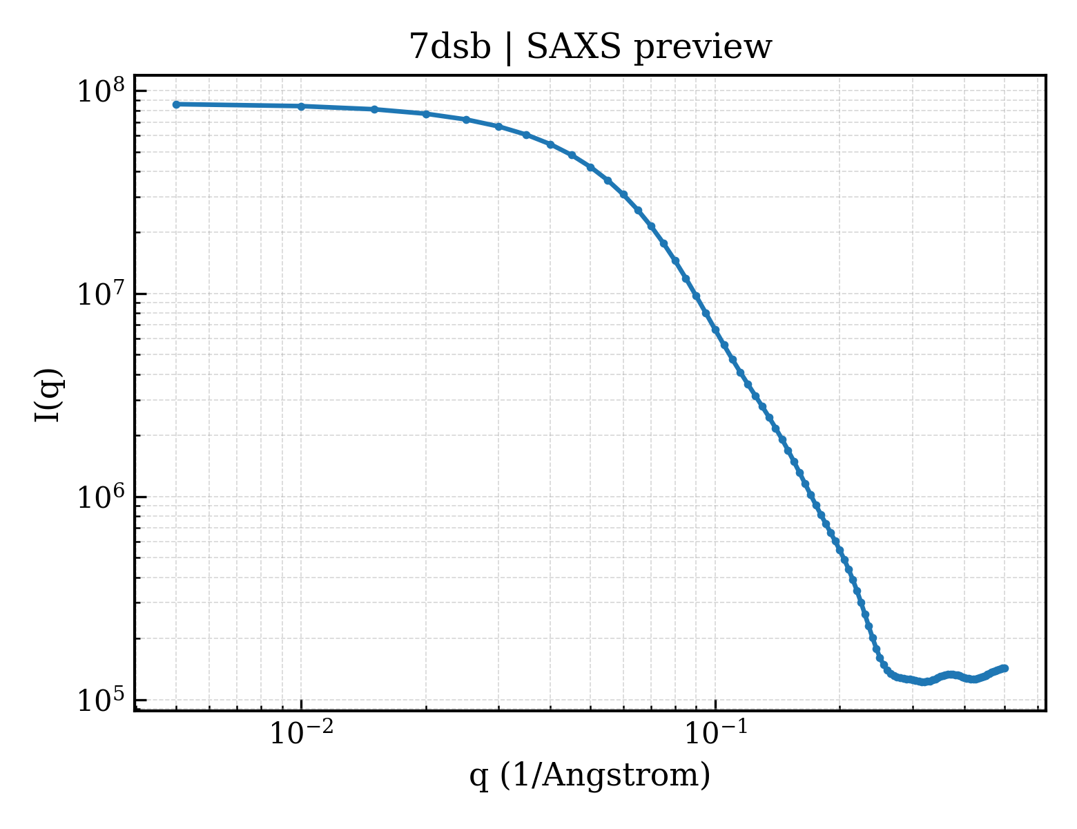
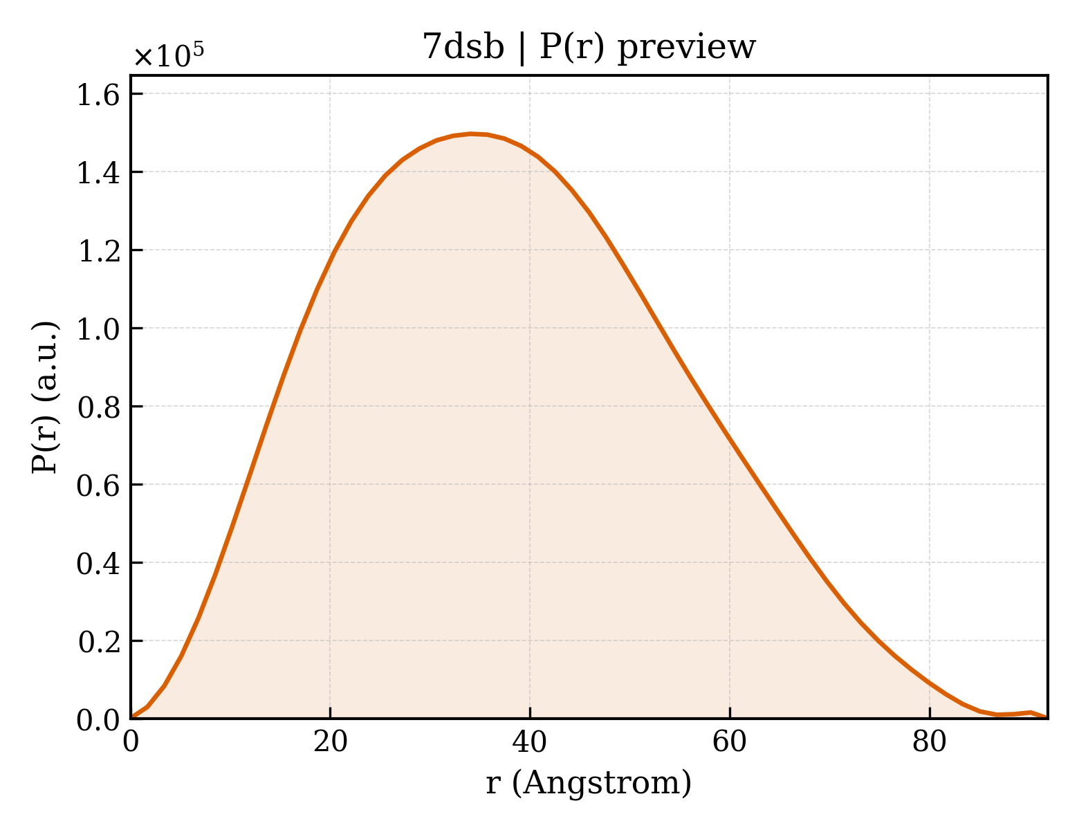
F-actin-capping protein subunit alpha-1
Gallus gallus
State in the Current Structure
| Assembly | Oligomeric State | Construct | Mutations and Modifications | Ligands, Ions and Associated Components | Method and Experimental Conditions | Structure Quality |
|---|---|---|---|---|---|---|
| 1 | Protein heterocomplex Heteromer Protein × 4 PDB declaration: tetrameric(4) Consistent with protein copy count | Chain A; UniProt 1–286 | Not recorded | F-actin-capping protein subunit beta isoforms 1 × 1 (P14315) Myotrophin × 1 (P58546) Twinfilin-1 × 1 (Q91YR1) | X-RAY DIFFRACTION X-ray crystallization conditions:VAPOR DIFFUSION, HANGING DROP;pH 7.5;293 K;10% (w/v) PEG 3350, 5% (v/v) isopropanol, 0.1M HEPES-NaOH (pH = 7.5) | Resolution 2.44 Å R-free 0.248 |
Other States of the Same Protein in the Database
Each row is a biological assembly of the same UniProt protein in another PDB entry. The “Difference from current entry” column identifies evidence-level differences; no tag means the currently parsed fields agree.
| Other PDB | Difference from Current Entry 7DSB | Assembly / Oligomeric State | Construct | Mutations and Modifications | Ligands, Ions and Non-polymers | Method and Experimental Conditions | Structure Quality |
|---|---|---|---|---|---|---|---|
| 1IZN Crystal Structure of Actin Filament Capping Protein CapZ Deposited 2002-10-10 | Different oligomeric state Different ligand/ion Different experimental conditions Different structure-quality metrics | Assembly 1 Protein heterocomplex Heteromer;Protein × 2 PDB declaration: dimeric |
Chain A
1–286(286 aa)
|
Not recorded | NO3 NITRATE ION × 2 |
X-RAY DIFFRACTION
X-ray crystallization conditions
VAPOR DIFFUSION, HANGING DROP;pH 5;293 K;PEG3350, magnesium nitrate, MES-NAOH, Jeffamine M-600, pH 5.0, VAPOR DIFFUSION, HANGING DROP, temperature 293.0K
|
Resolution 2.10 Å R-free 0.262 |
| 1IZN Crystal Structure of Actin Filament Capping Protein CapZ Deposited 2002-10-10 | Different oligomeric state Different ligand/ion Different experimental conditions Different structure-quality metrics | Assembly 2 Protein heterocomplex Heteromer;Protein × 2 PDB declaration: dimeric |
Chain C
1–286(286 aa)
|
Not recorded | NO3 NITRATE ION × 2 |
X-RAY DIFFRACTION
X-ray crystallization conditions
VAPOR DIFFUSION, HANGING DROP;pH 5;293 K;PEG3350, magnesium nitrate, MES-NAOH, Jeffamine M-600, pH 5.0, VAPOR DIFFUSION, HANGING DROP, temperature 293.0K
|
Resolution 2.10 Å R-free 0.262 |
| 2KXP Solution NMR structure of V-1 bound to capping protein (CP) Deposited 2010-05-11 | Different construct Different oligomeric state Different experimental method Different experimental conditions Different structure-quality metrics | Assembly 1 Protein heterocomplex Heteromer;Protein × 3 PDB declaration: trimeric |
Chain A
7–281(275 aa)
|
Not recorded | No recorded non-water small molecule |
SOLUTION NMR
NMR measurement conditions
pH 6.5;305 K;Ionic strength (raw mmCIF value) 0.1;Pressure ambient
NMR sample composition
0.35 mM [U-100% 15N; U-99% 2H] capping protein alpha1 subunit, 0.35 mM [U-100% 15N; U-99% 2H] capping protein beta1 subunit, 0.35 mM [U-99% 2H] V-1, 90% H2O/10% D2O | 90% H2O/10% D2O
NMR sample composition
0.35 mM [U-99% 2H] capping protein alpha1 subunit, 0.35 mM [U-99% 2H] capping protein beta1 subunit, 0.35 mM [U-100% 15N; U-99% 2H] V-1, 90% H2O/10% D2O | 90% H2O/10% D2O
NMR sample composition
0.35 mM [U-100% 15N; U-99% 2H] capping protein alpha1 subunit, 0.35 mM [U-100% 15N; U-99% 2H] capping protein beta1 subunit, 0.35 mM [U-99% 2H] M7C V-1, 90% H2O/10% D2O | 90% H2O/10% D2O
|
Resolution not provided |
| 2KZ7 Solution structure of the CARMIL CAH3a/b domain bound to capping protein (CP) Deposited 2010-06-12 | Different oligomeric state Different experimental method Different experimental conditions Different structure-quality metrics | Assembly 1 Protein heterocomplex Heteromer;Protein × 3 PDB declaration: trimeric |
Chain A
1–286(286 aa)
|
Not recorded | No recorded non-water small molecule |
SOLUTION NMR
NMR measurement conditions
pH 6.5;305 K;Ionic strength (raw mmCIF value) 100;Pressure ambient
NMR sample composition
0.35 mM [U-99% 15N; U-80% 2H] CPalpha subunit, 0.35 mM [U-99% 15N; U-80% 2H] CPbeta subunit, 0.39 mM [U-2H] CARMIL CAH3a/b domain, 93% H2O/7% D2O | 93% H2O/7% D2O
NMR sample composition
0.35 mM [U-99% 15N; U-80% 2H] CARMIL CAH3a/b domain, 0.39 mM [U-2H] CPalpha subunit, 0.39 mM [U-2H] CPbeta subunit, 93% H2O/7% D2O | 93% H2O/7% D2O
NMR sample composition
0.35 mM [U-99% 15N; U-80% 2H] CPalpha subunit, 0.35 mM [U-99% 15N; U-80% 2H] CPbeta subunit, 0.39 mM [U-2H; spin labeled at 1 or 5 postions] CARMIL CAH3a/b domain, 93% H2O/7% D2O | 93% H2O/7% D2O
|
Resolution not provided |
| 3AA0 Crystal structure of Actin Capping Protein in complex with the Cp-binding motif derived from CARMIL Deposited 2009-11-11 | Different oligomeric state Different ligand/ion Different experimental conditions Different structure-quality metrics | Assembly 1 Protein heterocomplex Heteromer;Protein × 3 PDB declaration: trimeric |
Chain A
1–286(286 aa)
|
Not recorded | CO3 CARBONATE ION × 1 |
X-RAY DIFFRACTION
X-ray crystallization conditions
VAPOR DIFFUSION, HANGING DROP;pH 6;293 K;18% PEG 400, 40MM BACL2, 100MM MES-NAOH, PH 6.0, VAPOR DIFFUSION, HANGING DROP, temperature 293.0K
|
Resolution 1.70 Å R-free 0.239 |
| 3AA1 Crystal structure of Actin capping protein in complex with the Cp-binding motif derived from CKIP-1 Deposited 2009-11-11 | Different oligomeric state Different ligand/ion Different experimental conditions Different structure-quality metrics | Assembly 1 Protein heterocomplex Heteromer;Protein × 3 PDB declaration: trimeric |
Chain A
1–286(286 aa)
|
Not recorded | MES 2-(N-MORPHOLINO)-ETHANESULFONIC ACID × 2 |
X-RAY DIFFRACTION
X-ray crystallization conditions
VAPOR DIFFUSION, HANGING DROP;pH 6;293 K;17.5% PEG 400, 30MM BACL2, 100MM MES-NAOH, PH 6.0, VAPOR DIFFUSION, HANGING DROP, temperature 293.0K
|
Resolution 1.90 Å R-free 0.263 |
| 3AA6 Crystal structure of Actin capping protein in complex with the Cp-binding motif derived from CD2AP Deposited 2009-11-11 | Different oligomeric state Different ligand/ion Different experimental conditions Different structure-quality metrics | Assembly 1 Protein heterocomplex Heteromer;Protein × 3 PDB declaration: trimeric |
Chain A
1–286(286 aa)
|
Not recorded | BA BARIUM ION × 1 |
X-RAY DIFFRACTION
X-ray crystallization conditions
VAPOR DIFFUSION, HANGING DROP;pH 6;293 K;10% PEG 400, 20MM BACL2, 100MM MES-NAOH, PH 6.5, VAPOR DIFFUSION, HANGING DROP, temperature 293.0K
|
Resolution 1.90 Å R-free 0.238 |
| 3AA7 Crystal structure of Actin capping protein Deposited 2009-11-11 | Different oligomeric state Different ligand/ion Different experimental conditions Different structure-quality metrics | Assembly 1 Protein heterocomplex Heteromer;Protein × 2 PDB declaration: dimeric |
Chain A
1–286(286 aa)
|
Not recorded | BA BARIUM ION × 1 MES 2-(N-MORPHOLINO)-ETHANESULFONIC ACID × 1 |
X-RAY DIFFRACTION
X-ray crystallization conditions
VAPOR DIFFUSION, HANGING DROP;pH 6;293 K;12.5% PEG 400, 20MM BACL2, 100MM MES-NAOH, PH 6.0, VAPOR DIFFUSION, HANGING DROP, temperature 293.0K
|
Resolution 1.90 Å R-free 0.261 |
| 3AAA Crystal Structure of Actin capping protein in complex with V-1 Deposited 2009-11-12 | Different oligomeric state Different ligand/ion Different experimental conditions Different structure-quality metrics | Assembly 1 Protein heterocomplex Heteromer;Protein × 2 PDB declaration: dimeric |
Chain A
1–286(286 aa)
|
Not recorded | IPA ISOPROPYL ALCOHOL × 2 |
X-RAY DIFFRACTION
X-ray crystallization conditions
VAPOR DIFFUSION, HANGING DROP;pH 8.35;293 K;10% PEG 4000, 20% ISOPROPANOL, 20MM EDTA, 0.1M TRIS-HCL, pH 8.35, VAPOR DIFFUSION, HANGING DROP, temperature 293.0K
|
Resolution 2.20 Å R-free 0.237 |
| 3AAE Crystal structure of Actin capping protein in complex with CARMIL fragment Deposited 2009-11-16 | Different oligomeric state Different experimental conditions Different structure-quality metrics | Assembly 1 Protein heterocomplex Heteromer;Protein × 3 PDB declaration: trimeric |
Chain A
1–286(286 aa)
|
Not recorded | No recorded non-water small molecule |
X-RAY DIFFRACTION
X-ray crystallization conditions
VAPOR DIFFUSION, HANGING DROP;pH 6.5;293 K;14% PEG 3350, 180MM TRI-AMMONIUM CITRATE, 100MM MES-NAOH, pH 6.5, VAPOR DIFFUSION, HANGING DROP, temperature 293K
|
Resolution 3.30 Å R-free 0.271 |
| 3AAE Crystal structure of Actin capping protein in complex with CARMIL fragment Deposited 2009-11-16 | Different oligomeric state Different experimental conditions Different structure-quality metrics | Assembly 2 Protein heterocomplex Heteromer;Protein × 3 PDB declaration: trimeric |
Chain C
1–286(286 aa)
|
Not recorded | No recorded non-water small molecule |
X-RAY DIFFRACTION
X-ray crystallization conditions
VAPOR DIFFUSION, HANGING DROP;pH 6.5;293 K;14% PEG 3350, 180MM TRI-AMMONIUM CITRATE, 100MM MES-NAOH, pH 6.5, VAPOR DIFFUSION, HANGING DROP, temperature 293K
|
Resolution 3.30 Å R-free 0.271 |
| 3AAE Crystal structure of Actin capping protein in complex with CARMIL fragment Deposited 2009-11-16 | Different oligomeric state Different experimental conditions Different structure-quality metrics | Assembly 3 Protein heterocomplex Heteromer;Protein × 3 PDB declaration: trimeric |
Chain E
1–286(286 aa)
|
Not recorded | No recorded non-water small molecule |
X-RAY DIFFRACTION
X-ray crystallization conditions
VAPOR DIFFUSION, HANGING DROP;pH 6.5;293 K;14% PEG 3350, 180MM TRI-AMMONIUM CITRATE, 100MM MES-NAOH, pH 6.5, VAPOR DIFFUSION, HANGING DROP, temperature 293K
|
Resolution 3.30 Å R-free 0.271 |
| 3AAE Crystal structure of Actin capping protein in complex with CARMIL fragment Deposited 2009-11-16 | Different oligomeric state Different experimental conditions Different structure-quality metrics | Assembly 4 Protein heterocomplex Heteromer;Protein × 3 PDB declaration: trimeric |
Chain G
1–286(286 aa)
|
Not recorded | No recorded non-water small molecule |
X-RAY DIFFRACTION
X-ray crystallization conditions
VAPOR DIFFUSION, HANGING DROP;pH 6.5;293 K;14% PEG 3350, 180MM TRI-AMMONIUM CITRATE, 100MM MES-NAOH, pH 6.5, VAPOR DIFFUSION, HANGING DROP, temperature 293K
|
Resolution 3.30 Å R-free 0.271 |
| 3AAE Crystal structure of Actin capping protein in complex with CARMIL fragment Deposited 2009-11-16 | Different oligomeric state Different experimental conditions Different structure-quality metrics | Assembly 5 Protein heterocomplex Heteromer;Protein × 3 PDB declaration: trimeric |
Chain I
1–286(286 aa)
|
Not recorded | No recorded non-water small molecule |
X-RAY DIFFRACTION
X-ray crystallization conditions
VAPOR DIFFUSION, HANGING DROP;pH 6.5;293 K;14% PEG 3350, 180MM TRI-AMMONIUM CITRATE, 100MM MES-NAOH, pH 6.5, VAPOR DIFFUSION, HANGING DROP, temperature 293K
|
Resolution 3.30 Å R-free 0.271 |
| 3LK2 Crystal structure of CapZ bound to the uncapping motif from CARMIL Deposited 2010-01-27 | Different oligomeric state Different experimental conditions Different structure-quality metrics | Assembly 1 Protein heterocomplex Heteromer;Protein × 3 PDB declaration: trimeric |
Chain A
1–286(286 aa)
|
Not recorded | No recorded non-water small molecule |
X-RAY DIFFRACTION
X-ray crystallization conditions
VAPOR DIFFUSION, SITTING DROP;pH 6.5;277 K;10% w/v PEG 20000, 100mM MES, pH 6.5, VAPOR DIFFUSION, SITTING DROP, temperature 277K
|
Resolution 2.20 Å R-free 0.268 |
| 3LK3 Crystal structure of CapZ bound to the CPI and CSI uncapping motifs from CARMIL Deposited 2010-01-27 | Different oligomeric state Different experimental conditions Different structure-quality metrics | Assembly 1 Protein heterocomplex Heteromer;Protein × 3 PDB declaration: trimeric |
Chain A
1–286(286 aa)
|
Not recorded | No recorded non-water small molecule |
X-RAY DIFFRACTION
X-ray crystallization conditions
VAPOR DIFFUSION, SITTING DROP;pH 8;293 K;0.2M KCl, 20% (w/v) PEG 3350 , pH 8.0, VAPOR DIFFUSION, SITTING DROP, temperature 293K
|
Resolution 2.68 Å R-free 0.248 |
| 3LK4 Crystal structure of CapZ bound to the uncapping motif from CD2AP Deposited 2010-01-27 | Different oligomeric state Different experimental conditions Different structure-quality metrics | Assembly 1 Protein heterocomplex Heteromer;Protein × 3 PDB declaration: trimeric |
Chain A
1–286(286 aa)
|
Not recorded | No recorded non-water small molecule |
X-RAY DIFFRACTION
X-ray crystallization conditions
VAPOR DIFFUSION, SITTING DROP;pH 6.5;277 K;30% PEG 400, 0.1M MES, pH 6.5, VAPOR DIFFUSION, SITTING DROP, temperature 277K
|
Resolution 1.99 Å R-free 0.272 |
| 3LK4 Crystal structure of CapZ bound to the uncapping motif from CD2AP Deposited 2010-01-27 | Different oligomeric state Different experimental conditions Different structure-quality metrics | Assembly 10 Protein heterocomplex Heteromer;Protein × 3 PDB declaration: trimeric |
Chain 1
1–286(286 aa)
|
Not recorded | No recorded non-water small molecule |
X-RAY DIFFRACTION
X-ray crystallization conditions
VAPOR DIFFUSION, SITTING DROP;pH 6.5;277 K;30% PEG 400, 0.1M MES, pH 6.5, VAPOR DIFFUSION, SITTING DROP, temperature 277K
|
Resolution 1.99 Å R-free 0.272 |
| 3LK4 Crystal structure of CapZ bound to the uncapping motif from CD2AP Deposited 2010-01-27 | Different oligomeric state Different experimental conditions Different structure-quality metrics | Assembly 11 Protein heterocomplex Heteromer;Protein × 3 PDB declaration: trimeric |
Chain 4
1–286(286 aa)
|
Not recorded | No recorded non-water small molecule |
X-RAY DIFFRACTION
X-ray crystallization conditions
VAPOR DIFFUSION, SITTING DROP;pH 6.5;277 K;30% PEG 400, 0.1M MES, pH 6.5, VAPOR DIFFUSION, SITTING DROP, temperature 277K
|
Resolution 1.99 Å R-free 0.272 |
| 3LK4 Crystal structure of CapZ bound to the uncapping motif from CD2AP Deposited 2010-01-27 | Different oligomeric state Different experimental conditions Different structure-quality metrics | Assembly 12 Protein heterocomplex Heteromer;Protein × 3 PDB declaration: trimeric |
Chain 7
1–286(286 aa)
|
Not recorded | No recorded non-water small molecule |
X-RAY DIFFRACTION
X-ray crystallization conditions
VAPOR DIFFUSION, SITTING DROP;pH 6.5;277 K;30% PEG 400, 0.1M MES, pH 6.5, VAPOR DIFFUSION, SITTING DROP, temperature 277K
|
Resolution 1.99 Å R-free 0.272 |
| 3LK4 Crystal structure of CapZ bound to the uncapping motif from CD2AP Deposited 2010-01-27 | Different oligomeric state Different experimental conditions Different structure-quality metrics | Assembly 2 Protein heterocomplex Heteromer;Protein × 3 PDB declaration: trimeric |
Chain D
1–286(286 aa)
|
Not recorded | No recorded non-water small molecule |
X-RAY DIFFRACTION
X-ray crystallization conditions
VAPOR DIFFUSION, SITTING DROP;pH 6.5;277 K;30% PEG 400, 0.1M MES, pH 6.5, VAPOR DIFFUSION, SITTING DROP, temperature 277K
|
Resolution 1.99 Å R-free 0.272 |
| 3LK4 Crystal structure of CapZ bound to the uncapping motif from CD2AP Deposited 2010-01-27 | Different oligomeric state Different experimental conditions Different structure-quality metrics | Assembly 3 Protein heterocomplex Heteromer;Protein × 3 PDB declaration: trimeric |
Chain G
1–286(286 aa)
|
Not recorded | No recorded non-water small molecule |
X-RAY DIFFRACTION
X-ray crystallization conditions
VAPOR DIFFUSION, SITTING DROP;pH 6.5;277 K;30% PEG 400, 0.1M MES, pH 6.5, VAPOR DIFFUSION, SITTING DROP, temperature 277K
|
Resolution 1.99 Å R-free 0.272 |
| 3LK4 Crystal structure of CapZ bound to the uncapping motif from CD2AP Deposited 2010-01-27 | Different oligomeric state Different experimental conditions Different structure-quality metrics | Assembly 4 Protein heterocomplex Heteromer;Protein × 3 PDB declaration: trimeric |
Chain J
1–286(286 aa)
|
Not recorded | No recorded non-water small molecule |
X-RAY DIFFRACTION
X-ray crystallization conditions
VAPOR DIFFUSION, SITTING DROP;pH 6.5;277 K;30% PEG 400, 0.1M MES, pH 6.5, VAPOR DIFFUSION, SITTING DROP, temperature 277K
|
Resolution 1.99 Å R-free 0.272 |
| 3LK4 Crystal structure of CapZ bound to the uncapping motif from CD2AP Deposited 2010-01-27 | Different oligomeric state Different experimental conditions Different structure-quality metrics | Assembly 5 Protein heterocomplex Heteromer;Protein × 3 PDB declaration: trimeric |
Chain M
1–286(286 aa)
|
Not recorded | No recorded non-water small molecule |
X-RAY DIFFRACTION
X-ray crystallization conditions
VAPOR DIFFUSION, SITTING DROP;pH 6.5;277 K;30% PEG 400, 0.1M MES, pH 6.5, VAPOR DIFFUSION, SITTING DROP, temperature 277K
|
Resolution 1.99 Å R-free 0.272 |
| 3LK4 Crystal structure of CapZ bound to the uncapping motif from CD2AP Deposited 2010-01-27 | Different oligomeric state Different experimental conditions Different structure-quality metrics | Assembly 6 Protein heterocomplex Heteromer;Protein × 3 PDB declaration: trimeric |
Chain P
1–286(286 aa)
|
Not recorded | No recorded non-water small molecule |
X-RAY DIFFRACTION
X-ray crystallization conditions
VAPOR DIFFUSION, SITTING DROP;pH 6.5;277 K;30% PEG 400, 0.1M MES, pH 6.5, VAPOR DIFFUSION, SITTING DROP, temperature 277K
|
Resolution 1.99 Å R-free 0.272 |
| 3LK4 Crystal structure of CapZ bound to the uncapping motif from CD2AP Deposited 2010-01-27 | Different oligomeric state Different experimental conditions Different structure-quality metrics | Assembly 7 Protein heterocomplex Heteromer;Protein × 3 PDB declaration: trimeric |
Chain S
1–286(286 aa)
|
Not recorded | No recorded non-water small molecule |
X-RAY DIFFRACTION
X-ray crystallization conditions
VAPOR DIFFUSION, SITTING DROP;pH 6.5;277 K;30% PEG 400, 0.1M MES, pH 6.5, VAPOR DIFFUSION, SITTING DROP, temperature 277K
|
Resolution 1.99 Å R-free 0.272 |
| 3LK4 Crystal structure of CapZ bound to the uncapping motif from CD2AP Deposited 2010-01-27 | Different oligomeric state Different experimental conditions Different structure-quality metrics | Assembly 8 Protein heterocomplex Heteromer;Protein × 3 PDB declaration: trimeric |
Chain V
1–286(286 aa)
|
Not recorded | No recorded non-water small molecule |
X-RAY DIFFRACTION
X-ray crystallization conditions
VAPOR DIFFUSION, SITTING DROP;pH 6.5;277 K;30% PEG 400, 0.1M MES, pH 6.5, VAPOR DIFFUSION, SITTING DROP, temperature 277K
|
Resolution 1.99 Å R-free 0.272 |
| 3LK4 Crystal structure of CapZ bound to the uncapping motif from CD2AP Deposited 2010-01-27 | Different oligomeric state Different experimental conditions Different structure-quality metrics | Assembly 9 Protein heterocomplex Heteromer;Protein × 3 PDB declaration: trimeric |
Chain Y
1–286(286 aa)
|
Not recorded | No recorded non-water small molecule |
X-RAY DIFFRACTION
X-ray crystallization conditions
VAPOR DIFFUSION, SITTING DROP;pH 6.5;277 K;30% PEG 400, 0.1M MES, pH 6.5, VAPOR DIFFUSION, SITTING DROP, temperature 277K
|
Resolution 1.99 Å R-free 0.272 |
| 7DS2 Crystal structure of actin capping protein in complex with twinflin-1 C-terminus tail Deposited 2020-12-30 | Different oligomeric state Different experimental conditions Different structure-quality metrics | Assembly 1 Protein heterocomplex Heteromer;Protein × 3 PDB declaration: trimeric |
Chain A
1–286(286 aa)
|
Not recorded | No recorded non-water small molecule |
X-RAY DIFFRACTION
X-ray crystallization conditions
VAPOR DIFFUSION, HANGING DROP;pH 7;293 K;22.5% (w/v) PEG 400, 0.1M HEPES-NaOH (pH = 7.0)
|
Resolution 1.95 Å R-free 0.183 |
| 7DS3 Crystal structure of actin capping protein in complex with twinflin-2 C-terminus tail Deposited 2020-12-30 | Different oligomeric state Different experimental conditions Different structure-quality metrics | Assembly 1 Protein heterocomplex Heteromer;Protein × 3 PDB declaration: trimeric |
Chain A
1–286(286 aa)
|
Not recorded | No recorded non-water small molecule |
X-RAY DIFFRACTION
X-ray crystallization conditions
VAPOR DIFFUSION, HANGING DROP;293 K;5% (w/v) PEG 3350, 50mM Tris-HCl (pH = 7.0)
|
Resolution 2.09 Å R-free 0.251 |
| 7DS4 Crystal structure of actin capping protein in complex with twinflin-1 C-terminus tail F323K mutant Deposited 2020-12-30 | Different oligomeric state Different experimental conditions Different structure-quality metrics | Assembly 1 Protein heterocomplex Heteromer;Protein × 3 PDB declaration: trimeric |
Chain A
1–286(286 aa)
|
Not recorded | No recorded non-water small molecule |
X-RAY DIFFRACTION
X-ray crystallization conditions
VAPOR DIFFUSION, HANGING DROP;293 K;5% (w/v) PEG 3350, 5% (v/v) ethanol, 50mM Tris-HCl (pH = 7.0)
|
Resolution 1.85 Å R-free 0.259 |
| 7DS6 Crystal structure of actin capping protein in complex with twinflin-1/CD2AP CPI chimera peptide (TWN-CDC) Deposited 2020-12-30 | Different oligomeric state Different experimental conditions Different structure-quality metrics | Assembly 1 Insufficient information Heteromer;Protein × 3 PDB declaration: trimeric |
Chain A
1–286(286 aa)
|
Not recorded | No recorded non-water small molecule |
X-RAY DIFFRACTION
X-ray crystallization conditions
VAPOR DIFFUSION, HANGING DROP;pH 7;293 K;5% (w/v) PEG 3350, 5% (v/v) ethanol, 50mM Tris-HCl (pH = 7.0)
|
Resolution 1.69 Å R-free 0.253 |
| 7DS8 Crystal structure of actin capping protein in complex with twinflin-1/CD2AP CPI chimera peptide (CDN-TWC) Deposited 2020-12-30 | Different oligomeric state Different experimental conditions Different structure-quality metrics | Assembly 1 Insufficient information Heteromer;Protein × 3 PDB declaration: trimeric |
Chain A
1–286(286 aa)
|
Not recorded | No recorded non-water small molecule |
X-RAY DIFFRACTION
X-ray crystallization conditions
VAPOR DIFFUSION, HANGING DROP;pH 7;293 K;4% (w/v) PEG 3350, 50mM Tris-HCl (pH = 7.0)
|
Resolution 1.95 Å R-free 0.233 |
| 7DSA Crystal structure of actin capping protein in complex with V-1 (space group P62) Deposited 2020-12-30 | Different oligomeric state Different ligand/ion Different experimental conditions Different structure-quality metrics | Assembly 1 Protein heterocomplex Heteromer;Protein × 3 PDB declaration: trimeric |
Chain A
1–286(286 aa)
|
Not recorded | MG MAGNESIUM ION × 2 |
X-RAY DIFFRACTION
X-ray crystallization conditions
VAPOR DIFFUSION, HANGING DROP;293 K;20% (w/v) PEG 3350, 0.2M Magnesium Acetate
|
Resolution 2.80 Å R-free 0.254 |
| 9BLI Crystal structure of Actin capping protein in complex with a fragment of Legionella pneumophila RavB Deposited 2024-04-30 | Different oligomeric state Different ligand/ion Different experimental conditions Different structure-quality metrics | Assembly 1 Protein heterocomplex Heteromer;Protein × 3 PDB declaration: trimeric |
Chain A
1–286(286 aa)
|
Not recorded | FMT FORMIC ACID × 3 |
X-RAY DIFFRACTION
X-ray crystallization conditions
VAPOR DIFFUSION, SITTING DROP;pH 8;293 K;10 mM Tris pH 8.0, 0.2 M ammonium formate, 0.15 M NaCl, 21% w/v PEG 3350, 30% ethylene glycol
|
Resolution 2.00 Å R-free 0.208 |
19 other PDB entries and 35 assemblies. Open the comparison page and filter oligomeric states
View Construct and Data Evidence
| UniProt name | CAZA1_CHICK |
| Isoform | — |
| PDB entities | 1 |
| Chains and sequence ranges | Author chain A; PDBConstruct 1–286; UniProt 1–286 |