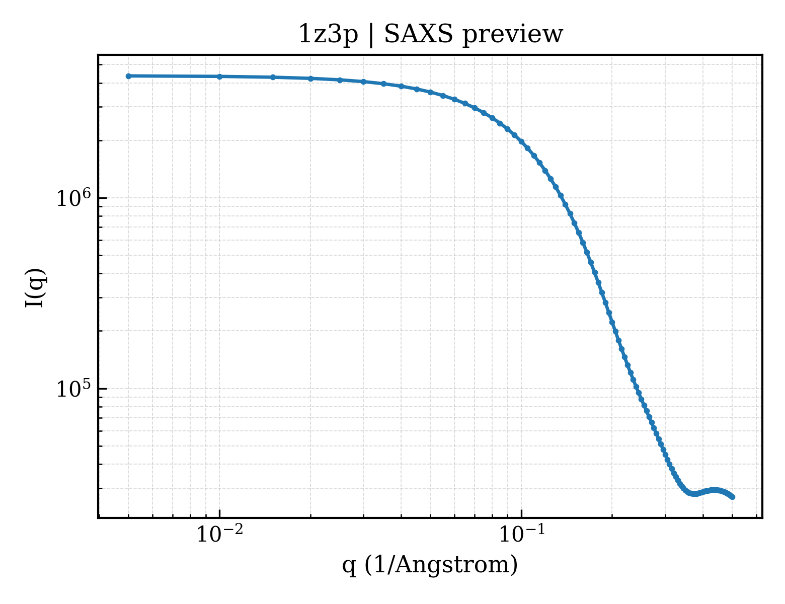
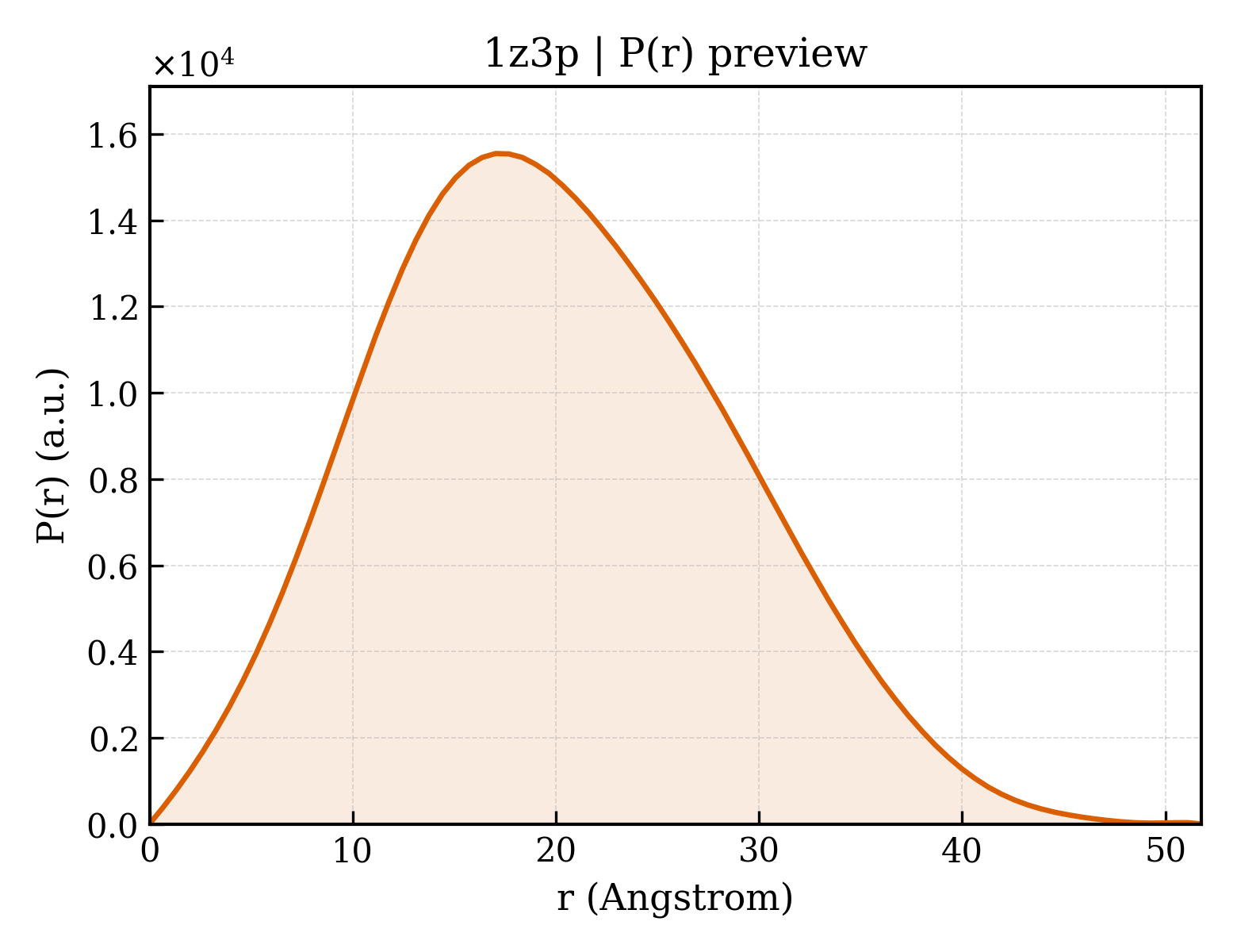
Ribonuclease pancreatic, S-Peptide
OrganismNot specified
State in the Current Structure
| Assembly | Oligomeric State | Construct | Mutations and Modifications | Ligands, Ions and Associated Components | Method and Experimental Conditions | Structure Quality |
|---|---|---|---|---|---|---|
| 1 | Protein homooligomer Homooligomer Protein × 2 PDB declaration: dimeric(2) Consistent with protein copy count | Chain E; UniProt 47–150 Chain S; UniProt 27–41 | Mutation:M13NVA Non-standard monomer:Yes (specific site not provided by mmCIF) | SO4 SULFATE ION × 2 | X-RAY DIFFRACTION X-ray crystallization conditions:Batch method;pH 5.2;293 K;ammonium sulfate, cesium chloride, sodium acetate, pH 5.2, Batch method, temperature 293K | Resolution 2.00 Å R-free 0.233 |
Other States of the Same Protein in the Database
Each row is a biological assembly of the same UniProt protein in another PDB entry. The “Difference from current entry” column identifies evidence-level differences; no tag means the currently parsed fields agree.
| Other PDB | Difference from Current Entry 1Z3P | Assembly / Oligomeric State | Construct | Mutations and Modifications | Ligands, Ions and Non-polymers | Method and Experimental Conditions | Structure Quality |
|---|---|---|---|---|---|---|---|
| 1A2W CRYSTAL STRUCTURE OF A 3D DOMAIN-SWAPPED DIMER OF BOVINE PANCREATIC RIBONUCLEASE A Deposited 1998-01-12 | Different construct Different mutation/modification Different ligand/ion Different experimental conditions Different structure-quality metrics | Assembly 1 Protein homooligomer Homooligomer;Protein × 2 PDB declaration: dimeric |
Chain A
27–150(124 aa)
Fragment:SWAPPED HELICAL DOMAIN
Chain B
27–150(124 aa)
Fragment:SWAPPED HELICAL DOMAIN
|
Not recorded | CL CHLORIDE ION × 2 SO4 SULFATE ION × 1 |
X-RAY DIFFRACTION
X-ray crystallization conditions
pH 7.5;100 MM PHOSPHATE BUFFER (PH 7.5) 1.6 M AMMONIUM SULFATE 0.6 M NACL 2% PEG400
|
Resolution 2.10 Å R-free 0.256 |
| 1A5P C[40,95]A VARIANT OF BOVINE PANCREATIC RIBONUCLEASE A Deposited 1998-02-17 | Different construct Different mutation/modification Different oligomeric state Different ligand/ion Different experimental conditions Different structure-quality metrics | Assembly 1 Protein monomer Monomer;Protein × 1 PDB declaration: monomeric |
Chain A
27–150(124 aa)
|
Mutation:C40A, C95A | SO4 SULFATE ION × 1 |
X-RAY DIFFRACTION
X-ray crystallization conditions
pH 6;PROTEIN WAS CRYSTALLIZED FROM 100 MM CACODYLATE PH 6.0, 2.2 M CSCL, AND 1.5 M AMM SO4. THEN IT WAS SOAKED IN 100 MM CACODYLATE PH 6.0, 80% AMM SO4 FOR 24 HOURS TO REMOVE THE CS IONS.
|
Resolution 1.60 Å R-free 0.272 |
| 1A5Q P93A VARIANT OF BOVINE PANCREATIC RIBONUCLEASE A Deposited 1998-02-17 | Different construct Different mutation/modification Different oligomeric state Different ligand/ion Different experimental conditions Different structure-quality metrics | Assembly 1 Protein monomer Monomer;Protein × 1 PDB declaration: monomeric |
Chain A
27–150(124 aa)
|
Mutation:P93A | SO4 SULFATE ION × 1 |
X-RAY DIFFRACTION
X-ray crystallization conditions
pH 6.8;PROTEIN WAS CRYSTALLIZED FROM 100 MM HEPES PH 7.0, 2.2 M CSCL, AND 1.2 M AMM SO4. THEN IT WAS SOAKED IN 100 MM HEPES PH 7.0, 80% AMM SO4 FOR 24 HOURS TO REMOVE THE CS IONS., pH 6.8
|
Resolution 2.30 Å R-free 0.273 |
| 1AFK CRYSTAL STRUCTURE OF RIBONUCLEASE A IN COMPLEX WITH 5'-DIPHOSPHOADENOSINE-3'-PHOSPHATE Deposited 1997-03-08 | Different construct Different mutation/modification Different oligomeric state Different ligand/ion Different experimental conditions Different structure-quality metrics | Assembly 1 Protein monomer Monomer;Protein × 1 PDB declaration: monomeric |
Chain A
27–150(124 aa)
|
Not recorded | PAP 3'-PHOSPHATE-ADENOSINE-5'-DIPHOSPHATE × 1 |
X-RAY DIFFRACTION
X-ray crystallization conditions
pH 5.5;289 K;PROTEIN WAS CRYSTALLIZED FROM 20% PEG 4000, 20 MM SODIUM CITRATE, PH 5.5 AND THEN SOAKED WITH 1MM 5' DIPHOSPHOADENOSINE 3'-PHOSPHATE FOR 2.5 HRS AT 16 C, temperature 289K
|
Resolution 1.70 Å R-free 0.266 |
| 1AFK CRYSTAL STRUCTURE OF RIBONUCLEASE A IN COMPLEX WITH 5'-DIPHOSPHOADENOSINE-3'-PHOSPHATE Deposited 1997-03-08 | Different construct Different mutation/modification Different oligomeric state Different ligand/ion Different experimental conditions Different structure-quality metrics | Assembly 2 Protein monomer Monomer;Protein × 1 PDB declaration: monomeric |
Chain B
27–150(124 aa)
|
Not recorded | PAP 3'-PHOSPHATE-ADENOSINE-5'-DIPHOSPHATE × 1 |
X-RAY DIFFRACTION
X-ray crystallization conditions
pH 5.5;289 K;PROTEIN WAS CRYSTALLIZED FROM 20% PEG 4000, 20 MM SODIUM CITRATE, PH 5.5 AND THEN SOAKED WITH 1MM 5' DIPHOSPHOADENOSINE 3'-PHOSPHATE FOR 2.5 HRS AT 16 C, temperature 289K
|
Resolution 1.70 Å R-free 0.266 |
| 1AFL RIBONUCLEASE A IN COMPLEX WITH 5'-DIPHOSPHOADENOSINE 2'-PHOSPHATE AT 1.7 ANGSTROM RESOLUTION Deposited 1997-03-08 | Different construct Different mutation/modification Different oligomeric state Different ligand/ion Different experimental conditions Different structure-quality metrics | Assembly 1 Protein monomer Monomer;Protein × 1 PDB declaration: monomeric |
Chain A
27–150(124 aa)
|
Not recorded | ATR 2'-MONOPHOSPHOADENOSINE-5'-DIPHOSPHATE × 1 CIT CITRIC ACID × 1 |
X-RAY DIFFRACTION
X-ray crystallization conditions
pH 5.5;289 K;PROTEIN WAS CRYSTALLIZED FROM 10% PEG 4000, 10 MM SODIUM CITRATE, PH 5.5 AND THEN SOAKED WITH 10 MM 5-DIPHOSPHOADENOSINE 2'- PHOSPHATE FOR 3 HRS AT 16 C, temperature 289K
|
Resolution 1.70 Å R-free 0.278 |
| 1AFL RIBONUCLEASE A IN COMPLEX WITH 5'-DIPHOSPHOADENOSINE 2'-PHOSPHATE AT 1.7 ANGSTROM RESOLUTION Deposited 1997-03-08 | Different construct Different mutation/modification Different oligomeric state Different ligand/ion Different experimental conditions Different structure-quality metrics | Assembly 2 Protein monomer Monomer;Protein × 1 PDB declaration: monomeric |
Chain B
27–150(124 aa)
|
Not recorded | ATR 2'-MONOPHOSPHOADENOSINE-5'-DIPHOSPHATE × 1 CIT CITRIC ACID × 1 |
X-RAY DIFFRACTION
X-ray crystallization conditions
pH 5.5;289 K;PROTEIN WAS CRYSTALLIZED FROM 10% PEG 4000, 10 MM SODIUM CITRATE, PH 5.5 AND THEN SOAKED WITH 10 MM 5-DIPHOSPHOADENOSINE 2'- PHOSPHATE FOR 3 HRS AT 16 C, temperature 289K
|
Resolution 1.70 Å R-free 0.278 |
| 1AFU STRUCTURE OF RIBONUCLEASE A AT 2.0 ANGSTROMS FROM MONOCLINIC CRYSTALS Deposited 1997-03-14 | Different construct Different mutation/modification Different oligomeric state Different ligand/ion Different experimental conditions Different structure-quality metrics | Assembly 1 Protein monomer Monomer;Protein × 1 PDB declaration: monomeric |
Chain A
27–150(124 aa)
|
Not recorded | No recorded non-water small molecule |
X-RAY DIFFRACTION
X-ray crystallization conditions
pH 5.5;PROTEIN WAS CRYSTALLIZED FROM 20 MM SODIUM CITRATE BUFFER PH 5.5 AND 20 % PEG 4000.
|
Resolution 2.00 Å R-free 0.285 |
| 1AFU STRUCTURE OF RIBONUCLEASE A AT 2.0 ANGSTROMS FROM MONOCLINIC CRYSTALS Deposited 1997-03-14 | Different construct Different mutation/modification Different oligomeric state Different ligand/ion Different experimental conditions Different structure-quality metrics | Assembly 2 Protein monomer Monomer;Protein × 1 PDB declaration: monomeric |
Chain B
27–150(124 aa)
|
Not recorded | No recorded non-water small molecule |
X-RAY DIFFRACTION
X-ray crystallization conditions
pH 5.5;PROTEIN WAS CRYSTALLIZED FROM 20 MM SODIUM CITRATE BUFFER PH 5.5 AND 20 % PEG 4000.
|
Resolution 2.00 Å R-free 0.285 |
| 1AQP RIBONUCLEASE A COPPER COMPLEX Deposited 1997-07-31 | Different construct Different mutation/modification Different oligomeric state Different ligand/ion Different experimental conditions Different structure-quality metrics | Assembly 1 Protein monomer Monomer;Protein × 1 PDB declaration: monomeric |
Chain A
27–150(124 aa)
|
Not recorded | CU COPPER (II) ION × 2 |
X-RAY DIFFRACTION
X-ray crystallization conditions
pH 5.5;ENZYME WAS CRYSTALLIZED FROM MPD WITH 6 MOLAR EXCESS OF COPPER, pH 5.5
|
Resolution 2.00 Å |
| 1B6V CRYSTAL STRUCTURE OF A HYBRID BETWEEN RIBONUCLEASE A AND BOVINE SEMINAL RIBONUCLEASE Deposited 1999-01-18 | Different construct Different mutation/modification Different oligomeric state Different ligand/ion Different experimental conditions Different structure-quality metrics | Assembly 1 Protein monomer Monomer;Protein × 1 PDB declaration: monomeric |
Chain A
27–150(124 aa)
|
Mutation:Q55K, N62K, A64T, Y76K, S80R, E111G, N113K | No recorded non-water small molecule |
X-RAY DIFFRACTION
X-ray crystallization conditions
pH 4.3;pH 4.3
|
Resolution 2.00 Å R-free 0.256 |
| 1B6V CRYSTAL STRUCTURE OF A HYBRID BETWEEN RIBONUCLEASE A AND BOVINE SEMINAL RIBONUCLEASE Deposited 1999-01-18 | Different construct Different mutation/modification Different oligomeric state Different ligand/ion Different experimental conditions Different structure-quality metrics | Assembly 2 Protein monomer Monomer;Protein × 1 PDB declaration: monomeric |
Chain B
27–150(124 aa)
|
Mutation:Q55K, N62K, A64T, Y76K, S80R, E111G, N113K | No recorded non-water small molecule |
X-RAY DIFFRACTION
X-ray crystallization conditions
pH 4.3;pH 4.3
|
Resolution 2.00 Å R-free 0.256 |
| 1BEL HYDROLASE PHOSPHORIC DIESTER, RNA Deposited 1995-12-21 | Different construct Different mutation/modification Different oligomeric state Different ligand/ion Different experimental conditions Different structure-quality metrics | Assembly 1 Protein monomer Monomer;Protein × 1 PDB declaration: monomeric |
Chain A
27–150(124 aa)
|
Not recorded | SO4 SULFATE ION × 2 MOH METHANOL × 4 |
X-RAY DIFFRACTION
X-ray crystallization conditions
pH 4.5;60% METHANOL, pH 4.50
|
Resolution 1.60 Å |
| 1BZQ COMPLEX OF A DROMEDARY SINGLE-DOMAIN VHH ANTIBODY FRAGMENT WITH RNASE A Deposited 1998-11-03 | Different construct Different mutation/modification Different oligomeric state Different ligand/ion Different experimental conditions Different structure-quality metrics | Assembly 1 Protein heterocomplex Heteromer;Protein × 2 PDB declaration: dimeric |
Chain A
27–150(124 aa)
|
Not recorded | PO4 PHOSPHATE ION × 1 |
X-RAY DIFFRACTION
X-ray crystallization conditions
VAPOR DIFFUSION, HANGING DROP;pH 6.4;HANGING DROP METHOD. 5 MG/ML OF THE COMPLEX RESERVOIR SOLUTION IS 0.1 M POTASSIUM PHOSPHATE PH 6.4 15 TO 20% W/V PEG 8000, vapor diffusion - hanging drop
|
Resolution 2.80 Å R-free 0.282 |
| 1BZQ COMPLEX OF A DROMEDARY SINGLE-DOMAIN VHH ANTIBODY FRAGMENT WITH RNASE A Deposited 1998-11-03 | Different construct Different mutation/modification Different oligomeric state Different ligand/ion Different experimental conditions Different structure-quality metrics | Assembly 2 Protein heterocomplex Heteromer;Protein × 2 PDB declaration: dimeric |
Chain B
27–150(124 aa)
|
Not recorded | PO4 PHOSPHATE ION × 1 |
X-RAY DIFFRACTION
X-ray crystallization conditions
VAPOR DIFFUSION, HANGING DROP;pH 6.4;HANGING DROP METHOD. 5 MG/ML OF THE COMPLEX RESERVOIR SOLUTION IS 0.1 M POTASSIUM PHOSPHATE PH 6.4 15 TO 20% W/V PEG 8000, vapor diffusion - hanging drop
|
Resolution 2.80 Å R-free 0.282 |
| 1BZQ COMPLEX OF A DROMEDARY SINGLE-DOMAIN VHH ANTIBODY FRAGMENT WITH RNASE A Deposited 1998-11-03 | Different construct Different mutation/modification Different oligomeric state Different ligand/ion Different experimental conditions Different structure-quality metrics | Assembly 3 Protein heterocomplex Heteromer;Protein × 2 PDB declaration: dimeric |
Chain C
27–150(124 aa)
|
Not recorded | PO4 PHOSPHATE ION × 1 |
X-RAY DIFFRACTION
X-ray crystallization conditions
VAPOR DIFFUSION, HANGING DROP;pH 6.4;HANGING DROP METHOD. 5 MG/ML OF THE COMPLEX RESERVOIR SOLUTION IS 0.1 M POTASSIUM PHOSPHATE PH 6.4 15 TO 20% W/V PEG 8000, vapor diffusion - hanging drop
|
Resolution 2.80 Å R-free 0.282 |
| 1BZQ COMPLEX OF A DROMEDARY SINGLE-DOMAIN VHH ANTIBODY FRAGMENT WITH RNASE A Deposited 1998-11-03 | Different construct Different mutation/modification Different oligomeric state Different ligand/ion Different experimental conditions Different structure-quality metrics | Assembly 4 Protein heterocomplex Heteromer;Protein × 2 PDB declaration: dimeric |
Chain D
27–150(124 aa)
|
Not recorded | PO4 PHOSPHATE ION × 1 |
X-RAY DIFFRACTION
X-ray crystallization conditions
VAPOR DIFFUSION, HANGING DROP;pH 6.4;HANGING DROP METHOD. 5 MG/ML OF THE COMPLEX RESERVOIR SOLUTION IS 0.1 M POTASSIUM PHOSPHATE PH 6.4 15 TO 20% W/V PEG 8000, vapor diffusion - hanging drop
|
Resolution 2.80 Å R-free 0.282 |
| 1C0B BOVINE PANCREATIC RIBONUCLEASE A DESICCATED FOR 2.5 DAYS Deposited 1999-07-15 | Different construct Different mutation/modification Different oligomeric state Different ligand/ion Different experimental conditions Different structure-quality metrics | Assembly 1 Protein monomer Monomer;Protein × 1 PDB declaration: monomeric |
Chain A
1–128(128 aa)
|
Not recorded | SO4 SULFATE ION × 3 |
X-RAY DIFFRACTION
X-ray crystallization conditions
SMALL TUBES;pH 4.5;295 K;58% methanol (v/v), pH 4.5, SMALL TUBES, temperature 295K
|
Resolution 1.90 Å R-free 0.272 |
| 1C0C BOVINE PANCREATIC RIBONUCLEASE A DESICCATED FOR 4.0 DAYS Deposited 1999-07-15 | Different construct Different mutation/modification Different oligomeric state Different ligand/ion Different experimental conditions Different structure-quality metrics | Assembly 1 Protein monomer Monomer;Protein × 1 PDB declaration: monomeric |
Chain A
1–128(128 aa)
|
Not recorded | SO4 SULFATE ION × 3 |
X-RAY DIFFRACTION
X-ray crystallization conditions
SMALL TUBES;pH 4.5;295 K;58% methanol (v/v), pH 4.5, SMALL TUBES, temperature 295K
|
Resolution 2.00 Å R-free 0.268 |
| 1C8W THR45GLY VARIANT OF RIBONUCLEASE A Deposited 1999-07-30 | Different construct Different mutation/modification Different oligomeric state Different ligand/ion Different experimental conditions Different structure-quality metrics | Assembly 1 Protein monomer Monomer;Protein × 1 PDB declaration: monomeric |
Chain A
2–125(124 aa)
Fragment:RNASE A
|
Mutation:T45G | ACT ACETATE ION × 1 CL CHLORIDE ION × 3 |
X-RAY DIFFRACTION
X-ray crystallization conditions
VAPOR DIFFUSION, HANGING DROP;pH 5.6;293 K;Ammonium sulfate, sodium chloride, sodium acetate, pH 5.6, VAPOR DIFFUSION, HANGING DROP, temperature 293K
|
Resolution 1.80 Å |
| 1C9V H12A VARIANT OF RIBONUCLEASE A Deposited 1999-08-03 | Different construct Different mutation/modification Different oligomeric state Different ligand/ion Different experimental conditions Different structure-quality metrics | Assembly 1 Protein monomer Monomer;Protein × 1 PDB declaration: monomeric |
Chain A
27–150(124 aa)
|
Mutation:H12A | CL CHLORIDE ION × 2 |
X-RAY DIFFRACTION
X-ray crystallization conditions
VAPOR DIFFUSION, HANGING DROP;pH 6;293 K;30% ammonium sulfate, 50% sodium chloride, 100mM sodium acetate, pH 6.0, VAPOR DIFFUSION, HANGING DROP, temperature 293K
|
Resolution 1.70 Å |
| 1C9X H119A VARIANT OF RIBONUCLEASE A Deposited 1999-08-03 | Different construct Different mutation/modification Different oligomeric state Different ligand/ion Different experimental conditions Different structure-quality metrics | Assembly 1 Protein monomer Monomer;Protein × 1 PDB declaration: monomeric |
Chain A
27–150(124 aa)
Fragment:RIBONUCLEASE A
|
Mutation:H119A | CL CHLORIDE ION × 3 |
X-RAY DIFFRACTION
X-ray crystallization conditions
VAPOR DIFFUSION, HANGING DROP;pH 6;293 K;29% ammonium sulfate, 45% sodium chloride, 100mM sodium acetate, pH 6.0, VAPOR DIFFUSION, HANGING DROP, temperature 293K
|
Resolution 1.80 Å |
| 1CJQ X-RAY CRYSTALLOGRAPHIC STUDIES OF THE DENATURATION OF THE DENATURATION OF RIBONUCLEASE S. Deposited 1999-04-19 | Different construct Different mutation/modification Different ligand/ion Different experimental conditions Different structure-quality metrics | Assembly 1 Protein homooligomer Homooligomer;Protein × 2 PDB declaration: dimeric |
Chain A
27–41(15 aa)
Fragment:S PEPTIDE
Chain B
50–150(101 aa)
Fragment:S PROTEIN
|
Not recorded | SO4 SULFATE ION × 1 |
X-RAY DIFFRACTION
X-ray crystallization conditions
pH 4.75;THE RNASE S CRYSTAL HAS BEEN STABILIZED BY GLUTARALDEHYDE CROSSLINKING AND
THEN THE CRYSTAL HAS BEEN SOAKED IN 5M UREA., pH 4.75
DATA COLLECTED ON A CRYSTAL SOAKED IN 5M UREA. THE CRYSTAL
WAS STABILIZED BY GLUTARALDEHYDE CROSSLINKING
|
Resolution 3.00 Å R-free 0.253 |
| 1CJQ X-RAY CRYSTALLOGRAPHIC STUDIES OF THE DENATURATION OF THE DENATURATION OF RIBONUCLEASE S. Deposited 1999-04-19 | Different construct Different mutation/modification Different oligomeric state Different experimental conditions Different structure-quality metrics | Assembly 2 Protein homooligomer Homooligomer;Protein × 4 PDB declaration: tetrameric |
Chain A
27–41(15 aa)
Fragment:S PEPTIDE
Chain B
50–150(101 aa)
Fragment:S PROTEIN
|
Not recorded | SO4 SULFATE ION × 2 |
X-RAY DIFFRACTION
X-ray crystallization conditions
pH 4.75;THE RNASE S CRYSTAL HAS BEEN STABILIZED BY GLUTARALDEHYDE CROSSLINKING AND
THEN THE CRYSTAL HAS BEEN SOAKED IN 5M UREA., pH 4.75
DATA COLLECTED ON A CRYSTAL SOAKED IN 5M UREA. THE CRYSTAL
WAS STABILIZED BY GLUTARALDEHYDE CROSSLINKING
|
Resolution 3.00 Å R-free 0.253 |
| 1CJR X-RAY CRYSTALLOGRAPHIC STUDIES OF DENATURATION IN RIBONUCLEASE S Deposited 1999-04-19 | Different construct Different mutation/modification Different ligand/ion Different experimental conditions Different structure-quality metrics | Assembly 1 Protein homooligomer Homooligomer;Protein × 2 PDB declaration: dimeric |
Chain A
27–41(15 aa)
Fragment:S PEPTIDE
Chain B
50–150(101 aa)
Fragment:S PROTEIN
|
Not recorded | SO4 SULFATE ION × 1 |
X-RAY DIFFRACTION
X-ray crystallization conditions
pH 2;pH 2.0
THE CRYSTALS WERE SOAKED AT PH=2.0 AND WERE STABLIZED BY
GLUTARALDEHYDE CROSSLINKING.
|
Resolution 2.30 Å R-free 0.270 |
| 1D5D The role of phenylalanine 8 in the stabilization of the s protein-s peptide interaction: packing and cavities Deposited 1999-10-07 | Different construct Different mutation/modification Different oligomeric state Different ligand/ion Different experimental conditions Different structure-quality metrics | Assembly 1 Protein heterocomplex Heteromer;Protein × 2 PDB declaration: dimeric |
Chain A
1–15(15 aa)
Fragment:RESIDUES 1-15
|
Mutation:F8M Non-standard monomer:Yes (specific site not provided by mmCIF) | SO4 SULFATE ION × 1 |
X-RAY DIFFRACTION
X-ray crystallization conditions
VAPOR DIFFUSION, SITTING DROP;pH 5.75;293 K;3M CSCL, 35% AMMONIUM SULFATE. 0.1 M SODIUM ACETATE, pH 5.75, VAPOR DIFFUSION, SITTING DROP, temperature 20K
|
Resolution 2.25 Å R-free 0.218 |
| 1D5E The role of phenylalanine 8 in the stabilization of the S protein-S peptide interaction: Packing and cavities Deposited 1999-10-07 | Different construct Different mutation/modification Different oligomeric state Different ligand/ion Different experimental conditions Different structure-quality metrics | Assembly 1 Protein heterocomplex Heteromer;Protein × 2 PDB declaration: dimeric |
Chain A
1–15(15 aa)
Fragment:RESIDUES 1-15
|
Mutation:F8(NLE) Non-standard monomer:Yes (specific site not provided by mmCIF) | SO4 SULFATE ION × 1 |
X-RAY DIFFRACTION
X-ray crystallization conditions
VAPOR DIFFUSION, SITTING DROP;pH 5.75;293 K;3M CsCl, 35% Ammonium Sulfate. 0.1 M Sodium Acetate, pH 5.75, VAPOR DIFFUSION, SITTING DROP, temperature 293K
|
Resolution 2.25 Å R-free 0.231 |
| 1D5H Rnase s(f8a). mutant ribonucleasE S. Deposited 1999-10-07 | Different construct Different mutation/modification Different oligomeric state Different ligand/ion Different experimental conditions Different structure-quality metrics | Assembly 1 Protein heterocomplex Heteromer;Protein × 2 PDB declaration: dimeric |
Chain A
1–15(15 aa)
Fragment:RESIDUES 1-15
|
Mutation:F8A Non-standard monomer:Yes (specific site not provided by mmCIF) | SO4 SULFATE ION × 1 |
X-RAY DIFFRACTION
X-ray crystallization conditions
VAPOR DIFFUSION, SITTING DROP;pH 5.75;293 K;3M CsCl, 35% Ammonium Sulfate. 0.1 M Sodium Acetate, pH 5.75, VAPOR DIFFUSION, SITTING DROP, temperature 20K
|
Resolution 2.25 Å R-free 0.257 |
| 1DFJ RIBONUCLEASE INHIBITOR COMPLEXED WITH RIBONUCLEASE A Deposited 1996-06-29 | Different construct Different mutation/modification Different oligomeric state Different ligand/ion Different experimental conditions Different structure-quality metrics | Assembly 1 Protein heterocomplex Heteromer;Protein × 2 PDB declaration: dimeric |
Chain E
27–150(124 aa)
|
Not recorded | SO4 SULFATE ION × 1 | X-RAY DIFFRACTION mmCIF provides none of the parsed conditions | Resolution 2.50 Å |
| 1DY5 Deamidated derivative of bovine pancreatic ribonuclease Deposited 2000-01-27 | Different construct Different mutation/modification Different oligomeric state Different ligand/ion Different experimental conditions Different structure-quality metrics | Assembly 1 Protein monomer Monomer;Protein × 1 PDB declaration: monomeric |
Chain A
27–150(124 aa)
|
Mutation:YES Non-standard monomer:Yes (specific site not provided by mmCIF) | SO4 SULFATE ION × 1 ACT ACETATE ION × 1 IPA ISOPROPYL ALCOHOL × 4 |
X-RAY DIFFRACTION
X-ray crystallization conditions
LIQUID DIFFUSION;pH 5.7;FREE LIQUID DIFFUSION METHOD USED. THE PROTEIN WAS CRYSTALLIZED FROM 50% ISOPROPANOL, 0.05 M AMMONIUM ACETATE, PH 5.7, CONC.=10 MG/ML
|
Resolution 0.87 Å R-free 0.120 |
| 1DY5 Deamidated derivative of bovine pancreatic ribonuclease Deposited 2000-01-27 | Different construct Different mutation/modification Different oligomeric state Different ligand/ion Different experimental conditions Different structure-quality metrics | Assembly 2 Protein monomer Monomer;Protein × 1 PDB declaration: monomeric |
Chain B
27–150(124 aa)
|
Mutation:YES Non-standard monomer:Yes (specific site not provided by mmCIF) | SO4 SULFATE ION × 2 ACT ACETATE ION × 3 IPA ISOPROPYL ALCOHOL × 4 |
X-RAY DIFFRACTION
X-ray crystallization conditions
LIQUID DIFFUSION;pH 5.7;FREE LIQUID DIFFUSION METHOD USED. THE PROTEIN WAS CRYSTALLIZED FROM 50% ISOPROPANOL, 0.05 M AMMONIUM ACETATE, PH 5.7, CONC.=10 MG/ML
|
Resolution 0.87 Å R-free 0.120 |
| 1EIC CRYSTAL STRUCTURE OF F120A MUTANT OF BOVINE PANCREATIC RIBONUCLEASE A Deposited 2000-02-25 | Different construct Different mutation/modification Different oligomeric state Different ligand/ion Different experimental conditions Different structure-quality metrics | Assembly 1 Protein monomer Monomer;Protein × 1 PDB declaration: monomeric |
Chain A
27–150(124 aa)
|
Mutation:F120A | No recorded non-water small molecule |
X-RAY DIFFRACTION
X-ray crystallization conditions
VAPOR DIFFUSION, HANGING DROP;pH 6;298 K;ammonium sulfate, sodium chloride, sodium acetate, pH 6.0, VAPOR DIFFUSION, HANGING DROP, temperature 298K
|
Resolution 1.40 Å R-free 0.254 |
| 1EID CRYSTAL STRUCTURE OF F120G MUTANT OF BOVINE PANCREATIC RIBONUCLEASE A Deposited 2000-02-25 | Different construct Different mutation/modification Different oligomeric state Different ligand/ion Different experimental conditions Different structure-quality metrics | Assembly 1 Protein monomer Monomer;Protein × 1 PDB declaration: monomeric |
Chain A
27–150(124 aa)
|
Mutation:F120G | No recorded non-water small molecule |
X-RAY DIFFRACTION
X-ray crystallization conditions
VAPOR DIFFUSION, HANGING DROP;pH 6;298 K;ammonium sulfate, sodium chloride, sodium acetate, pH 6.0, VAPOR DIFFUSION, HANGING DROP, temperature 298K
|
Resolution 1.40 Å R-free 0.265 |
| 1EIE CRYSTAL STRUCTURE OF F120W MUTANT OF BOVINE PANCREATIC RIBONUCLEASE A Deposited 2000-02-25 | Different construct Different mutation/modification Different oligomeric state Different ligand/ion Different experimental conditions Different structure-quality metrics | Assembly 1 Protein monomer Monomer;Protein × 1 PDB declaration: monomeric |
Chain A
27–150(124 aa)
|
Mutation:F120W | No recorded non-water small molecule |
X-RAY DIFFRACTION
X-ray crystallization conditions
VAPOR DIFFUSION, HANGING DROP;pH 6;298 K;ammonium sulfate, sodium chloride, sodium acetate, pH 6.0, VAPOR DIFFUSION, HANGING DROP, temperature 298K
|
Resolution 1.40 Å R-free 0.272 |
| 1EOS CRYSTAL STRUCTURE OF RIBONUCLEASE A COMPLEXED WITH URIDYLYL(2',5')GUANOSINE (PRODUCTIVE BINDING) Deposited 2000-03-24 | Different construct Different mutation/modification Different oligomeric state Different ligand/ion Different experimental conditions Different structure-quality metrics | Assembly 1 Protein monomer Monomer;Protein × 1 PDB declaration: monomeric |
Chain A
27–150(124 aa)
|
Not recorded | U2G URIDYLYL-2'-5'-PHOSPHO-GUANOSINE × 1 |
X-RAY DIFFRACTION
X-ray crystallization conditions
VAPOR DIFFUSION, HANGING DROP;pH 5;298 K;PEG4000, sodium citrate, pH 5.0, VAPOR DIFFUSION, HANGING DROP, temperature 298.0K
|
Resolution 2.00 Å |
| 1EOS CRYSTAL STRUCTURE OF RIBONUCLEASE A COMPLEXED WITH URIDYLYL(2',5')GUANOSINE (PRODUCTIVE BINDING) Deposited 2000-03-24 | Different construct Different mutation/modification Different oligomeric state Different ligand/ion Different experimental conditions Different structure-quality metrics | Assembly 2 Protein monomer Monomer;Protein × 1 PDB declaration: monomeric |
Chain B
27–150(124 aa)
|
Not recorded | No recorded non-water small molecule |
X-RAY DIFFRACTION
X-ray crystallization conditions
VAPOR DIFFUSION, HANGING DROP;pH 5;298 K;PEG4000, sodium citrate, pH 5.0, VAPOR DIFFUSION, HANGING DROP, temperature 298.0K
|
Resolution 2.00 Å |
| 1EOW CRYSTAL STRUCTURE OF RIBONUCLEASE A COMPLEXED WITH URIDYLYL(2',5')GUANOSINE (NON-PRODUCTIVE BINDING) Deposited 2000-03-24 | Different construct Different mutation/modification Different oligomeric state Different ligand/ion Different experimental conditions Different structure-quality metrics | Assembly 1 Protein monomer Monomer;Protein × 1 PDB declaration: monomeric |
Chain A
27–150(124 aa)
|
Not recorded | SO4 SULFATE ION × 1 U2G URIDYLYL-2'-5'-PHOSPHO-GUANOSINE × 1 |
X-RAY DIFFRACTION
X-ray crystallization conditions
FREE INTERFACE DIFFUSION;pH 5.5;298 K;2-METHYL-2-PROPANOL, pH 5.5, FREE INTERFACE DIFFUSION, temperature 298.0K
|
Resolution 2.00 Å |
| 1F0V Crystal structure of an Rnase A dimer displaying a new type of 3D domain swapping Deposited 2000-05-17 | Different construct Different mutation/modification Different oligomeric state Different ligand/ion Different experimental conditions Different structure-quality metrics | Assembly 1 Protein–DNA Monomer;Protein × 1 PDB declaration: dimeric |
Chain A
27–150(124 aa)
|
Not recorded | PO4 PHOSPHATE ION × 1 GOL GLYCEROL × 6 |
X-RAY DIFFRACTION
X-ray crystallization conditions
VAPOR DIFFUSION, HANGING DROP;pH 6.5;293 K;0.1 M Phosphate buffer, 16 % PEG 4000, 2 % Dioxane, pH 6.5, VAPOR DIFFUSION, HANGING DROP, temperature 293K
|
Resolution 1.70 Å R-free 0.213 |
| 1F0V Crystal structure of an Rnase A dimer displaying a new type of 3D domain swapping Deposited 2000-05-17 | Different construct Different mutation/modification Different oligomeric state Different ligand/ion Different experimental conditions Different structure-quality metrics | Assembly 2 Protein–DNA Monomer;Protein × 1 PDB declaration: dimeric |
Chain B
27–150(124 aa)
|
Not recorded | PO4 PHOSPHATE ION × 1 GOL GLYCEROL × 9 |
X-RAY DIFFRACTION
X-ray crystallization conditions
VAPOR DIFFUSION, HANGING DROP;pH 6.5;293 K;0.1 M Phosphate buffer, 16 % PEG 4000, 2 % Dioxane, pH 6.5, VAPOR DIFFUSION, HANGING DROP, temperature 293K
|
Resolution 1.70 Å R-free 0.213 |
| 1F0V Crystal structure of an Rnase A dimer displaying a new type of 3D domain swapping Deposited 2000-05-17 | Different construct Different mutation/modification Different oligomeric state Different ligand/ion Different experimental conditions Different structure-quality metrics | Assembly 3 Protein–DNA Monomer;Protein × 1 PDB declaration: dimeric |
Chain C
27–150(124 aa)
|
Not recorded | PO4 PHOSPHATE ION × 1 GOL GLYCEROL × 4 |
X-RAY DIFFRACTION
X-ray crystallization conditions
VAPOR DIFFUSION, HANGING DROP;pH 6.5;293 K;0.1 M Phosphate buffer, 16 % PEG 4000, 2 % Dioxane, pH 6.5, VAPOR DIFFUSION, HANGING DROP, temperature 293K
|
Resolution 1.70 Å R-free 0.213 |
| 1F0V Crystal structure of an Rnase A dimer displaying a new type of 3D domain swapping Deposited 2000-05-17 | Different construct Different mutation/modification Different oligomeric state Different ligand/ion Different experimental conditions Different structure-quality metrics | Assembly 4 Protein–DNA Monomer;Protein × 1 PDB declaration: dimeric |
Chain D
27–150(124 aa)
|
Not recorded | PO4 PHOSPHATE ION × 1 GOL GLYCEROL × 5 |
X-RAY DIFFRACTION
X-ray crystallization conditions
VAPOR DIFFUSION, HANGING DROP;pH 6.5;293 K;0.1 M Phosphate buffer, 16 % PEG 4000, 2 % Dioxane, pH 6.5, VAPOR DIFFUSION, HANGING DROP, temperature 293K
|
Resolution 1.70 Å R-free 0.213 |
| 1FEV CRYSTAL STRUCTURE OF THE ALA4AIB MUTATION IN RNASE S Deposited 2000-07-23 | Different construct Different mutation/modification Different ligand/ion Different experimental conditions Different structure-quality metrics | Assembly 1 Protein homooligomer Homooligomer;Protein × 2 PDB declaration: dimeric |
Chain A
27–41(15 aa)
Chain B
50–150(101 aa)
|
Non-standard monomer:Yes (specific site not provided by mmCIF) | SO4 SULFATE ION × 1 |
X-RAY DIFFRACTION
X-ray crystallization conditions
VAPOR DIFFUSION, SITTING DROP;pH 5.75;298 K;3M CsCl, 35% Ammonium Sulphate, 50 mM Sodium Acetate, pH=5.75 , VAPOR DIFFUSION, SITTING DROP, temperature 298K
|
Resolution 2.25 Å R-free 0.229 |
| 1FEV CRYSTAL STRUCTURE OF THE ALA4AIB MUTATION IN RNASE S Deposited 2000-07-23 | Different construct Different mutation/modification Different oligomeric state Different experimental conditions Different structure-quality metrics | Assembly 2 Protein homooligomer Homooligomer;Protein × 4 PDB declaration: tetrameric |
Chain A
27–41(15 aa)
Chain B
50–150(101 aa)
|
Non-standard monomer:Yes (specific site not provided by mmCIF) | SO4 SULFATE ION × 2 |
X-RAY DIFFRACTION
X-ray crystallization conditions
VAPOR DIFFUSION, SITTING DROP;pH 5.75;298 K;3M CsCl, 35% Ammonium Sulphate, 50 mM Sodium Acetate, pH=5.75 , VAPOR DIFFUSION, SITTING DROP, temperature 298K
|
Resolution 2.25 Å R-free 0.229 |
| 1FS3 CRYSTAL STRUCTURE OF WILD-TYPE BOVINE PANCREATIC RIBONUCLEASE A Deposited 2000-09-08 | Different construct Different mutation/modification Different oligomeric state Different ligand/ion Different experimental conditions Different structure-quality metrics | Assembly 1 Protein monomer Monomer;Protein × 1 PDB declaration: monomeric |
Chain A
27–150(124 aa)
|
Not recorded | No recorded non-water small molecule |
X-RAY DIFFRACTION
X-ray crystallization conditions
VAPOR DIFFUSION, HANGING DROP;pH 6;298 K;ammonium sulfate, sodium chloride, sodium acetate, pH 6.0, VAPOR DIFFUSION, HANGING DROP, temperature 298K
|
Resolution 1.40 Å R-free 0.255 |
| 1IZP F46L mutant of bovine pancreatic ribonuclease A Deposited 2002-10-11 | Different construct Different mutation/modification Different oligomeric state Different ligand/ion Different experimental conditions Different structure-quality metrics | Assembly 1 Protein monomer Monomer;Protein × 1 PDB declaration: monomeric |
Chain A
27–150(124 aa)
|
Mutation:F46L | No recorded non-water small molecule |
X-RAY DIFFRACTION
X-ray crystallization conditions
VAPOR DIFFUSION, HANGING DROP;pH 6;298 K;ammonium sulfate, sodium chloride, sodium acetate, pH 6.0, VAPOR DIFFUSION, HANGING DROP, temperature 298K
|
Resolution 1.50 Å R-free 0.225 |
| 1IZQ F46V mutant of bovine pancreatic ribonuclease A Deposited 2002-10-11 | Different construct Different mutation/modification Different oligomeric state Different ligand/ion Different experimental conditions Different structure-quality metrics | Assembly 1 Protein monomer Monomer;Protein × 1 PDB declaration: monomeric |
Chain A
27–150(124 aa)
|
Mutation:F46V | No recorded non-water small molecule |
X-RAY DIFFRACTION
X-ray crystallization conditions
VAPOR DIFFUSION, HANGING DROP;pH 6;298 K;ammonium sulfate, sodium chloride, sodium acetate, pH 6.0, VAPOR DIFFUSION, HANGING DROP, temperature 298K
|
Resolution 1.80 Å R-free 0.228 |
| 1IZR F46A mutant of bovine pancreatic ribonuclease A Deposited 2002-10-11 | Different construct Different mutation/modification Different oligomeric state Different ligand/ion Different experimental conditions Different structure-quality metrics | Assembly 1 Protein monomer Monomer;Protein × 1 PDB declaration: monomeric |
Chain A
27–150(124 aa)
|
Mutation:F46A | No recorded non-water small molecule |
X-RAY DIFFRACTION
X-ray crystallization conditions
VAPOR DIFFUSION, HANGING DROP;pH 6;298 K;ammonium sulfate, sodium chloride, sodium acetate, pH 6.0, VAPOR DIFFUSION, HANGING DROP, temperature 298K
|
Resolution 1.50 Å R-free 0.226 |
| 1J7Z Osmolyte Stabilization of Ribonuclease Deposited 2001-05-19 | Different construct Different mutation/modification Different ligand/ion Different experimental conditions Different structure-quality metrics | Assembly 1 Protein homooligomer Homooligomer;Protein × 2 PDB declaration: dimeric |
Chain A
27–41(15 aa)
Fragment:S PEPTIDE
Chain B
47–150(104 aa)
Fragment:S PROTEIN
|
Non-standard monomer:Yes (specific site not provided by mmCIF) | SO4 SULFATE ION × 1 |
X-RAY DIFFRACTION
X-ray crystallization conditions
VAPOR DIFFUSION, SITTING DROP;pH 4.75;293 K;Ammonium Sulfate, cesium Chloride, pH 4.75, VAPOR DIFFUSION, SITTING DROP, temperature 293.0K
|
Resolution 2.25 Å R-free 0.296 |
| 1J7Z Osmolyte Stabilization of Ribonuclease Deposited 2001-05-19 | Different construct Different mutation/modification Different oligomeric state Different experimental conditions Different structure-quality metrics | Assembly 2 Protein homooligomer Homooligomer;Protein × 4 PDB declaration: tetrameric |
Chain A
27–41(15 aa)
Fragment:S PEPTIDE
Chain B
47–150(104 aa)
Fragment:S PROTEIN
|
Non-standard monomer:Yes (specific site not provided by mmCIF) | SO4 SULFATE ION × 2 |
X-RAY DIFFRACTION
X-ray crystallization conditions
VAPOR DIFFUSION, SITTING DROP;pH 4.75;293 K;Ammonium Sulfate, cesium Chloride, pH 4.75, VAPOR DIFFUSION, SITTING DROP, temperature 293.0K
|
Resolution 2.25 Å R-free 0.296 |
| 1J80 Osmolyte Stabilization of RNase Deposited 2001-05-19 | Different construct Different mutation/modification Different ligand/ion Different experimental conditions Different structure-quality metrics | Assembly 1 Protein homooligomer Homooligomer;Protein × 2 PDB declaration: dimeric |
Chain A
27–41(15 aa)
Fragment:S PEPTIDE
Chain B
47–150(104 aa)
Fragment:S PROTEIN
|
Non-standard monomer:Yes (specific site not provided by mmCIF) | SO4 SULFATE ION × 1 |
X-RAY DIFFRACTION
X-ray crystallization conditions
VAPOR DIFFUSION, HANGING DROP;pH 4.75;293 K;Ammonium Sulfate, cesium Chloride, pH 4.75, VAPOR DIFFUSION, HANGING DROP, temperature 293.0K
|
Resolution 2.10 Å R-free 0.282 |
| 1J80 Osmolyte Stabilization of RNase Deposited 2001-05-19 | Different construct Different mutation/modification Different oligomeric state Different experimental conditions Different structure-quality metrics | Assembly 2 Protein homooligomer Homooligomer;Protein × 4 PDB declaration: tetrameric |
Chain A
27–41(15 aa)
Fragment:S PEPTIDE
Chain B
47–150(104 aa)
Fragment:S PROTEIN
|
Non-standard monomer:Yes (specific site not provided by mmCIF) | SO4 SULFATE ION × 2 |
X-RAY DIFFRACTION
X-ray crystallization conditions
VAPOR DIFFUSION, HANGING DROP;pH 4.75;293 K;Ammonium Sulfate, cesium Chloride, pH 4.75, VAPOR DIFFUSION, HANGING DROP, temperature 293.0K
|
Resolution 2.10 Å R-free 0.282 |
| 1J81 Osmolyte Stabilization of RNase Deposited 2001-05-19 | Different construct Different mutation/modification Different ligand/ion Different experimental conditions Different structure-quality metrics | Assembly 1 Protein homooligomer Homooligomer;Protein × 2 PDB declaration: dimeric |
Chain A
27–41(15 aa)
Fragment:S PEPTIDE
Chain B
47–150(104 aa)
Fragment:S PROTEIN
|
Non-standard monomer:Yes (specific site not provided by mmCIF) | SO4 SULFATE ION × 1 |
X-RAY DIFFRACTION
X-ray crystallization conditions
VAPOR DIFFUSION, SITTING DROP;pH 4.75;293 K;Ammonium Sulfate, cesium Chloride, pH 4.75, VAPOR DIFFUSION, SITTING DROP, temperature 293.0K
|
Resolution 2.20 Å R-free 0.250 |
| 1J81 Osmolyte Stabilization of RNase Deposited 2001-05-19 | Different construct Different mutation/modification Different oligomeric state Different experimental conditions Different structure-quality metrics | Assembly 2 Protein homooligomer Homooligomer;Protein × 4 PDB declaration: tetrameric |
Chain A
27–41(15 aa)
Fragment:S PEPTIDE
Chain B
47–150(104 aa)
Fragment:S PROTEIN
|
Non-standard monomer:Yes (specific site not provided by mmCIF) | SO4 SULFATE ION × 2 |
X-RAY DIFFRACTION
X-ray crystallization conditions
VAPOR DIFFUSION, SITTING DROP;pH 4.75;293 K;Ammonium Sulfate, cesium Chloride, pH 4.75, VAPOR DIFFUSION, SITTING DROP, temperature 293.0K
|
Resolution 2.20 Å R-free 0.250 |
| 1J82 Osmolyte Stabilization of RNase Deposited 2001-05-19 | Different construct Different mutation/modification Different ligand/ion Different experimental conditions Different structure-quality metrics | Assembly 1 Protein homooligomer Homooligomer;Protein × 2 PDB declaration: dimeric |
Chain A
27–41(15 aa)
Fragment:S PEPTIDE
Chain B
47–150(104 aa)
Fragment:S PROTEIN
|
Non-standard monomer:Yes (specific site not provided by mmCIF) | SO4 SULFATE ION × 1 |
X-RAY DIFFRACTION
X-ray crystallization conditions
VAPOR DIFFUSION, SITTING DROP;pH 4.75;298 K;Ammonium Sulfate, cesium Chloride, pH 4.75, VAPOR DIFFUSION, SITTING DROP, temperature 298.0K
|
Resolution 2.30 Å R-free 0.256 |
| 1JN4 The Crystal Structure of Ribonuclease A in complex with 2'-deoxyuridine 3'-pyrophosphate (P'-5') adenosine Deposited 2001-07-23 | Different construct Different mutation/modification Different oligomeric state Different ligand/ion Different experimental conditions Different structure-quality metrics | Assembly 1 Protein monomer Monomer;Protein × 1 PDB declaration: monomeric |
Chain A
27–150(124 aa)
|
Not recorded | 139 ADENOSINE-5'-[TRIHYDROGEN DIPHOSPHATE] P'-3'-ESTER WITH 2'-DEOXYURIDINE × 1 |
X-RAY DIFFRACTION
X-ray crystallization conditions
VAPOR DIFFUSION, HANGING DROP;pH 5.5;289 K;PEG 4000, sodium citrate, pH 5.5, VAPOR DIFFUSION, HANGING DROP at 289K
|
Resolution 1.80 Å R-free 0.295 |
| 1JN4 The Crystal Structure of Ribonuclease A in complex with 2'-deoxyuridine 3'-pyrophosphate (P'-5') adenosine Deposited 2001-07-23 | Different construct Different mutation/modification Different oligomeric state Different ligand/ion Different experimental conditions Different structure-quality metrics | Assembly 2 Protein monomer Monomer;Protein × 1 PDB declaration: monomeric |
Chain B
27–150(124 aa)
|
Not recorded | No recorded non-water small molecule |
X-RAY DIFFRACTION
X-ray crystallization conditions
VAPOR DIFFUSION, HANGING DROP;pH 5.5;289 K;PEG 4000, sodium citrate, pH 5.5, VAPOR DIFFUSION, HANGING DROP at 289K
|
Resolution 1.80 Å R-free 0.295 |
| 1JS0 Crystal Structure of 3D Domain-swapped RNase A Minor Trimer Deposited 2001-08-15 | Different construct Different mutation/modification Different oligomeric state Different ligand/ion Different experimental conditions Different structure-quality metrics | Assembly 1 Protein homooligomer Homooligomer;Protein × 3 PDB declaration: trimeric |
Chain A
27–150(124 aa)
Chain B
27–150(124 aa)
Chain C
27–150(124 aa)
|
Not recorded | SO4 SULFATE ION × 4 |
X-RAY DIFFRACTION
X-ray crystallization conditions
VAPOR DIFFUSION, HANGING DROP;pH 3.7;277 K;PEG 10,000, amonium sulfate, pH 3.7, VAPOR DIFFUSION, HANGING DROP, temperature 277K
|
Resolution 2.20 Å R-free 0.257 |
| 1JVT CRYSTAL STRUCTURE OF RIBONUCLEASE A (LIGAND-FREE FORM) Deposited 2001-08-31 | Different construct Different mutation/modification Different oligomeric state Different ligand/ion Different experimental conditions Different structure-quality metrics | Assembly 1 Protein monomer Monomer;Protein × 1 PDB declaration: monomeric |
Chain A
27–150(124 aa)
|
Not recorded | No recorded non-water small molecule |
X-RAY DIFFRACTION
X-ray crystallization conditions
VAPOR DIFFUSION, HANGING DROP;pH 5;298 K;PEG 4000, sodium citrate, pH 5.00, VAPOR DIFFUSION, HANGING DROP at 298K
|
Resolution 2.05 Å R-free 0.240 |
| 1JVT CRYSTAL STRUCTURE OF RIBONUCLEASE A (LIGAND-FREE FORM) Deposited 2001-08-31 | Different construct Different mutation/modification Different oligomeric state Different ligand/ion Different experimental conditions Different structure-quality metrics | Assembly 2 Protein monomer Monomer;Protein × 1 PDB declaration: monomeric |
Chain B
27–150(124 aa)
|
Not recorded | No recorded non-water small molecule |
X-RAY DIFFRACTION
X-ray crystallization conditions
VAPOR DIFFUSION, HANGING DROP;pH 5;298 K;PEG 4000, sodium citrate, pH 5.00, VAPOR DIFFUSION, HANGING DROP at 298K
|
Resolution 2.05 Å R-free 0.240 |
| 1JVU CRYSTAL STRUCTURE OF RIBONUCLEASE A (COMPLEXED FORM) Deposited 2001-08-31 | Different construct Different mutation/modification Different oligomeric state Different ligand/ion Different experimental conditions Different structure-quality metrics | Assembly 1 Protein monomer Monomer;Protein × 1 PDB declaration: monomeric |
Chain A
27–150(124 aa)
|
Not recorded | C2P CYTIDINE-2'-MONOPHOSPHATE × 1 |
X-RAY DIFFRACTION
X-ray crystallization conditions
VAPOR DIFFUSION, HANGING DROP;pH 5;298 K;PEG 4000, sodium citrate, pH 5.00, VAPOR DIFFUSION, HANGING DROP at 298K
|
Resolution 1.78 Å R-free 0.240 |
| 1JVU CRYSTAL STRUCTURE OF RIBONUCLEASE A (COMPLEXED FORM) Deposited 2001-08-31 | Different construct Different mutation/modification Different oligomeric state Different ligand/ion Different experimental conditions Different structure-quality metrics | Assembly 2 Protein monomer Monomer;Protein × 1 PDB declaration: monomeric |
Chain B
27–150(124 aa)
|
Not recorded | No recorded non-water small molecule |
X-RAY DIFFRACTION
X-ray crystallization conditions
VAPOR DIFFUSION, HANGING DROP;pH 5;298 K;PEG 4000, sodium citrate, pH 5.00, VAPOR DIFFUSION, HANGING DROP at 298K
|
Resolution 1.78 Å R-free 0.240 |
| 1JVV CRYSTAL STRUCTURE OF RIBONUCLEASE A (RETRO-SOAKED FORM) Deposited 2001-08-31 | Different construct Different mutation/modification Different oligomeric state Different ligand/ion Different experimental conditions Different structure-quality metrics | Assembly 1 Protein monomer Monomer;Protein × 1 PDB declaration: monomeric |
Chain A
27–150(124 aa)
|
Not recorded | No recorded non-water small molecule |
X-RAY DIFFRACTION
X-ray crystallization conditions
VAPOR DIFFUSION, HANGING DROP;pH 5;298 K;PEG 4000, sodium citrate, pH 5.00, VAPOR DIFFUSION, HANGING DROP at 298K.
THE CRYSTAL WAS PREVIOUSLY SOAKED WITH 2'-CMP AND THEN RETRO-SOAKED BY
WASHING OUT THE NUCLEOTIDE
|
Resolution 2.20 Å R-free 0.260 |
| 1JVV CRYSTAL STRUCTURE OF RIBONUCLEASE A (RETRO-SOAKED FORM) Deposited 2001-08-31 | Different construct Different mutation/modification Different oligomeric state Different ligand/ion Different experimental conditions Different structure-quality metrics | Assembly 2 Protein monomer Monomer;Protein × 1 PDB declaration: monomeric |
Chain B
27–150(124 aa)
|
Not recorded | No recorded non-water small molecule |
X-RAY DIFFRACTION
X-ray crystallization conditions
VAPOR DIFFUSION, HANGING DROP;pH 5;298 K;PEG 4000, sodium citrate, pH 5.00, VAPOR DIFFUSION, HANGING DROP at 298K.
THE CRYSTAL WAS PREVIOUSLY SOAKED WITH 2'-CMP AND THEN RETRO-SOAKED BY
WASHING OUT THE NUCLEOTIDE
|
Resolution 2.20 Å R-free 0.260 |
| 1KF2 Atomic Resolution Structure of RNase A at pH 5.2 Deposited 2001-11-19 | Different construct Different mutation/modification Different oligomeric state Different ligand/ion Different experimental conditions Different structure-quality metrics | Assembly 1 Protein monomer Monomer;Protein × 1 PDB declaration: monomeric |
Chain A
27–150(124 aa)
|
Not recorded | SO4 SULFATE ION × 1 |
X-RAY DIFFRACTION
X-ray crystallization conditions
LIQUID DIFFUSION;pH 5.2;298 K;2-propanol, pH 5.2, LIQUID DIFFUSION, temperature 298K
|
Resolution 1.10 Å |
| 1KF3 Atomic Resolution Structure of RNase A at pH 5.9 Deposited 2001-11-19 | Different construct Different mutation/modification Different oligomeric state Different ligand/ion Different experimental conditions Different structure-quality metrics | Assembly 1 Protein monomer Monomer;Protein × 1 PDB declaration: monomeric |
Chain A
27–150(124 aa)
|
Not recorded | SO4 SULFATE ION × 1 |
X-RAY DIFFRACTION
X-ray crystallization conditions
LIQUID DIFFUSION;pH 5.9;298 K;2-propanol, pH 5.9, LIQUID DIFFUSION, temperature 298K
|
Resolution 1.05 Å |
| 1KF4 Atomic Resolution Structure of RNase A at pH 6.3 Deposited 2001-11-19 | Different construct Different mutation/modification Different oligomeric state Different ligand/ion Different experimental conditions Different structure-quality metrics | Assembly 1 Protein monomer Monomer;Protein × 1 PDB declaration: monomeric |
Chain A
27–150(124 aa)
|
Not recorded | SO4 SULFATE ION × 1 |
X-RAY DIFFRACTION
X-ray crystallization conditions
LIQUID DIFFUSION;pH 6.3;298 K;2-propanol, pH 6.3, LIQUID DIFFUSION, temperature 298.0K
|
Resolution 1.10 Å |
| 1KF5 Atomic Resolution Structure of RNase A at pH 7.1 Deposited 2001-11-19 | Different construct Different mutation/modification Different oligomeric state Different ligand/ion Different experimental conditions Different structure-quality metrics | Assembly 1 Protein monomer Monomer;Protein × 1 PDB declaration: monomeric |
Chain A
27–150(124 aa)
|
Not recorded | No recorded non-water small molecule |
X-RAY DIFFRACTION
X-ray crystallization conditions
LIQUID DIFFUSION;pH 7.1;298 K;2-propanol, pH 7.1, LIQUID DIFFUSION, temperature 298.0K
|
Resolution 1.15 Å |
| 1KF7 Atomic Resolution Structure of RNase A at pH 8.0 Deposited 2001-11-19 | Different construct Different mutation/modification Different oligomeric state Different ligand/ion Different experimental conditions Different structure-quality metrics | Assembly 1 Protein monomer Monomer;Protein × 1 PDB declaration: monomeric |
Chain A
27–150(124 aa)
|
Not recorded | No recorded non-water small molecule |
X-RAY DIFFRACTION
X-ray crystallization conditions
LIQUID DIFFUSION;pH 8;298 K;2-propanol, pH 8.0, LIQUID DIFFUSION, temperature 298.0K
|
Resolution 1.15 Å |
| 1KF8 Atomic resolution structure of RNase A at pH 8.8 Deposited 2001-11-19 | Different construct Different mutation/modification Different oligomeric state Different ligand/ion Different experimental conditions Different structure-quality metrics | Assembly 1 Protein monomer Monomer;Protein × 1 PDB declaration: monomeric |
Chain A
27–150(124 aa)
|
Not recorded | No recorded non-water small molecule |
X-RAY DIFFRACTION
X-ray crystallization conditions
LIQUID DIFFUSION;pH 8.8;298 K;2-propanol, pH 8.8, LIQUID DIFFUSION, temperature 298.0K
|
Resolution 1.15 Å |
| 1KH8 Structure of a cis-proline (P114) to glycine variant of Ribonuclease A Deposited 2001-11-29 | Different construct Different mutation/modification Different oligomeric state Different ligand/ion Different experimental conditions Different structure-quality metrics | Assembly 1 Protein monomer Monomer;Protein × 1 PDB declaration: monomeric |
Chain A
27–150(124 aa)
|
Mutation:P114G | SO4 SULFATE ION × 1 CS CESIUM ION × 1 |
X-RAY DIFFRACTION
X-ray crystallization conditions
VAPOR DIFFUSION, HANGING DROP;pH 6.6;303 K;NaPO4, CsCl2, ammonium sulfate, pH 6.6, VAPOR DIFFUSION, HANGING DROP, temperature 303K
|
Resolution 2.00 Å R-free 0.219 |
| 1LSQ RIBONUCLEASE A WITH ASN 67 REPLACED BY A BETA-ASPARTYL RESIDUE Deposited 1996-06-25 | Different construct Different mutation/modification Different oligomeric state Different ligand/ion Different experimental conditions Different structure-quality metrics | Assembly 1 Protein monomer Monomer;Protein × 1 PDB declaration: monomeric |
Chain A
27–150(124 aa)
|
Non-standard monomer:Yes (specific site not provided by mmCIF) | SO4 SULFATE ION × 1 | X-RAY DIFFRACTION mmCIF provides none of the parsed conditions | Resolution 1.90 Å |
| 1LSQ RIBONUCLEASE A WITH ASN 67 REPLACED BY A BETA-ASPARTYL RESIDUE Deposited 1996-06-25 | Different construct Different mutation/modification Different oligomeric state Different ligand/ion Different experimental conditions Different structure-quality metrics | Assembly 2 Protein monomer Monomer;Protein × 1 PDB declaration: monomeric |
Chain B
27–150(124 aa)
|
Non-standard monomer:Yes (specific site not provided by mmCIF) | SO4 SULFATE ION × 1 | X-RAY DIFFRACTION mmCIF provides none of the parsed conditions | Resolution 1.90 Å |
| 1O0F RNASE A in complex with 3',5'-ADP Deposited 2003-02-21 | Different construct Different mutation/modification Different oligomeric state Different ligand/ion Different experimental conditions Different structure-quality metrics | Assembly 1 Protein monomer Monomer;Protein × 1 PDB declaration: monomeric |
Chain A
27–150(124 aa)
|
Not recorded | A3P ADENOSINE-3'-5'-DIPHOSPHATE × 1 |
X-RAY DIFFRACTION
X-ray crystallization conditions
VAPOR DIFFUSION, HANGING DROP;pH 5.5;289 K;20% PEG 4000, 20MM SODIUM CITRATE, pH 5.5, VAPOR DIFFUSION, HANGING DROP, temperature 289K
|
Resolution 1.50 Å R-free 0.271 |
| 1O0F RNASE A in complex with 3',5'-ADP Deposited 2003-02-21 | Different construct Different mutation/modification Different oligomeric state Different ligand/ion Different experimental conditions Different structure-quality metrics | Assembly 2 Protein monomer Monomer;Protein × 1 PDB declaration: monomeric |
Chain B
27–150(124 aa)
|
Not recorded | A3P ADENOSINE-3'-5'-DIPHOSPHATE × 1 |
X-RAY DIFFRACTION
X-ray crystallization conditions
VAPOR DIFFUSION, HANGING DROP;pH 5.5;289 K;20% PEG 4000, 20MM SODIUM CITRATE, pH 5.5, VAPOR DIFFUSION, HANGING DROP, temperature 289K
|
Resolution 1.50 Å R-free 0.271 |
| 1O0H Ribonuclease A in complex with 5'-ADP Deposited 2003-02-21 | Different construct Different mutation/modification Different oligomeric state Different ligand/ion Different experimental conditions Different structure-quality metrics | Assembly 1 Protein monomer Monomer;Protein × 1 PDB declaration: monomeric |
Chain A
27–150(124 aa)
|
Not recorded | ADP ADENOSINE-5'-DIPHOSPHATE × 1 |
X-RAY DIFFRACTION
X-ray crystallization conditions
VAPOR DIFFUSION, HANGING DROP;pH 5.5;289 K;20mM SODIUM CITRATE BUFFER, 20% PEG 4000, pH 5.5, VAPOR DIFFUSION, HANGING DROP, temperature 289K
|
Resolution 1.20 Å R-free 0.221 |
| 1O0H Ribonuclease A in complex with 5'-ADP Deposited 2003-02-21 | Different construct Different mutation/modification Different oligomeric state Different ligand/ion Different experimental conditions Different structure-quality metrics | Assembly 2 Protein monomer Monomer;Protein × 1 PDB declaration: monomeric |
Chain B
27–150(124 aa)
|
Not recorded | ADP ADENOSINE-5'-DIPHOSPHATE × 1 |
X-RAY DIFFRACTION
X-ray crystallization conditions
VAPOR DIFFUSION, HANGING DROP;pH 5.5;289 K;20mM SODIUM CITRATE BUFFER, 20% PEG 4000, pH 5.5, VAPOR DIFFUSION, HANGING DROP, temperature 289K
|
Resolution 1.20 Å R-free 0.221 |
| 1O0M Ribonuclease A in complex with uridine-2'-phosphate Deposited 2003-02-24 | Different construct Different mutation/modification Different oligomeric state Different ligand/ion Different experimental conditions Different structure-quality metrics | Assembly 1 Protein monomer Monomer;Protein × 1 PDB declaration: monomeric |
Chain A
27–150(124 aa)
|
Not recorded | U2P PHOSPHORIC ACID MONO-[2-(2,4-DIOXO-3,4-DIHYDRO-2H-PYRIMIDIN-1-YL)-4-HYDROXY-5-HYDROXYMETHYL-TETRAHYDRO-FURAN-3-YL] ESTER × 1 |
X-RAY DIFFRACTION
X-ray crystallization conditions
VAPOR DIFFUSION, HANGING DROP;pH 5.5;289 K;20mM SODIUM CITRATE BUFFER, 20% PEG 4000, pH 5.5, VAPOR DIFFUSION, HANGING DROP, temperature 289K
|
Resolution 1.50 Å R-free 0.244 |
| 1O0M Ribonuclease A in complex with uridine-2'-phosphate Deposited 2003-02-24 | Different construct Different mutation/modification Different oligomeric state Different ligand/ion Different experimental conditions Different structure-quality metrics | Assembly 2 Protein monomer Monomer;Protein × 1 PDB declaration: monomeric |
Chain B
27–150(124 aa)
|
Not recorded | U2P PHOSPHORIC ACID MONO-[2-(2,4-DIOXO-3,4-DIHYDRO-2H-PYRIMIDIN-1-YL)-4-HYDROXY-5-HYDROXYMETHYL-TETRAHYDRO-FURAN-3-YL] ESTER × 1 |
X-RAY DIFFRACTION
X-ray crystallization conditions
VAPOR DIFFUSION, HANGING DROP;pH 5.5;289 K;20mM SODIUM CITRATE BUFFER, 20% PEG 4000, pH 5.5, VAPOR DIFFUSION, HANGING DROP, temperature 289K
|
Resolution 1.50 Å R-free 0.244 |
| 1O0N Ribonuclease A in complex with uridine-3'-phosphate Deposited 2003-02-24 | Different construct Different mutation/modification Different oligomeric state Different ligand/ion Different experimental conditions Different structure-quality metrics | Assembly 1 Protein monomer Monomer;Protein × 1 PDB declaration: monomeric |
Chain A
27–150(124 aa)
|
Not recorded | U3P 3'-URIDINEMONOPHOSPHATE × 1 |
X-RAY DIFFRACTION
X-ray crystallization conditions
VAPOR DIFFUSION, HANGING DROP;pH 5.5;289 K;20mM SODIUM CITRATE BUFFER, 20% PEG 4000, pH 5.5, VAPOR DIFFUSION, HANGING DROP, temperature 289K
|
Resolution 1.50 Å R-free 0.241 |
| 1O0N Ribonuclease A in complex with uridine-3'-phosphate Deposited 2003-02-24 | Different construct Different mutation/modification Different oligomeric state Different ligand/ion Different experimental conditions Different structure-quality metrics | Assembly 2 Protein monomer Monomer;Protein × 1 PDB declaration: monomeric |
Chain B
27–150(124 aa)
|
Not recorded | U3P 3'-URIDINEMONOPHOSPHATE × 1 |
X-RAY DIFFRACTION
X-ray crystallization conditions
VAPOR DIFFUSION, HANGING DROP;pH 5.5;289 K;20mM SODIUM CITRATE BUFFER, 20% PEG 4000, pH 5.5, VAPOR DIFFUSION, HANGING DROP, temperature 289K
|
Resolution 1.50 Å R-free 0.241 |
| 1O0O Ribonuclease A in complex with adenosine-2',5'-diphosphate Deposited 2003-02-24 | Different construct Different mutation/modification Different oligomeric state Different ligand/ion Different experimental conditions Different structure-quality metrics | Assembly 1 Protein monomer Monomer;Protein × 1 PDB declaration: monomeric |
Chain A
27–150(124 aa)
|
Not recorded | A2P ADENOSINE-2'-5'-DIPHOSPHATE × 1 |
X-RAY DIFFRACTION
X-ray crystallization conditions
VAPOR DIFFUSION, HANGING DROP;pH 5.5;289 K;20mM Sodium citrate Buffer, 20% PEG 4000, pH 5.5, VAPOR DIFFUSION, HANGING DROP, temperature 289K
|
Resolution 1.20 Å R-free 0.228 |
| 1O0O Ribonuclease A in complex with adenosine-2',5'-diphosphate Deposited 2003-02-24 | Different construct Different mutation/modification Different oligomeric state Different ligand/ion Different experimental conditions Different structure-quality metrics | Assembly 2 Protein monomer Monomer;Protein × 1 PDB declaration: monomeric |
Chain B
27–150(124 aa)
|
Not recorded | A2P ADENOSINE-2'-5'-DIPHOSPHATE × 1 |
X-RAY DIFFRACTION
X-ray crystallization conditions
VAPOR DIFFUSION, HANGING DROP;pH 5.5;289 K;20mM Sodium citrate Buffer, 20% PEG 4000, pH 5.5, VAPOR DIFFUSION, HANGING DROP, temperature 289K
|
Resolution 1.20 Å R-free 0.228 |
| 1QHC CRYSTAL STRUCTURE OF RIBONUCLEASE A IN COMPLEX WITH 5'-PHOSPHO-2'-DEOXYURIDINE-3'-PYROPHOSPHATE ADENOSINE-3'-PHOSPHATE Deposited 1999-05-12 | Different construct Different mutation/modification Different oligomeric state Different ligand/ion Different experimental conditions Different structure-quality metrics | Assembly 1 Protein monomer Monomer;Protein × 1 PDB declaration: monomeric |
Chain A
27–150(124 aa)
|
Not recorded | PUA ADENYLATE-3'-PHOSPHATE-[[2'-DEOXY-URIDINE-5'-PHOSPHATE]-3'-PHOSPHATE] × 1 |
X-RAY DIFFRACTION
X-ray crystallization conditions
VAPOR DIFFUSION, HANGING DROP;pH 5.5;CRYSTALLIZED FROM 20% PEG 4000, 20 MM SODIUM CITRATE, PH 5.5, VAPOR DIFFUSION, HANGING DROP
|
Resolution 1.70 Å R-free 0.260 |
| 1QHC CRYSTAL STRUCTURE OF RIBONUCLEASE A IN COMPLEX WITH 5'-PHOSPHO-2'-DEOXYURIDINE-3'-PYROPHOSPHATE ADENOSINE-3'-PHOSPHATE Deposited 1999-05-12 | Different construct Different mutation/modification Different oligomeric state Different ligand/ion Different experimental conditions Different structure-quality metrics | Assembly 2 Protein monomer Monomer;Protein × 1 PDB declaration: monomeric |
Chain B
27–150(124 aa)
|
Not recorded | PUA ADENYLATE-3'-PHOSPHATE-[[2'-DEOXY-URIDINE-5'-PHOSPHATE]-3'-PHOSPHATE] × 1 |
X-RAY DIFFRACTION
X-ray crystallization conditions
VAPOR DIFFUSION, HANGING DROP;pH 5.5;CRYSTALLIZED FROM 20% PEG 4000, 20 MM SODIUM CITRATE, PH 5.5, VAPOR DIFFUSION, HANGING DROP
|
Resolution 1.70 Å R-free 0.260 |
| 1RAR CRYSTAL STRUCTURE OF A FLUORESCENT DERIVATIVE OF RNASE A Deposited 1993-03-02 | Different construct Different mutation/modification Different oligomeric state Different ligand/ion Different experimental conditions Different structure-quality metrics | Assembly 1 Protein monomer Monomer;Protein × 1 PDB declaration: monomeric |
Chain A
28–150(123 aa)
|
Not recorded | CL CHLORIDE ION × 3 AEN 5-(1-SULFONAPHTHYL)-ACETYLAMINO-ETHYLAMINE × 1 | X-RAY DIFFRACTION mmCIF provides none of the parsed conditions | Resolution 1.90 Å |
| 1RAS CRYSTAL STRUCTURE OF A FLUORESCENT DERIVATIVE OF RNASE A Deposited 1993-03-02 | Different construct Different mutation/modification Different oligomeric state Different ligand/ion Different experimental conditions Different structure-quality metrics | Assembly 1 Protein monomer Monomer;Protein × 1 PDB declaration: monomeric |
Chain A
28–150(123 aa)
|
Not recorded | AEN 5-(1-SULFONAPHTHYL)-ACETYLAMINO-ETHYLAMINE × 1 |
X-RAY DIFFRACTION
X-ray crystallization conditions
CSCL WAS WASHED OUT IN 80% AMMONIUM SULFATE BEFORE DATA
COLLECTION.
|
Resolution 1.70 Å |
| 1RAT EFFECTS OF TEMPERATURE ON PROTEIN STRUCTURE AND DYNAMICS: X-RAY CRYSTALLOGRAPHIC STUDIES OF THE PROTEIN RIBONUCLEASE-A AT NINE DIFFERENT TEMPERATURES FROM 98 TO 320 K Deposited 1991-08-13 | Different construct Different mutation/modification Different oligomeric state Different ligand/ion Different experimental conditions Different structure-quality metrics | Assembly 1 Protein monomer Monomer;Protein × 1 PDB declaration: monomeric |
Chain A
27–150(124 aa)
|
Not recorded | No recorded non-water small molecule | X-RAY DIFFRACTION mmCIF provides none of the parsed conditions | Resolution 1.50 Å |
| 1RBB THE CRYSTAL STRUCTURE OF RIBONUCLEASE B AT 2.5-ANGSTROMS RESOLUTION Deposited 1987-09-14 | Different construct Different mutation/modification Different ligand/ion Different experimental conditions Different structure-quality metrics | Assembly 1 Protein homooligomer Homooligomer;Protein × 2 PDB declaration: dimeric |
Chain A
27–150(124 aa)
Chain B
27–150(124 aa)
|
Not recorded | No recorded non-water small molecule | X-RAY DIFFRACTION mmCIF provides none of the parsed conditions | Resolution 2.50 Å |
| 1RBC CRYSTALLOGRAPHIC STRUCTURES OF RIBONUCLEASE S VARIANTS WITH NONPOLAR SUBSTITUTION AT POSITION 13: PACKING AND CAVITIES Deposited 1992-06-12 | Different mutation/modification Different ligand/ion Different experimental conditions Different structure-quality metrics | Assembly 1 Protein homooligomer Homooligomer;Protein × 2 PDB declaration: dimeric |
Chain A
47–150(104 aa)
Chain S
27–41(15 aa)
|
Non-standard monomer:Yes (specific site not provided by mmCIF) | SO4 SULFATE ION × 1 | X-RAY DIFFRACTION mmCIF provides none of the parsed conditions | Resolution 2.00 Å |
| 1RBD CRYSTALLOGRAPHIC STRUCTURES OF RIBONUCLEASE S VARIANTS WITH NONPOLAR SUBSTITUTION AT POSITION 13: PACKING AND CAVITIES Deposited 1992-06-12 | Different mutation/modification Different ligand/ion Different experimental conditions Different structure-quality metrics | Assembly 1 Protein homooligomer Homooligomer;Protein × 2 PDB declaration: dimeric |
Chain A
47–150(104 aa)
Chain S
27–41(15 aa)
|
Non-standard monomer:Yes (specific site not provided by mmCIF) | SO4 SULFATE ION × 1 | X-RAY DIFFRACTION mmCIF provides none of the parsed conditions | Resolution 1.70 Å |
| 1RBE CRYSTALLOGRAPHIC STRUCTURES OF RIBONUCLEASE S VARIANTS WITH NONPOLAR SUBSTITUTION AT POSITION 13: PACKING AND CAVITIES Deposited 1992-06-12 | Different mutation/modification Different ligand/ion Different experimental conditions Different structure-quality metrics | Assembly 1 Protein homooligomer Homooligomer;Protein × 2 PDB declaration: dimeric |
Chain A
47–150(104 aa)
Chain S
27–41(15 aa)
|
Non-standard monomer:Yes (specific site not provided by mmCIF) | SO4 SULFATE ION × 1 | X-RAY DIFFRACTION mmCIF provides none of the parsed conditions | Resolution 1.75 Å |
| 1RBE CRYSTALLOGRAPHIC STRUCTURES OF RIBONUCLEASE S VARIANTS WITH NONPOLAR SUBSTITUTION AT POSITION 13: PACKING AND CAVITIES Deposited 1992-06-12 | Different mutation/modification Different oligomeric state Different experimental conditions Different structure-quality metrics | Assembly 2 Protein homooligomer Homooligomer;Protein × 4 PDB declaration: tetrameric |
Chain A
47–150(104 aa)
Chain S
27–41(15 aa)
|
Non-standard monomer:Yes (specific site not provided by mmCIF) | SO4 SULFATE ION × 2 | X-RAY DIFFRACTION mmCIF provides none of the parsed conditions | Resolution 1.75 Å |
| 1RBF CRYSTALLOGRAPHIC STRUCTURES OF RIBONUCLEASE S VARIANTS WITH NONPOLAR SUBSTITUTION AT POSITION 13: PACKING AND CAVITIES Deposited 1992-06-12 | Different mutation/modification Different ligand/ion Different experimental conditions Different structure-quality metrics | Assembly 1 Protein homooligomer Homooligomer;Protein × 2 PDB declaration: dimeric |
Chain A
47–150(104 aa)
Chain S
27–41(15 aa)
|
Non-standard monomer:Yes (specific site not provided by mmCIF) | SO4 SULFATE ION × 1 | X-RAY DIFFRACTION mmCIF provides none of the parsed conditions | Resolution 1.80 Å |
| 1RBG CRYSTALLOGRAPHIC STRUCTURES OF RIBONUCLEASE S VARIANTS WITH NONPOLAR SUBSTITUTION AT POSITION 13: PACKING AND CAVITIES Deposited 1992-06-12 | Different mutation/modification Different ligand/ion Different experimental conditions Different structure-quality metrics | Assembly 1 Protein homooligomer Homooligomer;Protein × 2 PDB declaration: dimeric |
Chain A
47–150(104 aa)
Chain S
27–41(15 aa)
|
Non-standard monomer:Yes (specific site not provided by mmCIF) | SO4 SULFATE ION × 1 | X-RAY DIFFRACTION mmCIF provides none of the parsed conditions | Resolution 1.80 Å |
| 1RBG CRYSTALLOGRAPHIC STRUCTURES OF RIBONUCLEASE S VARIANTS WITH NONPOLAR SUBSTITUTION AT POSITION 13: PACKING AND CAVITIES Deposited 1992-06-12 | Different mutation/modification Different oligomeric state Different experimental conditions Different structure-quality metrics | Assembly 2 Protein homooligomer Homooligomer;Protein × 4 PDB declaration: tetrameric |
Chain A
47–150(104 aa)
Chain S
27–41(15 aa)
|
Non-standard monomer:Yes (specific site not provided by mmCIF) | SO4 SULFATE ION × 2 | X-RAY DIFFRACTION mmCIF provides none of the parsed conditions | Resolution 1.80 Å |
| 1RBH CRYSTALLOGRAPHIC STRUCTURES OF RIBONUCLEASE S VARIANTS WITH NONPOLAR SUBSTITUTION AT POSITION 13: PACKING AND CAVITIES Deposited 1992-06-12 | Different mutation/modification Different ligand/ion Different experimental conditions Different structure-quality metrics | Assembly 1 Protein homooligomer Homooligomer;Protein × 2 PDB declaration: dimeric |
Chain A
47–150(104 aa)
Chain S
27–41(15 aa)
|
Non-standard monomer:Yes (specific site not provided by mmCIF) | SO4 SULFATE ION × 1 | X-RAY DIFFRACTION mmCIF provides none of the parsed conditions | Resolution 1.70 Å |
| 1RBH CRYSTALLOGRAPHIC STRUCTURES OF RIBONUCLEASE S VARIANTS WITH NONPOLAR SUBSTITUTION AT POSITION 13: PACKING AND CAVITIES Deposited 1992-06-12 | Different mutation/modification Different oligomeric state Different experimental conditions Different structure-quality metrics | Assembly 2 Protein homooligomer Homooligomer;Protein × 4 PDB declaration: tetrameric |
Chain A
47–150(104 aa)
Chain S
27–41(15 aa)
|
Non-standard monomer:Yes (specific site not provided by mmCIF) | SO4 SULFATE ION × 2 | X-RAY DIFFRACTION mmCIF provides none of the parsed conditions | Resolution 1.70 Å |
| 1RBI CRYSTALLOGRAPHIC STRUCTURES OF RIBONUCLEASE S VARIANTS WITH NONPOLAR SUBSTITUTION AT POSITION 13: PACKING AND CAVITIES Deposited 1992-06-12 | Different mutation/modification Different ligand/ion Different experimental conditions Different structure-quality metrics | Assembly 1 Protein homooligomer Homooligomer;Protein × 2 PDB declaration: dimeric |
Chain A
47–150(104 aa)
Chain S
27–41(15 aa)
|
Non-standard monomer:Yes (specific site not provided by mmCIF) | SO4 SULFATE ION × 1 | X-RAY DIFFRACTION mmCIF provides none of the parsed conditions | Resolution 1.80 Å |
| 1RBJ RIBONUCLEASE B COMPLEX WITH D(TETRA-(DEOXY-ADENYLATE)) Deposited 1995-05-22 | Different construct Different mutation/modification Different oligomeric state Different ligand/ion Different experimental conditions Different structure-quality metrics | Assembly 1 Protein–DNA Monomer;Protein × 1 PDB declaration: dimeric |
Chain A
27–150(124 aa)
|
Not recorded | No recorded non-water small molecule |
X-RAY DIFFRACTION
X-ray crystallization conditions
VAPOR DIFFUSION;277 K;VAPOR DIFFUSION, temperature 277.00K
|
Resolution 2.70 Å |
| 1RBN THE STRUCTURE OF RIBONUCLEASE A DERIVATIVE II AT 2.1 ANGSTROMS RESOLUTION Deposited 1994-03-29 | Different construct Different mutation/modification Different oligomeric state Different ligand/ion Different experimental conditions Different structure-quality metrics | Assembly 1 Protein monomer Monomer;Protein × 1 PDB declaration: monomeric |
Chain A
27–150(124 aa)
|
Not recorded | P5P PURINE RIBOSIDE-5'-MONOPHOSPHATE × 1 | X-RAY DIFFRACTION mmCIF provides none of the parsed conditions | Resolution 2.10 Å |
| 1RBW RIBONUCLEASE A (E.C.3.1.27.5) WITH GUANIDINIUM Deposited 1995-07-21 | Different construct Different mutation/modification Different oligomeric state Different ligand/ion Different experimental conditions Different structure-quality metrics | Assembly 1 Protein monomer Monomer;Protein × 1 PDB declaration: monomeric |
Chain A
27–150(124 aa)
|
Not recorded | GAI GUANIDINE × 4 | X-RAY DIFFRACTION mmCIF provides none of the parsed conditions | Resolution 1.69 Å R-free 0.206 |
| 1RBX RIBONUCLEASE A (E.C.3.1.27.5) CONTROL Deposited 1995-07-21 | Different construct Different mutation/modification Different oligomeric state Different ligand/ion Different experimental conditions Different structure-quality metrics | Assembly 1 Protein monomer Monomer;Protein × 1 PDB declaration: monomeric |
Chain A
27–150(124 aa)
|
Not recorded | No recorded non-water small molecule | X-RAY DIFFRACTION mmCIF provides none of the parsed conditions | Resolution 1.69 Å R-free 0.198 |
| 1RCA STRUCTURE OF THE CRYSTALLINE COMPLEX OF DEOXYCYTIDYLYL-3',5'-GUANOSINE (3',5'-DCPDG) CO-CRYSTALISED WITH RIBONUCLEASE AT 1.9 ANGSTROMS RESOLUTION. RETROBINDING IN PANCREATIC RNASEA IS INDEPENDENT OF MODE OF INHIBITOR INTROMISSION Deposited 1994-07-18 | Different construct Different mutation/modification Different oligomeric state Different ligand/ion Different experimental conditions Different structure-quality metrics | Assembly 1 Protein monomer Monomer;Protein × 1 PDB declaration: monomeric |
Chain A
27–150(124 aa)
|
Not recorded | PO4 PHOSPHATE ION × 1 CGP 2'-DEOXYCYTIDINE-2'-DEOXYGUANOSINE-3',5'-MONOPHOSPHATE × 2 | X-RAY DIFFRACTION mmCIF provides none of the parsed conditions | Resolution 1.90 Å |
| 1RCN CRYSTAL STRUCTURE OF THE RIBONUCLEASE A D(APTPAPAPG) COMPLEX : DIRECT EVIDENCE FOR EXTENDED SUBSTRATE RECOGNITION Deposited 1994-05-27 | Different construct Different mutation/modification Different oligomeric state Different ligand/ion Different experimental conditions Different structure-quality metrics | Assembly 1 Protein–DNA Monomer;Protein × 1 PDB declaration: dimeric |
Chain E
27–150(124 aa)
|
Not recorded | No recorded non-water small molecule | X-RAY DIFFRACTION mmCIF provides none of the parsed conditions | Resolution 2.32 Å |
| 1RHA WATER DEPENDENT DOMAIN MOTION AND FLEXIBILITY IN RIBONUCLEASE A AND THE INVARIANT FEATURES IN ITS HYDRATION SHELL. AN X-RAY STUDY OF TWO LOW HUMIDITY CRYSTAL FORMS OF THE ENZYME Deposited 1994-11-13 | Different construct Different mutation/modification Different oligomeric state Different ligand/ion Different experimental conditions Different structure-quality metrics | Assembly 1 Protein monomer Monomer;Protein × 1 PDB declaration: monomeric |
Chain A
27–150(124 aa)
|
Not recorded | No recorded non-water small molecule | X-RAY DIFFRACTION mmCIF provides none of the parsed conditions | Resolution 1.80 Å |
| 1RHB WATER DEPENDENT DOMAIN MOTION AND FLEXIBILITY IN RIBONUCLEASE A AND THE INVARIANT FEATURES IN ITS HYDRATION SHELL. AN X-RAY STUDY OF TWO LOW HUMIDITY CRYSTAL FORMS OF THE ENZYME Deposited 1994-11-13 | Different construct Different mutation/modification Different oligomeric state Different ligand/ion Different experimental conditions Different structure-quality metrics | Assembly 1 Protein monomer Monomer;Protein × 1 PDB declaration: monomeric |
Chain A
27–150(124 aa)
|
Not recorded | No recorded non-water small molecule | X-RAY DIFFRACTION mmCIF provides none of the parsed conditions | Resolution 1.50 Å |
| 1RNC NEWLY OBSERVED BINDING MODE IN PANCREATIC RIBONUCLEASE Deposited 1991-10-21 | Different construct Different mutation/modification Different oligomeric state Different ligand/ion Different experimental conditions Different structure-quality metrics | Assembly 1 Protein monomer Monomer;Protein × 1 PDB declaration: monomeric |
Chain A
27–150(124 aa)
|
Not recorded | SO4 SULFATE ION × 1 5GP GUANOSINE-5'-MONOPHOSPHATE × 1 | X-RAY DIFFRACTION mmCIF provides none of the parsed conditions | Resolution 1.50 Å |
| 1RND NEWLY OBSERVED BINDING MODE IN PANCREATIC RIBONUCLEASE Deposited 1991-10-21 | Different construct Different mutation/modification Different oligomeric state Different ligand/ion Different experimental conditions Different structure-quality metrics | Assembly 1 Protein monomer Monomer;Protein × 1 PDB declaration: monomeric |
Chain A
27–150(124 aa)
|
Not recorded | SO4 SULFATE ION × 1 DGP 2'-DEOXYGUANOSINE-5'-MONOPHOSPHATE × 1 | X-RAY DIFFRACTION mmCIF provides none of the parsed conditions | Resolution 1.50 Å |
| 1RNM RIBONUCLEASE A COMPLEX WITH CYTIDYLIC ACID (5'CMP) CRYSTALLIZED FROM 80% AMMONIUM SULPHATE Deposited 1995-11-08 | Different construct Different mutation/modification Different oligomeric state Different ligand/ion Different experimental conditions Different structure-quality metrics | Assembly 1 Protein monomer Monomer;Protein × 1 PDB declaration: monomeric |
Chain E
27–150(124 aa)
|
Not recorded | SO4 SULFATE ION × 3 C5P CYTIDINE-5'-MONOPHOSPHATE × 1 |
X-RAY DIFFRACTION
X-ray crystallization conditions
pH 5.5;pH 5.5
|
Resolution 2.00 Å |
| 1RNM RIBONUCLEASE A COMPLEX WITH CYTIDYLIC ACID (5'CMP) CRYSTALLIZED FROM 80% AMMONIUM SULPHATE Deposited 1995-11-08 | Different construct Different mutation/modification Different ligand/ion Different experimental conditions Different structure-quality metrics | Assembly 2 Protein homooligomer Homooligomer;Protein × 2 PDB declaration: dimeric |
Chain E
27–150(124 aa)
|
Not recorded | SO4 SULFATE ION × 6 C5P CYTIDINE-5'-MONOPHOSPHATE × 2 |
X-RAY DIFFRACTION
X-ray crystallization conditions
pH 5.5;pH 5.5
|
Resolution 2.00 Å |
| 1RNN RIBONUCLEASE A COMPLEX WITH CYTIDYLIC ACID (5'CMP) CRYSTALLIZED FROM 8M SODIUM FORMATE Deposited 1995-11-08 | Different construct Different mutation/modification Different oligomeric state Different ligand/ion Different experimental conditions Different structure-quality metrics | Assembly 1 Protein monomer Monomer;Protein × 1 PDB declaration: monomeric |
Chain E
27–150(124 aa)
|
Not recorded | C5P CYTIDINE-5'-MONOPHOSPHATE × 1 FMT FORMIC ACID × 1 |
X-RAY DIFFRACTION
X-ray crystallization conditions
pH 5.5;pH 5.5
|
Resolution 1.80 Å |
| 1RNO RIBONUCLEASE A CRYSTALLIZED FROM 80% AMMONIUM SULPHATE Deposited 1995-11-08 | Different construct Different mutation/modification Different oligomeric state Different experimental conditions Different structure-quality metrics | Assembly 1 Protein monomer Monomer;Protein × 1 PDB declaration: monomeric |
Chain A
27–150(124 aa)
|
Not recorded | SO4 SULFATE ION × 2 |
X-RAY DIFFRACTION
X-ray crystallization conditions
pH 5.5;pH 5.5
|
Resolution 1.90 Å |
| 1RNQ RIBONUCLEASE A CRYSTALLIZED FROM 8M SODIUM FORMATE Deposited 1995-11-08 | Different construct Different mutation/modification Different oligomeric state Different ligand/ion Different experimental conditions Different structure-quality metrics | Assembly 1 Protein monomer Monomer;Protein × 1 PDB declaration: monomeric |
Chain A
27–150(124 aa)
|
Not recorded | FMT FORMIC ACID × 2 |
X-RAY DIFFRACTION
X-ray crystallization conditions
pH 5.5;pH 5.5
|
Resolution 2.00 Å |
| 1RNU REFINEMENT OF THE CRYSTAL STRUCTURE OF RIBONUCLEASE S. COMPARISON WITH AND BETWEEN THE VARIOUS RIBONUCLEASE A STRUCTURES Deposited 1992-02-19 | Different construct Different mutation/modification Different oligomeric state Different ligand/ion Different experimental conditions Different structure-quality metrics | Assembly 1 Protein monomer Monomer;Protein × 1 PDB declaration: monomeric |
Chain A
27–150(124 aa)
|
Not recorded | SO4 SULFATE ION × 1 | X-RAY DIFFRACTION mmCIF provides none of the parsed conditions | Resolution 1.60 Å |
| 1RNV REFINEMENT OF THE CRYSTAL STRUCTURE OF RIBONUCLEASE S. COMPARISON WITH AND BETWEEN THE VARIOUS RIBONUCLEASE A STRUCTURES Deposited 1992-02-19 | Different construct Different mutation/modification Different oligomeric state Different ligand/ion Different experimental conditions Different structure-quality metrics | Assembly 1 Protein monomer Monomer;Protein × 1 PDB declaration: monomeric |
Chain A
27–150(124 aa)
|
Not recorded | SO4 SULFATE ION × 1 | X-RAY DIFFRACTION mmCIF provides none of the parsed conditions | Resolution 1.60 Å |
| 1RNW RECOMBINANT RIBONUCLEASE A CRYSTALLIZED FROM 80% AMMONIUM SULPHATE Deposited 1996-11-08 | Different construct Different mutation/modification Different oligomeric state Different experimental conditions Different structure-quality metrics | Assembly 1 Protein monomer Monomer;Protein × 1 PDB declaration: monomeric |
Chain A
27–150(124 aa)
|
Not recorded | SO4 SULFATE ION × 2 |
X-RAY DIFFRACTION
X-ray crystallization conditions
pH 5.5;CRYSTALLIZED FROM 80% AMMONIUM SULFATE, pH 5.5
|
Resolution 1.80 Å |
| 1RNX RIBONUCLEASE A CRYSTALLIZED FROM 3M SODIUM CHLORIDE, 30% AMMONIUM SULFATE Deposited 1996-04-23 | Different construct Different mutation/modification Different oligomeric state Different ligand/ion Different experimental conditions Different structure-quality metrics | Assembly 1 Protein monomer Monomer;Protein × 1 PDB declaration: monomeric |
Chain A
27–150(124 aa)
|
Not recorded | CL CHLORIDE ION × 4 |
X-RAY DIFFRACTION
X-ray crystallization conditions
pH 5.5;CRYSTALLIZED FROM 80% AMMONIUM SULFATE (PDB ENTRY 1RNO), pH 5.5
|
Resolution 1.90 Å |
| 1RNY RIBONUCLEASE A CRYSTALLIZED FROM 3M CESIUM CHLORIDE, 30% AMMONIUM SULFATE Deposited 1996-04-23 | Different construct Different mutation/modification Different oligomeric state Different ligand/ion Different experimental conditions Different structure-quality metrics | Assembly 1 Protein monomer Monomer;Protein × 1 PDB declaration: monomeric |
Chain A
27–150(124 aa)
|
Not recorded | CL CHLORIDE ION × 4 CS CESIUM ION × 1 |
X-RAY DIFFRACTION
X-ray crystallization conditions
pH 5.5;CRYSTALLIZED FROM 80% AMMONIUM SULFATE (PDB ENTRY 1RNO), pH 5.5
|
Resolution 2.00 Å |
| 1RNZ RIBONUCLEASE A CRYSTALLIZED FROM 2.5M SODIUM CHLORIDE, 3.3M SODIUM FORMATE Deposited 1996-11-08 | Different construct Different mutation/modification Different oligomeric state Different ligand/ion Different experimental conditions Different structure-quality metrics | Assembly 1 Protein monomer Monomer;Protein × 1 PDB declaration: monomeric |
Chain A
27–150(124 aa)
|
Not recorded | CL CHLORIDE ION × 3 |
X-RAY DIFFRACTION
X-ray crystallization conditions
pH 5.5;CRYSTALLIZED FROM 2.5M SODIUM CHLORIDE, 3.3M SODIUM FORMATE, pH 5.5
|
Resolution 1.90 Å |
| 1ROB STRUCTURE OF THE CRYSTALLINE COMPLEX OF CYTIDYLIC ACID (2'-CMP) WITH RIBONUCLEASE AT 1.6 ANGSTROMS RESOLUTION Deposited 1993-08-23 | Different construct Different mutation/modification Different oligomeric state Different ligand/ion Different experimental conditions Different structure-quality metrics | Assembly 1 Protein monomer Monomer;Protein × 1 PDB declaration: monomeric |
Chain A
27–150(124 aa)
|
Not recorded | C2P CYTIDINE-2'-MONOPHOSPHATE × 1 | X-RAY DIFFRACTION mmCIF provides none of the parsed conditions | Resolution 1.60 Å |
| 1RPF THE STRUCTURES OF RNASE COMPLEXED WITH 3'-CMP AND D(CPA): ACTIVE SITE CONFORMATION AND CONSERVED WATER MOLECULES Deposited 1994-08-29 | Different construct Different mutation/modification Different oligomeric state Different ligand/ion Different experimental conditions Different structure-quality metrics | Assembly 1 Protein monomer Monomer;Protein × 1 PDB declaration: monomeric |
Chain A
27–150(124 aa)
|
Not recorded | C3P CYTIDINE-3'-MONOPHOSPHATE × 1 | X-RAY DIFFRACTION mmCIF provides none of the parsed conditions | Resolution 2.20 Å |
| 1RPG STRUCTURES OF RNASE A COMPLEXED WITH 3'-CMP AND D(CPA): ACTIVE SITE CONFORMATION AND CONSERVED WATER MOLECULES Deposited 1994-08-29 | Different construct Different mutation/modification Different oligomeric state Different ligand/ion Different experimental conditions Different structure-quality metrics | Assembly 1 Protein monomer Monomer;Protein × 1 PDB declaration: monomeric |
Chain A
27–150(124 aa)
|
Not recorded | CPA 2'-DEOXYCYTIDINE-2'-DEOXYADENOSINE-3',5'-MONOPHOSPHATE × 1 MPD (4S)-2-METHYL-2,4-PENTANEDIOL × 1 | X-RAY DIFFRACTION mmCIF provides none of the parsed conditions | Resolution 1.40 Å |
| 1RPH STRUCTURES OF RNASE A COMPLEXED WITH 3'-CMP AND D(CPA): ACTIVE SITE CONFORMATION AND CONSERVED WATER MOLECULES Deposited 1994-08-29 | Different construct Different mutation/modification Different oligomeric state Different ligand/ion Different experimental conditions Different structure-quality metrics | Assembly 1 Protein monomer Monomer;Protein × 1 PDB declaration: monomeric |
Chain A
27–150(124 aa)
|
Not recorded | SO4 SULFATE ION × 1 | X-RAY DIFFRACTION mmCIF provides none of the parsed conditions | Resolution 2.20 Å |
| 1RSM THE 2-ANGSTROMS RESOLUTION STRUCTURE OF A THERMOSTABLE RIBONUCLEASE A CHEMICALLY CROSS-LINKED BETWEEN LYSINE RESIDUES 7 AND 41 Deposited 1985-08-27 | Different construct Different mutation/modification Different oligomeric state Different ligand/ion Different experimental conditions Different structure-quality metrics | Assembly 1 Protein monomer Monomer;Protein × 1 PDB declaration: monomeric |
Chain A
27–150(124 aa)
|
Not recorded | NIN DINITROPHENYLENE × 1 | X-RAY DIFFRACTION mmCIF provides none of the parsed conditions | Resolution 2.00 Å |
| 1RTA CRYSTAL STRUCTURE DISPOSITION OF THYMIDYLIC ACID TETRAMER IN COMPLEX WITH RIBONUCLEASE A Deposited 1992-08-28 | Different construct Different mutation/modification Different oligomeric state Different ligand/ion Different experimental conditions Different structure-quality metrics | Assembly 1 Protein–DNA Monomer;Protein × 1 PDB declaration: dimeric |
Chain E
27–150(124 aa)
|
Not recorded | No recorded non-water small molecule |
X-RAY DIFFRACTION
X-ray crystallization conditions
THE COMPLEX WAS FORMED BY DIFFUSION OF DNA INTO THE NATIVE
CRYSTALS.
|
Resolution 2.50 Å |
| 1RTB CRYSTAL STRUCTURE DISPOSITION OF THYMIDYLIC ACID TETRAMER IN COMPLEX WITH RIBONUCLEASE A Deposited 1992-08-28 | Different construct Different mutation/modification Different oligomeric state Different ligand/ion Different experimental conditions Different structure-quality metrics | Assembly 1 Protein monomer Monomer;Protein × 1 PDB declaration: monomeric |
Chain A
27–150(124 aa)
|
Not recorded | No recorded non-water small molecule | X-RAY DIFFRACTION mmCIF provides none of the parsed conditions | Resolution 2.50 Å |
| 1RUV RIBONUCLEASE A-URIDINE VANADATE COMPLEX: HIGH RESOLUTION RESOLUTION X-RAY STRUCTURE (1.3 A) Deposited 1995-07-27 | Different construct Different mutation/modification Different oligomeric state Different ligand/ion Different experimental conditions Different structure-quality metrics | Assembly 1 Protein monomer Monomer;Protein × 1 PDB declaration: monomeric |
Chain A
27–150(124 aa)
|
Not recorded | UVC URIDINE-2',3'-VANADATE × 1 TBU TERTIARY-BUTYL ALCOHOL × 1 | X-RAY DIFFRACTION mmCIF provides none of the parsed conditions | Resolution 1.25 Å |
| 1SRN THE REFINED CRYSTAL STRUCTURE OF A FULLY ACTIVE SEMISYNTHETIC RIBONUCLEASE AT 1.8 ANGSTROMS RESOLUTION Deposited 1990-10-05 | Different construct Different mutation/modification Different ligand/ion Different experimental conditions Different structure-quality metrics | Assembly 1 Protein homooligomer Homooligomer;Protein × 2 PDB declaration: dimeric |
Chain A
27–144(118 aa)
Chain B
137–150(14 aa)
|
Not recorded | SO4 SULFATE ION × 1 |
X-RAY DIFFRACTION
X-ray crystallization conditions
THE MOTHER LIQUOR WAS 80 PER CENT SATURATED AMMONIUM
SULFATE, PH 5.2.
|
Resolution 1.80 Å |
| 1SSA A STRUCTURAL INVESTIGATION OF CATALYTICALLY MODIFIED F12OL AND F12OY SEMISYNTHETIC RIBONUCLEASES Deposited 1993-08-03 | Different construct Different mutation/modification Different ligand/ion Different experimental conditions Different structure-quality metrics | Assembly 1 Protein homooligomer Homooligomer;Protein × 2 PDB declaration: dimeric |
Chain A
27–144(118 aa)
Chain B
135–148(14 aa)
|
Not recorded | SO4 SULFATE ION × 1 |
X-RAY DIFFRACTION
X-ray crystallization conditions
THE MOTHER LIQUOR WAS 80 % SATURATED AMMONIUM SULFATE, PH
5.2.
|
Resolution 2.00 Å |
| 1SSB A STRUCTURAL INVESTIGATION OF CATALYTICALLY MODIFIED F12OL AND F12OY SEMISYNTHETIC RIBONUCLEASES Deposited 1993-08-03 | Different construct Different mutation/modification Different ligand/ion Different experimental conditions Different structure-quality metrics | Assembly 1 Protein homooligomer Homooligomer;Protein × 2 PDB declaration: dimeric |
Chain A
27–144(118 aa)
Chain B
111–124(14 aa)
|
Not recorded | SO4 SULFATE ION × 1 |
X-RAY DIFFRACTION
X-ray crystallization conditions
THE MOTHER LIQUOR WAS 80 % SATURATED AMMONIUM SULFATE, PH
5.2.
|
Resolution 2.00 Å |
| 1SSC THE 1.6 ANGSTROMS STRUCTURE OF A SEMISYNTHETIC RIBONUCLEASE CRYSTALLIZED FROM AQUEOUS ETHANOL. COMPARISON WITH CRYSTALS FROM SALT SOLUTIONS AND WITH RNASE A FROM AQUEOUS ALCOHOL SOLUTIONS Deposited 1994-10-05 | Different construct Different mutation/modification Different ligand/ion Different experimental conditions Different structure-quality metrics | Assembly 1 Protein homooligomer Homooligomer;Protein × 2 PDB declaration: dimeric |
Chain A
27–138(112 aa)
Chain B
140–150(11 aa)
|
Not recorded | PO4 PHOSPHATE ION × 1 | X-RAY DIFFRACTION mmCIF provides none of the parsed conditions | Resolution 2.00 Å |
| 1U1B Structure of bovine pancreatic Ribonuclease A in complex with 3'-phosphothymidine (3'-5')-pyrophosphate adenosine 3'-phosphate Deposited 2004-07-15 | Different construct Different mutation/modification Different oligomeric state Different ligand/ion Different experimental conditions Different structure-quality metrics | Assembly 1 Protein monomer Monomer;Protein × 1 PDB declaration: monomeric |
Chain A
27–150(124 aa)
|
Not recorded | PAX 5'-PHOSPHOTHYMIDINE (3'-5')-PYROPHOSPHATE ADENOSINE 3'-PHOSPHATE × 1 |
X-RAY DIFFRACTION
X-ray crystallization conditions
VAPOR DIFFUSION, HANGING DROP;pH 5.5;298 K;27% PEG 4000, 20 mM sodium citrate, pH 5.5, VAPOR DIFFUSION, HANGING DROP, temperature 298K
|
Resolution 2.00 Å R-free 0.258 |
| 1U1B Structure of bovine pancreatic Ribonuclease A in complex with 3'-phosphothymidine (3'-5')-pyrophosphate adenosine 3'-phosphate Deposited 2004-07-15 | Different construct Different mutation/modification Different oligomeric state Different ligand/ion Different experimental conditions Different structure-quality metrics | Assembly 2 Protein monomer Monomer;Protein × 1 PDB declaration: monomeric |
Chain B
27–150(124 aa)
|
Not recorded | PAX 5'-PHOSPHOTHYMIDINE (3'-5')-PYROPHOSPHATE ADENOSINE 3'-PHOSPHATE × 1 |
X-RAY DIFFRACTION
X-ray crystallization conditions
VAPOR DIFFUSION, HANGING DROP;pH 5.5;298 K;27% PEG 4000, 20 mM sodium citrate, pH 5.5, VAPOR DIFFUSION, HANGING DROP, temperature 298K
|
Resolution 2.00 Å R-free 0.258 |
| 1W4O Binding of Nonnatural 3'-Nucleotides to Ribonuclease A Deposited 2004-07-27 | Different construct Different mutation/modification Different oligomeric state Different ligand/ion Different experimental conditions Different structure-quality metrics | Assembly 1 Protein monomer Monomer;Protein × 1 PDB declaration: monomeric |
Chain A
27–150(124 aa)
|
Not recorded | UA3 URACIL ARABINOSE-3'-PHOSPHATE × 1 |
X-RAY DIFFRACTION
X-ray crystallization conditions
pH 5.5;20MM SODIUM CITRATE BUFFER, PH5.5, PEG 4000 (25% W/V), pH 5.50
|
Resolution 1.60 Å R-free 0.248 |
| 1W4O Binding of Nonnatural 3'-Nucleotides to Ribonuclease A Deposited 2004-07-27 | Different construct Different mutation/modification Different oligomeric state Different ligand/ion Different experimental conditions Different structure-quality metrics | Assembly 2 Protein monomer Monomer;Protein × 1 PDB declaration: monomeric |
Chain B
27–150(124 aa)
|
Not recorded | No recorded non-water small molecule |
X-RAY DIFFRACTION
X-ray crystallization conditions
pH 5.5;20MM SODIUM CITRATE BUFFER, PH5.5, PEG 4000 (25% W/V), pH 5.50
|
Resolution 1.60 Å R-free 0.248 |
| 1W4P Binding of Nonnatural 3'-Nucleotides to Ribonuclease A Deposited 2004-07-27 | Different construct Different mutation/modification Different oligomeric state Different ligand/ion Different experimental conditions Different structure-quality metrics | Assembly 1 Protein monomer Monomer;Protein × 1 PDB declaration: monomeric |
Chain A
27–150(124 aa)
|
Not recorded | UM3 2'-DEOXYURIDINE 3'-MONOPHOSPHATE × 1 |
X-RAY DIFFRACTION
X-ray crystallization conditions
pH 5.5;20MM SODIUM CITRATE BUFFER, PH5.5, CONTAINING PEG4000 (25% W/V), pH 5.50
|
Resolution 1.69 Å R-free 0.226 |
| 1W4P Binding of Nonnatural 3'-Nucleotides to Ribonuclease A Deposited 2004-07-27 | Different construct Different mutation/modification Different oligomeric state Different ligand/ion Different experimental conditions Different structure-quality metrics | Assembly 2 Protein monomer Monomer;Protein × 1 PDB declaration: monomeric |
Chain B
27–150(124 aa)
|
Not recorded | UM3 2'-DEOXYURIDINE 3'-MONOPHOSPHATE × 1 |
X-RAY DIFFRACTION
X-ray crystallization conditions
pH 5.5;20MM SODIUM CITRATE BUFFER, PH5.5, CONTAINING PEG4000 (25% W/V), pH 5.50
|
Resolution 1.69 Å R-free 0.226 |
| 1W4Q Binding of Nonnatural 3'-Nucleotides to Ribonuclease A Deposited 2004-07-27 | Different construct Different mutation/modification Different oligomeric state Different ligand/ion Different experimental conditions Different structure-quality metrics | Assembly 1 Protein monomer Monomer;Protein × 1 PDB declaration: monomeric |
Chain A
27–150(124 aa)
|
Not recorded | UMF 2'-FLUORO-2'-DEOXYURIDINE 3'-MONOPHOSPHATE × 1 |
X-RAY DIFFRACTION
X-ray crystallization conditions
pH 5.5;20MM SODIUM CITRATE, BUFFER PH 5.5, PEG 4000(25% W/V)
|
Resolution 1.68 Å R-free 0.241 |
| 1W4Q Binding of Nonnatural 3'-Nucleotides to Ribonuclease A Deposited 2004-07-27 | Different construct Different mutation/modification Different oligomeric state Different ligand/ion Different experimental conditions Different structure-quality metrics | Assembly 2 Protein monomer Monomer;Protein × 1 PDB declaration: monomeric |
Chain B
27–150(124 aa)
|
Not recorded | UMF 2'-FLUORO-2'-DEOXYURIDINE 3'-MONOPHOSPHATE × 1 |
X-RAY DIFFRACTION
X-ray crystallization conditions
pH 5.5;20MM SODIUM CITRATE, BUFFER PH 5.5, PEG 4000(25% W/V)
|
Resolution 1.68 Å R-free 0.241 |
| 1XPS BOVINE RIBONUCLEASE A (PHOSPHATE-FREE) (93 % HUMIDITY) Deposited 1998-02-23 | Different construct Different mutation/modification Different oligomeric state Different ligand/ion Different experimental conditions Different structure-quality metrics | Assembly 1 Protein monomer Monomer;Protein × 1 PDB declaration: monomeric |
Chain A
27–150(124 aa)
|
Not recorded | No recorded non-water small molecule | X-RAY DIFFRACTION mmCIF provides none of the parsed conditions | Resolution 1.80 Å R-free 0.248 |
| 1XPS BOVINE RIBONUCLEASE A (PHOSPHATE-FREE) (93 % HUMIDITY) Deposited 1998-02-23 | Different construct Different mutation/modification Different oligomeric state Different ligand/ion Different experimental conditions Different structure-quality metrics | Assembly 2 Protein monomer Monomer;Protein × 1 PDB declaration: monomeric |
Chain B
27–150(124 aa)
|
Not recorded | No recorded non-water small molecule | X-RAY DIFFRACTION mmCIF provides none of the parsed conditions | Resolution 1.80 Å R-free 0.248 |
| 1XPT BOVINE RIBONUCLEASE A (PHOSPHATE-FREE) Deposited 1998-02-23 | Different construct Different mutation/modification Different oligomeric state Different ligand/ion Different experimental conditions Different structure-quality metrics | Assembly 1 Protein monomer Monomer;Protein × 1 PDB declaration: monomeric |
Chain A
27–150(124 aa)
|
Not recorded | No recorded non-water small molecule | X-RAY DIFFRACTION mmCIF provides none of the parsed conditions | Resolution 1.90 Å R-free 0.222 |
| 1XPT BOVINE RIBONUCLEASE A (PHOSPHATE-FREE) Deposited 1998-02-23 | Different construct Different mutation/modification Different oligomeric state Different ligand/ion Different experimental conditions Different structure-quality metrics | Assembly 2 Protein monomer Monomer;Protein × 1 PDB declaration: monomeric |
Chain B
27–150(124 aa)
|
Not recorded | No recorded non-water small molecule | X-RAY DIFFRACTION mmCIF provides none of the parsed conditions | Resolution 1.90 Å R-free 0.222 |
| 1YMN The study of reductive unfolding pathways of RNase A (Y92L mutant) Deposited 2005-01-21 | Different construct Different mutation/modification Different oligomeric state Different ligand/ion Different experimental conditions Different structure-quality metrics | Assembly 1 Protein monomer Monomer;Protein × 1 PDB declaration: monomeric |
Chain A
27–150(124 aa)
|
Mutation:Y92L | No recorded non-water small molecule |
X-RAY DIFFRACTION
X-ray crystallization conditions
VAPOR DIFFUSION, HANGING DROP;pH 4.9;301 K;Ammonium sulfate, Sodium chloride, pH 4.9, VAPOR DIFFUSION, HANGING DROP, temperature 301K
|
Resolution 1.45 Å R-free 0.219 |
| 1YMR The study of reductive unfolding pathways of RNase A (Y92A mutant) Deposited 2005-01-21 | Different construct Different mutation/modification Different oligomeric state Different ligand/ion Different experimental conditions Different structure-quality metrics | Assembly 1 Protein monomer Monomer;Protein × 1 PDB declaration: monomeric |
Chain A
27–150(124 aa)
|
Mutation:Y92A | No recorded non-water small molecule |
X-RAY DIFFRACTION
X-ray crystallization conditions
VAPOR DIFFUSION, HANGING DROP;pH 4.9;301 K;Ammonium sulfate, Sodium chloride, pH 4.9, VAPOR DIFFUSION, HANGING DROP, temperature 301K
|
Resolution 1.50 Å R-free 0.261 |
| 1YMW The study of reductive unfolding pathways of RNase A (Y92G mutant) Deposited 2005-01-21 | Different construct Different mutation/modification Different oligomeric state Different ligand/ion Different experimental conditions Different structure-quality metrics | Assembly 1 Protein monomer Monomer;Protein × 1 PDB declaration: monomeric |
Chain A
27–150(124 aa)
|
Mutation:Y92G | No recorded non-water small molecule |
X-RAY DIFFRACTION
X-ray crystallization conditions
VAPOR DIFFUSION, HANGING DROP;pH 4.9;301 K;Ammonium sulfate, Sodium chloride, pH 4.9, VAPOR DIFFUSION, HANGING DROP, temperature 301K
|
Resolution 1.50 Å R-free 0.244 |
| 1Z3L X-Ray Crystal Structure of a Mutant Ribonuclease S (F8Anb) Deposited 2005-03-14 | Different mutation/modification Different experimental conditions Different structure-quality metrics | Assembly 1 Protein homooligomer Homooligomer;Protein × 2 PDB declaration: dimeric |
Chain E
47–150(104 aa)
Chain S
27–41(15 aa)
|
Mutation:F8AA3, M13NLE Non-standard monomer:Yes (specific site not provided by mmCIF) | SO4 SULFATE ION × 2 |
X-RAY DIFFRACTION
X-ray crystallization conditions
Batch method;pH 5.75;293 K;ammonium sulfate, cesium chloride, sodium acetate, pH 5.75, Batch method, temperature 293K
|
Resolution 1.80 Å R-free 0.226 |
| 1Z3L X-Ray Crystal Structure of a Mutant Ribonuclease S (F8Anb) Deposited 2005-03-14 | Different mutation/modification Different oligomeric state Different ligand/ion Different experimental conditions Different structure-quality metrics | Assembly 2 Protein homooligomer Homooligomer;Protein × 4 PDB declaration: tetrameric |
Chain E
47–150(104 aa)
Chain S
27–41(15 aa)
|
Mutation:F8AA3, M13NLE Non-standard monomer:Yes (specific site not provided by mmCIF) | SO4 SULFATE ION × 4 |
X-RAY DIFFRACTION
X-ray crystallization conditions
Batch method;pH 5.75;293 K;ammonium sulfate, cesium chloride, sodium acetate, pH 5.75, Batch method, temperature 293K
|
Resolution 1.80 Å R-free 0.226 |
| 1Z3M Crystal structure of mutant Ribonuclease S (F8Nva) Deposited 2005-03-14 | Different mutation/modification Different experimental conditions Different structure-quality metrics | Assembly 1 Protein homooligomer Homooligomer;Protein × 2 PDB declaration: dimeric |
Chain E
47–150(104 aa)
Chain S
27–41(15 aa)
|
Mutation:F8NVA, M13NLE Non-standard monomer:Yes (specific site not provided by mmCIF) | SO4 SULFATE ION × 2 |
X-RAY DIFFRACTION
X-ray crystallization conditions
Batch method;pH 5.75;293 K;ammonium sulfate, cesium chloride, sodium acetate, pH 5.75, Batch method, temperature 293K
|
Resolution 2.00 Å R-free 0.247 |
| 1Z3M Crystal structure of mutant Ribonuclease S (F8Nva) Deposited 2005-03-14 | Different mutation/modification Different oligomeric state Different ligand/ion Different experimental conditions Different structure-quality metrics | Assembly 2 Protein homooligomer Homooligomer;Protein × 4 PDB declaration: tetrameric |
Chain E
47–150(104 aa)
Chain S
27–41(15 aa)
|
Mutation:F8NVA, M13NLE Non-standard monomer:Yes (specific site not provided by mmCIF) | SO4 SULFATE ION × 4 |
X-RAY DIFFRACTION
X-ray crystallization conditions
Batch method;pH 5.75;293 K;ammonium sulfate, cesium chloride, sodium acetate, pH 5.75, Batch method, temperature 293K
|
Resolution 2.00 Å R-free 0.247 |
| 1Z6D Ribonuclease A- IMP complex Deposited 2005-03-22 | Different construct Different mutation/modification Different oligomeric state Different ligand/ion Different experimental conditions Different structure-quality metrics | Assembly 1 Protein monomer Monomer;Protein × 1 PDB declaration: monomeric |
Chain A
27–150(124 aa)
|
Not recorded | IMP INOSINIC ACID × 3 |
X-RAY DIFFRACTION
X-ray crystallization conditions
VAPOR DIFFUSION, HANGING DROP;pH 5.5;289 K;20mM sodium citrate, 20% PEG4000, pH 5.5, VAPOR DIFFUSION, HANGING DROP, temperature 289K
|
Resolution 1.54 Å R-free 0.234 |
| 1Z6D Ribonuclease A- IMP complex Deposited 2005-03-22 | Different construct Different mutation/modification Different oligomeric state Different ligand/ion Different experimental conditions Different structure-quality metrics | Assembly 2 Protein monomer Monomer;Protein × 1 PDB declaration: monomeric |
Chain B
27–150(124 aa)
|
Not recorded | IMP INOSINIC ACID × 2 |
X-RAY DIFFRACTION
X-ray crystallization conditions
VAPOR DIFFUSION, HANGING DROP;pH 5.5;289 K;20mM sodium citrate, 20% PEG4000, pH 5.5, VAPOR DIFFUSION, HANGING DROP, temperature 289K
|
Resolution 1.54 Å R-free 0.234 |
| 1Z6D Ribonuclease A- IMP complex Deposited 2005-03-22 | Different construct Different mutation/modification Different ligand/ion Different experimental conditions Different structure-quality metrics | Assembly 3 Protein homooligomer Homooligomer;Protein × 2 PDB declaration: dimeric |
Chain B
27–150(124 aa)
|
Not recorded | IMP INOSINIC ACID × 4 |
X-RAY DIFFRACTION
X-ray crystallization conditions
VAPOR DIFFUSION, HANGING DROP;pH 5.5;289 K;20mM sodium citrate, 20% PEG4000, pH 5.5, VAPOR DIFFUSION, HANGING DROP, temperature 289K
|
Resolution 1.54 Å R-free 0.234 |
| 1Z6S Ribonuclease A- AMP complex Deposited 2005-03-23 | Different construct Different mutation/modification Different oligomeric state Different ligand/ion Different experimental conditions Different structure-quality metrics | Assembly 1 Protein monomer Monomer;Protein × 1 PDB declaration: monomeric |
Chain A
27–150(124 aa)
|
Not recorded | AMP ADENOSINE MONOPHOSPHATE × 1 |
X-RAY DIFFRACTION
X-ray crystallization conditions
VAPOR DIFFUSION, HANGING DROP;pH 5.5;289 K;Sodium citrate, PEG 4000, pH 5.5, VAPOR DIFFUSION, HANGING DROP, temperature 289K
|
Resolution 1.50 Å R-free 0.231 |
| 1Z6S Ribonuclease A- AMP complex Deposited 2005-03-23 | Different construct Different mutation/modification Different oligomeric state Different ligand/ion Different experimental conditions Different structure-quality metrics | Assembly 2 Protein monomer Monomer;Protein × 1 PDB declaration: monomeric |
Chain B
27–150(124 aa)
|
Not recorded | AMP ADENOSINE MONOPHOSPHATE × 1 |
X-RAY DIFFRACTION
X-ray crystallization conditions
VAPOR DIFFUSION, HANGING DROP;pH 5.5;289 K;Sodium citrate, PEG 4000, pH 5.5, VAPOR DIFFUSION, HANGING DROP, temperature 289K
|
Resolution 1.50 Å R-free 0.231 |
| 2AAS HIGH-RESOLUTION THREE-DIMENSIONAL STRUCTURE OF RIBONUCLEASE A IN SOLUTION BY NUCLEAR MAGNETIC RESONANCE SPECTROSCOPY Deposited 1992-11-20 | Different construct Different mutation/modification Different oligomeric state Different ligand/ion Different experimental method Different experimental conditions Different structure-quality metrics | Assembly 1 Protein monomer Monomer;Protein × 1 PDB declaration: monomeric |
Chain A
27–150(124 aa)
|
Not recorded | No recorded non-water small molecule | SOLUTION NMR mmCIF provides none of the parsed conditions | Resolution not provided |
| 2APQ Crystal Structure of an Active Site Mutant of Bovine Pancreatic Ribonuclease A (H119A-RNase A) with a 10-Glutamine expansion in the C-terminal hinge-loop. Deposited 2005-08-16 | Different construct Different mutation/modification Different oligomeric state Different ligand/ion Different experimental conditions Different structure-quality metrics | Assembly 1 Protein monomer Monomer;Protein × 1 PDB declaration: monomeric |
Chain A
27–150(124 aa)
|
Mutation:H119A - GQ10G expansion between G112 and N113 | PO4 PHOSPHATE ION × 2 |
X-RAY DIFFRACTION
X-ray crystallization conditions
VAPOR DIFFUSION, HANGING DROP;pH 7.3;293 K;0.4M Potassium Sodium tartrate tetrahydrate, pH 7.3, VAPOR DIFFUSION, HANGING DROP, temperature 293K
|
Resolution 1.80 Å R-free 0.215 |
| 2BLP RNase before unattenuated X-RAY burn Deposited 2005-03-08 | Different construct Different mutation/modification Different oligomeric state Different ligand/ion Different experimental conditions Different structure-quality metrics | Assembly 1 Protein monomer Monomer;Protein × 1 PDB declaration: monomeric |
Chain A
27–150(124 aa)
|
Not recorded | CL CHLORIDE ION × 1 |
X-RAY DIFFRACTION
X-ray crystallization conditions
pH 5.5;30 % PEG 5000 MME,1 M SODIUM CHLORIDE,50 MM PH 4.5 SODIUM ACETATE
|
Resolution 1.40 Å R-free 0.176 |
| 2BLZ RNAse after a high dose X-ray "burn" Deposited 2005-03-08 | Different construct Different mutation/modification Different oligomeric state Different ligand/ion Different experimental conditions Different structure-quality metrics | Assembly 1 Protein monomer Monomer;Protein × 1 PDB declaration: monomeric |
Chain A
27–150(124 aa)
|
Not recorded | CL CHLORIDE ION × 1 |
X-RAY DIFFRACTION
X-ray crystallization conditions
30 % PEG 5000 MME, 1 M SODIUM CHLORIDE,50 MM PH 4.5 SODIUM ACETATE
|
Resolution 1.40 Å R-free 0.181 |
| 2E33 Structural basis for selection of glycosylated substrate by SCFFbs1 ubiquitin ligase Deposited 2006-11-20 | Different construct Different mutation/modification Different oligomeric state Different ligand/ion Different experimental conditions Different structure-quality metrics | Assembly 1 Other combination Heteromer;Protein × 2 PDB declaration: dimeric |
Chain B
27–150(124 aa)
|
Not recorded | No recorded non-water small molecule |
X-RAY DIFFRACTION
X-ray crystallization conditions
VAPOR DIFFUSION, SITTING DROP;pH 7.5;293 K;2.0% (v/v) PEG 400, 0.1M HEPES, 2.1M ammonium sulfate, pH 7.5, VAPOR DIFFUSION, SITTING DROP, temperature 293K
|
Resolution 2.70 Å R-free 0.288 |
| 2E3W X-ray structure of native RNase A Deposited 2006-11-30 | Different construct Different mutation/modification Different oligomeric state Different ligand/ion Different experimental conditions Different structure-quality metrics | Assembly 1 Protein monomer Monomer;Protein × 1 PDB declaration: monomeric |
Chain A
27–150(124 aa)
|
Not recorded | CL CHLORIDE ION × 3 |
X-RAY DIFFRACTION
X-ray crystallization conditions
VAPOR DIFFUSION, HANGING DROP;pH 5.89;292 K;Well solution: Combination of Hampton Index Reagents 3 and 9, Final solution: 0.1 M Bis-tris, 1.0 M ammonium sulfate, 1.5 M NaCl, pH 5.89,
Drop: 2 microliter well solution + 2 microliter 10 mg/ml protein solution in water, VAPOR DIFFUSION, HANGING DROP, temperature 292K
|
Resolution 1.05 Å R-free 0.208 |
| 2G4W anomalous substructure of ribonuclease A (C2) Deposited 2006-02-22 | Different construct Different mutation/modification Different oligomeric state Different ligand/ion Different experimental conditions Different structure-quality metrics | Assembly 1 Protein monomer Monomer;Protein × 1 PDB declaration: monomeric |
Chain A
27–150(124 aa)
|
Not recorded | SO4 SULFATE ION × 1 | X-RAY DIFFRACTION mmCIF provides none of the parsed conditions | Resolution 1.84 Å R-free 0.274 |
| 2G4W anomalous substructure of ribonuclease A (C2) Deposited 2006-02-22 | Different construct Different mutation/modification Different oligomeric state Different ligand/ion Different experimental conditions Different structure-quality metrics | Assembly 2 Protein monomer Monomer;Protein × 1 PDB declaration: monomeric |
Chain B
27–150(124 aa)
|
Not recorded | CL CHLORIDE ION × 1 | X-RAY DIFFRACTION mmCIF provides none of the parsed conditions | Resolution 1.84 Å R-free 0.274 |
| 2G4X Anomalous substructure od ribonuclease A (P3221) Deposited 2006-02-22 | Different construct Different mutation/modification Different oligomeric state Different ligand/ion Different experimental conditions Different structure-quality metrics | Assembly 1 Protein monomer Monomer;Protein × 1 PDB declaration: monomeric |
Chain A
27–150(124 aa)
|
Not recorded | CL CHLORIDE ION × 5 SO4 SULFATE ION × 1 | X-RAY DIFFRACTION mmCIF provides none of the parsed conditions | Resolution 1.95 Å R-free 0.224 |
| 2G8Q The crystal structure of RNase A from monoclinic crystals at 100 K Deposited 2006-03-03 | Different construct Different mutation/modification Different oligomeric state Different ligand/ion Different experimental conditions Different structure-quality metrics | Assembly 1 Protein monomer Monomer;Protein × 1 PDB declaration: monomeric |
Chain A
27–150(124 aa)
|
Not recorded | No recorded non-water small molecule |
X-RAY DIFFRACTION
X-ray crystallization conditions
VAPOR DIFFUSION, HANGING DROP;pH 5.5;279 K;20 MM SODIUM CITRATE BUFFER AND 20 % PEG 4000, pH 5.5, VAPOR DIFFUSION, HANGING DROP, temperature 279K
|
Resolution 1.50 Å R-free 0.218 |
| 2G8Q The crystal structure of RNase A from monoclinic crystals at 100 K Deposited 2006-03-03 | Different construct Different mutation/modification Different oligomeric state Different ligand/ion Different experimental conditions Different structure-quality metrics | Assembly 2 Protein monomer Monomer;Protein × 1 PDB declaration: monomeric |
Chain B
27–150(124 aa)
|
Not recorded | No recorded non-water small molecule |
X-RAY DIFFRACTION
X-ray crystallization conditions
VAPOR DIFFUSION, HANGING DROP;pH 5.5;279 K;20 MM SODIUM CITRATE BUFFER AND 20 % PEG 4000, pH 5.5, VAPOR DIFFUSION, HANGING DROP, temperature 279K
|
Resolution 1.50 Å R-free 0.218 |
| 2G8R The crystal structure of the RNase A- 3-N-piperidine-4-carboxyl-3-deoxy-ara-uridine complex Deposited 2006-03-03 | Different construct Different mutation/modification Different oligomeric state Different ligand/ion Different experimental conditions Different structure-quality metrics | Assembly 1 Protein monomer Monomer;Protein × 1 PDB declaration: monomeric |
Chain A
27–150(124 aa)
|
Not recorded | N3E 1-[3-(4-CARBOXYPIPERIDIN-1-YL)-3-DEOXY-BETA-D-ARABINOFURANOSYL]PYRIMIDINE-2,4(1H,3H)-DIONE × 2 |
X-RAY DIFFRACTION
X-ray crystallization conditions
VAPOR DIFFUSION, HANGING DROP;pH 5.5;279 K;20mM SODIUM CITRATE BUFFER AND 20% PEG 4000, pH 5.5, VAPOR DIFFUSION, HANGING DROP, temperature 279K
|
Resolution 1.70 Å R-free 0.230 |
| 2G8R The crystal structure of the RNase A- 3-N-piperidine-4-carboxyl-3-deoxy-ara-uridine complex Deposited 2006-03-03 | Different construct Different mutation/modification Different oligomeric state Different ligand/ion Different experimental conditions Different structure-quality metrics | Assembly 2 Protein monomer Monomer;Protein × 1 PDB declaration: monomeric |
Chain B
27–150(124 aa)
|
Not recorded | No recorded non-water small molecule |
X-RAY DIFFRACTION
X-ray crystallization conditions
VAPOR DIFFUSION, HANGING DROP;pH 5.5;279 K;20mM SODIUM CITRATE BUFFER AND 20% PEG 4000, pH 5.5, VAPOR DIFFUSION, HANGING DROP, temperature 279K
|
Resolution 1.70 Å R-free 0.230 |
| 2NUI X-ray Structure of synthetic [D83A]RNase A Deposited 2006-11-09 | Different construct Different mutation/modification Different oligomeric state Different ligand/ion Different experimental conditions Different structure-quality metrics | Assembly 1 Protein monomer Monomer;Protein × 1 PDB declaration: monomeric |
Chain A
27–150(124 aa)
|
Mutation:D83A | CL CHLORIDE ION × 3 BTB 2-[BIS-(2-HYDROXY-ETHYL)-AMINO]-2-HYDROXYMETHYL-PROPANE-1,3-DIOL × 1 |
X-RAY DIFFRACTION
X-ray crystallization conditions
VAPOR DIFFUSION, HANGING DROP;pH 5.89;0.1 M Bis-Tris, 1.0 M ammonium sulfate, 1.5 M NaCl, pH 5.89,
Drop 1:1 well:protein solution in H2O, VAPOR DIFFUSION, HANGING DROP
|
Resolution 1.10 Å R-free 0.193 |
| 2OP2 Crystal structure of RNase double-mutant V43C R85C with extra disulphide bond Deposited 2007-01-26 | Different construct Different mutation/modification Different oligomeric state Different ligand/ion Different experimental conditions Different structure-quality metrics | Assembly 1 Protein monomer Monomer;Protein × 1 PDB declaration: monomeric |
Chain A
27–150(124 aa)
|
Mutation:V43C, R85C | No recorded non-water small molecule |
X-RAY DIFFRACTION
X-ray crystallization conditions
VAPOR DIFFUSION, HANGING DROP;pH 4.9;295 K;AMMONIUM SULFATE, SODIUM CHLORIDE, pH 4.9, VAPOR DIFFUSION, HANGING DROP, temperature 295K
|
Resolution 1.60 Å R-free 0.220 |
| 2OQF Structure of a synthetic, non-natural analogue of RNase A: [N71K(Ade), D83A]RNase A Deposited 2007-01-31 | Different construct Different mutation/modification Different oligomeric state Different ligand/ion Different experimental conditions Different structure-quality metrics | Assembly 1 Protein monomer Monomer;Protein × 1 PDB declaration: monomeric |
Chain A
27–150(124 aa)
|
Mutation:N71K(Ade), D83A Non-standard monomer:Yes (specific site not provided by mmCIF) | No recorded non-water small molecule | X-RAY DIFFRACTION mmCIF provides none of the parsed conditions | Resolution 2.30 Å R-free 0.291 |
| 2OQF Structure of a synthetic, non-natural analogue of RNase A: [N71K(Ade), D83A]RNase A Deposited 2007-01-31 | Different construct Different mutation/modification Different oligomeric state Different ligand/ion Different experimental conditions Different structure-quality metrics | Assembly 2 Protein monomer Monomer;Protein × 1 PDB declaration: monomeric |
Chain B
27–150(124 aa)
|
Mutation:N71K(Ade), D83A Non-standard monomer:Yes (specific site not provided by mmCIF) | No recorded non-water small molecule | X-RAY DIFFRACTION mmCIF provides none of the parsed conditions | Resolution 2.30 Å R-free 0.291 |
| 2OQF Structure of a synthetic, non-natural analogue of RNase A: [N71K(Ade), D83A]RNase A Deposited 2007-01-31 | Different construct Different mutation/modification Different oligomeric state Different ligand/ion Different experimental conditions Different structure-quality metrics | Assembly 3 Protein monomer Monomer;Protein × 1 PDB declaration: monomeric |
Chain C
27–150(124 aa)
|
Mutation:N71K(Ade), D83A Non-standard monomer:Yes (specific site not provided by mmCIF) | No recorded non-water small molecule | X-RAY DIFFRACTION mmCIF provides none of the parsed conditions | Resolution 2.30 Å R-free 0.291 |
| 2OQF Structure of a synthetic, non-natural analogue of RNase A: [N71K(Ade), D83A]RNase A Deposited 2007-01-31 | Different construct Different mutation/modification Different oligomeric state Different ligand/ion Different experimental conditions Different structure-quality metrics | Assembly 4 Protein monomer Monomer;Protein × 1 PDB declaration: monomeric |
Chain D
27–150(124 aa)
|
Mutation:N71K(Ade), D83A Non-standard monomer:Yes (specific site not provided by mmCIF) | No recorded non-water small molecule | X-RAY DIFFRACTION mmCIF provides none of the parsed conditions | Resolution 2.30 Å R-free 0.291 |
| 2P42 Complex of a camelid single-domain vhh antibody fragment with RNASE A at 1.8A resolution: SE3-mono-2 crystal form with three se-met sites (M34, M51, M83) in vhh scaffold Deposited 2007-03-11 | Different construct Different mutation/modification Different oligomeric state Different ligand/ion Different experimental conditions Different structure-quality metrics | Assembly 1 Protein heterocomplex Heteromer;Protein × 2 PDB declaration: dimeric |
Chain A
27–150(124 aa)
|
Not recorded | MG MAGNESIUM ION × 1 |
X-RAY DIFFRACTION
X-ray crystallization conditions
VAPOR DIFFUSION;pH 7.5;295 K;HAMPTON RESEARCH INDEX SCREEN, SOLUTION #85: MGCL2, PEG3350, HEPES BUFFER, PH 7.5, VAPOR DIFFUSION, temperature 295K
|
Resolution 1.80 Å R-free 0.215 |
| 2P42 Complex of a camelid single-domain vhh antibody fragment with RNASE A at 1.8A resolution: SE3-mono-2 crystal form with three se-met sites (M34, M51, M83) in vhh scaffold Deposited 2007-03-11 | Different construct Different mutation/modification Different oligomeric state Different ligand/ion Different experimental conditions Different structure-quality metrics | Assembly 2 Protein heterocomplex Heteromer;Protein × 2 PDB declaration: dimeric |
Chain C
27–150(124 aa)
|
Not recorded | No recorded non-water small molecule |
X-RAY DIFFRACTION
X-ray crystallization conditions
VAPOR DIFFUSION;pH 7.5;295 K;HAMPTON RESEARCH INDEX SCREEN, SOLUTION #85: MGCL2, PEG3350, HEPES BUFFER, PH 7.5, VAPOR DIFFUSION, temperature 295K
|
Resolution 1.80 Å R-free 0.215 |
| 2P43 Complex of a camelid single-domain vhh antibody fragment with RNASE A at 1.65A resolution: SE3-mono-1 crystal form with three se-met sites (M34, M51, M83) in vhh scaffold Deposited 2007-03-11 | Different construct Different mutation/modification Different oligomeric state Different ligand/ion Different experimental conditions Different structure-quality metrics | Assembly 1 Protein heterocomplex Heteromer;Protein × 2 PDB declaration: dimeric |
Chain A
27–150(124 aa)
|
Not recorded | No recorded non-water small molecule |
X-RAY DIFFRACTION
X-ray crystallization conditions
VAPOR DIFFUSION;pH 7;295 K;HAMPTON RESEARCH INDEX SCREEN, SOLUTION #89: PEG 3350, SUCCINIC ACID, PH 7.0, VAPOR DIFFUSION, temperature 295K
|
Resolution 1.65 Å R-free 0.202 |
| 2P44 Complex of a camelid single-domain vhh antibody fragment with RNASE A at 1.8A resolution: SE5A-mono-1 crystal form with five se-met sites (M34, M51, F68M, M83, L86M) in vhh scaffold Deposited 2007-03-11 | Different construct Different mutation/modification Different oligomeric state Different ligand/ion Different experimental conditions Different structure-quality metrics | Assembly 1 Protein heterocomplex Heteromer;Protein × 2 PDB declaration: dimeric |
Chain A
27–150(124 aa)
|
Not recorded | No recorded non-water small molecule |
X-RAY DIFFRACTION
X-ray crystallization conditions
VAPOR DIFFUSION;pH 8.5;295 K;HAMPTON RESEARCH INDEX SCREEN, SOLUTION #85: MGCL2, PEG 3350,
TRIS BUFFER, PH 8.5, VAPOR DIFFUSION, temperature 295K
|
Resolution 1.80 Å R-free 0.238 |
| 2P45 Complex of a camelid single-domain vhh antibody fragment with RNASE A at 1.1A resolution: SE5B-ORTHO-1 crystal form with five se-met sites (L4M, M34, M51, F68M, M83) in vhh scaffold. Deposited 2007-03-11 | Different construct Different mutation/modification Different oligomeric state Different experimental conditions Different structure-quality metrics | Assembly 1 Protein heterocomplex Heteromer;Protein × 2 PDB declaration: dimeric |
Chain A
27–150(124 aa)
|
Not recorded | SO4 SULFATE ION × 2 |
X-RAY DIFFRACTION
X-ray crystallization conditions
VAPOR DIFFUSION;pH 5.5;295 K;HAMPTON RESEARCH INDEX SCREEN, SOLUTION #74: LI2SO4, PEG 3350, BIS-TRIS BUFFER, PH 5.5, VAPOR DIFFUSION, temperature 295K
|
Resolution 1.10 Å R-free 0.195 |
| 2P46 Complex of a camelid single-domain vhh antibody fragment with RNASE A at 2.5A resolution: se5b-ortho-2 crystal form with five se-met sites (L4M, M34, M51, F68M, M83) in vhh scaffold. Deposited 2007-03-11 | Different construct Different mutation/modification Different oligomeric state Different ligand/ion Different experimental conditions Different structure-quality metrics | Assembly 1 Protein heterocomplex Heteromer;Protein × 2 PDB declaration: dimeric |
Chain A
27–150(124 aa)
|
Not recorded | ZN ZINC ION × 6 |
X-RAY DIFFRACTION
X-ray crystallization conditions
VAPOR DIFFUSION;pH 8;295 K;HAMPTON RESEARCH INDEX SCREEN, SOLUTION #93: ZN(CH2H3O2)2,
PEG 3350, pH 8.0, VAPOR DIFFUSION, temperature 295K
|
Resolution 2.50 Å R-free 0.299 |
| 2P46 Complex of a camelid single-domain vhh antibody fragment with RNASE A at 2.5A resolution: se5b-ortho-2 crystal form with five se-met sites (L4M, M34, M51, F68M, M83) in vhh scaffold. Deposited 2007-03-11 | Different construct Different mutation/modification Different oligomeric state Different ligand/ion Different experimental conditions Different structure-quality metrics | Assembly 2 Protein heterocomplex Heteromer;Protein × 2 PDB declaration: dimeric |
Chain C
27–150(124 aa)
|
Not recorded | ZN ZINC ION × 5 |
X-RAY DIFFRACTION
X-ray crystallization conditions
VAPOR DIFFUSION;pH 8;295 K;HAMPTON RESEARCH INDEX SCREEN, SOLUTION #93: ZN(CH2H3O2)2,
PEG 3350, pH 8.0, VAPOR DIFFUSION, temperature 295K
|
Resolution 2.50 Å R-free 0.299 |
| 2P47 Complex of a camelid single-domain vhh antibody fragment with RNASE A at 2.5A resolution: SE5B-TRI crystal form with five se-met sites (L4M, M34, M51, F68M, M83) in vhh scaffold. Deposited 2007-03-11 | Different construct Different mutation/modification Different oligomeric state Different ligand/ion Different experimental conditions Different structure-quality metrics | Assembly 1 Protein heterocomplex Heteromer;Protein × 2 PDB declaration: dimeric |
Chain A
27–150(124 aa)
|
Not recorded | No recorded non-water small molecule |
X-RAY DIFFRACTION
X-ray crystallization conditions
VAPOR DIFFUSION;pH 7.5;295 K;HAMPTON RESEARCH INDEX SCREEN, SOLUTION #68: (NH4)SO4,
PEG 3350, HEPES BUFFER, PH 7.5, VAPOR DIFFUSION, temperature 295K
|
Resolution 2.50 Å R-free 0.294 |
| 2P48 Complex of a camelid single-domain vhh antibody fragment with RNASE A at 2.3A resolution: SE5B-tetra crystal form with five se-met sites (L4M, M34, M51, F68M, M83) in vhh scaffold. Deposited 2007-03-11 | Different construct Different mutation/modification Different oligomeric state Different experimental conditions Different structure-quality metrics | Assembly 1 Protein heterocomplex Heteromer;Protein × 2 PDB declaration: dimeric |
Chain A
27–150(124 aa)
|
Not recorded | SO4 SULFATE ION × 2 |
X-RAY DIFFRACTION
X-ray crystallization conditions
VAPOR DIFFUSION;pH 5.5;295 K;HAMPTON RESEARCH INDEX SCREEN, SOLUTION #66: (NH4)2SO4,
PEG 3350, BIS-TRIS, PH 5.5, VAPOR DIFFUSION, temperature 295K
|
Resolution 2.30 Å R-free 0.297 |
| 2P49 Complex of a camelid single-domain vhh antibody fragment with RNASE A at 1.4A resolution: native mono_1 crystal form Deposited 2007-03-11 | Different construct Different mutation/modification Different oligomeric state Different ligand/ion Different experimental conditions Different structure-quality metrics | Assembly 1 Protein heterocomplex Heteromer;Protein × 2 PDB declaration: dimeric |
Chain A
27–150(124 aa)
|
Not recorded | PO4 PHOSPHATE ION × 1 |
X-RAY DIFFRACTION
X-ray crystallization conditions
VAPOR DIFFUSION;pH 8.5;295 K;HAMPTON RESEARCH INDEX SCREEN, SOLUTION #85: MGCL2, PEG 3350, TRIS BUFFER PH 8.5, VAPOR DIFFUSION, temperature 295K
|
Resolution 1.38 Å R-free 0.185 |
| 2P4A X-ray structure of a camelid affinity matured single-domain vhh antibody fragment in complex with RNASE A Deposited 2007-03-11 | Different construct Different mutation/modification Different oligomeric state Different ligand/ion Different experimental conditions Different structure-quality metrics | Assembly 1 Protein heterocomplex Heteromer;Protein × 2 PDB declaration: dimeric |
Chain A
27–150(124 aa)
|
Not recorded | SO4 SULFATE ION × 1 |
X-RAY DIFFRACTION
X-ray crystallization conditions
VAPOR DIFFUSION;pH 5.5;295 K;HAMPTON RESEARCH INDEX SCREEN, SOLUTION #67, pH 5.5, VAPOR DIFFUSION, temperature 295K
|
Resolution 1.90 Å R-free 0.218 |
| 2P4A X-ray structure of a camelid affinity matured single-domain vhh antibody fragment in complex with RNASE A Deposited 2007-03-11 | Different construct Different mutation/modification Different oligomeric state Different ligand/ion Different experimental conditions Different structure-quality metrics | Assembly 2 Protein heterocomplex Heteromer;Protein × 2 PDB declaration: dimeric |
Chain C
27–150(124 aa)
|
Not recorded | SO4 SULFATE ION × 1 |
X-RAY DIFFRACTION
X-ray crystallization conditions
VAPOR DIFFUSION;pH 5.5;295 K;HAMPTON RESEARCH INDEX SCREEN, SOLUTION #67, pH 5.5, VAPOR DIFFUSION, temperature 295K
|
Resolution 1.90 Å R-free 0.218 |
| 2QCA A New Crystal Form of Bovine Pancreatic RNase A in Complex with 2'-Deoxyguanosine-5'-monophosphate Deposited 2007-06-19 | Different construct Different mutation/modification Different oligomeric state Different ligand/ion Different experimental conditions Different structure-quality metrics | Assembly 1 Protein monomer Monomer;Protein × 1 PDB declaration: monomeric |
Chain A
27–150(124 aa)
|
Not recorded | DGP 2'-DEOXYGUANOSINE-5'-MONOPHOSPHATE × 2 |
X-RAY DIFFRACTION
X-ray crystallization conditions
pH 7;295 K;Reservoir: 0.6 ml of 30% PEG 3350 in 0.1 M HEPES at pH 7.0. Sample: 2 ul 10-40 mg/ml Protein in 0.1 M HEPES at pH 7.0 and 2 ul of 0.1 M DGP, cholesterol, thymine and oxamic acid in 15% PEG 3350 and 9.1 M HEPES at pH 7.0, VAPOR DIFFUSION, SITTING DROP, temperature 295K, pH 7.00
|
Resolution 1.33 Å R-free 0.177 |
| 2RAT EFFECTS OF TEMPERATURE ON PROTEIN STRUCTURE AND DYNAMICS: X-RAY CRYSTALLOGRAPHIC STUDIES OF THE PROTEIN RIBONUCLEASE-A AT NINE DIFFERENT TEMPERATURES FROM 98 TO 320 K Deposited 1991-08-13 | Different construct Different mutation/modification Different oligomeric state Different ligand/ion Different experimental conditions Different structure-quality metrics | Assembly 1 Protein monomer Monomer;Protein × 1 PDB declaration: monomeric |
Chain A
27–150(124 aa)
|
Not recorded | No recorded non-water small molecule | X-RAY DIFFRACTION mmCIF provides none of the parsed conditions | Resolution 1.50 Å |
| 2RNS REFINEMENT OF THE CRYSTAL STRUCTURE OF RIBONUCLEASE S. COMPARISON WITH AND BETWEEN THE VARIOUS RIBONUCLEASE A STRUCTURES Deposited 1992-02-19 | Different construct Different mutation/modification Different oligomeric state Different ligand/ion Different experimental conditions Different structure-quality metrics | Assembly 1 Protein monomer Monomer;Protein × 1 PDB declaration: monomeric |
Chain A
27–150(124 aa)
|
Not recorded | SO4 SULFATE ION × 1 | X-RAY DIFFRACTION mmCIF provides none of the parsed conditions | Resolution 1.60 Å |
| 2W5G RNASE A-5'-ATP COMPLEX Deposited 2008-12-10 | Different construct Different mutation/modification Different oligomeric state Different ligand/ion Different experimental conditions Different structure-quality metrics | Assembly 1 Protein monomer Monomer;Protein × 1 PDB declaration: monomeric |
Chain A
27–150(124 aa)
|
Not recorded | ATP ADENOSINE-5'-TRIPHOSPHATE × 1 |
X-RAY DIFFRACTION
X-ray crystallization conditions
pH 5.5;20% PEG 4000, 0.02M SODIUM CITRATE BUFFER, PH 5.5
|
Resolution 1.70 Å R-free 0.230 |
| 2W5G RNASE A-5'-ATP COMPLEX Deposited 2008-12-10 | Different construct Different mutation/modification Different oligomeric state Different ligand/ion Different experimental conditions Different structure-quality metrics | Assembly 2 Protein monomer Monomer;Protein × 1 PDB declaration: monomeric |
Chain B
27–150(124 aa)
|
Not recorded | ATP ADENOSINE-5'-TRIPHOSPHATE × 1 |
X-RAY DIFFRACTION
X-ray crystallization conditions
pH 5.5;20% PEG 4000, 0.02M SODIUM CITRATE BUFFER, PH 5.5
|
Resolution 1.70 Å R-free 0.230 |
| 2W5I RNASE A-AP3A COMPLEX Deposited 2008-12-10 | Different construct Different mutation/modification Different oligomeric state Different ligand/ion Different experimental conditions Different structure-quality metrics | Assembly 1 Protein monomer Monomer;Protein × 1 PDB declaration: monomeric |
Chain A
27–150(124 aa)
|
Not recorded | ATP ADENOSINE-5'-TRIPHOSPHATE × 1 |
X-RAY DIFFRACTION
X-ray crystallization conditions
pH 5.5;20% PEG 4000, 0.02M SODIUM CITRATE BUFFER, PH 5.5
|
Resolution 2.40 Å R-free 0.273 |
| 2W5I RNASE A-AP3A COMPLEX Deposited 2008-12-10 | Different construct Different mutation/modification Different oligomeric state Different ligand/ion Different experimental conditions Different structure-quality metrics | Assembly 2 Protein monomer Monomer;Protein × 1 PDB declaration: monomeric |
Chain B
27–150(124 aa)
|
Not recorded | ATP ADENOSINE-5'-TRIPHOSPHATE × 1 |
X-RAY DIFFRACTION
X-ray crystallization conditions
pH 5.5;20% PEG 4000, 0.02M SODIUM CITRATE BUFFER, PH 5.5
|
Resolution 2.40 Å R-free 0.273 |
| 2W5K RNASE A-NADPH COMPLEX Deposited 2008-12-10 | Different construct Different mutation/modification Different oligomeric state Different ligand/ion Different experimental conditions Different structure-quality metrics | Assembly 1 Protein monomer Monomer;Protein × 1 PDB declaration: monomeric |
Chain A
27–150(124 aa)
|
Not recorded | NDP NADPH DIHYDRO-NICOTINAMIDE-ADENINE-DINUCLEOTIDE PHOSPHATE × 1 |
X-RAY DIFFRACTION
X-ray crystallization conditions
pH 5.5;20% PEG 4000, 0.02M SODIUM CITRATE BUFFER, PH 5.5
|
Resolution 1.70 Å R-free 0.205 |
| 2W5K RNASE A-NADPH COMPLEX Deposited 2008-12-10 | Different construct Different mutation/modification Different oligomeric state Different ligand/ion Different experimental conditions Different structure-quality metrics | Assembly 2 Protein monomer Monomer;Protein × 1 PDB declaration: monomeric |
Chain B
27–150(124 aa)
|
Not recorded | NDP NADPH DIHYDRO-NICOTINAMIDE-ADENINE-DINUCLEOTIDE PHOSPHATE × 1 |
X-RAY DIFFRACTION
X-ray crystallization conditions
pH 5.5;20% PEG 4000, 0.02M SODIUM CITRATE BUFFER, PH 5.5
|
Resolution 1.70 Å R-free 0.205 |
| 2W5L RNASE A-NADP COMPLEX Deposited 2008-12-10 | Different construct Different mutation/modification Different oligomeric state Different ligand/ion Different experimental conditions Different structure-quality metrics | Assembly 1 Protein monomer Monomer;Protein × 1 PDB declaration: monomeric |
Chain A
27–150(124 aa)
|
Not recorded | NAP NADP NICOTINAMIDE-ADENINE-DINUCLEOTIDE PHOSPHATE × 1 |
X-RAY DIFFRACTION
X-ray crystallization conditions
pH 5.5;20% PEG 4000, 0.02M SODIUM CITRATE BUFFER, PH 5.5
|
Resolution 1.70 Å R-free 0.227 |
| 2W5L RNASE A-NADP COMPLEX Deposited 2008-12-10 | Different construct Different mutation/modification Different oligomeric state Different ligand/ion Different experimental conditions Different structure-quality metrics | Assembly 2 Protein monomer Monomer;Protein × 1 PDB declaration: monomeric |
Chain B
27–150(124 aa)
|
Not recorded | NAP NADP NICOTINAMIDE-ADENINE-DINUCLEOTIDE PHOSPHATE × 1 |
X-RAY DIFFRACTION
X-ray crystallization conditions
pH 5.5;20% PEG 4000, 0.02M SODIUM CITRATE BUFFER, PH 5.5
|
Resolution 1.70 Å R-free 0.227 |
| 2W5M RNASE A-PYROPHOSPHATE ION COMPLEX Deposited 2008-12-10 | Different construct Different mutation/modification Different oligomeric state Different ligand/ion Different experimental conditions Different structure-quality metrics | Assembly 1 Protein monomer Monomer;Protein × 1 PDB declaration: monomeric |
Chain A
27–150(124 aa)
|
Not recorded | POP PYROPHOSPHATE 2- × 1 |
X-RAY DIFFRACTION
X-ray crystallization conditions
pH 5.5;20% PEG 4000, 0.02M SODIUM CITRATE BUFFER, PH 5.5
|
Resolution 1.80 Å R-free 0.222 |
| 2W5M RNASE A-PYROPHOSPHATE ION COMPLEX Deposited 2008-12-10 | Different construct Different mutation/modification Different oligomeric state Different ligand/ion Different experimental conditions Different structure-quality metrics | Assembly 2 Protein monomer Monomer;Protein × 1 PDB declaration: monomeric |
Chain B
27–150(124 aa)
|
Not recorded | POP PYROPHOSPHATE 2- × 1 |
X-RAY DIFFRACTION
X-ray crystallization conditions
pH 5.5;20% PEG 4000, 0.02M SODIUM CITRATE BUFFER, PH 5.5
|
Resolution 1.80 Å R-free 0.222 |
| 2XOG Functional and Structural Analyses of N-Acylsulfonamide-Linked Dinucleoside Inhibitors of Ribonuclease A Deposited 2010-08-16 | Different construct Different mutation/modification Different oligomeric state Different ligand/ion Different experimental conditions Different structure-quality metrics | Assembly 1 Protein monomer Monomer;Protein × 1 PDB declaration: monomeric |
Chain A
27–150(124 aa)
|
Not recorded | SFB (2S,3S,4R,5R)-5-(6-AMINOPURIN-9-YL)-N-[[(2S,3S,4R,5R)-5-(2,4-DIOXOPYRIMIDIN-1-YL)-4-HYDROXY-2-(HYDROXYMETHYL)OXOLAN-3-YL]METHYLSULFONYL]-3,4-DIHYDROXY-OXOLANE-2-CARBOXAMIDE × 1 |
X-RAY DIFFRACTION
X-ray crystallization conditions
pH 5;25 % PEG 4000, 20 MM NA CITRATE PH 5.0
|
Resolution 1.72 Å R-free 0.246 |
| 2XOG Functional and Structural Analyses of N-Acylsulfonamide-Linked Dinucleoside Inhibitors of Ribonuclease A Deposited 2010-08-16 | Different construct Different mutation/modification Different oligomeric state Different ligand/ion Different experimental conditions Different structure-quality metrics | Assembly 2 Protein monomer Monomer;Protein × 1 PDB declaration: monomeric |
Chain B
27–150(124 aa)
|
Not recorded | SFB (2S,3S,4R,5R)-5-(6-AMINOPURIN-9-YL)-N-[[(2S,3S,4R,5R)-5-(2,4-DIOXOPYRIMIDIN-1-YL)-4-HYDROXY-2-(HYDROXYMETHYL)OXOLAN-3-YL]METHYLSULFONYL]-3,4-DIHYDROXY-OXOLANE-2-CARBOXAMIDE × 1 |
X-RAY DIFFRACTION
X-ray crystallization conditions
pH 5;25 % PEG 4000, 20 MM NA CITRATE PH 5.0
|
Resolution 1.72 Å R-free 0.246 |
| 2XOI Functional and Structural Analyses of N-Acylsulfonamide-Linked Dinucleoside Inhibitors of Ribonuclease A Deposited 2010-08-17 | Different construct Different mutation/modification Different oligomeric state Different ligand/ion Different experimental conditions Different structure-quality metrics | Assembly 1 Protein monomer Monomer;Protein × 1 PDB declaration: monomeric |
Chain A
27–150(124 aa)
|
Not recorded | SFB (2S,3S,4R,5R)-5-(6-AMINOPURIN-9-YL)-N-[[(2S,3S,4R,5R)-5-(2,4-DIOXOPYRIMIDIN-1-YL)-4-HYDROXY-2-(HYDROXYMETHYL)OXOLAN-3-YL]METHYLSULFONYL]-3,4-DIHYDROXY-OXOLANE-2-CARBOXAMIDE × 1 |
X-RAY DIFFRACTION
X-ray crystallization conditions
pH 5;25 % PEG 4000, 20 MM NA CITRATE PH 5.0
|
Resolution 1.72 Å R-free 0.246 |
| 2XOI Functional and Structural Analyses of N-Acylsulfonamide-Linked Dinucleoside Inhibitors of Ribonuclease A Deposited 2010-08-17 | Different construct Different mutation/modification Different oligomeric state Different ligand/ion Different experimental conditions Different structure-quality metrics | Assembly 2 Protein monomer Monomer;Protein × 1 PDB declaration: monomeric |
Chain B
27–150(124 aa)
|
Not recorded | SFB (2S,3S,4R,5R)-5-(6-AMINOPURIN-9-YL)-N-[[(2S,3S,4R,5R)-5-(2,4-DIOXOPYRIMIDIN-1-YL)-4-HYDROXY-2-(HYDROXYMETHYL)OXOLAN-3-YL]METHYLSULFONYL]-3,4-DIHYDROXY-OXOLANE-2-CARBOXAMIDE × 1 |
X-RAY DIFFRACTION
X-ray crystallization conditions
pH 5;25 % PEG 4000, 20 MM NA CITRATE PH 5.0
|
Resolution 1.72 Å R-free 0.246 |
| 3A1R Neutron crystal structure analysis of bovine pancreatic ribonuclease A Deposited 2009-04-21 | Different construct Different mutation/modification Different oligomeric state Different ligand/ion Different experimental method Different experimental conditions Different structure-quality metrics | Assembly 1 Protein monomer Monomer;Protein × 1 PDB declaration: monomeric |
Chain A
27–150(124 aa)
|
Not recorded | No recorded non-water small molecule |
NEUTRON DIFFRACTION
X-ray crystallization conditions
pH 6.2;298 K;50% tert-butyl alcohol, temperature 298K, then the crystal was soaked in heavy water solution, PH6.2, for two months, BATCH
|
Resolution 1.70 Å R-free 0.238 |
| 3D6O The RNase A- 5'-Deoxy-5'-N-(ethyl isonipecotatyl)uridine complex Deposited 2008-05-20 | Different construct Different mutation/modification Different oligomeric state Different ligand/ion Different experimental conditions Different structure-quality metrics | Assembly 1 Protein monomer Monomer;Protein × 1 PDB declaration: monomeric |
Chain A
27–150(124 aa)
|
Not recorded | U1S 1-{5-deoxy-5-[4-(ethoxycarbonyl)piperidin-1-yl]-alpha-L-arabinofuranosyl}pyrimidine-2,4(1H,3H)-dione × 1 |
X-RAY DIFFRACTION
X-ray crystallization conditions
VAPOR DIFFUSION, HANGING DROP;pH 5.5;289 K;PEG 4000, Sodium citrate, pH 5.5, VAPOR DIFFUSION, HANGING DROP, temperature 289K
|
Resolution 1.58 Å R-free 0.227 |
| 3D6O The RNase A- 5'-Deoxy-5'-N-(ethyl isonipecotatyl)uridine complex Deposited 2008-05-20 | Different construct Different mutation/modification Different oligomeric state Different ligand/ion Different experimental conditions Different structure-quality metrics | Assembly 2 Protein monomer Monomer;Protein × 1 PDB declaration: monomeric |
Chain B
27–150(124 aa)
|
Not recorded | No recorded non-water small molecule |
X-RAY DIFFRACTION
X-ray crystallization conditions
VAPOR DIFFUSION, HANGING DROP;pH 5.5;289 K;PEG 4000, Sodium citrate, pH 5.5, VAPOR DIFFUSION, HANGING DROP, temperature 289K
|
Resolution 1.58 Å R-free 0.227 |
| 3D6P RNase A- 5'-Deoxy-5'-N-morpholinouridine complex Deposited 2008-05-20 | Different construct Different mutation/modification Different oligomeric state Different ligand/ion Different experimental conditions Different structure-quality metrics | Assembly 1 Protein monomer Monomer;Protein × 1 PDB declaration: monomeric |
Chain A
27–150(124 aa)
|
Not recorded | U2S 1-(5-deoxy-5-morpholin-4-yl-alpha-L-arabinofuranosyl)pyrimidine-2,4(1H,3H)-dione × 1 |
X-RAY DIFFRACTION
X-ray crystallization conditions
VAPOR DIFFUSION, HANGING DROP;pH 5.5;289 K;PEG 4000, SODIUM CITRATE, pH 5.5, VAPOR DIFFUSION, HANGING DROP, temperature 289K
|
Resolution 1.60 Å R-free 0.219 |
| 3D6P RNase A- 5'-Deoxy-5'-N-morpholinouridine complex Deposited 2008-05-20 | Different construct Different mutation/modification Different oligomeric state Different ligand/ion Different experimental conditions Different structure-quality metrics | Assembly 2 Protein monomer Monomer;Protein × 1 PDB declaration: monomeric |
Chain B
27–150(124 aa)
|
Not recorded | No recorded non-water small molecule |
X-RAY DIFFRACTION
X-ray crystallization conditions
VAPOR DIFFUSION, HANGING DROP;pH 5.5;289 K;PEG 4000, SODIUM CITRATE, pH 5.5, VAPOR DIFFUSION, HANGING DROP, temperature 289K
|
Resolution 1.60 Å R-free 0.219 |
| 3D6Q The RNase A- 5'-Deoxy-5'-N-piperidinouridine complex Deposited 2008-05-20 | Different construct Different mutation/modification Different oligomeric state Different ligand/ion Different experimental conditions Different structure-quality metrics | Assembly 1 Protein monomer Monomer;Protein × 1 PDB declaration: monomeric |
Chain A
27–150(124 aa)
|
Not recorded | FLC CITRATE ANION × 2 U3S 1-(5-deoxy-5-piperidin-1-yl-alpha-L-arabinofuranosyl)pyrimidine-2,4(1H,3H)-dione × 1 |
X-RAY DIFFRACTION
X-ray crystallization conditions
VAPOR DIFFUSION, HANGING DROP;pH 5.5;298 K;PEG 4000, SODIUM CITRATE, pH 5.5, VAPOR DIFFUSION, HANGING DROP, temperature 298K
|
Resolution 1.60 Å R-free 0.209 |
| 3D6Q The RNase A- 5'-Deoxy-5'-N-piperidinouridine complex Deposited 2008-05-20 | Different construct Different mutation/modification Different oligomeric state Different ligand/ion Different experimental conditions Different structure-quality metrics | Assembly 2 Protein monomer Monomer;Protein × 1 PDB declaration: monomeric |
Chain B
27–150(124 aa)
|
Not recorded | No recorded non-water small molecule |
X-RAY DIFFRACTION
X-ray crystallization conditions
VAPOR DIFFUSION, HANGING DROP;pH 5.5;298 K;PEG 4000, SODIUM CITRATE, pH 5.5, VAPOR DIFFUSION, HANGING DROP, temperature 298K
|
Resolution 1.60 Å R-free 0.209 |
| 3D7B The Ribonuclease A- 5'-Deoxy-5'-N-pyrrolidinouridine complex Deposited 2008-05-21 | Different construct Different mutation/modification Different oligomeric state Different ligand/ion Different experimental conditions Different structure-quality metrics | Assembly 1 Protein monomer Monomer;Protein × 1 PDB declaration: monomeric |
Chain A
27–150(124 aa)
|
Not recorded | FLC CITRATE ANION × 2 U4S 1-(5-deoxy-5-pyrrolidin-1-yl-alpha-L-arabinofuranosyl)pyrimidine-2,4(1H,3H)-dione × 1 |
X-RAY DIFFRACTION
X-ray crystallization conditions
VAPOR DIFFUSION, HANGING DROP;pH 5.5;298 K;PEG 4000, SODIUM CITRATE, pH 5.5, VAPOR DIFFUSION, HANGING DROP, temperature 298K
|
Resolution 1.60 Å R-free 0.213 |
| 3D7B The Ribonuclease A- 5'-Deoxy-5'-N-pyrrolidinouridine complex Deposited 2008-05-21 | Different construct Different mutation/modification Different oligomeric state Different ligand/ion Different experimental conditions Different structure-quality metrics | Assembly 2 Protein monomer Monomer;Protein × 1 PDB declaration: monomeric |
Chain B
27–150(124 aa)
|
Not recorded | No recorded non-water small molecule |
X-RAY DIFFRACTION
X-ray crystallization conditions
VAPOR DIFFUSION, HANGING DROP;pH 5.5;298 K;PEG 4000, SODIUM CITRATE, pH 5.5, VAPOR DIFFUSION, HANGING DROP, temperature 298K
|
Resolution 1.60 Å R-free 0.213 |
| 3D8Y RNase A- 5'-Deoxy-5'-N-piperidinothymidine complex Deposited 2008-05-26 | Different construct Different mutation/modification Different oligomeric state Different ligand/ion Different experimental conditions Different structure-quality metrics | Assembly 1 Protein monomer Monomer;Protein × 1 PDB declaration: monomeric |
Chain A
27–150(124 aa)
|
Not recorded | FLC CITRATE ANION × 2 T3S 5'-deoxy-5'-piperidin-1-ylthymidine × 1 |
X-RAY DIFFRACTION
X-ray crystallization conditions
VAPOR DIFFUSION, HANGING DROP;pH 5.5;289 K;PEG 4000, SODIUM CITRATE, pH 5.5, VAPOR DIFFUSION, HANGING DROP, temperature 289K
|
Resolution 1.72 Å R-free 0.230 |
| 3D8Y RNase A- 5'-Deoxy-5'-N-piperidinothymidine complex Deposited 2008-05-26 | Different construct Different mutation/modification Different oligomeric state Different ligand/ion Different experimental conditions Different structure-quality metrics | Assembly 2 Protein monomer Monomer;Protein × 1 PDB declaration: monomeric |
Chain B
27–150(124 aa)
|
Not recorded | No recorded non-water small molecule |
X-RAY DIFFRACTION
X-ray crystallization conditions
VAPOR DIFFUSION, HANGING DROP;pH 5.5;289 K;PEG 4000, SODIUM CITRATE, pH 5.5, VAPOR DIFFUSION, HANGING DROP, temperature 289K
|
Resolution 1.72 Å R-free 0.230 |
| 3D8Z RNase A- 5'-Deoxy-5'-N-pyrrolidinothymidine complex Deposited 2008-05-26 | Different construct Different mutation/modification Different oligomeric state Different ligand/ion Different experimental conditions Different structure-quality metrics | Assembly 1 Protein monomer Monomer;Protein × 1 PDB declaration: monomeric |
Chain A
27–150(124 aa)
|
Not recorded | TXS 1-(2,5-dideoxy-5-pyrrolidin-1-yl-beta-L-erythro-pentofuranosyl)-5-methylpyrimidine-2,4(1H,3H)-dione × 1 |
X-RAY DIFFRACTION
X-ray crystallization conditions
VAPOR DIFFUSION, HANGING DROP;pH 5.5;289 K;PEG 4000, SODIUM CITRATE, pH 5.5, VAPOR DIFFUSION, HANGING DROP, temperature 289K
|
Resolution 1.98 Å R-free 0.240 |
| 3D8Z RNase A- 5'-Deoxy-5'-N-pyrrolidinothymidine complex Deposited 2008-05-26 | Different construct Different mutation/modification Different oligomeric state Different ligand/ion Different experimental conditions Different structure-quality metrics | Assembly 2 Protein monomer Monomer;Protein × 1 PDB declaration: monomeric |
Chain B
27–150(124 aa)
|
Not recorded | FLC CITRATE ANION × 1 |
X-RAY DIFFRACTION
X-ray crystallization conditions
VAPOR DIFFUSION, HANGING DROP;pH 5.5;289 K;PEG 4000, SODIUM CITRATE, pH 5.5, VAPOR DIFFUSION, HANGING DROP, temperature 289K
|
Resolution 1.98 Å R-free 0.240 |
| 3DH5 Crystal structure of bovine pancreatic ribonuclease A (wild-type) Deposited 2008-06-17 | Different construct Different mutation/modification Different oligomeric state Different ligand/ion Different experimental conditions Different structure-quality metrics | Assembly 1 Protein monomer Monomer;Protein × 1 PDB declaration: monomeric |
Chain A
27–150(124 aa)
|
Not recorded | CL CHLORIDE ION × 4 SO4 SULFATE ION × 1 |
X-RAY DIFFRACTION
X-ray crystallization conditions
VAPOR DIFFUSION, HANGING DROP;pH 6;298 K;ammonium sulfate, sodium chloride, sodium acetate, pH 6.0, vapor diffusion, hanging drop, temperature 298K
|
Resolution 1.60 Å R-free 0.215 |
| 3DH5 Crystal structure of bovine pancreatic ribonuclease A (wild-type) Deposited 2008-06-17 | Different construct Different mutation/modification Different ligand/ion Different experimental conditions Different structure-quality metrics | Assembly 2 Protein homooligomer Homooligomer;Protein × 2 PDB declaration: dimeric |
Chain A
27–150(124 aa)
|
Not recorded | CL CHLORIDE ION × 8 SO4 SULFATE ION × 2 |
X-RAY DIFFRACTION
X-ray crystallization conditions
VAPOR DIFFUSION, HANGING DROP;pH 6;298 K;ammonium sulfate, sodium chloride, sodium acetate, pH 6.0, vapor diffusion, hanging drop, temperature 298K
|
Resolution 1.60 Å R-free 0.215 |
| 3DH6 Crystal structure of bovine pancreatic ribonuclease A variant (V47A) Deposited 2008-06-17 | Different construct Different mutation/modification Different oligomeric state Different ligand/ion Different experimental conditions Different structure-quality metrics | Assembly 1 Protein monomer Monomer;Protein × 1 PDB declaration: monomeric |
Chain A
27–150(124 aa)
|
Mutation:V47A | CL CHLORIDE ION × 4 SO4 SULFATE ION × 1 |
X-RAY DIFFRACTION
X-ray crystallization conditions
VAPOR DIFFUSION, HANGING DROP;pH 6;298 K;ammonium sulfate, sodium chloride, sodium acetate, pH 6.0, vapor diffusion, hanging drop, temperature 298K
|
Resolution 1.60 Å R-free 0.260 |
| 3DH6 Crystal structure of bovine pancreatic ribonuclease A variant (V47A) Deposited 2008-06-17 | Different construct Different mutation/modification Different ligand/ion Different experimental conditions Different structure-quality metrics | Assembly 2 Protein homooligomer Homooligomer;Protein × 2 PDB declaration: dimeric |
Chain A
27–150(124 aa)
|
Mutation:V47A | CL CHLORIDE ION × 8 SO4 SULFATE ION × 2 |
X-RAY DIFFRACTION
X-ray crystallization conditions
VAPOR DIFFUSION, HANGING DROP;pH 6;298 K;ammonium sulfate, sodium chloride, sodium acetate, pH 6.0, vapor diffusion, hanging drop, temperature 298K
|
Resolution 1.60 Å R-free 0.260 |
| 3DI7 Crystal structure of bovine pancreatic ribonuclease A variant (V54A) Deposited 2008-06-20 | Different construct Different mutation/modification Different oligomeric state Different ligand/ion Different experimental conditions Different structure-quality metrics | Assembly 1 Protein monomer Monomer;Protein × 1 PDB declaration: monomeric |
Chain A
27–150(124 aa)
|
Mutation:V54A | CL CHLORIDE ION × 3 SO4 SULFATE ION × 1 |
X-RAY DIFFRACTION
X-ray crystallization conditions
VAPOR DIFFUSION, HANGING DROP;pH 6;298 K;ammonium sulfate, sodium chloride, sodium acetate, pH 6.0, vapor diffusion, hanging drop, temperature 298K
|
Resolution 1.60 Å R-free 0.240 |
| 3DI7 Crystal structure of bovine pancreatic ribonuclease A variant (V54A) Deposited 2008-06-20 | Different construct Different mutation/modification Different ligand/ion Different experimental conditions Different structure-quality metrics | Assembly 2 Protein homooligomer Homooligomer;Protein × 2 PDB declaration: dimeric |
Chain A
27–150(124 aa)
|
Mutation:V54A | CL CHLORIDE ION × 6 SO4 SULFATE ION × 2 |
X-RAY DIFFRACTION
X-ray crystallization conditions
VAPOR DIFFUSION, HANGING DROP;pH 6;298 K;ammonium sulfate, sodium chloride, sodium acetate, pH 6.0, vapor diffusion, hanging drop, temperature 298K
|
Resolution 1.60 Å R-free 0.240 |
| 3DI8 Crystal structure of bovine pancreatic ribonuclease A variant (V57A) Deposited 2008-06-20 | Different construct Different mutation/modification Different oligomeric state Different ligand/ion Different experimental conditions Different structure-quality metrics | Assembly 1 Protein monomer Monomer;Protein × 1 PDB declaration: monomeric |
Chain A
27–150(124 aa)
|
Mutation:V57A | CL CHLORIDE ION × 4 SO4 SULFATE ION × 1 |
X-RAY DIFFRACTION
X-ray crystallization conditions
VAPOR DIFFUSION, HANGING DROP;pH 6;298 K;ammonium sulfate, sodium chloride, sodium acetate, pH 6.0, vapor diffusion, hanging drop, temperature 298K
|
Resolution 1.60 Å R-free 0.226 |
| 3DI8 Crystal structure of bovine pancreatic ribonuclease A variant (V57A) Deposited 2008-06-20 | Different construct Different mutation/modification Different ligand/ion Different experimental conditions Different structure-quality metrics | Assembly 2 Protein homooligomer Homooligomer;Protein × 2 PDB declaration: dimeric |
Chain A
27–150(124 aa)
|
Mutation:V57A | CL CHLORIDE ION × 8 SO4 SULFATE ION × 2 |
X-RAY DIFFRACTION
X-ray crystallization conditions
VAPOR DIFFUSION, HANGING DROP;pH 6;298 K;ammonium sulfate, sodium chloride, sodium acetate, pH 6.0, vapor diffusion, hanging drop, temperature 298K
|
Resolution 1.60 Å R-free 0.226 |
| 3DI9 Crystal structure of bovine pancreatic ribonuclease A variant (I81A) Deposited 2008-06-20 | Different construct Different mutation/modification Different oligomeric state Different ligand/ion Different experimental conditions Different structure-quality metrics | Assembly 1 Protein monomer Monomer;Protein × 1 PDB declaration: monomeric |
Chain A
27–150(124 aa)
|
Mutation:I81A | CL CHLORIDE ION × 2 SO4 SULFATE ION × 1 |
X-RAY DIFFRACTION
X-ray crystallization conditions
VAPOR DIFFUSION, HANGING DROP;pH 6;298 K;ammonium sulfate, sodium chloride, sodium acetate, pH 6.0, vapor diffusion, hanging drop, temperature 298K
|
Resolution 2.00 Å R-free 0.243 |
| 3DI9 Crystal structure of bovine pancreatic ribonuclease A variant (I81A) Deposited 2008-06-20 | Different construct Different mutation/modification Different ligand/ion Different experimental conditions Different structure-quality metrics | Assembly 2 Protein homooligomer Homooligomer;Protein × 2 PDB declaration: dimeric |
Chain A
27–150(124 aa)
|
Mutation:I81A | CL CHLORIDE ION × 4 SO4 SULFATE ION × 2 |
X-RAY DIFFRACTION
X-ray crystallization conditions
VAPOR DIFFUSION, HANGING DROP;pH 6;298 K;ammonium sulfate, sodium chloride, sodium acetate, pH 6.0, vapor diffusion, hanging drop, temperature 298K
|
Resolution 2.00 Å R-free 0.243 |
| 3DIB Crystal structure of bovine pancreatic ribonuclease A variant (I106A) Deposited 2008-06-20 | Different construct Different mutation/modification Different oligomeric state Different ligand/ion Different experimental conditions Different structure-quality metrics | Assembly 1 Protein monomer Monomer;Protein × 1 PDB declaration: monomeric |
Chain A
27–150(124 aa)
|
Mutation:I106A | CL CHLORIDE ION × 5 SO4 SULFATE ION × 1 |
X-RAY DIFFRACTION
X-ray crystallization conditions
VAPOR DIFFUSION, HANGING DROP;pH 6;298 K;ammonium sulfate, sodium chloride, sodium acetate, pH 6.0, vapor diffusion, hanging drop, temperature 298K
|
Resolution 1.40 Å R-free 0.218 |
| 3DIB Crystal structure of bovine pancreatic ribonuclease A variant (I106A) Deposited 2008-06-20 | Different construct Different mutation/modification Different ligand/ion Different experimental conditions Different structure-quality metrics | Assembly 2 Protein homooligomer Homooligomer;Protein × 2 PDB declaration: dimeric |
Chain A
27–150(124 aa)
|
Mutation:I106A | CL CHLORIDE ION × 10 SO4 SULFATE ION × 2 |
X-RAY DIFFRACTION
X-ray crystallization conditions
VAPOR DIFFUSION, HANGING DROP;pH 6;298 K;ammonium sulfate, sodium chloride, sodium acetate, pH 6.0, vapor diffusion, hanging drop, temperature 298K
|
Resolution 1.40 Å R-free 0.218 |
| 3DIC Crystal structure of bovine pancreatic ribonuclease A variant (V108A) Deposited 2008-06-20 | Different construct Different mutation/modification Different oligomeric state Different ligand/ion Different experimental conditions Different structure-quality metrics | Assembly 1 Protein monomer Monomer;Protein × 1 PDB declaration: monomeric |
Chain A
27–150(124 aa)
|
Mutation:V108A | CL CHLORIDE ION × 3 SO4 SULFATE ION × 1 |
X-RAY DIFFRACTION
X-ray crystallization conditions
VAPOR DIFFUSION, HANGING DROP;pH 6;298 K;ammonium sulfate, sodium chloride, sodium acetate, pH 6.0, vapor diffusion, hanging drop, temperature 298K
|
Resolution 1.60 Å R-free 0.210 |
| 3DIC Crystal structure of bovine pancreatic ribonuclease A variant (V108A) Deposited 2008-06-20 | Different construct Different mutation/modification Different ligand/ion Different experimental conditions Different structure-quality metrics | Assembly 2 Protein homooligomer Homooligomer;Protein × 2 PDB declaration: dimeric |
Chain A
27–150(124 aa)
|
Mutation:V108A | CL CHLORIDE ION × 6 SO4 SULFATE ION × 2 |
X-RAY DIFFRACTION
X-ray crystallization conditions
VAPOR DIFFUSION, HANGING DROP;pH 6;298 K;ammonium sulfate, sodium chloride, sodium acetate, pH 6.0, vapor diffusion, hanging drop, temperature 298K
|
Resolution 1.60 Å R-free 0.210 |
| 3DXG Ribonuclease A- uridine 5' phosphate complex Deposited 2008-07-24 | Different construct Different mutation/modification Different oligomeric state Different ligand/ion Different experimental conditions Different structure-quality metrics | Assembly 1 Protein monomer Monomer;Protein × 1 PDB declaration: monomeric |
Chain A
27–150(124 aa)
|
Not recorded | U5P URIDINE-5'-MONOPHOSPHATE × 2 |
X-RAY DIFFRACTION
X-ray crystallization conditions
VAPOR DIFFUSION, HANGING DROP;pH 5.5;289 K;PEG 4000, sodium citrate, pH 5.5, VAPOR DIFFUSION, HANGING DROP, temperature 289K
|
Resolution 1.39 Å R-free 0.254 |
| 3DXG Ribonuclease A- uridine 5' phosphate complex Deposited 2008-07-24 | Different construct Different mutation/modification Different oligomeric state Different ligand/ion Different experimental conditions Different structure-quality metrics | Assembly 2 Protein monomer Monomer;Protein × 1 PDB declaration: monomeric |
Chain B
27–150(124 aa)
|
Not recorded | U5P URIDINE-5'-MONOPHOSPHATE × 1 |
X-RAY DIFFRACTION
X-ray crystallization conditions
VAPOR DIFFUSION, HANGING DROP;pH 5.5;289 K;PEG 4000, sodium citrate, pH 5.5, VAPOR DIFFUSION, HANGING DROP, temperature 289K
|
Resolution 1.39 Å R-free 0.254 |
| 3DXH Ribonuclease A uridine 5' diphosphate complex Deposited 2008-07-24 | Different construct Different mutation/modification Different oligomeric state Different ligand/ion Different experimental conditions Different structure-quality metrics | Assembly 1 Protein monomer Monomer;Protein × 1 PDB declaration: monomeric |
Chain A
27–150(124 aa)
|
Not recorded | UDP URIDINE-5'-DIPHOSPHATE × 2 |
X-RAY DIFFRACTION
X-ray crystallization conditions
VAPOR DIFFUSION, HANGING DROP;pH 5.5;289 K;PEG 4000, sodium citrate, pH 5.5, VAPOR DIFFUSION, HANGING DROP, temperature 289K
|
Resolution 1.40 Å R-free 0.225 |
| 3DXH Ribonuclease A uridine 5' diphosphate complex Deposited 2008-07-24 | Different construct Different mutation/modification Different oligomeric state Different ligand/ion Different experimental conditions Different structure-quality metrics | Assembly 2 Protein monomer Monomer;Protein × 1 PDB declaration: monomeric |
Chain B
27–150(124 aa)
|
Not recorded | UDP URIDINE-5'-DIPHOSPHATE × 2 |
X-RAY DIFFRACTION
X-ray crystallization conditions
VAPOR DIFFUSION, HANGING DROP;pH 5.5;289 K;PEG 4000, sodium citrate, pH 5.5, VAPOR DIFFUSION, HANGING DROP, temperature 289K
|
Resolution 1.40 Å R-free 0.225 |
| 3EUX Crystal Structure of Crosslinked Ribonuclease A Deposited 2008-10-12 | Different construct Different mutation/modification Different oligomeric state Different ligand/ion Different experimental conditions Different structure-quality metrics | Assembly 1 Protein monomer Monomer;Protein × 1 PDB declaration: monomeric |
Chain A
27–150(124 aa)
|
Not recorded | No recorded non-water small molecule |
X-RAY DIFFRACTION
X-ray crystallization conditions
VAPOR DIFFUSION, HANGING DROP;pH 5;291 K;PEG 4000, sodium citrate, pH 5.0, vapor diffusion, hanging drop, temperature 291K
|
Resolution 1.65 Å R-free 0.249 |
| 3EUX Crystal Structure of Crosslinked Ribonuclease A Deposited 2008-10-12 | Different construct Different mutation/modification Different oligomeric state Different ligand/ion Different experimental conditions Different structure-quality metrics | Assembly 2 Protein monomer Monomer;Protein × 1 PDB declaration: monomeric |
Chain B
27–150(124 aa)
|
Not recorded | No recorded non-water small molecule |
X-RAY DIFFRACTION
X-ray crystallization conditions
VAPOR DIFFUSION, HANGING DROP;pH 5;291 K;PEG 4000, sodium citrate, pH 5.0, vapor diffusion, hanging drop, temperature 291K
|
Resolution 1.65 Å R-free 0.249 |
| 3EUY Crystal Structure of Ribonuclease A in 50% Dioxane Deposited 2008-10-12 | Different construct Different mutation/modification Different oligomeric state Different ligand/ion Different experimental conditions Different structure-quality metrics | Assembly 1 Protein monomer Monomer;Protein × 1 PDB declaration: monomeric |
Chain A
27–150(124 aa)
|
Not recorded | DIO 1,4-DIETHYLENE DIOXIDE × 3 |
X-RAY DIFFRACTION
X-ray crystallization conditions
VAPOR DIFFUSION, HANGING DROP;pH 5;291 K;PEG 4000, sodium citrate, pH 5.0, vapor diffusion, hanging drop, temperature 291K
|
Resolution 1.95 Å R-free 0.241 |
| 3EUY Crystal Structure of Ribonuclease A in 50% Dioxane Deposited 2008-10-12 | Different construct Different mutation/modification Different oligomeric state Different ligand/ion Different experimental conditions Different structure-quality metrics | Assembly 2 Protein monomer Monomer;Protein × 1 PDB declaration: monomeric |
Chain B
27–150(124 aa)
|
Not recorded | DIO 1,4-DIETHYLENE DIOXIDE × 2 |
X-RAY DIFFRACTION
X-ray crystallization conditions
VAPOR DIFFUSION, HANGING DROP;pH 5;291 K;PEG 4000, sodium citrate, pH 5.0, vapor diffusion, hanging drop, temperature 291K
|
Resolution 1.95 Å R-free 0.241 |
| 3EUZ Crystal Structure of Ribonuclease A in 50% Dimethylformamide Deposited 2008-10-12 | Different construct Different mutation/modification Different oligomeric state Different ligand/ion Different experimental conditions Different structure-quality metrics | Assembly 1 Protein monomer Monomer;Protein × 1 PDB declaration: monomeric |
Chain A
27–150(124 aa)
|
Not recorded | DMF DIMETHYLFORMAMIDE × 1 |
X-RAY DIFFRACTION
X-ray crystallization conditions
VAPOR DIFFUSION, HANGING DROP;pH 5;291 K;PEG 4000, sodium citrate, pH 5.0, vapor diffusion, hanging drop, temperature 291K
|
Resolution 1.84 Å R-free 0.229 |
| 3EUZ Crystal Structure of Ribonuclease A in 50% Dimethylformamide Deposited 2008-10-12 | Different construct Different mutation/modification Different oligomeric state Different ligand/ion Different experimental conditions Different structure-quality metrics | Assembly 2 Protein monomer Monomer;Protein × 1 PDB declaration: monomeric |
Chain B
27–150(124 aa)
|
Not recorded | No recorded non-water small molecule |
X-RAY DIFFRACTION
X-ray crystallization conditions
VAPOR DIFFUSION, HANGING DROP;pH 5;291 K;PEG 4000, sodium citrate, pH 5.0, vapor diffusion, hanging drop, temperature 291K
|
Resolution 1.84 Å R-free 0.229 |
| 3EV0 Crystal Structure of Ribonuclease A in 70% Dimethyl Sulfoxide Deposited 2008-10-12 | Different construct Different mutation/modification Different oligomeric state Different ligand/ion Different experimental conditions Different structure-quality metrics | Assembly 1 Protein monomer Monomer;Protein × 1 PDB declaration: monomeric |
Chain A
27–150(124 aa)
|
Not recorded | DMS DIMETHYL SULFOXIDE × 3 |
X-RAY DIFFRACTION
X-ray crystallization conditions
VAPOR DIFFUSION, HANGING DROP;pH 5;291 K;PEG 4000, sodium citrate, pH 5.0, vapor diffusion, hanging drop, temperature 291K
|
Resolution 1.76 Å R-free 0.237 |
| 3EV0 Crystal Structure of Ribonuclease A in 70% Dimethyl Sulfoxide Deposited 2008-10-12 | Different construct Different mutation/modification Different oligomeric state Different ligand/ion Different experimental conditions Different structure-quality metrics | Assembly 2 Protein monomer Monomer;Protein × 1 PDB declaration: monomeric |
Chain B
27–150(124 aa)
|
Not recorded | DMS DIMETHYL SULFOXIDE × 7 |
X-RAY DIFFRACTION
X-ray crystallization conditions
VAPOR DIFFUSION, HANGING DROP;pH 5;291 K;PEG 4000, sodium citrate, pH 5.0, vapor diffusion, hanging drop, temperature 291K
|
Resolution 1.76 Å R-free 0.237 |
| 3EV1 Crystal Structure of Ribonuclease A in 70% Hexanediol Deposited 2008-10-12 | Different construct Different mutation/modification Different oligomeric state Different ligand/ion Different experimental conditions Different structure-quality metrics | Assembly 1 Protein monomer Monomer;Protein × 1 PDB declaration: monomeric |
Chain A
27–150(124 aa)
|
Not recorded | HEZ HEXANE-1,6-DIOL × 3 |
X-RAY DIFFRACTION
X-ray crystallization conditions
VAPOR DIFFUSION, HANGING DROP;pH 5;291 K;PEG 4000, sodium citrate, pH 5.0, vapor diffusion, hanging drop, temperature 291K
|
Resolution 2.00 Å R-free 0.294 |
| 3EV1 Crystal Structure of Ribonuclease A in 70% Hexanediol Deposited 2008-10-12 | Different construct Different mutation/modification Different oligomeric state Different ligand/ion Different experimental conditions Different structure-quality metrics | Assembly 2 Protein monomer Monomer;Protein × 1 PDB declaration: monomeric |
Chain B
27–150(124 aa)
|
Not recorded | HEZ HEXANE-1,6-DIOL × 1 |
X-RAY DIFFRACTION
X-ray crystallization conditions
VAPOR DIFFUSION, HANGING DROP;pH 5;291 K;PEG 4000, sodium citrate, pH 5.0, vapor diffusion, hanging drop, temperature 291K
|
Resolution 2.00 Å R-free 0.294 |
| 3EV2 Crystal Structure of Ribonuclease A in 70% Isopropanol Deposited 2008-10-12 | Different construct Different mutation/modification Different oligomeric state Different ligand/ion Different experimental conditions Different structure-quality metrics | Assembly 1 Protein monomer Monomer;Protein × 1 PDB declaration: monomeric |
Chain A
27–150(124 aa)
|
Not recorded | IPA ISOPROPYL ALCOHOL × 2 |
X-RAY DIFFRACTION
X-ray crystallization conditions
VAPOR DIFFUSION, HANGING DROP;pH 5;291 K;PEG 4000, sodium citrate, pH 5.0, vapor diffusion, hanging drop, temperature 291K
|
Resolution 2.02 Å R-free 0.246 |
| 3EV2 Crystal Structure of Ribonuclease A in 70% Isopropanol Deposited 2008-10-12 | Different construct Different mutation/modification Different oligomeric state Different ligand/ion Different experimental conditions Different structure-quality metrics | Assembly 2 Protein monomer Monomer;Protein × 1 PDB declaration: monomeric |
Chain B
27–150(124 aa)
|
Not recorded | IPA ISOPROPYL ALCOHOL × 2 |
X-RAY DIFFRACTION
X-ray crystallization conditions
VAPOR DIFFUSION, HANGING DROP;pH 5;291 K;PEG 4000, sodium citrate, pH 5.0, vapor diffusion, hanging drop, temperature 291K
|
Resolution 2.02 Å R-free 0.246 |
| 3EV3 Crystal Structure of Ribonuclease A in 70% t-Butanol Deposited 2008-10-12 | Different construct Different mutation/modification Different oligomeric state Different ligand/ion Different experimental conditions Different structure-quality metrics | Assembly 1 Protein monomer Monomer;Protein × 1 PDB declaration: monomeric |
Chain A
27–150(124 aa)
|
Not recorded | No recorded non-water small molecule |
X-RAY DIFFRACTION
X-ray crystallization conditions
VAPOR DIFFUSION, HANGING DROP;pH 5;291 K;PEG 4000, sodium citrate, pH 5.0, vapor diffusion, hanging drop, temperature 291K
|
Resolution 1.68 Å R-free 0.233 |
| 3EV3 Crystal Structure of Ribonuclease A in 70% t-Butanol Deposited 2008-10-12 | Different construct Different mutation/modification Different oligomeric state Different ligand/ion Different experimental conditions Different structure-quality metrics | Assembly 2 Protein monomer Monomer;Protein × 1 PDB declaration: monomeric |
Chain B
27–150(124 aa)
|
Not recorded | TBU TERTIARY-BUTYL ALCOHOL × 1 |
X-RAY DIFFRACTION
X-ray crystallization conditions
VAPOR DIFFUSION, HANGING DROP;pH 5;291 K;PEG 4000, sodium citrate, pH 5.0, vapor diffusion, hanging drop, temperature 291K
|
Resolution 1.68 Å R-free 0.233 |
| 3EV4 Crystal Structure of Ribonuclease A in 50% Trifluoroethanol Deposited 2008-10-12 | Different construct Different mutation/modification Different oligomeric state Different ligand/ion Different experimental conditions Different structure-quality metrics | Assembly 1 Protein monomer Monomer;Protein × 1 PDB declaration: monomeric |
Chain A
27–150(124 aa)
|
Not recorded | ETF TRIFLUOROETHANOL × 3 |
X-RAY DIFFRACTION
X-ray crystallization conditions
VAPOR DIFFUSION, HANGING DROP;pH 5;291 K;PEG 4000, sodium citrate, pH 5.0, vapor diffusion, hanging drop, temperature 291K
|
Resolution 1.93 Å R-free 0.219 |
| 3EV4 Crystal Structure of Ribonuclease A in 50% Trifluoroethanol Deposited 2008-10-12 | Different construct Different mutation/modification Different oligomeric state Different ligand/ion Different experimental conditions Different structure-quality metrics | Assembly 2 Protein monomer Monomer;Protein × 1 PDB declaration: monomeric |
Chain B
27–150(124 aa)
|
Not recorded | ETF TRIFLUOROETHANOL × 3 |
X-RAY DIFFRACTION
X-ray crystallization conditions
VAPOR DIFFUSION, HANGING DROP;pH 5;291 K;PEG 4000, sodium citrate, pH 5.0, vapor diffusion, hanging drop, temperature 291K
|
Resolution 1.93 Å R-free 0.219 |
| 3EV5 Crystal Structure of Ribonuclease A in 1M Trimethylamine N-Oxide Deposited 2008-10-12 | Different construct Different mutation/modification Different oligomeric state Different ligand/ion Different experimental conditions Different structure-quality metrics | Assembly 1 Protein monomer Monomer;Protein × 1 PDB declaration: monomeric |
Chain A
27–150(124 aa)
|
Not recorded | TMO trimethylamine oxide × 2 |
X-RAY DIFFRACTION
X-ray crystallization conditions
VAPOR DIFFUSION, HANGING DROP;pH 5;291 K;PEG 4000, sodium citrate, pH 5.0, vapor diffusion, hanging drop, temperature 291K
|
Resolution 1.68 Å R-free 0.261 |
| 3EV5 Crystal Structure of Ribonuclease A in 1M Trimethylamine N-Oxide Deposited 2008-10-12 | Different construct Different mutation/modification Different oligomeric state Different ligand/ion Different experimental conditions Different structure-quality metrics | Assembly 2 Protein monomer Monomer;Protein × 1 PDB declaration: monomeric |
Chain B
27–150(124 aa)
|
Not recorded | No recorded non-water small molecule |
X-RAY DIFFRACTION
X-ray crystallization conditions
VAPOR DIFFUSION, HANGING DROP;pH 5;291 K;PEG 4000, sodium citrate, pH 5.0, vapor diffusion, hanging drop, temperature 291K
|
Resolution 1.68 Å R-free 0.261 |
| 3EV6 Crystal Structure of Ribonuclease A in 50% R,S,R-Bisfuranol Deposited 2008-10-12 | Different construct Different mutation/modification Different oligomeric state Different ligand/ion Different experimental conditions Different structure-quality metrics | Assembly 1 Protein monomer Monomer;Protein × 1 PDB declaration: monomeric |
Chain A
27–150(124 aa)
|
Not recorded | RSF (3R,3aS,6aR)-hexahydrofuro[2,3-b]furan-3-ol × 2 |
X-RAY DIFFRACTION
X-ray crystallization conditions
VAPOR DIFFUSION, HANGING DROP;pH 5;291 K;PEG 4000, sodium citrate, pH 5.0, vapor diffusion, hanging drop, temperature 291K
|
Resolution 1.76 Å R-free 0.249 |
| 3EV6 Crystal Structure of Ribonuclease A in 50% R,S,R-Bisfuranol Deposited 2008-10-12 | Different construct Different mutation/modification Different oligomeric state Different ligand/ion Different experimental conditions Different structure-quality metrics | Assembly 2 Protein monomer Monomer;Protein × 1 PDB declaration: monomeric |
Chain B
27–150(124 aa)
|
Not recorded | RSF (3R,3aS,6aR)-hexahydrofuro[2,3-b]furan-3-ol × 4 |
X-RAY DIFFRACTION
X-ray crystallization conditions
VAPOR DIFFUSION, HANGING DROP;pH 5;291 K;PEG 4000, sodium citrate, pH 5.0, vapor diffusion, hanging drop, temperature 291K
|
Resolution 1.76 Å R-free 0.249 |
| 3FKZ X-ray structure of the non covalent swapped form of the S16G/T17N/A19P/A20S/K31C/S32C mutant of bovine pancreatic ribonuclease Deposited 2008-12-18 | Different construct Different mutation/modification Different ligand/ion Different experimental conditions Different structure-quality metrics | Assembly 1 Protein homooligomer Homooligomer;Protein × 2 PDB declaration: dimeric |
Chain A
27–150(124 aa)
Chain B
27–150(124 aa)
|
Mutation:S16G, T17N, A19P, A20S, K31C, S32C Non-standard monomer:Yes (specific site not provided by mmCIF) Mutation:S16G, T17N, A19P, A20S, K31C, S32C Non-standard monomer:Yes (specific site not provided by mmCIF) | No recorded non-water small molecule |
X-RAY DIFFRACTION
X-ray crystallization conditions
VAPOR DIFFUSION, HANGING DROP;pH 8.5;293 K;30% PEG 4000, 0.2M lithium sulfate, 0.1M Tris/HCl, pH 8.5, VAPOR DIFFUSION, HANGING DROP, temperature 293K
|
Resolution 1.99 Å R-free 0.246 |
| 3FL0 X-ray structure of the non covalent swapped form of the Q28L/K31C/S32C mutant of bovine pancreatic ribonuclease in complex with 2'-DEOXYCYTIDINE-2'-DEOXYGUANOSINE-3',5'-MONOPHOSPHATE Deposited 2008-12-18 | Different construct Different mutation/modification Different ligand/ion Different experimental conditions Different structure-quality metrics | Assembly 1 Protein homooligomer Homooligomer;Protein × 2 PDB declaration: dimeric |
Chain A
27–150(124 aa)
Chain B
27–150(124 aa)
|
Mutation:Q28L, K31C, S32C Non-standard monomer:Yes (specific site not provided by mmCIF) Mutation:Q28L, K31C, S32C Non-standard monomer:Yes (specific site not provided by mmCIF) | CGP 2'-DEOXYCYTIDINE-2'-DEOXYGUANOSINE-3',5'-MONOPHOSPHATE × 1 |
X-RAY DIFFRACTION
X-ray crystallization conditions
VAPOR DIFFUSION, HANGING DROP;pH 8.5;293 K;21% PEG 35000, 0.2M sodium acetate, 0.1M Tris/HCl, pH 8.5, VAPOR DIFFUSION, HANGING DROP, temperature 293K
|
Resolution 1.94 Å R-free 0.227 |
| 3FL1 X-ray structure of the non covalent swapped form of the A19P/Q28L/K31C/S32C mutant of bovine pancreatic ribonuclease in complex with 2'-DEOXYCYTIDINE-2'-DEOXYGUANOSINE-3',5'-MONOPHOSPHATE Deposited 2008-12-18 | Different construct Different mutation/modification Different oligomeric state Different ligand/ion Different experimental conditions Different structure-quality metrics | Assembly 1 Other combination Homooligomer;Protein × 2 PDB declaration: dimeric |
Chain A
27–150(124 aa)
Chain B
27–150(124 aa)
|
Mutation:A19P, Q28L, K31C, S32C Non-standard monomer:Yes (specific site not provided by mmCIF) Mutation:A19P, Q28L, K31C, S32C Non-standard monomer:Yes (specific site not provided by mmCIF) | CGP 2'-DEOXYCYTIDINE-2'-DEOXYGUANOSINE-3',5'-MONOPHOSPHATE × 1 SO4 SULFATE ION × 1 |
X-RAY DIFFRACTION
X-ray crystallization conditions
VAPOR DIFFUSION, HANGING DROP;pH 8.5;293 K;26% PEG 4000, 0.2M ammonium sulfate, 0.1M Tris/HCl, pH 8.5, VAPOR DIFFUSION, HANGING DROP, temperature 293K
|
Resolution 1.90 Å R-free 0.223 |
| 3FL3 X-ray structure of the ligand free non covalent swapped form of the A19P/Q28L/K31C/S32C mutant of bovine pancreatic ribonuclease Deposited 2008-12-18 | Different construct Different mutation/modification Different oligomeric state Different ligand/ion Different experimental conditions Different structure-quality metrics | Assembly 1 Other combination Homooligomer;Protein × 2 PDB declaration: dimeric |
Chain A
27–150(124 aa)
Chain B
27–150(124 aa)
|
Mutation:A19P, Q28L, K31C, S32C Non-standard monomer:Yes (specific site not provided by mmCIF) Mutation:A19P, Q28L, K31C, S32C Non-standard monomer:Yes (specific site not provided by mmCIF) | No recorded non-water small molecule |
X-RAY DIFFRACTION
X-ray crystallization conditions
VAPOR DIFFUSION, HANGING DROP;pH 8.5;293 K;26% PEG 4000, 0.2M ammonium sulfate, 0.1M Tris/HCl, pH 8.5, VAPOR DIFFUSION, HANGING DROP, temperature 293K
|
Resolution 1.60 Å R-free 0.217 |
| 3I67 Ribonuclease A by LB nanotemplate method after high X-Ray dose on ESRF ID14-2 beamline Deposited 2009-07-06 | Different construct Different mutation/modification Different ligand/ion Different experimental conditions Different structure-quality metrics | Assembly 1 Protein homooligomer Homooligomer;Protein × 2 PDB declaration: dimeric |
Chain A
27–150(124 aa)
|
Not recorded | CL CHLORIDE ION × 2 |
X-RAY DIFFRACTION
X-ray crystallization conditions
VAPOR DIFFUSION, HANGING DROP;pH 5.5;293 K;1.75M ammonium sulphate, 2.0M Sodium Chloride, 100mM Na-acetate, pH 5.5, VAPOR DIFFUSION, HANGING DROP, temperature 293K
|
Resolution 1.30 Å R-free 0.237 |
| 3I6F Ribonuclease A by Classical hanging drop method before high X-Ray dose on ESRF ID14-2 beamline Deposited 2009-07-07 | Different construct Different mutation/modification Different ligand/ion Different experimental conditions Different structure-quality metrics | Assembly 1 Protein homooligomer Homooligomer;Protein × 2 PDB declaration: dimeric |
Chain A
27–150(124 aa)
|
Not recorded | CL CHLORIDE ION × 2 |
X-RAY DIFFRACTION
X-ray crystallization conditions
VAPOR DIFFUSION, HANGING DROP;pH 7;pH 7.0, VAPOR DIFFUSION, HANGING DROP
|
Resolution 1.30 Å R-free 0.220 |
| 3I6H Ribonuclease A by LB nanotemplate method before high X-Ray dose on ESRF ID14-2 beamline Deposited 2009-07-07 | Different construct Different mutation/modification Different ligand/ion Different experimental conditions Different structure-quality metrics | Assembly 1 Protein homooligomer Homooligomer;Protein × 2 PDB declaration: dimeric |
Chain A
27–150(124 aa)
|
Not recorded | CL CHLORIDE ION × 2 |
X-RAY DIFFRACTION
X-ray crystallization conditions
VAPOR DIFFUSION, HANGING DROP;pH 5.5;293 K;1.75M ammonium sulphate, 2.0M Sodium Chloride, 100mM Na-acetate, pH 5.5, VAPOR DIFFUSION, HANGING DROP, temperature 293K
|
Resolution 1.30 Å R-free 0.233 |
| 3I6J Ribonuclease A by Classical hanging drop method after high X-Ray dose on ESRF ID14-2 beamline Deposited 2009-07-07 | Different construct Different mutation/modification Different ligand/ion Different experimental conditions Different structure-quality metrics | Assembly 1 Protein homooligomer Homooligomer;Protein × 2 PDB declaration: dimeric |
Chain A
27–150(124 aa)
|
Not recorded | CL CHLORIDE ION × 2 |
X-RAY DIFFRACTION
X-ray crystallization conditions
VAPOR DIFFUSION, HANGING DROP;pH 5.5;293 K;1.75M ammonium sulphate, 2.0M Sodium Chloride, 100mM Na-acetate, pH 5.5, VAPOR DIFFUSION, HANGING DROP, temperature 293K
|
Resolution 1.30 Å R-free 0.234 |
| 3I7W High pressure structure of wild-type RNase A (0.67 GPa) Deposited 2009-07-09 | Different construct Different mutation/modification Different oligomeric state Different ligand/ion Different experimental conditions Different structure-quality metrics | Assembly 1 Protein monomer Monomer;Protein × 1 PDB declaration: monomeric |
Chain A
27–150(124 aa)
|
Not recorded | CL CHLORIDE ION × 2 |
X-RAY DIFFRACTION
X-ray crystallization conditions
VAPOR DIFFUSION, HANGING DROP;pH 6;298 K;ammonium sulfate, sodium chloride, sodium acetate, pH 6.0, VAPOR DIFFUSION, HANGING DROP, temperature 298K
|
Resolution 2.35 Å R-free 0.295 |
| 3I7X High pressure structure of I106A RNase A variant (0.35 GPa) Deposited 2009-07-09 | Different construct Different mutation/modification Different oligomeric state Different ligand/ion Different experimental conditions Different structure-quality metrics | Assembly 1 Protein monomer Monomer;Protein × 1 PDB declaration: monomeric |
Chain A
27–150(124 aa)
|
Mutation:I106A | CL CHLORIDE ION × 2 |
X-RAY DIFFRACTION
X-ray crystallization conditions
VAPOR DIFFUSION, HANGING DROP;pH 6;298 K;ammonium sulfate, sodium chloride, sodium acetate, pH 6.0, VAPOR DIFFUSION, HANGING DROP, temperature 298.0K
|
Resolution 2.60 Å R-free 0.241 |
| 3I7Y High pressure structure of I106A variant of RNase A (0.48 GPa) Deposited 2009-07-09 | Different construct Different mutation/modification Different oligomeric state Different ligand/ion Different experimental conditions Different structure-quality metrics | Assembly 1 Protein monomer Monomer;Protein × 1 PDB declaration: monomeric |
Chain A
27–150(124 aa)
|
Mutation:I106A | CL CHLORIDE ION × 3 | X-RAY DIFFRACTION mmCIF provides none of the parsed conditions | Resolution 2.40 Å R-free 0.241 |
| 3JW1 Crystal Structure of Bovine Pancreatic Ribonuclease Complexed with Uridine-5'-monophosphate at 1.60 A Resolution Deposited 2009-09-17 | Different construct Different mutation/modification Different oligomeric state Different ligand/ion Different experimental conditions Different structure-quality metrics | Assembly 1 Protein monomer Monomer;Protein × 1 PDB declaration: monomeric |
Chain A
27–150(124 aa)
|
Not recorded | U5P URIDINE-5'-MONOPHOSPHATE × 1 |
X-RAY DIFFRACTION
X-ray crystallization conditions
pH 7;298 K;0.1 M HEPES BUFFER; Reservoir of 25% PEG3350 in water; droplets were 5-10 mM in basic fuchsin, tobramycin, uridine-5'-monophosphate and 1 mM in ribonuclease A, pH 7.0, VAPOR DIFFUSION, SITTING DROP, temperature 298K
|
Resolution 1.60 Å R-free 0.253 |
| 3JW1 Crystal Structure of Bovine Pancreatic Ribonuclease Complexed with Uridine-5'-monophosphate at 1.60 A Resolution Deposited 2009-09-17 | Different construct Different mutation/modification Different oligomeric state Different ligand/ion Different experimental conditions Different structure-quality metrics | Assembly 2 Protein monomer Monomer;Protein × 1 PDB declaration: monomeric |
Chain B
27–150(124 aa)
|
Not recorded | U5P URIDINE-5'-MONOPHOSPHATE × 1 |
X-RAY DIFFRACTION
X-ray crystallization conditions
pH 7;298 K;0.1 M HEPES BUFFER; Reservoir of 25% PEG3350 in water; droplets were 5-10 mM in basic fuchsin, tobramycin, uridine-5'-monophosphate and 1 mM in ribonuclease A, pH 7.0, VAPOR DIFFUSION, SITTING DROP, temperature 298K
|
Resolution 1.60 Å R-free 0.253 |
| 3LXO The crystal structure of ribonuclease A in complex with thymidine-3'-monophosphate Deposited 2010-02-25 | Different construct Different mutation/modification Different oligomeric state Different ligand/ion Different experimental conditions Different structure-quality metrics | Assembly 1 Protein monomer Monomer;Protein × 1 PDB declaration: monomeric |
Chain A
27–150(124 aa)
|
Not recorded | T3P THYMIDINE-3'-PHOSPHATE × 1 |
X-RAY DIFFRACTION
X-ray crystallization conditions
VAPOR DIFFUSION, HANGING DROP;pH 5.5;294 K;20% ammonium sulfate, 2M NaCl, pH 5.5., VAPOR DIFFUSION, HANGING DROP, temperature 294K
|
Resolution 1.55 Å R-free 0.181 |
| 3LXO The crystal structure of ribonuclease A in complex with thymidine-3'-monophosphate Deposited 2010-02-25 | Different construct Different mutation/modification Different ligand/ion Different experimental conditions Different structure-quality metrics | Assembly 2 Protein homooligomer Homooligomer;Protein × 2 PDB declaration: dimeric |
Chain A
27–150(124 aa)
|
Not recorded | T3P THYMIDINE-3'-PHOSPHATE × 2 |
X-RAY DIFFRACTION
X-ray crystallization conditions
VAPOR DIFFUSION, HANGING DROP;pH 5.5;294 K;20% ammonium sulfate, 2M NaCl, pH 5.5., VAPOR DIFFUSION, HANGING DROP, temperature 294K
|
Resolution 1.55 Å R-free 0.181 |
| 3MWQ Crystal structure of ribonuclease A tandem enzymes and their interaction with the cytosolic ribonuclease inhibitor Deposited 2010-05-06 | Different construct Different mutation/modification Different oligomeric state Different ligand/ion Different experimental conditions Different structure-quality metrics | Assembly 1 Protein monomer Monomer;Protein × 1 PDB declaration: monomeric |
Chain A
27–150(124 aa)
|
Not recorded | SO4 SULFATE ION × 5 GOL GLYCEROL × 2 |
X-RAY DIFFRACTION
X-ray crystallization conditions
VAPOR DIFFUSION, HANGING DROP;pH 5.9;286 K;30% PEG 8000, 0.2M ammonium sulfate, pH 5.9, VAPOR DIFFUSION, HANGING DROP, temperature 286K
|
Resolution 1.68 Å R-free 0.216 |
| 3MWR Crystal structure of ribonuclease A tandem enzymes and their interaction with the cytosolic ribonuclease inhibitor Deposited 2010-05-06 | Different construct Different mutation/modification Different oligomeric state Different ligand/ion Different experimental conditions Different structure-quality metrics | Assembly 1 Protein monomer Monomer;Protein × 1 PDB declaration: monomeric |
Chain A
27–150(124 aa)
|
Not recorded | SO4 SULFATE ION × 4 GOL GLYCEROL × 1 |
X-RAY DIFFRACTION
X-ray crystallization conditions
VAPOR DIFFUSION, HANGING DROP;pH 5.9;286 K;30% PEG 8000, 0.2M ammonium sulfate, pH 5.9, VAPOR DIFFUSION, HANGING DROP, temperature 286K
|
Resolution 1.85 Å R-free 0.207 |
| 3MX8 Crystal structure of ribonuclease A tandem enzymes and their interaction with the cytosolic ribonuclease inhibitor Deposited 2010-05-07 | Different construct Different mutation/modification Different oligomeric state Different ligand/ion Different experimental conditions Different structure-quality metrics | Assembly 1 Protein monomer Monomer;Protein × 1 PDB declaration: monomeric |
Chain A
27–150(124 aa)
|
Not recorded | SO4 SULFATE ION × 5 CL CHLORIDE ION × 1 |
X-RAY DIFFRACTION
X-ray crystallization conditions
VAPOR DIFFUSION, HANGING DROP;pH 5.9;286 K;30% PEG 8000, 0.2M ammonium sulfate, pH 5.9, VAPOR DIFFUSION, HANGING DROP, temperature 286K
|
Resolution 2.10 Å R-free 0.258 |
| 3MZQ RNase crystals grown by the hanging drop method Deposited 2010-05-12 | Different construct Different mutation/modification Different oligomeric state Different ligand/ion Different experimental conditions Different structure-quality metrics | Assembly 1 Protein monomer Monomer;Protein × 1 PDB declaration: monomeric |
Chain A
23–150(128 aa)
|
Not recorded | CL CHLORIDE ION × 4 SO4 SULFATE ION × 1 |
X-RAY DIFFRACTION
X-ray crystallization conditions
VAPOR DIFFUSION, HANGING DROP;pH 5.5;295 K;1.7M Ammonium sulfate, 2M NaCal, 0.1M Sodium acetate, pH 5.5, VAPOR DIFFUSION, HANGING DROP, temperature 295K
|
Resolution 1.50 Å R-free 0.193 |
| 3MZQ RNase crystals grown by the hanging drop method Deposited 2010-05-12 | Different construct Different mutation/modification Different ligand/ion Different experimental conditions Different structure-quality metrics | Assembly 2 Protein homooligomer Homooligomer;Protein × 2 PDB declaration: dimeric |
Chain A
23–150(128 aa)
|
Not recorded | CL CHLORIDE ION × 8 SO4 SULFATE ION × 2 |
X-RAY DIFFRACTION
X-ray crystallization conditions
VAPOR DIFFUSION, HANGING DROP;pH 5.5;295 K;1.7M Ammonium sulfate, 2M NaCal, 0.1M Sodium acetate, pH 5.5, VAPOR DIFFUSION, HANGING DROP, temperature 295K
|
Resolution 1.50 Å R-free 0.193 |
| 3MZR RNase crystals grown in loops/micromounts Deposited 2010-05-12 | Different construct Different mutation/modification Different oligomeric state Different ligand/ion Different experimental conditions Different structure-quality metrics | Assembly 1 Protein monomer Monomer;Protein × 1 PDB declaration: monomeric |
Chain A
23–150(128 aa)
|
Not recorded | CL CHLORIDE ION × 4 SO4 SULFATE ION × 1 |
X-RAY DIFFRACTION
X-ray crystallization conditions
Crystals grown in loops;pH 5.5;295 K;1.7M Ammonium sulfate, 2M NaCal, 0.1M Sodium acetate, pH 5.5, Crystals grown in loops, temperature 295K
|
Resolution 1.50 Å R-free 0.193 |
| 3MZR RNase crystals grown in loops/micromounts Deposited 2010-05-12 | Different construct Different mutation/modification Different ligand/ion Different experimental conditions Different structure-quality metrics | Assembly 2 Protein homooligomer Homooligomer;Protein × 2 PDB declaration: dimeric |
Chain A
23–150(128 aa)
|
Not recorded | CL CHLORIDE ION × 8 SO4 SULFATE ION × 2 |
X-RAY DIFFRACTION
X-ray crystallization conditions
Crystals grown in loops;pH 5.5;295 K;1.7M Ammonium sulfate, 2M NaCal, 0.1M Sodium acetate, pH 5.5, Crystals grown in loops, temperature 295K
|
Resolution 1.50 Å R-free 0.193 |
| 3OQY Semi-synthetic ribonuclease S: para-cyano-phenylalanine at position 8 Deposited 2010-09-04 | Different construct Different mutation/modification Different ligand/ion Different experimental conditions Different structure-quality metrics | Assembly 1 Protein homooligomer Homooligomer;Protein × 2 PDB declaration: dimeric |
Chain B
47–150(104 aa)
Fragment:UNP residues 47-150
Chain b
27–41(15 aa)
Fragment:UNP residues 27-41
|
Non-standard monomer:Yes (specific site not provided by mmCIF) | No recorded non-water small molecule |
X-RAY DIFFRACTION
X-ray crystallization conditions
VAPOR DIFFUSION, HANGING DROP;pH 5.6;279 K;25% PEG 4000, 0.08M ammonium acetate, 0.1 M sodium citrate, pH 5.6, VAPOR DIFFUSION, HANGING DROP, temperature 279K
|
Resolution 1.49 Å R-free 0.222 |
| 3OQY Semi-synthetic ribonuclease S: para-cyano-phenylalanine at position 8 Deposited 2010-09-04 | Different construct Different mutation/modification Different ligand/ion Different experimental conditions Different structure-quality metrics | Assembly 2 Protein homooligomer Homooligomer;Protein × 2 PDB declaration: dimeric |
Chain A
47–150(104 aa)
Fragment:UNP residues 47-150
Chain a
27–41(15 aa)
Fragment:UNP residues 27-41
|
Non-standard monomer:Yes (specific site not provided by mmCIF) | No recorded non-water small molecule |
X-RAY DIFFRACTION
X-ray crystallization conditions
VAPOR DIFFUSION, HANGING DROP;pH 5.6;279 K;25% PEG 4000, 0.08M ammonium acetate, 0.1 M sodium citrate, pH 5.6, VAPOR DIFFUSION, HANGING DROP, temperature 279K
|
Resolution 1.49 Å R-free 0.222 |
| 3OQZ Semi-synthetic ribonuclease S: meta-cyano-phenylalanine at position 8 Deposited 2010-09-04 | Different construct Different mutation/modification Different ligand/ion Different experimental conditions Different structure-quality metrics | Assembly 1 Protein homooligomer Homooligomer;Protein × 2 PDB declaration: dimeric |
Chain A
47–150(104 aa)
Fragment:UNP residues 47-150
Chain a
27–41(15 aa)
Fragment:UNP residues 27-41
|
Non-standard monomer:Yes (specific site not provided by mmCIF) | No recorded non-water small molecule |
X-RAY DIFFRACTION
X-ray crystallization conditions
VAPOR DIFFUSION, HANGING DROP;pH 5.6;279 K;25% PEG 4000, 0.08M ammonium acetate, 0.1 M sodium citrate, pH 5.6, VAPOR DIFFUSION, HANGING DROP, temperature 279K
|
Resolution 2.50 Å R-free 0.268 |
| 3OQZ Semi-synthetic ribonuclease S: meta-cyano-phenylalanine at position 8 Deposited 2010-09-04 | Different construct Different mutation/modification Different ligand/ion Different experimental conditions Different structure-quality metrics | Assembly 2 Protein homooligomer Homooligomer;Protein × 2 PDB declaration: dimeric |
Chain B
47–150(104 aa)
Fragment:UNP residues 47-150
Chain b
27–41(15 aa)
Fragment:UNP residues 27-41
|
Non-standard monomer:Yes (specific site not provided by mmCIF) | No recorded non-water small molecule |
X-RAY DIFFRACTION
X-ray crystallization conditions
VAPOR DIFFUSION, HANGING DROP;pH 5.6;279 K;25% PEG 4000, 0.08M ammonium acetate, 0.1 M sodium citrate, pH 5.6, VAPOR DIFFUSION, HANGING DROP, temperature 279K
|
Resolution 2.50 Å R-free 0.268 |
| 3OR0 Semi-synthetic ribonuclease S: cyanylated homocysteine at position 13 Deposited 2010-09-05 | Different construct Different mutation/modification Different ligand/ion Different experimental conditions Different structure-quality metrics | Assembly 1 Protein homooligomer Homooligomer;Protein × 2 PDB declaration: dimeric |
Chain A
47–150(104 aa)
Fragment:UNP residues 47-150
Chain a
27–41(15 aa)
Fragment:UNP residues 27-41
|
Non-standard monomer:Yes (specific site not provided by mmCIF) | No recorded non-water small molecule |
X-RAY DIFFRACTION
X-ray crystallization conditions
VAPOR DIFFUSION, HANGING DROP;pH 5.6;279 K;25% PEG 4000, 0.08M ammonium acetate, 0.1 M sodium citrate, pH 5.6, VAPOR DIFFUSION, HANGING DROP, temperature 279K
|
Resolution 2.30 Å R-free 0.271 |
| 3OR0 Semi-synthetic ribonuclease S: cyanylated homocysteine at position 13 Deposited 2010-09-05 | Different construct Different mutation/modification Different ligand/ion Different experimental conditions Different structure-quality metrics | Assembly 2 Protein homooligomer Homooligomer;Protein × 2 PDB declaration: dimeric |
Chain B
47–150(104 aa)
Fragment:UNP residues 47-150
Chain b
27–41(15 aa)
Fragment:UNP residues 27-41
|
Non-standard monomer:Yes (specific site not provided by mmCIF) | No recorded non-water small molecule |
X-RAY DIFFRACTION
X-ray crystallization conditions
VAPOR DIFFUSION, HANGING DROP;pH 5.6;279 K;25% PEG 4000, 0.08M ammonium acetate, 0.1 M sodium citrate, pH 5.6, VAPOR DIFFUSION, HANGING DROP, temperature 279K
|
Resolution 2.30 Å R-free 0.271 |
| 3QL1 Crystal Structure of Ribonuclease A Variant A4C/D83E/V118C Deposited 2011-02-02 | Different construct Different mutation/modification Different oligomeric state Different ligand/ion Different experimental conditions Different structure-quality metrics | Assembly 1 Protein monomer Monomer;Protein × 1 PDB declaration: monomeric |
Chain A
27–150(124 aa)
|
Mutation:A4C, D83E, V118C | SO4 SULFATE ION × 1 EDO 1,2-ETHANEDIOL × 1 ACT ACETATE ION × 1 |
X-RAY DIFFRACTION
X-ray crystallization conditions
VAPOR DIFFUSION, HANGING DROP;pH 4.8;293 K;0.1M sodium acetate, 0.2 M ammonium sulfate, 25% (w/v) PEG 4000, 10mM copper chloride, pH 4.8, VAPOR DIFFUSION, HANGING DROP, temperature 293K
|
Resolution 1.29 Å R-free 0.190 |
| 3QL2 Crystal Structure of Ribonuclease A Variant A4C/D83E/V118C Deposited 2011-02-02 | Different construct Different mutation/modification Different oligomeric state Different ligand/ion Different experimental conditions Different structure-quality metrics | Assembly 1 Protein monomer Monomer;Protein × 1 PDB declaration: monomeric |
Chain A
27–150(124 aa)
|
Mutation:A4C, D83E, V118C | SO4 SULFATE ION × 1 |
X-RAY DIFFRACTION
X-ray crystallization conditions
VAPOR DIFFUSION, HANGING DROP;pH 4.8;293 K;0.1M sodium acetate, 0.2M ammonium sulfate, 25% (w/v) PEG 4000, 10mM calcium chloride, pH 4.8, VAPOR DIFFUSION, HANGING DROP, temperature 293K
|
Resolution 1.49 Å R-free 0.209 |
| 3QL2 Crystal Structure of Ribonuclease A Variant A4C/D83E/V118C Deposited 2011-02-02 | Different construct Different mutation/modification Different oligomeric state Different ligand/ion Different experimental conditions Different structure-quality metrics | Assembly 2 Protein monomer Monomer;Protein × 1 PDB declaration: monomeric |
Chain B
27–150(124 aa)
|
Mutation:A4C, D83E, V118C | SO4 SULFATE ION × 1 |
X-RAY DIFFRACTION
X-ray crystallization conditions
VAPOR DIFFUSION, HANGING DROP;pH 4.8;293 K;0.1M sodium acetate, 0.2M ammonium sulfate, 25% (w/v) PEG 4000, 10mM calcium chloride, pH 4.8, VAPOR DIFFUSION, HANGING DROP, temperature 293K
|
Resolution 1.49 Å R-free 0.209 |
| 3QSK 5 Histidine Variant of the anti-RNase A VHH in Complex with RNAse A Deposited 2011-02-21 | Different construct Different mutation/modification Different oligomeric state Different ligand/ion Different experimental conditions Different structure-quality metrics | Assembly 1 Protein heterocomplex Heteromer;Protein × 2 PDB declaration: dimeric |
Chain A
27–150(124 aa)
|
Not recorded | No recorded non-water small molecule |
X-RAY DIFFRACTION
X-ray crystallization conditions
VAPOR DIFFUSION, HANGING DROP;pH 5.5;293 K;25% PEG 3.350, 0.2M lithium sulfate monohydrate, 0.1M bis-tris, pH 5.5, VAPOR DIFFUSION, HANGING DROP, temperature 293K
|
Resolution 1.75 Å R-free 0.246 |
| 3RAT EFFECTS OF TEMPERATURE ON PROTEIN STRUCTURE AND DYNAMICS: X-RAY CRYSTALLOGRAPHIC STUDIES OF THE PROTEIN RIBONUCLEASE-A AT NINE DIFFERENT TEMPERATURES FROM 98 TO 320 K Deposited 1991-08-13 | Different construct Different mutation/modification Different oligomeric state Different ligand/ion Different experimental conditions Different structure-quality metrics | Assembly 1 Protein monomer Monomer;Protein × 1 PDB declaration: monomeric |
Chain A
27–150(124 aa)
|
Not recorded | No recorded non-water small molecule | X-RAY DIFFRACTION mmCIF provides none of the parsed conditions | Resolution 1.50 Å |
| 3RH1 X-ray Structure of a cis-proline (P114) to alanine variant of Ribonuclease A Deposited 2011-04-11 | Different construct Different mutation/modification Different ligand/ion Different experimental conditions Different structure-quality metrics | Assembly 1 Protein homooligomer Homooligomer;Protein × 2 PDB declaration: dimeric |
Chain A
27–150(124 aa)
|
Mutation:P114A | CL CHLORIDE ION × 2 |
X-RAY DIFFRACTION
X-ray crystallization conditions
VAPOR DIFFUSION, HANGING DROP;pH 6.6;293 K;35% ammonium sulphate, 50% NaCl, 0.1M sodium acetate pH 6.6, VAPOR DIFFUSION, HANGING DROP, temperature 293.0K
|
Resolution 2.10 Å R-free 0.242 |
| 3RID X-ray structure of the C-terminal swapped dimer of P114A variant of Ribonuclease A Deposited 2011-04-13 | Different construct Different mutation/modification Different ligand/ion Different experimental conditions Different structure-quality metrics | Assembly 1 Protein homooligomer Homooligomer;Protein × 2 PDB declaration: dimeric |
Chain A
27–150(124 aa)
Chain B
27–150(124 aa)
|
Mutation:P114A Mutation:P114A | CGP 2'-DEOXYCYTIDINE-2'-DEOXYGUANOSINE-3',5'-MONOPHOSPHATE × 2 PO4 PHOSPHATE ION × 2 |
X-RAY DIFFRACTION
X-ray crystallization conditions
VAPOR DIFFUSION, SITTING DROP;pH 6.5;293 K;17-19% PEG 20000, 0.1M cacodylate, 100-150 mg/ml trehalose, 11mM dCpdG, pH 6.5, VAPOR DIFFUSION, SITTING DROP, temperature 293.0K
|
Resolution 2.18 Å R-free 0.249 |
| 3RID X-ray structure of the C-terminal swapped dimer of P114A variant of Ribonuclease A Deposited 2011-04-13 | Different construct Different mutation/modification Different ligand/ion Different experimental conditions Different structure-quality metrics | Assembly 2 Protein homooligomer Homooligomer;Protein × 2 PDB declaration: dimeric |
Chain C
27–150(124 aa)
Chain D
27–150(124 aa)
|
Mutation:P114A Mutation:P114A | CGP 2'-DEOXYCYTIDINE-2'-DEOXYGUANOSINE-3',5'-MONOPHOSPHATE × 2 PO4 PHOSPHATE ION × 2 |
X-RAY DIFFRACTION
X-ray crystallization conditions
VAPOR DIFFUSION, SITTING DROP;pH 6.5;293 K;17-19% PEG 20000, 0.1M cacodylate, 100-150 mg/ml trehalose, 11mM dCpdG, pH 6.5, VAPOR DIFFUSION, SITTING DROP, temperature 293.0K
|
Resolution 2.18 Å R-free 0.249 |
| 3RN3 SEGMENTED ANISOTROPIC REFINEMENT OF BOVINE RIBONUCLEASE A BY THE APPLICATION OF THE RIGID-BODY TLS MODEL Deposited 1991-10-30 | Different construct Different mutation/modification Different oligomeric state Different ligand/ion Different experimental conditions Different structure-quality metrics | Assembly 1 Protein monomer Monomer;Protein × 1 PDB declaration: monomeric |
Chain A
27–150(124 aa)
|
Not recorded | SO4 SULFATE ION × 1 | X-RAY DIFFRACTION mmCIF provides none of the parsed conditions | Resolution 1.45 Å |
| 3RSD STRUCTURE OF THE D121N VARIANT OF RIBONUCLEASE A Deposited 1998-02-05 | Different construct Different mutation/modification Different oligomeric state Different ligand/ion Different experimental conditions Different structure-quality metrics | Assembly 1 Protein monomer Monomer;Protein × 1 PDB declaration: monomeric |
Chain A
27–150(124 aa)
|
Mutation:D121N | No recorded non-water small molecule |
X-RAY DIFFRACTION
X-ray crystallization conditions
batch method;pH 5.2;LYOPHILIZED D121N RNASE A WAS DISSOLVED IN UNBUFFERED WATER TO A CONCENTRATION OF 60MG/ ML. CRYSTALLIZATION WAS PERFORMED IN SMALL SILICONIZED TUBES USING THE BATCH METHOD. A SOLUTION (25UL) OF THE ENZYME WAS MIXED WITH AN EQUAL VOLUME OF 95% (V/V) 2-METHYL-2,4-PENTANEDIOL IN 0.10 M MES BUFFER, PH 5.2. CRYSTALS APPEARED WITHIN SEVERAL MONTHS., batch method
|
Resolution 1.60 Å |
| 3RSK STRUCTURE OF THE K7A/R10A/K66A VARIANT OF RIBONUCLEASE A Deposited 1998-04-09 | Different construct Different mutation/modification Different oligomeric state Different ligand/ion Different experimental conditions Different structure-quality metrics | Assembly 1 Protein monomer Monomer;Protein × 1 PDB declaration: monomeric |
Chain A
27–150(124 aa)
|
Mutation:K7A, R10A, K66A | ACT ACETATE ION × 1 |
X-RAY DIFFRACTION
X-ray crystallization conditions
VAPOR DIFFUSION, HANGING DROP;pH 4.5;CRYSTALS WERE PREPARED USING THE HANGING DROP METHOD. LYOPHILIZED K7A/R10A/K66A RNASE A WAS DISSOLVED IN UNBUFFERED WATER TO A CONCENTRATION OF 60MG/ML. DROPS CONSISTING OF PROTEIN SOLUTION (1.5UL), WATER (1.5UL), AND RESERVOIR SOLUTION (3.0UL) WERE SUSPENDED OVER 0.5 ML OF RESERVOIR SOLUTION [0.1M SODIUM ACETATE BUFFER, PH 4.5, CONTAINING 36% PEG 4000., vapor diffusion - hanging drop
|
Resolution 2.00 Å |
| 3RSP STRUCTURE OF THE P93G VARIANT OF RIBONUCLEASE A Deposited 1997-10-20 | Different construct Different mutation/modification Different oligomeric state Different ligand/ion Different experimental conditions Different structure-quality metrics | Assembly 1 Protein monomer Monomer;Protein × 1 PDB declaration: monomeric |
Chain A
27–150(124 aa)
|
Mutation:P93G | CL CHLORIDE ION × 2 |
X-RAY DIFFRACTION
X-ray crystallization conditions
VAPOR DIFFUSION, HANGING DROP;pH 6;6UL DROPS COMPOSED OF 15MG/ML PROTEIN,50MM SODIUM ACETATE AT PH 6.0, 15% SATURATED AMMONIUM SULFATE AND 25% SATURATED SODIUM CHLORIDE WERE SUSPENDED OVER 0.5ML WELLS CONTAINING 100MM SODIUM ACETATE, PH 6.0, 30% SATURATED AMMONIUM SULFATE AND 50% SATURATED SODIUM CHLORIDE. TEMPERATURE: 20C., vapor diffusion - hanging drop
|
Resolution 1.70 Å |
| 3SRN STRUCTURAL CHANGES THAT ACCOMPANY THE REDUCED CATALYTIC EFFICIENCY OF TWO SEMISYNTHETIC RIBONUCLEASE ANALOGS Deposited 1991-05-20 | Different construct Different mutation/modification Different ligand/ion Different experimental conditions Different structure-quality metrics | Assembly 1 Protein homooligomer Homooligomer;Protein × 2 PDB declaration: dimeric |
Chain A
27–139(113 aa)
Chain B
98–106(9 aa)
|
Not recorded | SO4 SULFATE ION × 1 |
X-RAY DIFFRACTION
X-ray crystallization conditions
THE MOTHER LIQUOR WAS 80 PERCENT SATURATED AMMONIUM
SULFATE, PH 5.2.
|
Resolution 2.00 Å |
| 4AO1 High resolution crystal structure of bovine pancreatic ribonuclease crystallized using ionic liquid Deposited 2012-03-23 | Different construct Different mutation/modification Different ligand/ion Different experimental conditions Different structure-quality metrics | Assembly 1 Protein homooligomer Homooligomer;Protein × 2 PDB declaration: dimeric |
Chain A
27–150(124 aa)
|
Not recorded | CL CHLORIDE ION × 4 SO4 SULFATE ION × 2 |
X-RAY DIFFRACTION
X-ray crystallization conditions
VAPOR DIFFUSION, SITTING DROP;pH 4.5;RIBONUCLEASE A FROM BOVINE PANCREAS (SIGMA) HAS BEEN DISSOLVED IN 50 MM SODIUM ACETATE BUFFER (PH 4.57) AND CRYSTALLIZED BY THE SITTING DROP DIFFUSION METHOD
|
Resolution 1.58 Å R-free 0.196 |
| 4G8V Crystal structure of Ribonuclease A in complex with 5a Deposited 2012-07-23 | Different construct Different mutation/modification Different oligomeric state Different ligand/ion Different experimental conditions Different structure-quality metrics | Assembly 1 Protein monomer Monomer;Protein × 1 PDB declaration: monomeric |
Chain A
27–150(124 aa)
|
Not recorded | 0EY 1-{[1-(alpha-L-arabinofuranosyl)-1H-1,2,3-triazol-4-yl]methyl}-2,4-dioxo-1,2,3,4-tetrahydropyrimidine × 1 |
X-RAY DIFFRACTION
X-ray crystallization conditions
VAPOR DIFFUSION, HANGING DROP;289 K;25% PEG 4000, 20 mM Nacitrate, pH 5,5, VAPOR DIFFUSION, HANGING DROP, temperature 289K
|
Resolution 1.70 Å R-free 0.237 |
| 4G8V Crystal structure of Ribonuclease A in complex with 5a Deposited 2012-07-23 | Different construct Different mutation/modification Different oligomeric state Different ligand/ion Different experimental conditions Different structure-quality metrics | Assembly 2 Protein monomer Monomer;Protein × 1 PDB declaration: monomeric |
Chain B
27–150(124 aa)
|
Not recorded | No recorded non-water small molecule |
X-RAY DIFFRACTION
X-ray crystallization conditions
VAPOR DIFFUSION, HANGING DROP;289 K;25% PEG 4000, 20 mM Nacitrate, pH 5,5, VAPOR DIFFUSION, HANGING DROP, temperature 289K
|
Resolution 1.70 Å R-free 0.237 |
| 4G8Y Crystal structure of Ribonuclease A in complex with 5b Deposited 2012-07-23 | Different construct Different mutation/modification Different oligomeric state Different ligand/ion Different experimental conditions Different structure-quality metrics | Assembly 1 Protein monomer Monomer;Protein × 1 PDB declaration: monomeric |
Chain A
27–150(124 aa)
|
Not recorded | 0FT 1-{[1-(alpha-L-arabinofuranosyl)-1H-1,2,3-triazol-4-yl]methyl}-5-methyl-2,4-dioxo-1,2,3,4-tetrahydropyrimidine × 1 |
X-RAY DIFFRACTION
X-ray crystallization conditions
VAPOR DIFFUSION, HANGING DROP;pH 5.5;289 K;25% PEG 4000, 20 mM Nacitrate, pH 5.5, VAPOR DIFFUSION, HANGING DROP, temperature 289K
|
Resolution 1.80 Å R-free 0.257 |
| 4G8Y Crystal structure of Ribonuclease A in complex with 5b Deposited 2012-07-23 | Different construct Different mutation/modification Different oligomeric state Different ligand/ion Different experimental conditions Different structure-quality metrics | Assembly 2 Protein monomer Monomer;Protein × 1 PDB declaration: monomeric |
Chain B
27–150(124 aa)
|
Not recorded | No recorded non-water small molecule |
X-RAY DIFFRACTION
X-ray crystallization conditions
VAPOR DIFFUSION, HANGING DROP;pH 5.5;289 K;25% PEG 4000, 20 mM Nacitrate, pH 5.5, VAPOR DIFFUSION, HANGING DROP, temperature 289K
|
Resolution 1.80 Å R-free 0.257 |
| 4G90 Crystal structure of Ribonuclease A in complex with 5e Deposited 2012-07-23 | Different construct Different mutation/modification Different oligomeric state Different ligand/ion Different experimental conditions Different structure-quality metrics | Assembly 1 Protein monomer Monomer;Protein × 1 PDB declaration: monomeric |
Chain A
27–150(124 aa)
|
Not recorded | 0G0 1-{[1-(alpha-L-arabinofuranosyl)-1H-1,2,3-triazol-4-yl]methyl}-5-fluoro-2,4-dioxo-1,2,3,4-tetrahydropyrimidine × 1 |
X-RAY DIFFRACTION
X-ray crystallization conditions
VAPOR DIFFUSION, HANGING DROP;pH 5.5;289 K;25% PEG 4000, 20 mM Nacitrate, pH 5.5, VAPOR DIFFUSION, HANGING DROP, temperature 289K
|
Resolution 1.90 Å R-free 0.236 |
| 4G90 Crystal structure of Ribonuclease A in complex with 5e Deposited 2012-07-23 | Different construct Different mutation/modification Different oligomeric state Different ligand/ion Different experimental conditions Different structure-quality metrics | Assembly 2 Protein monomer Monomer;Protein × 1 PDB declaration: monomeric |
Chain B
27–150(124 aa)
|
Not recorded | No recorded non-water small molecule |
X-RAY DIFFRACTION
X-ray crystallization conditions
VAPOR DIFFUSION, HANGING DROP;pH 5.5;289 K;25% PEG 4000, 20 mM Nacitrate, pH 5.5, VAPOR DIFFUSION, HANGING DROP, temperature 289K
|
Resolution 1.90 Å R-free 0.236 |
| 4J5Z Crystal structure of Ribonuclease A in aqueous solution: One of twelve in MSCS set Deposited 2013-02-11 | Different construct Different mutation/modification Different ligand/ion Different experimental conditions Different structure-quality metrics | Assembly 1 Protein homooligomer Homooligomer;Protein × 2 PDB declaration: dimeric |
Chain A
27–150(124 aa)
|
Not recorded | SO4 SULFATE ION × 4 |
X-RAY DIFFRACTION
X-ray crystallization conditions
VAPOR DIFFUSION, HANGING DROP;pH 6;291 K;35% ammonium sulfate, 1.5 M sodium chloride, 35 mg/mL protein in 20 mM sodium phosphate buffer, pH 6.0, vapor diffusion, hanging drop, temperature 291K
|
Resolution 1.80 Å R-free 0.210 |
| 4J60 Crystal structure of Ribonuclease A soaked in 25% Cyclopentanol: One of twelve in MSCS set Deposited 2013-02-11 | Different construct Different mutation/modification Different ligand/ion Different experimental conditions Different structure-quality metrics | Assembly 1 Protein homooligomer Homooligomer;Protein × 2 PDB declaration: dimeric |
Chain A
27–150(124 aa)
|
Not recorded | SO4 SULFATE ION × 2 YEG cyclopentanol × 6 |
X-RAY DIFFRACTION
X-ray crystallization conditions
VAPOR DIFFUSION, HANGING DROP;pH 6;291 K;35% ammonium sulfate, 1.5 M sodium chloride, 35 mg/mL protein in 20 mM sodium phosphate buffer, pH 6.0, vapor diffusion, hanging drop, temperature 291K
|
Resolution 1.69 Å R-free 0.195 |
| 4J61 Crystal structure of Ribonuclease A soaked in 40% Cyclopentanone: One of twelve in MSCS set Deposited 2013-02-11 | Different construct Different mutation/modification Different ligand/ion Different experimental conditions Different structure-quality metrics | Assembly 1 Protein homooligomer Homooligomer;Protein × 2 PDB declaration: dimeric |
Chain A
27–150(124 aa)
|
Not recorded | SO4 SULFATE ION × 2 MFW cyclopentanone × 8 |
X-RAY DIFFRACTION
X-ray crystallization conditions
VAPOR DIFFUSION, HANGING DROP;pH 6;291 K;35% ammonium sulfate, 1.5 M sodium chloride, 35 mg/mL protein in 20 mM sodium phosphate buffer, pH 6.0, vapor diffusion, hanging drop, temperature 291K
|
Resolution 1.69 Å R-free 0.194 |
| 4J62 Crystal structure of Ribonuclease A soaked in 40% Cyclohexanol: One of twelve in MSCS set Deposited 2013-02-11 | Different construct Different mutation/modification Different oligomeric state Different ligand/ion Different experimental conditions Different structure-quality metrics | Assembly 1 Protein monomer Monomer;Protein × 1 PDB declaration: monomeric |
Chain A
27–150(124 aa)
|
Not recorded | SO4 SULFATE ION × 1 CXL CYCLOHEXANOL × 4 |
X-RAY DIFFRACTION
X-ray crystallization conditions
VAPOR DIFFUSION, HANGING DROP;pH 6;291 K;35% ammonium sulfate, 1.5 M sodium chloride, 35 mg/mL protein in 20 mM sodium phosphate buffer, pH 6.0, vapor diffusion, hanging drop, temperature 291K
|
Resolution 2.04 Å R-free 0.226 |
| 4J63 Crystal structure of Ribonuclease A soaked in 40% Cyclohexanone: One of twelve in MSCS set Deposited 2013-02-11 | Different construct Different mutation/modification Different oligomeric state Different ligand/ion Different experimental conditions Different structure-quality metrics | Assembly 1 Protein monomer Monomer;Protein × 1 PDB declaration: monomeric |
Chain A
27–150(124 aa)
|
Not recorded | SO4 SULFATE ION × 2 CYH CYCLOHEXANONE × 2 |
X-RAY DIFFRACTION
X-ray crystallization conditions
VAPOR DIFFUSION, HANGING DROP;pH 6;291 K;35% ammonium sulfate, 1.5 M sodium chloride, 35 mg/mL protein in 20 mM sodium phosphate buffer, pH 6.0, vapor diffusion, hanging drop, temperature 291K
|
Resolution 2.04 Å R-free 0.219 |
| 4J64 Crystal structure of Ribonuclease A soaked in 40% Dioxane: One of twelve in MSCS set Deposited 2013-02-11 | Different construct Different mutation/modification Different oligomeric state Different ligand/ion Different experimental conditions Different structure-quality metrics | Assembly 1 Protein monomer Monomer;Protein × 1 PDB declaration: monomeric |
Chain A
27–150(124 aa)
|
Not recorded | DIO 1,4-DIETHYLENE DIOXIDE × 4 SO4 SULFATE ION × 1 |
X-RAY DIFFRACTION
X-ray crystallization conditions
VAPOR DIFFUSION, HANGING DROP;pH 6;291 K;35% ammonium sulfate, 1.5 M sodium chloride, 35 mg/mL protein in 20 mM sodium phosphate buffer, pH 6.0, vapor diffusion, hanging drop, temperature 291K
|
Resolution 1.78 Å R-free 0.209 |
| 4J65 Crystal structure of Ribonuclease A soaked in 40% Dimethylformamide: One of twelve in MSCS set Deposited 2013-02-11 | Different construct Different mutation/modification Different oligomeric state Different ligand/ion Different experimental conditions Different structure-quality metrics | Assembly 1 Protein monomer Monomer;Protein × 1 PDB declaration: monomeric |
Chain A
27–150(124 aa)
|
Not recorded | SO4 SULFATE ION × 1 DMF DIMETHYLFORMAMIDE × 5 |
X-RAY DIFFRACTION
X-ray crystallization conditions
VAPOR DIFFUSION, HANGING DROP;pH 6;291 K;35% ammonium sulfate, 1.5 M sodium chloride, 35 mg/mL protein in 20 mM sodium phosphate buffer, pH 6.0, vapor diffusion, hanging drop, temperature 291K
|
Resolution 1.96 Å R-free 0.234 |
| 4J66 Crystal structure of Ribonuclease A soaked in 25% Dimethyl sulfoxide: One of twelve in MSCS set Deposited 2013-02-11 | Different construct Different mutation/modification Different oligomeric state Different ligand/ion Different experimental conditions Different structure-quality metrics | Assembly 1 Protein monomer Monomer;Protein × 1 PDB declaration: monomeric |
Chain A
27–150(124 aa)
|
Not recorded | SO4 SULFATE ION × 1 DMS DIMETHYL SULFOXIDE × 2 |
X-RAY DIFFRACTION
X-ray crystallization conditions
VAPOR DIFFUSION, HANGING DROP;pH 6;291 K;35% ammonium sulfate, 1.5 M sodium chloride, 35 mg/mL protein in 20 mM sodium phosphate buffer, pH 6.0, vapor diffusion, hanging drop, temperature 291K
|
Resolution 2.04 Å R-free 0.238 |
| 4J67 Crystal structure of Ribonuclease A soaked in 50% 1,6-Hexanediol: One of twelve in MSCS set Deposited 2013-02-11 | Different construct Different mutation/modification Different oligomeric state Different ligand/ion Different experimental conditions Different structure-quality metrics | Assembly 1 Protein monomer Monomer;Protein × 1 PDB declaration: monomeric |
Chain A
27–150(124 aa)
|
Not recorded | SO4 SULFATE ION × 1 HEZ HEXANE-1,6-DIOL × 2 |
X-RAY DIFFRACTION
X-ray crystallization conditions
VAPOR DIFFUSION, HANGING DROP;pH 6;291 K;35% ammonium sulfate, 1.5 M sodium chloride, 35 mg/mL protein in 20 mM sodium phosphate buffer, pH 6.0, vapor diffusion, hanging drop, temperature 291K
|
Resolution 1.86 Å R-free 0.220 |
| 4J68 Crystal structure of Ribonuclease A soaked in 40% Isopropanol: One of twelve in MSCS set Deposited 2013-02-11 | Different construct Different mutation/modification Different oligomeric state Different ligand/ion Different experimental conditions Different structure-quality metrics | Assembly 1 Protein monomer Monomer;Protein × 1 PDB declaration: monomeric |
Chain A
27–150(124 aa)
|
Not recorded | SO4 SULFATE ION × 1 IPA ISOPROPYL ALCOHOL × 3 |
X-RAY DIFFRACTION
X-ray crystallization conditions
VAPOR DIFFUSION, HANGING DROP;pH 6;291 K;35% ammonium sulfate, 1.5 M sodium chloride, 35 mg/mL protein in 20 mM sodium phosphate buffer, pH 6.0, vapor diffusion, hanging drop, temperature 291K
|
Resolution 1.78 Å R-free 0.208 |
| 4J69 Crystal structure of Ribonuclease A soaked in 50% S,R,S-bisfuranol: One of twelve in MSCS set Deposited 2013-02-11 | Different construct Different mutation/modification Different ligand/ion Different experimental conditions Different structure-quality metrics | Assembly 1 Protein homooligomer Homooligomer;Protein × 2 PDB declaration: dimeric |
Chain A
27–150(124 aa)
|
Not recorded | SO4 SULFATE ION × 4 RSG (3S,3aR,6aS)-hexahydrofuro[2,3-b]furan-3-ol × 18 |
X-RAY DIFFRACTION
X-ray crystallization conditions
VAPOR DIFFUSION, HANGING DROP;pH 6;291 K;35% ammonium sulfate, 1.5 M sodium chloride, 35 mg/mL protein in 20 mM sodium phosphate buffer, pH 6.0, vapor diffusion, hanging drop, temperature 291K
|
Resolution 1.89 Å R-free 0.222 |
| 4J6A Crystal structure of Ribonuclease A soaked in 40% 2,2,2-Trifluoroethanol: One of twelve in MSCS set Deposited 2013-02-11 | Different construct Different mutation/modification Different ligand/ion Different experimental conditions Different structure-quality metrics | Assembly 1 Protein homooligomer Homooligomer;Protein × 2 PDB declaration: dimeric |
Chain A
27–150(124 aa)
|
Not recorded | SO4 SULFATE ION × 2 ETF TRIFLUOROETHANOL × 4 |
X-RAY DIFFRACTION
X-ray crystallization conditions
VAPOR DIFFUSION, HANGING DROP;pH 6;291 K;35% ammonium sulfate, 1.5 M sodium chloride, 35 mg/mL protein in 20 mM sodium phosphate buffer, pH 6.0, VAPOR DIFFUSION, HANGING DROP, temperature 291K
|
Resolution 2.04 Å R-free 0.204 |
| 4K7L Crystal structure of RNase S variant (K7C/Q11C) Deposited 2013-04-17 | Different construct Different mutation/modification Different experimental conditions Different structure-quality metrics | Assembly 1 Protein homooligomer Homooligomer;Protein × 2 PDB declaration: dimeric |
Chain A
27–41(15 aa)
Fragment:unp residues 27-41
Chain B
47–150(104 aa)
Fragment:unp residues 47-150
|
Mutation:K7C, Q11C | SO4 SULFATE ION × 2 |
X-RAY DIFFRACTION
X-ray crystallization conditions
pH 4;292 K;0.1 M Citric Acid,
2.4 M Ammonium Sulfate, pH 4.0, VAPOR DIFFUSION, HANGING DROP, temperature 292K
|
Resolution 1.38 Å R-free 0.188 |
| 4K7M Crystal structure of RNase S variant (K7C/Q11C) with bound mercury ions Deposited 2013-04-17 | Different construct Different mutation/modification Different oligomeric state Different ligand/ion Different experimental conditions Different structure-quality metrics | Assembly 1 Protein monomer Monomer;Protein × 1 PDB declaration: monomeric |
Chain A
27–150(124 aa)
Fragment:unp residues 27-150
|
Mutation:K7C, Q11C | HG MERCURY (II) ION × 2 SO4 SULFATE ION × 2 |
X-RAY DIFFRACTION
X-ray crystallization conditions
VAPOR DIFFUSION, HANGING DROP;pH 3.9;292 K;0.1 M Citric Acid,
2.1 M Ammonium Sulfate, pH 3.9, VAPOR DIFFUSION, HANGING DROP, temperature 292K
|
Resolution 1.80 Å R-free 0.205 |
| 4K7M Crystal structure of RNase S variant (K7C/Q11C) with bound mercury ions Deposited 2013-04-17 | Different construct Different mutation/modification Different ligand/ion Different experimental conditions Different structure-quality metrics | Assembly 2 Protein homooligomer Homooligomer;Protein × 2 PDB declaration: dimeric |
Chain A
27–150(124 aa)
Fragment:unp residues 27-150
|
Mutation:K7C, Q11C | HG MERCURY (II) ION × 4 SO4 SULFATE ION × 4 |
X-RAY DIFFRACTION
X-ray crystallization conditions
VAPOR DIFFUSION, HANGING DROP;pH 3.9;292 K;0.1 M Citric Acid,
2.1 M Ammonium Sulfate, pH 3.9, VAPOR DIFFUSION, HANGING DROP, temperature 292K
|
Resolution 1.80 Å R-free 0.205 |
| 4L55 X-ray structure of the adduct between bovine pancreatic ribonuclease and AziRu Deposited 2013-06-10 | Different construct Different mutation/modification Different oligomeric state Different ligand/ion Different experimental conditions Different structure-quality metrics | Assembly 1 Protein monomer Monomer;Protein × 1 PDB declaration: monomeric |
Chain A
27–150(124 aa)
|
Not recorded | RU RUTHENIUM ION × 1 |
X-RAY DIFFRACTION
X-ray crystallization conditions
VAPOR DIFFUSION, HANGING DROP;pH 5;298 K;20% PEG 4000, sodium citrate, pH 5.00, VAPOR DIFFUSION, HANGING DROP at 298K soaked in a satured solution of AziRu
|
Resolution 1.65 Å R-free 0.244 |
| 4L55 X-ray structure of the adduct between bovine pancreatic ribonuclease and AziRu Deposited 2013-06-10 | Different construct Different mutation/modification Different oligomeric state Different ligand/ion Different experimental conditions Different structure-quality metrics | Assembly 2 Protein monomer Monomer;Protein × 1 PDB declaration: monomeric |
Chain B
27–150(124 aa)
|
Not recorded | RU RUTHENIUM ION × 1 |
X-RAY DIFFRACTION
X-ray crystallization conditions
VAPOR DIFFUSION, HANGING DROP;pH 5;298 K;20% PEG 4000, sodium citrate, pH 5.00, VAPOR DIFFUSION, HANGING DROP at 298K soaked in a satured solution of AziRu
|
Resolution 1.65 Å R-free 0.244 |
| 4MXF X-ray structure of the adduct between bovine pancreatic ribonuclease and Auoxo6, a dinuclear gold(III) complex with -dioxo bridges linking the two metal centers Deposited 2013-09-26 | Different construct Different mutation/modification Different oligomeric state Different ligand/ion Different experimental conditions Different structure-quality metrics | Assembly 1 Protein monomer Monomer;Protein × 1 PDB declaration: monomeric |
Chain A
27–150(124 aa)
|
Not recorded | AU GOLD ION × 1 |
X-RAY DIFFRACTION
X-ray crystallization conditions
VAPOR DIFFUSION, HANGING DROP;pH 5;298 K;20% PEG4000 and 20 mM sodium citrate buffer pH 5.0. Crystals appeared within 7 days. After two weeks, crystals were soaked in a solution of the gold-based drug dissolved in DMSO and in 50 mM sodium citrate buffer pH 5.0. At the final, concentrations of metallodrugs and protein were in at least 1:1 ratio., VAPOR DIFFUSION, HANGING DROP, temperature 298K
|
Resolution 2.25 Å R-free 0.268 |
| 4MXF X-ray structure of the adduct between bovine pancreatic ribonuclease and Auoxo6, a dinuclear gold(III) complex with -dioxo bridges linking the two metal centers Deposited 2013-09-26 | Different construct Different mutation/modification Different oligomeric state Different ligand/ion Different experimental conditions Different structure-quality metrics | Assembly 2 Protein monomer Monomer;Protein × 1 PDB declaration: monomeric |
Chain B
27–150(124 aa)
|
Not recorded | No recorded non-water small molecule |
X-RAY DIFFRACTION
X-ray crystallization conditions
VAPOR DIFFUSION, HANGING DROP;pH 5;298 K;20% PEG4000 and 20 mM sodium citrate buffer pH 5.0. Crystals appeared within 7 days. After two weeks, crystals were soaked in a solution of the gold-based drug dissolved in DMSO and in 50 mM sodium citrate buffer pH 5.0. At the final, concentrations of metallodrugs and protein were in at least 1:1 ratio., VAPOR DIFFUSION, HANGING DROP, temperature 298K
|
Resolution 2.25 Å R-free 0.268 |
| 4O36 Semisynthetic RNase S1-15-H7/11-Q10 Deposited 2013-12-18 | Different construct Different mutation/modification Different ligand/ion Different experimental conditions Different structure-quality metrics | Assembly 1 Protein homooligomer Homooligomer;Protein × 2 PDB declaration: dimeric |
Chain A
27–41(15 aa)
Fragment:unp residues 27-41
Chain B
47–150(104 aa)
Fragment:unp residues 47-150
|
Mutation:K7H, R10Q, Q11H | SO4 SULFATE ION × 1 CL CHLORIDE ION × 5 |
X-RAY DIFFRACTION
X-ray crystallization conditions
VAPOR DIFFUSION, HANGING DROP;pH 3.8;292 K;100 mM sodium citrate
2.4 M ammonium sulfate, pH 3.8, VAPOR DIFFUSION, HANGING DROP, temperature 292K
|
Resolution 1.22 Å R-free 0.226 |
| 4O37 seminsynthetic RNase S1-15-3Pl-7/11 Deposited 2013-12-18 | Different construct Different mutation/modification Different ligand/ion Different experimental conditions Different structure-quality metrics | Assembly 1 Protein homooligomer Homooligomer;Protein × 2 PDB declaration: dimeric |
Chain A
27–41(15 aa)
Fragment:S-peptide
Chain B
47–150(104 aa)
Fragment:S-protein
|
Mutation:K7-> 3Pl, Q11-> 3Pl, M13 -> Nle Non-standard monomer:Yes (specific site not provided by mmCIF) Mutation:wild type | CL CHLORIDE ION × 1 SO4 SULFATE ION × 2 |
X-RAY DIFFRACTION
X-ray crystallization conditions
VAPOR DIFFUSION, HANGING DROP;pH 3.8;292 K;100 mM sodium citrate
2.2 M ammonium sulfate, pH 3.8, VAPOR DIFFUSION, HANGING DROP, temperature 292K
|
Resolution 1.40 Å R-free 0.211 |
| 4OKF RNase S in complex with an artificial peptide Deposited 2014-01-22 | Different construct Different mutation/modification Different ligand/ion Different experimental conditions Different structure-quality metrics | Assembly 1 Protein homooligomer Homooligomer;Protein × 2 PDB declaration: dimeric |
Chain A
27–41(15 aa)
Fragment:S-peptide: unp residues 27-41
Chain B
47–150(104 aa)
Fragment:S-protein: unp residues 47-150
|
Mutation:A6(DHA), M13(NLE) Non-standard monomer:Yes (specific site not provided by mmCIF) | SO4 SULFATE ION × 2 CL CHLORIDE ION × 12 |
X-RAY DIFFRACTION
X-ray crystallization conditions
VAPOR DIFFUSION, HANGING DROP;pH 4.1;292 K;0.1 M sodium citrate, 2.4 M ammoniumsulfate, pH 4.1, VAPOR DIFFUSION, HANGING DROP, temperature 292K
|
Resolution 1.54 Å R-free 0.210 |
| 4OOH Structure of RIBONUCLEASE A at 40C Deposited 2014-02-02 | Different construct Different mutation/modification Different oligomeric state Different ligand/ion Different experimental conditions Different structure-quality metrics | Assembly 1 Protein monomer Monomer;Protein × 1 PDB declaration: monomeric |
Chain A
27–150(124 aa)
Fragment:UNP residues27-150
|
Not recorded | No recorded non-water small molecule |
X-RAY DIFFRACTION
X-ray crystallization conditions
VAPOR DIFFUSION, HANGING DROP;pH 5.5;313 K;3M sodium chloride, 30% Ammonium Sulphate, 0.1M sodium acetate, pH 5.5, VAPOR DIFFUSION, HANGING DROP, temperature 313K
|
Resolution 1.89 Å R-free 0.221 |
| 4OOH Structure of RIBONUCLEASE A at 40C Deposited 2014-02-02 | Different construct Different mutation/modification Different ligand/ion Different experimental conditions Different structure-quality metrics | Assembly 2 Protein homooligomer Homooligomer;Protein × 2 PDB declaration: dimeric |
Chain A
27–150(124 aa)
Fragment:UNP residues27-150
|
Not recorded | No recorded non-water small molecule |
X-RAY DIFFRACTION
X-ray crystallization conditions
VAPOR DIFFUSION, HANGING DROP;pH 5.5;313 K;3M sodium chloride, 30% Ammonium Sulphate, 0.1M sodium acetate, pH 5.5, VAPOR DIFFUSION, HANGING DROP, temperature 313K
|
Resolution 1.89 Å R-free 0.221 |
| 4OT4 X-ray Structure of the Adduct formed between cisplatin and Ribonuclease A Deposited 2014-02-13 | Different construct Different mutation/modification Different oligomeric state Different ligand/ion Different experimental conditions Different structure-quality metrics | Assembly 1 Protein monomer Monomer;Protein × 1 PDB declaration: monomeric |
Chain A
27–150(124 aa)
|
Not recorded | CPT Cisplatin × 2 |
X-RAY DIFFRACTION
X-ray crystallization conditions
VAPOR DIFFUSION, HANGING DROP;pH 5;298 K;Crystals of RNase A were grown by hanging-drop vapor diffusion technique at 298 K mixing a protein solution containing 20 mg mL-1 of RNase A with equal volumes of reservoir solution. Well diffracting crystals appeared within 15days from the following conditions:20% PEG4000 and 20 mM sodium citrate buffer pH 5.0. , VAPOR DIFFUSION, HANGING DROP
|
Resolution 1.85 Å R-free 0.247 |
| 4OT4 X-ray Structure of the Adduct formed between cisplatin and Ribonuclease A Deposited 2014-02-13 | Different construct Different mutation/modification Different oligomeric state Different ligand/ion Different experimental conditions Different structure-quality metrics | Assembly 2 Protein monomer Monomer;Protein × 1 PDB declaration: monomeric |
Chain B
27–150(124 aa)
|
Not recorded | CPT Cisplatin × 1 |
X-RAY DIFFRACTION
X-ray crystallization conditions
VAPOR DIFFUSION, HANGING DROP;pH 5;298 K;Crystals of RNase A were grown by hanging-drop vapor diffusion technique at 298 K mixing a protein solution containing 20 mg mL-1 of RNase A with equal volumes of reservoir solution. Well diffracting crystals appeared within 15days from the following conditions:20% PEG4000 and 20 mM sodium citrate buffer pH 5.0. , VAPOR DIFFUSION, HANGING DROP
|
Resolution 1.85 Å R-free 0.247 |
| 4PEQ Structure of bovine ribonuclease inhibitor complexed with bovine ribonuclease I Deposited 2014-04-24 | Different construct Different mutation/modification Different oligomeric state Different ligand/ion Different experimental conditions Different structure-quality metrics | Assembly 1 Protein heterocomplex Heteromer;Protein × 2 PDB declaration: dimeric |
Chain A
27–150(124 aa)
Fragment:UNP residues 27-150
|
Not recorded | No recorded non-water small molecule |
X-RAY DIFFRACTION
X-ray crystallization conditions
VAPOR DIFFUSION, HANGING DROP;pH 4;298 K;1:1 protein solution (20 mM HEPES-NaOH, 10 mM DTT, 2% w/v glycerol, pH 7.5) to well solution (100 mM malic acid/MES/Tris, pH 4.0, 25% PEG1500), cryoprotectant: 100 mM malic acid/MES/Tris, pH 4.0, 25% PEG1500, 15% ethylene glycol
|
Resolution 2.21 Å R-free 0.226 |
| 4PEQ Structure of bovine ribonuclease inhibitor complexed with bovine ribonuclease I Deposited 2014-04-24 | Different construct Different mutation/modification Different oligomeric state Different ligand/ion Different experimental conditions Different structure-quality metrics | Assembly 2 Protein heterocomplex Heteromer;Protein × 2 PDB declaration: dimeric |
Chain C
27–150(124 aa)
Fragment:UNP residues 27-150
|
Not recorded | No recorded non-water small molecule |
X-RAY DIFFRACTION
X-ray crystallization conditions
VAPOR DIFFUSION, HANGING DROP;pH 4;298 K;1:1 protein solution (20 mM HEPES-NaOH, 10 mM DTT, 2% w/v glycerol, pH 7.5) to well solution (100 mM malic acid/MES/Tris, pH 4.0, 25% PEG1500), cryoprotectant: 100 mM malic acid/MES/Tris, pH 4.0, 25% PEG1500, 15% ethylene glycol
|
Resolution 2.21 Å R-free 0.226 |
| 4POU VHH-metal in Complex with RNase A Deposited 2014-02-26 | Different construct Different mutation/modification Different oligomeric state Different ligand/ion Different experimental conditions Different structure-quality metrics | Assembly 1 Protein heterocomplex Heteromer;Protein × 2 PDB declaration: dimeric |
Chain A
27–150(124 aa)
|
Not recorded | No recorded non-water small molecule |
X-RAY DIFFRACTION
X-ray crystallization conditions
VAPOR DIFFUSION, HANGING DROP;pH 8.5;298.15 K;25% PEG 3,350, 0.1M Bis-Tris, pH 8.5, VAPOR DIFFUSION, HANGING DROP, temperature 298.15K
|
Resolution 1.75 Å R-free 0.230 |
| 4QH3 X-ray structure of the adduct formed between bovine pancreatic ribonuclease and trans-dimethylamine methylamine dichlorido platinum(II) Deposited 2014-05-26 | Different construct Different mutation/modification Different oligomeric state Different ligand/ion Different experimental conditions Different structure-quality metrics | Assembly 1 Protein monomer Monomer;Protein × 1 PDB declaration: monomeric |
Chain A
27–150(124 aa)
|
Not recorded | I83 bis(chloranyl)-(dimethylamino)-(methylamino)platinum × 4 |
X-RAY DIFFRACTION
X-ray crystallization conditions
VAPOR DIFFUSION, HANGING DROP;pH 5;293 K;20% PEG4000, 10 mM sodium citrate, pH 5.0, VAPOR DIFFUSION, HANGING DROP, temperature 293K
|
Resolution 2.00 Å R-free 0.275 |
| 4QH3 X-ray structure of the adduct formed between bovine pancreatic ribonuclease and trans-dimethylamine methylamine dichlorido platinum(II) Deposited 2014-05-26 | Different construct Different mutation/modification Different oligomeric state Different ligand/ion Different experimental conditions Different structure-quality metrics | Assembly 2 Protein monomer Monomer;Protein × 1 PDB declaration: monomeric |
Chain B
27–150(124 aa)
|
Not recorded | I83 bis(chloranyl)-(dimethylamino)-(methylamino)platinum × 4 |
X-RAY DIFFRACTION
X-ray crystallization conditions
VAPOR DIFFUSION, HANGING DROP;pH 5;293 K;20% PEG4000, 10 mM sodium citrate, pH 5.0, VAPOR DIFFUSION, HANGING DROP, temperature 293K
|
Resolution 2.00 Å R-free 0.275 |
| 4RAT EFFECTS OF TEMPERATURE ON PROTEIN STRUCTURE AND DYNAMICS: X-RAY CRYSTALLOGRAPHIC STUDIES OF THE PROTEIN RIBONUCLEASE-A AT NINE DIFFERENT TEMPERATURES FROM 98 TO 320 K Deposited 1991-08-13 | Different construct Different mutation/modification Different oligomeric state Different ligand/ion Different experimental conditions Different structure-quality metrics | Assembly 1 Protein monomer Monomer;Protein × 1 PDB declaration: monomeric |
Chain A
27–150(124 aa)
|
Not recorded | No recorded non-water small molecule | X-RAY DIFFRACTION mmCIF provides none of the parsed conditions | Resolution 1.50 Å |
| 4RSD STRUCTURE OF THE D121A VARIANT OF RIBONUCLEASE A Deposited 1998-02-05 | Different construct Different mutation/modification Different oligomeric state Different ligand/ion Different experimental conditions Different structure-quality metrics | Assembly 1 Protein monomer Monomer;Protein × 1 PDB declaration: monomeric |
Chain A
27–150(124 aa)
|
Mutation:D121A | ACT ACETATE ION × 1 CL CHLORIDE ION × 2 |
X-RAY DIFFRACTION
X-ray crystallization conditions
pH 5.3;DROPS CONSISTING OF 1.5UL OF A 60MG/ML PROTEIN SOLUTION, 1.5UL OF H2O AND 3UL OF RESERVOIR SOLUTION WERE SUSPENDED OVER A 0.5ML RESERVOIR CONTAINING A SOLUTION OF 2.5M NAOAC, PH 5.3 IN SATURATING NACL. TEMP. 20C.
|
Resolution 1.60 Å |
| 4RSK STRUCTURE OF THE K7A/R10A/K66A VARIANT OF RIBONUCLEASE A COMPLEXED WITH 3'-UMP Deposited 1998-04-09 | Different construct Different mutation/modification Different oligomeric state Different ligand/ion Different experimental conditions Different structure-quality metrics | Assembly 1 Protein monomer Monomer;Protein × 1 PDB declaration: monomeric |
Chain A
27–150(124 aa)
|
Mutation:K7A, R10A, K66A | U3P 3'-URIDINEMONOPHOSPHATE × 1 |
X-RAY DIFFRACTION
X-ray crystallization conditions
VAPOR DIFFUSION, HANGING DROP;pH 4.5;CRYSTALS WERE PREPARED USING THE HANGING DROP METHOD. LYOPHILIZED K7A/R10A/K66A RNASE A WAS DISSOLVED IN UNBUFFERED WATER TO A CONCENTRATION OF 60MG/ML. DROPS CONSISTING OF PROTEIN SOLUTION (1.5MICROLITER), WATER (1.5MICROLITER), AND RESERVOIR SOLUTION (3.0MICROLITER) WERE SUSPENDED OVER 0.5 ML OF RESERVOIR SOLUTION (0.1M SODIUM ACETATE BUFFER, PH 4.5, CONTAINING 36% PEG 4000). THE COMPLEX WITH 3'-UMP WAS PREPARED BY SOAKING CRYSTALS IN MOTHER LIQUOR CONTAINING 5MM 3'-UMP FOR 2 DAYS., vapor diffusion - hanging drop
|
Resolution 2.10 Å |
| 4RTE The X-ray structure of bovine pancreatic ribonuclease incubated in the presence of an excess of cisplatin (1:10 ratio) Deposited 2014-11-14 | Different construct Different mutation/modification Different oligomeric state Different ligand/ion Different experimental conditions Different structure-quality metrics | Assembly 1 Protein monomer Monomer;Protein × 1 PDB declaration: monomeric |
Chain A
27–150(124 aa)
|
Not recorded | CPT Cisplatin × 4 CL CHLORIDE ION × 3 |
X-RAY DIFFRACTION
X-ray crystallization conditions
VAPOR DIFFUSION, HANGING DROP;pH 7;298 K;protein concentration 15 mg mL-1 and a precipitant solution containing 30 % ammonium sulfate and 3.0 M sodium chloride, pH 7, VAPOR DIFFUSION, HANGING DROP, temperature 298K
|
Resolution 1.95 Å R-free 0.224 |
| 4RTE The X-ray structure of bovine pancreatic ribonuclease incubated in the presence of an excess of cisplatin (1:10 ratio) Deposited 2014-11-14 | Different construct Different mutation/modification Different ligand/ion Different experimental conditions Different structure-quality metrics | Assembly 2 Protein homooligomer Homooligomer;Protein × 2 PDB declaration: dimeric |
Chain A
27–150(124 aa)
|
Not recorded | CPT Cisplatin × 8 CL CHLORIDE ION × 6 |
X-RAY DIFFRACTION
X-ray crystallization conditions
VAPOR DIFFUSION, HANGING DROP;pH 7;298 K;protein concentration 15 mg mL-1 and a precipitant solution containing 30 % ammonium sulfate and 3.0 M sodium chloride, pH 7, VAPOR DIFFUSION, HANGING DROP, temperature 298K
|
Resolution 1.95 Å R-free 0.224 |
| 4S0Q The X-ray structure of the adduct formed in the reaction between bovine pancreatic ribonuclease and carboplatin Deposited 2015-01-04 | Different construct Different mutation/modification Different oligomeric state Different ligand/ion Different experimental conditions Different structure-quality metrics | Assembly 1 Protein monomer Monomer;Protein × 1 PDB declaration: monomeric |
Chain A
27–150(124 aa)
|
Not recorded | QPT carboplatin × 1 |
X-RAY DIFFRACTION
X-ray crystallization conditions
VAPOR DIFFUSION, HANGING DROP;pH 5;298 K;Crystals of the RNase A-carboplatin adduct have been obtained by soaking experiments where pre-grown monoclinic protein crystals were incubated with an excess of the platinum drug (at a protein to platinum ratio 1:10). Crystals of RNase A were grown by hanging-drop vapor mixing 1 L of RNase A at 20 mg mL-1 with equal volumes of reservoir solution containing 20% PEG4000 and 20 mM sodium citrate buffer pH 5.0 at 298 K. Two weeks after their appearance, crystals were soaked for four days in a solution of Carboplatin dissolved in 10 L of reservoir. At the final, Pt drug:protein concentrations were in 10:1 ratio., VAPOR DIFFUSION, HANGING DROP
|
Resolution 2.09 Å R-free 0.262 |
| 4S0Q The X-ray structure of the adduct formed in the reaction between bovine pancreatic ribonuclease and carboplatin Deposited 2015-01-04 | Different construct Different mutation/modification Different oligomeric state Different ligand/ion Different experimental conditions Different structure-quality metrics | Assembly 2 Protein monomer Monomer;Protein × 1 PDB declaration: monomeric |
Chain B
27–150(124 aa)
|
Not recorded | QPT carboplatin × 1 |
X-RAY DIFFRACTION
X-ray crystallization conditions
VAPOR DIFFUSION, HANGING DROP;pH 5;298 K;Crystals of the RNase A-carboplatin adduct have been obtained by soaking experiments where pre-grown monoclinic protein crystals were incubated with an excess of the platinum drug (at a protein to platinum ratio 1:10). Crystals of RNase A were grown by hanging-drop vapor mixing 1 L of RNase A at 20 mg mL-1 with equal volumes of reservoir solution containing 20% PEG4000 and 20 mM sodium citrate buffer pH 5.0 at 298 K. Two weeks after their appearance, crystals were soaked for four days in a solution of Carboplatin dissolved in 10 L of reservoir. At the final, Pt drug:protein concentrations were in 10:1 ratio., VAPOR DIFFUSION, HANGING DROP
|
Resolution 2.09 Å R-free 0.262 |
| 4S18 The X-ray structure of the adduct formed in the reaction between bovine pancreatic ribonuclease and oxaliplatin Deposited 2015-01-09 | Different construct Different mutation/modification Different oligomeric state Different ligand/ion Different experimental conditions Different structure-quality metrics | Assembly 1 Protein monomer Monomer;Protein × 1 PDB declaration: monomeric |
Chain A
27–150(124 aa)
|
Not recorded | 1PT CYCLOHEXANE-1(R),2(R)-DIAMINE-PLATINUM(II) × 3 |
X-RAY DIFFRACTION
X-ray crystallization conditions
VAPOR DIFFUSION, HANGING DROP;pH 5;298 K;RNase A was crystallized at room temperature mixing 1 L solution of protein at 20 mg mL-1 with an equal volume of reservoir solution containing 20% PEG4000 and 20 mM sodium citrate buffer pH 5.0. Two weeks after their appearance, single crystals were soaked for four days in a solution of Oxaliplatin dissolved in 10 L of reservoir. At the final, Pt drugs:protein concentrations were in 10:1 ratio, VAPOR DIFFUSION, HANGING DROP, temperature 298K
|
Resolution 2.27 Å R-free 0.301 |
| 4S18 The X-ray structure of the adduct formed in the reaction between bovine pancreatic ribonuclease and oxaliplatin Deposited 2015-01-09 | Different construct Different mutation/modification Different oligomeric state Different ligand/ion Different experimental conditions Different structure-quality metrics | Assembly 2 Protein monomer Monomer;Protein × 1 PDB declaration: monomeric |
Chain B
27–150(124 aa)
|
Not recorded | 1PT CYCLOHEXANE-1(R),2(R)-DIAMINE-PLATINUM(II) × 3 |
X-RAY DIFFRACTION
X-ray crystallization conditions
VAPOR DIFFUSION, HANGING DROP;pH 5;298 K;RNase A was crystallized at room temperature mixing 1 L solution of protein at 20 mg mL-1 with an equal volume of reservoir solution containing 20% PEG4000 and 20 mM sodium citrate buffer pH 5.0. Two weeks after their appearance, single crystals were soaked for four days in a solution of Oxaliplatin dissolved in 10 L of reservoir. At the final, Pt drugs:protein concentrations were in 10:1 ratio, VAPOR DIFFUSION, HANGING DROP, temperature 298K
|
Resolution 2.27 Å R-free 0.301 |
| 4SRN STRUCTURAL CHANGES THAT ACCOMPANY THE REDUCED CATALYTIC EFFICIENCY OF TWO SEMISYNTHETIC RIBONUCLEASE ANALOGS Deposited 1991-05-20 | Different construct Different mutation/modification Different ligand/ion Different experimental conditions Different structure-quality metrics | Assembly 1 Protein homooligomer Homooligomer;Protein × 2 PDB declaration: dimeric |
Chain A
27–139(113 aa)
Chain B
140–150(11 aa)
|
Not recorded | SO4 SULFATE ION × 1 |
X-RAY DIFFRACTION
X-ray crystallization conditions
THE MOTHER LIQUOR WAS 80 PERCENT SATURATED AMMONIUM
SULFATE, PH 5.2.
|
Resolution 2.00 Å |
| 4WYN The crystal structure of the A109G mutant of RNase A Deposited 2014-11-17 | Different construct Different mutation/modification Different oligomeric state Different ligand/ion Different experimental conditions Different structure-quality metrics | Assembly 1 Protein monomer Monomer;Protein × 1 PDB declaration: monomeric |
Chain A
27–150(124 aa)
|
Mutation:A109G | No recorded non-water small molecule |
X-RAY DIFFRACTION
X-ray crystallization conditions
VAPOR DIFFUSION, SITTING DROP;294 K;0.1 M MIB buffer, 20% (w/v) PEG 1,500
|
Resolution 1.80 Å R-free 0.203 |
| 4WYN The crystal structure of the A109G mutant of RNase A Deposited 2014-11-17 | Different construct Different mutation/modification Different oligomeric state Different ligand/ion Different experimental conditions Different structure-quality metrics | Assembly 2 Protein monomer Monomer;Protein × 1 PDB declaration: monomeric |
Chain B
27–150(124 aa)
|
Mutation:A109G | No recorded non-water small molecule |
X-RAY DIFFRACTION
X-ray crystallization conditions
VAPOR DIFFUSION, SITTING DROP;294 K;0.1 M MIB buffer, 20% (w/v) PEG 1,500
|
Resolution 1.80 Å R-free 0.203 |
| 4WYP The crystal structure of the A109G mutant of RNase A in complex with 5'AMP Deposited 2014-11-17 | Different construct Different mutation/modification Different ligand/ion Different experimental conditions Different structure-quality metrics | Assembly 1 Protein homooligomer Homooligomer;Protein × 2 PDB declaration: dimeric |
Chain A
27–150(124 aa)
Chain B
27–150(124 aa)
|
Mutation:A109G Mutation:A109G | AMP ADENOSINE MONOPHOSPHATE × 2 |
X-RAY DIFFRACTION
X-ray crystallization conditions
VAPOR DIFFUSION, SITTING DROP;pH 7;294 K;MIB Buffer, PEG 6,000
|
Resolution 1.50 Å R-free 0.186 |
| 4WYZ The crystal structure of the A109G mutant of RNase A in complex with 3'UMP Deposited 2014-11-18 | Different construct Different mutation/modification Different oligomeric state Different ligand/ion Different experimental conditions Different structure-quality metrics | Assembly 1 Protein monomer Monomer;Protein × 1 PDB declaration: monomeric |
Chain A
27–150(124 aa)
Fragment:residues 27-150
|
Mutation:A109G | U3P 3'-URIDINEMONOPHOSPHATE × 1 |
X-RAY DIFFRACTION
X-ray crystallization conditions
VAPOR DIFFUSION, SITTING DROP;pH 6;293 K;MIB buffer, PEG 1500
|
Resolution 1.45 Å R-free 0.171 |
| 4WYZ The crystal structure of the A109G mutant of RNase A in complex with 3'UMP Deposited 2014-11-18 | Different construct Different mutation/modification Different oligomeric state Different ligand/ion Different experimental conditions Different structure-quality metrics | Assembly 2 Protein monomer Monomer;Protein × 1 PDB declaration: monomeric |
Chain B
27–150(124 aa)
Fragment:residues 27-150
|
Mutation:A109G | U3P 3'-URIDINEMONOPHOSPHATE × 1 |
X-RAY DIFFRACTION
X-ray crystallization conditions
VAPOR DIFFUSION, SITTING DROP;pH 6;293 K;MIB buffer, PEG 1500
|
Resolution 1.45 Å R-free 0.171 |
| 4ZZ4 Neutron crystal structure of ribonuclease A determined by the real space D/H contrast method Deposited 2015-05-22 | Different construct Different mutation/modification Different oligomeric state Different ligand/ion Different experimental method Different experimental conditions Different structure-quality metrics | Assembly 1 Protein monomer Monomer;Protein × 1 PDB declaration: monomeric |
Chain A
27–150(124 aa)
|
Not recorded | IPA ISOPROPYL ALCOHOL × 1 |
NEUTRON DIFFRACTION
X-ray crystallization conditions
LIQUID DIFFUSION;293 K;a H2O-solvent crystal was sealed in a capillary with D2O-buffer containing 20 mg/mL ribonuclease A, 0.05 M sodium acetate, and 50% 2-d8-propanol (pD 5.8) to exchange hydrogen atoms in the crystal with deuterium atoms.
|
Resolution 1.80 Å R-free 0.221 |
| 5D6U X-ray crystal structure of ribonuclease A determined for the real space D/H contrast method Deposited 2015-08-13 | Different construct Different mutation/modification Different oligomeric state Different ligand/ion Different experimental conditions Different structure-quality metrics | Assembly 1 Protein monomer Monomer;Protein × 1 PDB declaration: monomeric |
Chain A
27–150(124 aa)
|
Not recorded | IPA ISOPROPYL ALCOHOL × 1 |
X-RAY DIFFRACTION
X-ray crystallization conditions
LIQUID DIFFUSION;pH 5.8;293 K;20 mg/mL ribonuclease A, 0.05 M sodium acetate, 50% 2-propanol
|
Resolution 1.52 Å R-free 0.156 |
| 5D97 Neutron crystal structure of H2O-solvent ribonuclease A Deposited 2015-08-18 | Different construct Different mutation/modification Different oligomeric state Different ligand/ion Different experimental method Different experimental conditions Different structure-quality metrics | Assembly 1 Protein monomer Monomer;Protein × 1 PDB declaration: monomeric |
Chain A
27–150(124 aa)
|
Not recorded | IPA ISOPROPYL ALCOHOL × 1 |
NEUTRON DIFFRACTION
X-ray crystallization conditions
LIQUID DIFFUSION;pH 5.8;293 K;20 mg/mL ribonuclease A, 0.05 M sodium acetate, 50% 2-propanol
|
Resolution 1.80 Å R-free 0.222 |
| 5E5E X-ray structure of the adduct formed in the reaction between RNase A and a neutral organometallic derivative [Pt(pbi)(Me)(DMSO)], pbi=2-(2'-pyridil)benzimidazole (compound 3) Deposited 2015-10-08 | Different construct Different mutation/modification Different oligomeric state Different ligand/ion Different experimental conditions Different structure-quality metrics | Assembly 1 Protein monomer Monomer;Protein × 1 PDB declaration: monomeric |
Chain A
27–150(124 aa)
|
Not recorded | PT PLATINUM (II) ION × 2 DMS DIMETHYL SULFOXIDE × 1 |
X-RAY DIFFRACTION
X-ray crystallization conditions
VAPOR DIFFUSION, HANGING DROP;pH 5.1;298 K;RNase A crystals were grown by hanging drop vapor diffusion method using a reservoir solution containing 22-24 % (w/v) PEG4K, 10 mM sodium citrate pH 5.1-5.3. The crystals are monoclinic, space group C2, with two molecules in the asymmetric unit. The adduct was prepared by a three-days soaking in a solution that comprised the standard mother liquor used for the crystal growth to which a 2.5 mM solution of compound 3was added
|
Resolution 1.98 Å R-free 0.254 |
| 5E5E X-ray structure of the adduct formed in the reaction between RNase A and a neutral organometallic derivative [Pt(pbi)(Me)(DMSO)], pbi=2-(2'-pyridil)benzimidazole (compound 3) Deposited 2015-10-08 | Different construct Different mutation/modification Different oligomeric state Different ligand/ion Different experimental conditions Different structure-quality metrics | Assembly 2 Protein monomer Monomer;Protein × 1 PDB declaration: monomeric |
Chain B
27–150(124 aa)
|
Not recorded | No recorded non-water small molecule |
X-RAY DIFFRACTION
X-ray crystallization conditions
VAPOR DIFFUSION, HANGING DROP;pH 5.1;298 K;RNase A crystals were grown by hanging drop vapor diffusion method using a reservoir solution containing 22-24 % (w/v) PEG4K, 10 mM sodium citrate pH 5.1-5.3. The crystals are monoclinic, space group C2, with two molecules in the asymmetric unit. The adduct was prepared by a three-days soaking in a solution that comprised the standard mother liquor used for the crystal growth to which a 2.5 mM solution of compound 3was added
|
Resolution 1.98 Å R-free 0.254 |
| 5E5F X-ray structure of the complex between RNase A and compound 4-PF6 ([(PPh3)Au(mi-pbi)Pt(Me)(DMSO)][PF6]), the heterobimetallic derivative obtained in the reaction between the organometallic compound [Pt(pbi)(Me)(DMSO)], pbi=2-(2'-pyridil)benzimidazole (compound 3) and the gold(I) compound [Au(Ph3P)][PF6] Deposited 2015-10-08 | Different construct Different mutation/modification Different oligomeric state Different ligand/ion Different experimental conditions Different structure-quality metrics | Assembly 1 Protein monomer Monomer;Protein × 1 PDB declaration: monomeric |
Chain A
27–150(124 aa)
|
Not recorded | AU GOLD ION × 1 SO4 SULFATE ION × 1 ACT ACETATE ION × 1 |
X-RAY DIFFRACTION
X-ray crystallization conditions
VAPOR DIFFUSION, HANGING DROP;298 K;RNase A crystals were obtained using the procedure previously described by Vitagliano et al., 200028. Briefly, crystals of the protein were grown by hanging drop vapor diffusion method using a reservoir solution containing 22-24 % (w/v) PEG4K, 10 mM sodium citrate pH 5.1-5.3. The crystals are monoclinic, space group C2, with two molecules in the asymmetric unit. The adducts were prepared by a three-days soaking in a solution that comprised the standard mother liquor used for the crystal growth to which a 2.5-5 mM solution of each compound was added. Crystal soaked in this solution were mounted for the X-ray data collection without using a cryoprotectant, as done in previous studies30.
|
Resolution 1.68 Å R-free 0.234 |
| 5E5F X-ray structure of the complex between RNase A and compound 4-PF6 ([(PPh3)Au(mi-pbi)Pt(Me)(DMSO)][PF6]), the heterobimetallic derivative obtained in the reaction between the organometallic compound [Pt(pbi)(Me)(DMSO)], pbi=2-(2'-pyridil)benzimidazole (compound 3) and the gold(I) compound [Au(Ph3P)][PF6] Deposited 2015-10-08 | Different construct Different mutation/modification Different oligomeric state Different ligand/ion Different experimental conditions Different structure-quality metrics | Assembly 2 Protein monomer Monomer;Protein × 1 PDB declaration: monomeric |
Chain B
27–150(124 aa)
|
Not recorded | No recorded non-water small molecule |
X-RAY DIFFRACTION
X-ray crystallization conditions
VAPOR DIFFUSION, HANGING DROP;298 K;RNase A crystals were obtained using the procedure previously described by Vitagliano et al., 200028. Briefly, crystals of the protein were grown by hanging drop vapor diffusion method using a reservoir solution containing 22-24 % (w/v) PEG4K, 10 mM sodium citrate pH 5.1-5.3. The crystals are monoclinic, space group C2, with two molecules in the asymmetric unit. The adducts were prepared by a three-days soaking in a solution that comprised the standard mother liquor used for the crystal growth to which a 2.5-5 mM solution of each compound was added. Crystal soaked in this solution were mounted for the X-ray data collection without using a cryoprotectant, as done in previous studies30.
|
Resolution 1.68 Å R-free 0.234 |
| 5ET4 Structure of RNase A-K7H/R10H in complex with 3'-CMP Deposited 2015-11-17 | Different construct Different mutation/modification Different oligomeric state Different ligand/ion Different experimental conditions Different structure-quality metrics | Assembly 1 Protein homooligomer Homooligomer;Protein × 4 PDB declaration: tetrameric |
Chain A
27–150(124 aa)
Chain B
27–150(124 aa)
Chain C
27–150(124 aa)
Chain D
27–150(124 aa)
|
Mutation:K7H, R10H Mutation:K7H, R10H Mutation:K7H, R10H Mutation:K7H, R10H | C3P CYTIDINE-3'-MONOPHOSPHATE × 4 MPD (4S)-2-METHYL-2,4-PENTANEDIOL × 1 |
X-RAY DIFFRACTION
X-ray crystallization conditions
VAPOR DIFFUSION, HANGING DROP;pH 5;289 K;800 uL crystallisation condition reservoir formed by 27% PEG4000 and 20 mM sodium cacodylate buffer, pH 5.0. Crystals grew from droplets of 1 uL of protein solution and an equal volume of reservoir solution.
|
Resolution 2.10 Å R-free 0.321 |
| 5JLG The X-ray structure of the adduct formed in the reaction between bovine pancreatic ribonuclease and compound I, a piano-stool organometallic Ru(II) arene compound containing an O,S-chelating ligand Deposited 2016-04-27 | Different construct Different mutation/modification Different oligomeric state Different ligand/ion Different experimental conditions Different structure-quality metrics | Assembly 1 Protein monomer Monomer;Protein × 1 PDB declaration: monomeric |
Chain A
27–150(124 aa)
|
Not recorded | RU RUTHENIUM ION × 1 6L6 methyl (2Z)-3-hydroxy-3-(3-hydroxyphenyl)prop-2-ene(dithioate) × 1 DMS DIMETHYL SULFOXIDE × 1 |
X-RAY DIFFRACTION
X-ray crystallization conditions
VAPOR DIFFUSION, HANGING DROP;pH 5.1;298 K;22% PEG 4k
10 mM sodium citrate pH 5.1
|
Resolution 1.79 Å R-free 0.236 |
| 5JLG The X-ray structure of the adduct formed in the reaction between bovine pancreatic ribonuclease and compound I, a piano-stool organometallic Ru(II) arene compound containing an O,S-chelating ligand Deposited 2016-04-27 | Different construct Different mutation/modification Different oligomeric state Different ligand/ion Different experimental conditions Different structure-quality metrics | Assembly 2 Protein monomer Monomer;Protein × 1 PDB declaration: monomeric |
Chain B
27–150(124 aa)
|
Not recorded | RU RUTHENIUM ION × 1 6L6 methyl (2Z)-3-hydroxy-3-(3-hydroxyphenyl)prop-2-ene(dithioate) × 1 DMS DIMETHYL SULFOXIDE × 2 |
X-RAY DIFFRACTION
X-ray crystallization conditions
VAPOR DIFFUSION, HANGING DROP;pH 5.1;298 K;22% PEG 4k
10 mM sodium citrate pH 5.1
|
Resolution 1.79 Å R-free 0.236 |
| 5JMG X-ray structure of the complex between bovine pancreatic ribonuclease and pentachlorocarbonyliridate(III) (4 days of soaking) Deposited 2016-04-29 | Different construct Different mutation/modification Different oligomeric state Different ligand/ion Different experimental conditions Different structure-quality metrics | Assembly 1 Protein monomer Monomer;Protein × 1 PDB declaration: monomeric |
Chain A
27–150(124 aa)
|
Not recorded | IR IRIDIUM ION × 4 CL CHLORIDE ION × 2 CMO CARBON MONOXIDE × 2 |
X-RAY DIFFRACTION
X-ray crystallization conditions
VAPOR DIFFUSION, HANGING DROP;pH 5.1;293 K;22% PEG4K, 10 mM sodium citrate pH 5.1
|
Resolution 1.85 Å R-free 0.226 |
| 5JMG X-ray structure of the complex between bovine pancreatic ribonuclease and pentachlorocarbonyliridate(III) (4 days of soaking) Deposited 2016-04-29 | Different construct Different mutation/modification Different oligomeric state Different ligand/ion Different experimental conditions Different structure-quality metrics | Assembly 2 Protein monomer Monomer;Protein × 1 PDB declaration: monomeric |
Chain B
27–150(124 aa)
|
Not recorded | IR IRIDIUM ION × 1 CL CHLORIDE ION × 3 CMO CARBON MONOXIDE × 1 |
X-RAY DIFFRACTION
X-ray crystallization conditions
VAPOR DIFFUSION, HANGING DROP;pH 5.1;293 K;22% PEG4K, 10 mM sodium citrate pH 5.1
|
Resolution 1.85 Å R-free 0.226 |
| 5JML X-ray structure of the complex between bovine pancreatic ribonuclease and penthachlorocarbonyliridate(III) (2 months of soaking) Deposited 2016-04-29 | Different construct Different mutation/modification Different oligomeric state Different ligand/ion Different experimental conditions Different structure-quality metrics | Assembly 1 Protein monomer Monomer;Protein × 1 PDB declaration: monomeric |
Chain A
27–150(124 aa)
|
Not recorded | IR IRIDIUM ION × 5 O OXYGEN ATOM × 1 CL CHLORIDE ION × 2 CMO CARBON MONOXIDE × 2 |
X-RAY DIFFRACTION
X-ray crystallization conditions
VAPOR DIFFUSION, HANGING DROP;pH 5.1;293 K;22% PEG4K, 10 mM soium citrate pH 5.1
|
Resolution 2.29 Å R-free 0.294 |
| 5JML X-ray structure of the complex between bovine pancreatic ribonuclease and penthachlorocarbonyliridate(III) (2 months of soaking) Deposited 2016-04-29 | Different construct Different mutation/modification Different oligomeric state Different ligand/ion Different experimental conditions Different structure-quality metrics | Assembly 2 Protein monomer Monomer;Protein × 1 PDB declaration: monomeric |
Chain B
27–150(124 aa)
|
Not recorded | IR IRIDIUM ION × 3 CL CHLORIDE ION × 3 CMO CARBON MONOXIDE × 2 |
X-RAY DIFFRACTION
X-ray crystallization conditions
VAPOR DIFFUSION, HANGING DROP;pH 5.1;293 K;22% PEG4K, 10 mM soium citrate pH 5.1
|
Resolution 2.29 Å R-free 0.294 |
| 5NA9 The X-ray structure of bovine pancreatic ribonuclease incubated in the presence of an excess of carboplatin (1:10 ratio) Deposited 2017-02-27 | Different construct Different mutation/modification Different oligomeric state Different ligand/ion Different experimental conditions Different structure-quality metrics | Assembly 1 Protein monomer Monomer;Protein × 1 PDB declaration: monomeric |
Chain A
27–150(124 aa)
|
Not recorded | QPT carboplatin × 1 CS CESIUM ION × 4 CL CHLORIDE ION × 9 |
X-RAY DIFFRACTION
X-ray crystallization conditions
VAPOR DIFFUSION, HANGING DROP;277 K;1.5 M ammonium sulfate - 0.1 M sodium acetate pH 5.5 - 3.0 M cesium chloride
|
Resolution 2.07 Å R-free 0.244 |
| 5NJ7 The X-ray structure of the adduct formed in the reaction between bovine pancreatic ribonuclease and arsenoplatin-1 Deposited 2017-03-28 | Different construct Different mutation/modification Different oligomeric state Different ligand/ion Different experimental conditions Different structure-quality metrics | Assembly 1 Protein monomer Monomer;Protein × 1 PDB declaration: monomeric |
Chain A
27–150(124 aa)
|
Not recorded | A6R arsenoplatin-1 × 2 |
X-RAY DIFFRACTION
X-ray crystallization conditions
VAPOR DIFFUSION, HANGING DROP;pH 5.1;298 K;22% w/v PEG4K, 10 mM sodium acetate pH 5.1. Crystals of RNase A-AP-1 were formed by soaking pre-grown RNase A crystals for 3 h in a solution containing 5 mM AP-1 dissolved in DMSO and then added to the reservoir.
|
Resolution 2.15 Å R-free 0.250 |
| 5NJ7 The X-ray structure of the adduct formed in the reaction between bovine pancreatic ribonuclease and arsenoplatin-1 Deposited 2017-03-28 | Different construct Different mutation/modification Different oligomeric state Different ligand/ion Different experimental conditions Different structure-quality metrics | Assembly 2 Protein monomer Monomer;Protein × 1 PDB declaration: monomeric |
Chain B
27–150(124 aa)
|
Not recorded | A6R arsenoplatin-1 × 2 |
X-RAY DIFFRACTION
X-ray crystallization conditions
VAPOR DIFFUSION, HANGING DROP;pH 5.1;298 K;22% w/v PEG4K, 10 mM sodium acetate pH 5.1. Crystals of RNase A-AP-1 were formed by soaking pre-grown RNase A crystals for 3 h in a solution containing 5 mM AP-1 dissolved in DMSO and then added to the reservoir.
|
Resolution 2.15 Å R-free 0.250 |
| 5OBC X-ray structure of the adduct formed upon reaction of ribonuclease A with the compound fac-[RuII(CO)3Cl2(N3-IM), IM=imidazole Deposited 2017-06-26 | Different construct Different mutation/modification Different oligomeric state Different ligand/ion Different experimental conditions Different structure-quality metrics | Assembly 1 Protein monomer Monomer;Protein × 1 PDB declaration: monomeric |
Chain A
27–150(124 aa)
|
Not recorded | RU2 pentakis(oxidaniumyl)-(oxidaniumylidynemethyl)ruthenium × 1 PO4 PHOSPHATE ION × 2 |
X-RAY DIFFRACTION
X-ray crystallization conditions
VAPOR DIFFUSION, HANGING DROP;298 K;18% PEG4K, 10 mM sodium citrate pH 5.1
Soaking: 4 days
Ligand concen-tration: 5 mM
|
Resolution 2.07 Å R-free 0.272 |
| 5OBC X-ray structure of the adduct formed upon reaction of ribonuclease A with the compound fac-[RuII(CO)3Cl2(N3-IM), IM=imidazole Deposited 2017-06-26 | Different construct Different mutation/modification Different oligomeric state Different ligand/ion Different experimental conditions Different structure-quality metrics | Assembly 2 Protein monomer Monomer;Protein × 1 PDB declaration: monomeric |
Chain B
27–150(124 aa)
|
Not recorded | RU2 pentakis(oxidaniumyl)-(oxidaniumylidynemethyl)ruthenium × 1 9Q8 tris(oxidaniumylidynemethyl)-tris(oxidanyl)ruthenium × 1 |
X-RAY DIFFRACTION
X-ray crystallization conditions
VAPOR DIFFUSION, HANGING DROP;298 K;18% PEG4K, 10 mM sodium citrate pH 5.1
Soaking: 4 days
Ligand concen-tration: 5 mM
|
Resolution 2.07 Å R-free 0.272 |
| 5OBD X-ray structure of the adduct formed upon reaction of ribonuclease A with the compound fac-[RuII(CO)3Cl2(N3-MIM), MIM=methyl-imidazole Deposited 2017-06-26 | Different construct Different mutation/modification Different oligomeric state Different ligand/ion Different experimental conditions Different structure-quality metrics | Assembly 1 Protein monomer Monomer;Protein × 1 PDB declaration: monomeric |
Chain A
27–150(124 aa)
|
Not recorded | 9Q8 tris(oxidaniumylidynemethyl)-tris(oxidanyl)ruthenium × 1 PO4 PHOSPHATE ION × 2 |
X-RAY DIFFRACTION
X-ray crystallization conditions
VAPOR DIFFUSION, HANGING DROP;298 K;22% PEG4K, 10 mM sodium cit-rate pH 5.1
Soaking: 14 days
Ligand concentration: 5 mM
|
Resolution 1.98 Å R-free 0.279 |
| 5OBD X-ray structure of the adduct formed upon reaction of ribonuclease A with the compound fac-[RuII(CO)3Cl2(N3-MIM), MIM=methyl-imidazole Deposited 2017-06-26 | Different construct Different mutation/modification Different oligomeric state Different ligand/ion Different experimental conditions Different structure-quality metrics | Assembly 2 Protein monomer Monomer;Protein × 1 PDB declaration: monomeric |
Chain B
27–150(124 aa)
|
Not recorded | 9Q8 tris(oxidaniumylidynemethyl)-tris(oxidanyl)ruthenium × 1 |
X-RAY DIFFRACTION
X-ray crystallization conditions
VAPOR DIFFUSION, HANGING DROP;298 K;22% PEG4K, 10 mM sodium cit-rate pH 5.1
Soaking: 14 days
Ligand concentration: 5 mM
|
Resolution 1.98 Å R-free 0.279 |
| 5OBE X-ray structure of the adduct formed upon reaction of ribonuclease A with the compound fac-[RuII(CO)3Cl2(N3-MBI), MBI=methyl-benzimidazole Deposited 2017-06-26 | Different construct Different mutation/modification Different oligomeric state Different ligand/ion Different experimental conditions Different structure-quality metrics | Assembly 1 Protein monomer Monomer;Protein × 1 PDB declaration: monomeric |
Chain A
27–150(124 aa)
|
Not recorded | RU2 pentakis(oxidaniumyl)-(oxidaniumylidynemethyl)ruthenium × 1 PO4 PHOSPHATE ION × 2 |
X-RAY DIFFRACTION
X-ray crystallization conditions
VAPOR DIFFUSION, HANGING DROP;298 K;22% PEG4K, 10 mM sodium citrate pH 5.1
Soaking: 3 days
Ligand con-centration: 5 mM
|
Resolution 1.82 Å R-free 0.236 |
| 5OBE X-ray structure of the adduct formed upon reaction of ribonuclease A with the compound fac-[RuII(CO)3Cl2(N3-MBI), MBI=methyl-benzimidazole Deposited 2017-06-26 | Different construct Different mutation/modification Different oligomeric state Different ligand/ion Different experimental conditions Different structure-quality metrics | Assembly 2 Protein monomer Monomer;Protein × 1 PDB declaration: monomeric |
Chain B
27–150(124 aa)
|
Not recorded | 9Q8 tris(oxidaniumylidynemethyl)-tris(oxidanyl)ruthenium × 1 |
X-RAY DIFFRACTION
X-ray crystallization conditions
VAPOR DIFFUSION, HANGING DROP;298 K;22% PEG4K, 10 mM sodium citrate pH 5.1
Soaking: 3 days
Ligand con-centration: 5 mM
|
Resolution 1.82 Å R-free 0.236 |
| 5OGH Structure of RNase A at high resolution (1.16 A) in complex with 3'-CMP and sulphate ions Deposited 2017-07-13 | Different construct Different mutation/modification Different oligomeric state Different ligand/ion Different experimental conditions Different structure-quality metrics | Assembly 1 Protein monomer Monomer;Protein × 1 PDB declaration: monomeric |
Chain A
27–150(124 aa)
|
Not recorded | C3P CYTIDINE-3'-MONOPHOSPHATE × 2 SO4 SULFATE ION × 1 CL CHLORIDE ION × 5 NA SODIUM ION × 2 |
X-RAY DIFFRACTION
X-ray crystallization conditions
VAPOR DIFFUSION, HANGING DROP;pH 6;293 K;0.54 M ammonium sulphate, 1.25 M sodium chloride, 0.1 M sodium acetate pH 6.0
|
Resolution 1.16 Å R-free 0.135 |
| 5OLD X-ray structure of the adduct formed upon reaction of ribonuclease A with a tetranuclear Pt-thiosemicarbazone compound Deposited 2017-07-27 | Different construct Different mutation/modification Different oligomeric state Different ligand/ion Different experimental conditions Different structure-quality metrics | Assembly 1 Protein monomer Monomer;Protein × 1 PDB declaration: monomeric |
Chain A
27–150(124 aa)
|
Not recorded | PT PLATINUM (II) ION × 2 DMS DIMETHYL SULFOXIDE × 3 |
X-RAY DIFFRACTION
X-ray crystallization conditions
VAPOR DIFFUSION, HANGING DROP;293 K;22% PEG4K
0.01 M sodium citrate pH 5.1
|
Resolution 1.78 Å R-free 0.250 |
| 5OLD X-ray structure of the adduct formed upon reaction of ribonuclease A with a tetranuclear Pt-thiosemicarbazone compound Deposited 2017-07-27 | Different construct Different mutation/modification Different oligomeric state Different ligand/ion Different experimental conditions Different structure-quality metrics | Assembly 2 Protein monomer Monomer;Protein × 1 PDB declaration: monomeric |
Chain B
27–150(124 aa)
|
Not recorded | PT PLATINUM (II) ION × 2 DMS DIMETHYL SULFOXIDE × 2 |
X-RAY DIFFRACTION
X-ray crystallization conditions
VAPOR DIFFUSION, HANGING DROP;293 K;22% PEG4K
0.01 M sodium citrate pH 5.1
|
Resolution 1.78 Å R-free 0.250 |
| 5RAT Effects of temperature on protein structure and dynamics: X-ray crystallographic studies of the protein ribonuclease-A at nine different temperatures from 98 TO 320 K Deposited 1991-08-13 | Different construct Different mutation/modification Different oligomeric state Different ligand/ion Different experimental conditions Different structure-quality metrics | Assembly 1 Protein monomer Monomer;Protein × 1 PDB declaration: monomeric |
Chain A
27–150(124 aa)
|
Not recorded | No recorded non-water small molecule | X-RAY DIFFRACTION mmCIF provides none of the parsed conditions | Resolution 1.50 Å |
| 5RSA COMPARISON OF TWO INDEPENDENTLY REFINED MODELS OF RIBONUCLEASE-A Deposited 1985-04-29 | Different construct Different mutation/modification Different oligomeric state Different ligand/ion Different experimental method Different experimental conditions Different structure-quality metrics | Assembly 1 Protein monomer Monomer;Protein × 1 PDB declaration: monomeric |
Chain A
27–150(124 aa)
|
Not recorded | PO4 PHOSPHATE ION × 1 | Experimental method not declared mmCIF provides none of the parsed conditions | Resolution not provided |
| 6ETK Atomic resolution structure of RNase A (data collection 1) Deposited 2017-10-27 | Different construct Different mutation/modification Different oligomeric state Different ligand/ion Different experimental conditions Different structure-quality metrics | Assembly 1 Protein monomer Monomer;Protein × 1 PDB declaration: monomeric |
Chain A
27–150(124 aa)
|
Not recorded | IPA ISOPROPYL ALCOHOL × 2 |
X-RAY DIFFRACTION
X-ray crystallization conditions
LIQUID DIFFUSION;pH 5.3;293 K;Free-interface diffusion of a 10 uL of 30 mg ml-1 protein solution, 20 mM sodium citrate at pH 5.3 agaist 10 ul isopropanol 99.9% isopropanol.
|
Resolution 0.85 Å R-free 0.150 |
| 6ETL Atomic resolution structure of RNase A (data collection 2) Deposited 2017-10-27 | Different construct Different mutation/modification Different oligomeric state Different ligand/ion Different experimental conditions Different structure-quality metrics | Assembly 1 Protein monomer Monomer;Protein × 1 PDB declaration: monomeric |
Chain A
27–150(124 aa)
|
Not recorded | IPA ISOPROPYL ALCOHOL × 3 |
X-RAY DIFFRACTION
X-ray crystallization conditions
LIQUID DIFFUSION;pH 5.3;293 K;Free-interface diffusion of a 10 uL of 30 mg ml-1 protein solution, 20 mM sodium citrate at pH 5.3 agaist 10 ul isopropanol 99.9% isopropanol.
|
Resolution 0.85 Å R-free 0.122 |
| 6ETM Atomic resolution structure of RNase A (data collection 3) Deposited 2017-10-27 | Different construct Different mutation/modification Different oligomeric state Different ligand/ion Different experimental conditions Different structure-quality metrics | Assembly 1 Protein monomer Monomer;Protein × 1 PDB declaration: monomeric |
Chain A
27–150(124 aa)
|
Not recorded | IPA ISOPROPYL ALCOHOL × 3 |
X-RAY DIFFRACTION
X-ray crystallization conditions
LIQUID DIFFUSION;293 K;Free-interface diffusion of a 10 uL of 30 mg ml-1 protein solution, 20 mM sodium citrate at pH 5.3 agaist 10 ul isopropanol 99.9% isopropanol.
|
Resolution 0.92 Å R-free 0.136 |
| 6ETN Atomic resolution structure of RNase A (data collection 4) Deposited 2017-10-27 | Different construct Different mutation/modification Different oligomeric state Different ligand/ion Different experimental conditions Different structure-quality metrics | Assembly 1 Protein monomer Monomer;Protein × 1 PDB declaration: monomeric |
Chain A
27–150(124 aa)
|
Not recorded | IPA ISOPROPYL ALCOHOL × 4 |
X-RAY DIFFRACTION
X-ray crystallization conditions
LIQUID DIFFUSION;293 K;Free-interface diffusion of a 10 uL of 30 mg ml-1 protein solution, 20 mM sodium citrate at pH 5.3 agaist 10 ul isopropanol 99.9% isopropanol.
|
Resolution 0.92 Å R-free 0.141 |
| 6ETO Atomic resolution structure of RNase A (data collection 5) Deposited 2017-10-27 | Different construct Different mutation/modification Different oligomeric state Different ligand/ion Different experimental conditions Different structure-quality metrics | Assembly 1 Protein monomer Monomer;Protein × 1 PDB declaration: monomeric |
Chain A
27–150(124 aa)
|
Not recorded | IPA ISOPROPYL ALCOHOL × 3 |
X-RAY DIFFRACTION
X-ray crystallization conditions
LIQUID DIFFUSION;293 K;Free-interface diffusion of a 10 uL of 30 mg ml-1 protein solution, 20 mM sodium citrate at pH 5.3 agaist 10 ul isopropanol 99.9% isopropanol.
|
Resolution 1.02 Å R-free 0.165 |
| 6ETP Atomic resolution structure of RNase A (data collection 6) Deposited 2017-10-27 | Different construct Different mutation/modification Different oligomeric state Different ligand/ion Different experimental conditions Different structure-quality metrics | Assembly 1 Protein monomer Monomer;Protein × 1 PDB declaration: monomeric |
Chain A
27–150(124 aa)
|
Not recorded | IPA ISOPROPYL ALCOHOL × 4 |
X-RAY DIFFRACTION
X-ray crystallization conditions
LIQUID DIFFUSION;293 K;Free-interface diffusion of a 10 uL of 30 mg ml-1 protein solution, 20 mM sodium citrate at pH 5.3 agaist 10 ul isopropanol 99.9% isopropanol.
|
Resolution 1.02 Å R-free 0.178 |
| 6ETQ Atomic resolution structure of RNase A (data collection 7) Deposited 2017-10-27 | Different construct Different mutation/modification Different oligomeric state Different ligand/ion Different experimental conditions Different structure-quality metrics | Assembly 1 Protein monomer Monomer;Protein × 1 PDB declaration: monomeric |
Chain A
27–150(124 aa)
|
Not recorded | IPA ISOPROPYL ALCOHOL × 3 |
X-RAY DIFFRACTION
X-ray crystallization conditions
LIQUID DIFFUSION;293 K;Free-interface diffusion of a 10 uL of 30 mg ml-1 protein solution, 20 mM sodium citrate at pH 5.3 agaist 10 ul isopropanol 99.9% isopropanol.
|
Resolution 1.08 Å R-free 0.212 |
| 6ETR Atomic resolution structure of RNase A (data collection 8) Deposited 2017-10-27 | Different construct Different mutation/modification Different oligomeric state Different ligand/ion Different experimental conditions Different structure-quality metrics | Assembly 1 Protein monomer Monomer;Protein × 1 PDB declaration: monomeric |
Chain A
27–150(124 aa)
|
Not recorded | IPA ISOPROPYL ALCOHOL × 3 |
X-RAY DIFFRACTION
X-ray crystallization conditions
LIQUID DIFFUSION;293 K;Free-interface diffusion of a 10 uL of 30 mg ml-1 protein solution, 20 mM sodium citrate at pH 5.3 agaist 10 ul isopropanol 99.9% isopropanol.
|
Resolution 1.17 Å R-free 0.231 |
| 6F60 The x-ray structure of bovine pancreatic ribonuclease in complex with a five-coordinate platinum(II) compound containing a sugar ligand Deposited 2017-12-04 | Different construct Different mutation/modification Different ligand/ion Different experimental conditions Different structure-quality metrics | Assembly 1 Protein homooligomer Homooligomer;Protein × 2 PDB declaration: dimeric |
Chain A
27–150(124 aa)
Chain B
27–150(124 aa)
|
Not recorded | SO4 SULFATE ION × 1 DVW five-coordinate platinum(II) compound × 1 DW5 five-coordinate platinum(II) compound × 1 |
X-RAY DIFFRACTION
X-ray crystallization conditions
VAPOR DIFFUSION, HANGING DROP;293.15 K;22% PEG 4K
0.01 M sodium citrate pH 5.1
|
Resolution 1.14 Å R-free 0.194 |
| 6GOK X-ray structure of the adduct formed upon reaction of bovine pancreatic ribonuclease with a Pd(II) complex bearing N,N-pyridylbenzimidazole derivative with an alkylated sulphonate side chain Deposited 2018-06-01 | Different construct Different mutation/modification Different oligomeric state Different ligand/ion Different experimental conditions Different structure-quality metrics | Assembly 1 Protein monomer Monomer;Protein × 1 PDB declaration: monomeric |
Chain A
27–150(124 aa)
|
Not recorded | PD PALLADIUM ION × 1 F6Q N,N-pyridylbenzimidazole derivative-Pd complex × 1 |
X-RAY DIFFRACTION
X-ray crystallization conditions
VAPOR DIFFUSION, HANGING DROP;pH 5.1;293 K;20% PEG4000
10 mM sodium citrate buffer pH 5.1
|
Resolution 2.65 Å R-free 0.255 |
| 6GOK X-ray structure of the adduct formed upon reaction of bovine pancreatic ribonuclease with a Pd(II) complex bearing N,N-pyridylbenzimidazole derivative with an alkylated sulphonate side chain Deposited 2018-06-01 | Different construct Different mutation/modification Different oligomeric state Different ligand/ion Different experimental conditions Different structure-quality metrics | Assembly 2 Protein monomer Monomer;Protein × 1 PDB declaration: monomeric |
Chain B
27–150(124 aa)
|
Not recorded | PD PALLADIUM ION × 1 F6Q N,N-pyridylbenzimidazole derivative-Pd complex × 1 |
X-RAY DIFFRACTION
X-ray crystallization conditions
VAPOR DIFFUSION, HANGING DROP;pH 5.1;293 K;20% PEG4000
10 mM sodium citrate buffer pH 5.1
|
Resolution 2.65 Å R-free 0.255 |
| 6PVU RNase A in complex with hexametaphosphate Deposited 2019-07-21 | Different construct Different mutation/modification Different oligomeric state Different ligand/ion Different experimental conditions Different structure-quality metrics | Assembly 1 Protein monomer Monomer;Protein × 1 PDB declaration: monomeric |
Chain A
27–150(124 aa)
|
Not recorded | P0S 2,4,6,8,10,12-hexahydroxy-2lambda~5~,4lambda~5~,6lambda~5~,8lambda~5~,10lambda~5~,12lambda~5~-cyclohexaphosphoxane-2,4,6,8,10,12-hexone × 1 |
X-RAY DIFFRACTION
X-ray crystallization conditions
VAPOR DIFFUSION, HANGING DROP;pH 5.5;298 K;20 mM sodium citrate, 20% PEG 4000
|
Resolution 1.49 Å R-free 0.236 |
| 6PVU RNase A in complex with hexametaphosphate Deposited 2019-07-21 | Different construct Different mutation/modification Different oligomeric state Different ligand/ion Different experimental conditions Different structure-quality metrics | Assembly 2 Protein monomer Monomer;Protein × 1 PDB declaration: monomeric |
Chain B
27–150(124 aa)
|
Not recorded | P0S 2,4,6,8,10,12-hexahydroxy-2lambda~5~,4lambda~5~,6lambda~5~,8lambda~5~,10lambda~5~,12lambda~5~-cyclohexaphosphoxane-2,4,6,8,10,12-hexone × 1 |
X-RAY DIFFRACTION
X-ray crystallization conditions
VAPOR DIFFUSION, HANGING DROP;pH 5.5;298 K;20 mM sodium citrate, 20% PEG 4000
|
Resolution 1.49 Å R-free 0.236 |
| 6PVV RNase A in complex with p5A Deposited 2019-07-21 | Different construct Different mutation/modification Different oligomeric state Different ligand/ion Different experimental conditions Different structure-quality metrics | Assembly 1 Protein monomer Monomer;Protein × 1 PDB declaration: monomeric |
Chain A
27–150(124 aa)
|
Not recorded | 5FA ADENOSINE-5'-PENTAPHOSPHATE × 1 |
X-RAY DIFFRACTION
X-ray crystallization conditions
VAPOR DIFFUSION, HANGING DROP;pH 5.5;298 K;20 mM sodium citrate, 20% PEG 4000
|
Resolution 1.65 Å R-free 0.223 |
| 6PVV RNase A in complex with p5A Deposited 2019-07-21 | Different construct Different mutation/modification Different oligomeric state Different ligand/ion Different experimental conditions Different structure-quality metrics | Assembly 2 Protein monomer Monomer;Protein × 1 PDB declaration: monomeric |
Chain B
27–150(124 aa)
|
Not recorded | 5FA ADENOSINE-5'-PENTAPHOSPHATE × 1 |
X-RAY DIFFRACTION
X-ray crystallization conditions
VAPOR DIFFUSION, HANGING DROP;pH 5.5;298 K;20 mM sodium citrate, 20% PEG 4000
|
Resolution 1.65 Å R-free 0.223 |
| 6PVW RNase A in complex with cp4pA Deposited 2019-07-21 | Different construct Different mutation/modification Different oligomeric state Different ligand/ion Different experimental conditions Different structure-quality metrics | Assembly 1 Protein monomer Monomer;Protein × 1 PDB declaration: monomeric |
Chain A
27–150(124 aa)
|
Not recorded | OZV 5'-O-[(R)-hydroxy{[(4R,8S)-4,6,8-trihydroxy-2,4,6,8-tetraoxo-1,3,5,7,2lambda~5~,4lambda~5~,6lambda~5~,8lambda~5~-tetroxatetraphosphocan-2-yl]oxy}phosphoryl]adenosine × 1 |
X-RAY DIFFRACTION
X-ray crystallization conditions
VAPOR DIFFUSION, HANGING DROP;pH 5.5;298 K;20 mM sodium citrate, 20% PEG 4000
|
Resolution 1.60 Å R-free 0.226 |
| 6PVW RNase A in complex with cp4pA Deposited 2019-07-21 | Different construct Different mutation/modification Different oligomeric state Different ligand/ion Different experimental conditions Different structure-quality metrics | Assembly 2 Protein monomer Monomer;Protein × 1 PDB declaration: monomeric |
Chain B
27–150(124 aa)
|
Not recorded | P0J 2,4,6,8-tetrahydroxy-1,3,5,7,2lambda~5~,4lambda~5~,6lambda~5~,8lambda~5~-tetroxatetraphosphocane-2,4,6,8-tetrone × 1 |
X-RAY DIFFRACTION
X-ray crystallization conditions
VAPOR DIFFUSION, HANGING DROP;pH 5.5;298 K;20 mM sodium citrate, 20% PEG 4000
|
Resolution 1.60 Å R-free 0.226 |
| 6PVX RNase A in complex with p5U Deposited 2019-07-21 | Different construct Different mutation/modification Different oligomeric state Different ligand/ion Different experimental conditions Different structure-quality metrics | Assembly 1 Protein monomer Monomer;Protein × 1 PDB declaration: monomeric |
Chain A
27–150(124 aa)
|
Not recorded | U5F 5'-O-[(R)-hydroxy{[(S)-hydroxy{[(S)-hydroxy{[(S)-hydroxy(phosphonooxy)phosphoryl]oxy}phosphoryl]oxy}phosphoryl]oxy}phosphoryl]uridine × 2 |
X-RAY DIFFRACTION
X-ray crystallization conditions
VAPOR DIFFUSION, HANGING DROP;pH 5.5;298 K;20 mM sodium citrate, 20% (v/v) PEG 4000
|
Resolution 1.55 Å R-free 0.228 |
| 6PVX RNase A in complex with p5U Deposited 2019-07-21 | Different construct Different mutation/modification Different oligomeric state Different ligand/ion Different experimental conditions Different structure-quality metrics | Assembly 2 Protein monomer Monomer;Protein × 1 PDB declaration: monomeric |
Chain B
27–150(124 aa)
|
Not recorded | U5F 5'-O-[(R)-hydroxy{[(S)-hydroxy{[(S)-hydroxy{[(S)-hydroxy(phosphonooxy)phosphoryl]oxy}phosphoryl]oxy}phosphoryl]oxy}phosphoryl]uridine × 1 |
X-RAY DIFFRACTION
X-ray crystallization conditions
VAPOR DIFFUSION, HANGING DROP;pH 5.5;298 K;20 mM sodium citrate, 20% (v/v) PEG 4000
|
Resolution 1.55 Å R-free 0.228 |
| 6QE9 The X-ray structure of the adduct formed in the reaction between bovine pancreatic ribonuclease and complex I, a pentacoordinate Pt(II) compound containing 2,9-dimethyl-1,10-phenanthroline, dimethylfumarate, methyl and iodine as ligands Deposited 2019-01-07 | Different construct Different mutation/modification Different oligomeric state Different ligand/ion Different experimental conditions Different structure-quality metrics | Assembly 1 Protein monomer Monomer;Protein × 1 PDB declaration: monomeric |
Chain A
27–150(124 aa)
|
Not recorded | SO4 SULFATE ION × 1 J9H pentacoordinate Pt(II) compound × 1 |
X-RAY DIFFRACTION
X-ray crystallization conditions
VAPOR DIFFUSION, HANGING DROP;pH 5.1;298 K;22% PEG4K
0.01 M sodium citrate pH 5.1
|
Resolution 2.03 Å R-free 0.254 |
| 6QE9 The X-ray structure of the adduct formed in the reaction between bovine pancreatic ribonuclease and complex I, a pentacoordinate Pt(II) compound containing 2,9-dimethyl-1,10-phenanthroline, dimethylfumarate, methyl and iodine as ligands Deposited 2019-01-07 | Different construct Different mutation/modification Different oligomeric state Different ligand/ion Different experimental conditions Different structure-quality metrics | Assembly 2 Protein monomer Monomer;Protein × 1 PDB declaration: monomeric |
Chain B
27–150(124 aa)
|
Not recorded | J9H pentacoordinate Pt(II) compound × 1 |
X-RAY DIFFRACTION
X-ray crystallization conditions
VAPOR DIFFUSION, HANGING DROP;pH 5.1;298 K;22% PEG4K
0.01 M sodium citrate pH 5.1
|
Resolution 2.03 Å R-free 0.254 |
| 6RAT EFFECTS OF TEMPERATURE ON PROTEIN STRUCTURE AND DYNAMICS: X-RAY CRYSTALLOGRAPHIC STUDIES OF THE PROTEIN RIBONUCLEASE-A AT NINE DIFFERENT TEMPERATURES FROM 98 TO 320 K Deposited 1991-08-13 | Different construct Different mutation/modification Different oligomeric state Different ligand/ion Different experimental conditions Different structure-quality metrics | Assembly 1 Protein monomer Monomer;Protein × 1 PDB declaration: monomeric |
Chain A
27–150(124 aa)
|
Not recorded | No recorded non-water small molecule | X-RAY DIFFRACTION mmCIF provides none of the parsed conditions | Resolution 1.50 Å |
| 6XHC Structure of glycinyl 5'-O-adenosine phosphoramidate Deposited 2020-06-18 | Different construct Different mutation/modification Different oligomeric state Different ligand/ion Different experimental conditions Different structure-quality metrics | Assembly 1 Protein monomer Monomer;Protein × 1 PDB declaration: monomeric |
Chain A
27–150(124 aa)
|
Not recorded | V2G 2-[[[(2~{R},3~{S},4~{R},5~{R})-5-(6-aminopurin-9-yl)-3,4-bis(oxidanyl)oxolan-2-yl]methoxy-oxidanyl-phosphoryl]amino]ethanoic acid × 1 |
X-RAY DIFFRACTION
X-ray crystallization conditions
VAPOR DIFFUSION, HANGING DROP;pH 5.5;291 K;PROTEIN WAS CRYSTALLIZED FROM 25%, PEG 3350, 20 MM SODIUM CITRATE, PH 5.5, Glycinyl-O5'-adenosine phosphoramidate was soaked into the crystals using a stock solution of 100 mM, to achieve ~35 mM in the soaking solution
|
Resolution 1.60 Å R-free 0.237 |
| 6XHC Structure of glycinyl 5'-O-adenosine phosphoramidate Deposited 2020-06-18 | Different construct Different mutation/modification Different oligomeric state Different ligand/ion Different experimental conditions Different structure-quality metrics | Assembly 2 Protein monomer Monomer;Protein × 1 PDB declaration: monomeric |
Chain B
27–150(124 aa)
|
Not recorded | V2G 2-[[[(2~{R},3~{S},4~{R},5~{R})-5-(6-aminopurin-9-yl)-3,4-bis(oxidanyl)oxolan-2-yl]methoxy-oxidanyl-phosphoryl]amino]ethanoic acid × 1 |
X-RAY DIFFRACTION
X-ray crystallization conditions
VAPOR DIFFUSION, HANGING DROP;pH 5.5;291 K;PROTEIN WAS CRYSTALLIZED FROM 25%, PEG 3350, 20 MM SODIUM CITRATE, PH 5.5, Glycinyl-O5'-adenosine phosphoramidate was soaked into the crystals using a stock solution of 100 mM, to achieve ~35 mM in the soaking solution
|
Resolution 1.60 Å R-free 0.237 |
| 6XHD Structure of Prolinyl-5'-O-adenosine phosphoramidate Deposited 2020-06-18 | Different construct Different mutation/modification Different oligomeric state Different ligand/ion Different experimental conditions Different structure-quality metrics | Assembly 1 Protein monomer Monomer;Protein × 1 PDB declaration: monomeric |
Chain A
27–150(124 aa)
|
Not recorded | K POTASSIUM ION × 1 |
X-RAY DIFFRACTION
X-ray crystallization conditions
VAPOR DIFFUSION, HANGING DROP;pH 5.5;291 K;PROTEIN WAS CRYSTALLIZED FROM 25%
PEG 3350, 20 MM SODIUM CITRATE, PH 5.5 Prolinyl-5'-O-adenosine phosphoramidate soaking was achieved as follows. 1 uL of a stock solution of 100 mM ligand was added to 2uL of reservoir solution, to achieve a concentration of ~33 mM in the soaking solution. A few RNase A crystals were soaked for 45 - 90 minutes in the soaking solution
|
Resolution 1.51 Å R-free 0.217 |
| 6XHD Structure of Prolinyl-5'-O-adenosine phosphoramidate Deposited 2020-06-18 | Different construct Different mutation/modification Different oligomeric state Different ligand/ion Different experimental conditions Different structure-quality metrics | Assembly 2 Protein monomer Monomer;Protein × 1 PDB declaration: monomeric |
Chain B
27–150(124 aa)
|
Not recorded | V2P (2~{S})-1-[[(2~{R},3~{S},4~{R},5~{R})-5-(6-aminopurin-9-yl)-3,4-bis(oxidanyl)oxolan-2-yl]methoxy-oxidanyl-phosphoryl]pyrrolidine-2-carboxylic acid × 1 MPD (4S)-2-METHYL-2,4-PENTANEDIOL × 1 |
X-RAY DIFFRACTION
X-ray crystallization conditions
VAPOR DIFFUSION, HANGING DROP;pH 5.5;291 K;PROTEIN WAS CRYSTALLIZED FROM 25%
PEG 3350, 20 MM SODIUM CITRATE, PH 5.5 Prolinyl-5'-O-adenosine phosphoramidate soaking was achieved as follows. 1 uL of a stock solution of 100 mM ligand was added to 2uL of reservoir solution, to achieve a concentration of ~33 mM in the soaking solution. A few RNase A crystals were soaked for 45 - 90 minutes in the soaking solution
|
Resolution 1.51 Å R-free 0.217 |
| 6XHE Structure of beta-prolinyl 5'-O-adenosine phosphoramidate Deposited 2020-06-18 | Different construct Different mutation/modification Different oligomeric state Different ligand/ion Different experimental conditions Different structure-quality metrics | Assembly 1 Protein monomer Monomer;Protein × 1 PDB declaration: monomeric |
Chain A
27–150(124 aa)
|
Not recorded | AMP ADENOSINE MONOPHOSPHATE × 1 |
X-RAY DIFFRACTION
X-ray crystallization conditions
VAPOR DIFFUSION, HANGING DROP;pH 5.5;291 K;PROTEIN WAS CRYSTALLIZED FROM 25%
PEG 3350, 20 MM SODIUM CITRATE, PH 5.5, Glycinyl-5'-O-adenosine phosphoramidate soaking was achieved as follows. 1 uL of a stock solution of 50 mM ligand was added to 2uL of reservoir solution, to achieve a concentration of ~25 mM in the soaking solution. A few RNase A crystals were soaked for 45 - 90 minutes in the soaking solution
|
Resolution 1.88 Å R-free 0.265 |
| 6XHE Structure of beta-prolinyl 5'-O-adenosine phosphoramidate Deposited 2020-06-18 | Different construct Different mutation/modification Different oligomeric state Different ligand/ion Different experimental conditions Different structure-quality metrics | Assembly 2 Protein monomer Monomer;Protein × 1 PDB declaration: monomeric |
Chain B
27–150(124 aa)
|
Not recorded | V2J 5'-O-[(S)-[(3S)-3-carboxypyrrolidin-1-yl](hydroxy)phosphoryl]adenosine × 1 |
X-RAY DIFFRACTION
X-ray crystallization conditions
VAPOR DIFFUSION, HANGING DROP;pH 5.5;291 K;PROTEIN WAS CRYSTALLIZED FROM 25%
PEG 3350, 20 MM SODIUM CITRATE, PH 5.5, Glycinyl-5'-O-adenosine phosphoramidate soaking was achieved as follows. 1 uL of a stock solution of 50 mM ligand was added to 2uL of reservoir solution, to achieve a concentration of ~25 mM in the soaking solution. A few RNase A crystals were soaked for 45 - 90 minutes in the soaking solution
|
Resolution 1.88 Å R-free 0.265 |
| 6XHF Structure of Phenylalanyl-5'-O-adenosine phosphoramidate Deposited 2020-06-18 | Different construct Different mutation/modification Different oligomeric state Different ligand/ion Different experimental conditions Different structure-quality metrics | Assembly 1 Protein monomer Monomer;Protein × 1 PDB declaration: monomeric |
Chain A
27–150(124 aa)
|
Not recorded | V2S (2~{S})-2-[[[(2~{R},3~{S},4~{R},5~{R})-5-(6-aminopurin-9-yl)-3,4-bis(oxidanyl)oxolan-2-yl]methoxy-oxidanyl-phosphoryl]amino]-3-phenyl-propanoic acid × 1 CL CHLORIDE ION × 1 |
X-RAY DIFFRACTION
X-ray crystallization conditions
VAPOR DIFFUSION, HANGING DROP;pH 5.5;291 K;PROTEIN WAS CRYSTALLIZED FROM 25%
PEG 3350, 20 MM SODIUM CITRATE, PH 5.5
Phenylalanyl-O5'-adenosine phosphoramidate was soaked into the crystals using a stock solution of 100 mM, to achieve ~35 mM in the soaking solution
|
Resolution 1.45 Å R-free 0.231 |
| 6XHF Structure of Phenylalanyl-5'-O-adenosine phosphoramidate Deposited 2020-06-18 | Different construct Different mutation/modification Different oligomeric state Different ligand/ion Different experimental conditions Different structure-quality metrics | Assembly 2 Protein monomer Monomer;Protein × 1 PDB declaration: monomeric |
Chain B
27–150(124 aa)
|
Not recorded | V2S (2~{S})-2-[[[(2~{R},3~{S},4~{R},5~{R})-5-(6-aminopurin-9-yl)-3,4-bis(oxidanyl)oxolan-2-yl]methoxy-oxidanyl-phosphoryl]amino]-3-phenyl-propanoic acid × 1 CL CHLORIDE ION × 1 |
X-RAY DIFFRACTION
X-ray crystallization conditions
VAPOR DIFFUSION, HANGING DROP;pH 5.5;291 K;PROTEIN WAS CRYSTALLIZED FROM 25%
PEG 3350, 20 MM SODIUM CITRATE, PH 5.5
Phenylalanyl-O5'-adenosine phosphoramidate was soaked into the crystals using a stock solution of 100 mM, to achieve ~35 mM in the soaking solution
|
Resolution 1.45 Å R-free 0.231 |
| 6XVX X-ray structure obtained upon reaction of dirhodium tetraacetate with RNase A (high resolution) Deposited 2020-01-22 | Different construct Different mutation/modification Different oligomeric state Different ligand/ion Different experimental conditions Different structure-quality metrics | Assembly 1 Protein monomer Monomer;Protein × 1 PDB declaration: monomeric |
Chain AAA
27–150(124 aa)
|
Not recorded | ACT ACETATE ION × 8 RH3 RHODIUM(III) ION × 4 |
X-RAY DIFFRACTION
X-ray crystallization conditions
VAPOR DIFFUSION, HANGING DROP;pH 5.1;298 K;22% PEG 4K
10 mM sodium citrate buffer pH 5.1
|
Resolution 1.40 Å R-free 0.233 |
| 6XVX X-ray structure obtained upon reaction of dirhodium tetraacetate with RNase A (high resolution) Deposited 2020-01-22 | Different construct Different mutation/modification Different oligomeric state Different ligand/ion Different experimental conditions Different structure-quality metrics | Assembly 2 Protein monomer Monomer;Protein × 1 PDB declaration: monomeric |
Chain BBB
27–150(124 aa)
|
Not recorded | ACT ACETATE ION × 9 RH3 RHODIUM(III) ION × 4 |
X-RAY DIFFRACTION
X-ray crystallization conditions
VAPOR DIFFUSION, HANGING DROP;pH 5.1;298 K;22% PEG 4K
10 mM sodium citrate buffer pH 5.1
|
Resolution 1.40 Å R-free 0.233 |
| 6XW0 X-ray structure obtained upon reaction of dirhodium tetraacetate with RNase A (low resolution) Deposited 2020-01-22 | Different construct Different mutation/modification Different oligomeric state Different ligand/ion Different experimental conditions Different structure-quality metrics | Assembly 1 Protein monomer Monomer;Protein × 1 PDB declaration: monomeric |
Chain AAA
27–150(124 aa)
|
Not recorded | RH3 RHODIUM(III) ION × 4 ACT ACETATE ION × 8 |
X-RAY DIFFRACTION
X-ray crystallization conditions
VAPOR DIFFUSION, HANGING DROP;pH 5.1;298 K;22% PEG4K
10 mM sodium citrate pH 5.1
|
Resolution 1.80 Å R-free 0.218 |
| 6XW0 X-ray structure obtained upon reaction of dirhodium tetraacetate with RNase A (low resolution) Deposited 2020-01-22 | Different construct Different mutation/modification Different oligomeric state Different ligand/ion Different experimental conditions Different structure-quality metrics | Assembly 2 Protein monomer Monomer;Protein × 1 PDB declaration: monomeric |
Chain BBB
27–150(124 aa)
|
Not recorded | RH3 RHODIUM(III) ION × 4 ACT ACETATE ION × 4 |
X-RAY DIFFRACTION
X-ray crystallization conditions
VAPOR DIFFUSION, HANGING DROP;pH 5.1;298 K;22% PEG4K
10 mM sodium citrate pH 5.1
|
Resolution 1.80 Å R-free 0.218 |
| 6YO1 Crystal structure of ribonuclease A solved by vanadium SAD phasing Deposited 2020-04-14 | Different construct Different mutation/modification Different oligomeric state Different ligand/ion Different experimental conditions Different structure-quality metrics | Assembly 1 Protein monomer Monomer;Protein × 1 PDB declaration: monomeric |
Chain A
27–150(124 aa)
|
Not recorded | UVC URIDINE-2',3'-VANADATE × 1 |
X-RAY DIFFRACTION
X-ray crystallization conditions
VAPOR DIFFUSION, HANGING DROP;pH 5.5;289 K;20 mM sodium citrate pH 5.5, 28 % PEG 4000 with 20 mM uridine/ortho-vanadate
|
Resolution 1.90 Å R-free 0.213 |
| 6YO1 Crystal structure of ribonuclease A solved by vanadium SAD phasing Deposited 2020-04-14 | Different construct Different mutation/modification Different oligomeric state Different ligand/ion Different experimental conditions Different structure-quality metrics | Assembly 2 Protein monomer Monomer;Protein × 1 PDB declaration: monomeric |
Chain B
27–150(124 aa)
|
Not recorded | UVC URIDINE-2',3'-VANADATE × 1 |
X-RAY DIFFRACTION
X-ray crystallization conditions
VAPOR DIFFUSION, HANGING DROP;pH 5.5;289 K;20 mM sodium citrate pH 5.5, 28 % PEG 4000 with 20 mM uridine/ortho-vanadate
|
Resolution 1.90 Å R-free 0.213 |
| 7P4R Ultra High Resolution X-ray Structure of Orthorhombic Bovine Pancreatic Ribonuclease at 100K Deposited 2021-07-12 | Different construct Different mutation/modification Different oligomeric state Different ligand/ion Different experimental conditions Different structure-quality metrics | Assembly 1 Protein monomer Monomer;Protein × 1 PDB declaration: monomeric |
Chain AAA
27–150(124 aa)
|
Mutation:None | EOH ETHANOL × 11 SO4 SULFATE ION × 7 |
X-RAY DIFFRACTION
X-ray crystallization conditions
EVAPORATION;pH 7;277 K;100mg of powdered Ribonuclease A in 1ml of distilled water dissolved in 2ml of ice cold absolute ethanol.
|
Resolution 0.85 Å R-free 0.129 |
| 7P8R X-ray structure of the adduct of a vanadium picolinate complex with RNase A at pH 5.1 Deposited 2021-07-23 | Different construct Different mutation/modification Different oligomeric state Different ligand/ion Different experimental conditions Different structure-quality metrics | Assembly 1 Protein monomer Monomer;Protein × 1 PDB declaration: monomeric |
Chain A
27–150(124 aa)
|
Not recorded | AIV hydroxy(oxo)bis(pyridine-2-carboxylato-kappa~2~N,O)vanadium(3+) × 1 SO4 SULFATE ION × 1 |
X-RAY DIFFRACTION
X-ray crystallization conditions
VAPOR DIFFUSION, HANGING DROP;pH 5.1;293 K;22% PEG 4K
10 mM sodium citrate pH 5.1
|
Resolution 1.27 Å R-free 0.222 |
| 7P8R X-ray structure of the adduct of a vanadium picolinate complex with RNase A at pH 5.1 Deposited 2021-07-23 | Different construct Different mutation/modification Different oligomeric state Different ligand/ion Different experimental conditions Different structure-quality metrics | Assembly 2 Protein monomer Monomer;Protein × 1 PDB declaration: monomeric |
Chain B
27–150(124 aa)
|
Not recorded | SO4 SULFATE ION × 1 |
X-RAY DIFFRACTION
X-ray crystallization conditions
VAPOR DIFFUSION, HANGING DROP;pH 5.1;293 K;22% PEG 4K
10 mM sodium citrate pH 5.1
|
Resolution 1.27 Å R-free 0.222 |
| 7PNI X-ray structure of the adduct formed upon reaction of Pt(II) complex 2c with ribonuclease A Deposited 2021-09-07 | Different construct Different mutation/modification Different oligomeric state Different ligand/ion Different experimental conditions Different structure-quality metrics | Assembly 1 Protein monomer Monomer;Protein × 1 PDB declaration: monomeric |
Chain AAA
27–150(124 aa)
|
Not recorded | PT PLATINUM (II) ION × 3 |
X-RAY DIFFRACTION
X-ray crystallization conditions
VAPOR DIFFUSION, HANGING DROP;pH 5.1;293 K;22% PEG4K
10 mM sodium citrate pH 5.1
|
Resolution 2.13 Å R-free 0.275 |
| 7PNI X-ray structure of the adduct formed upon reaction of Pt(II) complex 2c with ribonuclease A Deposited 2021-09-07 | Different construct Different mutation/modification Different oligomeric state Different ligand/ion Different experimental conditions Different structure-quality metrics | Assembly 2 Protein monomer Monomer;Protein × 1 PDB declaration: monomeric |
Chain BBB
27–150(124 aa)
|
Not recorded | PT PLATINUM (II) ION × 2 |
X-RAY DIFFRACTION
X-ray crystallization conditions
VAPOR DIFFUSION, HANGING DROP;pH 5.1;293 K;22% PEG4K
10 mM sodium citrate pH 5.1
|
Resolution 2.13 Å R-free 0.275 |
| 7Q75 Room temperature structure of RNase A at atmospheric pressure Deposited 2021-11-09 | Different construct Different mutation/modification Different oligomeric state Different ligand/ion Different experimental conditions Different structure-quality metrics | Assembly 1 Protein monomer Monomer;Protein × 1 PDB declaration: monomeric |
Chain A
27–150(124 aa)
|
Not recorded | SO4 SULFATE ION × 2 CL CHLORIDE ION × 3 NA SODIUM ION × 1 |
X-RAY DIFFRACTION
X-ray crystallization conditions
VAPOR DIFFUSION, HANGING DROP;pH 4.5;295 K;10-20 mg/ml protein dissolved in 0.1 M sodium acetate pH 4.5 was mixed in a 1:1 ratio with precipitant containing 0.1 M sodium acetate pH 4.5, 1.2-1.5 M ammonium sulphate and 2-2.5 M sodium chloride and equilibrated for one to two days at room-temperature.
|
Resolution 1.64 Å R-free 0.189 |
| 7Q76 Room temperature structure of RNase A at 22 MPa helium gas pressure in a sapphire capillary Deposited 2021-11-09 | Different construct Different mutation/modification Different oligomeric state Different ligand/ion Different experimental conditions Different structure-quality metrics | Assembly 1 Protein monomer Monomer;Protein × 1 PDB declaration: monomeric |
Chain A
27–150(124 aa)
|
Not recorded | SO4 SULFATE ION × 2 CL CHLORIDE ION × 3 NA SODIUM ION × 1 |
X-RAY DIFFRACTION
X-ray crystallization conditions
VAPOR DIFFUSION, HANGING DROP;pH 4.5;295 K;10-20 mg/ml protein dissolved in 0.1 M sodium acetate pH 4.5 was mixed in a 1:1 ratio
with precipitant containing 0.1 M sodium acetate pH 4.5, 1.2-1.5 M ammonium sulphate
and 2-2.5 M sodium chloride and equilibrated for one to two days at room-temperature.
|
Resolution 1.48 Å R-free 0.180 |
| 7Q77 Room temperature structure of RNase A at 50 MPa helium gas pressure in a sapphire capillary Deposited 2021-11-09 | Different construct Different mutation/modification Different oligomeric state Different ligand/ion Different experimental conditions Different structure-quality metrics | Assembly 1 Protein monomer Monomer;Protein × 1 PDB declaration: monomeric |
Chain A
27–150(124 aa)
|
Not recorded | SO4 SULFATE ION × 2 CL CHLORIDE ION × 3 NA SODIUM ION × 1 |
X-RAY DIFFRACTION
X-ray crystallization conditions
VAPOR DIFFUSION, HANGING DROP;pH 4.5;295 K;10-20 mg/ml protein dissolved in 0.1 M sodium acetate pH 4.5 was mixed in a 1:1 ratio with precipitant containing 0.1 M sodium acetate pH 4.5, 1.2-1.5 M ammonium sulphate and 2-2.5 M sodium chloride and equilibrated for one to two days at room-temperature.
|
Resolution 1.60 Å R-free 0.188 |
| 7Q78 Room temperature structure of RNase A at 72 MPa helium gas pressure in a sapphire capillary Deposited 2021-11-09 | Different construct Different mutation/modification Different oligomeric state Different ligand/ion Different experimental conditions Different structure-quality metrics | Assembly 1 Protein monomer Monomer;Protein × 1 PDB declaration: monomeric |
Chain A
27–150(124 aa)
|
Not recorded | SO4 SULFATE ION × 2 CL CHLORIDE ION × 3 NA SODIUM ION × 1 |
X-RAY DIFFRACTION
X-ray crystallization conditions
VAPOR DIFFUSION, HANGING DROP;pH 4.5;295 K;10-20 mg/ml protein dissolved in 0.1 M sodium acetate pH 4.5 was mixed in a 1:1 ratio with precipitant containing 0.1 M sodium acetate pH 4.5, 1.2-1.5 M ammonium sulphate and 2-2.5 M sodium chloride and equilibrated for one to two days at room-temperature.
|
Resolution 1.52 Å R-free 0.183 |
| 7Q79 Room temperature structure of RNase A at 100 MPa helium gas pressure in a sapphire capillary Deposited 2021-11-09 | Different construct Different mutation/modification Different oligomeric state Different ligand/ion Different experimental conditions Different structure-quality metrics | Assembly 1 Protein monomer Monomer;Protein × 1 PDB declaration: monomeric |
Chain A
27–150(124 aa)
|
Not recorded | SO4 SULFATE ION × 2 CL CHLORIDE ION × 3 NA SODIUM ION × 1 |
X-RAY DIFFRACTION
X-ray crystallization conditions
VAPOR DIFFUSION, HANGING DROP;pH 4.5;295 K;10-20 mg/ml protein dissolved in 0.1 M sodium acetate pH 4.5 was mixed in a 1:1 ratio with precipitant containing 0.1 M sodium acetate pH 4.5, 1.2-1.5 M ammonium sulphate and 2-2.5 M sodium chloride and equilibrated for one to two days at room temperature.
|
Resolution 1.55 Å R-free 0.188 |
| 7Q7A Room temperature structure of RNase A at 120 MPa helium gas pressure in a sapphire capillary Deposited 2021-11-09 | Different construct Different mutation/modification Different oligomeric state Different ligand/ion Different experimental conditions Different structure-quality metrics | Assembly 1 Protein monomer Monomer;Protein × 1 PDB declaration: monomeric |
Chain A
27–150(124 aa)
|
Not recorded | SO4 SULFATE ION × 2 CL CHLORIDE ION × 3 NA SODIUM ION × 1 |
X-RAY DIFFRACTION
X-ray crystallization conditions
VAPOR DIFFUSION, HANGING DROP;pH 4.5;295 K;10-20 mg/ml protein dissolved in 0.1 M sodium acetate pH 4.5 was mixed in a 1:1 ratio with precipitant containing 0.1 M sodium acetate pH 4.5, 1.2-1.5 M ammonium sulphate and 2-2.5 M sodium chloride and equilibrated for one to two days at room temperature.
|
Resolution 1.56 Å R-free 0.190 |
| 7Q7B Room temperature structure of RNase A at atmospheric pressure in a sapphire capillary after high helium gas pressure release Deposited 2021-11-09 | Different construct Different mutation/modification Different oligomeric state Different ligand/ion Different experimental conditions Different structure-quality metrics | Assembly 1 Protein monomer Monomer;Protein × 1 PDB declaration: monomeric |
Chain A
27–150(124 aa)
|
Not recorded | SO4 SULFATE ION × 2 CL CHLORIDE ION × 3 NA SODIUM ION × 1 |
X-RAY DIFFRACTION
X-ray crystallization conditions
VAPOR DIFFUSION, HANGING DROP;pH 4.5;295 K;10-20 mg/ml protein dissolved in 0.1 M sodium acetate pH 4.5 was mixed in a 1:1 ratio with precipitant containing 0.1 M sodium acetate pH 4.5, 1.2-1.5 M ammonium sulphate and 2-2.5 M sodium chloride and equilibrated for one to two days at room temperature.
|
Resolution 1.78 Å R-free 0.184 |
| 7QHR Unexpected imidazole coordination to dirhodium tetraacetate complex in a protein environment: insights from X-ray crystallography and quantum chemistry Deposited 2021-12-13 | Different construct Different mutation/modification Different oligomeric state Different ligand/ion Different experimental conditions Different structure-quality metrics | Assembly 1 Protein monomer Monomer;Protein × 1 PDB declaration: monomeric |
Chain A
27–150(124 aa)
|
Not recorded | D1O tri-(mi2-acetato-(O, O')-diaqua-dirhodium(II, II) × 1 D3I octaaqua-dirhodium (II, II) fragment × 1 CJI cis-mi2-acetato-(O, O')-N-imidazyl-pentaaqua-dirhodium(II, II) × 1 |
X-RAY DIFFRACTION
X-ray crystallization conditions
VAPOR DIFFUSION, HANGING DROP;pH 5.1;298 K;22% PEG 4000
10 mM sodium citrate pH 5.1
|
Resolution 1.40 Å R-free 0.231 |
| 7QHR Unexpected imidazole coordination to dirhodium tetraacetate complex in a protein environment: insights from X-ray crystallography and quantum chemistry Deposited 2021-12-13 | Different construct Different mutation/modification Different oligomeric state Different ligand/ion Different experimental conditions Different structure-quality metrics | Assembly 2 Protein monomer Monomer;Protein × 1 PDB declaration: monomeric |
Chain B
27–150(124 aa)
|
Not recorded | D1O tri-(mi2-acetato-(O, O')-diaqua-dirhodium(II, II) × 1 D5A trans-mi2-acetato-(O, O')-N-imidazyl-pentaaqua-dirhodium(II, II) × 1 |
X-RAY DIFFRACTION
X-ray crystallization conditions
VAPOR DIFFUSION, HANGING DROP;pH 5.1;298 K;22% PEG 4000
10 mM sodium citrate pH 5.1
|
Resolution 1.40 Å R-free 0.231 |
| 7QPW X-ray structure of the adduct obtained upon reaction of [cis-Rh2(OCOCH3)2(OCOCF3)2] with RNase A (1) Deposited 2022-01-05 | Different construct Different mutation/modification Different oligomeric state Different ligand/ion Different experimental conditions Different structure-quality metrics | Assembly 1 Protein monomer Monomer;Protein × 1 PDB declaration: monomeric |
Chain AAA
27–150(124 aa)
|
Not recorded | F5T cis-bis(mi2-acetato-O, O')-(mi2-trifluoroacetato-O, O')-diaquo-dirhodium (II) × 1 F3I (mi2-acetato-O, O')-hexaaquo-dirhodium (II) × 1 |
X-RAY DIFFRACTION
X-ray crystallization conditions
VAPOR DIFFUSION, HANGING DROP;pH 5.1;298 K;22 % PEG 4K
0.01 M sodium citrate pH 5.1
|
Resolution 1.15 Å R-free 0.165 |
| 7QPW X-ray structure of the adduct obtained upon reaction of [cis-Rh2(OCOCH3)2(OCOCF3)2] with RNase A (1) Deposited 2022-01-05 | Different construct Different mutation/modification Different oligomeric state Different ligand/ion Different experimental conditions Different structure-quality metrics | Assembly 2 Protein monomer Monomer;Protein × 1 PDB declaration: monomeric |
Chain BBB
27–150(124 aa)
|
Not recorded | F5I cis-bis(mi2-acetato-O, O')-tetraaquo-dirhodium(II) × 1 |
X-RAY DIFFRACTION
X-ray crystallization conditions
VAPOR DIFFUSION, HANGING DROP;pH 5.1;298 K;22 % PEG 4K
0.01 M sodium citrate pH 5.1
|
Resolution 1.15 Å R-free 0.165 |
| 7QPY X-ray structure of the adduct obtained upon reaction of [cis-Rh2(OCOCH3)2(OCOCF3)2] with RNase A (3) Deposited 2022-01-05 | Different construct Different mutation/modification Different oligomeric state Different ligand/ion Different experimental conditions Different structure-quality metrics | Assembly 1 Protein monomer Monomer;Protein × 1 PDB declaration: monomeric |
Chain A
27–150(124 aa)
|
Not recorded | F5T cis-bis(mi2-acetato-O, O')-(mi2-trifluoroacetato-O, O')-diaquo-dirhodium (II) × 1 F3I (mi2-acetato-O, O')-hexaaquo-dirhodium (II) × 1 |
X-RAY DIFFRACTION
X-ray crystallization conditions
VAPOR DIFFUSION, SITTING DROP;pH 5.1;298 K;22 % PEG 4K
0.01 M sodium citrate pH 5.1
|
Resolution 1.42 Å R-free 0.212 |
| 7QPY X-ray structure of the adduct obtained upon reaction of [cis-Rh2(OCOCH3)2(OCOCF3)2] with RNase A (3) Deposited 2022-01-05 | Different construct Different mutation/modification Different oligomeric state Different ligand/ion Different experimental conditions Different structure-quality metrics | Assembly 2 Protein monomer Monomer;Protein × 1 PDB declaration: monomeric |
Chain B
27–150(124 aa)
|
Not recorded | F5I cis-bis(mi2-acetato-O, O')-tetraaquo-dirhodium(II) × 2 |
X-RAY DIFFRACTION
X-ray crystallization conditions
VAPOR DIFFUSION, SITTING DROP;pH 5.1;298 K;22 % PEG 4K
0.01 M sodium citrate pH 5.1
|
Resolution 1.42 Å R-free 0.212 |
| 7QPZ X-ray structure of the adduct obtained upon reaction of [cis-Rh2(OCOCH3)2(OCOCF3)2] with RNase A (4) Deposited 2022-01-05 | Different construct Different mutation/modification Different oligomeric state Different ligand/ion Different experimental conditions Different structure-quality metrics | Assembly 1 Protein monomer Monomer;Protein × 1 PDB declaration: monomeric |
Chain A
27–150(124 aa)
|
Not recorded | F5T cis-bis(mi2-acetato-O, O')-(mi2-trifluoroacetato-O, O')-diaquo-dirhodium (II) × 1 F5I cis-bis(mi2-acetato-O, O')-tetraaquo-dirhodium(II) × 1 |
X-RAY DIFFRACTION
X-ray crystallization conditions
VAPOR DIFFUSION, HANGING DROP;pH 5.1;298 K;22 % PEG 4K
0.01 M sodium citrate pH 5.1
|
Resolution 1.45 Å R-free 0.202 |
| 7QPZ X-ray structure of the adduct obtained upon reaction of [cis-Rh2(OCOCH3)2(OCOCF3)2] with RNase A (4) Deposited 2022-01-05 | Different construct Different mutation/modification Different oligomeric state Different ligand/ion Different experimental conditions Different structure-quality metrics | Assembly 2 Protein monomer Monomer;Protein × 1 PDB declaration: monomeric |
Chain B
27–150(124 aa)
|
Not recorded | F5I cis-bis(mi2-acetato-O, O')-tetraaquo-dirhodium(II) × 1 |
X-RAY DIFFRACTION
X-ray crystallization conditions
VAPOR DIFFUSION, HANGING DROP;pH 5.1;298 K;22 % PEG 4K
0.01 M sodium citrate pH 5.1
|
Resolution 1.45 Å R-free 0.202 |
| 7QQ0 X-ray structure of the adduct obtained upon reaction of [cis-Rh2(OCOCH3)2(OCOCF3)2] with RNase A (2) Deposited 2022-01-05 | Different construct Different mutation/modification Different oligomeric state Different ligand/ion Different experimental conditions Different structure-quality metrics | Assembly 1 Protein monomer Monomer;Protein × 1 PDB declaration: monomeric |
Chain AAA
27–150(124 aa)
|
Not recorded | F3I (mi2-acetato-O, O')-hexaaquo-dirhodium (II) × 1 RHF RHODIUM(II) ION × 2 |
X-RAY DIFFRACTION
X-ray crystallization conditions
VAPOR DIFFUSION, HANGING DROP;pH 5.1;298 K;22% PEG 4K
10 mM sodium citrate buffer pH 5.1
|
Resolution 1.32 Å R-free 0.243 |
| 7QQ0 X-ray structure of the adduct obtained upon reaction of [cis-Rh2(OCOCH3)2(OCOCF3)2] with RNase A (2) Deposited 2022-01-05 | Different construct Different mutation/modification Different oligomeric state Different ligand/ion Different experimental conditions Different structure-quality metrics | Assembly 2 Protein monomer Monomer;Protein × 1 PDB declaration: monomeric |
Chain BBB
27–150(124 aa)
|
Not recorded | F3I (mi2-acetato-O, O')-hexaaquo-dirhodium (II) × 1 |
X-RAY DIFFRACTION
X-ray crystallization conditions
VAPOR DIFFUSION, HANGING DROP;pH 5.1;298 K;22% PEG 4K
10 mM sodium citrate buffer pH 5.1
|
Resolution 1.32 Å R-free 0.243 |
| 7QWH X-ray structure of the adduct formed upon reaction of a vanadium hydroxyquinoline complex with RNase A Deposited 2022-01-25 | Different construct Different mutation/modification Different oligomeric state Different ligand/ion Different experimental conditions Different structure-quality metrics | Assembly 1 Protein monomer Monomer;Protein × 1 PDB declaration: monomeric |
Chain AAA
27–150(124 aa)
|
Not recorded | I3Y 2,2-bis($l^{1}-oxidanyl)-3-oxa-1$l^{4}-aza-2$l^{4}-vanadatricyclo[6.3.1.0^{4,12}]dodeca-1(12),4,6,8,10-pentaene × 1 |
X-RAY DIFFRACTION
X-ray crystallization conditions
VAPOR DIFFUSION, HANGING DROP;pH 5.1;293 K;22% PEG4K, 10 mM sodium citrate buffer pH 5.1
|
Resolution 1.57 Å R-free 0.222 |
| 7QWH X-ray structure of the adduct formed upon reaction of a vanadium hydroxyquinoline complex with RNase A Deposited 2022-01-25 | Different construct Different mutation/modification Different oligomeric state Different ligand/ion Different experimental conditions Different structure-quality metrics | Assembly 2 Protein monomer Monomer;Protein × 1 PDB declaration: monomeric |
Chain BBB
27–150(124 aa)
|
Not recorded | No recorded non-water small molecule |
X-RAY DIFFRACTION
X-ray crystallization conditions
VAPOR DIFFUSION, HANGING DROP;pH 5.1;293 K;22% PEG4K, 10 mM sodium citrate buffer pH 5.1
|
Resolution 1.57 Å R-free 0.222 |
| 7R1P X-ray structure of the adduct formed upon reaction of the gold(I) N-heterocyclic carbene complex Au1 with RNase A Deposited 2022-02-03 | Different construct Different mutation/modification Different oligomeric state Different ligand/ion Different experimental conditions Different structure-quality metrics | Assembly 1 Protein monomer Monomer;Protein × 1 PDB declaration: monomeric |
Chain AAA
27–150(124 aa)
|
Not recorded | AU GOLD ION × 1 SO4 SULFATE ION × 1 |
X-RAY DIFFRACTION
X-ray crystallization conditions
VAPOR DIFFUSION, HANGING DROP;pH 5.1;293 K;22% PEG4K
10 mM sodium citrate pH 5.1
|
Resolution 1.42 Å R-free 0.219 |
| 7R1P X-ray structure of the adduct formed upon reaction of the gold(I) N-heterocyclic carbene complex Au1 with RNase A Deposited 2022-02-03 | Different construct Different mutation/modification Different oligomeric state Different ligand/ion Different experimental conditions Different structure-quality metrics | Assembly 2 Protein monomer Monomer;Protein × 1 PDB declaration: monomeric |
Chain BBB
27–150(124 aa)
|
Not recorded | AU GOLD ION × 1 |
X-RAY DIFFRACTION
X-ray crystallization conditions
VAPOR DIFFUSION, HANGING DROP;pH 5.1;293 K;22% PEG4K
10 mM sodium citrate pH 5.1
|
Resolution 1.42 Å R-free 0.219 |
| 7RAT EFFECTS OF TEMPERATURE ON PROTEIN STRUCTURE AND DYNAMICS: X-RAY CRYSTALLOGRAPHIC STUDIES OF THE PROTEIN RIBONUCLEASE-A AT NINE DIFFERENT TEMPERATURES FROM 98 TO 320 K Deposited 1991-08-13 | Different construct Different mutation/modification Different oligomeric state Different ligand/ion Different experimental conditions Different structure-quality metrics | Assembly 1 Protein monomer Monomer;Protein × 1 PDB declaration: monomeric |
Chain A
27–150(124 aa)
|
Not recorded | No recorded non-water small molecule | X-RAY DIFFRACTION mmCIF provides none of the parsed conditions | Resolution 1.50 Å |
| 7Z6D X-ray structure of the adduct obtained upon reaction of [Rh2(OCOCH3)(OCOCF3)3] with RNase A (1) Deposited 2022-03-11 | Different construct Different mutation/modification Different oligomeric state Different ligand/ion Different experimental conditions Different structure-quality metrics | Assembly 1 Protein monomer Monomer;Protein × 1 PDB declaration: monomeric |
Chain A
27–150(124 aa)
|
Not recorded | ACT ACETATE ION × 1 F3I (mi2-acetato-O, O')-hexaaquo-dirhodium (II) × 1 RH Rhodium × 2 |
X-RAY DIFFRACTION
X-ray crystallization conditions
VAPOR DIFFUSION, HANGING DROP;pH 5.1;298 K;0.01 M sodium citrate pH 5.1;
22% PEG 4K
|
Resolution 1.45 Å R-free 0.232 |
| 7Z6D X-ray structure of the adduct obtained upon reaction of [Rh2(OCOCH3)(OCOCF3)3] with RNase A (1) Deposited 2022-03-11 | Different construct Different mutation/modification Different oligomeric state Different ligand/ion Different experimental conditions Different structure-quality metrics | Assembly 2 Protein monomer Monomer;Protein × 1 PDB declaration: monomeric |
Chain B
27–150(124 aa)
|
Not recorded | RH Rhodium × 2 |
X-RAY DIFFRACTION
X-ray crystallization conditions
VAPOR DIFFUSION, HANGING DROP;pH 5.1;298 K;0.01 M sodium citrate pH 5.1;
22% PEG 4K
|
Resolution 1.45 Å R-free 0.232 |
| 7Z6G X-ray structure of the adduct obtained upon reaction of [Rh2(OCOCH3)(OCOCF3)3] with RNase A (2) Deposited 2022-03-11 | Different construct Different mutation/modification Different oligomeric state Different ligand/ion Different experimental conditions Different structure-quality metrics | Assembly 1 Protein monomer Monomer;Protein × 1 PDB declaration: monomeric |
Chain A
27–150(124 aa)
|
Not recorded | F3I (mi2-acetato-O, O')-hexaaquo-dirhodium (II) × 1 RH Rhodium × 2 |
X-RAY DIFFRACTION
X-ray crystallization conditions
VAPOR DIFFUSION, HANGING DROP;pH 5.1;298 K;0.01 M sodium cintrate buffer pH 5.1
22% PEG4K
|
Resolution 1.71 Å R-free 0.236 |
| 7Z6G X-ray structure of the adduct obtained upon reaction of [Rh2(OCOCH3)(OCOCF3)3] with RNase A (2) Deposited 2022-03-11 | Different construct Different mutation/modification Different oligomeric state Different ligand/ion Different experimental conditions Different structure-quality metrics | Assembly 2 Protein monomer Monomer;Protein × 1 PDB declaration: monomeric |
Chain B
27–150(124 aa)
|
Not recorded | RH Rhodium × 2 |
X-RAY DIFFRACTION
X-ray crystallization conditions
VAPOR DIFFUSION, HANGING DROP;pH 5.1;298 K;0.01 M sodium cintrate buffer pH 5.1
22% PEG4K
|
Resolution 1.71 Å R-free 0.236 |
| 8C3B X-ray structure of RNase A upon reaction with a Ruthenium(II)-arene Complexed with Glycosylated Carbene Ligands (5) Deposited 2022-12-23 | Different construct Different mutation/modification Different ligand/ion Different experimental conditions Different structure-quality metrics | Assembly 1 Protein homooligomer Homooligomer;Protein × 2 PDB declaration: dimeric |
Chain AAA
27–150(124 aa)
Chain BBB
27–150(124 aa)
|
Not recorded | SO4 SULFATE ION × 1 R6U (1,3-dimethylimidazol-1-ium-2-yl)-tris(oxidanyl)ruthenium × 1 T9L (1,3-dimethylimidazol-1-ium-2-yl)-tetrakis(oxidanyl)ruthenium × 1 T9U (1,3-dimethyl-2~{H}-imidazol-2-yl)-oxidanyl-oxidanylidene-ruthenium × 1 |
X-RAY DIFFRACTION
X-ray crystallization conditions
VAPOR DIFFUSION, HANGING DROP;pH 5.1;293 K;22% PEG4k, 10 mM sodium citrate pH 5.1
|
Resolution 1.24 Å R-free 0.227 |
| 8FHM RNase A-Uridine 5'-Hexaphosphate (RNaseA.p6U) Deposited 2022-12-14 | Different construct Different mutation/modification Different oligomeric state Different ligand/ion Different experimental method Different experimental conditions Different structure-quality metrics | Assembly 1 Protein monomer Monomer;Protein × 1 PDB declaration: monomeric |
Chain A
27–150(124 aa)
|
Not recorded | U6F 5'-O-[(S)-hydroxy{[(S)-hydroxy{[(R)-hydroxy{[(S)-hydroxy{[(R)-hydroxy(phosphonooxy)phosphoryl]oxy}phosphoryl]oxy}phosphoryl]oxy}phosphoryl]oxy}phosphoryl]uridine × 1 |
Experimental method not declared
X-ray crystallization conditions
VAPOR DIFFUSION, HANGING DROP;pH 6;295 K;25% PEG4000, 20 mM citrate, pH 6, crystal soaked in 50 mM ligand, 30% PEG4000, 25% glycerol, 20 mM citrate
|
Resolution 1.79 Å R-free 0.231 |
| 8FHM RNase A-Uridine 5'-Hexaphosphate (RNaseA.p6U) Deposited 2022-12-14 | Different construct Different mutation/modification Different oligomeric state Different ligand/ion Different experimental method Different experimental conditions Different structure-quality metrics | Assembly 2 Protein monomer Monomer;Protein × 1 PDB declaration: monomeric |
Chain B
27–150(124 aa)
|
Not recorded | U6F 5'-O-[(S)-hydroxy{[(S)-hydroxy{[(R)-hydroxy{[(S)-hydroxy{[(R)-hydroxy(phosphonooxy)phosphoryl]oxy}phosphoryl]oxy}phosphoryl]oxy}phosphoryl]oxy}phosphoryl]uridine × 2 |
Experimental method not declared
X-ray crystallization conditions
VAPOR DIFFUSION, HANGING DROP;pH 6;295 K;25% PEG4000, 20 mM citrate, pH 6, crystal soaked in 50 mM ligand, 30% PEG4000, 25% glycerol, 20 mM citrate
|
Resolution 1.79 Å R-free 0.231 |
| 8GC9 RNase A-Uridine 5'-Heptaphosphate (RNase A.p7U) Deposited 2023-03-01 | Different construct Different mutation/modification Different oligomeric state Different ligand/ion Different experimental conditions Different structure-quality metrics | Assembly 1 Protein monomer Monomer;Protein × 1 PDB declaration: monomeric |
Chain A
27–150(124 aa)
Fragment:UNP residues 27-150
|
Not recorded | YWQ uridine 5'-heptaphosphate × 1 |
X-RAY DIFFRACTION
X-ray crystallization conditions
VAPOR DIFFUSION, HANGING DROP;pH 6;295 K;25% PEG4000, 20 mM citrate, pH 6, crystal soaked in 50 mM ligand, 30% PEG4000, 25% glycerol, 20 mM citrate
|
Resolution 1.85 Å R-free 0.239 |
| 8GC9 RNase A-Uridine 5'-Heptaphosphate (RNase A.p7U) Deposited 2023-03-01 | Different construct Different mutation/modification Different oligomeric state Different ligand/ion Different experimental conditions Different structure-quality metrics | Assembly 2 Protein monomer Monomer;Protein × 1 PDB declaration: monomeric |
Chain B
27–150(124 aa)
Fragment:UNP residues 27-150
|
Not recorded | YWQ uridine 5'-heptaphosphate × 1 |
X-RAY DIFFRACTION
X-ray crystallization conditions
VAPOR DIFFUSION, HANGING DROP;pH 6;295 K;25% PEG4000, 20 mM citrate, pH 6, crystal soaked in 50 mM ligand, 30% PEG4000, 25% glycerol, 20 mM citrate
|
Resolution 1.85 Å R-free 0.239 |
| 8GGG RNase A-Adenosine 5'-Hexaphosphate (RNaseA.p6A) Deposited 2023-03-08 | Different construct Different mutation/modification Different oligomeric state Different ligand/ion Different experimental conditions Different structure-quality metrics | Assembly 1 Protein monomer Monomer;Protein × 1 PDB declaration: monomeric |
Chain A
27–150(124 aa)
|
Not recorded | ZF9 adenosine 5'-hexaphosphate × 2 GOL GLYCEROL × 1 |
X-RAY DIFFRACTION
X-ray crystallization conditions
VAPOR DIFFUSION, HANGING DROP;pH 6;295 K;25% PEG4000, 20 mM citrate, pH 6, crystal soaked in 50 mM ligand, 30% PEG4000, 25% glycerol, 20 mM citrate
|
Resolution 1.86 Å R-free 0.249 |
| 8GGG RNase A-Adenosine 5'-Hexaphosphate (RNaseA.p6A) Deposited 2023-03-08 | Different construct Different mutation/modification Different oligomeric state Different ligand/ion Different experimental conditions Different structure-quality metrics | Assembly 2 Protein monomer Monomer;Protein × 1 PDB declaration: monomeric |
Chain B
27–150(124 aa)
|
Not recorded | ZF9 adenosine 5'-hexaphosphate × 1 GOL GLYCEROL × 2 |
X-RAY DIFFRACTION
X-ray crystallization conditions
VAPOR DIFFUSION, HANGING DROP;pH 6;295 K;25% PEG4000, 20 mM citrate, pH 6, crystal soaked in 50 mM ligand, 30% PEG4000, 25% glycerol, 20 mM citrate
|
Resolution 1.86 Å R-free 0.249 |
| 8OQC Dirhodium tetraacetate/ribonuclease A adduct in the P3221 space group (1 h soaking) Deposited 2023-04-12 | Different construct Different mutation/modification Different oligomeric state Different ligand/ion Different experimental conditions Different structure-quality metrics | Assembly 1 Protein monomer Monomer;Protein × 1 PDB declaration: monomeric |
Chain A
27–150(124 aa)
|
Not recorded | D1O tri-(mi2-acetato-(O, O')-diaqua-dirhodium(II, II) × 1 FMT FORMIC ACID × 1 CL CHLORIDE ION × 2 VVU dirhodium (II) tetraacetate × 1 |
X-RAY DIFFRACTION
X-ray crystallization conditions
VAPOR DIFFUSION, HANGING DROP;pH 5.2;293 K;2.5 M NaCl
3.3 M HCOONa
0.1 M Acetate buffer
|
Resolution 1.50 Å R-free 0.215 |
| 8OQD Dirhodium tetraacetate/ribonuclease A adduct in the P3221 space group (1 h soaking) Deposited 2023-04-12 | Different construct Different mutation/modification Different oligomeric state Different ligand/ion Different experimental conditions Different structure-quality metrics | Assembly 1 Protein monomer Monomer;Protein × 1 PDB declaration: monomeric |
Chain A
27–150(124 aa)
|
Not recorded | D1O tri-(mi2-acetato-(O, O')-diaqua-dirhodium(II, II) × 1 FMT FORMIC ACID × 1 CL CHLORIDE ION × 2 VVU dirhodium (II) tetraacetate × 1 |
X-RAY DIFFRACTION
X-ray crystallization conditions
VAPOR DIFFUSION, HANGING DROP;pH 5.2;293 K;2.5 M NaCl
3.3 M HCOONa
0.1 M Acetate buffer
|
Resolution 1.54 Å R-free 0.222 |
| 8OQE Dirhodium tetraacetate/ribonuclease A adduct in the P3221 space group (6 h soaking) Deposited 2023-04-12 | Different construct Different mutation/modification Different oligomeric state Different ligand/ion Different experimental conditions Different structure-quality metrics | Assembly 1 Protein monomer Monomer;Protein × 1 PDB declaration: monomeric |
Chain A
27–150(124 aa)
|
Not recorded | VVU dirhodium (II) tetraacetate × 1 D1O tri-(mi2-acetato-(O, O')-diaqua-dirhodium(II, II) × 1 FMT FORMIC ACID × 1 CL CHLORIDE ION × 2 |
X-RAY DIFFRACTION
X-ray crystallization conditions
VAPOR DIFFUSION, HANGING DROP;pH 5.2;293 K;2.5 M NaCl
3.3 M HCOONa
0.1 M Acetate buffer
|
Resolution 1.50 Å R-free 0.216 |
| 8OQF Cross-linked crystal of Dirhodium tetraacetate/ribonuclease A adduct in the P3221 space group (low temperature data collection) Deposited 2023-04-12 | Different construct Different mutation/modification Different oligomeric state Different ligand/ion Different experimental conditions Different structure-quality metrics | Assembly 1 Protein monomer Monomer;Protein × 1 PDB declaration: monomeric |
Chain A
27–150(124 aa)
|
Not recorded | FMT FORMIC ACID × 2 F3I (mi2-acetato-O, O')-hexaaquo-dirhodium (II) × 1 VVU dirhodium (II) tetraacetate × 1 CL CHLORIDE ION × 1 |
X-RAY DIFFRACTION
X-ray crystallization conditions
VAPOR DIFFUSION, HANGING DROP;pH 5.2;293 K;2.5 M NaCl
3.3 M HCOONa
0.1 M Acetate buffer
|
Resolution 1.50 Å R-free 0.224 |
| 8OQG Cross-linked crystal of dirhodium tetraacetate/ribonuclease A adduct in the P3221 space group (high temperature data collection) Deposited 2023-04-12 | Different construct Different mutation/modification Different oligomeric state Different ligand/ion Different experimental conditions Different structure-quality metrics | Assembly 1 Protein monomer Monomer;Protein × 1 PDB declaration: monomeric |
Chain A
27–150(124 aa)
|
Not recorded | VVU dirhodium (II) tetraacetate × 1 VW5 dirhodium (II) oxide × 1 FMT FORMIC ACID × 1 CL CHLORIDE ION × 1 |
X-RAY DIFFRACTION
X-ray crystallization conditions
VAPOR DIFFUSION, HANGING DROP;pH 5.2;293 K;2.5 M NaCl
3.3 M HCOONa
0.1 M Acetate buffer
|
Resolution 1.60 Å R-free 0.202 |
| 8PX0 Structure of ribonuclease A, solved at wavelength 2.75 A Deposited 2023-07-22 | Different construct Different mutation/modification Different oligomeric state Different ligand/ion Different experimental conditions Different structure-quality metrics | Assembly 1 Protein monomer Monomer;Protein × 1 PDB declaration: monomeric |
Chain A
27–150(124 aa)
|
Not recorded | 0U L-URIDINE-5'-MONOPHOSPHATE × 1 |
X-RAY DIFFRACTION
X-ray crystallization conditions
VAPOR DIFFUSION, SITTING DROP;293 K;20 mM sodium citrate pH 5.5, 28 % PEG 4000
|
Resolution 1.80 Å R-free 0.239 |
| 8PX0 Structure of ribonuclease A, solved at wavelength 2.75 A Deposited 2023-07-22 | Different construct Different mutation/modification Different oligomeric state Different ligand/ion Different experimental conditions Different structure-quality metrics | Assembly 2 Protein monomer Monomer;Protein × 1 PDB declaration: monomeric |
Chain B
27–150(124 aa)
|
Not recorded | No recorded non-water small molecule |
X-RAY DIFFRACTION
X-ray crystallization conditions
VAPOR DIFFUSION, SITTING DROP;293 K;20 mM sodium citrate pH 5.5, 28 % PEG 4000
|
Resolution 1.80 Å R-free 0.239 |
| 8R5V Crystal structure of bovine pancreatic ribonuclease A in complex with [Sp-PS]-mU-dT dinucleotide Deposited 2023-11-17 | Different construct Different mutation/modification Different ligand/ion Different experimental conditions Different structure-quality metrics | Assembly 1 Protein homooligomer Homooligomer;Protein × 2 PDB declaration: dimeric |
Chain A
1–150(150 aa)
Chain B
1–150(150 aa)
|
Not recorded | Y4I 1-[(2~{R},4~{S},5~{R})-5-[[[(2~{R},6~{S})-2-[2,4-bis(oxidanylidene)pyrimidin-1-yl]-6-(hydroxymethyl)morpholin-4-yl]-oxidanyl-phosphinothioyl]oxymethyl]-4-oxidanyl-oxolan-2-yl]-5-methyl-pyrimidine-2,4-dione × 1 TF4 DICHLORO-ACETIC ACID × 2 |
X-RAY DIFFRACTION
X-ray crystallization conditions
VAPOR DIFFUSION, HANGING DROP;pH 5.5;281 K;22-25 % (w/v) PEG 4000, 20 mM sodium citrate pH 5.5
|
Resolution 1.80 Å R-free 0.230 |
| 8RAT EFFECTS OF TEMPERATURE ON PROTEIN STRUCTURE AND DYNAMICS: X-RAY CRYSTALLOGRAPHIC STUDIES OF THE PROTEIN RIBONUCLEASE-A AT NINE DIFFERENT TEMPERATURES FROM 98 TO 320 K Deposited 1991-08-13 | Different construct Different mutation/modification Different oligomeric state Different ligand/ion Different experimental conditions Different structure-quality metrics | Assembly 1 Protein monomer Monomer;Protein × 1 PDB declaration: monomeric |
Chain A
27–150(124 aa)
|
Not recorded | No recorded non-water small molecule | X-RAY DIFFRACTION mmCIF provides none of the parsed conditions | Resolution 1.50 Å |
| 8RSA CRYSTAL STRUCTURE OF TWO COVALENT NUCLEOSIDE DERIVATIVES OF RIBONUCLEASE A Deposited 1989-08-28 | Different construct Different mutation/modification Different ligand/ion Different experimental conditions Different structure-quality metrics | Assembly 1 Protein homooligomer Homooligomer;Protein × 2 PDB declaration: dimeric |
Chain A
27–150(124 aa)
Chain B
27–150(124 aa)
|
Not recorded | ADT 3'-DEOXY-3'-ACETAMIDO-THYMIDINE × 2 | X-RAY DIFFRACTION mmCIF provides none of the parsed conditions | Resolution 1.80 Å |
| 8S96 RNase A-Adenosine 5'-Heptaphosphate (RNaseA.p7A) Deposited 2023-03-27 | Different construct Different mutation/modification Different oligomeric state Different ligand/ion Different experimental conditions Different structure-quality metrics | Assembly 1 Protein monomer Monomer;Protein × 1 PDB declaration: monomeric |
Chain A
27–150(124 aa)
|
Not recorded | ZSF adenosine 5'-heptaphosphate × 1 |
X-RAY DIFFRACTION
X-ray crystallization conditions
VAPOR DIFFUSION, HANGING DROP;pH 6;295 K;25% PEG4000, 20 mM citrate, pH 6, crystal soaked in 50 mM ligand, 30% PEG4000, 25% glycerol, 20 mM citrate
|
Resolution 1.68 Å R-free 0.237 |
| 8S96 RNase A-Adenosine 5'-Heptaphosphate (RNaseA.p7A) Deposited 2023-03-27 | Different construct Different mutation/modification Different oligomeric state Different ligand/ion Different experimental conditions Different structure-quality metrics | Assembly 2 Protein monomer Monomer;Protein × 1 PDB declaration: monomeric |
Chain B
27–150(124 aa)
|
Not recorded | ZSF adenosine 5'-heptaphosphate × 1 |
X-RAY DIFFRACTION
X-ray crystallization conditions
VAPOR DIFFUSION, HANGING DROP;pH 6;295 K;25% PEG4000, 20 mM citrate, pH 6, crystal soaked in 50 mM ligand, 30% PEG4000, 25% glycerol, 20 mM citrate
|
Resolution 1.68 Å R-free 0.237 |
| 8UAX Structure of Alanyl-5'-O-adenosine phosphoramidate/RNase A Deposited 2023-09-22 | Different construct Different mutation/modification Different oligomeric state Different ligand/ion Different experimental conditions Different structure-quality metrics | Assembly 1 Protein monomer Monomer;Protein × 1 PDB declaration: monomeric |
Chain A
27–150(124 aa)
|
Not recorded | PEG DI(HYDROXYETHYL)ETHER × 3 |
X-RAY DIFFRACTION
X-ray crystallization conditions
VAPOR DIFFUSION, HANGING DROP;pH 5.5;291 K;PROTEIN WAS CRYSTALLIZED FROM 25 percent PEG 3350, 20 MM SODIUM CITRATE, PH 5.5. Alanyl-5'-O-adenosine phosphoramidate soaking was achieved as follows. 1 uL of a stock solution of 100 mM ligand was added to 2uL of reservoir solution, to achieve a concentration of ~35mM in the soaking solution. A few RNase A crystals were soaked for 120 - 130 minutes in the soaking solution.
|
Resolution 1.90 Å R-free 0.273 |
| 8UAX Structure of Alanyl-5'-O-adenosine phosphoramidate/RNase A Deposited 2023-09-22 | Different construct Different mutation/modification Different oligomeric state Different ligand/ion Different experimental conditions Different structure-quality metrics | Assembly 2 Protein monomer Monomer;Protein × 1 PDB declaration: monomeric |
Chain B
27–150(124 aa)
|
Not recorded | WFH (2S)-2-{[(R)-{[(2R,3S,4R,5R)-5-(6-amino-9H-purin-9-yl)-3,4-dihydroxyoxolan-2-yl]methoxy}(hydroxy)phosphoryl]amino}propanoic acid (non-preferred name) × 1 |
X-RAY DIFFRACTION
X-ray crystallization conditions
VAPOR DIFFUSION, HANGING DROP;pH 5.5;291 K;PROTEIN WAS CRYSTALLIZED FROM 25 percent PEG 3350, 20 MM SODIUM CITRATE, PH 5.5. Alanyl-5'-O-adenosine phosphoramidate soaking was achieved as follows. 1 uL of a stock solution of 100 mM ligand was added to 2uL of reservoir solution, to achieve a concentration of ~35mM in the soaking solution. A few RNase A crystals were soaked for 120 - 130 minutes in the soaking solution.
|
Resolution 1.90 Å R-free 0.273 |
| 8UAY Structure of Arginyl-5'-O-adenosine phosphoramidate/RNase A Deposited 2023-09-22 | Different construct Different mutation/modification Different oligomeric state Different ligand/ion Different experimental conditions Different structure-quality metrics | Assembly 1 Protein monomer Monomer;Protein × 1 PDB declaration: monomeric |
Chain A
27–150(124 aa)
|
Not recorded | WF9 (2S)-2-{[(R)-{[(2R,3S,4R,5R)-5-(6-amino-9H-purin-9-yl)-3,4-dihydroxyoxolan-2-yl]methoxy}(hydroxy)phosphoryl]amino}-5-carbamimidamidopentanoic acid (non-preferred name) × 1 |
X-RAY DIFFRACTION
X-ray crystallization conditions
VAPOR DIFFUSION, HANGING DROP;pH 5.5;291 K;PROTEIN WAS CRYSTALLIZED FROM 25 percent PEG 3350, 20 MM SODIUM CITRATE, PH 5.5. Arginyl-5'-O-adenosine phosphoramidate soaking was achieved as follows. 1 uL of a stock solution of 100 mM ligand was added to 2uL of reservoir solution, to achieve a concentration of ~33 mM in the soaking solution. A few RNase A crystals were soaked for 110 - 130 minutes in the soaking solution.
|
Resolution 1.80 Å R-free 0.286 |
| 8UAY Structure of Arginyl-5'-O-adenosine phosphoramidate/RNase A Deposited 2023-09-22 | Different construct Different mutation/modification Different oligomeric state Different ligand/ion Different experimental conditions Different structure-quality metrics | Assembly 2 Protein monomer Monomer;Protein × 1 PDB declaration: monomeric |
Chain B
27–150(124 aa)
|
Not recorded | PEG DI(HYDROXYETHYL)ETHER × 1 |
X-RAY DIFFRACTION
X-ray crystallization conditions
VAPOR DIFFUSION, HANGING DROP;pH 5.5;291 K;PROTEIN WAS CRYSTALLIZED FROM 25 percent PEG 3350, 20 MM SODIUM CITRATE, PH 5.5. Arginyl-5'-O-adenosine phosphoramidate soaking was achieved as follows. 1 uL of a stock solution of 100 mM ligand was added to 2uL of reservoir solution, to achieve a concentration of ~33 mM in the soaking solution. A few RNase A crystals were soaked for 110 - 130 minutes in the soaking solution.
|
Resolution 1.80 Å R-free 0.286 |
| 8UAZ Structure of Glutamyl-5'-O-adenosine phosphoramidate/RNase A Deposited 2023-09-22 | Different construct Different mutation/modification Different oligomeric state Different ligand/ion Different experimental conditions Different structure-quality metrics | Assembly 1 Protein monomer Monomer;Protein × 1 PDB declaration: monomeric |
Chain A
27–150(124 aa)
|
Not recorded | WFU (2S)-2-{[(S)-{[(2R,3S,4R,5R)-5-(6-amino-9H-purin-9-yl)-3,4-dihydroxyoxolan-2-yl]methoxy}(hydroxy)phosphoryl]amino}pentanedioic acid (non-preferred name) × 1 PEG DI(HYDROXYETHYL)ETHER × 1 |
X-RAY DIFFRACTION
X-ray crystallization conditions
VAPOR DIFFUSION, HANGING DROP;pH 5.5;291 K;PROTEIN WAS CRYSTALLIZED FROM 25 percent PEG 3350, 20 MM SODIUM CITRATE, PH 5.5. Glutaminyl-5'-O-adenosine phosphoramidate soaking was achieved as follows. 1 uL of a stock solution of 100 mM ligand was added to 2uL of reservoir solution, to achieve a concentration of ~35 mM in the soaking solution. A few RNase A crystals were soaked for 110 - 130 minutes in the soaking solution.
|
Resolution 1.76 Å R-free 0.249 |
| 8UAZ Structure of Glutamyl-5'-O-adenosine phosphoramidate/RNase A Deposited 2023-09-22 | Different construct Different mutation/modification Different oligomeric state Different ligand/ion Different experimental conditions Different structure-quality metrics | Assembly 2 Protein monomer Monomer;Protein × 1 PDB declaration: monomeric |
Chain B
27–150(124 aa)
|
Not recorded | WFU (2S)-2-{[(S)-{[(2R,3S,4R,5R)-5-(6-amino-9H-purin-9-yl)-3,4-dihydroxyoxolan-2-yl]methoxy}(hydroxy)phosphoryl]amino}pentanedioic acid (non-preferred name) × 1 |
X-RAY DIFFRACTION
X-ray crystallization conditions
VAPOR DIFFUSION, HANGING DROP;pH 5.5;291 K;PROTEIN WAS CRYSTALLIZED FROM 25 percent PEG 3350, 20 MM SODIUM CITRATE, PH 5.5. Glutaminyl-5'-O-adenosine phosphoramidate soaking was achieved as follows. 1 uL of a stock solution of 100 mM ligand was added to 2uL of reservoir solution, to achieve a concentration of ~35 mM in the soaking solution. A few RNase A crystals were soaked for 110 - 130 minutes in the soaking solution.
|
Resolution 1.76 Å R-free 0.249 |
| 8UB0 Structure of Histidine-5'-O-adenosine phosphoramidate/RNase A Deposited 2023-09-22 | Different construct Different mutation/modification Different oligomeric state Different ligand/ion Different experimental conditions Different structure-quality metrics | Assembly 1 Protein monomer Monomer;Protein × 1 PDB declaration: monomeric |
Chain A
27–150(124 aa)
|
Not recorded | AMP ADENOSINE MONOPHOSPHATE × 1 PEG DI(HYDROXYETHYL)ETHER × 2 |
X-RAY DIFFRACTION
X-ray crystallization conditions
VAPOR DIFFUSION, HANGING DROP;pH 5.5;291 K;25% PEG 3350, 20 mM sodium citrate, pH 5.5,
Histidyl-5'-O-adenosine phosphoramidate was soaked into the crystals using a stock solution of 100 mM, to achieve ~35 mM in the soaking solution
|
Resolution 2.00 Å R-free 0.301 |
| 8UB0 Structure of Histidine-5'-O-adenosine phosphoramidate/RNase A Deposited 2023-09-22 | Different construct Different mutation/modification Different oligomeric state Different ligand/ion Different experimental conditions Different structure-quality metrics | Assembly 2 Protein monomer Monomer;Protein × 1 PDB declaration: monomeric |
Chain B
27–150(124 aa)
|
Not recorded | A1BA4 (2S)-2-{[(R)-{[(2R,3S,4R,5R)-5-(6-amino-9H-purin-9-yl)-3,4-dihydroxyoxolan-2-yl]methoxy}(hydroxy)phosphoryl]amino}-3-(1H-imidazol-4-yl)propanoic acid (non-preferred name) × 1 |
X-RAY DIFFRACTION
X-ray crystallization conditions
VAPOR DIFFUSION, HANGING DROP;pH 5.5;291 K;25% PEG 3350, 20 mM sodium citrate, pH 5.5,
Histidyl-5'-O-adenosine phosphoramidate was soaked into the crystals using a stock solution of 100 mM, to achieve ~35 mM in the soaking solution
|
Resolution 2.00 Å R-free 0.301 |
| 8UB1 Structure of Butyl-5'-O-adenosine phosphoramidate/RNase A Deposited 2023-09-22 | Different construct Different mutation/modification Different oligomeric state Different ligand/ion Different experimental conditions Different structure-quality metrics | Assembly 1 Protein monomer Monomer;Protein × 1 PDB declaration: monomeric |
Chain A
27–150(124 aa)
|
Not recorded | No recorded non-water small molecule |
X-RAY DIFFRACTION
X-ray crystallization conditions
VAPOR DIFFUSION, HANGING DROP;pH 5.5;291 K;25% PEG 3350, 20 MM SODIUM CITRATE
PROTEIN WAS CRYSTALLIZED FROM 25%
PEG 3350, 20 MM SODIUM CITRATE, PH 5.5
Butyl-5'-O-adenosine phosphoramidate was soaked into the crystals using a stock solution of 100 mM, to achieve ~35 mM in the soaking solution.
|
Resolution 1.53 Å R-free 0.297 |
| 8UB1 Structure of Butyl-5'-O-adenosine phosphoramidate/RNase A Deposited 2023-09-22 | Different construct Different mutation/modification Different oligomeric state Different ligand/ion Different experimental conditions Different structure-quality metrics | Assembly 2 Protein monomer Monomer;Protein × 1 PDB declaration: monomeric |
Chain B
27–150(124 aa)
|
Not recorded | WFL 5'-O-[(R)-(butylamino)(hydroxy)phosphoryl]adenosine × 1 |
X-RAY DIFFRACTION
X-ray crystallization conditions
VAPOR DIFFUSION, HANGING DROP;pH 5.5;291 K;25% PEG 3350, 20 MM SODIUM CITRATE
PROTEIN WAS CRYSTALLIZED FROM 25%
PEG 3350, 20 MM SODIUM CITRATE, PH 5.5
Butyl-5'-O-adenosine phosphoramidate was soaked into the crystals using a stock solution of 100 mM, to achieve ~35 mM in the soaking solution.
|
Resolution 1.53 Å R-free 0.297 |
| 8UB2 Structure of Adenosine monophosphate/RNase A Deposited 2023-09-22 | Different construct Different mutation/modification Different oligomeric state Different ligand/ion Different experimental conditions Different structure-quality metrics | Assembly 1 Protein monomer Monomer;Protein × 1 PDB declaration: monomeric |
Chain A
27–150(124 aa)
|
Not recorded | AMP ADENOSINE MONOPHOSPHATE × 1 PEG DI(HYDROXYETHYL)ETHER × 2 |
X-RAY DIFFRACTION
X-ray crystallization conditions
VAPOR DIFFUSION, HANGING DROP;pH 5.5;291 K;PROTEIN WAS CRYSTALLIZED FROM 25 percent PEG 3350, 20 MM SODIUM CITRATE, PH 5.5.
5'-O-adenosine monophoshate soaking was achieved as follows. 1 uL of a stock solution of 100 mM ligand was added to 2uL of reservoir solution, to achieve a concentration of ~33 mM in the soaking solution. A few RNase A crystals were soaked for 160 - 180 minutes in the soaking solution
|
Resolution 1.60 Å R-free 0.262 |
| 8UB2 Structure of Adenosine monophosphate/RNase A Deposited 2023-09-22 | Different construct Different mutation/modification Different oligomeric state Different ligand/ion Different experimental conditions Different structure-quality metrics | Assembly 2 Protein monomer Monomer;Protein × 1 PDB declaration: monomeric |
Chain B
27–150(124 aa)
|
Not recorded | PEG DI(HYDROXYETHYL)ETHER × 3 |
X-RAY DIFFRACTION
X-ray crystallization conditions
VAPOR DIFFUSION, HANGING DROP;pH 5.5;291 K;PROTEIN WAS CRYSTALLIZED FROM 25 percent PEG 3350, 20 MM SODIUM CITRATE, PH 5.5.
5'-O-adenosine monophoshate soaking was achieved as follows. 1 uL of a stock solution of 100 mM ligand was added to 2uL of reservoir solution, to achieve a concentration of ~33 mM in the soaking solution. A few RNase A crystals were soaked for 160 - 180 minutes in the soaking solution
|
Resolution 1.60 Å R-free 0.262 |
| 9EO5 X-ray structure of the adduct formed upon reaction of picoplatin with bovine pancreatic ribonuclease (structure C) Deposited 2024-03-14 | Different construct Different mutation/modification Different oligomeric state Different ligand/ion Different experimental conditions Different structure-quality metrics | Assembly 1 Protein monomer Monomer;Protein × 1 PDB declaration: monomeric |
Chain AAA
27–150(124 aa)
|
Not recorded | NH3 AMMONIA × 2 PT PLATINUM (II) ION × 2 |
X-RAY DIFFRACTION
X-ray crystallization conditions
VAPOR DIFFUSION, HANGING DROP;pH 4.5;298 K;22% PEG4000, 0.1 M sodium citrate pH 5.1
|
Resolution 1.99 Å R-free 0.243 |
| 9EO5 X-ray structure of the adduct formed upon reaction of picoplatin with bovine pancreatic ribonuclease (structure C) Deposited 2024-03-14 | Different construct Different mutation/modification Different oligomeric state Different ligand/ion Different experimental conditions Different structure-quality metrics | Assembly 2 Protein monomer Monomer;Protein × 1 PDB declaration: monomeric |
Chain BBB
27–150(124 aa)
|
Not recorded | NH3 AMMONIA × 3 PT PLATINUM (II) ION × 3 |
X-RAY DIFFRACTION
X-ray crystallization conditions
VAPOR DIFFUSION, HANGING DROP;pH 4.5;298 K;22% PEG4000, 0.1 M sodium citrate pH 5.1
|
Resolution 1.99 Å R-free 0.243 |
| 9FYW X-ray structure of the adduct formed upon reaction of RNase A with [Ru2Cl(D-p-CNPhF)(O2CCH3)3] Deposited 2024-07-04 | Different construct Different mutation/modification Different oligomeric state Different ligand/ion Different experimental conditions Different structure-quality metrics | Assembly 1 Protein monomer Monomer;Protein × 1 PDB declaration: monomeric |
Chain A
1–150(150 aa)
|
Not recorded | SO4 SULFATE ION × 1 |
X-RAY DIFFRACTION
X-ray crystallization conditions
VAPOR DIFFUSION, HANGING DROP;pH 5.1;293 K;Free-interface diffusion of a 10 uL of 30 mg/ml protein solution, 20 mM sodium citrate at pH 5.3 agaist 10 ul isopropanol 99.9% isopropanol
|
Resolution 1.40 Å R-free 0.214 |
| 9FYW X-ray structure of the adduct formed upon reaction of RNase A with [Ru2Cl(D-p-CNPhF)(O2CCH3)3] Deposited 2024-07-04 | Different construct Different mutation/modification Different oligomeric state Different ligand/ion Different experimental conditions Different structure-quality metrics | Assembly 2 Protein monomer Monomer;Protein × 1 PDB declaration: monomeric |
Chain B
1–150(150 aa)
|
Not recorded | A1IHW 4-[11-(4-cyanophenyl)-1,5-bis($l^{1}-oxidanyl)-3,7-dimethyl-2,4,6,8-tetraoxa-9,11-diaza-1$l^{5},5$l^{5}-diruthenatricyclo[3.3.3.0^{1,5}]undecan-9-yl]benzenecarbonitrile × 1 |
X-RAY DIFFRACTION
X-ray crystallization conditions
VAPOR DIFFUSION, HANGING DROP;pH 5.1;293 K;Free-interface diffusion of a 10 uL of 30 mg/ml protein solution, 20 mM sodium citrate at pH 5.3 agaist 10 ul isopropanol 99.9% isopropanol
|
Resolution 1.40 Å R-free 0.214 |
| 9GF9 S-Protease complexed with stapled peptide-like ligand Deposited 2024-08-08 | Different construct Different mutation/modification Different ligand/ion Different experimental conditions Different structure-quality metrics | Assembly 1 Protein homooligomer Homooligomer;Protein × 2 PDB declaration: dimeric |
Chain A
50–150(101 aa)
Chain G
27–41(15 aa)
|
Non-standard monomer:Yes (specific site not provided by mmCIF) | SO4 SULFATE ION × 5 |
X-RAY DIFFRACTION
X-ray crystallization conditions
VAPOR DIFFUSION, HANGING DROP;292 K;32.5% PEG4000, 0.2M Li2SO4
|
Resolution 1.76 Å R-free 0.252 |
| 9GF9 S-Protease complexed with stapled peptide-like ligand Deposited 2024-08-08 | Different construct Different mutation/modification Different experimental conditions Different structure-quality metrics | Assembly 2 Protein homooligomer Homooligomer;Protein × 2 PDB declaration: dimeric |
Chain B
50–150(101 aa)
Chain H
27–41(15 aa)
|
Non-standard monomer:Yes (specific site not provided by mmCIF) | SO4 SULFATE ION × 2 |
X-RAY DIFFRACTION
X-ray crystallization conditions
VAPOR DIFFUSION, HANGING DROP;292 K;32.5% PEG4000, 0.2M Li2SO4
|
Resolution 1.76 Å R-free 0.252 |
| 9GF9 S-Protease complexed with stapled peptide-like ligand Deposited 2024-08-08 | Different construct Different mutation/modification Different experimental conditions Different structure-quality metrics | Assembly 3 Protein homooligomer Homooligomer;Protein × 2 PDB declaration: dimeric |
Chain C
50–150(101 aa)
Chain I
27–41(15 aa)
|
Non-standard monomer:Yes (specific site not provided by mmCIF) | SO4 SULFATE ION × 2 |
X-RAY DIFFRACTION
X-ray crystallization conditions
VAPOR DIFFUSION, HANGING DROP;292 K;32.5% PEG4000, 0.2M Li2SO4
|
Resolution 1.76 Å R-free 0.252 |
| 9GF9 S-Protease complexed with stapled peptide-like ligand Deposited 2024-08-08 | Different construct Different mutation/modification Different ligand/ion Different experimental conditions Different structure-quality metrics | Assembly 4 Protein homooligomer Homooligomer;Protein × 2 PDB declaration: dimeric |
Chain D
50–150(101 aa)
Chain J
27–41(15 aa)
|
Non-standard monomer:Yes (specific site not provided by mmCIF) | SO4 SULFATE ION × 4 |
X-RAY DIFFRACTION
X-ray crystallization conditions
VAPOR DIFFUSION, HANGING DROP;292 K;32.5% PEG4000, 0.2M Li2SO4
|
Resolution 1.76 Å R-free 0.252 |
| 9GF9 S-Protease complexed with stapled peptide-like ligand Deposited 2024-08-08 | Different construct Different mutation/modification Different ligand/ion Different experimental conditions Different structure-quality metrics | Assembly 5 Protein homooligomer Homooligomer;Protein × 2 PDB declaration: dimeric |
Chain E
50–150(101 aa)
Chain K
27–41(15 aa)
|
Non-standard monomer:Yes (specific site not provided by mmCIF) | SO4 SULFATE ION × 3 |
X-RAY DIFFRACTION
X-ray crystallization conditions
VAPOR DIFFUSION, HANGING DROP;292 K;32.5% PEG4000, 0.2M Li2SO4
|
Resolution 1.76 Å R-free 0.252 |
| 9GF9 S-Protease complexed with stapled peptide-like ligand Deposited 2024-08-08 | Different construct Different mutation/modification Different experimental conditions Different structure-quality metrics | Assembly 6 Protein homooligomer Homooligomer;Protein × 2 PDB declaration: dimeric |
Chain F
50–150(101 aa)
Chain L
27–41(15 aa)
|
Non-standard monomer:Yes (specific site not provided by mmCIF) | SO4 SULFATE ION × 2 |
X-RAY DIFFRACTION
X-ray crystallization conditions
VAPOR DIFFUSION, HANGING DROP;292 K;32.5% PEG4000, 0.2M Li2SO4
|
Resolution 1.76 Å R-free 0.252 |
| 9GYS X-ray structure of the adduct formed upon reaction of RNase A with [Ru2(D-p-FPhF)(O2CCH3)2(O2CO)] complex Deposited 2024-10-02 | Different construct Different mutation/modification Different oligomeric state Different ligand/ion Different experimental conditions Different structure-quality metrics | Assembly 1 Protein monomer Monomer;Protein × 1 PDB declaration: monomeric |
Chain A
27–150(124 aa)
|
Not recorded | SO4 SULFATE ION × 1 A1IQW [Ru2-N,N'-diphenylformamidinate(O2CCH3)2(O2CO)] complex × 3 |
X-RAY DIFFRACTION
X-ray crystallization conditions
VAPOR DIFFUSION, HANGING DROP;pH 5.1;293 K;22% PEG4K, 10 mM sodium citrate buffer at pH 5.1
|
Resolution 1.74 Å R-free 0.260 |
| 9GYS X-ray structure of the adduct formed upon reaction of RNase A with [Ru2(D-p-FPhF)(O2CCH3)2(O2CO)] complex Deposited 2024-10-02 | Different construct Different mutation/modification Different oligomeric state Different ligand/ion Different experimental conditions Different structure-quality metrics | Assembly 2 Protein monomer Monomer;Protein × 1 PDB declaration: monomeric |
Chain B
27–150(124 aa)
|
Not recorded | A1IQW [Ru2-N,N'-diphenylformamidinate(O2CCH3)2(O2CO)] complex × 2 |
X-RAY DIFFRACTION
X-ray crystallization conditions
VAPOR DIFFUSION, HANGING DROP;pH 5.1;293 K;22% PEG4K, 10 mM sodium citrate buffer at pH 5.1
|
Resolution 1.74 Å R-free 0.260 |
| 9HN6 X-ray structure of the adduct formed upon reaction of the diiodido analogue of picoplatin with ribonuclease A Deposited 2024-12-10 | Different construct Different mutation/modification Different ligand/ion Different experimental conditions Different structure-quality metrics | Assembly 1 Protein homooligomer Homooligomer;Protein × 2 PDB declaration: dimeric |
Chain AAA
27–150(124 aa)
Chain BBB
27–150(124 aa)
|
Not recorded | NH3 AMMONIA × 6 PT PLATINUM (II) ION × 13 IOD IODIDE ION × 10 CL CHLORIDE ION × 1 |
X-RAY DIFFRACTION
X-ray crystallization conditions
VAPOR DIFFUSION, HANGING DROP;pH 5.1;293 K;20% PEG4000, and 0.1 M sodium citrate pH 5.1
|
Resolution 1.77 Å R-free 0.276 |
| 9JWD Crystal structure of RNAs A treated with sodium cyanide Deposited 2024-10-10 | Different construct Different mutation/modification Different oligomeric state Different ligand/ion Different experimental conditions Different structure-quality metrics | Assembly 1 Protein monomer Monomer;Protein × 1 PDB declaration: monomeric |
Chain A
27–150(124 aa)
|
Not recorded | CYN CYANIDE ION × 1 GOL GLYCEROL × 1 CL CHLORIDE ION × 3 SO4 SULFATE ION × 1 |
X-RAY DIFFRACTION
X-ray crystallization conditions
VAPOR DIFFUSION, HANGING DROP;pH 5.5;292 K;0.1M sodium acetate buffer pH 5.5. reservoir solution containing 24-34% (NH4)2SO4, 0.1M sodium acetate buffer pH 5.5, and super-saturated solution of NaCl.
|
Resolution 2.22 Å R-free 0.258 |
| 9JWH Crystal structure of native bovine pancreatic RNAs A Deposited 2024-10-10 | Different construct Different mutation/modification Different oligomeric state Different ligand/ion Different experimental conditions Different structure-quality metrics | Assembly 1 Protein monomer Monomer;Protein × 1 PDB declaration: monomeric |
Chain A
27–150(124 aa)
|
Not recorded | CL CHLORIDE ION × 4 SO4 SULFATE ION × 2 |
X-RAY DIFFRACTION
X-ray crystallization conditions
VAPOR DIFFUSION, HANGING DROP;pH 5.5;292 K;24-34% (NH4)2SO4, 0.1M sodium acetate buffer, super saturated NaCl
|
Resolution 1.85 Å R-free 0.223 |
| 9NCS RNase A in complex with Uridine Vanadate and decavanadates Deposited 2025-02-17 | Different construct Different mutation/modification Different ligand/ion Different experimental conditions Different structure-quality metrics | Assembly 1 Protein homooligomer Homooligomer;Protein × 2 PDB declaration: dimeric |
Chain A
27–150(124 aa)
Chain B
27–150(124 aa)
|
Not recorded | DVT DECAVANADATE × 4 URI URIDINE × 1 VVO oxovanadium(2+) × 1 |
X-RAY DIFFRACTION
X-ray crystallization conditions
VAPOR DIFFUSION, HANGING DROP;pH 5.5;289 K;Crystallization: 50mM Imidazole pH 5.5, 20% PEG 4000 seeded with crystals grown in 20mM Sodium Citrate pH 5.5, 20% PEG 4000
Ligand soaking: 25mM Imidazole pH 5.5, 25% PEG 4000, 7.5mM Uridine Vanadate
|
Resolution 1.83 Å R-free 0.209 |
| 9O4V RNase A in complex with Pseudouridine Vanadate and decavanadates Deposited 2025-04-08 | Different construct Different mutation/modification Different oligomeric state Different ligand/ion Different experimental conditions Different structure-quality metrics | Assembly 1 Protein monomer Monomer;Protein × 1 PDB declaration: monomeric |
Chain A
27–150(124 aa)
|
Not recorded | DVT DECAVANADATE × 1 |
X-RAY DIFFRACTION
X-ray crystallization conditions
VAPOR DIFFUSION, HANGING DROP;pH 5.5;289 K;Crystallization: 20mM Sodium Citrate pH 5.5, 25% PEG 4000 seeded with crystals grown in 20mM Sodium Citrate pH 5.5, 20% PEG 4000
Ligand soaking: 16.7mM Imidazole pH 5.5, 30% PEG 4000, 5% glycerol, 5mM Pseudouridine Vanadate
|
Resolution 1.70 Å R-free 0.205 |
| 9O4V RNase A in complex with Pseudouridine Vanadate and decavanadates Deposited 2025-04-08 | Different construct Different mutation/modification Different oligomeric state Different ligand/ion Different experimental conditions Different structure-quality metrics | Assembly 2 Protein monomer Monomer;Protein × 1 PDB declaration: monomeric |
Chain B
27–150(124 aa)
|
Not recorded | DVT DECAVANADATE × 3 FJF 5-[(2~{S},3~{R},4~{S},5~{R})-5-(hydroxymethyl)-3,4-bis(oxidanyl)oxolan-2-yl]-1~{H}-pyrimidine-2,4-dione × 1 VVO oxovanadium(2+) × 1 |
X-RAY DIFFRACTION
X-ray crystallization conditions
VAPOR DIFFUSION, HANGING DROP;pH 5.5;289 K;Crystallization: 20mM Sodium Citrate pH 5.5, 25% PEG 4000 seeded with crystals grown in 20mM Sodium Citrate pH 5.5, 20% PEG 4000
Ligand soaking: 16.7mM Imidazole pH 5.5, 30% PEG 4000, 5% glycerol, 5mM Pseudouridine Vanadate
|
Resolution 1.70 Å R-free 0.205 |
| 9O5B RNase A in complex with N1-Methylpseudouridine Vanadate and decavanadates Deposited 2025-04-09 | Different construct Different mutation/modification Different ligand/ion Different experimental conditions Different structure-quality metrics | Assembly 1 Protein homooligomer Homooligomer;Protein × 2 PDB declaration: dimeric |
Chain A
27–150(124 aa)
Chain B
27–150(124 aa)
|
Not recorded | 8IZ 5-[(2S,3R,4S,5R)-5-(hydroxymethyl)-3,4-bis(oxidanyl)oxolan-2-yl]-1-methyl-pyrimidine-2,4-dione × 1 DVT DECAVANADATE × 4 VVO oxovanadium(2+) × 1 |
X-RAY DIFFRACTION
X-ray crystallization conditions
VAPOR DIFFUSION, HANGING DROP;pH 5.5;289 K;Crystallization: 20mM Sodium Citrate pH 5.5, 25% PEG 4000 seeded with crystals grown in 20mM Sodium Citrate pH 5.5, 20% PEG 4000
Ligand soaking: 16.7mM Imidazole pH 5.5, 30% PEG 4000, 5% glycerol, 5mM N1-methylpseudouridine Vanadate
|
Resolution 1.71 Å R-free 0.219 |
| 9RAT EFFECTS OF TEMPERATURE ON PROTEIN STRUCTURE AND DYNAMICS: X-RAY CRYSTALLOGRAPHIC STUDIES OF THE PROTEIN RIBONUCLEASE-A AT NINE DIFFERENT TEMPERATURES FROM 98 TO 320 K Deposited 1991-08-13 | Different construct Different mutation/modification Different oligomeric state Different ligand/ion Different experimental conditions Different structure-quality metrics | Assembly 1 Protein monomer Monomer;Protein × 1 PDB declaration: monomeric |
Chain A
27–150(124 aa)
|
Not recorded | No recorded non-water small molecule | X-RAY DIFFRACTION mmCIF provides none of the parsed conditions | Resolution 1.50 Å |
| 9RSA CRYSTAL STRUCTURE OF TWO COVALENT NUCLEOSIDE DERIVATIVES OF RIBONUCLEASE A Deposited 1989-08-28 | Different construct Different mutation/modification Different ligand/ion Different experimental conditions Different structure-quality metrics | Assembly 1 Protein homooligomer Homooligomer;Protein × 2 PDB declaration: dimeric |
Chain A
27–150(124 aa)
Chain B
27–150(124 aa)
|
Not recorded | ADU 3'-DEOXY-3'-ACETAMIDO-URIDINE × 2 | X-RAY DIFFRACTION mmCIF provides none of the parsed conditions | Resolution 1.80 Å |
333 other PDB entries and 495 assemblies. Open the comparison page and filter oligomeric states
View Construct and Data Evidence
| UniProt name | RNP_BOVIN |
| Isoform | — |
| PDB entities | 1, 2 |
| Chains and sequence ranges | Author chain S; PDBConstruct 1–15; UniProt 27–41 Author chain E; PDBConstruct 1–104; UniProt 47–150 |