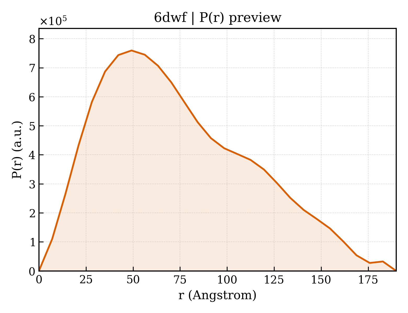
Cationic trypsin
OrganismNot specified
State in the Current Structure
| Assembly | Oligomeric State | Construct | Mutations and Modifications | Ligands, Ions and Associated Components | Method and Experimental Conditions | Structure Quality |
|---|---|---|---|---|---|---|
| 1 | Protein heterocomplex Heteromer Protein × 2 PDB declaration: dimeric(2) Consistent with protein copy count | Chain A; UniProt 24–246 | Fragment:residues 24-246 | Kunitz-type inihibitor × 1 (Q6VEQ7) | X-RAY DIFFRACTION X-ray crystallization conditions:VAPOR DIFFUSION, HANGING DROP;293 K;1.6M Ammonium Sulfate, 0.1M Sodium Citrate at pH 4.2 | Resolution 1.94 Å R-free 0.223 |
| 2 | Protein heterocomplex Heteromer Protein × 2 PDB declaration: dimeric(2) Consistent with protein copy count | Chain B; UniProt 24–246 | Fragment:residues 24-246 | Kunitz-type inihibitor × 1 (Q6VEQ7) | X-RAY DIFFRACTION X-ray crystallization conditions:VAPOR DIFFUSION, HANGING DROP;293 K;1.6M Ammonium Sulfate, 0.1M Sodium Citrate at pH 4.2 | Resolution 1.94 Å R-free 0.223 |
| 3 | Protein heterocomplex Heteromer Protein × 2 PDB declaration: dimeric(2) Consistent with protein copy count | Chain C; UniProt 24–246 | Fragment:residues 24-246 | Kunitz-type inihibitor × 1 (Q6VEQ7) | X-RAY DIFFRACTION X-ray crystallization conditions:VAPOR DIFFUSION, HANGING DROP;293 K;1.6M Ammonium Sulfate, 0.1M Sodium Citrate at pH 4.2 | Resolution 1.94 Å R-free 0.223 |
| 4 | Protein heterocomplex Heteromer Protein × 2 PDB declaration: dimeric(2) Consistent with protein copy count | Chain D; UniProt 24–246 | Fragment:residues 24-246 | Kunitz-type inihibitor × 1 (Q6VEQ7) | X-RAY DIFFRACTION X-ray crystallization conditions:VAPOR DIFFUSION, HANGING DROP;293 K;1.6M Ammonium Sulfate, 0.1M Sodium Citrate at pH 4.2 | Resolution 1.94 Å R-free 0.223 |
| 5 | Protein heterocomplex Heteromer Protein × 2 PDB declaration: dimeric(2) Consistent with protein copy count | Chain E; UniProt 24–246 | Fragment:residues 24-246 | Kunitz-type inihibitor × 1 (Q6VEQ7) | X-RAY DIFFRACTION X-ray crystallization conditions:VAPOR DIFFUSION, HANGING DROP;293 K;1.6M Ammonium Sulfate, 0.1M Sodium Citrate at pH 4.2 | Resolution 1.94 Å R-free 0.223 |
| 6 | Protein heterocomplex Heteromer Protein × 2 PDB declaration: dimeric(2) Consistent with protein copy count | Chain F; UniProt 24–246 | Fragment:residues 24-246 | Kunitz-type inihibitor × 1 (Q6VEQ7) | X-RAY DIFFRACTION X-ray crystallization conditions:VAPOR DIFFUSION, HANGING DROP;293 K;1.6M Ammonium Sulfate, 0.1M Sodium Citrate at pH 4.2 | Resolution 1.94 Å R-free 0.223 |
Other States of the Same Protein in the Database
Each row is a biological assembly of the same UniProt protein in another PDB entry. The “Difference from current entry” column identifies evidence-level differences; no tag means the currently parsed fields agree.
| Other PDB | Difference from Current Entry 6DWF | Assembly / Oligomeric State | Construct | Mutations and Modifications | Ligands, Ions and Non-polymers | Method and Experimental Conditions | Structure Quality |
|---|---|---|---|---|---|---|---|
| 1AQ7 TRYPSIN WITH INHIBITOR AERUGINOSIN 98-B Deposited 1997-08-07 | Different construct Different experimental conditions Different structure-quality metrics | Assembly 1 Protein heterocomplex Heteromer;Protein × 2 PDB declaration: dimeric |
Chain A
21–243(223 aa)
|
Not recorded | No recorded non-water small molecule |
X-RAY DIFFRACTION
X-ray crystallization conditions
pH 8.5;PROTEIN WAS CRYSTALLIZED FROM 10MG/ML TRYPSIN-LIGAND AGAINST 20% PEG-4000 AND 10% ISOPROPANOL IN 100MM PH 8.5 HEPES
|
Resolution 2.20 Å R-free 0.237 |
| 1AUJ BOVINE TRYPSIN COMPLEXED TO META-CYANO-BENZYLIC INHIBITOR Deposited 1997-08-28 | Different construct Different oligomeric state Different ligand/ion Different experimental conditions Different structure-quality metrics | Assembly 1 Protein monomer Monomer;Protein × 1 PDB declaration: monomeric |
Chain A
21–243(223 aa)
|
Not recorded | CA CALCIUM ION × 1 PPB 1-{[1-(2-AMINO-3-PHENYL-PROPIONYL)-PYRROLIDINE-2-CARBONYL]-AMINO}-2-(3-CYANO-PHENYL)-ETHANEBORONIC ACID × 1 |
X-RAY DIFFRACTION
X-ray crystallization conditions
pH 6.6;pH 6.6
|
Resolution 2.10 Å |
| 1AZ8 BOVINE TRYPSIN COMPLEXED TO BIS-PHENYLAMIDINE INHIBITOR Deposited 1997-11-26 | Different construct Different oligomeric state Different ligand/ion Different experimental conditions Different structure-quality metrics | Assembly 1 Protein monomer Monomer;Protein × 1 PDB declaration: monomeric |
Chain A
21–243(223 aa)
|
Not recorded | IN4 +/-METHYL 4-(AMINOIMINOMETHYL)-BETA-[3- INH (AMINOIMINO)PHENYL]BENZENE PENTANOATE × 1 |
X-RAY DIFFRACTION
X-ray crystallization conditions
pH 8.2;pH 8.2
|
Resolution 1.80 Å |
| 1BJU BETA-TRYPSIN COMPLEXED WITH ACPU Deposited 1998-06-29 | Different construct Different oligomeric state Different ligand/ion Different experimental conditions Different structure-quality metrics | Assembly 1 Protein monomer Monomer;Protein × 1 PDB declaration: monomeric |
Chain A
21–243(223 aa)
|
Not recorded | CA CALCIUM ION × 1 SO4 SULFATE ION × 1 GP6 1-(4-AMIDINOPHENYL)-3-(4-CHLOROPHENYL)UREA × 1 DMS DIMETHYL SULFOXIDE × 1 |
X-RAY DIFFRACTION
X-ray crystallization conditions
pH 6;0.9 M AMMONIUM SULFATE, 0.005 M MES, 0.001 M CACL2, 0.0025 M BENZAMIDINE, PH 6.0
|
Resolution 1.80 Å |
| 1BJV BETA-TRYPSIN COMPLEXED WITH APPU Deposited 1998-06-29 | Different construct Different oligomeric state Different ligand/ion Different experimental conditions Different structure-quality metrics | Assembly 1 Protein monomer Monomer;Protein × 1 PDB declaration: monomeric |
Chain A
21–243(223 aa)
|
Not recorded | CA CALCIUM ION × 1 SO4 SULFATE ION × 2 DMS DIMETHYL SULFOXIDE × 1 GP8 1-(2-AMIDINOPHENYL)-3-(PHENOXYPHENYL)UREA × 1 |
X-RAY DIFFRACTION
X-ray crystallization conditions
pH 6;0.9 M AMMONIUM SULFATE, 0.005 M MES, 0.001 M CACL2 0.0025 M BENZAMIDINE PH 6.0
|
Resolution 1.80 Å |
| 1BTP UNIQUE BINDING OF A NOVEL SYNTHETIC INHIBITOR, N-[3-[4-[4-(AMIDINOPHENOXY)-CARBONYL]PHENYL]-2-METHYL-2-PROPENOYL]-N-ALLYLGLYCINE METHANESULFONATE TO BOVINE TRYPSIN, REVEALED BY THE CRYSTAL STRUCTURE OF THE COMPLEX Deposited 1995-08-11 | Different construct Different oligomeric state Different ligand/ion Different experimental conditions Different structure-quality metrics | Assembly 1 Protein monomer Monomer;Protein × 1 PDB declaration: monomeric |
Chain A
15–243(229 aa)
|
Not recorded | CA CALCIUM ION × 1 | X-RAY DIFFRACTION mmCIF provides none of the parsed conditions | Resolution 2.20 Å |
| 1BTW Episelection: novel KI ~nanomolar inhibitors of serine proteases selected by binding or chemistry on an enzyme surface Deposited 1995-05-17 | Different construct Different oligomeric state Different ligand/ion Different experimental conditions Different structure-quality metrics | Assembly 1 Protein monomer Monomer;Protein × 1 PDB declaration: monomeric |
Chain A
15–243(229 aa)
|
Not recorded | 0ZW N-(tert-butoxycarbonyl)-L-alanyl-N-{(1S)-5-ammonio-1-[hydroxy(3-hydroxypropoxy)boranyl]pentyl}-L-valinamide × 1 CA CALCIUM ION × 1 | X-RAY DIFFRACTION mmCIF provides none of the parsed conditions | Resolution 1.70 Å |
| 1BTX Episelection: Novel Ki ~Nanomolar Inhibitors of Serine Proteases Selected by Binding or Chemistry on an Enzyme Surface Deposited 1995-05-17 | Different construct Different oligomeric state Different ligand/ion Different experimental conditions Different structure-quality metrics | Assembly 1 Protein monomer Monomer;Protein × 1 PDB declaration: monomeric |
Chain A
15–243(229 aa)
|
Not recorded | 0ZX N-(tert-butoxycarbonyl)-L-alanyl-N-[(1S)-5-amino-1-(diethoxyboranyl)pentyl]-L-valinamide × 1 CA CALCIUM ION × 1 | X-RAY DIFFRACTION mmCIF provides none of the parsed conditions | Resolution 1.70 Å |
| 1BTY Crystal structure of beta-trypsin in complex with benzamidine Deposited 1995-05-17 | Different construct Different oligomeric state Different ligand/ion Different experimental conditions Different structure-quality metrics | Assembly 1 Protein monomer Monomer;Protein × 1 PDB declaration: monomeric |
Chain A
15–243(229 aa)
|
Not recorded | CA CALCIUM ION × 1 BEN BENZAMIDINE × 1 | X-RAY DIFFRACTION mmCIF provides none of the parsed conditions | Resolution 1.50 Å |
| 1BTZ Episelection: novel KI ~nanomolar inhibitors of serine proteases selected by binding or chemistry on an enzyme surface Deposited 1995-05-17 | Different construct Different oligomeric state Different ligand/ion Different experimental conditions Different structure-quality metrics | Assembly 1 Protein monomer Monomer;Protein × 1 PDB declaration: monomeric |
Chain A
15–243(229 aa)
|
Not recorded | 0ZY N-(tert-butoxycarbonyl)-L-alanyl-N-{(1R)-5-ammonio-1-[hydroxy(methoxy)boranyl]pentyl}-L-valinamide × 1 CA CALCIUM ION × 1 | X-RAY DIFFRACTION mmCIF provides none of the parsed conditions | Resolution 2.00 Å |
| 1C1N RECRUITING ZINC TO MEDIATE POTENT, SPECIFIC INHIBITION OF SERINE PROTEASES Deposited 1999-07-21 | Different construct Different oligomeric state Different ligand/ion Different experimental conditions Different structure-quality metrics | Assembly 1 Protein monomer Monomer;Protein × 1 PDB declaration: monomeric |
Chain A
21–243(223 aa)
|
Not recorded | CA CALCIUM ION × 1 ZN ZINC ION × 1 SO4 SULFATE ION × 1 BEN BENZAMIDINE × 1 |
X-RAY DIFFRACTION
X-ray crystallization conditions
pH 5.11;TRYPSIN-BENZAMIDINE, P3(1) 2 1 WERE GROWN BY VAPOR DIFFUSION, AS DESCRIBED FOR
P2(1) 2(1) 2(1) (LARGE CELL) (MANGEL, ET AL., BIOCHEMISTRY 29, 8351-8357, 1990)
THE CRYSTAL WAS SOAKED FOR 10 DAYS IN A CHLORIDE-FREE SOLUTION OF 1.02 M
MGSO4 . 7 H2O,1.62 M ZNSO4 . 7 H2O, 0.50 M ZINC ACETATE DIHYDRATE, PH 5.11.
THE SOAKING SOLUTION WAS PERIODICALLY REPLACED.
|
Resolution 1.40 Å R-free 0.214 |
| 1C1O RECRUITING ZINC TO MEDIATE POTENT, SPECIFIC INHIBITION OF SERINE PROTEASES Deposited 1999-07-21 | Different construct Different oligomeric state Different ligand/ion Different experimental conditions Different structure-quality metrics | Assembly 1 Protein monomer Monomer;Protein × 1 PDB declaration: monomeric |
Chain A
21–243(223 aa)
|
Not recorded | CA CALCIUM ION × 1 MG MAGNESIUM ION × 1 SO4 SULFATE ION × 2 |
X-RAY DIFFRACTION
X-ray crystallization conditions
pH 4.98;TRYPSIN-BENZAMIDINE, P3(1) 2 1 WERE GROWN BY VAPOR DIFFUSION, AS DESCRIBED FOR
P2(1) 2(1) 2(1) (LARGE CELL) (MANGEL, ET AL., BIOCHEMISTRY 29, 8351-8357, 1990)
THE CRYSTAL WAS TRANSFERRED TO 0.25 M MES, 1.53 M MGSO4 . 7 H2O, PH 4.98 AND
SOAKED FOR MANY DAYS DURING WHICH THE SOAKING SOLUTION WAS REPLACED TO REMOVE
BOUND BENZAMIDINE.
|
Resolution 1.40 Å R-free 0.231 |
| 1C1P RECRUITING ZINC TO MEDIATE POTENT, SPECIFIC INHIBITION OF SERINE PROTEASES Deposited 1999-07-21 | Different construct Different oligomeric state Different ligand/ion Different experimental conditions Different structure-quality metrics | Assembly 1 Protein monomer Monomer;Protein × 1 PDB declaration: monomeric |
Chain A
21–243(223 aa)
|
Not recorded | CA CALCIUM ION × 1 MG MAGNESIUM ION × 1 SO4 SULFATE ION × 2 BAI (5-AMIDINO-2-BENZIMIDAZOLYL)(2-BENZIMIDAZOLYL)METHANE × 1 DMS DIMETHYL SULFOXIDE × 1 |
X-RAY DIFFRACTION
X-ray crystallization conditions
pH 5.9;TRYPSIN-BENZAMIDINE, P3(1) 2 1 WERE GROWN BY VAPOR DIFFUSION, AS DESCRIBED FOR
P2(1) 2(1) 2(1) (LARGE CELL) (MANGEL, ET AL., BIOCHEMISTRY 29, 8351-8357, 1990)
THE CRYSTAL WAS SOAKED IN A SOLUTION OF 0.10 M MES, 2.02 M MGSO4 . 7 H2O, PH
5.90, 2.0 % DMSO, SATURATED IN HEMI-BABIM OVER A PERIOD OF SEVERAL DAYS WITH
SEVERAL REPLACEMENTS OF THE SOAKING SOLUTION.
|
Resolution 1.37 Å R-free 0.192 |
| 1C1Q RECRUITING ZINC TO MEDIATE POTENT, SPECIFIC INHIBITION OF SERINE PROTEASES Deposited 1999-07-21 | Different construct Different oligomeric state Different ligand/ion Different experimental conditions Different structure-quality metrics | Assembly 1 Protein monomer Monomer;Protein × 1 PDB declaration: monomeric |
Chain A
21–243(223 aa)
|
Not recorded | CA CALCIUM ION × 1 MG MAGNESIUM ION × 1 SO4 SULFATE ION × 1 BAI (5-AMIDINO-2-BENZIMIDAZOLYL)(2-BENZIMIDAZOLYL)METHANE × 1 |
X-RAY DIFFRACTION
X-ray crystallization conditions
pH 8.21;TRYPSIN-BENZAMIDINE, P3(1) 2 1 WERE GROWN BY VAPOR DIFFUSION, AS DESCRIBED FOR
P2(1) 2(1) 2(1) (LARGE CELL) (MANGEL, ET AL., BIOCHEMISTRY 29, 8351-8357, 1990)
THE CRYSTAL WAS SOAKED IN A SOLUTION OF 0.10 M TRIS, 2.02 M MGSO4 . 7 H2O, PH
8.21, 2.0 % DMSO, SATURATED IN HEMI-BABIM OVER A PERIOD OF SEVERAL DAYS WITH
SEVERAL REPLACEMENTS OF THE SOAKING SOLUTION.
|
Resolution 1.37 Å R-free 0.193 |
| 1C1R RECRUITING ZINC TO MEDIATE POTENT, SPECIFIC INHIBITION OF SERINE PROTEASES Deposited 1999-07-21 | Different construct Different oligomeric state Different ligand/ion Different experimental conditions Different structure-quality metrics | Assembly 1 Protein monomer Monomer;Protein × 1 PDB declaration: monomeric |
Chain A
21–243(223 aa)
|
Not recorded | CA CALCIUM ION × 1 MG MAGNESIUM ION × 1 SO4 SULFATE ION × 1 ZN ZINC ION × 1 BAI (5-AMIDINO-2-BENZIMIDAZOLYL)(2-BENZIMIDAZOLYL)METHANE × 1 DMS DIMETHYL SULFOXIDE × 1 |
X-RAY DIFFRACTION
X-ray crystallization conditions
pH 8.2;TRYPSIN-BENZAMIDINE, P3(1) 2 1 WERE GROWN BY VAPOR DIFFUSION, AS DESCRIBED FOR
P2(1) 2(1) 2(1) (LARGE CELL) (MANGEL, ET AL., BIOCHEMISTRY 29, 8351-8357, 1990)
THE CRYSTAL WAS SOAKED IN A SOLUTION OF 0.24 M TRIS, 1.90 M MGSO4 . 7 H2O,
5.0 MM ZNSO4 . 7 H2O, PH 8.20 SATURATED IN HEMI-BABIM OVER A PERIOD OF SEVERAL
DAYS WITH SEVERAL REPLACEMENTS OF THE SOAKING SOLUTION.
|
Resolution 1.37 Å R-free 0.219 |
| 1C1S RECRUITING ZINC TO MEDIATE POTENT, SPECIFIC INHIBITION OF SERINE PROTEASES Deposited 1999-07-21 | Different construct Different oligomeric state Different ligand/ion Different experimental conditions Different structure-quality metrics | Assembly 1 Protein monomer Monomer;Protein × 1 PDB declaration: monomeric |
Chain A
21–243(223 aa)
|
Not recorded | CA CALCIUM ION × 1 NA SODIUM ION × 1 PO4 PHOSPHATE ION × 3 BAB BIS(5-AMIDINO-BENZIMIDAZOLYL)METHANE × 1 |
X-RAY DIFFRACTION
X-ray crystallization conditions
pH 6.1;TRYPSIN-BENZAMIDINE, P3(1) 2 1 WERE GROWN BY VAPOR DIFFUSION, AS DESCRIBED FOR
P2(1) 2(1) 2(1) (LARGE CELL) (MANGEL, ET AL., BIOCHEMISTRY 29, 8351-8357, 1990)
THE CRYSTAL WAS SOAKED IN A SOLUTION OF 1.25 M NA2PO4 . 7 H2O, 1.75 M KH2PO4,
2.0 % DMSO, SATURATED IN BABIM OVER A PERIOD OF 5 DAYS WITH SEVERAL
REPLACEMENTS OF THE SOAKING SOLUTION., pH 6.1
|
Resolution 1.63 Å R-free 0.206 |
| 1C1T RECRUITING ZINC TO MEDIATE POTENT, SPECIFIC INHIBITION OF SERINE PROTEASES Deposited 1999-07-21 | Different construct Different oligomeric state Different ligand/ion Different experimental conditions Different structure-quality metrics | Assembly 1 Protein monomer Monomer;Protein × 1 PDB declaration: monomeric |
Chain A
21–243(223 aa)
|
Not recorded | CA CALCIUM ION × 1 MG MAGNESIUM ION × 1 SO4 SULFATE ION × 1 BAB BIS(5-AMIDINO-BENZIMIDAZOLYL)METHANE × 1 |
X-RAY DIFFRACTION
X-ray crystallization conditions
pH 8.18;TRYPSIN-BENZAMIDINE, P3(1) 2 1 WERE GROWN BY VAPOR DIFFUSION, AS DESCRIBED FOR
P2(1) 2(1) 2(1) (LARGE CELL) (MANGEL, ET AL., BIOCHEMISTRY 29, 8351-8357, 1990)
THE CRYSTAL WAS SOAKED IN A SOLUTION OF 0.10 M TRIS, 2.02 M MGSO4 . 7 H2O, PH
8.18, 2.0 % DMSO, SATURATED IN BABIM OVER A PERIOD OF SEVERAL DAYS WITH
SEVERAL REPLACEMENTS OF THE SOAKING SOLUTION.
|
Resolution 1.37 Å R-free 0.194 |
| 1C2D RECRUITING ZINC TO MEDIATE POTENT, SPECIFIC INHIBITION OF SERINE PROTEASES Deposited 1999-07-21 | Different construct Different oligomeric state Different ligand/ion Different experimental conditions Different structure-quality metrics | Assembly 1 Protein monomer Monomer;Protein × 1 PDB declaration: monomeric |
Chain A
21–243(223 aa)
|
Not recorded | CA CALCIUM ION × 1 MG MAGNESIUM ION × 1 ZN ZINC ION × 2 BAK BIS(5-AMIDINO-2-BENZIMIDAZOLYL)METHANE KETONE × 1 |
X-RAY DIFFRACTION
X-ray crystallization conditions
pH 9.93;BATCH, 1.51 M MGSO4.7H2O, 0.59 MM TRYPSIN, 1.0 MM ZN(II) 5.0 % DMSO:
CRYSTALLIZATION SOLUTION WAS SATURATED IN KETO-BABIM. CRYSTAL WAS TRANSFERRED
TO SYNTHETIC MOTHER LIQUOR CONTAINING ~0.4 MM ZN(II), PH 9.93 AND SOAKED FOR
SEVERAL DAYS WITH SEVERAL REPLACEMENTS OF SOAKING SOLUTIONS. THE SOLUTION WAS
SATURATED IN KETO-BABIM AND CONTAINED 2.0 % DMSO TO INCREASE INHIBITOR
SOLUBILITY.
|
Resolution 1.65 Å R-free 0.211 |
| 1C2E RECRUITING ZINC TO MEDIATE POTENT, SPECIFIC INHIBITION OF SERINE PROTEASES Deposited 1999-07-22 | Different construct Different oligomeric state Different ligand/ion Different experimental conditions Different structure-quality metrics | Assembly 1 Protein monomer Monomer;Protein × 1 PDB declaration: monomeric |
Chain A
21–243(223 aa)
|
Not recorded | CA CALCIUM ION × 1 MG MAGNESIUM ION × 1 ZN ZINC ION × 1 BAK BIS(5-AMIDINO-2-BENZIMIDAZOLYL)METHANE KETONE × 1 |
X-RAY DIFFRACTION
X-ray crystallization conditions
pH 9;BATCH, 1.51 M MGSO4 . 7 H2O, 0.59 MM TRYPSIN, 1.0 MM ZN(II) 5.0 % DMSO:
CRYSTALLIZATION SOLUTION WAS SATURATED IN KETO-BABIM. CRYSTAL WAS TRANSFERRED
TO SYNTHETIC MOTHER LIQUOR CONTAINING ~0.4 MM ZN(II), PH 9.00 AND SOAKED FOR
SEVERAL DAYS WITH SEVERAL REPLACEMENTS OF SOAKING SOLUTIONS. THE SOLUTION WAS
SATURATED IN KETO-BABIM AND CONTAINED 2.0 % DMSO TO INCREASE INHIBITOR
SOLUBILITY.
|
Resolution 1.65 Å R-free 0.212 |
| 1C2F RECRUITING ZINC TO MEDIATE POTENT, SPECIFIC INHIBITION OF SERINE PROTEASES Deposited 1999-07-21 | Different construct Different oligomeric state Different ligand/ion Different experimental conditions Different structure-quality metrics | Assembly 1 Protein monomer Monomer;Protein × 1 PDB declaration: monomeric |
Chain A
21–243(223 aa)
|
Not recorded | CA CALCIUM ION × 1 MG MAGNESIUM ION × 1 ZN ZINC ION × 3 BAH BIS(5-AMIDINO-2-BENZIMIDAZOLYL)METHANE KETONE HYDRATE × 1 DMS DIMETHYL SULFOXIDE × 1 |
X-RAY DIFFRACTION
X-ray crystallization conditions
pH 8;BATCH, 1.51 M MGSO4 . 7 H2O, 0.59 MM TRYPSIN, 1.0 MM ZN(II) 5.0 % DMSO:
CRYSTALLIZATION SOLUTION WAS SATURATED IN KETO-BABIM. CRYSTAL WAS TRANSFERRED
TO SYNTHETIC MOTHER LIQUOR CONTAINING 9.0 MM ZN(II), PH 8.00 AND SOAKED FOR
SEVERAL DAYS WITH SEVERAL REPLACEMENTS OF SOAKING SOLUTIONS. THE SOLUTION WAS
SATURATED IN KETO-BABIM AND CONTAINED 2.0 % DMSO TO INCREASE INHIBITOR
SOLUBILITY.
|
Resolution 1.70 Å R-free 0.241 |
| 1C2G RECRUITING ZINC TO MEDIATE POTENT, SPECIFIC INHIBITION OF SERINE PROTEASES Deposited 1999-07-21 | Different construct Different oligomeric state Different ligand/ion Different experimental conditions Different structure-quality metrics | Assembly 1 Protein monomer Monomer;Protein × 1 PDB declaration: monomeric |
Chain A
21–243(223 aa)
|
Not recorded | CA CALCIUM ION × 1 MG MAGNESIUM ION × 1 ZN ZINC ION × 3 BAH BIS(5-AMIDINO-2-BENZIMIDAZOLYL)METHANE KETONE HYDRATE × 1 DMS DIMETHYL SULFOXIDE × 1 |
X-RAY DIFFRACTION
X-ray crystallization conditions
pH 7;BATCH, 1.51 M MGSO4 . 7 H2O, 0.59 MM TRYPSIN, 1.0 MM ZN(II) 5.0 % DMSO:
CRYSTALLIZATION SOLUTION WAS SATURATED IN KETO-BABIM. CRYSTAL WAS TRANSFERRED
TO SYNTHETIC MOTHER LIQUOR CONTAINING 46 MM ZN(II), PH 7.00 AND SOAKED FOR
SEVERAL DAYS WITH SEVERAL REPLACEMENTS OF SOAKING SOLUTIONS. THE SOLUTION WAS
SATURATED IN KETO-BABIM AND CONTAINED 2.0 % DMSO TO INCREASE INHIBITOR
SOLUBILITY.
|
Resolution 1.65 Å R-free 0.200 |
| 1C2H RECRUITING ZINC TO MEDIATE POTENT, SPECIFIC INHIBITION OF SERINE PROTEASES Deposited 1999-07-21 | Different construct Different oligomeric state Different ligand/ion Different experimental conditions Different structure-quality metrics | Assembly 1 Protein monomer Monomer;Protein × 1 PDB declaration: monomeric |
Chain A
21–243(223 aa)
|
Not recorded | CA CALCIUM ION × 1 MG MAGNESIUM ION × 1 ZN ZINC ION × 3 BAK BIS(5-AMIDINO-2-BENZIMIDAZOLYL)METHANE KETONE × 1 |
X-RAY DIFFRACTION
X-ray crystallization conditions
pH 6.04;BATCH, 1.51 M MGSO4 . 7 H2O, 0.59 MM TRYPSIN, 1.0 MM ZN(II) 5.0 % DMSO:
CRYSTALLIZATION SOLUTION WAS SATURATED IN KETO-BABIM. CRYSTAL WAS TRANSFERRED
TO SYNTHETIC MOTHER LIQUOR CONTAINING 46 MM ZN(II), PH 6.04 AND SOAKED FOR
SEVERAL DAYS WITH SEVERAL REPLACEMENTS OF SOAKING SOLUTIONS. THE SOLUTION WAS
SATURATED IN KETO-BABIM AND CONTAINED 2.0 % DMSO TO INCREASE INHIBITOR
SOLUBILITY.
|
Resolution 1.40 Å R-free 0.218 |
| 1C2I RECRUITING ZINC TO MEDIATE POTENT, SPECIFIC INHIBITION OF SERINE PROTEASES Deposited 1999-07-21 | Different construct Different oligomeric state Different ligand/ion Different experimental conditions Different structure-quality metrics | Assembly 1 Protein monomer Monomer;Protein × 1 PDB declaration: monomeric |
Chain A
21–243(223 aa)
|
Not recorded | CA CALCIUM ION × 1 CL CHLORIDE ION × 1 ZN ZINC ION × 3 BAK BIS(5-AMIDINO-2-BENZIMIDAZOLYL)METHANE KETONE × 1 DMS DIMETHYL SULFOXIDE × 1 |
X-RAY DIFFRACTION
X-ray crystallization conditions
pH 5.5;BATCH, 1.51 M MGSO4 . 7 H2O, 0.59 MM TRYPSIN, 1.0 MM ZN(II) 5.0 % DMSO:
CRYSTALLIZATION SOLUTION WAS SATURATED IN KETO-BABIM. CRYSTAL WAS TRANSFERRED
TO SYNTHETIC MOTHER LIQUOR CONTAINING 46 MM ZN(II), PH 5.50 AND SOAKED FOR
SEVERAL DAYS WITH SEVERAL REPLACEMENTS OF SOAKING SOLUTIONS. THE SOLUTION WAS
SATURATED IN KETO-BABIM AND CONTAINED 2.0 % DMSO TO INCREASE INHIBITOR
SOLUBILITY.
|
Resolution 1.47 Å R-free 0.208 |
| 1C2J RECRUITING ZINC TO MEDIATE POTENT, SPECIFIC INHIBITION OF SERINE PROTEASES Deposited 1999-07-21 | Different construct Different oligomeric state Different ligand/ion Different experimental conditions Different structure-quality metrics | Assembly 1 Protein monomer Monomer;Protein × 1 PDB declaration: monomeric |
Chain A
21–243(223 aa)
|
Not recorded | CA CALCIUM ION × 1 MG MAGNESIUM ION × 1 SO4 SULFATE ION × 1 ZN ZINC ION × 1 BAK BIS(5-AMIDINO-2-BENZIMIDAZOLYL)METHANE KETONE × 1 |
X-RAY DIFFRACTION
X-ray crystallization conditions
pH 7.06;BATCH, 1.51 M MGSO4 . 7 H2O, 0.59 MM TRYPSIN, 1.0 MM ZN(II) 5.0 % DMSO:
CRYSTALLIZATION SOLUTION WAS SATURATED IN KETO-BABIM. CRYSTAL WAS TRANSFERRED
TO SYNTHETIC MOTHER LIQUOR CONTAINING 100 NM ZN(II), PH 7.06 AND SOAKED FOR
SEVERAL DAYS WITH SEVERAL REPLACEMENTS OF SOAKING SOLUTIONS. THE SOLUTION WAS
SATURATED IN KETO-BABIM AND CONTAINED 2.0 % DMSO TO INCREASE INHIBITOR
SOLUBILITY.
|
Resolution 1.40 Å R-free 0.219 |
| 1C2K RECRUITING ZINC TO MEDIATE POTENT, SPECIFIC INHIBITION OF SERINE PROTEASES Deposited 1999-07-21 | Different construct Different oligomeric state Different ligand/ion Different experimental conditions Different structure-quality metrics | Assembly 1 Protein monomer Monomer;Protein × 1 PDB declaration: monomeric |
Chain A
21–243(223 aa)
|
Not recorded | CA CALCIUM ION × 1 CL CHLORIDE ION × 1 ZN ZINC ION × 2 ABI 5-AMIDINO-BENZIMIDAZOLE × 1 |
X-RAY DIFFRACTION
X-ray crystallization conditions
pH 3.85;BATCH, 1.51 M MGSO4 . 7 H2O, 0.59 MM TRYPSIN, 1.0 MM ZN(II) 5.0 % DMSO:
CRYSTALLIZATION SOLUTION WAS SATURATED IN KETO-BABIM. CRYSTAL WAS TRANSFERRED
TO SYNTHETIC MOTHER LIQUOR CONTAINING 2.11 M ZN(II), PH 3.85 AND SOAKED FOR
SEVERAL DAYS WITH SEVERAL REPLACEMENTS OF SOAKING SOLUTIONS. THE SOLUTION WAS
SATURATED IN KETO-BABIM AND CONTAINED 2.0 % DMSO TO INCREASE INHIBITOR
SOLUBILITY. UNDER THIS CONDITION KETO-BABIM UNDERGOES DECOMPOSITION TO 5-
AMIDINO-BENZIMIDAZOLE.
|
Resolution 1.65 Å R-free 0.243 |
| 1C2L RECRUITING ZINC TO MEDIATE POTENT, SPECIFIC INHIBITION OF SERINE PROTEASES Deposited 1999-07-21 | Different construct Different oligomeric state Different ligand/ion Different experimental conditions Different structure-quality metrics | Assembly 1 Protein monomer Monomer;Protein × 1 PDB declaration: monomeric |
Chain A
21–243(223 aa)
|
Not recorded | CA CALCIUM ION × 1 CL CHLORIDE ION × 1 ZN ZINC ION × 2 |
X-RAY DIFFRACTION
X-ray crystallization conditions
pH 4.86;TRYPSIN-BENZAMIDINE, P3(1) 2 1 WERE GROWN BY VAPOR DIFFUSION, AS DESCRIBED FOR
P2(1) 2(1) 2(1) (LARGE CELL) (MANGEL, ET AL., BIOCHEMISTRY 29, 8351-8357, 1990)
A CRYSTAL WAS SOAKED FOR 25 DAYS IN 2.11 M ZN+2, 250 MM CL-, PH 4.86 IN THE
ABSENCE OF AN INHIBITOR. THIS SOLUTION WAS REPLACED ONCE EVERY DAY OR TWO TO
REMOVE BOUND BENZAMIDINE AND TO INCORPORATE ZN+2 AT THE ACTIVE SITE.
|
Resolution 1.50 Å R-free 0.224 |
| 1C2M RECRUITING ZINC TO MEDIATE POTENT, SPECIFIC INHIBITION OF SERINE PROTEASES Deposited 1999-07-21 | Different construct Different oligomeric state Different ligand/ion Different experimental conditions Different structure-quality metrics | Assembly 1 Protein monomer Monomer;Protein × 1 PDB declaration: monomeric |
Chain A
21–243(223 aa)
|
Not recorded | CA CALCIUM ION × 1 ZN ZINC ION × 1 MG MAGNESIUM ION × 1 SO4 SULFATE ION × 1 |
X-RAY DIFFRACTION
X-ray crystallization conditions
pH 4.98;TRYPSIN-BENZAMIDINE, P3(1) 2 1 WERE GROWN BY VAPOR DIFFUSION, AS DESCRIBED FOR
P2(1) 2(1) 2(1) (LARGE CELL) (MANGEL, ET AL., BIOCHEMISTRY 29, 8351-8357, 1990)
THE CRYSTAL WAS SOAKED FOR 25 DAYS IN 2.11 M ZN+2, 250 MM CL-, PH 4.86 IN THE
ABSENCE OF AN INHIBITOR. THIS SOLUTION WAS REPLACED ONCE EVERY DAY OR TWO TO
REMOVE BOUND BENZAMIDINE AND TO INCORPORATE ZN+2 AT THE ACTIVE SITE. IT WAS
THEN SOAKED IN CHLORIDE-FREE SOLUTION OF 1.02 MGSO4 . 7H2O, 1.62 M ZNSO4 . 7
H2O, 0.50 M ZINC ACETATE DIHYDRATE, PH 4.98 FOR 10 DAYS DURING WHICH THE
SOAKING SOLUTION WAS REPLACED ONCE A DAY.
|
Resolution 1.40 Å R-free 0.234 |
| 1C5P STRUCTURAL BASIS FOR SELECTIVITY OF A SMALL MOLECULE, S1-BINDING, SUB-MICROMOLAR INHIBITOR OF UROKINASE TYPE PLASMINOGEN ACTIVATOR Deposited 1999-12-22 | Different construct Different oligomeric state Different ligand/ion Different experimental conditions Different structure-quality metrics | Assembly 1 Protein monomer Monomer;Protein × 1 PDB declaration: monomeric |
Chain A
7–229(223 aa)
|
Not recorded | CA CALCIUM ION × 1 MG MAGNESIUM ION × 1 SO4 SULFATE ION × 2 BEN BENZAMIDINE × 1 |
X-RAY DIFFRACTION
X-ray crystallization conditions
VAPOR DIFFUSION;pH 7.5;trypsin-benzamidine, P2(1)2(1)2(1) were grown by
vapor diffusion, as described for P2(1) 2(1) 2(1)
(large cell) (Mangel, et al., Biochemistry 29, 8351-8357, 1990)
The crystal was soaked in a solution of 85 % saturated MgSO4 . 7 H2O,
1 mM CaCl2, 2.0 % DMSO, 100 mM Tris, pH 7.50., VAPOR DIFFUSION
|
Resolution 1.43 Å R-free 0.221 |
| 1C5Q STRUCTURAL BASIS FOR SELECTIVITY OF A SMALL MOLECULE, S1-BINDING, SUB-MICROMOLAR INHIBITOR OF UROKINASE TYPE PLASMINOGEN ACTIVATOR Deposited 1999-12-22 | Different construct Different oligomeric state Different ligand/ion Different experimental conditions Different structure-quality metrics | Assembly 1 Protein monomer Monomer;Protein × 1 PDB declaration: monomeric |
Chain A
7–229(223 aa)
|
Not recorded | CA CALCIUM ION × 1 CL CHLORIDE ION × 1 ESI 4-IODOBENZO[B]THIOPHENE-2-CARBOXAMIDINE × 1 |
X-RAY DIFFRACTION
X-ray crystallization conditions
pH 8.2;trypsin-benzamidine, P2(1)2(1)2(1) were grown by
vapor diffusion, as described for P2(1) 2(1) 2(1)
(large cell) (Mangel, et al., Biochemistry 29, 8351-8357, 1990)
The crystal was soaked in a solution of 85 % MgSO4 . 7 H2O,
1 mM CaCl2, 2.0 % DMSO, saturated with inhibitor, 100 mM Tris,
pH 8.20 over a period of several days with several replacements
of the soaking solution.
|
Resolution 1.43 Å R-free 0.221 |
| 1C5R STRUCTURAL BASIS FOR SELECTIVITY OF A SMALL MOLECULE, S1-BINDING, SUB-MICROMOLAR INHIBITOR OF UROKINASE TYPE PLASMINOGEN ACTIVATOR Deposited 1999-12-22 | Different construct Different oligomeric state Different ligand/ion Different experimental conditions Different structure-quality metrics | Assembly 1 Protein monomer Monomer;Protein × 1 PDB declaration: monomeric |
Chain A
7–229(223 aa)
|
Not recorded | FLC CITRATE ANION × 1 CA CALCIUM ION × 1 SO4 SULFATE ION × 2 ESI 4-IODOBENZO[B]THIOPHENE-2-CARBOXAMIDINE × 1 |
X-RAY DIFFRACTION
X-ray crystallization conditions
pH 5.5;trypsin-benzamidine, P3(1) 2 1 were grown by vapor
diffusion, as described for P2(1) 2(1) 2(1)
(large cell) (Mangel, et al., Biochemistry 29, 8351-8357, 1990)
The crystal was soaked in a solution of 85 % saturated citrate,
1 mM CaCl2, 2.0 % DMSO, saturated with inhibitor, pH 5.50 over
a period of several days with several replacements of the
soaking solution.
|
Resolution 1.47 Å R-free 0.229 |
| 1C5S STRUCTURAL BASIS FOR SELECTIVITY OF A SMALL MOLECULE, S1-BINDING, SUB-MICROMOLAR INHIBITOR OF UROKINASE TYPE PLASMINOGEN ACTIVATOR Deposited 1999-12-22 | Different construct Different oligomeric state Different ligand/ion Different experimental conditions Different structure-quality metrics | Assembly 1 Protein monomer Monomer;Protein × 1 PDB declaration: monomeric |
Chain A
7–229(223 aa)
|
Not recorded | CA CALCIUM ION × 1 SO4 SULFATE ION × 6 ESX BENZO[B]THIOPHENE-2-CARBOXAMIDINE × 1 |
X-RAY DIFFRACTION
X-ray crystallization conditions
VAPOR DIFFUSION;pH 8.2;trypsin-benzamidine, P3(1) 2 1 were grown by
vapor diffusion, as described for P2(1) 2(1) 2(1) (large cell)
(Mangel, et al., Biochemistry 29, 8351-8357, 1990) The crystal was soaked
in a solution of 85 % saturated MgSO4 . 7 H2O, 100 mM Tris, 1 mM CaCl2,
2.0 % DMSO,saturated with inhibitor, pH 8.20 over a period of several
days with several replacements of the soaking solution., VAPOR DIFFUSION
|
Resolution 1.36 Å R-free 0.224 |
| 1C5T STRUCTURAL BASIS FOR SELECTIVITY OF A SMALL MOLECULE, S1-BINDING, SUB-MICROMOLAR INHIBITOR OF UROKINASE TYPE PLASMINOGEN ACTIVATOR Deposited 1999-12-22 | Different construct Different oligomeric state Different ligand/ion Different experimental conditions Different structure-quality metrics | Assembly 1 Protein monomer Monomer;Protein × 1 PDB declaration: monomeric |
Chain A
7–229(223 aa)
|
Not recorded | CA CALCIUM ION × 1 ESP THIENO[2,3-B]PYRIDINE-2-CARBOXAMIDINE × 1 |
X-RAY DIFFRACTION
X-ray crystallization conditions
pH 8.2;trypsin-benzamidine, P3(1) 2 1 were grown by
vapor diffusion, as described for P2(1) 2(1) 2(1)
(large cell) (Mangel, et al., Biochemistry 29, 8351-8357, 1990)
The crystal was soaked in a solution of 1.73 M MgSO4 . 7 H2O,
150 mM Tris, 1 mM CaCl2, 2.0 % DMSO,saturated with inhibitor,
pH 8.20 over a period of several days with several replacements
of the soaking solution.
|
Resolution 1.37 Å R-free 0.195 |
| 1C5U STRUCTURAL BASIS FOR SELECTIVITY OF A SMALL MOLECULE, S1-BINDING, SUB-MICROMOLAR INHIBITOR OF UROKINASE TYPE PLASMINOGEN ACTIVATOR Deposited 1999-12-22 | Different construct Different oligomeric state Different ligand/ion Different experimental conditions Different structure-quality metrics | Assembly 1 Protein monomer Monomer;Protein × 1 PDB declaration: monomeric |
Chain A
7–229(223 aa)
|
Not recorded | CA CALCIUM ION × 1 MG MAGNESIUM ION × 1 SO4 SULFATE ION × 2 ESP THIENO[2,3-B]PYRIDINE-2-CARBOXAMIDINE × 1 |
X-RAY DIFFRACTION
X-ray crystallization conditions
VAPOR DIFFUSION;pH 5.5;trypsin-benzamidine, P3(1) 2 1 were grown by vapor
diffusion, as described for P2(1) 2(1) 2(1) (large cell)
(Mangel, et al., Biochemistry 29, 8351-8357, 1990) The crystal was
soaked in a solution of 1.73 M MgSO4 . 7 H2O, 150 mM MES, 1 mM CaCl2,
2.0 % DMSO,saturated with inhibitor, pH 5.50 over a period of several
days with several replacements of the soaking solution., VAPOR DIFFUSION
|
Resolution 1.37 Å R-free 0.195 |
| 1C5V STRUCTURAL BASIS FOR SELECTIVITY OF A SMALL MOLECULE, S1-BINDING, SUB-MICROMOLAR INHIBITOR OF UROKINASE TYPE PLASMINOGEN ACTIVATOR Deposited 1999-12-22 | Different construct Different oligomeric state Different ligand/ion Different experimental conditions Different structure-quality metrics | Assembly 1 Protein monomer Monomer;Protein × 1 PDB declaration: monomeric |
Chain A
7–229(223 aa)
|
Not recorded | CA CALCIUM ION × 1 MG MAGNESIUM ION × 1 |
X-RAY DIFFRACTION
X-ray crystallization conditions
pH 7.72;trypsin-benzamidine, P3(1) 2 1 crystals were grown
by vapor diffusion, as described for P2(1) 2(1) 2(1) (large cell)
(Mangel, et al., Biochemistry 29, 8351-8357, 1990) The crystal was soaked in
inhibitor-free solution of 1.84 M MgSO4 . 7 H2O, 150 mM Tris, 1 mM CaCl2,
pH 7.72 over a period of several weeks with several replacements of the
soaking solution.
|
Resolution 1.48 Å R-free 0.223 |
| 1C9T COMPLEX OF BDELLASTASIN WITH BOVINE TRYPSIN Deposited 1999-08-03 | Different construct Different experimental conditions Different structure-quality metrics | Assembly 1 Protein heterocomplex Heteromer;Protein × 2 PDB declaration: dimeric |
Chain A
21–243(223 aa)
|
Not recorded | No recorded non-water small molecule |
X-RAY DIFFRACTION
X-ray crystallization conditions
VAPOR DIFFUSION, SITTING DROP;pH 5.6;293 K;50MM MES, 2.0M AMMONIUM SULFATE, 10MM MAGNESIM CHLORIDE, pH 5.6, VAPOR DIFFUSION, SITTING DROP, temperature 293K
X-ray crystallization conditions
VAPOR DIFFUSION, SITTING DROP;pH 5.6;293 K;50MM MES, 2.2M AMMONIUM SULFATE, 10MM MAGNESIM CHLORIDE, pH 5.6, VAPOR DIFFUSION, SITTING DROP, temperature 293K
|
Resolution 3.30 Å R-free 0.287 |
| 1C9T COMPLEX OF BDELLASTASIN WITH BOVINE TRYPSIN Deposited 1999-08-03 | Different construct Different experimental conditions Different structure-quality metrics | Assembly 2 Protein heterocomplex Heteromer;Protein × 2 PDB declaration: dimeric |
Chain B
21–243(223 aa)
|
Not recorded | No recorded non-water small molecule |
X-RAY DIFFRACTION
X-ray crystallization conditions
VAPOR DIFFUSION, SITTING DROP;pH 5.6;293 K;50MM MES, 2.0M AMMONIUM SULFATE, 10MM MAGNESIM CHLORIDE, pH 5.6, VAPOR DIFFUSION, SITTING DROP, temperature 293K
X-ray crystallization conditions
VAPOR DIFFUSION, SITTING DROP;pH 5.6;293 K;50MM MES, 2.2M AMMONIUM SULFATE, 10MM MAGNESIM CHLORIDE, pH 5.6, VAPOR DIFFUSION, SITTING DROP, temperature 293K
|
Resolution 3.30 Å R-free 0.287 |
| 1C9T COMPLEX OF BDELLASTASIN WITH BOVINE TRYPSIN Deposited 1999-08-03 | Different construct Different experimental conditions Different structure-quality metrics | Assembly 3 Protein heterocomplex Heteromer;Protein × 2 PDB declaration: dimeric |
Chain C
21–243(223 aa)
|
Not recorded | No recorded non-water small molecule |
X-RAY DIFFRACTION
X-ray crystallization conditions
VAPOR DIFFUSION, SITTING DROP;pH 5.6;293 K;50MM MES, 2.0M AMMONIUM SULFATE, 10MM MAGNESIM CHLORIDE, pH 5.6, VAPOR DIFFUSION, SITTING DROP, temperature 293K
X-ray crystallization conditions
VAPOR DIFFUSION, SITTING DROP;pH 5.6;293 K;50MM MES, 2.2M AMMONIUM SULFATE, 10MM MAGNESIM CHLORIDE, pH 5.6, VAPOR DIFFUSION, SITTING DROP, temperature 293K
|
Resolution 3.30 Å R-free 0.287 |
| 1C9T COMPLEX OF BDELLASTASIN WITH BOVINE TRYPSIN Deposited 1999-08-03 | Different construct Different experimental conditions Different structure-quality metrics | Assembly 4 Protein heterocomplex Heteromer;Protein × 2 PDB declaration: dimeric |
Chain D
21–243(223 aa)
|
Not recorded | No recorded non-water small molecule |
X-RAY DIFFRACTION
X-ray crystallization conditions
VAPOR DIFFUSION, SITTING DROP;pH 5.6;293 K;50MM MES, 2.0M AMMONIUM SULFATE, 10MM MAGNESIM CHLORIDE, pH 5.6, VAPOR DIFFUSION, SITTING DROP, temperature 293K
X-ray crystallization conditions
VAPOR DIFFUSION, SITTING DROP;pH 5.6;293 K;50MM MES, 2.2M AMMONIUM SULFATE, 10MM MAGNESIM CHLORIDE, pH 5.6, VAPOR DIFFUSION, SITTING DROP, temperature 293K
|
Resolution 3.30 Å R-free 0.287 |
| 1C9T COMPLEX OF BDELLASTASIN WITH BOVINE TRYPSIN Deposited 1999-08-03 | Different construct Different experimental conditions Different structure-quality metrics | Assembly 5 Protein heterocomplex Heteromer;Protein × 2 PDB declaration: dimeric |
Chain E
21–243(223 aa)
|
Not recorded | No recorded non-water small molecule |
X-RAY DIFFRACTION
X-ray crystallization conditions
VAPOR DIFFUSION, SITTING DROP;pH 5.6;293 K;50MM MES, 2.0M AMMONIUM SULFATE, 10MM MAGNESIM CHLORIDE, pH 5.6, VAPOR DIFFUSION, SITTING DROP, temperature 293K
X-ray crystallization conditions
VAPOR DIFFUSION, SITTING DROP;pH 5.6;293 K;50MM MES, 2.2M AMMONIUM SULFATE, 10MM MAGNESIM CHLORIDE, pH 5.6, VAPOR DIFFUSION, SITTING DROP, temperature 293K
|
Resolution 3.30 Å R-free 0.287 |
| 1C9T COMPLEX OF BDELLASTASIN WITH BOVINE TRYPSIN Deposited 1999-08-03 | Different construct Different experimental conditions Different structure-quality metrics | Assembly 6 Protein heterocomplex Heteromer;Protein × 2 PDB declaration: dimeric |
Chain F
21–243(223 aa)
|
Not recorded | No recorded non-water small molecule |
X-RAY DIFFRACTION
X-ray crystallization conditions
VAPOR DIFFUSION, SITTING DROP;pH 5.6;293 K;50MM MES, 2.0M AMMONIUM SULFATE, 10MM MAGNESIM CHLORIDE, pH 5.6, VAPOR DIFFUSION, SITTING DROP, temperature 293K
X-ray crystallization conditions
VAPOR DIFFUSION, SITTING DROP;pH 5.6;293 K;50MM MES, 2.2M AMMONIUM SULFATE, 10MM MAGNESIM CHLORIDE, pH 5.6, VAPOR DIFFUSION, SITTING DROP, temperature 293K
|
Resolution 3.30 Å R-free 0.287 |
| 1CE5 BOVINE PANCREAS BETA-TRYPSIN IN COMPLEX WITH BENZAMIDINE Deposited 1999-03-16 | Different construct Different oligomeric state Different ligand/ion Different experimental conditions Different structure-quality metrics | Assembly 1 Protein monomer Monomer;Protein × 1 PDB declaration: monomeric |
Chain A
21–243(223 aa)
|
Not recorded | CA CALCIUM ION × 1 CL CHLORIDE ION × 1 SO4 SULFATE ION × 1 BEN BENZAMIDINE × 1 |
X-RAY DIFFRACTION
X-ray crystallization conditions
pH 6;pH 6.0
|
Resolution 1.90 Å R-free 0.186 |
| 1D6R CRYSTAL STRUCTURE OF CANCER CHEMOPREVENTIVE BOWMAN-BIRK INHIBITOR IN TERNARY COMPLEX WITH BOVINE TRYPSIN AT 2.3 A RESOLUTION. STRUCTURAL BASIS OF JANUS-FACED SERINE PROTEASE INHIBITOR SPECIFICITY Deposited 1999-10-15 | Different construct Different oligomeric state Different experimental conditions Different structure-quality metrics | Assembly 1 Protein heterocomplex Heteromer;Protein × 4 PDB declaration: tetrameric |
Chain A
21–243(223 aa)
Fragment:CATIONIC PRECURSOR
|
Not recorded | No recorded non-water small molecule |
X-RAY DIFFRACTION
X-ray crystallization conditions
VAPOR DIFFUSION, SITTING DROP;pH 8;291 K;PROTEIN SOLUTION: TRIS/CL, CACL2, MGSO4.7H2O, NAN3 PLUS PRECIPITANT
PRECIPITANTS: (NH4)2SO4, MDP, pH 8.0, VAPOR DIFFUSION, SITTING DROP, temperature 291.0K
|
Resolution 2.30 Å R-free 0.226 |
| 1EB2 Trypsin inhibitor complex (BPO) Deposited 2001-07-18 | Different construct Different oligomeric state Different ligand/ion Different experimental conditions Different structure-quality metrics | Assembly 1 Protein monomer Monomer;Protein × 1 PDB declaration: monomeric |
Chain A
21–243(223 aa)
|
Not recorded | SO4 SULFATE ION × 1 CA CALCIUM ION × 1 BPO 3-[(Z)-AMINO(IMINO)METHYL]-N-[2-(4-BENZOYL-1-PIPERIDINYL)-2-OXO-1-PHENYLETHYL]BENZAMIDE × 1 |
X-RAY DIFFRACTION
X-ray crystallization conditions
pH 8.15;2.1 M AMMONIUM SULPHATE, 0.05 M TRIS PH 8.15
|
Resolution 2.00 Å R-free 0.244 |
| 1EJM CRYSTAL STRUCTURE OF THE BPTI ALA16LEU MUTANT IN COMPLEX WITH BOVINE TRYPSIN Deposited 2000-03-03 | Different construct Different ligand/ion Different experimental conditions Different structure-quality metrics | Assembly 1 Protein heterocomplex Heteromer;Protein × 2 PDB declaration: dimeric |
Chain A
21–243(223 aa)
|
Not recorded | SO4 SULFATE ION × 4 |
X-RAY DIFFRACTION
X-ray crystallization conditions
VAPOR DIFFUSION, HANGING DROP;pH 7.5;310 K;48% saturated ammonium sulphate, 0.1 M Hepes buffer, pH 7.5, VAPOR DIFFUSION, HANGING DROP, temperature 37K
|
Resolution 1.85 Å R-free 0.233 |
| 1EJM CRYSTAL STRUCTURE OF THE BPTI ALA16LEU MUTANT IN COMPLEX WITH BOVINE TRYPSIN Deposited 2000-03-03 | Different construct Different ligand/ion Different experimental conditions Different structure-quality metrics | Assembly 2 Protein heterocomplex Heteromer;Protein × 2 PDB declaration: dimeric |
Chain C
21–243(223 aa)
|
Not recorded | SO4 SULFATE ION × 5 |
X-RAY DIFFRACTION
X-ray crystallization conditions
VAPOR DIFFUSION, HANGING DROP;pH 7.5;310 K;48% saturated ammonium sulphate, 0.1 M Hepes buffer, pH 7.5, VAPOR DIFFUSION, HANGING DROP, temperature 37K
|
Resolution 1.85 Å R-free 0.233 |
| 1EJM CRYSTAL STRUCTURE OF THE BPTI ALA16LEU MUTANT IN COMPLEX WITH BOVINE TRYPSIN Deposited 2000-03-03 | Different construct Different ligand/ion Different experimental conditions Different structure-quality metrics | Assembly 3 Protein heterocomplex Heteromer;Protein × 2 PDB declaration: dimeric |
Chain E
21–243(223 aa)
|
Not recorded | SO4 SULFATE ION × 3 |
X-RAY DIFFRACTION
X-ray crystallization conditions
VAPOR DIFFUSION, HANGING DROP;pH 7.5;310 K;48% saturated ammonium sulphate, 0.1 M Hepes buffer, pH 7.5, VAPOR DIFFUSION, HANGING DROP, temperature 37K
|
Resolution 1.85 Å R-free 0.233 |
| 1EZX CRYSTAL STRUCTURE OF A SERPIN:PROTEASE COMPLEX Deposited 2000-05-12 | Different construct Different oligomeric state Different experimental conditions Different structure-quality metrics | Assembly 1 Protein heterocomplex Heteromer;Protein × 3 PDB declaration: trimeric |
Chain C
1–243(243 aa)
|
Not recorded | No recorded non-water small molecule |
X-RAY DIFFRACTION
X-ray crystallization conditions
VAPOR DIFFUSION, HANGING DROP;pH 8;277 K;PEG 3350, sodium citrate , pH 8.0, VAPOR DIFFUSION, HANGING DROP, temperature 277K
|
Resolution 2.60 Å R-free 0.239 |
| 1F0T BOVINE TRYPSIN COMPLEXED WITH RPR131247 Deposited 2000-05-17 | Different construct Different oligomeric state Different ligand/ion Different experimental conditions Different structure-quality metrics | Assembly 1 Protein monomer Monomer;Protein × 1 PDB declaration: monomeric |
Chain A
1–243(243 aa)
|
Not recorded | CA CALCIUM ION × 1 SO4 SULFATE ION × 2 PR1 4-HYDROXY-3-[2-OXO-3-(THIENO[3,2-B]PYRIDINE-2-SULFONYLAMINO)-PYRROLIDIN-1-YLMETHYL]-BENZAMIDINE × 1 |
X-RAY DIFFRACTION
X-ray crystallization conditions
VAPOR DIFFUSION, HANGING DROP;pH 6;292 K;1.85-1.9M ammonium sulfate, 50 mM Mes-NaOH pH 6.0, VAPOR DIFFUSION, HANGING DROP, temperature 292K
|
Resolution 1.80 Å |
| 1F0U BOVINE TRYPSIN COMPLEXED WITH RPR128515 Deposited 2000-05-17 | Different construct Different oligomeric state Different ligand/ion Different experimental conditions Different structure-quality metrics | Assembly 1 Protein monomer Monomer;Protein × 1 PDB declaration: monomeric |
Chain A
1–243(243 aa)
|
Not recorded | CA CALCIUM ION × 1 SO4 SULFATE ION × 1 RPR 3-[(3'-AMINOMETHYL-BIPHENYL-4-CARBONYL)-AMINO]-2-(3-CARBAMIMIDOYL-BENZYL)-BUTYRIC ACID METHYL ESTER × 1 |
X-RAY DIFFRACTION
X-ray crystallization conditions
VAPOR DIFFUSION, HANGING DROP;pH 6;292 K;1.85-1.9M ammonium sulfate, 50 mM Mes-NaOH pH 6.0, VAPOR DIFFUSION, HANGING DROP, temperature 292K
|
Resolution 1.90 Å |
| 1F2S CRYSTAL STRUCTURE OF THE COMPLEX FORMED BETWEEN BOVINE BETA-TRYPSIN AND MCTI-A, A TRYPSIN INHIBITOR OF SQUASH FAMILY AT 1.8 A RESOLUTION Deposited 2000-05-29 | Different construct Different ligand/ion Different experimental conditions Different structure-quality metrics | Assembly 1 Protein heterocomplex Heteromer;Protein × 2 PDB declaration: dimeric |
Chain E
21–243(223 aa)
|
Not recorded | CA CALCIUM ION × 1 |
X-RAY DIFFRACTION
X-ray crystallization conditions
VAPOR DIFFUSION, HANGING DROP;pH 5.1;295 K;PEG6000, soduim phosphate buffer, pH 5.10, VAPOR DIFFUSION, HANGING DROP, temperature 295K
|
Resolution 1.79 Å R-free 0.226 |
| 1G36 TRYPSIN INHIBITOR COMPLEX Deposited 2000-10-23 | Different construct Different oligomeric state Different ligand/ion Different experimental conditions Different structure-quality metrics | Assembly 1 Protein monomer Monomer;Protein × 1 PDB declaration: monomeric |
Chain A
21–243(223 aa)
|
Not recorded | CA CALCIUM ION × 1 SO4 SULFATE ION × 1 R11 4-{[1-METHYL-5-(2-METHYL-BENZOIMIDAZOL-1-YLMETHYL)-1H-BENZOIMIDAZOL-2-YLMETHYL]-AMINO}-BENZAMIDINE × 1 | X-RAY DIFFRACTION mmCIF provides none of the parsed conditions | Resolution 1.90 Å R-free 0.216 |
| 1G3B BOVINE BETA-TRYPSIN BOUND TO META-AMIDINO SCHIFF BASE MAGNESIUM(II) CHELATE Deposited 2000-10-23 | Different construct Different oligomeric state Different ligand/ion Different experimental conditions Different structure-quality metrics | Assembly 1 Protein monomer Monomer;Protein × 1 PDB declaration: monomeric |
Chain A
11–238(228 aa)
Fragment:MATURE ENZYME
|
Not recorded | MG MAGNESIUM ION × 1 CA CALCIUM ION × 1 SO4 SULFATE ION × 1 108 2-(5-CARBAMIMIDOYL-2-HYDROXY-BENZYLAMINO)-PROPIONIC ACID × 1 |
X-RAY DIFFRACTION
X-ray crystallization conditions
VAPOR DIFFUSION, HANGING DROP;pH 8.3;298 K;magnesium sulfate, Tris-Cl, calcium chloride, pH 8.3, VAPOR DIFFUSION, HANGING DROP, temperature 298K
|
Resolution 1.80 Å R-free 0.193 |
| 1G3C BOVINE BETA-TRYPSIN BOUND TO PARA-AMIDINO SCHIFF BASE IRON(III) CHELATE Deposited 2000-10-23 | Different construct Different oligomeric state Different ligand/ion Different experimental conditions Different structure-quality metrics | Assembly 1 Protein monomer Monomer;Protein × 1 PDB declaration: monomeric |
Chain A
11–238(228 aa)
Fragment:MATURE ENZYME
|
Not recorded | FE FE (III) ION × 1 CA CALCIUM ION × 1 SO4 SULFATE ION × 1 109 2-(4-CARBAMIMIDOYL-2-HYDROXY-BENZYLAMINO)-PROPIONIC ACID × 2 |
X-RAY DIFFRACTION
X-ray crystallization conditions
VAPOR DIFFUSION, HANGING DROP;pH 8.3;298 K;magnesium sulfate, Tris-Cl, calcium chloride, pH 8.3, VAPOR DIFFUSION, HANGING DROP, temperature 298K
|
Resolution 1.80 Å R-free 0.188 |
| 1G3D BOVINE BETA-TRYPSIN BOUND TO META-AMIDINO SCHIFF BASE COPPER (II) CHELATE Deposited 2000-10-23 | Different construct Different oligomeric state Different ligand/ion Different experimental conditions Different structure-quality metrics | Assembly 1 Protein monomer Monomer;Protein × 1 PDB declaration: monomeric |
Chain A
11–238(228 aa)
Fragment:MATURE ENZYME
|
Not recorded | CU COPPER (II) ION × 1 CA CALCIUM ION × 1 SO4 SULFATE ION × 1 108 2-(5-CARBAMIMIDOYL-2-HYDROXY-BENZYLAMINO)-PROPIONIC ACID × 1 |
X-RAY DIFFRACTION
X-ray crystallization conditions
VAPOR DIFFUSION, HANGING DROP;pH 8.3;298 K;magnesium sulfate, Tris-Cl, calcium chloride, pH 8.3, VAPOR DIFFUSION, HANGING DROP, temperature 298K
|
Resolution 1.80 Å R-free 0.189 |
| 1G3E BOVINE BETA-TRYPSIN BOUND TO PARA-AMIDINO SCHIFF-BASE COPPER (II) CHELATE Deposited 2000-10-23 | Different construct Different oligomeric state Different ligand/ion Different experimental conditions Different structure-quality metrics | Assembly 1 Protein monomer Monomer;Protein × 1 PDB declaration: monomeric |
Chain A
11–238(228 aa)
Fragment:MATURE ENZYME
|
Not recorded | CU COPPER (II) ION × 1 CA CALCIUM ION × 1 SO4 SULFATE ION × 1 109 2-(4-CARBAMIMIDOYL-2-HYDROXY-BENZYLAMINO)-PROPIONIC ACID × 1 |
X-RAY DIFFRACTION
X-ray crystallization conditions
VAPOR DIFFUSION, HANGING DROP;pH 8.3;298 K;magnesium sulfate, Tris-Cl, calcium chloride, pH 8.3, VAPOR DIFFUSION, HANGING DROP, temperature 298K
|
Resolution 1.80 Å R-free 0.191 |
| 1G9I CRYSTAL STRUCTURE OF BETA-TRYSIN COMPLEX IN CYCLOHEXANE Deposited 2000-11-24 | Different construct Different ligand/ion Different experimental conditions Different structure-quality metrics | Assembly 1 Protein heterocomplex Heteromer;Protein × 2 PDB declaration: dimeric |
Chain E
21–243(223 aa)
Fragment:BETA-TRYPSIN
|
Not recorded | CA CALCIUM ION × 1 SO4 SULFATE ION × 2 |
X-RAY DIFFRACTION
X-ray crystallization conditions
VAPOR DIFFUSION, HANGING DROP;pH 6.4;297 K;2M ammonium sulphate, 0.2M phosphate buffer, pH 6.4, VAPOR DIFFUSION, HANGING DROP, temperature 297K
|
Resolution 2.20 Å R-free 0.243 |
| 1GBT STRUCTURE OF AN ACYL-ENZYME INTERMEDIATE DURING CATALYSIS: (GUANIDINOBENZOYL) TRYPSIN Deposited 1991-09-17 | Different construct Different oligomeric state Different ligand/ion Different experimental conditions Different structure-quality metrics | Assembly 1 Protein monomer Monomer;Protein × 1 PDB declaration: monomeric |
Chain A
21–243(223 aa)
|
Not recorded | CA CALCIUM ION × 1 SO4 SULFATE ION × 2 GBS 4-carbamimidamidobenzoic acid × 1 | X-RAY DIFFRACTION mmCIF provides none of the parsed conditions | Resolution 2.00 Å |
| 1GHZ A NOVEL SERINE PROTEASE INHIBITION MOTIF INVOLVING A MULTI-CENTERED SHORT HYDROGEN BONDING NETWORK AT THE ACTIVE SITE Deposited 2001-01-22 | Different construct Different oligomeric state Different ligand/ion Different experimental conditions Different structure-quality metrics | Assembly 1 Protein monomer Monomer;Protein × 1 PDB declaration: monomeric |
Chain A
21–243(223 aa)
|
Not recorded | CA CALCIUM ION × 1 MG MAGNESIUM ION × 1 SO4 SULFATE ION × 3 120 2-(2-OXO-1,2-DIHYDRO-PYRIDIN-3-YL)-1H-BENZOIMIDAZOLE-5-CARBOXAMIDINE × 1 |
X-RAY DIFFRACTION
X-ray crystallization conditions
VAPOR DIFFUSION;pH 8.1;298 K;magnesium sulfate soak at pH 7.0. vapor diffusion at 298 K, pH 8.10
|
Resolution 1.39 Å R-free 0.212 |
| 1GI0 A NOVEL SERINE PROTEASE INHIBITION MOTIF INVOLVING A MULTI-CENTERED SHORT HYDROGEN BONDING NETWORK AT THE ACTIVE SITE Deposited 2001-01-22 | Different construct Different oligomeric state Different ligand/ion Different experimental conditions Different structure-quality metrics | Assembly 1 Protein monomer Monomer;Protein × 1 PDB declaration: monomeric |
Chain A
21–243(223 aa)
|
Not recorded | CA CALCIUM ION × 1 MG MAGNESIUM ION × 1 SO4 SULFATE ION × 3 BMZ 2-(2-HYDROXY-PHENYL)-1H-BENZOIMIDAZOLE-5-CARBOXAMIDINE × 1 |
X-RAY DIFFRACTION
X-ray crystallization conditions
VAPOR DIFFUSION;pH 8.1;298 K;magnesium sulfate soak at pH 3.50. vapor diffusion at 298 K', pH 8.10
|
Resolution 1.42 Å R-free 0.219 |
| 1GI1 A NOVEL SERINE PROTEASE INHIBITION MOTIF INVOLVING A MULTI-CENTERED SHORT HYDROGEN BONDING NETWORK AT THE ACTIVE SITE Deposited 2001-01-22 | Different construct Different oligomeric state Different ligand/ion Different experimental conditions Different structure-quality metrics | Assembly 1 Protein monomer Monomer;Protein × 1 PDB declaration: monomeric |
Chain A
21–243(223 aa)
|
Not recorded | CA CALCIUM ION × 1 SO4 SULFATE ION × 1 BMZ 2-(2-HYDROXY-PHENYL)-1H-BENZOIMIDAZOLE-5-CARBOXAMIDINE × 1 |
X-RAY DIFFRACTION
X-ray crystallization conditions
VAPOR DIFFUSION;pH 8.1;298 K;magnesium sulfate soak at target pH (6.4). vapor diffusion at 298 K, pH 8.10
|
Resolution 1.42 Å R-free 0.186 |
| 1GI2 A NOVEL SERINE PROTEASE INHIBITION MOTIF INVOLVING A MULTI-CENTERED SHORT HYDROGEN BONDING NETWORK AT THE ACTIVE SITE Deposited 2001-01-22 | Different construct Different oligomeric state Different ligand/ion Different experimental conditions Different structure-quality metrics | Assembly 1 Protein monomer Monomer;Protein × 1 PDB declaration: monomeric |
Chain A
21–243(223 aa)
|
Not recorded | CA CALCIUM ION × 1 SO4 SULFATE ION × 1 122 2-(2-HYDROXY-PHENYL)-3H-BENZOIMIDAZOLE-5-CARBOXAMIDINE × 1 |
X-RAY DIFFRACTION
X-ray crystallization conditions
VAPOR DIFFUSION;pH 8.1;298 K;magnesium sulfate soak at target pH (8.1). vapor diffusion at 298 K, pH 8.10
|
Resolution 1.38 Å R-free 0.205 |
| 1GI3 A NOVEL SERINE PROTEASE INHIBITION MOTIF INVOLVING A MULTI-CENTERED SHORT HYDROGEN BONDING NETWORK AT THE ACTIVE SITE Deposited 2001-01-22 | Different construct Different oligomeric state Different ligand/ion Different experimental conditions Different structure-quality metrics | Assembly 1 Protein monomer Monomer;Protein × 1 PDB declaration: monomeric |
Chain A
21–243(223 aa)
|
Not recorded | CA CALCIUM ION × 1 BMZ 2-(2-HYDROXY-PHENYL)-1H-BENZOIMIDAZOLE-5-CARBOXAMIDINE × 1 |
X-RAY DIFFRACTION
X-ray crystallization conditions
VAPOR DIFFUSION;pH 8.1;298 K;magnesium sulfate soak at pH 9.52. vapor diffusion at 298 K, pH 8.10
|
Resolution 1.44 Å R-free 0.235 |
| 1GI4 A NOVEL SERINE PROTEASE INHIBITION MOTIF INVOLVING A MULTI-CENTERED SHORT HYDROGEN BONDING NETWORK AT THE ACTIVE SITE Deposited 2001-01-22 | Different construct Different oligomeric state Different ligand/ion Different experimental conditions Different structure-quality metrics | Assembly 1 Protein monomer Monomer;Protein × 1 PDB declaration: monomeric |
Chain A
21–243(223 aa)
|
Not recorded | CA CALCIUM ION × 1 ZN ZINC ION × 1 SO4 SULFATE ION × 1 122 2-(2-HYDROXY-PHENYL)-3H-BENZOIMIDAZOLE-5-CARBOXAMIDINE × 1 |
X-RAY DIFFRACTION
X-ray crystallization conditions
VAPOR DIFFUSION;pH 8.1;298 K;magnesium sulfate soak at target pH. vapor diffusion at 298 K, pH 8.10
|
Resolution 1.37 Å R-free 0.214 |
| 1GI5 A NOVEL SERINE PROTEASE INHIBITION MOTIF INVOLVING A MULTI-CENTERED SHORT HYDROGEN BONDING NETWORK AT THE ACTIVE SITE Deposited 2001-01-22 | Different construct Different oligomeric state Different ligand/ion Different experimental conditions Different structure-quality metrics | Assembly 1 Protein monomer Monomer;Protein × 1 PDB declaration: monomeric |
Chain A
21–243(223 aa)
|
Not recorded | CA CALCIUM ION × 1 SO4 SULFATE ION × 2 123 2-(2-HYDROXY-5-METHOXY-PHENYL)-1H-BENZOIMIDAZOLE-5-CARBOXAMIDINE × 1 |
X-RAY DIFFRACTION
X-ray crystallization conditions
VAPOR DIFFUSION;pH 8.1;298 K;magnesium sulfate soak at target pH (10.3). vapor diffusion at 298 K, pH 8.10
|
Resolution 1.60 Å R-free 0.216 |
| 1GI6 A NOVEL SERINE PROTEASE INHIBITION MOTIF INVOLVING A MULTI-CENTERED SHORT HYDROGEN BONDING NETWORK AT THE ACTIVE SITE Deposited 2001-01-22 | Different construct Different oligomeric state Different ligand/ion Different experimental conditions Different structure-quality metrics | Assembly 1 Protein monomer Monomer;Protein × 1 PDB declaration: monomeric |
Chain A
21–243(223 aa)
|
Not recorded | CA CALCIUM ION × 1 CL CHLORIDE ION × 1 SO4 SULFATE ION × 1 124 2-(2-HYDROXY-PHENYL)-1H-INDOLE-5-CARBOXAMIDINE × 1 |
X-RAY DIFFRACTION
X-ray crystallization conditions
VAPOR DIFFUSION;pH 8.1;298 K;magnesium sulfate soak at target pH (8.66). vapor diffusion at 298 K, pH 8.10
|
Resolution 1.49 Å R-free 0.209 |
| 1GJ6 ENGINEERING INHIBITORS HIGHLY SELECTIVE FOR THE S1 SITES OF SER190 TRYPSIN-LIKE SERINE PROTEASE DRUG TARGETS Deposited 2001-04-27 | Different construct Different oligomeric state Different ligand/ion Different experimental conditions Different structure-quality metrics | Assembly 1 Protein monomer Monomer;Protein × 1 PDB declaration: monomeric |
Chain A
21–243(223 aa)
|
Not recorded | CA CALCIUM ION × 1 132 6-CHLORO-2-(2-HYDROXY-BIPHENYL-3-YL)-1H-INDOLE-5-CARBOXAMIDINE × 1 |
X-RAY DIFFRACTION
X-ray crystallization conditions
VAPOR DIFFUSION;pH 8.1;298 K;magnesium sulfate soak at pH 9.05, vapor diffusion at 298 K, pH 8.10
|
Resolution 1.50 Å R-free 0.206 |
| 1HJ9 Atomic resolution structures of trypsin provide insight into structural radiation damage Deposited 2001-01-10 | Different construct Different oligomeric state Different ligand/ion Different experimental conditions Different structure-quality metrics | Assembly 1 Protein monomer Monomer;Protein × 1 PDB declaration: monomeric |
Chain A
21–243(223 aa)
Fragment:RESIDUES 21-243
|
Not recorded | ANL ANILINE × 1 SO4 SULFATE ION × 3 GOL GLYCEROL × 1 CA CALCIUM ION × 1 |
X-RAY DIFFRACTION
X-ray crystallization conditions
pH 8;pH 8.00
|
Resolution 0.95 Å R-free 0.140 |
| 1J8A CRYSTAL STRUCTURE OF BENZAMIDINE INHIBITED BOVINE PANCREATIC TRYPSIN AT 105K TO 1.21A RESOLUTION FROM LABORATORY SOURCE WITH HIGH NUMBER OF WATERS MODELLED Deposited 2001-05-21 | Different construct Different oligomeric state Different ligand/ion Different experimental conditions Different structure-quality metrics | Assembly 1 Protein monomer Monomer;Protein × 1 PDB declaration: monomeric |
Chain A
21–243(223 aa)
|
Not recorded | CA CALCIUM ION × 1 SO4 SULFATE ION × 3 BEN BENZAMIDINE × 1 TRS 2-AMINO-2-HYDROXYMETHYL-PROPANE-1,3-DIOL × 1 GOL GLYCEROL × 2 |
X-RAY DIFFRACTION
X-ray crystallization conditions
VAPOR DIFFUSION, HANGING DROP;pH 7.4;293.15 K;PEG 6000, ammonium sulfate, tris-HCl, benzamidine, calcium chloride, pH 7.4, VAPOR DIFFUSION, HANGING DROP, temperature 293.15K
|
Resolution 1.21 Å R-free 0.178 |
| 1JIR Crystal Structure of Trypsin Complex with Amylamine in Cyclohexane Deposited 2001-07-02 | Different construct Different oligomeric state Different ligand/ion Different experimental conditions Different structure-quality metrics | Assembly 1 Protein monomer Monomer;Protein × 1 PDB declaration: monomeric |
Chain A
21–243(223 aa)
|
Not recorded | CA CALCIUM ION × 1 SO4 SULFATE ION × 4 AML AMYLAMINE × 2 |
X-RAY DIFFRACTION
X-ray crystallization conditions
VAPOR DIFFUSION, HANGING DROP;pH 6;295 K;ammonium sulfate, pH 6.0, VAPOR DIFFUSION, HANGING DROP, temperature 295K
|
Resolution 2.00 Å R-free 0.210 |
| 1JRS HEMIACETAL COMPLEX BETWEEN LEUPEPTIN AND TRYPSIN Deposited 1996-02-07 | Different construct Different ligand/ion Different experimental conditions Different structure-quality metrics | Assembly 1 Protein heterocomplex Heteromer;Protein × 2 PDB declaration: dimeric |
Chain A
21–243(223 aa)
|
Not recorded | CA CALCIUM ION × 1 |
X-RAY DIFFRACTION
X-ray crystallization conditions
VAPOR DIFFUSION, HANGING DROP;ORTHORHOMBIC SPACE GROUP; CRYSTALLIZATION CONDITIONS: 24-26% PEG8000, 0.02M INHIBITOR, 0.2M (NH4)2 SO4, 50MM TRIS-HCL, ROOM TEMPERATURE, HANGING DROP., vapor diffusion - hanging drop
|
Resolution 1.80 Å R-free 0.207 |
| 1JRT HEMIACETAL COMPLEX BETWEEN LEUPEPTIN AND TRYPSIN Deposited 1996-02-07 | Different construct Different ligand/ion Different experimental conditions Different structure-quality metrics | Assembly 1 Protein heterocomplex Heteromer;Protein × 2 PDB declaration: dimeric |
Chain A
21–243(223 aa)
|
Not recorded | CA CALCIUM ION × 1 |
X-RAY DIFFRACTION
X-ray crystallization conditions
VAPOR DIFFUSION, SITTING DROP;TRIGONAL SPACE GROUP; 24-26% PEG8000, 0.02M INHIBITOR, 0.2M (NH4)2 SO4, 50MM TRIS-HCL, ROOM TEMPERATURE, SITTING DROP., vapor diffusion - sitting drop
|
Resolution 1.70 Å R-free 0.218 |
| 1K1I BOVINE TRYPSIN-INHIBITOR COMPLEX Deposited 2001-09-25 | Different construct Different oligomeric state Different ligand/ion Different experimental conditions Different structure-quality metrics | Assembly 1 Protein monomer Monomer;Protein × 1 PDB declaration: monomeric |
Chain A
21–243(223 aa)
|
Not recorded | CA CALCIUM ION × 1 FD1 N-ALPHA-(2-NAPHTHYLSULFONYL)-N-(3-AMIDINO-L-PHENYLALANINYL)-D-PIPECOLINIC ACID × 1 |
X-RAY DIFFRACTION
X-ray crystallization conditions
VAPOR DIFFUSION;pH 7;291 K;ammonium sulfate, peg8000, imidazole, pH 7, VAPOR DIFFUSION, temperature 291K
|
Resolution 2.20 Å |
| 1K1J BOVINE TRYPSIN-INHIBITOR COMPLEX Deposited 2001-09-25 | Different construct Different oligomeric state Different ligand/ion Different experimental conditions Different structure-quality metrics | Assembly 1 Protein monomer Monomer;Protein × 1 PDB declaration: monomeric |
Chain A
21–243(223 aa)
|
Not recorded | CA CALCIUM ION × 1 SO4 SULFATE ION × 1 FD2 N-ALPHA-(2-NAPHTHYLSULFONYL)-N(3-AMIDINO-L-PHENYLALANINYL)ISOPIPECOLINIC ACID METHYL ESTER × 1 | X-RAY DIFFRACTION mmCIF provides none of the parsed conditions | Resolution 2.20 Å |
| 1K1L BOVINE TRYPSIN-INHIBITOR COMPLEX Deposited 2001-09-25 | Different construct Different oligomeric state Different ligand/ion Different experimental conditions Different structure-quality metrics | Assembly 1 Protein monomer Monomer;Protein × 1 PDB declaration: monomeric |
Chain A
21–243(223 aa)
|
Not recorded | SO4 SULFATE ION × 1 CA CALCIUM ION × 1 FD3 N-ALPHA-(2-NAPHTHYLSULFONYL)-3-AMIDINO-L-PHENYLALANINE PIPERAZIDE × 1 | X-RAY DIFFRACTION mmCIF provides none of the parsed conditions | Resolution 2.50 Å |
| 1K1M BOVINE TRYPSIN-INHIBITOR COMPLEX Deposited 2001-09-25 | Different construct Different oligomeric state Different ligand/ion Different experimental conditions Different structure-quality metrics | Assembly 1 Protein monomer Monomer;Protein × 1 PDB declaration: monomeric |
Chain A
21–243(223 aa)
|
Not recorded | CA CALCIUM ION × 1 SO4 SULFATE ION × 1 FD4 N-ALPHA-(2-NAPHTHYLSULFONYL)-N(3-AMIDINO-L-PHENYLALANINYL)-4-ACETYL-PIPERAZINE × 1 | X-RAY DIFFRACTION mmCIF provides none of the parsed conditions | Resolution 2.20 Å |
| 1K1N BOVINE TRYPSIN-INHIBITOR COMPLEX Deposited 2001-09-25 | Different construct Different oligomeric state Different ligand/ion Different experimental conditions Different structure-quality metrics | Assembly 1 Protein monomer Monomer;Protein × 1 PDB declaration: monomeric |
Chain A
21–243(223 aa)
|
Not recorded | CA CALCIUM ION × 1 CCR [N-[N-(4-METHOXY-2,3,6-TRIMETHYLPHENYLSULFONYL)-L-ASPARTYL]-D-(4-AMIDINO-PHENYLALANYL)]-PIPERIDINE × 1 | X-RAY DIFFRACTION mmCIF provides none of the parsed conditions | Resolution 2.00 Å |
| 1K1O BOVINE TRYPSIN-INHIBITOR COMPLEX Deposited 2001-09-25 | Different construct Different oligomeric state Different ligand/ion Different experimental conditions Different structure-quality metrics | Assembly 1 Protein monomer Monomer;Protein × 1 PDB declaration: monomeric |
Chain A
21–243(223 aa)
|
Not recorded | CA CALCIUM ION × 1 IGN {[(1R)-2-((2S)-2-{[(3-{[AMINO(IMINO)METHYL]AMINO}PROPYL)AMINO]CARBONYL}PIPERIDINYL)-1-(CYCLOHEXYLMETHYL)-2-OXOETHYL]AMINO}ACETIC ACID × 1 | X-RAY DIFFRACTION mmCIF provides none of the parsed conditions | Resolution 2.00 Å |
| 1K1P BOVINE TRYPSIN-INHIBITOR COMPLEX Deposited 2001-09-25 | Different construct Different oligomeric state Different ligand/ion Different experimental conditions Different structure-quality metrics | Assembly 1 Protein monomer Monomer;Protein × 1 PDB declaration: monomeric |
Chain A
21–243(223 aa)
|
Not recorded | CA CALCIUM ION × 1 MEL [((1R)-2-{(2S)-2-[({4-[AMINO(IMINO)METHYL]BENZYL}AMINO)CARBONYL]AZETIDINYL}-1-CYCLOHEXYL-2-OXOETHYL)AMINO]ACETIC ACID × 1 | X-RAY DIFFRACTION mmCIF provides none of the parsed conditions | Resolution 1.90 Å |
| 1LQE CRYSTAL STRUCTURE OF TRYPSIN IN COMPLEX WITH 79. Deposited 2002-05-10 | Different construct Different oligomeric state Different ligand/ion Different experimental conditions Different structure-quality metrics | Assembly 1 Protein monomer Monomer;Protein × 1 PDB declaration: monomeric |
Chain A
1–243(243 aa)
|
Not recorded | CA CALCIUM ION × 1 SO4 SULFATE ION × 1 IMA [4-({[5-BENZYLOXY-1-(3-CARBAMIMIDOYL-BENZYL)-1H-INDOLE-2-CARBONYL]-AMINO}-METHYL)-PHENYL]-TRIMETHYL-AMMONIUM × 1 |
X-RAY DIFFRACTION
X-ray crystallization conditions
VAPOR DIFFUSION, HANGING DROP;pH 6;292 K;Ammonium sulfate, CaCl2, pH 6.0, VAPOR DIFFUSION, HANGING DROP, temperature 292K
|
Resolution 2.20 Å |
| 1MAX BETA-TRYPSIN PHOSPHONATE INHIBITED Deposited 1996-02-06 | Different construct Different oligomeric state Different ligand/ion Different experimental conditions Different structure-quality metrics | Assembly 1 Protein monomer Monomer;Protein × 1 PDB declaration: monomeric |
Chain A
21–243(223 aa)
|
Not recorded | CA CALCIUM ION × 1 ZAP [N-(BENZYLOXYCARBONYL)AMINO](4-AMIDINOPHENYL)METHANE-PHOSPHONATE × 1 | X-RAY DIFFRACTION mmCIF provides none of the parsed conditions | Resolution 1.80 Å |
| 1MAY BETA-TRYPSIN PHOSPHONATE INHIBITED Deposited 1996-02-06 | Different construct Different oligomeric state Different ligand/ion Different experimental conditions Different structure-quality metrics | Assembly 1 Protein monomer Monomer;Protein × 1 PDB declaration: monomeric |
Chain A
21–243(223 aa)
|
Not recorded | CA CALCIUM ION × 1 ZAP [N-(BENZYLOXYCARBONYL)AMINO](4-AMIDINOPHENYL)METHANE-PHOSPHONATE × 1 | X-RAY DIFFRACTION mmCIF provides none of the parsed conditions | Resolution 1.80 Å |
| 1MTS FACTOR XA SPECIFIC INHIBITOR IN COMPLEX WITH BOVINE TRYPSIN Deposited 1997-05-16 | Different construct Different oligomeric state Different ligand/ion Different experimental conditions Different structure-quality metrics | Assembly 1 Protein monomer Monomer;Protein × 1 PDB declaration: monomeric |
Chain A
21–243(223 aa)
|
Not recorded | CA CALCIUM ION × 1 BX3 (+)-2-[4-[(-1-ACETIMIDOYL-4-PIPERIDINYL)OXY]-3-(7-AMIDINO-2-NAPHTHYL)PROPIONIC ACID × 1 |
X-RAY DIFFRACTION
X-ray crystallization conditions
pH 6;1.8M AMMONIUM SULFATE, PH 6.0
|
Resolution 1.90 Å |
| 1MTU FACTOR XA SPECIFIC INHIBITOR IN COMPLEX WITH BOVINE TRYPSIN Deposited 1997-05-16 | Different construct Different oligomeric state Different ligand/ion Different experimental conditions Different structure-quality metrics | Assembly 1 Protein monomer Monomer;Protein × 1 PDB declaration: monomeric |
Chain A
21–243(223 aa)
|
Not recorded | CA CALCIUM ION × 1 BX3 (+)-2-[4-[(-1-ACETIMIDOYL-4-PIPERIDINYL)OXY]-3-(7-AMIDINO-2-NAPHTHYL)PROPIONIC ACID × 1 |
X-RAY DIFFRACTION
X-ray crystallization conditions
pH 6;1.8M AMMONIUM SULFATE, PH 6.0
|
Resolution 1.90 Å |
| 1MTV FACTOR XA SPECIFIC INHIBITOR IN COMPLEX WITH BOVINE TRYPSIN Deposited 1997-05-16 | Different construct Different oligomeric state Different ligand/ion Different experimental conditions Different structure-quality metrics | Assembly 1 Protein monomer Monomer;Protein × 1 PDB declaration: monomeric |
Chain A
21–243(223 aa)
|
Not recorded | CA CALCIUM ION × 1 BX3 (+)-2-[4-[(-1-ACETIMIDOYL-4-PIPERIDINYL)OXY]-3-(7-AMIDINO-2-NAPHTHYL)PROPIONIC ACID × 1 |
X-RAY DIFFRACTION
X-ray crystallization conditions
pH 7;0.1M AMMONIUM SULFATE, PH 7.0, 20% PEG 8000
|
Resolution 1.90 Å |
| 1MTW FACTOR XA SPECIFIC INHIBITOR IN COMPLEX WITH BOVINE TRYPSIN Deposited 1997-05-16 | Different construct Different oligomeric state Different ligand/ion Different experimental conditions Different structure-quality metrics | Assembly 1 Protein monomer Monomer;Protein × 1 PDB declaration: monomeric |
Chain A
21–243(223 aa)
|
Not recorded | CA CALCIUM ION × 1 DX9 (2S)-3-(7-carbamimidoylnaphthalen-2-yl)-2-[4-({(3R)-1-[(1Z)-ethanimidoyl]pyrrolidin-3-yl}oxy)phenyl]propanoic acid × 1 |
X-RAY DIFFRACTION
X-ray crystallization conditions
pH 8;0.3M AMMONIUM SULFATE, PH 8.0, 30% PEG 8000
|
Resolution 1.90 Å |
| 1N6X RIP-phasing on Bovine Trypsin Deposited 2002-11-12 | Different construct Different oligomeric state Different ligand/ion Different experimental conditions Different structure-quality metrics | Assembly 1 Protein monomer Monomer;Protein × 1 PDB declaration: monomeric |
Chain A
21–243(223 aa)
|
Not recorded | SO4 SULFATE ION × 2 CA CALCIUM ION × 1 ABN BENZYLAMINE × 1 GOL GLYCEROL × 1 |
X-RAY DIFFRACTION
X-ray crystallization conditions
VAPOR DIFFUSION, SITTING DROP;pH 8;298 K;25% polyethylene glycol (PEG) 8000, 0.2 M ammonium sulphate, 0.1 M Tris buffer, pH 8, and 100 mM benzylamine, VAPOR DIFFUSION, SITTING DROP, temperature 298K
|
Resolution 1.40 Å R-free 0.165 |
| 1N6Y RIP-phasing on Bovine Trypsin Deposited 2002-11-12 | Different construct Different oligomeric state Different ligand/ion Different experimental conditions Different structure-quality metrics | Assembly 1 Protein monomer Monomer;Protein × 1 PDB declaration: monomeric |
Chain A
21–243(223 aa)
|
Not recorded | SO4 SULFATE ION × 2 CA CALCIUM ION × 1 ABN BENZYLAMINE × 1 GOL GLYCEROL × 1 |
X-RAY DIFFRACTION
X-ray crystallization conditions
VAPOR DIFFUSION, SITTING DROP;pH 8;298 K;25% polyethylene glycol (PEG) 8000, 0.2 M ammonium sulphate, 0.1 M Tris buffer, pH 8, and 100 mM benzylamine, VAPOR DIFFUSION, SITTING DROP, temperature 298K
|
Resolution 1.40 Å R-free 0.170 |
| 1NC6 Potent, small molecule inhibitors of human mast cell tryptase. Anti-asthmatic action of a dipeptide-based transition state analogue containing benzothiazole ketone Deposited 2002-12-04 | Different construct Different oligomeric state Different ligand/ion Different experimental conditions Different structure-quality metrics | Assembly 1 Protein monomer Monomer;Protein × 1 PDB declaration: monomeric |
Chain A
21–243(223 aa)
|
Not recorded | CA CALCIUM ION × 1 SO4 SULFATE ION × 1 ABB (2S,4R)-1-ACETYL-N-[(1S)-4-[(AMINOIMINOMETHYL)AMINO]-1-(2-BENZOTHIAZOLYLCARBONYL)BUTYL]-4-HYDROXY-2-PYRROLIDINECARBOXAMIDE × 1 |
X-RAY DIFFRACTION
X-ray crystallization conditions
VAPOR DIFFUSION, HANGING DROP;pH 7.75;298 K;magnesium chloride, pH 7.75, VAPOR DIFFUSION, HANGING DROP, temperature 298K
|
Resolution 1.90 Å R-free 0.206 |
| 1NTP USE OF THE NEUTRON DIFFRACTION H/D EXCHANGE TECHNIQUE TO DETERMINE THE CONFORMATIONAL DYNAMICS OF TRYPSIN Deposited 1987-09-16 | Different construct Different oligomeric state Different ligand/ion Different experimental method Different experimental conditions Different structure-quality metrics | Assembly 1 Protein monomer Monomer;Protein × 1 PDB declaration: monomeric |
Chain A
21–243(223 aa)
|
Not recorded | ISP PHOSPHORYLISOPROPANE × 1 | NEUTRON DIFFRACTION mmCIF provides none of the parsed conditions | Resolution 1.80 Å |
| 1O2H Elaborate Manifold of Short Hydrogen Bond Arrays Mediating Binding of Active Site-Directed Serine Protease Inhibitors Deposited 2003-03-06 | Different construct Different oligomeric state Different ligand/ion Different experimental conditions Different structure-quality metrics | Assembly 1 Protein monomer Monomer;Protein × 1 PDB declaration: monomeric |
Chain A
21–243(223 aa)
|
Not recorded | CA CALCIUM ION × 1 CR3 2-{5-[AMINO(IMINIO)METHYL]-1H-INDOL-2-YL}-6-(CYCLOPENTYLOXY)BENZENOLATE × 1 |
X-RAY DIFFRACTION
X-ray crystallization conditions
VAPOR DIFFUSION;pH 8.1;298 K;sodium citrate soak at target pH (10.92). vapor diffusion at 298 K, pH 8.10
|
Resolution 1.77 Å R-free 0.227 |
| 1O2I Elaborate Manifold of Short Hydrogen Bond Arrays Mediating Binding of Active Site-Directed Serine Protease Inhibitors Deposited 2003-03-06 | Different construct Different oligomeric state Different ligand/ion Different experimental conditions Different structure-quality metrics | Assembly 1 Protein monomer Monomer;Protein × 1 PDB declaration: monomeric |
Chain A
21–243(223 aa)
|
Not recorded | CA CALCIUM ION × 1 SO4 SULFATE ION × 1 655 2-{5-[AMINO(IMINIO)METHYL]-1H-BENZIMIDAZOL-2-YL}-6-(CYCLOPENTYLOXY)BENZENOLATE × 1 |
X-RAY DIFFRACTION
X-ray crystallization conditions
VAPOR DIFFUSION;pH 8.1;298 K;magnesium sulfate soak at target pH (6.54). vapor diffusion at 298 K, pH 8.10
|
Resolution 1.50 Å R-free 0.211 |
| 1O2J Elaborate Manifold of Short Hydrogen Bond Arrays Mediating Binding of Active Site-Directed Serine Protease Inhibitors Deposited 2003-03-06 | Different construct Different oligomeric state Different ligand/ion Different experimental conditions Different structure-quality metrics | Assembly 1 Protein monomer Monomer;Protein × 1 PDB declaration: monomeric |
Chain A
21–243(223 aa)
|
Not recorded | CA CALCIUM ION × 1 CL CHLORIDE ION × 1 SO4 SULFATE ION × 1 656 2-{5-[AMINO(IMINIO)METHYL]-1H-BENZIMIDAZOL-2-YL}-6-ISOBUTOXYBENZENOLATE × 1 |
X-RAY DIFFRACTION
X-ray crystallization conditions
VAPOR DIFFUSION;pH 8.1;298 K;magnesium sulfate soak at target pH (6.28). vapor diffusion at 298 K, pH 8.10
|
Resolution 1.65 Å R-free 0.201 |
| 1O2K Elaborate Manifold of Short Hydrogen Bond Arrays Mediating Binding of Active Site-Directed Serine Protease Inhibitors Deposited 2003-03-06 | Different construct Different oligomeric state Different ligand/ion Different experimental conditions Different structure-quality metrics | Assembly 1 Protein monomer Monomer;Protein × 1 PDB declaration: monomeric |
Chain A
21–243(223 aa)
|
Not recorded | CA CALCIUM ION × 1 CL CHLORIDE ION × 1 SO4 SULFATE ION × 1 656 2-{5-[AMINO(IMINIO)METHYL]-1H-BENZIMIDAZOL-2-YL}-6-ISOBUTOXYBENZENOLATE × 1 |
X-RAY DIFFRACTION
X-ray crystallization conditions
VAPOR DIFFUSION;pH 8.1;298 K;magnesium sulfate soak at target pH (7.09). vapor diffusion at 298 K, pH 8.10
|
Resolution 1.63 Å R-free 0.201 |
| 1O2L Elaborate Manifold of Short Hydrogen Bond Arrays Mediating Binding of Active Site-Directed Serine Protease Inhibitors Deposited 2003-03-06 | Different construct Different oligomeric state Different ligand/ion Different experimental conditions Different structure-quality metrics | Assembly 1 Protein monomer Monomer;Protein × 1 PDB declaration: monomeric |
Chain A
21–243(223 aa)
|
Not recorded | CA CALCIUM ION × 1 762 3-{5-[AMINO(IMINIO)METHYL]-6-CHLORO-1H-BENZIMIDAZOL-2-YL}-1,1'-BIPHENYL-2-OLATE × 1 |
X-RAY DIFFRACTION
X-ray crystallization conditions
VAPOR DIFFUSION;pH 8.1;298 K;sodium citrate soak at target pH (10.38). vapor diffusion at 298 K, pH 8.10
|
Resolution 1.68 Å R-free 0.219 |
| 1O2M Elaborate Manifold of Short Hydrogen Bond Arrays Mediating Binding of Active Site-Directed Serine Protease Inhibitors Deposited 2003-03-06 | Different construct Different oligomeric state Different ligand/ion Different experimental conditions Different structure-quality metrics | Assembly 1 Protein monomer Monomer;Protein × 1 PDB declaration: monomeric |
Chain A
21–243(223 aa)
|
Not recorded | CA CALCIUM ION × 1 CL CHLORIDE ION × 1 762 3-{5-[AMINO(IMINIO)METHYL]-6-CHLORO-1H-BENZIMIDAZOL-2-YL}-1,1'-BIPHENYL-2-OLATE × 1 |
X-RAY DIFFRACTION
X-ray crystallization conditions
VAPOR DIFFUSION;pH 8.1;298 K;magnesium sulfate soak at target pH (4.88). vapor diffusion at 298 K, pH 8.10
|
Resolution 1.69 Å R-free 0.226 |
| 1O2N Elaborate Manifold of Short Hydrogen Bond Arrays Mediating Binding of Active Site-Directed Serine Protease Inhibitors Deposited 2003-03-06 | Different construct Different oligomeric state Different ligand/ion Different experimental conditions Different structure-quality metrics | Assembly 1 Protein monomer Monomer;Protein × 1 PDB declaration: monomeric |
Chain A
21–243(223 aa)
|
Not recorded | CA CALCIUM ION × 1 SO4 SULFATE ION × 1 762 3-{5-[AMINO(IMINIO)METHYL]-6-CHLORO-1H-BENZIMIDAZOL-2-YL}-1,1'-BIPHENYL-2-OLATE × 1 |
X-RAY DIFFRACTION
X-ray crystallization conditions
VAPOR DIFFUSION;pH 8.1;298 K;magnesium sulfate soak at target pH (5.65). vapor diffusion at 298 K, pH 8.10
|
Resolution 1.50 Å R-free 0.224 |
| 1O2O Elaborate Manifold of Short Hydrogen Bond Arrays Mediating Binding of Active Site-Directed Serine Protease Inhibitors Deposited 2003-03-06 | Different construct Different oligomeric state Different ligand/ion Different experimental conditions Different structure-quality metrics | Assembly 1 Protein monomer Monomer;Protein × 1 PDB declaration: monomeric |
Chain A
21–243(223 aa)
|
Not recorded | CA CALCIUM ION × 1 SO4 SULFATE ION × 1 950 2-{5-[AMINO(IMINIO)METHYL]-6-FLUORO-1H-BENZIMIDAZOL-2-YL}-6-ISOBUTOXYBENZENOLATE × 1 |
X-RAY DIFFRACTION
X-ray crystallization conditions
VAPOR DIFFUSION;pH 8.1;298 K;sodium citrate soak at target pH (9.13). vapor diffusion at 298 K, pH 8.10
|
Resolution 1.63 Å R-free 0.212 |
| 1O2P Elaborate Manifold of Short Hydrogen Bond Arrays Mediating Binding of Active Site-Directed Serine Protease Inhibitors Deposited 2003-03-06 | Different construct Different oligomeric state Different ligand/ion Different experimental conditions Different structure-quality metrics | Assembly 1 Protein monomer Monomer;Protein × 1 PDB declaration: monomeric |
Chain A
21–243(223 aa)
|
Not recorded | CA CALCIUM ION × 1 CL CHLORIDE ION × 1 SO4 SULFATE ION × 1 972 2-{5-[AMINO(IMINIO)METHYL]-6-CHLORO-1H-BENZIMIDAZOL-2-YL}-6-ISOBUTOXYBENZENOLATE × 1 |
X-RAY DIFFRACTION
X-ray crystallization conditions
VAPOR DIFFUSION;pH 8.1;298 K;magnesium sulfate soak at target pH (7.98). vapor diffusion at 298 K, pH 8.10
|
Resolution 1.47 Å R-free 0.205 |
| 1O2Q Elaborate Manifold of Short Hydrogen Bond Arrays Mediating Binding of Active Site-Directed Serine Protease Inhibitors Deposited 2003-03-06 | Different construct Different oligomeric state Different ligand/ion Different experimental conditions Different structure-quality metrics | Assembly 1 Protein monomer Monomer;Protein × 1 PDB declaration: monomeric |
Chain A
21–243(223 aa)
|
Not recorded | CA CALCIUM ION × 1 SO4 SULFATE ION × 1 991 2-{5-[AMINO(IMINIO)METHYL]-6-CHLORO-1H-INDOL-2-YL}-6-(CYCLOPENTYLOXY)BENZENOLATE × 1 |
X-RAY DIFFRACTION
X-ray crystallization conditions
VAPOR DIFFUSION;pH 8.1;298 K;magnesium sulfate soak at target pH (8.32). vapor diffusion at 298 K, pH 8.10
|
Resolution 1.50 Å R-free 0.188 |
| 1O2R Elaborate Manifold of Short Hydrogen Bond Arrays Mediating Binding of Active Site-Directed Serine Protease Inhibitors Deposited 2003-03-06 | Different construct Different oligomeric state Different ligand/ion Different experimental conditions Different structure-quality metrics | Assembly 1 Protein monomer Monomer;Protein × 1 PDB declaration: monomeric |
Chain A
21–243(223 aa)
|
Not recorded | CA CALCIUM ION × 1 CL CHLORIDE ION × 1 SO4 SULFATE ION × 1 CR9 2-{5-[AMINO(IMINIO)METHYL]-6-FLUORO-1H-BENZIMIDAZOL-2-YL}-6-[(2-METHYLCYCLOHEXYL)OXY]BENZENOLATE × 1 |
X-RAY DIFFRACTION
X-ray crystallization conditions
VAPOR DIFFUSION;pH 8.1;298 K;magnesium sulfate soak at target pH (8.04). vapor diffusion at 298 K, pH 8.10
|
Resolution 1.45 Å R-free 0.219 |
| 1O2S Elaborate Manifold of Short Hydrogen Bond Arrays Mediating Binding of Active Site-Directed Serine Protease Inhibitors Deposited 2003-03-06 | Different construct Different oligomeric state Different ligand/ion Different experimental conditions Different structure-quality metrics | Assembly 1 Protein monomer Monomer;Protein × 1 PDB declaration: monomeric |
Chain A
21–243(223 aa)
|
Not recorded | CA CALCIUM ION × 1 CL CHLORIDE ION × 1 CR4 2-{5-[AMINO(IMINIO)METHYL]-1H-BENZIMIDAZOL-2-YL}BENZENOLATE × 1 |
X-RAY DIFFRACTION
X-ray crystallization conditions
VAPOR DIFFUSION;pH 8.1;298 K;sodium citrate soak at target pH (10.15). vapor diffusion at 298 K, pH 8.10
|
Resolution 1.65 Å R-free 0.201 |
| 1O2T Elaborate Manifold of Short Hydrogen Bond Arrays Mediating Binding of Active Site-Directed Serine Protease Inhibitors Deposited 2003-03-06 | Different construct Different oligomeric state Different ligand/ion Different experimental conditions Different structure-quality metrics | Assembly 1 Protein monomer Monomer;Protein × 1 PDB declaration: monomeric |
Chain A
21–243(223 aa)
|
Not recorded | CA CALCIUM ION × 1 CL CHLORIDE ION × 1 783 3-{5-[AMINO(IMINIO)METHYL]-1H-INDOL-2-YL}-5-METHOXY-1,1'-BIPHENYL-2-OLATE × 1 |
X-RAY DIFFRACTION
X-ray crystallization conditions
VAPOR DIFFUSION;pH 8.1;298 K;magnesium sulfate soak at target pH (8.99). vapor diffusion at 298 K, pH 8.10
|
Resolution 1.62 Å R-free 0.202 |
| 1O2U Elaborate Manifold of Short Hydrogen Bond Arrays Mediating Binding of Active Site-Directed Serine Protease Inhibitors Deposited 2003-03-06 | Different construct Different oligomeric state Different ligand/ion Different experimental conditions Different structure-quality metrics | Assembly 1 Protein monomer Monomer;Protein × 1 PDB declaration: monomeric |
Chain A
21–243(223 aa)
|
Not recorded | CA CALCIUM ION × 1 847 2-(3-{5-[AMINO(IMINIO)METHYL]-1H-BENZIMIDAZOL-2-YL}-5-BROMO-4-OXIDOPHENYL)SUCCINATE × 1 |
X-RAY DIFFRACTION
X-ray crystallization conditions
VAPOR DIFFUSION;pH 8.1;298 K;sodium citrate soak at target pH (10.34). vapor diffusion at 298 K, pH 8.10
|
Resolution 1.41 Å R-free 0.207 |
| 1O2V Elaborate Manifold of Short Hydrogen Bond Arrays Mediating Binding of Active Site-Directed Serine Protease Inhibitors Deposited 2003-03-06 | Different construct Different oligomeric state Different ligand/ion Different experimental conditions Different structure-quality metrics | Assembly 1 Protein monomer Monomer;Protein × 1 PDB declaration: monomeric |
Chain A
21–243(223 aa)
|
Not recorded | CA CALCIUM ION × 1 CL CHLORIDE ION × 1 847 2-(3-{5-[AMINO(IMINIO)METHYL]-1H-BENZIMIDAZOL-2-YL}-5-BROMO-4-OXIDOPHENYL)SUCCINATE × 1 |
X-RAY DIFFRACTION
X-ray crystallization conditions
VAPOR DIFFUSION;pH 8.1;298 K;magnesium sulfate soak at target pH (6.36). vapor diffusion at 298 K, pH 8.10
|
Resolution 1.50 Å R-free 0.231 |
| 1O2W Elaborate Manifold of Short Hydrogen Bond Arrays Mediating Binding of Active Site-Directed Serine Protease Inhibitors Deposited 2003-03-06 | Different construct Different oligomeric state Different ligand/ion Different experimental conditions Different structure-quality metrics | Assembly 1 Protein monomer Monomer;Protein × 1 PDB declaration: monomeric |
Chain A
21–243(223 aa)
|
Not recorded | CA CALCIUM ION × 1 CL CHLORIDE ION × 1 847 2-(3-{5-[AMINO(IMINIO)METHYL]-1H-BENZIMIDAZOL-2-YL}-5-BROMO-4-OXIDOPHENYL)SUCCINATE × 1 |
X-RAY DIFFRACTION
X-ray crystallization conditions
VAPOR DIFFUSION;pH 8.1;298 K;magnesium sulfate soak at target pH (7.02). vapor diffusion at 298 K, pH 8.10
|
Resolution 1.38 Å R-free 0.228 |
| 1O2X Elaborate Manifold of Short Hydrogen Bond Arrays Mediating Binding of Active Site-Directed Serine Protease Inhibitors Deposited 2003-03-06 | Different construct Different oligomeric state Different ligand/ion Different experimental conditions Different structure-quality metrics | Assembly 1 Protein monomer Monomer;Protein × 1 PDB declaration: monomeric |
Chain A
21–243(223 aa)
|
Not recorded | CA CALCIUM ION × 1 CL CHLORIDE ION × 1 847 2-(3-{5-[AMINO(IMINIO)METHYL]-1H-BENZIMIDAZOL-2-YL}-5-BROMO-4-OXIDOPHENYL)SUCCINATE × 1 |
X-RAY DIFFRACTION
X-ray crystallization conditions
VAPOR DIFFUSION;pH 8.1;298 K;magnesium sulfate soak at target pH (7.57). vapor diffusion at 298 K, pH 8.10
|
Resolution 1.46 Å R-free 0.231 |
| 1O2Y Elaborate Manifold of Short Hydrogen Bond Arrays Mediating Binding of Active Site-Directed Serine Protease Inhibitors Deposited 2003-03-06 | Different construct Different oligomeric state Different ligand/ion Different experimental conditions Different structure-quality metrics | Assembly 1 Protein monomer Monomer;Protein × 1 PDB declaration: monomeric |
Chain A
21–243(223 aa)
|
Not recorded | CA CALCIUM ION × 1 847 2-(3-{5-[AMINO(IMINIO)METHYL]-1H-BENZIMIDAZOL-2-YL}-5-BROMO-4-OXIDOPHENYL)SUCCINATE × 1 |
X-RAY DIFFRACTION
X-ray crystallization conditions
VAPOR DIFFUSION;pH 8.1;298 K;sodium citrate soak at target pH (9.08). vapor diffusion at 298 K, pH 8.10
|
Resolution 1.45 Å R-free 0.224 |
| 1O2Z Elaborate Manifold of Short Hydrogen Bond Arrays Mediating Binding of Active Site-Directed Serine Protease Inhibitors Deposited 2003-03-06 | Different construct Different oligomeric state Different ligand/ion Different experimental conditions Different structure-quality metrics | Assembly 1 Protein monomer Monomer;Protein × 1 PDB declaration: monomeric |
Chain A
21–243(223 aa)
|
Not recorded | CA CALCIUM ION × 1 312 2-(5-{5-[AMINO(IMINIO)METHYL]-1H-BENZIMIDAZOL-2-YL}-2'-METHOXY-6-OXIDO-1,1'-BIPHENYL-3-YL)SUCCINATE × 1 |
X-RAY DIFFRACTION
X-ray crystallization conditions
VAPOR DIFFUSION;pH 8.1;298 K;sodium citrate soak at target pH (8.99). vapor diffusion at 298 K, pH 8.10
|
Resolution 1.65 Å R-free 0.234 |
| 1O30 Elaborate Manifold of Short Hydrogen Bond Arrays Mediating Binding of Active Site-Directed Serine Protease Inhibitors Deposited 2003-03-06 | Different construct Different oligomeric state Different ligand/ion Different experimental conditions Different structure-quality metrics | Assembly 1 Protein monomer Monomer;Protein × 1 PDB declaration: monomeric |
Chain A
21–243(223 aa)
|
Not recorded | CA CALCIUM ION × 1 CL CHLORIDE ION × 1 693 2-(5-{5-[AMINO(IMINIO)METHYL]-1H-BENZIMIDAZOL-2-YL}-2'-FLUORO-6-OXIDO-1,1'-BIPHENYL-3-YL)SUCCINATE × 1 |
X-RAY DIFFRACTION
X-ray crystallization conditions
VAPOR DIFFUSION;pH 8.1;298 K;magnesium sulfate soak at target pH (7.12). vapor diffusion at 298 K, pH 8.10
|
Resolution 1.55 Å R-free 0.199 |
| 1O31 Elaborate Manifold of Short Hydrogen Bond Arrays Mediating Binding of Active Site-Directed Serine Protease Inhibitors Deposited 2003-03-06 | Different construct Different oligomeric state Different ligand/ion Different experimental conditions Different structure-quality metrics | Assembly 1 Protein monomer Monomer;Protein × 1 PDB declaration: monomeric |
Chain A
21–243(223 aa)
|
Not recorded | CA CALCIUM ION × 1 801 2-{5-[AMINO(IMINIO)METHYL]-1H-BENZIMIDAZOL-2-YL}PYRIDIN-3-OLATE × 1 |
X-RAY DIFFRACTION
X-ray crystallization conditions
VAPOR DIFFUSION;pH 8.1;298 K;sodium citrate soak at target pH (10.05). vapor diffusion at 298 K, pH 8.10
|
Resolution 1.66 Å R-free 0.195 |
| 1O32 Elaborate Manifold of Short Hydrogen Bond Arrays Mediating Binding of Active Site-Directed Serine Protease Inhibitors Deposited 2003-03-06 | Different construct Different oligomeric state Different ligand/ion Different experimental conditions Different structure-quality metrics | Assembly 1 Protein monomer Monomer;Protein × 1 PDB declaration: monomeric |
Chain A
21–243(223 aa)
|
Not recorded | CA CALCIUM ION × 1 801 2-{5-[AMINO(IMINIO)METHYL]-1H-BENZIMIDAZOL-2-YL}PYRIDIN-3-OLATE × 1 |
X-RAY DIFFRACTION
X-ray crystallization conditions
VAPOR DIFFUSION;pH 8.1;298 K;sodium citrate soak at target pH (10.32). vapor diffusion at 298 K, pH 8.10
|
Resolution 1.78 Å R-free 0.209 |
| 1O33 Elaborate Manifold of Short Hydrogen Bond Arrays Mediating Binding of Active Site-Directed Serine Protease Inhibitors Deposited 2003-03-06 | Different construct Different oligomeric state Different ligand/ion Different experimental conditions Different structure-quality metrics | Assembly 1 Protein monomer Monomer;Protein × 1 PDB declaration: monomeric |
Chain A
21–243(223 aa)
|
Not recorded | CA CALCIUM ION × 1 CL CHLORIDE ION × 1 801 2-{5-[AMINO(IMINIO)METHYL]-1H-BENZIMIDAZOL-2-YL}PYRIDIN-3-OLATE × 1 |
X-RAY DIFFRACTION
X-ray crystallization conditions
VAPOR DIFFUSION;pH 8.1;298 K;magnesium sulfate soak at target pH (7.24). vapor diffusion at 298 K, pH 8.10
|
Resolution 1.46 Å R-free 0.194 |
| 1O34 Elaborate Manifold of Short Hydrogen Bond Arrays Mediating Binding of Active Site-Directed Serine Protease Inhibitors Deposited 2003-03-06 | Different construct Different oligomeric state Different ligand/ion Different experimental conditions Different structure-quality metrics | Assembly 1 Protein monomer Monomer;Protein × 1 PDB declaration: monomeric |
Chain A
21–243(223 aa)
|
Not recorded | CA CALCIUM ION × 1 CL CHLORIDE ION × 1 801 2-{5-[AMINO(IMINIO)METHYL]-1H-BENZIMIDAZOL-2-YL}PYRIDIN-3-OLATE × 1 |
X-RAY DIFFRACTION
X-ray crystallization conditions
VAPOR DIFFUSION;pH 8.1;298 K;magnesium sulfate soak at target pH (8.11). vapor diffusion at 298 K, pH 8.10
|
Resolution 1.50 Å R-free 0.211 |
| 1O35 Elaborate Manifold of Short Hydrogen Bond Arrays Mediating Binding of Active Site-Directed Serine Protease Inhibitors Deposited 2003-03-06 | Different construct Different oligomeric state Different ligand/ion Different experimental conditions Different structure-quality metrics | Assembly 1 Protein monomer Monomer;Protein × 1 PDB declaration: monomeric |
Chain A
21–243(223 aa)
|
Not recorded | CA CALCIUM ION × 1 SO4 SULFATE ION × 1 802 2-{5-[AMINO(IMINIO)METHYL]-1H-BENZIMIDAZOL-2-YL}-4-FLUOROBENZENOLATE × 1 |
X-RAY DIFFRACTION
X-ray crystallization conditions
VAPOR DIFFUSION;pH 8.1;298 K;magnesium sulfate soak at target pH (6.36). vapor diffusion at 298 K, pH 8.10
|
Resolution 1.41 Å R-free 0.198 |
| 1O36 Elaborate Manifold of Short Hydrogen Bond Arrays Mediating Binding of Active Site-Directed Serine Protease Inhibitors Deposited 2003-03-06 | Different construct Different oligomeric state Different ligand/ion Different experimental conditions Different structure-quality metrics | Assembly 1 Protein monomer Monomer;Protein × 1 PDB declaration: monomeric |
Chain A
21–243(223 aa)
|
Not recorded | CA CALCIUM ION × 1 CL CHLORIDE ION × 1 607 2-(2'-AMINO-5-{5-[AMINO(IMINIO)METHYL]-1H-BENZIMIDAZOL-2-YL}-6-OXIDO-1,1'-BIPHENYL-3-YL)SUCCINATE × 1 |
X-RAY DIFFRACTION
X-ray crystallization conditions
VAPOR DIFFUSION;pH 8.1;298 K;magnesium sulfate soak at target pH (7.03). vapor diffusion at 298 K, pH 8.10
|
Resolution 1.70 Å R-free 0.216 |
| 1O37 Elaborate Manifold of Short Hydrogen Bond Arrays Mediating Binding of Active Site-Directed Serine Protease Inhibitors Deposited 2003-03-06 | Different construct Different oligomeric state Different ligand/ion Different experimental conditions Different structure-quality metrics | Assembly 1 Protein monomer Monomer;Protein × 1 PDB declaration: monomeric |
Chain A
21–243(223 aa)
|
Not recorded | CA CALCIUM ION × 1 653 5-(2-AMINOETHYL)-3-{5-[AMINO(IMINIO)METHYL]-1H-BENZIMIDAZOL-2-YL}-1,1'-BIPHENYL-2-OLATE × 1 |
X-RAY DIFFRACTION
X-ray crystallization conditions
VAPOR DIFFUSION;pH 8.1;298 K;sodium citrate soak at target pH (10.30). vapor diffusion at 298 K, pH 8.10
|
Resolution 1.45 Å R-free 0.241 |
| 1O38 Elaborate Manifold of Short Hydrogen Bond Arrays Mediating Binding of Active Site-Directed Serine Protease Inhibitors Deposited 2003-03-06 | Different construct Different oligomeric state Different ligand/ion Different experimental conditions Different structure-quality metrics | Assembly 1 Protein monomer Monomer;Protein × 1 PDB declaration: monomeric |
Chain A
21–243(223 aa)
|
Not recorded | CA CALCIUM ION × 1 SO4 SULFATE ION × 1 653 5-(2-AMINOETHYL)-3-{5-[AMINO(IMINIO)METHYL]-1H-BENZIMIDAZOL-2-YL}-1,1'-BIPHENYL-2-OLATE × 1 |
X-RAY DIFFRACTION
X-ray crystallization conditions
VAPOR DIFFUSION;pH 8.1;298 K;magnesium sulfate soak at target pH (7.50). vapor diffusion at 298 K, pH 8.10
|
Resolution 1.38 Å R-free 0.197 |
| 1O39 Elaborate Manifold of Short Hydrogen Bond Arrays Mediating Binding of Active Site-Directed Serine Protease Inhibitors Deposited 2003-03-06 | Different construct Different oligomeric state Different ligand/ion Different experimental conditions Different structure-quality metrics | Assembly 1 Protein monomer Monomer;Protein × 1 PDB declaration: monomeric |
Chain A
21–243(223 aa)
|
Not recorded | CA CALCIUM ION × 1 780 3-{5-[AMINO(IMINIO)METHYL]-1H-BENZIMIDAZOL-2-YL}-1,1'-BIPHENYL-2-OLATE × 1 |
X-RAY DIFFRACTION
X-ray crystallization conditions
VAPOR DIFFUSION;pH 8.1;298 K;sodium citrate soak at target pH (10.35). vapor diffusion at 298 K, pH 8.10
|
Resolution 1.59 Å R-free 0.195 |
| 1O3A Elaborate Manifold of Short Hydrogen Bond Arrays Mediating Binding of Active Site-Directed Serine Protease Inhibitors Deposited 2003-03-06 | Different construct Different oligomeric state Different ligand/ion Different experimental conditions Different structure-quality metrics | Assembly 1 Protein monomer Monomer;Protein × 1 PDB declaration: monomeric |
Chain A
21–243(223 aa)
|
Not recorded | CA CALCIUM ION × 1 780 3-{5-[AMINO(IMINIO)METHYL]-1H-BENZIMIDAZOL-2-YL}-1,1'-BIPHENYL-2-OLATE × 1 |
X-RAY DIFFRACTION
X-ray crystallization conditions
VAPOR DIFFUSION;pH 8.1;298 K;magnesium sulfate soak at target pH (4.69). vapor diffusion at 298 K, pH 8.10
|
Resolution 2.00 Å R-free 0.229 |
| 1O3B Elaborate Manifold of Short Hydrogen Bond Arrays Mediating Binding of Active Site-Directed Serine Protease Inhibitors Deposited 2003-03-06 | Different construct Different oligomeric state Different ligand/ion Different experimental conditions Different structure-quality metrics | Assembly 1 Protein monomer Monomer;Protein × 1 PDB declaration: monomeric |
Chain A
21–243(223 aa)
|
Not recorded | CA CALCIUM ION × 1 CL CHLORIDE ION × 1 780 3-{5-[AMINO(IMINIO)METHYL]-1H-BENZIMIDAZOL-2-YL}-1,1'-BIPHENYL-2-OLATE × 1 |
X-RAY DIFFRACTION
X-ray crystallization conditions
VAPOR DIFFUSION;pH 8.1;298 K;magnesium sulfate soak at target pH (5.65). vapor diffusion at 298 K, pH 8.10
|
Resolution 1.75 Å R-free 0.204 |
| 1O3C Elaborate Manifold of Short Hydrogen Bond Arrays Mediating Binding of Active Site-Directed Serine Protease Inhibitors Deposited 2003-03-06 | Different construct Different oligomeric state Different ligand/ion Different experimental conditions Different structure-quality metrics | Assembly 1 Protein monomer Monomer;Protein × 1 PDB declaration: monomeric |
Chain A
21–243(223 aa)
|
Not recorded | CA CALCIUM ION × 1 CL CHLORIDE ION × 1 780 3-{5-[AMINO(IMINIO)METHYL]-1H-BENZIMIDAZOL-2-YL}-1,1'-BIPHENYL-2-OLATE × 1 |
X-RAY DIFFRACTION
X-ray crystallization conditions
VAPOR DIFFUSION;pH 8.1;298 K;magnesium sulfate soak at target pH (6.44). vapor diffusion at 298 K, pH 8.10
|
Resolution 1.64 Å R-free 0.198 |
| 1O3D Elaborate Manifold of Short Hydrogen Bond Arrays Mediating Binding of Active Site-Directed Serine Protease Inhibitors Deposited 2003-03-06 | Different construct Different oligomeric state Different ligand/ion Different experimental conditions Different structure-quality metrics | Assembly 1 Protein monomer Monomer;Protein × 1 PDB declaration: monomeric |
Chain A
21–243(223 aa)
|
Not recorded | CA CALCIUM ION × 1 CL CHLORIDE ION × 1 780 3-{5-[AMINO(IMINIO)METHYL]-1H-BENZIMIDAZOL-2-YL}-1,1'-BIPHENYL-2-OLATE × 1 |
X-RAY DIFFRACTION
X-ray crystallization conditions
VAPOR DIFFUSION;pH 8.1;298 K;magnesium sulfate soak at target pH (8.20). vapor diffusion at 298 K, pH 8.10
|
Resolution 1.33 Å R-free 0.211 |
| 1O3E Elaborate Manifold of Short Hydrogen Bond Arrays Mediating Binding of Active Site-Directed Serine Protease Inhibitors Deposited 2003-03-06 | Different construct Different oligomeric state Different ligand/ion Different experimental conditions Different structure-quality metrics | Assembly 1 Protein monomer Monomer;Protein × 1 PDB declaration: monomeric |
Chain A
21–243(223 aa)
|
Not recorded | CA CALCIUM ION × 1 CL CHLORIDE ION × 1 696 3-{5-[AMINO(IMINIO)METHYL]-1H-INDOL-2-YL}-1,1'-BIPHENYL-2-OLATE × 1 |
X-RAY DIFFRACTION
X-ray crystallization conditions
VAPOR DIFFUSION;pH 8.1;298 K;magnesium sulfate soak at target pH (5.80). vapor diffusion at 298 K, pH 8.10
|
Resolution 1.64 Å R-free 0.190 |
| 1O3F Elaborate Manifold of Short Hydrogen Bond Arrays Mediating Binding of Active Site-Directed Serine Protease Inhibitors Deposited 2003-03-06 | Different construct Different oligomeric state Different ligand/ion Different experimental conditions Different structure-quality metrics | Assembly 1 Protein monomer Monomer;Protein × 1 PDB declaration: monomeric |
Chain A
21–243(223 aa)
|
Not recorded | CA CALCIUM ION × 1 CL CHLORIDE ION × 1 696 3-{5-[AMINO(IMINIO)METHYL]-1H-INDOL-2-YL}-1,1'-BIPHENYL-2-OLATE × 1 |
X-RAY DIFFRACTION
X-ray crystallization conditions
VAPOR DIFFUSION;pH 8.1;298 K;magnesium sulfate soak at target pH (6.08). vapor diffusion at 298 K, pH 8.10
|
Resolution 1.55 Å R-free 0.213 |
| 1O3G Elaborate Manifold of Short Hydrogen Bond Arrays Mediating Binding of Active Site-Directed Serine Protease Inhibitors Deposited 2003-03-06 | Different construct Different oligomeric state Different ligand/ion Different experimental conditions Different structure-quality metrics | Assembly 1 Protein monomer Monomer;Protein × 1 PDB declaration: monomeric |
Chain A
21–243(223 aa)
|
Not recorded | CA CALCIUM ION × 1 CL CHLORIDE ION × 1 696 3-{5-[AMINO(IMINIO)METHYL]-1H-INDOL-2-YL}-1,1'-BIPHENYL-2-OLATE × 1 |
X-RAY DIFFRACTION
X-ray crystallization conditions
VAPOR DIFFUSION;pH 8.1;298 K;magnesium sulfate soak at target pH (7.05). vapor diffusion at 298 K, pH 8.10
|
Resolution 1.55 Å R-free 0.205 |
| 1O3H Elaborate Manifold of Short Hydrogen Bond Arrays Mediating Binding of Active Site-Directed Serine Protease Inhibitors Deposited 2003-03-06 | Different construct Different oligomeric state Different ligand/ion Different experimental conditions Different structure-quality metrics | Assembly 1 Protein monomer Monomer;Protein × 1 PDB declaration: monomeric |
Chain A
21–243(223 aa)
|
Not recorded | CA CALCIUM ION × 1 CL CHLORIDE ION × 1 907 2-{5-[AMINO(IMINIO)METHYL]-1H-INDOL-2-YL}-6-BROMO-4-METHYLBENZENOLATE × 1 |
X-RAY DIFFRACTION
X-ray crystallization conditions
VAPOR DIFFUSION;pH 8.1;298 K;magnesium citrate soak at target pH (9.12). vapor diffusion at 298 K, pH 8.10
|
Resolution 1.53 Å R-free 0.208 |
| 1O3I Elaborate Manifold of Short Hydrogen Bond Arrays Mediating Binding of Active Site-Directed Serine Protease Inhibitors Deposited 2003-03-06 | Different construct Different oligomeric state Different ligand/ion Different experimental conditions Different structure-quality metrics | Assembly 1 Protein monomer Monomer;Protein × 1 PDB declaration: monomeric |
Chain A
21–243(223 aa)
|
Not recorded | CA CALCIUM ION × 1 907 2-{5-[AMINO(IMINIO)METHYL]-1H-INDOL-2-YL}-6-BROMO-4-METHYLBENZENOLATE × 1 |
X-RAY DIFFRACTION
X-ray crystallization conditions
VAPOR DIFFUSION;pH 8.1;298 K;sodium citrate soak at target pH (9.41). vapor diffusion at 298 K, pH 8.10
|
Resolution 1.51 Å R-free 0.202 |
| 1O3J Elaborate Manifold of Short Hydrogen Bond Arrays Mediating Binding of Active Site-Directed Serine Protease Inhibitors Deposited 2003-03-06 | Different construct Different oligomeric state Different ligand/ion Different experimental conditions Different structure-quality metrics | Assembly 1 Protein monomer Monomer;Protein × 1 PDB declaration: monomeric |
Chain A
21–243(223 aa)
|
Not recorded | CA CALCIUM ION × 1 334 2-{5-[AMINO(IMINIO)METHYL]-1H-BENZIMIDAZOL-2-YL}-6-BROMO-4-METHYLBENZENOLATE × 1 |
X-RAY DIFFRACTION
X-ray crystallization conditions
VAPOR DIFFUSION;pH 8.1;298 K;sodium citrate soak at target pH (10.13). vapor diffusion at 298 K, pH 8.10
|
Resolution 1.40 Å R-free 0.198 |
| 1O3K Elaborate Manifold of Short Hydrogen Bond Arrays Mediating Binding of Active Site-Directed Serine Protease Inhibitors Deposited 2003-03-06 | Different construct Different oligomeric state Different ligand/ion Different experimental conditions Different structure-quality metrics | Assembly 1 Protein monomer Monomer;Protein × 1 PDB declaration: monomeric |
Chain A
21–243(223 aa)
|
Not recorded | CA CALCIUM ION × 1 CL CHLORIDE ION × 1 334 2-{5-[AMINO(IMINIO)METHYL]-1H-BENZIMIDAZOL-2-YL}-6-BROMO-4-METHYLBENZENOLATE × 1 |
X-RAY DIFFRACTION
X-ray crystallization conditions
VAPOR DIFFUSION;pH 8.1;298 K;magnesium sulfate soak at target pH (8.60). vapor diffusion at 298 K, pH 8.10
|
Resolution 1.43 Å R-free 0.193 |
| 1O3L Elaborate Manifold of Short Hydrogen Bond Arrays Mediating Binding of Active Site-Directed Serine Protease Inhibitors Deposited 2003-03-06 | Different construct Different oligomeric state Different ligand/ion Different experimental conditions Different structure-quality metrics | Assembly 1 Protein monomer Monomer;Protein × 1 PDB declaration: monomeric |
Chain A
21–243(223 aa)
|
Not recorded | CA CALCIUM ION × 1 CL CHLORIDE ION × 1 678 (3-{5-[AMINO(IMINIO)METHYL]-1H-INDOL-2-YL}-5-BROMO-4-OXIDOPHENYL)ACETATE × 1 |
X-RAY DIFFRACTION
X-ray crystallization conditions
VAPOR DIFFUSION;pH 8.1;298 K;magnesium sulfate soak at target pH (7.64). vapor diffusion at 298 K, pH 8.10
|
Resolution 1.40 Å R-free 0.220 |
| 1O3M Elaborate Manifold of Short Hydrogen Bond Arrays Mediating Binding of Active Site-Directed Serine Protease Inhibitors Deposited 2003-03-06 | Different construct Different oligomeric state Different ligand/ion Different experimental conditions Different structure-quality metrics | Assembly 1 Protein monomer Monomer;Protein × 1 PDB declaration: monomeric |
Chain A
21–243(223 aa)
|
Not recorded | CA CALCIUM ION × 1 CL CHLORIDE ION × 1 785 2-{5-[AMINO(IMINIO)METHYL]-1H-BENZIMIDAZOL-2-YL}-4-(TRIFLUOROMETHOXY)BENZENOLATE × 1 |
X-RAY DIFFRACTION
X-ray crystallization conditions
VAPOR DIFFUSION;pH 8.1;298 K;magnesium sulfate soak at target pH (8.60). vapor diffusion at 298 K, pH 8.10
|
Resolution 1.55 Å R-free 0.200 |
| 1O3N Elaborate Manifold of Short Hydrogen Bond Arrays Mediating Binding of Active Site-Directed Serine Protease Inhibitors Deposited 2003-03-06 | Different construct Different oligomeric state Different ligand/ion Different experimental conditions Different structure-quality metrics | Assembly 1 Protein monomer Monomer;Protein × 1 PDB declaration: monomeric |
Chain A
21–243(223 aa)
|
Not recorded | CA CALCIUM ION × 1 785 2-{5-[AMINO(IMINIO)METHYL]-1H-BENZIMIDAZOL-2-YL}-4-(TRIFLUOROMETHOXY)BENZENOLATE × 1 |
X-RAY DIFFRACTION
X-ray crystallization conditions
VAPOR DIFFUSION;pH 8.1;298 K;magnesium sulfate soak at target pH (9.07). vapor diffusion at 298 K, pH 8.10
|
Resolution 1.55 Å R-free 0.195 |
| 1O3O Elaborate Manifold of Short Hydrogen Bond Arrays Mediating Binding of Active Site-Directed Serine Protease Inhibitors Deposited 2003-03-06 | Different construct Different oligomeric state Different ligand/ion Different experimental conditions Different structure-quality metrics | Assembly 1 Protein monomer Monomer;Protein × 1 PDB declaration: monomeric |
Chain A
21–243(223 aa)
|
Not recorded | CA CALCIUM ION × 1 785 2-{5-[AMINO(IMINIO)METHYL]-1H-BENZIMIDAZOL-2-YL}-4-(TRIFLUOROMETHOXY)BENZENOLATE × 1 |
X-RAY DIFFRACTION
X-ray crystallization conditions
VAPOR DIFFUSION;pH 8.1;298 K;sodium citrate soak at target pH (9.25). vapor diffusion at 298 K, pH 8.10
|
Resolution 1.55 Å R-free 0.209 |
| 1OPH NON-COVALENT COMPLEX BETWEEN ALPHA-1-PI-PITTSBURGH AND S195A TRYPSIN Deposited 2003-03-05 | Different construct Different mutation/modification Different experimental conditions Different structure-quality metrics | Assembly 1 Protein heterocomplex Heteromer;Protein × 2 PDB declaration: dimeric |
Chain B
1–243(243 aa)
|
Mutation:S195A | No recorded non-water small molecule |
X-RAY DIFFRACTION
X-ray crystallization conditions
VAPOR DIFFUSION, HANGING DROP;pH 8.3;291 K;0.2 M tri-K-citrate, 17.5% PEG 3350, 2% GLYCEROL, pH 8.30, VAPOR DIFFUSION, HANGING DROP, temperature 291K
|
Resolution 2.30 Å R-free 0.229 |
| 1OPH NON-COVALENT COMPLEX BETWEEN ALPHA-1-PI-PITTSBURGH AND S195A TRYPSIN Deposited 2003-03-05 | Different construct Different mutation/modification Different oligomeric state Different experimental conditions Different structure-quality metrics | Assembly 2 Protein heterocomplex Heteromer;Protein × 4 PDB declaration: tetrameric |
Chain B
1–243(243 aa)
|
Mutation:S195A | No recorded non-water small molecule |
X-RAY DIFFRACTION
X-ray crystallization conditions
VAPOR DIFFUSION, HANGING DROP;pH 8.3;291 K;0.2 M tri-K-citrate, 17.5% PEG 3350, 2% GLYCEROL, pH 8.30, VAPOR DIFFUSION, HANGING DROP, temperature 291K
|
Resolution 2.30 Å R-free 0.229 |
| 1OX1 crystal structure of the bovine trypsin complex with a synthetic 11 peptide inhibitor Deposited 2003-03-31 | Different construct Different ligand/ion Different experimental conditions Different structure-quality metrics | Assembly 1 Protein heterocomplex Heteromer;Protein × 2 PDB declaration: dimeric |
Chain A
21–243(223 aa)
|
Not recorded | CA CALCIUM ION × 1 |
X-RAY DIFFRACTION
X-ray crystallization conditions
VAPOR DIFFUSION, HANGING DROP;pH 6;296 K;sodium sulfate, sodium phosphate, pH 6.0, VAPOR DIFFUSION, HANGING DROP, temperature 296K
|
Resolution 2.00 Å R-free 0.185 |
| 1OYQ TRYPSIN INHIBITOR COMPLEX Deposited 2003-04-07 | Different construct Different oligomeric state Different ligand/ion Different experimental conditions Different structure-quality metrics | Assembly 1 Protein monomer Monomer;Protein × 1 PDB declaration: monomeric |
Chain A
21–243(223 aa)
|
Not recorded | CA CALCIUM ION × 1 SO4 SULFATE ION × 1 T87 [(1-{2[(4-CARBAMIMIDOYL-PHENYLAMINO)-METHYL]-1-METHYL-1H-BENZOIMIDAZOL-5-YL}-CYCLOPROPYL)-PYRIDIN-2-YL-METHYLENEAMINOOXY]-ACETIC ACID ETHYL ESTER × 1 | X-RAY DIFFRACTION mmCIF provides none of the parsed conditions | Resolution 1.90 Å R-free 0.221 |
| 1P2I Structural consequences of accommodation of four non-cognate amino-acid residues in the S1 pocket of bovine trypsin and chymotrypsin Deposited 2003-04-15 | Different construct Different ligand/ion Different experimental conditions Different structure-quality metrics | Assembly 1 Protein heterocomplex Heteromer;Protein × 2 PDB declaration: dimeric |
Chain A
21–243(223 aa)
|
Not recorded | CA CALCIUM ION × 1 SO4 SULFATE ION × 2 |
X-RAY DIFFRACTION
X-ray crystallization conditions
VAPOR DIFFUSION, HANGING DROP;pH 7.5;310 K;50% ammonium sulfate, 0.1M Hepes, pH 7.5, VAPOR DIFFUSION, HANGING DROP, temperature 310K
|
Resolution 1.65 Å R-free 0.210 |
| 1P2J Structural consequences of accommodation of four non-cognate amino-acid residues in the S1 pocket of bovine trypsin and chymotrypsin Deposited 2003-04-15 | Different construct Different ligand/ion Different experimental conditions Different structure-quality metrics | Assembly 1 Protein heterocomplex Heteromer;Protein × 2 PDB declaration: dimeric |
Chain A
21–243(223 aa)
|
Not recorded | CA CALCIUM ION × 1 SO4 SULFATE ION × 4 |
X-RAY DIFFRACTION
X-ray crystallization conditions
VAPOR DIFFUSION, HANGING DROP;pH 7.5;310 K;50% ammonium sulfate, 0.1M Hepes, pH 7.5, VAPOR DIFFUSION, HANGING DROP, temperature 310K
|
Resolution 1.35 Å R-free 0.194 |
| 1P2J Structural consequences of accommodation of four non-cognate amino-acid residues in the S1 pocket of bovine trypsin and chymotrypsin Deposited 2003-04-15 | Different construct Different oligomeric state Different ligand/ion Different experimental conditions Different structure-quality metrics | Assembly 2 Protein heterocomplex Heteromer;Protein × 8 PDB declaration: octameric |
Chain A
21–243(223 aa)
|
Not recorded | CA CALCIUM ION × 4 SO4 SULFATE ION × 16 |
X-RAY DIFFRACTION
X-ray crystallization conditions
VAPOR DIFFUSION, HANGING DROP;pH 7.5;310 K;50% ammonium sulfate, 0.1M Hepes, pH 7.5, VAPOR DIFFUSION, HANGING DROP, temperature 310K
|
Resolution 1.35 Å R-free 0.194 |
| 1P2J Structural consequences of accommodation of four non-cognate amino-acid residues in the S1 pocket of bovine trypsin and chymotrypsin Deposited 2003-04-15 | Different construct Different oligomeric state Different ligand/ion Different experimental conditions Different structure-quality metrics | Assembly 3 Protein heterocomplex Heteromer;Protein × 4 PDB declaration: tetrameric |
Chain A
21–243(223 aa)
|
Not recorded | CA CALCIUM ION × 2 SO4 SULFATE ION × 8 |
X-RAY DIFFRACTION
X-ray crystallization conditions
VAPOR DIFFUSION, HANGING DROP;pH 7.5;310 K;50% ammonium sulfate, 0.1M Hepes, pH 7.5, VAPOR DIFFUSION, HANGING DROP, temperature 310K
|
Resolution 1.35 Å R-free 0.194 |
| 1P2K Structural consequences of accommodation of four non-cognate amino-acid residues in the S1 pocket of bovine trypsin and chymotrypsin Deposited 2003-04-15 | Different construct Different ligand/ion Different experimental conditions Different structure-quality metrics | Assembly 1 Protein heterocomplex Heteromer;Protein × 2 PDB declaration: dimeric |
Chain A
21–243(223 aa)
|
Not recorded | CA CALCIUM ION × 1 SO4 SULFATE ION × 2 |
X-RAY DIFFRACTION
X-ray crystallization conditions
VAPOR DIFFUSION, HANGING DROP;pH 7.5;310 K;50% ammonium sulfate, 0.1M Hepes, pH 7.5, VAPOR DIFFUSION, HANGING DROP, temperature 310K
|
Resolution 1.60 Å R-free 0.212 |
| 1PPC GEOMETRY OF BINDING OF THE BENZAMIDINE-AND ARGININE-BASED INHIBITORS N-ALPHA-(2-NAPHTHYL-SULPHONYL-GLYCYL)-DL-P-AMIDINOPHENYLALANYL-PIPERIDINE (NAPAP) AND (2R,4R)-4-METHYL-1-[N-ALPHA-(3-METHYL-1,2,3,4-TETRAHYDRO-8-QUINOLINESULPHONYL)-L-ARGINYL]-2-PIPERIDINE CARBOXYLIC ACID (MQPA) TO HUMAN ALPHA-THROMBIN: X-RAY CRYSTALLOGRAPHIC DETERMINATION OF THE NAPAP-TRYPSIN COMPLEX AND MODELING OF NAPAP-THROMBIN AND MQPA-THROMBIN Deposited 1991-10-24 | Different construct Different oligomeric state Different ligand/ion Different experimental conditions Different structure-quality metrics | Assembly 1 Protein monomer Monomer;Protein × 1 PDB declaration: monomeric |
Chain E
21–243(223 aa)
|
Not recorded | MID 1-[N-(naphthalen-2-ylsulfonyl)glycyl-4-carbamimidoyl-D-phenylalanyl]piperidine × 1 CA CALCIUM ION × 1 | X-RAY DIFFRACTION mmCIF provides none of the parsed conditions | Resolution 1.80 Å |
| 1PPE THE REFINED 2.0 ANGSTROMS X-RAY CRYSTAL STRUCTURE OF THE COMPLEX FORMED BETWEEN BOVINE BETA-TRYPSIN AND CMTI-I, A TRYPSIN INHIBITOR FROM SQUASH SEEDS (CUCURBITA MAXIMA): TOPOLOGICAL SIMILARITY OF THE SQUASH SEED INHIBITORS WITH THE CARBOXYPEPTIDASE A INHIBITOR FROM POTATOES Deposited 1991-10-24 | Different construct Different experimental conditions Different structure-quality metrics | Assembly 1 Protein heterocomplex Heteromer;Protein × 2 PDB declaration: dimeric |
Chain E
21–243(223 aa)
|
Not recorded | No recorded non-water small molecule | X-RAY DIFFRACTION mmCIF provides none of the parsed conditions | Resolution 2.00 Å |
| 1PPH GEOMETRY OF BINDING OF THE NALPHA-TOSYLATED PIPERIDIDES OF M-AMIDINO-, P-AMIDINO-AND P-GUANIDINO PHENYLALANINE TO THROMBIN AND TRYPSIN: X-RAY CRYSTAL STRUCTURES OF THEIR TRYPSIN COMPLEXES AND MODELING OF THEIR THROMBIN COMPLEXES Deposited 1991-10-24 | Different construct Different oligomeric state Different ligand/ion Different experimental conditions Different structure-quality metrics | Assembly 1 Protein monomer Monomer;Protein × 1 PDB declaration: monomeric |
Chain E
21–243(223 aa)
|
Not recorded | CA CALCIUM ION × 1 SO4 SULFATE ION × 1 0ZG 3-[(2S)-2-{[(4-methylphenyl)sulfonyl]amino}-3-oxo-3-piperidin-1-ylpropyl]benzenecarboximidamide × 1 | X-RAY DIFFRACTION mmCIF provides none of the parsed conditions | Resolution 1.90 Å |
| 1QA0 BOVINE TRYPSIN 2-AMINOBENZIMIDAZOLE COMPLEX Deposited 1999-04-09 | Different construct Different mutation/modification Different oligomeric state Different ligand/ion Different experimental conditions Different structure-quality metrics | Assembly 1 Protein monomer Monomer;Protein × 1 PDB declaration: monomeric |
Chain A
1–223(223 aa)
Fragment:BOVINE TRYPSIN
|
Mutation:NONE | CA CALCIUM ION × 1 AX7 1H-benzimidazol-2-amine × 1 |
X-RAY DIFFRACTION
X-ray crystallization conditions
VAPOR DIFFUSION, HANGING DROP;pH 7.4;298 K;MICROCRYSTALS WERE INITIALLY GROWN USING THE
HANGING DROP METHOD IN LINBRO CULTURE PLATES. CRYSTALLIZATION RESERVOIRS
CONTAIN 1.8 - 2.0 M AMMONIUM SULFATE, 50 MM TRIS HCL, 10 MM CACL2, PH 7.4. SIX
UL OF 15 MG/ML BOVINE TRYPSIN IN 20 MM 2-AMINOBENZIMIDAZOLE WAS ADDED TO 6 UL
OF RESERVOIR. MACROSEEDING WAS USED TO GROW LARGE CRYSTALS (REF 2). SINGLE
CRYSTALS WERE WASHED IN 0.8 M AMMONIUM SULFATE, 50 MM TRIS HCL, 10 MM CACL2,
PH 7.4. THE WASHED CRYSTALS WERE PLACED IN A 18 HOUR OLD HANGING., VAPOR DIFFUSION, HANGING DROP, temperature 298K
|
Resolution 1.80 Å |
| 1QB1 Bovine Trypsin with 1-[2-[5-[amino(imino)methyl]-2-hydroxyphenoxy]-6-[3-(4,5-dihydro-1-methyl-1H-imidazol-2-yl)phenoxy]pyridin-4-yl]piperidine-3-carboxylic Acid (ZK-806974) Deposited 1999-04-28 | Different construct Different oligomeric state Different ligand/ion Different experimental conditions Different structure-quality metrics | Assembly 1 Protein monomer Monomer;Protein × 1 PDB declaration: monomeric |
Chain A
1–223(223 aa)
Fragment:BOVINE TRYPSIN
|
Not recorded | CA CALCIUM ION × 1 974 1-[2-[5-[AMINO(IMINO)METHYL]-2-HYDROXYPHENOXY]-6-[3-(4,5-DIHYDRO-1-METHYL-1H-IMIDAZOL-2-YL)PHENOXY] PYRIDIN-4-YL]PIPERIDINE-3-CARBOXYLIC ACID × 1 |
X-RAY DIFFRACTION
X-ray crystallization conditions
VAPOR DIFFUSION, HANGING DROP;pH 7.4;297 K;MICROCRYSTALS WERE INITIALLY GROWN USING THE HANGING DROP METHOD IN LINBRO
CULTURE PLATES. REFERENCES 7 (BODE, TURK & STURZEBACHER) AND 8 (BARTUNIK,
SUMMERS & BARTSH) HAVE DESCRIBED THE CRYSTALLIZATION OF THE ORTHORHOMBIC FORM
OF BOVINE TRYPSIN. CRYSTALS WERE SEEDING INTO DROPS CONTAINING 40 MG/ML
BOVINE
TRYPSIN, 0.0125 M BENZAMIDINE, 0.7 TO 0.9 M AS, 0.05 M MES PH 6.0, 0.0025 M
CACL2 AFTER EQUILIBRATION AGAINST THE CRYSTALLIZATION RESERVOIR FOR 1 DAY. THE
CRYSTALLIZATION RESERVOIRS CONTAINED 1.4 TO 1.8 M AS, 0.1 M MES PH 6.0, 0.005
M CACL2., pH 7.4, VAPOR DIFFUSION, HANGING DROP, temperature 297K
|
Resolution 1.80 Å R-free 0.237 |
| 1QB6 BOVINE TRYPSIN 3,3'-[3,5-DIFLUORO-4-METHYL-2, 6-PYRIDINEDIYLBIS(OXY)]BIS(BENZENECARBOXIMIDAMIDE) (ZK-805623) COMPLEX Deposited 1999-04-29 | Different construct Different oligomeric state Different ligand/ion Different experimental conditions Different structure-quality metrics | Assembly 1 Protein monomer Monomer;Protein × 1 PDB declaration: monomeric |
Chain A
1–223(223 aa)
Fragment:BOVINE TRYPSIN
|
Not recorded | CA CALCIUM ION × 1 K POTASSIUM ION × 1 623 3,3'-[3,5-DIFLUORO-4-METHYL-2,6-PYRIDYLENEBIS(OXY)]-BIS(BENZENECARBOXIMIDAMIDE) × 1 |
X-RAY DIFFRACTION
X-ray crystallization conditions
VAPOR DIFFUSION, HANGING DROP;pH 7.4;297 K;MICROCRYSTALS WERE INITIALLY GROWN USING THE HANGING DROP METHOD IN LINBRO
CULTURE PLATES. CRYSTALLIZATION RESERVOIRS CONTAIN 1.8 - 2.0 M AMMONIUM
SULFATE, 50 MM TRIS HCL, 10 MM CACL2, PH 7.4. SIX UL OF 15 MG/ML BOVINE
TRYPSIN IN 20 MM 2-AMINOBENZIMIDAZOLE WAS ADDED TO 6 UL OF RESERVOIR.
MACROSEEDING WAS USED TO GROW LARGE CRYSTALS (REF. 5). SINGLE CRYSTALS WERE
WASHED IN 0.8 M AMMONIUM SULFATE, 50 MM TRIS HCL, 10 MM CACL2, PH 7.4. THE
WASHED CRYSTALS WERE PLACED IN A 18 HOUR OLD HANGING, VAPOR DIFFUSION, HANGING DROP, temperature 297K
|
Resolution 1.80 Å R-free 0.224 |
| 1QB9 BOVINE TRYPSIN 7-[[2-[[1-(1-IMINOETHYL)PIPERIDIN-4-YL]OXY]-9H-CARBOZOL-9-YL] METHYL]NAPHTHALENE-2-CARBOXIMIDAMIDE (ZK-806450) COMPLEX Deposited 1999-04-30 | Different construct Different oligomeric state Different ligand/ion Different experimental conditions Different structure-quality metrics | Assembly 1 Protein monomer Monomer;Protein × 1 PDB declaration: monomeric |
Chain A
1–223(223 aa)
|
Not recorded | CA CALCIUM ION × 2 K POTASSIUM ION × 1 806 7-[[2-[[1-(1-IMINOETHYL)PIPERIDIN-4-YL]OXY]-9H-CARBOZOL-9-YL]METHYL]NAPHTHALENE-2-CARBOXIMIDAMID × 1 |
X-RAY DIFFRACTION
X-ray crystallization conditions
VAPOR DIFFUSION, HANGING DROP;pH 7.4;297 K;MICROCRYSTALS WERE INITIALLY GROWN USING THE HANGING DROP METHOD IN LINBRO
CULTURE PLATES. CRYSTALLIZATION RESERVOIRS CONTAIN 1.8 - 2.0 M AMMONIUM
SULFATE, 50 MM TRIS HCL, 10 MM CACL2, PH 7.4. SIX UL OF 15 MG/ML BOVINE
TRYPSIN IN 20 MM 2-AMINOBENZIMIDAZOLE WAS ADDED TO 6 UL OF RESERVOIR.
MACROSEEDING WAS USED TO GROW LARGE CRYSTALS (REF. 5). SINGLE CRYSTALS WERE
WASHED IN 0.8 M AMMONIUM SULFATE, 50 MM TRIS HCL, 10 MM CACL2, PH 7.4. THE
WASHED CRYSTALS WERE PLACED IN A 18 HOUR OLD HANGING, VAPOR DIFFUSION, HANGING DROP, temperature 297K
|
Resolution 1.80 Å R-free 0.278 |
| 1QBN Bovine Trypsin 2-[amino(imino)methyl]-2-hydroxyphenoxy]-6-[3-(4,5-dihydro-1H-imidazol-2-yl)phenoxy]pyridine-4-carboxylic Acid (ZK-806688) Complex Deposited 1999-04-25 | Different construct Different oligomeric state Different ligand/ion Different experimental conditions Different structure-quality metrics | Assembly 1 Protein monomer Monomer;Protein × 1 PDB declaration: monomeric |
Chain A
1–223(223 aa)
Fragment:BOVINE TRYPSIN
|
Not recorded | CA CALCIUM ION × 2 688 2-[AMINO(IMINO)METHYL]-2-HYDROXYPHENOXY]-6-[3-(4,5-DIHYDRO-1H-IMIDAZOL-2-YL)PHENOXY]PYRIDINE-4-CARBOXYLIC ACID × 1 |
X-RAY DIFFRACTION
X-ray crystallization conditions
VAPOR DIFFUSION, HANGING DROP;pH 7.4;297 K;MICROCRYSTALS GROWN USING THE HANGING DROP METHOD IN LINBRO
CULTURE PLATES. CRYSTALLIZATION RESERVOIRS CONTAIN 1.8 - 2.0 M AMMONIUM
SULFATE, 50 MM TRIS HCL, 10 MM CACL2, PH 7.4. SIX UL OF 15 MG/ML BOVINE
TRYPSIN IN 20 MM 2-AMINOBENZIMIDAZOLE WAS ADDED TO 6 UL OF RESERVOIR.
MACROSEEDING WAS USED TO GROW LARGE CRYSTALS (REF 5). SINGLE CRYSTALS WERE WASHED
IN 0.8 M AMMONIUM SULFATE, 50 MM TRIS HCL, 10 MM CACL2, PH 7.4. THE WASHED
CRYSTALS WERE PLACED IN A 18 HOUR OLD HANGING, VAPOR DIFFUSION, HANGING DROP, temperature 297K
|
Resolution 1.80 Å R-free 0.217 |
| 1QBO BOVINE TRYPSIN 7-[[6-[[1-(1-IMINOETHYL)PIPERIDIN-4-YL]OXY]-2-METHYL-BENZIMIDAZOL-1-YL]METHYL]NAPHTHALENE-2-CARBOXIMIDAMID ZK-806711 INHIBITOR COMPLEX Deposited 1999-04-26 | Different construct Different oligomeric state Different ligand/ion Different experimental conditions Different structure-quality metrics | Assembly 1 Protein monomer Monomer;Protein × 1 PDB declaration: monomeric |
Chain A
1–223(223 aa)
Fragment:BOVINE TRYPSIN
|
Not recorded | CA CALCIUM ION × 2 711 7-[[6-[[1-(1-IMINOETHYL)PIPERIDIN-4-YL]OXY]-2-METHYL-BENZIMIDAZOL-1-YL]METHYL]NAPHTHALENE-2-CARBOXIMIDAMID × 1 |
X-RAY DIFFRACTION
X-ray crystallization conditions
VAPOR DIFFUSION, HANGING DROP;pH 7.4;297 K;MICROCRYSTALS WERE INITIALLY GROWN USING THE HANGING DROP METHOD IN LINBRO
CULTURE PLATES. CRYSTALLIZATION RESERVOIRS CONTAIN 1.8 - 2.0 M AMMONIUM
SULFATE, 50 MM TRIS HCL, 10 MM CACL2, PH 7.4. SIX UL OF 15 MG/ML BOVINE
TRYPSIN IN 20 MM 2-AMINOBENZIMIDAZOLE WAS ADDED TO 6 UL OF RESERVOIR.
MACROSEEDING WAS USED TO GROW LARGE CRYSTALS (REF 5). SINGLE CRYSTALS WERE WASHED
IN 0.8 M AMMONIUM SULFATE, 50 MM TRIS HCL, 10 MM CACL2, PH 7.4. THE WASHED
CRYSTALS WERE PLACED IN A 18 HOUR OLD HANGING, VAPOR DIFFUSION, HANGING DROP, temperature 297K
|
Resolution 1.80 Å R-free 0.224 |
| 1QCP CRYSTAL STRUCTURE OF THE RWJ-51084 BOVINE PANCREATIC BETA-TRYPSIN AT 1.8 A Deposited 1999-05-10 | Different construct Different oligomeric state Different ligand/ion Different experimental conditions Different structure-quality metrics | Assembly 1 Protein monomer Monomer;Protein × 1 PDB declaration: monomeric |
Chain A
21–243(223 aa)
|
Not recorded | CA CALCIUM ION × 1 RWJ CYCLOPENTANECARBOXYLIC ACID [1-(BENZOTHIAZOLE-2-CARBONYL)-4-GUANIDINO-BUTYL]-AMIDE × 1 |
X-RAY DIFFRACTION
X-ray crystallization conditions
pH 8.5;VAPOR DIFFUSION, PH 8.5, 298K, AMMONIUM SULFATE, TRIS/HCL BUFFER
|
Resolution 1.80 Å R-free 0.237 |
| 1QL7 FACTOR XA SPECIFIC INHIBITOR IN COMPLEX WITH BOVINE TRYPSIN Deposited 1999-08-24 | Different construct Different oligomeric state Different ligand/ion Different experimental conditions Different structure-quality metrics | Assembly 1 Protein monomer Monomer;Protein × 1 PDB declaration: monomeric |
Chain A
21–243(223 aa)
|
Not recorded | CA CALCIUM ION × 1 SO4 SULFATE ION × 1 ZEN [4-(6-CHLORO-NAPHTHALENE-2-SULFONYL)-PIPERAZIN-1-YL]- (3,4,5,6-TETRAHYDRO-2H-[1,4']BIPYRIDINYL-4-YL)- METHANONE × 1 |
X-RAY DIFFRACTION
X-ray crystallization conditions
pH 8;0.1M IMIDAZOLE PH8.0, 0.1M AMMONIUM SULPHATE, 20% PEG8K, pH 8.00
|
Resolution 2.10 Å |
| 1QL8 FACTOR XA SPECIFIC INHIBITOR IN COMPLEX WITH BOVINE TRYPSIN Deposited 1999-08-24 | Different construct Different oligomeric state Different ligand/ion Different experimental conditions Different structure-quality metrics | Assembly 1 Protein monomer Monomer;Protein × 1 PDB declaration: monomeric |
Chain A
21–243(223 aa)
|
Not recorded | CA CALCIUM ION × 1 SO4 SULFATE ION × 1 ZEN [4-(6-CHLORO-NAPHTHALENE-2-SULFONYL)-PIPERAZIN-1-YL]- (3,4,5,6-TETRAHYDRO-2H-[1,4']BIPYRIDINYL-4-YL)- METHANONE × 1 |
X-RAY DIFFRACTION
X-ray crystallization conditions
pH 7;0.1M IMIDAZOLE PH7.0, 0.3M AMMONIUM SULPHATE, 30% PEG8K, pH 7.00
|
Resolution 3.00 Å |
| 1RXP STRUCTURE OF TRYPSIN (ORTHORHOMBIC) WITH 1-(4-TERT-BUTYLCARBAMOYL- PIPERAZINE-1-CARBONYL)-3-(3-GUANIDINO-PROPYL)-4-OXO-AZETIDINE-2-CARBOXYLIC ACID Deposited 2003-12-18 | Different construct Different oligomeric state Different ligand/ion Different experimental conditions Different structure-quality metrics | Assembly 1 Protein monomer Monomer;Protein × 1 PDB declaration: monomeric |
Chain A
21–243(223 aa)
|
Not recorded | CA CALCIUM ION × 1 SO4 SULFATE ION × 1 169 1-(4-TERT-BUTYLCARBAMOYL-PIPERAZINE-1-CARBONYL)-3-(3-GUANIDINO-PROPYL)-4-OXO-AZETIDINE-2-CARBOXYLIC ACID × 1 | X-RAY DIFFRACTION mmCIF provides none of the parsed conditions | Resolution 1.70 Å R-free 0.222 |
| 1S0Q Native Bovine Pancreatic Trypsin Deposited 2004-01-02 | Different construct Different oligomeric state Different ligand/ion Different experimental conditions Different structure-quality metrics | Assembly 1 Protein monomer Monomer;Protein × 1 PDB declaration: monomeric |
Chain A
21–243(223 aa)
|
Not recorded | CA CALCIUM ION × 1 |
X-RAY DIFFRACTION
X-ray crystallization conditions
VAPOR DIFFUSION, HANGING DROP;pH 8;PEG 6000, Ammonium Sulphate, Calcium Chloride, TRIS-HCl, pH 8, VAPOR DIFFUSION, HANGING DROP, temperature 100K
|
Resolution 1.02 Å R-free 0.142 |
| 1S0R Bovine Pancreatic Trypsin inhibited with Benzamidine at Atomic resolution Deposited 2004-01-02 | Different construct Different oligomeric state Different ligand/ion Different experimental conditions Different structure-quality metrics | Assembly 1 Protein monomer Monomer;Protein × 1 PDB declaration: monomeric |
Chain A
21–243(223 aa)
Fragment:Trypsin
|
Not recorded | CA CALCIUM ION × 1 BEN BENZAMIDINE × 1 |
X-RAY DIFFRACTION
X-ray crystallization conditions
VAPOR DIFFUSION, HANGING DROP;pH 8;PEG 6000, Benzamidine, Amonum Sulphate, Calcium Chloride, TRIS-HCl, pH 8, VAPOR DIFFUSION, HANGING DROP, temperature
100K
|
Resolution 1.02 Å R-free 0.137 |
| 1SBW CRYSTAL STRUCTURE OF MUNG BEAN INHIBITOR LYSINE ACTIVE FRAGMENT COMPLEX WITH BOVINE BETA-TRYPSIN AT 1.8A RESOLUTION Deposited 1999-04-29 | Different construct Different ligand/ion Different experimental conditions Different structure-quality metrics | Assembly 1 Protein heterocomplex Heteromer;Protein × 2 PDB declaration: dimeric |
Chain A
21–243(223 aa)
|
Not recorded | CA CALCIUM ION × 1 SO4 SULFATE ION × 1 |
X-RAY DIFFRACTION
X-ray crystallization conditions
pH 6;pH 6.0
|
Resolution 1.80 Å R-free 0.196 |
| 1SFI High resolution structure of a potent, cyclic protease inhibitor from sunflower seeds Deposited 1998-12-16 | Different construct Different ligand/ion Different experimental conditions Different structure-quality metrics | Assembly 1 Protein heterocomplex Heteromer;Protein × 2 PDB declaration: dimeric |
Chain A
21–243(223 aa)
|
Not recorded | SO4 SULFATE ION × 3 CA CALCIUM ION × 1 |
X-RAY DIFFRACTION
X-ray crystallization conditions
pH 8;pH 8.0
|
Resolution 1.65 Å R-free 0.204 |
| 1SMF Studies on an artificial trypsin inhibitor peptide derived from the mung bean inhibitor Deposited 1992-10-24 | Different construct Different ligand/ion Different experimental conditions Different structure-quality metrics | Assembly 1 Protein heterocomplex Heteromer;Protein × 2 PDB declaration: dimeric |
Chain E
21–243(223 aa)
|
Not recorded | CA CALCIUM ION × 1 | X-RAY DIFFRACTION mmCIF provides none of the parsed conditions | Resolution 2.10 Å |
| 1TAB STRUCTURE OF THE TRYPSIN-BINDING DOMAIN OF BOWMAN-BIRK TYPE PROTEASE INHIBITOR AND ITS INTERACTION WITH TRYPSIN Deposited 1990-10-15 | Different construct Different oligomeric state Different experimental conditions Different structure-quality metrics | Assembly 1 Protein heterocomplex Heteromer;Protein × 4 PDB declaration: tetrameric |
Chain E
21–243(223 aa)
|
Not recorded | No recorded non-water small molecule | X-RAY DIFFRACTION mmCIF provides none of the parsed conditions | Resolution 2.30 Å |
| 1TAW BOVINE TRYPSIN COMPLEXED TO APPI Deposited 1996-12-19 | Different construct Different oligomeric state Different ligand/ion Different experimental conditions Different structure-quality metrics | Assembly 1 Protein heterocomplex Heteromer;Protein × 4 PDB declaration: tetrameric |
Chain A
21–243(223 aa)
|
Not recorded | CA CALCIUM ION × 2 |
X-RAY DIFFRACTION
X-ray crystallization conditions
pH 6.5;pH 6.5
|
Resolution 1.80 Å |
| 1TGB CRYSTAL STRUCTURE OF BOVINE TRYPSINOGEN AT 1.8 ANGSTROMS RESOLUTION. II. CRYSTALLOGRAPHIC REFINEMENT, REFINED CRYSTAL STRUCTURE AND COMPARISON WITH BOVINE TRYPSIN Deposited 1979-03-07 | Different construct Different oligomeric state Different ligand/ion Different experimental conditions Different structure-quality metrics | Assembly 1 Protein monomer Monomer;Protein × 1 PDB declaration: monomeric |
Chain A
15–243(229 aa)
|
Not recorded | CA CALCIUM ION × 1 | X-RAY DIFFRACTION mmCIF provides none of the parsed conditions | Resolution 1.80 Å |
| 1TGC ON THE DISORDERED ACTIVATION DOMAIN IN TRYPSINOGEN. CHEMICAL LABELLING AND LOW-TEMPERATURE CRYSTALLOGRAPHY Deposited 1981-10-26 | Different construct Different oligomeric state Different ligand/ion Different experimental conditions Different structure-quality metrics | Assembly 1 Protein monomer Monomer;Protein × 1 PDB declaration: monomeric |
Chain A
15–243(229 aa)
|
Not recorded | CA CALCIUM ION × 1 | X-RAY DIFFRACTION mmCIF provides none of the parsed conditions | Resolution 1.80 Å |
| 1TGN STRUCTURE OF BOVINE TRYPSINOGEN AT 1.9 ANGSTROMS RESOLUTION Deposited 1979-09-19 | Different construct Different oligomeric state Different experimental conditions Different structure-quality metrics | Assembly 1 Protein monomer Monomer;Protein × 1 PDB declaration: monomeric |
Chain A
15–243(229 aa)
|
Not recorded | No recorded non-water small molecule | X-RAY DIFFRACTION mmCIF provides none of the parsed conditions | Resolution 1.65 Å |
| 1TGS THREE-DIMENSIONAL STRUCTURE OF THE COMPLEX BETWEEN PANCREATIC SECRETORY INHIBITOR (KAZAL TYPE) AND TRYPSINOGEN AT 1.8 ANGSTROMS RESOLUTION. STRUCTURE SOLUTION, CRYSTALLOGRAPHIC REFINEMENT AND PRELIMINARY STRUCTURAL INTERPRETATION Deposited 1982-09-27 | Different construct Different ligand/ion Different experimental conditions Different structure-quality metrics | Assembly 1 Protein heterocomplex Heteromer;Protein × 2 PDB declaration: dimeric |
Chain Z
15–243(229 aa)
|
Not recorded | SO4 SULFATE ION × 1 CA CALCIUM ION × 1 | X-RAY DIFFRACTION mmCIF provides none of the parsed conditions | Resolution 1.80 Å |
| 1TGT ON THE DISORDERED ACTIVATION DOMAIN IN TRYPSINOGEN. CHEMICAL LABELLING AND LOW-TEMPERATURE CRYSTALLOGRAPHY Deposited 1981-10-26 | Different construct Different oligomeric state Different ligand/ion Different experimental conditions Different structure-quality metrics | Assembly 1 Protein monomer Monomer;Protein × 1 PDB declaration: monomeric |
Chain A
15–243(229 aa)
|
Not recorded | CA CALCIUM ION × 1 MOH METHANOL × 2 | X-RAY DIFFRACTION mmCIF provides none of the parsed conditions | Resolution 1.70 Å |
| 1TIO HIGH PACKING DENSITY FORM OF BOVINE BETA-TRYPSIN IN CYCLOHEXANE Deposited 1998-09-17 | Different construct Different oligomeric state Different ligand/ion Different experimental conditions Different structure-quality metrics | Assembly 1 Protein monomer Monomer;Protein × 1 PDB declaration: monomeric |
Chain A
21–243(223 aa)
|
Not recorded | CA CALCIUM ION × 1 SO4 SULFATE ION × 6 BEN BENZAMIDINE × 2 CHX CYCLOHEXANE × 1 |
X-RAY DIFFRACTION
X-ray crystallization conditions
VAPOR DIFFUSION, HANGING DROP;pH 6.2;HANGING DROP METHOD: THE PROTEIN WAS DISSOLVED IN 10 MM HAC-NAAC BUFFER (PH 4.1) PRECIPITANT AND RESERVOIR SOLUTION WERE 0.5 M NA2SO4 AND 0.1 M SODIUM CACODYLATE-HCL BUFFER CONTAINING 15% (W/V) MGSO4 AND 1.5% (V/V) MPD AT PH 6.2., vapor diffusion - hanging drop
|
Resolution 1.93 Å R-free 0.232 |
| 1TLD CRYSTAL STRUCTURE OF BOVINE BETA-TRYPSIN AT 1.5 ANGSTROMS RESOLUTION IN A CRYSTAL FORM WITH LOW MOLECULAR PACKING DENSITY. ACTIVE SITE GEOMETRY, ION PAIRS AND SOLVENT STRUCTURE Deposited 1989-07-24 | Different construct Different oligomeric state Different ligand/ion Different experimental conditions Different structure-quality metrics | Assembly 1 Protein monomer Monomer;Protein × 1 PDB declaration: monomeric |
Chain A
21–243(223 aa)
|
Not recorded | CA CALCIUM ION × 1 SO4 SULFATE ION × 1 |
X-RAY DIFFRACTION
X-ray crystallization conditions
THE SOLVENT USED WAS 2.5 M AMMONIUM SULPHATE, 0.1 M
PHOSPHATE, 1 MM CACL AT PH 5.3.
|
Resolution 1.50 Å |
| 1TNG PREDICTION OF NOVEL SERINE PROTEASE INHIBITORS Deposited 1994-07-21 | Different construct Different oligomeric state Different ligand/ion Different experimental conditions Different structure-quality metrics | Assembly 1 Protein monomer Monomer;Protein × 1 PDB declaration: monomeric |
Chain A
15–243(229 aa)
|
Not recorded | CA CALCIUM ION × 1 AMC AMINOMETHYLCYCLOHEXANE × 1 | X-RAY DIFFRACTION mmCIF provides none of the parsed conditions | Resolution 1.80 Å |
| 1TNH PREDICTION OF NOVEL SERINE PROTEASE INHIBITORS Deposited 1994-07-21 | Different construct Different oligomeric state Different ligand/ion Different experimental conditions Different structure-quality metrics | Assembly 1 Protein monomer Monomer;Protein × 1 PDB declaration: monomeric |
Chain A
15–243(229 aa)
|
Not recorded | CA CALCIUM ION × 1 FBA 4-FLUOROBENZYLAMINE × 1 | X-RAY DIFFRACTION mmCIF provides none of the parsed conditions | Resolution 1.80 Å |
| 1TNI PREDICTION OF NOVEL SERINE PROTEASE INHIBITORS Deposited 1994-07-21 | Different construct Different oligomeric state Different ligand/ion Different experimental conditions Different structure-quality metrics | Assembly 1 Protein monomer Monomer;Protein × 1 PDB declaration: monomeric |
Chain A
15–243(229 aa)
|
Not recorded | CA CALCIUM ION × 1 PBN 4-PHENYLBUTYLAMINE × 1 | X-RAY DIFFRACTION mmCIF provides none of the parsed conditions | Resolution 1.90 Å |
| 1TNJ PREDICTION OF NOVEL SERINE PROTEASE INHIBITORS Deposited 1994-07-21 | Different construct Different oligomeric state Different ligand/ion Different experimental conditions Different structure-quality metrics | Assembly 1 Protein monomer Monomer;Protein × 1 PDB declaration: monomeric |
Chain A
15–243(229 aa)
|
Not recorded | CA CALCIUM ION × 1 PEA 2-PHENYLETHYLAMINE × 1 | X-RAY DIFFRACTION mmCIF provides none of the parsed conditions | Resolution 1.80 Å |
| 1TNK PREDICTION OF NOVEL SERINE PROTEASE INHIBITORS Deposited 1994-07-21 | Different construct Different oligomeric state Different ligand/ion Different experimental conditions Different structure-quality metrics | Assembly 1 Protein monomer Monomer;Protein × 1 PDB declaration: monomeric |
Chain A
15–243(229 aa)
|
Not recorded | CA CALCIUM ION × 1 PRA 3-PHENYLPROPYLAMINE × 1 | X-RAY DIFFRACTION mmCIF provides none of the parsed conditions | Resolution 1.80 Å |
| 1TNL PREDICTION OF NOVEL SERINE PROTEASE INHIBITORS Deposited 1994-07-21 | Different construct Different oligomeric state Different ligand/ion Different experimental conditions Different structure-quality metrics | Assembly 1 Protein monomer Monomer;Protein × 1 PDB declaration: monomeric |
Chain A
15–243(229 aa)
|
Not recorded | CA CALCIUM ION × 1 TPA TRANS-2-PHENYLCYCLOPROPYLAMINE × 1 | X-RAY DIFFRACTION mmCIF provides none of the parsed conditions | Resolution 1.90 Å |
| 1TPA THE GEOMETRY OF THE REACTIVE SITE AND OF THE PEPTIDE GROUPS IN TRYPSIN, TRYPSINOGEN AND ITS COMPLEXES WITH INHIBITORS Deposited 1982-09-27 | Different construct Different ligand/ion Different experimental conditions Different structure-quality metrics | Assembly 1 Protein heterocomplex Heteromer;Protein × 2 PDB declaration: dimeric |
Chain E
21–243(223 aa)
|
Not recorded | CA CALCIUM ION × 1 | X-RAY DIFFRACTION mmCIF provides none of the parsed conditions | Resolution 1.90 Å |
| 1TPA THE GEOMETRY OF THE REACTIVE SITE AND OF THE PEPTIDE GROUPS IN TRYPSIN, TRYPSINOGEN AND ITS COMPLEXES WITH INHIBITORS Deposited 1982-09-27 | Different construct Different oligomeric state Different ligand/ion Different experimental conditions Different structure-quality metrics | Assembly 2 Protein heterocomplex Heteromer;Protein × 8 PDB declaration: octameric |
Chain E
21–243(223 aa)
|
Not recorded | CA CALCIUM ION × 4 | X-RAY DIFFRACTION mmCIF provides none of the parsed conditions | Resolution 1.90 Å |
| 1TPO THE GEOMETRY OF THE REACTIVE SITE AND OF THE PEPTIDE GROUPS IN TRYPSIN, TRYPSINOGEN AND ITS COMPLEXES WITH INHIBITORS Deposited 1982-09-27 | Different construct Different oligomeric state Different ligand/ion Different experimental conditions Different structure-quality metrics | Assembly 1 Protein monomer Monomer;Protein × 1 PDB declaration: monomeric |
Chain A
21–243(223 aa)
|
Not recorded | CA CALCIUM ION × 1 | X-RAY DIFFRACTION mmCIF provides none of the parsed conditions | Resolution 1.70 Å |
| 1TPP THE GEOMETRY OF THE REACTIVE SITE AND OF THE PEPTIDE GROUPS IN TRYPSIN, TRYPSINOGEN AND ITS COMPLEXES WITH INHIBITORS Deposited 1982-09-27 | Different construct Different oligomeric state Different ligand/ion Different experimental conditions Different structure-quality metrics | Assembly 1 Protein monomer Monomer;Protein × 1 PDB declaration: monomeric |
Chain A
21–243(223 aa)
|
Not recorded | SO4 SULFATE ION × 1 CA CALCIUM ION × 1 APA (2S)-3-(4-carbamimidoylphenyl)-2-hydroxypropanoic acid × 1 | X-RAY DIFFRACTION mmCIF provides none of the parsed conditions | Resolution 1.40 Å |
| 1TPS ATOMIC STRUCTURE OF THE TRYPSIN-A90720A COMPLEX: A UNIFIED APPROACH TO STRUCTURE AND FUNCTION Deposited 1994-09-04 | Different construct Different ligand/ion Different experimental conditions Different structure-quality metrics | Assembly 1 Protein heterocomplex Heteromer;Protein × 2 PDB declaration: dimeric |
Chain A
21–243(223 aa)
|
Not recorded | CA CALCIUM ION × 1 | X-RAY DIFFRACTION mmCIF provides none of the parsed conditions | Resolution 1.90 Å R-free 0.197 |
| 1TX7 Bovine Trypsin complexed with p-amidinophenylmethylphosphinic acid (AMPA) Deposited 2004-07-02 | Different construct Different oligomeric state Different ligand/ion Different experimental conditions Different structure-quality metrics | Assembly 1 Protein monomer Monomer;Protein × 1 PDB declaration: monomeric |
Chain A
21–243(223 aa)
Fragment:beta Trypsin
|
Not recorded | CA CALCIUM ION × 1 4CM (4-CARBAMIMIDOYLPHENYL)-METHYL-PHOSPHINIC ACID × 1 | X-RAY DIFFRACTION mmCIF provides none of the parsed conditions | Resolution 1.75 Å R-free 0.229 |
| 1TX8 Bovine Trypsin complexed with AMSO Deposited 2004-07-02 | Different construct Different oligomeric state Different ligand/ion Different experimental conditions Different structure-quality metrics | Assembly 1 Protein monomer Monomer;Protein × 1 PDB declaration: monomeric |
Chain A
21–243(223 aa)
|
Not recorded | CA CALCIUM ION × 1 AM4 4-(METHYLSULFONYL)BENZENECARBOXIMIDAMIDE × 1 | X-RAY DIFFRACTION mmCIF provides none of the parsed conditions | Resolution 1.70 Å R-free 0.193 |
| 1TYN ATOMIC STRUCTURE OF THE TRYPSIN-CYCLOTHEONAMIDE A COMPLEX: LESSONS FOR THE DESIGN OF SERINE PROTEASE INHIBITORS Deposited 1994-09-19 | Different construct Different oligomeric state Different ligand/ion Different experimental conditions Different structure-quality metrics | Assembly 1 Protein monomer Monomer;Protein × 1 PDB declaration: monomeric |
Chain A
21–243(223 aa)
|
Not recorded | CTA CYCLOTHEONAMIDE A × 1 | X-RAY DIFFRACTION mmCIF provides none of the parsed conditions | Resolution 2.00 Å R-free 0.222 |
| 1UTN Trypsin specificity as elucidated by LIE calculations, X-ray structures and association constant measurements Deposited 2003-12-09 | Different construct Different oligomeric state Different ligand/ion Different experimental conditions Different structure-quality metrics | Assembly 1 Protein monomer Monomer;Protein × 1 PDB declaration: monomeric |
Chain A
1–243(243 aa)
|
Not recorded | ABN BENZYLAMINE × 2 SO4 SULFATE ION × 5 GOL GLYCEROL × 2 TRS 2-AMINO-2-HYDROXYMETHYL-PROPANE-1,3-DIOL × 1 CA CALCIUM ION × 1 |
X-RAY DIFFRACTION
X-ray crystallization conditions
pH 8;pH 8.00
|
Resolution 1.15 Å R-free 0.155 |
| 1UTO Trypsin specificity as elucidated by LIE calculations, X-ray structures and association constant measurements Deposited 2003-12-09 | Different construct Different oligomeric state Different ligand/ion Different experimental conditions Different structure-quality metrics | Assembly 1 Protein monomer Monomer;Protein × 1 PDB declaration: monomeric |
Chain A
1–243(243 aa)
|
Not recorded | PEA 2-PHENYLETHYLAMINE × 1 GOL GLYCEROL × 1 CA CALCIUM ION × 1 |
X-RAY DIFFRACTION
X-ray crystallization conditions
pH 8;pH 8.00
|
Resolution 1.15 Å R-free 0.187 |
| 1UTP Trypsin specificity as elucidated by LIE calculations, X-ray structures and association constant measurements Deposited 2003-12-09 | Different construct Different oligomeric state Different ligand/ion Different experimental conditions Different structure-quality metrics | Assembly 1 Protein monomer Monomer;Protein × 1 PDB declaration: monomeric |
Chain A
1–243(243 aa)
|
Not recorded | PBN 4-PHENYLBUTYLAMINE × 1 GOL GLYCEROL × 1 CA CALCIUM ION × 1 |
X-RAY DIFFRACTION
X-ray crystallization conditions
pH 8;pH 8.00
|
Resolution 1.30 Å R-free 0.183 |
| 1UTQ Trypsin specificity as elucidated by LIE calculations, X-ray structures and association constant measurements Deposited 2003-12-09 | Different construct Different oligomeric state Different ligand/ion Different experimental conditions Different structure-quality metrics | Assembly 1 Protein monomer Monomer;Protein × 1 PDB declaration: monomeric |
Chain A
1–243(243 aa)
|
Not recorded | GOL GLYCEROL × 1 CA CALCIUM ION × 1 |
X-RAY DIFFRACTION
X-ray crystallization conditions
pH 8;pH 8.00
|
Resolution 1.15 Å R-free 0.188 |
| 1V2J BENZAMIDINE IN COMPLEX WITH BOVINE TRYPSIN VARIANT X(SSRI)bT.C1 Deposited 2003-10-17 | Different construct Different mutation/modification Different oligomeric state Different ligand/ion Different experimental conditions Different structure-quality metrics | Assembly 1 Protein monomer Monomer;Protein × 1 PDB declaration: monomeric |
Chain T
21–243(223 aa)
|
Mutation:Y172S, P173S, G174R, Q175I | SO4 SULFATE ION × 1 CA CALCIUM ION × 1 BEN BENZAMIDINE × 1 |
X-RAY DIFFRACTION
X-ray crystallization conditions
VAPOR DIFFUSION, HANGING DROP;pH 6;295 K;ammonium sulphate, pH 6, VAPOR DIFFUSION, HANGING DROP, temperature 295K
|
Resolution 1.90 Å R-free 0.241 |
| 1V2K Factor XA specific Inhibitor in complex with bovine trypsin variant X(triple.Glu)bT.D2 Deposited 2003-10-17 | Different construct Different mutation/modification Different oligomeric state Different ligand/ion Different experimental conditions Different structure-quality metrics | Assembly 1 Protein monomer Monomer;Protein × 1 PDB declaration: monomeric |
Chain T
21–243(223 aa)
|
Mutation:N97E, L99Y, Y172S, P173S, G174F, Q175I, S190A, S217E | SO4 SULFATE ION × 1 CA CALCIUM ION × 1 ZEN [4-(6-CHLORO-NAPHTHALENE-2-SULFONYL)-PIPERAZIN-1-YL]- (3,4,5,6-TETRAHYDRO-2H-[1,4']BIPYRIDINYL-4-YL)- METHANONE × 1 |
X-RAY DIFFRACTION
X-ray crystallization conditions
VAPOR DIFFUSION, HANGING DROP;pH 6;pH 6, VAPOR DIFFUSION, HANGING DROP
|
Resolution 2.00 Å R-free 0.223 |
| 1V2L Benzamidine in complex with bovine trypsin variant X(triple.Glu)bT.D1 Deposited 2003-10-17 | Different construct Different mutation/modification Different oligomeric state Different ligand/ion Different experimental conditions Different structure-quality metrics | Assembly 1 Protein monomer Monomer;Protein × 1 PDB declaration: monomeric |
Chain T
21–243(223 aa)
|
Mutation:N97E, L99Y, Y172S, P173S, G174F, Q175I, S190A, S217E | SO4 SULFATE ION × 2 CA CALCIUM ION × 1 BEN BENZAMIDINE × 1 |
X-RAY DIFFRACTION
X-ray crystallization conditions
VAPOR DIFFUSION, HANGING DROP;pH 6;313 K;ammonium sulphate, pH 6, VAPOR DIFFUSION, HANGING DROP, temperature 313K
|
Resolution 1.60 Å R-free 0.199 |
| 1V2M Benzamidine in complex with bovine trypsin variant X(triple.Glu)bT.A1 Deposited 2003-10-17 | Different construct Different mutation/modification Different oligomeric state Different ligand/ion Different experimental conditions Different structure-quality metrics | Assembly 1 Protein monomer Monomer;Protein × 1 PDB declaration: monomeric |
Chain T
21–243(223 aa)
|
Mutation:N97E, L99T, Y172S, P173S, G174F, Q175I, S190A, S217E | SO4 SULFATE ION × 1 BEN BENZAMIDINE × 1 |
X-RAY DIFFRACTION
X-ray crystallization conditions
VAPOR DIFFUSION, HANGING DROP;pH 8;294 K;ammonium sulphate, pH 8, VAPOR DIFFUSION, HANGING DROP, temperature 294K
|
Resolution 1.65 Å R-free 0.237 |
| 1V2N Potent factor XA inhibitor in complex with bovine trypsin variant X(99/175/190)bT Deposited 2003-10-17 | Different construct Different mutation/modification Different oligomeric state Different ligand/ion Different experimental conditions Different structure-quality metrics | Assembly 1 Protein monomer Monomer;Protein × 1 PDB declaration: monomeric |
Chain T
21–243(223 aa)
|
Mutation:N97E, L99Y, Y172S, P173S, G174F, Q175I, S190A | CA CALCIUM ION × 1 BBA 2,7-BIS-(4-AMIDINOBENZYLIDENE)-CYCLOHEPTAN-1-ONE × 1 |
X-RAY DIFFRACTION
X-ray crystallization conditions
VAPOR DIFFUSION, HANGING DROP;pH 7;293 K;ammonium sulphate, pH 7, VAPOR DIFFUSION, HANGING DROP, temperature 293K
|
Resolution 1.80 Å R-free 0.196 |
| 1V2O Trypsin inhibitor in complex with bovine trypsin variant X(SSYI)bT.B4 Deposited 2003-10-17 | Different construct Different mutation/modification Different oligomeric state Different ligand/ion Different experimental conditions Different structure-quality metrics | Assembly 1 Protein monomer Monomer;Protein × 1 PDB declaration: monomeric |
Chain T
21–243(223 aa)
|
Mutation:Y172S, P173S, G174Y, Q175I | SO4 SULFATE ION × 1 CA CALCIUM ION × 1 ANH METHYL N-[(4-METHYLPHENYL)SULFONYL]GLYCYL-3-[AMINO(IMINO)METHYL]-D-PHENYLALANINATE × 1 |
X-RAY DIFFRACTION
X-ray crystallization conditions
VAPOR DIFFUSION, HANGING DROP;pH 8;293 K;ammonium sulphate, pH 8, VAPOR DIFFUSION, HANGING DROP, temperature 293K
|
Resolution 1.62 Å R-free 0.196 |
| 1V2P Trypsin inhibitor in complex with bovine trypsin variant X(SSYI)bT.A4 Deposited 2003-10-17 | Different construct Different mutation/modification Different oligomeric state Different ligand/ion Different experimental conditions Different structure-quality metrics | Assembly 1 Protein monomer Monomer;Protein × 1 PDB declaration: monomeric |
Chain T
21–243(223 aa)
|
Mutation:Y172S, P173S, G174Y, Q175I | SO4 SULFATE ION × 1 CA CALCIUM ION × 1 ANH METHYL N-[(4-METHYLPHENYL)SULFONYL]GLYCYL-3-[AMINO(IMINO)METHYL]-D-PHENYLALANINATE × 1 |
X-RAY DIFFRACTION
X-ray crystallization conditions
VAPOR DIFFUSION, HANGING DROP;pH 8;293 K;ammonium sulphate, pH 8, VAPOR DIFFUSION, HANGING DROP, temperature 293K
|
Resolution 1.92 Å R-free 0.255 |
| 1V2Q Trypsin inhibitor in complex with bovine trypsin variant X(SSWI)bT.B4 Deposited 2003-10-17 | Different construct Different mutation/modification Different oligomeric state Different ligand/ion Different experimental conditions Different structure-quality metrics | Assembly 1 Protein monomer Monomer;Protein × 1 PDB declaration: monomeric |
Chain T
21–243(223 aa)
|
Mutation:Y172S, P173S, G174W, Q175I | SO4 SULFATE ION × 1 CA CALCIUM ION × 1 ANH METHYL N-[(4-METHYLPHENYL)SULFONYL]GLYCYL-3-[AMINO(IMINO)METHYL]-D-PHENYLALANINATE × 1 |
X-RAY DIFFRACTION
X-ray crystallization conditions
VAPOR DIFFUSION, HANGING DROP;pH 7;294 K;ammonium sulphate, PEG8000, pH 7, VAPOR DIFFUSION, HANGING DROP, temperature 294K
|
Resolution 2.30 Å R-free 0.233 |
| 1V2R Trypsin inhibitor in complex with bovine trypsin variant X(SSRI)bT.B4 Deposited 2003-10-17 | Different construct Different mutation/modification Different oligomeric state Different ligand/ion Different experimental conditions Different structure-quality metrics | Assembly 1 Protein monomer Monomer;Protein × 1 PDB declaration: monomeric |
Chain T
21–243(223 aa)
|
Mutation:Y172S, P173S, G174R, Q175I | SO4 SULFATE ION × 1 CA CALCIUM ION × 1 ANH METHYL N-[(4-METHYLPHENYL)SULFONYL]GLYCYL-3-[AMINO(IMINO)METHYL]-D-PHENYLALANINATE × 1 |
X-RAY DIFFRACTION
X-ray crystallization conditions
VAPOR DIFFUSION, HANGING DROP;pH 7;297 K;ammonium sulphate, pH 7, VAPOR DIFFUSION, HANGING DROP, temperature 297K
|
Resolution 1.70 Å R-free 0.230 |
| 1V2S Benzamidine in complex with bovine trypsin variant X(SSFI.Glu)bT.D1 Deposited 2003-10-17 | Different construct Different mutation/modification Different oligomeric state Different ligand/ion Different experimental conditions Different structure-quality metrics | Assembly 1 Protein monomer Monomer;Protein × 1 PDB declaration: monomeric |
Chain T
21–243(223 aa)
|
Mutation:Y172S, P173S, G174F, Q175I, S217E | SO4 SULFATE ION × 2 CA CALCIUM ION × 1 BEN BENZAMIDINE × 1 |
X-RAY DIFFRACTION
X-ray crystallization conditions
VAPOR DIFFUSION, HANGING DROP;pH 6;294 K;ammonium sulphate, pH 6, VAPOR DIFFUSION, HANGING DROP, temperature 294K
|
Resolution 1.72 Å R-free 0.217 |
| 1V2T Trypsin inhibitor in complex with bovine trypsin variant X(SSFI.Glu)bT.B4 Deposited 2003-10-17 | Different construct Different mutation/modification Different oligomeric state Different ligand/ion Different experimental conditions Different structure-quality metrics | Assembly 1 Protein monomer Monomer;Protein × 1 PDB declaration: monomeric |
Chain T
21–243(223 aa)
|
Mutation:Y172S, P173S, G174F, Q175I, S217E | SO4 SULFATE ION × 1 CA CALCIUM ION × 1 ANH METHYL N-[(4-METHYLPHENYL)SULFONYL]GLYCYL-3-[AMINO(IMINO)METHYL]-D-PHENYLALANINATE × 1 |
X-RAY DIFFRACTION
X-ray crystallization conditions
VAPOR DIFFUSION, HANGING DROP;pH 7;294 K;ammonium sulphate, pH 7, VAPOR DIFFUSION, HANGING DROP, temperature 294K
|
Resolution 1.90 Å R-free 0.237 |
| 1V2U Benzamidine in complex with bovine trypsin varinat X(SSAI)bT.D1 Deposited 2003-10-17 | Different construct Different mutation/modification Different oligomeric state Different ligand/ion Different experimental conditions Different structure-quality metrics | Assembly 1 Protein monomer Monomer;Protein × 1 PDB declaration: monomeric |
Chain T
21–243(223 aa)
|
Mutation:Y172S, P173S, G174A, Q175I | SO4 SULFATE ION × 2 CA CALCIUM ION × 1 BEN BENZAMIDINE × 1 |
X-RAY DIFFRACTION
X-ray crystallization conditions
VAPOR DIFFUSION, HANGING DROP;pH 6;293 K;ammonium sulphate, pH 6, VAPOR DIFFUSION, HANGING DROP, temperature 293K
|
Resolution 1.80 Å R-free 0.225 |
| 1V2V Benzamidine in complex with bovine trypsin variant X(SSAI)bT.C1 Deposited 2003-10-17 | Different construct Different mutation/modification Different oligomeric state Different ligand/ion Different experimental conditions Different structure-quality metrics | Assembly 1 Protein monomer Monomer;Protein × 1 PDB declaration: monomeric |
Chain T
21–243(223 aa)
|
Mutation:Y172S, P173S, G174A, Q175I | SO4 SULFATE ION × 1 CA CALCIUM ION × 1 BEN BENZAMIDINE × 1 |
X-RAY DIFFRACTION
X-ray crystallization conditions
VAPOR DIFFUSION, SITTING DROP;pH 6;294 K;ammonium sulphate, MES, pH 6, VAPOR DIFFUSION, SITTING DROP, temperature 294K
|
Resolution 1.80 Å R-free 0.249 |
| 1V2W Trypsin inhibitor in complex with bovine trypsin variant X(SSAI)bT.B4 Deposited 2003-10-17 | Different construct Different mutation/modification Different oligomeric state Different ligand/ion Different experimental conditions Different structure-quality metrics | Assembly 1 Protein monomer Monomer;Protein × 1 PDB declaration: monomeric |
Chain T
21–243(223 aa)
|
Mutation:Y172S, P173S, G174A, Q175I | SO4 SULFATE ION × 1 CA CALCIUM ION × 1 ANH METHYL N-[(4-METHYLPHENYL)SULFONYL]GLYCYL-3-[AMINO(IMINO)METHYL]-D-PHENYLALANINATE × 1 |
X-RAY DIFFRACTION
X-ray crystallization conditions
VAPOR DIFFUSION, HANGING DROP;pH 7;294 K;ammonium sulphate, PEG 8000, pH 7, VAPOR DIFFUSION, HANGING DROP, temperature 294K
|
Resolution 1.75 Å R-free 0.223 |
| 1XUF TRYPSIN-BABIM-ZN+2, PH 8.2 Deposited 1997-10-10 | Different construct Different oligomeric state Different ligand/ion Different experimental conditions Different structure-quality metrics | Assembly 1 Protein monomer Monomer;Protein × 1 PDB declaration: monomeric |
Chain A
21–243(223 aa)
|
Not recorded | CA CALCIUM ION × 1 BAZ BIS(5-AMIDINO-BENZIMIDAZOLYL)METHANE ZINC × 1 |
X-RAY DIFFRACTION
X-ray crystallization conditions
pH 8.2;SYNTHETIC MOTHER LIQUOR = 75% SATURATED MAGNESIUM SULFATE, 25 % 1.0 M TRIS ADJUSTED TO PH 8.2; SATURATED IN BABIM, 5.0 MM ZN+2. COMPLEX PRODUCED BY SOAKING TRYPSIN-BENZAMIDINE CO-CRYSTAL.
|
Resolution 1.90 Å R-free 0.229 |
| 1XUG TRYPSIN-BABIM-ZN+2, PH 8.2 Deposited 1997-10-10 | Different construct Different oligomeric state Different ligand/ion Different experimental conditions Different structure-quality metrics | Assembly 1 Protein monomer Monomer;Protein × 1 PDB declaration: monomeric |
Chain A
21–243(223 aa)
|
Not recorded | CA CALCIUM ION × 1 ZN ZINC ION × 1 BAB BIS(5-AMIDINO-BENZIMIDAZOLYL)METHANE × 1 |
X-RAY DIFFRACTION
X-ray crystallization conditions
pH 8.2;SYNTHETIC MOTHER LIQUOR = 75% SATURATED MAGNESIUM SULFATE, 25 % 1.0 M TRIS ADJUSTED TO PH 8.2; SATURATED IN BABIM, 5.0 MM ZN+2. COMPLEX PRODUCED BY SOAKING TRYPSIN-BENZAMIDINE CO-CRYSTAL.
|
Resolution 1.50 Å R-free 0.226 |
| 1XUH TRYPSIN-KETO-BABIM-CO+2, PH 8.2 Deposited 1997-10-10 | Different construct Different oligomeric state Different ligand/ion Different experimental conditions Different structure-quality metrics | Assembly 1 Protein monomer Monomer;Protein × 1 PDB declaration: monomeric |
Chain A
21–243(223 aa)
|
Not recorded | CO COBALT (II) ION × 1 CA CALCIUM ION × 1 SO4 SULFATE ION × 1 BAO BIS(5-AMIDINO-2-BENZIMIDAZOLYL)METHANONE × 1 |
X-RAY DIFFRACTION
X-ray crystallization conditions
pH 8.2;SYNTHETIC MOTHER LIQUOR = 75% SATURATED MAGNESIUM SULFATE, 25 % 1.0 M TRIS ADJUSTED TO PH 8.2; SATURATED IN KETO-BABIM, 5.0 MM CO+2. COMPLEX PRODUCED BY SOAKING TRYPSIN-BENZAMIDINE CO-CRYSTAL.
|
Resolution 2.20 Å R-free 0.188 |
| 1XUI TRYPSIN-KETO-BABIM, ZN+2-FREE, PH 8.2 Deposited 1997-10-10 | Different construct Different oligomeric state Different ligand/ion Different experimental conditions Different structure-quality metrics | Assembly 1 Protein monomer Monomer;Protein × 1 PDB declaration: monomeric |
Chain A
21–243(223 aa)
|
Not recorded | CA CALCIUM ION × 1 NA SODIUM ION × 1 BAO BIS(5-AMIDINO-2-BENZIMIDAZOLYL)METHANONE × 1 |
X-RAY DIFFRACTION
X-ray crystallization conditions
pH 8.2;CRYSTALLIZED IN ABSENCE OF ZN+2, WITH MOTHER LIQUOR SATURATED IN KETO-BABIM., pH 8.2
|
Resolution 1.50 Å R-free 0.223 |
| 1XUJ TRYPSIN-KETO-BABIM-ZN+2, PH 8.2 Deposited 1997-10-10 | Different construct Different oligomeric state Different ligand/ion Different experimental conditions Different structure-quality metrics | Assembly 1 Protein monomer Monomer;Protein × 1 PDB declaration: monomeric |
Chain A
21–243(223 aa)
|
Not recorded | CA CALCIUM ION × 1 SO4 SULFATE ION × 1 BOZ BIS(5-AMIDINO-BENZIMIDAZOLYL)METHANONE ZINC × 1 |
X-RAY DIFFRACTION
X-ray crystallization conditions
pH 8.2;SYNTHETIC MOTHER LIQUOR = 75% SATURATED MAGNESIUM SULFATE, 25 % 1.0 M TRIS ADJUSTED TO PH 8.2; SATURATED IN KETO-BABIM, 5.0 MM ZN+2. COMPLEX PRODUCED BY SOAKING TRYPSIN-BENZAMIDINE CO-CRYSTAL.
|
Resolution 1.92 Å R-free 0.171 |
| 1XUK TRYPSIN-BABIM-SULFATE, PH 5.9 Deposited 1997-10-10 | Different construct Different oligomeric state Different ligand/ion Different experimental conditions Different structure-quality metrics | Assembly 1 Protein monomer Monomer;Protein × 1 PDB declaration: monomeric |
Chain A
21–243(223 aa)
|
Not recorded | CA CALCIUM ION × 1 SO4 SULFATE ION × 1 BAB BIS(5-AMIDINO-BENZIMIDAZOLYL)METHANE × 1 |
X-RAY DIFFRACTION
X-ray crystallization conditions
pH 5.9;SYNTHETIC MOTHER LIQUOR = 75% SATURATED MAGNESIUM SULFATE, 25 % 1.0 M MES ADJUSTED TO PH 5.9; SATURATED IN BABIM, COMPLEX PRODUCED BY SOAKING TRYPSIN-BENZAMIDINE CO-CRYSTAL.
|
Resolution 1.80 Å R-free 0.171 |
| 1Y3U TRYPSIN INHIBITOR COMPLEX Deposited 2004-11-26 | Different construct Different oligomeric state Different ligand/ion Different experimental conditions Different structure-quality metrics | Assembly 1 Protein monomer Monomer;Protein × 1 PDB declaration: monomeric |
Chain A
21–243(223 aa)
|
Not recorded | CA CALCIUM ION × 1 UIQ (1R,3AS,4R,8AS,8BR)-4-(2-BENZO[1,3]DIOXOL-5-YLMETHYL-1-ISOPROPYL-3-OXO-DECAHYDRO-PYRROLO[3,4-A]PYRROLIZIN-4-YL)-BENZAMIDINE × 1 |
X-RAY DIFFRACTION
X-ray crystallization conditions
VAPOR DIFFUSION, HANGING DROP;pH 6;298 K;ammonium sulfate, MES, calcium chloride, benzamidinium hydrochloride, pH 6.00, VAPOR DIFFUSION, HANGING DROP, temperature 298K
|
Resolution 1.80 Å R-free 0.201 |
| 1Y3V Trypsin Inhibitor Complex Deposited 2004-11-26 | Different construct Different oligomeric state Different ligand/ion Different experimental conditions Different structure-quality metrics | Assembly 1 Protein monomer Monomer;Protein × 1 PDB declaration: monomeric |
Chain A
21–243(223 aa)
|
Not recorded | CA CALCIUM ION × 1 UIR (1R,3AS,4R,8AS,8BR)-4-(2-BENZO[1,3]DIOXOL-5-YL-1-CYCLOPROPYL-3-OXO-DECAHYDRO-PYRROLO[3,4-A]PYRROLIZIN-4-YL)-BENZAMIDINE × 1 |
X-RAY DIFFRACTION
X-ray crystallization conditions
VAPOR DIFFUSION, HANGING DROP;pH 6;298 K;ammonium sulfate, MES, calcium chloride, benzamidinium hydrochloride, pH 6.00, VAPOR DIFFUSION, HANGING DROP, temperature 298K
|
Resolution 1.60 Å R-free 0.193 |
| 1Y3W TRYPSIN INHIBITOR COMPLEX Deposited 2004-11-26 | Different construct Different oligomeric state Different ligand/ion Different experimental conditions Different structure-quality metrics | Assembly 1 Protein monomer Monomer;Protein × 1 PDB declaration: monomeric |
Chain A
21–243(223 aa)
|
Not recorded | CA CALCIUM ION × 1 UIP (1R,3AS,4R,8AS,8BR)-4-(2-BENZO[1,3]DIOXOL-5-YLMETHYL-1-ETHYL-3-OXO-DECAHYDRO-PYRROLO[3,4-A]PYRROLIZIN-4-YL)-BENZAMIDINE × 1 |
X-RAY DIFFRACTION
X-ray crystallization conditions
VAPOR DIFFUSION, HANGING DROP;pH 6;298 K;ammonium sulfate, MES, calcium chloride, benzamidinium hydrochloride, pH 6.00, VAPOR DIFFUSION, HANGING DROP, temperature 298K
|
Resolution 1.80 Å R-free 0.237 |
| 1Y3X TRYPSIN INHIBITOR COMPLEX Deposited 2004-11-26 | Different construct Different oligomeric state Different ligand/ion Different experimental conditions Different structure-quality metrics | Assembly 1 Protein monomer Monomer;Protein × 1 PDB declaration: monomeric |
Chain A
21–243(223 aa)
|
Not recorded | CA CALCIUM ION × 1 UIB (1R,3AS,4R,8AS,8BR)-4-{5-(PHENYL[1,3]DIOXOL-5-YLMETHYL)-4-ETHYL-2,3,3-TRIMETHYL-6-OXO-OCTAHYDRO-PYRROLO[3,4-C]PYRROL-1-YL}-BENZAMIDINE × 1 |
X-RAY DIFFRACTION
X-ray crystallization conditions
VAPOR DIFFUSION, HANGING DROP;pH 6;298 K;ammonium sulfate, MES, calcium chloride, benzamidinium hydrochloride, pH 6.00, VAPOR DIFFUSION, HANGING DROP, temperature 298K
|
Resolution 1.70 Å R-free 0.204 |
| 1Y3Y TRYPSIN INHIBITOR COMPLEX Deposited 2004-11-26 | Different construct Different oligomeric state Different ligand/ion Different experimental conditions Different structure-quality metrics | Assembly 1 Protein monomer Monomer;Protein × 1 PDB declaration: monomeric |
Chain A
21–243(223 aa)
|
Not recorded | CA CALCIUM ION × 1 UIR (1R,3AS,4R,8AS,8BR)-4-(2-BENZO[1,3]DIOXOL-5-YL-1-CYCLOPROPYL-3-OXO-DECAHYDRO-PYRROLO[3,4-A]PYRROLIZIN-4-YL)-BENZAMIDINE × 1 |
X-RAY DIFFRACTION
X-ray crystallization conditions
VAPOR DIFFUSION, HANGING DROP;pH 7;298 K;ammonium sulfate, Imidazol, Calcium chloride, benzamidin hydrochloride, pH 7.0, VAPOR DIFFUSION, HANGING DROP, temperature 298K
|
Resolution 1.75 Å R-free 0.199 |
| 1Y59 Dianhydrosugar-based benzamidine, factor Xa specific inhibitor in complex with bovine trypsin mutant Deposited 2004-12-02 | Different construct Different mutation/modification Different oligomeric state Different ligand/ion Different experimental conditions Different structure-quality metrics | Assembly 1 Protein monomer Monomer;Protein × 1 PDB declaration: monomeric |
Chain T
21–243(223 aa)
|
Mutation:N97E, L99Y | CA CALCIUM ION × 1 SO4 SULFATE ION × 1 TL1 2,5-BIS-O-{3-[AMINO(IMINO)METHYL]PHENYL}-1,4:3,6-DIANHYDRO-D-GLUCITOL × 1 IMD IMIDAZOLE × 3 GOL GLYCEROL × 1 |
X-RAY DIFFRACTION
X-ray crystallization conditions
VAPOR DIFFUSION, HANGING DROP;pH 7;293.15 K;PEG 8000, Imidazole, Ammonium sulfate, pH 7.0, VAPOR DIFFUSION, HANGING DROP, temperature 293.15K
|
Resolution 1.20 Å R-free 0.167 |
| 1Y5A Dianhydrosugar-based benzamidine, factor Xa specific inhibitor in complex with bovine trypsin mutant Deposited 2004-12-02 | Different construct Different mutation/modification Different oligomeric state Different ligand/ion Different experimental conditions Different structure-quality metrics | Assembly 1 Protein monomer Monomer;Protein × 1 PDB declaration: monomeric |
Chain T
21–243(223 aa)
|
Mutation:N97E, L99Y | SO4 SULFATE ION × 1 CA CALCIUM ION × 1 TL2 2-O-{4-[AMINO(IMINO)METHYL]PHENYL}-5-O-{3-[AMINO(IMINO)METHYL]PHENYL}-1,4:3,6-DIANHYDRO-D-GLUCITOL × 1 IMD IMIDAZOLE × 2 GOL GLYCEROL × 2 |
X-RAY DIFFRACTION
X-ray crystallization conditions
VAPOR DIFFUSION, HANGING DROP;pH 7;293.15 K;PEG 8000, Imidazole, Ammonium sulfate, pH 7.0, VAPOR DIFFUSION, HANGING DROP, temperature 293.15K
|
Resolution 1.40 Å R-free 0.211 |
| 1Y5B Dianhydrosugar-based benzamidine, factor Xa specific inhibitor in complex with bovine trypsin mutant Deposited 2004-12-02 | Different construct Different mutation/modification Different oligomeric state Different ligand/ion Different experimental conditions Different structure-quality metrics | Assembly 1 Protein monomer Monomer;Protein × 1 PDB declaration: monomeric |
Chain T
21–243(223 aa)
|
Mutation:N97E, L99Y | SO4 SULFATE ION × 2 CA CALCIUM ION × 1 TL3 2,5-BIS-O-{4-[AMINO(IMINO)METHYL]PHENYL}-1,4:3,6-DIANHYDRO-D-GLUCITOL × 1 IMD IMIDAZOLE × 1 |
X-RAY DIFFRACTION
X-ray crystallization conditions
VAPOR DIFFUSION, HANGING DROP;pH 7;293.15 K;PEG 8000, Imidazole, Ammonium sulfate, pH 7.0, VAPOR DIFFUSION, HANGING DROP, temperature 293.15K
|
Resolution 1.65 Å R-free 0.221 |
| 1Y5U Dianhydrosugar-based benzamidine, factor Xa specific inhibitor in complex with bovine trypsin mutant Deposited 2004-12-03 | Different construct Different mutation/modification Different oligomeric state Different ligand/ion Different experimental conditions Different structure-quality metrics | Assembly 1 Protein monomer Monomer;Protein × 1 PDB declaration: monomeric |
Chain T
21–243(223 aa)
|
Mutation:N97E, L99Y | CA CALCIUM ION × 1 SO4 SULFATE ION × 2 TL4 2-O-{3-[AMINO(IMINO)METHYL]PHENYL}-5-O-{4-[AMINO(IMINO)METHYL]PHENYL}-1,4:3,6-DIANHYDRO-D-GLUCITOL × 1 IMD IMIDAZOLE × 1 GOL GLYCEROL × 1 PEG DI(HYDROXYETHYL)ETHER × 1 |
X-RAY DIFFRACTION
X-ray crystallization conditions
VAPOR DIFFUSION, HANGING DROP;pH 7;293.15 K;PEG 8000, Imidazole, Ammonium sulfate, pH 7.0, VAPOR DIFFUSION, HANGING DROP, temperature 293.15K
|
Resolution 1.60 Å R-free 0.215 |
| 1YP9 Trypsin Inhibitor Complex Deposited 2005-01-31 | Different construct Different oligomeric state Different ligand/ion Different experimental conditions Different structure-quality metrics | Assembly 1 Protein monomer Monomer;Protein × 1 PDB declaration: monomeric |
Chain A
21–243(223 aa)
|
Not recorded | CA CALCIUM ION × 1 UIZ (1R,3AS,4R,8AS,8BR)-4-(2-BENZO[1,3]DIOXOL-5-YLMETHYL)-1-BENZYL-3-OXO-DECAHYDRO-PYRROLO[3,4-A]PYRROZILIN-4-YL-BENZAMIDINE × 1 GOL GLYCEROL × 2 |
X-RAY DIFFRACTION
X-ray crystallization conditions
VAPOR DIFFUSION, HANGING DROP;pH 7;298 K;ammonium sulfate, Imidazol, Calcium chloride, benzamidin hydrochlorid, pH 7.0, VAPOR DIFFUSION, HANGING DROP, temperature 298K
|
Resolution 2.10 Å R-free 0.264 |
| 1YYY Trypsin inhibitors with rigid tripeptidyl aldehydes Deposited 1998-06-03 | Different construct Different oligomeric state Different ligand/ion Different experimental conditions Different structure-quality metrics | Assembly 1 Protein monomer Monomer;Protein × 1 PDB declaration: monomeric |
Chain 1
21–243(223 aa)
|
Not recorded | CA CALCIUM ION × 1 0KV 2-{(3S)-3-[(benzylsulfonyl)amino]-2-oxopiperidin-1-yl}-N-{(2S)-1-[(3S)-1-carbamimidoylpiperidin-3-yl]-3-oxopropan-2-yl}acetamide × 1 |
X-RAY DIFFRACTION
X-ray crystallization conditions
pH 7.5;pH 7.5
|
Resolution 2.10 Å |
| 1ZR0 Crystal Structure of Kunitz Domain 1 of Tissue Factor Pathway Inhibitor-2 with Bovine Trypsin Deposited 2005-05-18 | Different construct Different ligand/ion Different experimental conditions Different structure-quality metrics | Assembly 1 Protein heterocomplex Heteromer;Protein × 2 PDB declaration: dimeric |
Chain A
21–243(223 aa)
|
Not recorded | CA CALCIUM ION × 1 |
X-RAY DIFFRACTION
X-ray crystallization conditions
VAPOR DIFFUSION, SITTING DROP;pH 7.5;310 K;Hepes, ammonium sulfate, calcium chloride, pH 7.5, VAPOR DIFFUSION, SITTING DROP, temperature 310K
|
Resolution 1.80 Å R-free 0.295 |
| 1ZR0 Crystal Structure of Kunitz Domain 1 of Tissue Factor Pathway Inhibitor-2 with Bovine Trypsin Deposited 2005-05-18 | Different construct Different ligand/ion Different experimental conditions Different structure-quality metrics | Assembly 2 Protein heterocomplex Heteromer;Protein × 2 PDB declaration: dimeric |
Chain C
21–243(223 aa)
|
Not recorded | CA CALCIUM ION × 1 |
X-RAY DIFFRACTION
X-ray crystallization conditions
VAPOR DIFFUSION, SITTING DROP;pH 7.5;310 K;Hepes, ammonium sulfate, calcium chloride, pH 7.5, VAPOR DIFFUSION, SITTING DROP, temperature 310K
|
Resolution 1.80 Å R-free 0.295 |
| 1ZZZ Trypsin inhibitors with rigid tripeptidyl aldehydes Deposited 1998-06-02 | Different construct Different oligomeric state Different ligand/ion Different experimental conditions Different structure-quality metrics | Assembly 1 Protein monomer Monomer;Protein × 1 PDB declaration: monomeric |
Chain A
7–243(237 aa)
|
Not recorded | 0IV 2-{(3S)-3-[(benzylsulfonyl)amino]-2-oxopiperidin-1-yl}-N-{(2S)-1-[(3R)-1-carbamimidoylpiperidin-3-yl]-3-oxopropan-2-yl}acetamide × 1 CA CALCIUM ION × 1 |
X-RAY DIFFRACTION
X-ray crystallization conditions
pH 7.5;pH 7.5
|
Resolution 1.90 Å |
| 2A7H On the Routine Use of Soft X-Rays in Macromolecular Crystallography, Part III- The Optimal Data Collection Wavelength Deposited 2005-07-05 | Different construct Different oligomeric state Different ligand/ion Different experimental conditions Different structure-quality metrics | Assembly 1 Protein monomer Monomer;Protein × 1 PDB declaration: monomeric |
Chain A
21–243(223 aa)
|
Not recorded | CA CALCIUM ION × 1 CL CHLORIDE ION × 2 | X-RAY DIFFRACTION mmCIF provides none of the parsed conditions | Resolution 2.10 Å R-free 0.230 |
| 2AGE Succinyl-AAPR-trypsin acyl-enzyme at 1.15 A resolution Deposited 2005-07-26 | Different construct Different ligand/ion Different experimental conditions Different structure-quality metrics | Assembly 1 Protein heterocomplex Heteromer;Protein × 2 PDB declaration: dimeric |
Chain X
24–246(223 aa)
|
Not recorded | CA CALCIUM ION × 1 |
X-RAY DIFFRACTION
X-ray crystallization conditions
VAPOR DIFFUSION, HANGING DROP;pH 6;298 K;ammonium sulfate, bis-tris propane, calcium chloride, benzamidine, pH 6.0, VAPOR DIFFUSION, HANGING DROP, temperature 298K
|
Resolution 1.15 Å R-free 0.138 |
| 2AGG succinyl-AAPK-trypsin acyl-enzyme at 1.28 A resolution Deposited 2005-07-26 | Different construct Different ligand/ion Different experimental conditions Different structure-quality metrics | Assembly 1 Protein heterocomplex Heteromer;Protein × 2 PDB declaration: dimeric |
Chain X
24–246(223 aa)
|
Not recorded | SO4 SULFATE ION × 1 CA CALCIUM ION × 1 |
X-RAY DIFFRACTION
X-ray crystallization conditions
VAPOR DIFFUSION, HANGING DROP;pH 6;298 K;ammonium sulfate, bis-tris propane, calcium chloride, benzamidine, pH 6, VAPOR DIFFUSION, HANGING DROP, temperature 298K
|
Resolution 1.28 Å R-free 0.158 |
| 2AGI The leupeptin-trypsin covalent complex at 1.14 A resolution Deposited 2005-07-26 | Different construct Different ligand/ion Different experimental conditions Different structure-quality metrics | Assembly 1 Protein heterocomplex Heteromer;Protein × 2 PDB declaration: dimeric |
Chain X
21–243(223 aa)
|
Not recorded | SO4 SULFATE ION × 2 CA CALCIUM ION × 1 |
X-RAY DIFFRACTION
X-ray crystallization conditions
VAPOR DIFFUSION, HANGING DROP;pH 6;298 K;ammonium sulfate, bis-tris propane, calcium chloride, benzamidine, pH 6.0, VAPOR DIFFUSION, HANGING DROP, temperature 298K
|
Resolution 1.14 Å R-free 0.138 |
| 2AH4 guanidinobenzoyl-trypsin acyl-enzyme at 1.13 A resolution Deposited 2005-07-26 | Different construct Different oligomeric state Different ligand/ion Different experimental conditions Different structure-quality metrics | Assembly 1 Protein monomer Monomer;Protein × 1 PDB declaration: monomeric |
Chain X
21–243(223 aa)
|
Not recorded | CA CALCIUM ION × 1 SO4 SULFATE ION × 4 GBS 4-carbamimidamidobenzoic acid × 1 |
X-RAY DIFFRACTION
X-ray crystallization conditions
VAPOR DIFFUSION, HANGING DROP;pH 5;298 K;ammonium sulfate, bis-tris propane, calcium chloride, 4-nitrophenyl 4-guanidinobenzoate, pH 5.0, VAPOR DIFFUSION, HANGING DROP, temperature 298K
|
Resolution 1.13 Å R-free 0.146 |
| 2AYW Crystal Structure of the complex formed between trypsin and a designed synthetic highly potent inhibitor in the presence of benzamidine at 0.97 A resolution Deposited 2005-09-09 | Different construct Different oligomeric state Different ligand/ion Different experimental conditions Different structure-quality metrics | Assembly 1 Protein monomer Monomer;Protein × 1 PDB declaration: monomeric |
Chain A
21–243(223 aa)
|
Not recorded | CA CALCIUM ION × 1 1NJ 2-{2-[(4-carbamimidoylphenyl)carbamoyl]-6-methoxypyridin-3-yl}-5-{[(2S)-1-hydroxy-3,3-dimethylbutan-2-yl]carbamoyl}benzoic acid × 2 BEN BENZAMIDINE × 1 MES 2-(N-MORPHOLINO)-ETHANESULFONIC ACID × 1 GOL GLYCEROL × 1 |
X-RAY DIFFRACTION
X-ray crystallization conditions
VAPOR DIFFUSION, HANGING DROP;pH 6;290 K;5mM MES, 1mM CaCl2, 2.5mM benzamidine, pH 6.0, VAPOR DIFFUSION, HANGING DROP, temperature 290K
|
Resolution 0.97 Å R-free 0.157 |
| 2BLV Trypsin before a high dose x-ray "burn" Deposited 2005-03-08 | Different construct Different oligomeric state Different ligand/ion Different experimental conditions Different structure-quality metrics | Assembly 1 Protein monomer Monomer;Protein × 1 PDB declaration: monomeric |
Chain A
21–243(223 aa)
|
Not recorded | CA CALCIUM ION × 1 BEN BENZAMIDINE × 1 SO4 SULFATE ION × 4 GOL GLYCEROL × 1 | X-RAY DIFFRACTION mmCIF provides none of the parsed conditions | Resolution 1.20 Å R-free 0.128 |
| 2BLW Trypsin after a high dose X-ray "Burn" Deposited 2005-03-08 | Different construct Different oligomeric state Different ligand/ion Different experimental conditions Different structure-quality metrics | Assembly 1 Protein monomer Monomer;Protein × 1 PDB declaration: monomeric |
Chain A
21–243(223 aa)
|
Not recorded | CA CALCIUM ION × 1 BEN BENZAMIDINE × 1 SO4 SULFATE ION × 4 GOL GLYCEROL × 1 | X-RAY DIFFRACTION mmCIF provides none of the parsed conditions | Resolution 1.20 Å R-free 0.130 |
| 2BTC BOVINE TRYPSIN IN COMPLEX WITH SQUASH SEED INHIBITOR (CUCURBITA PEPO TRYPSIN INHIBITOR II) Deposited 1998-12-11 | Different construct Different ligand/ion Different experimental conditions Different structure-quality metrics | Assembly 1 Protein heterocomplex Heteromer;Protein × 2 PDB declaration: dimeric |
Chain E
21–243(223 aa)
|
Not recorded | CA CALCIUM ION × 1 |
X-RAY DIFFRACTION
X-ray crystallization conditions
pH 6;pH 6.0
|
Resolution 1.50 Å R-free 0.212 |
| 2BY5 Is radiation damage dependent on the dose-rate used during macromolecular crystallography data collection Deposited 2005-07-29 | Different construct Different mutation/modification Different oligomeric state Different ligand/ion Different experimental conditions Different structure-quality metrics | Assembly 1 Protein monomer Monomer;Protein × 1 PDB declaration: monomeric |
Chain X
1–39(39 aa)
Chain X
40–70(31 aa)
Chain X
71–127(57 aa)
Chain X
128–131(4 aa)
Chain X
132–184(53 aa)
Chain X
185–189(5 aa)
Chain X
190–206(17 aa)
Chain X
207–215(9 aa)
Chain X
216–218(3 aa)
Chain X
219–243(25 aa)
|
Not recorded | BEN BENZAMIDINE × 1 GOL GLYCEROL × 1 SO4 SULFATE ION × 2 CA CALCIUM ION × 1 |
X-RAY DIFFRACTION
X-ray crystallization conditions
25% PEG 8000, 0.2 M AMMONIUM SULPHATE, 0.1 M TRIS-HCL PH 8.0
|
Resolution 1.30 Å R-free 0.138 |
| 2BY6 Is radiation damage dependent on the dose-rate used during macromolecular crystallography data collection Deposited 2005-07-29 | Different construct Different mutation/modification Different oligomeric state Different ligand/ion Different experimental conditions Different structure-quality metrics | Assembly 1 Protein monomer Monomer;Protein × 1 PDB declaration: monomeric |
Chain X
1–39(39 aa)
Chain X
40–70(31 aa)
Chain X
71–127(57 aa)
Chain X
128–131(4 aa)
Chain X
132–184(53 aa)
Chain X
185–189(5 aa)
Chain X
190–206(17 aa)
Chain X
207–215(9 aa)
Chain X
216–218(3 aa)
Chain X
219–243(25 aa)
|
Not recorded | BEN BENZAMIDINE × 1 GOL GLYCEROL × 1 SO4 SULFATE ION × 2 CA CALCIUM ION × 1 |
X-RAY DIFFRACTION
X-ray crystallization conditions
25% PEG 8000, 0.2 M AMMONIUM SULPHATE, 0.1 M TRIS-HCL PH 8.0
|
Resolution 1.30 Å R-free 0.139 |
| 2BY7 Is radiation damage dependent on the dose-rate used during macromolecular crystallography data collection Deposited 2005-07-29 | Different construct Different mutation/modification Different oligomeric state Different ligand/ion Different experimental conditions Different structure-quality metrics | Assembly 1 Protein monomer Monomer;Protein × 1 PDB declaration: monomeric |
Chain X
1–39(39 aa)
Chain X
40–70(31 aa)
Chain X
71–127(57 aa)
Chain X
128–131(4 aa)
Chain X
132–184(53 aa)
Chain X
185–189(5 aa)
Chain X
190–206(17 aa)
Chain X
207–215(9 aa)
Chain X
216–218(3 aa)
Chain X
219–243(25 aa)
|
Not recorded | BEN BENZAMIDINE × 1 GOL GLYCEROL × 1 SO4 SULFATE ION × 2 CA CALCIUM ION × 1 |
X-RAY DIFFRACTION
X-ray crystallization conditions
25% PEG 8000, 0.2 M AMMONIUM SULPHATE AND 0.1 M TRIS-HCL PH 8.0
|
Resolution 1.30 Å R-free 0.144 |
| 2BY8 Is radiation damage dependent on the dose-rate used during macromolecular crystallography data collection Deposited 2005-07-29 | Different construct Different mutation/modification Different oligomeric state Different ligand/ion Different experimental conditions Different structure-quality metrics | Assembly 1 Protein monomer Monomer;Protein × 1 PDB declaration: monomeric |
Chain X
1–39(39 aa)
Chain X
40–70(31 aa)
Chain X
71–127(57 aa)
Chain X
128–131(4 aa)
Chain X
132–184(53 aa)
Chain X
185–189(5 aa)
Chain X
190–206(17 aa)
Chain X
207–215(9 aa)
Chain X
216–218(3 aa)
Chain X
219–243(25 aa)
|
Not recorded | BEN BENZAMIDINE × 1 GOL GLYCEROL × 1 SO4 SULFATE ION × 2 CA CALCIUM ION × 1 |
X-RAY DIFFRACTION
X-ray crystallization conditions
25% PEG 8000, 0.2 M AMMONIUM SULPHATE AND 0.1 M TRIS-HCL PH 8.0
|
Resolution 1.30 Å R-free 0.144 |
| 2BY9 Is radiation damage dependent on the dose-rate used during macromolecular crystallography data collection Deposited 2005-07-29 | Different construct Different mutation/modification Different oligomeric state Different ligand/ion Different experimental conditions Different structure-quality metrics | Assembly 1 Protein monomer Monomer;Protein × 1 PDB declaration: monomeric |
Chain X
1–39(39 aa)
Chain X
40–70(31 aa)
Chain X
71–127(57 aa)
Chain X
128–131(4 aa)
Chain X
132–184(53 aa)
Chain X
185–189(5 aa)
Chain X
190–206(17 aa)
Chain X
207–215(9 aa)
Chain X
216–218(3 aa)
Chain X
219–243(25 aa)
|
Not recorded | BEN BENZAMIDINE × 1 GOL GLYCEROL × 1 SO4 SULFATE ION × 2 CA CALCIUM ION × 1 |
X-RAY DIFFRACTION
X-ray crystallization conditions
25% PEG 8000, 0.2 M AMMONIUM SULPHATE AND 0.1 M TRIS-HCL PH 8.0
|
Resolution 1.30 Å R-free 0.139 |
| 2BYA Is radiation damage dependent on the dose-rate used during macromolecular crystallography data collection Deposited 2005-07-29 | Different construct Different mutation/modification Different oligomeric state Different ligand/ion Different experimental conditions Different structure-quality metrics | Assembly 1 Protein monomer Monomer;Protein × 1 PDB declaration: monomeric |
Chain X
1–39(39 aa)
Chain X
40–70(31 aa)
Chain X
71–127(57 aa)
Chain X
128–131(4 aa)
Chain X
132–184(53 aa)
Chain X
185–189(5 aa)
Chain X
190–206(17 aa)
Chain X
207–215(9 aa)
Chain X
216–218(3 aa)
Chain X
219–243(25 aa)
|
Not recorded | BEN BENZAMIDINE × 1 GOL GLYCEROL × 1 SO4 SULFATE ION × 2 CA CALCIUM ION × 1 |
X-RAY DIFFRACTION
X-ray crystallization conditions
25% PEG 8000, 0.2 M AMMONIUM SULPHATE AND 0.1 M TRIS-HCL PH 8.0
|
Resolution 1.30 Å R-free 0.142 |
| 2BZA BOVINE PANCREAS BETA-TRYPSIN IN COMPLEX WITH BENZYLAMINE Deposited 1999-03-16 | Different construct Different oligomeric state Different ligand/ion Different experimental conditions Different structure-quality metrics | Assembly 1 Protein monomer Monomer;Protein × 1 PDB declaration: monomeric |
Chain A
21–243(223 aa)
|
Not recorded | CA CALCIUM ION × 1 CL CHLORIDE ION × 1 SO4 SULFATE ION × 1 ABN BENZYLAMINE × 1 |
X-RAY DIFFRACTION
X-ray crystallization conditions
pH 6;pH 6.0
|
Resolution 1.90 Å R-free 0.195 |
| 2CMY Crystal complex between bovine trypsin and Veronica hederifolia trypsin inhibitor Deposited 2006-05-16 | Different construct Different ligand/ion Different experimental conditions Different structure-quality metrics | Assembly 1 Protein heterocomplex Heteromer;Protein × 2 PDB declaration: dimeric |
Chain A
21–243(223 aa)
|
Not recorded | SO4 SULFATE ION × 2 CA CALCIUM ION × 1 GOL GLYCEROL × 1 |
X-RAY DIFFRACTION
X-ray crystallization conditions
pH 8;CRYSTALS OF TRYPSIN WERE GROWN IN 2.5M AMMONIUM SULPHATE, 6MM CALCIUM CHLORIDE, 0.1M TRIS PH 8.15, 60MM BENZAMIDINE. THEY WERE BACKSOAKED IN 0.1M NA PHOSPHATE PH 5.8, 2.5M AMMONIUM SULPHATE TO REMOVE BENZAMIDINE. CRYSTALS WERE MOVED TO 2.5M AMMONIUM SULPHATE, 1MM CALCIUM CHLORDIE, 0.1M TRIS PH 8 AND PEPTIDE INHIBITOR ADDED AT 10MM AND INCUBATED FOR 16 HOURS.
|
Resolution 2.25 Å R-free 0.253 |
| 2D8W Structure of HYPER-VIL-trypsin Deposited 2005-12-08 | Different construct Different mutation/modification Different oligomeric state Different ligand/ion Different experimental conditions Different structure-quality metrics | Assembly 1 Protein monomer Monomer;Protein × 1 PDB declaration: monomeric |
Chain A
21–243(223 aa)
|
Non-standard monomer:Yes (specific site not provided by mmCIF) | CA CALCIUM ION × 1 IOD IODIDE ION × 3 |
X-RAY DIFFRACTION
X-ray crystallization conditions
VAPOR DIFFUSION, HANGING DROP;pH 7.5;293 K;2.0M ammonium sulfate, 60mM benzamidine, 6mM CaCl2, 50mM Tris/HCl, pH 7.5, VAPOR DIFFUSION, HANGING DROP, temperature 293K
|
Resolution 2.00 Å R-free 0.256 |
| 2F3C Crystal structure of infestin 1, a Kazal-type serineprotease inhibitor, in complex with trypsin Deposited 2005-11-20 | Different construct Different ligand/ion Different experimental conditions Different structure-quality metrics | Assembly 1 Protein heterocomplex Heteromer;Protein × 2 PDB declaration: dimeric |
Chain E
21–243(223 aa)
Fragment:residues 21-243
|
Not recorded | SO4 SULFATE ION × 4 CA CALCIUM ION × 1 |
X-RAY DIFFRACTION
X-ray crystallization conditions
VAPOR DIFFUSION, SITTING DROP;pH 8.5;303 K;30% PEG 4000, 0.1M Tris-HCl, 0.2 M lithium sulfate, pH 8.5, VAPOR DIFFUSION, SITTING DROP, temperature 303K
|
Resolution 2.50 Å R-free 0.252 |
| 2FI3 Crystal structure of a BPTI variant (Cys14->Ser, Cys38->Ser) in complex with trypsin Deposited 2005-12-27 | Different construct Different ligand/ion Different experimental conditions Different structure-quality metrics | Assembly 1 Protein heterocomplex Heteromer;Protein × 2 PDB declaration: dimeric |
Chain E
21–243(223 aa)
|
Not recorded | NA SODIUM ION × 1 CA CALCIUM ION × 2 SO4 SULFATE ION × 8 EDO 1,2-ETHANEDIOL × 6 |
X-RAY DIFFRACTION
X-ray crystallization conditions
VAPOR DIFFUSION, HANGING DROP;pH 7.5;296 K;1.75 M ammonium sulfate, 0.1 M HEPES, 0.02% sodium azide, pH 7.5, VAPOR DIFFUSION, HANGING DROP, temperature 296K
|
Resolution 1.58 Å R-free 0.218 |
| 2FI3 Crystal structure of a BPTI variant (Cys14->Ser, Cys38->Ser) in complex with trypsin Deposited 2005-12-27 | Different construct Different oligomeric state Different ligand/ion Different experimental conditions Different structure-quality metrics | Assembly 2 Protein heterocomplex Heteromer;Protein × 8 PDB declaration: octameric |
Chain E
21–243(223 aa)
|
Not recorded | NA SODIUM ION × 4 CA CALCIUM ION × 8 SO4 SULFATE ION × 32 EDO 1,2-ETHANEDIOL × 24 |
X-RAY DIFFRACTION
X-ray crystallization conditions
VAPOR DIFFUSION, HANGING DROP;pH 7.5;296 K;1.75 M ammonium sulfate, 0.1 M HEPES, 0.02% sodium azide, pH 7.5, VAPOR DIFFUSION, HANGING DROP, temperature 296K
|
Resolution 1.58 Å R-free 0.218 |
| 2FI3 Crystal structure of a BPTI variant (Cys14->Ser, Cys38->Ser) in complex with trypsin Deposited 2005-12-27 | Different construct Different oligomeric state Different ligand/ion Different experimental conditions Different structure-quality metrics | Assembly 3 Protein heterocomplex Heteromer;Protein × 4 PDB declaration: tetrameric |
Chain E
21–243(223 aa)
|
Not recorded | NA SODIUM ION × 2 CA CALCIUM ION × 4 SO4 SULFATE ION × 16 EDO 1,2-ETHANEDIOL × 12 |
X-RAY DIFFRACTION
X-ray crystallization conditions
VAPOR DIFFUSION, HANGING DROP;pH 7.5;296 K;1.75 M ammonium sulfate, 0.1 M HEPES, 0.02% sodium azide, pH 7.5, VAPOR DIFFUSION, HANGING DROP, temperature 296K
|
Resolution 1.58 Å R-free 0.218 |
| 2FI3 Crystal structure of a BPTI variant (Cys14->Ser, Cys38->Ser) in complex with trypsin Deposited 2005-12-27 | Different construct Different oligomeric state Different ligand/ion Different experimental conditions Different structure-quality metrics | Assembly 4 Protein heterocomplex Heteromer;Protein × 4 PDB declaration: tetrameric |
Chain E
21–243(223 aa)
|
Not recorded | NA SODIUM ION × 2 CA CALCIUM ION × 4 SO4 SULFATE ION × 16 EDO 1,2-ETHANEDIOL × 12 |
X-RAY DIFFRACTION
X-ray crystallization conditions
VAPOR DIFFUSION, HANGING DROP;pH 7.5;296 K;1.75 M ammonium sulfate, 0.1 M HEPES, 0.02% sodium azide, pH 7.5, VAPOR DIFFUSION, HANGING DROP, temperature 296K
|
Resolution 1.58 Å R-free 0.218 |
| 2FI3 Crystal structure of a BPTI variant (Cys14->Ser, Cys38->Ser) in complex with trypsin Deposited 2005-12-27 | Different construct Different oligomeric state Different ligand/ion Different experimental conditions Different structure-quality metrics | Assembly 5 Protein heterocomplex Heteromer;Protein × 4 PDB declaration: tetrameric |
Chain E
21–243(223 aa)
|
Not recorded | NA SODIUM ION × 2 CA CALCIUM ION × 4 SO4 SULFATE ION × 16 EDO 1,2-ETHANEDIOL × 12 |
X-RAY DIFFRACTION
X-ray crystallization conditions
VAPOR DIFFUSION, HANGING DROP;pH 7.5;296 K;1.75 M ammonium sulfate, 0.1 M HEPES, 0.02% sodium azide, pH 7.5, VAPOR DIFFUSION, HANGING DROP, temperature 296K
|
Resolution 1.58 Å R-free 0.218 |
| 2FI3 Crystal structure of a BPTI variant (Cys14->Ser, Cys38->Ser) in complex with trypsin Deposited 2005-12-27 | Different construct Different oligomeric state Different ligand/ion Different experimental conditions Different structure-quality metrics | Assembly 6 Protein heterocomplex Heteromer;Protein × 4 PDB declaration: tetrameric |
Chain E
21–243(223 aa)
|
Not recorded | NA SODIUM ION × 2 CA CALCIUM ION × 4 SO4 SULFATE ION × 16 EDO 1,2-ETHANEDIOL × 12 |
X-RAY DIFFRACTION
X-ray crystallization conditions
VAPOR DIFFUSION, HANGING DROP;pH 7.5;296 K;1.75 M ammonium sulfate, 0.1 M HEPES, 0.02% sodium azide, pH 7.5, VAPOR DIFFUSION, HANGING DROP, temperature 296K
|
Resolution 1.58 Å R-free 0.218 |
| 2FI3 Crystal structure of a BPTI variant (Cys14->Ser, Cys38->Ser) in complex with trypsin Deposited 2005-12-27 | Different construct Different ligand/ion Different experimental conditions Different structure-quality metrics | Assembly 7 Protein heterocomplex Heteromer;Protein × 2 PDB declaration: dimeric |
Chain E
21–243(223 aa)
|
Not recorded | NA SODIUM ION × 1 CA CALCIUM ION × 2 SO4 SULFATE ION × 8 EDO 1,2-ETHANEDIOL × 6 |
X-RAY DIFFRACTION
X-ray crystallization conditions
VAPOR DIFFUSION, HANGING DROP;pH 7.5;296 K;1.75 M ammonium sulfate, 0.1 M HEPES, 0.02% sodium azide, pH 7.5, VAPOR DIFFUSION, HANGING DROP, temperature 296K
|
Resolution 1.58 Å R-free 0.218 |
| 2FI4 Crystal structure of a BPTI variant (Cys14->Ser) in complex with trypsin Deposited 2005-12-27 | Different construct Different mutation/modification Different ligand/ion Different experimental conditions Different structure-quality metrics | Assembly 1 Protein heterocomplex Heteromer;Protein × 2 PDB declaration: dimeric |
Chain E
21–243(223 aa)
|
Mutation:N112D Non-standard monomer:Yes (specific site not provided by mmCIF) | NA SODIUM ION × 1 CA CALCIUM ION × 2 SO4 SULFATE ION × 8 EDO 1,2-ETHANEDIOL × 6 |
X-RAY DIFFRACTION
X-ray crystallization conditions
VAPOR DIFFUSION, HANGING DROP;pH 7.5;296 K;2 M ammonium sulfate, 0.1 M HEPES, 0.02% sodium azide , pH 7.5, VAPOR DIFFUSION, HANGING DROP, temperature 296K
|
Resolution 1.58 Å R-free 0.235 |
| 2FI4 Crystal structure of a BPTI variant (Cys14->Ser) in complex with trypsin Deposited 2005-12-27 | Different construct Different mutation/modification Different oligomeric state Different ligand/ion Different experimental conditions Different structure-quality metrics | Assembly 2 Protein heterocomplex Heteromer;Protein × 8 PDB declaration: octameric |
Chain E
21–243(223 aa)
|
Mutation:N112D Non-standard monomer:Yes (specific site not provided by mmCIF) | NA SODIUM ION × 4 CA CALCIUM ION × 8 SO4 SULFATE ION × 32 EDO 1,2-ETHANEDIOL × 24 |
X-RAY DIFFRACTION
X-ray crystallization conditions
VAPOR DIFFUSION, HANGING DROP;pH 7.5;296 K;2 M ammonium sulfate, 0.1 M HEPES, 0.02% sodium azide , pH 7.5, VAPOR DIFFUSION, HANGING DROP, temperature 296K
|
Resolution 1.58 Å R-free 0.235 |
| 2FI4 Crystal structure of a BPTI variant (Cys14->Ser) in complex with trypsin Deposited 2005-12-27 | Different construct Different mutation/modification Different oligomeric state Different ligand/ion Different experimental conditions Different structure-quality metrics | Assembly 3 Protein heterocomplex Heteromer;Protein × 4 PDB declaration: tetrameric |
Chain E
21–243(223 aa)
|
Mutation:N112D Non-standard monomer:Yes (specific site not provided by mmCIF) | NA SODIUM ION × 2 CA CALCIUM ION × 4 SO4 SULFATE ION × 16 EDO 1,2-ETHANEDIOL × 12 |
X-RAY DIFFRACTION
X-ray crystallization conditions
VAPOR DIFFUSION, HANGING DROP;pH 7.5;296 K;2 M ammonium sulfate, 0.1 M HEPES, 0.02% sodium azide , pH 7.5, VAPOR DIFFUSION, HANGING DROP, temperature 296K
|
Resolution 1.58 Å R-free 0.235 |
| 2FI4 Crystal structure of a BPTI variant (Cys14->Ser) in complex with trypsin Deposited 2005-12-27 | Different construct Different mutation/modification Different oligomeric state Different ligand/ion Different experimental conditions Different structure-quality metrics | Assembly 4 Protein heterocomplex Heteromer;Protein × 4 PDB declaration: tetrameric |
Chain E
21–243(223 aa)
|
Mutation:N112D Non-standard monomer:Yes (specific site not provided by mmCIF) | NA SODIUM ION × 2 CA CALCIUM ION × 4 SO4 SULFATE ION × 16 EDO 1,2-ETHANEDIOL × 12 |
X-RAY DIFFRACTION
X-ray crystallization conditions
VAPOR DIFFUSION, HANGING DROP;pH 7.5;296 K;2 M ammonium sulfate, 0.1 M HEPES, 0.02% sodium azide , pH 7.5, VAPOR DIFFUSION, HANGING DROP, temperature 296K
|
Resolution 1.58 Å R-free 0.235 |
| 2FI4 Crystal structure of a BPTI variant (Cys14->Ser) in complex with trypsin Deposited 2005-12-27 | Different construct Different mutation/modification Different oligomeric state Different ligand/ion Different experimental conditions Different structure-quality metrics | Assembly 5 Protein heterocomplex Heteromer;Protein × 4 PDB declaration: tetrameric |
Chain E
21–243(223 aa)
|
Mutation:N112D Non-standard monomer:Yes (specific site not provided by mmCIF) | NA SODIUM ION × 2 CA CALCIUM ION × 4 SO4 SULFATE ION × 16 EDO 1,2-ETHANEDIOL × 12 |
X-RAY DIFFRACTION
X-ray crystallization conditions
VAPOR DIFFUSION, HANGING DROP;pH 7.5;296 K;2 M ammonium sulfate, 0.1 M HEPES, 0.02% sodium azide , pH 7.5, VAPOR DIFFUSION, HANGING DROP, temperature 296K
|
Resolution 1.58 Å R-free 0.235 |
| 2FI4 Crystal structure of a BPTI variant (Cys14->Ser) in complex with trypsin Deposited 2005-12-27 | Different construct Different mutation/modification Different oligomeric state Different ligand/ion Different experimental conditions Different structure-quality metrics | Assembly 6 Protein heterocomplex Heteromer;Protein × 4 PDB declaration: tetrameric |
Chain E
21–243(223 aa)
|
Mutation:N112D Non-standard monomer:Yes (specific site not provided by mmCIF) | NA SODIUM ION × 2 CA CALCIUM ION × 4 SO4 SULFATE ION × 16 EDO 1,2-ETHANEDIOL × 12 |
X-RAY DIFFRACTION
X-ray crystallization conditions
VAPOR DIFFUSION, HANGING DROP;pH 7.5;296 K;2 M ammonium sulfate, 0.1 M HEPES, 0.02% sodium azide , pH 7.5, VAPOR DIFFUSION, HANGING DROP, temperature 296K
|
Resolution 1.58 Å R-free 0.235 |
| 2FI4 Crystal structure of a BPTI variant (Cys14->Ser) in complex with trypsin Deposited 2005-12-27 | Different construct Different mutation/modification Different ligand/ion Different experimental conditions Different structure-quality metrics | Assembly 7 Protein heterocomplex Heteromer;Protein × 2 PDB declaration: dimeric |
Chain E
21–243(223 aa)
|
Mutation:N112D Non-standard monomer:Yes (specific site not provided by mmCIF) | NA SODIUM ION × 1 CA CALCIUM ION × 2 SO4 SULFATE ION × 8 EDO 1,2-ETHANEDIOL × 6 |
X-RAY DIFFRACTION
X-ray crystallization conditions
VAPOR DIFFUSION, HANGING DROP;pH 7.5;296 K;2 M ammonium sulfate, 0.1 M HEPES, 0.02% sodium azide , pH 7.5, VAPOR DIFFUSION, HANGING DROP, temperature 296K
|
Resolution 1.58 Å R-free 0.235 |
| 2FI5 Crystal structure of a BPTI variant (Cys38->Ser) in complex with trypsin Deposited 2005-12-27 | Different construct Different mutation/modification Different ligand/ion Different experimental conditions Different structure-quality metrics | Assembly 1 Protein heterocomplex Heteromer;Protein × 2 PDB declaration: dimeric |
Chain E
21–243(223 aa)
|
Non-standard monomer:Yes (specific site not provided by mmCIF) | NA SODIUM ION × 1 CA CALCIUM ION × 2 SO4 SULFATE ION × 8 EDO 1,2-ETHANEDIOL × 7 |
X-RAY DIFFRACTION
X-ray crystallization conditions
VAPOR DIFFUSION, HANGING DROP;pH 7.5;296 K;1.5 M ammonium sulfate, 0.1 M HEPES, 0.02% sodium azide, pH 7.5, VAPOR DIFFUSION, HANGING DROP, temperature 296K
|
Resolution 1.58 Å R-free 0.223 |
| 2FI5 Crystal structure of a BPTI variant (Cys38->Ser) in complex with trypsin Deposited 2005-12-27 | Different construct Different mutation/modification Different oligomeric state Different ligand/ion Different experimental conditions Different structure-quality metrics | Assembly 2 Protein heterocomplex Heteromer;Protein × 8 PDB declaration: octameric |
Chain E
21–243(223 aa)
|
Non-standard monomer:Yes (specific site not provided by mmCIF) | NA SODIUM ION × 4 CA CALCIUM ION × 8 SO4 SULFATE ION × 32 EDO 1,2-ETHANEDIOL × 28 |
X-RAY DIFFRACTION
X-ray crystallization conditions
VAPOR DIFFUSION, HANGING DROP;pH 7.5;296 K;1.5 M ammonium sulfate, 0.1 M HEPES, 0.02% sodium azide, pH 7.5, VAPOR DIFFUSION, HANGING DROP, temperature 296K
|
Resolution 1.58 Å R-free 0.223 |
| 2FI5 Crystal structure of a BPTI variant (Cys38->Ser) in complex with trypsin Deposited 2005-12-27 | Different construct Different mutation/modification Different oligomeric state Different ligand/ion Different experimental conditions Different structure-quality metrics | Assembly 3 Protein heterocomplex Heteromer;Protein × 4 PDB declaration: tetrameric |
Chain E
21–243(223 aa)
|
Non-standard monomer:Yes (specific site not provided by mmCIF) | NA SODIUM ION × 2 CA CALCIUM ION × 4 SO4 SULFATE ION × 16 EDO 1,2-ETHANEDIOL × 14 |
X-RAY DIFFRACTION
X-ray crystallization conditions
VAPOR DIFFUSION, HANGING DROP;pH 7.5;296 K;1.5 M ammonium sulfate, 0.1 M HEPES, 0.02% sodium azide, pH 7.5, VAPOR DIFFUSION, HANGING DROP, temperature 296K
|
Resolution 1.58 Å R-free 0.223 |
| 2FI5 Crystal structure of a BPTI variant (Cys38->Ser) in complex with trypsin Deposited 2005-12-27 | Different construct Different mutation/modification Different oligomeric state Different ligand/ion Different experimental conditions Different structure-quality metrics | Assembly 4 Protein heterocomplex Heteromer;Protein × 4 PDB declaration: tetrameric |
Chain E
21–243(223 aa)
|
Non-standard monomer:Yes (specific site not provided by mmCIF) | NA SODIUM ION × 2 CA CALCIUM ION × 4 SO4 SULFATE ION × 16 EDO 1,2-ETHANEDIOL × 14 |
X-RAY DIFFRACTION
X-ray crystallization conditions
VAPOR DIFFUSION, HANGING DROP;pH 7.5;296 K;1.5 M ammonium sulfate, 0.1 M HEPES, 0.02% sodium azide, pH 7.5, VAPOR DIFFUSION, HANGING DROP, temperature 296K
|
Resolution 1.58 Å R-free 0.223 |
| 2FI5 Crystal structure of a BPTI variant (Cys38->Ser) in complex with trypsin Deposited 2005-12-27 | Different construct Different mutation/modification Different oligomeric state Different ligand/ion Different experimental conditions Different structure-quality metrics | Assembly 5 Protein heterocomplex Heteromer;Protein × 4 PDB declaration: tetrameric |
Chain E
21–243(223 aa)
|
Non-standard monomer:Yes (specific site not provided by mmCIF) | NA SODIUM ION × 2 CA CALCIUM ION × 4 SO4 SULFATE ION × 16 EDO 1,2-ETHANEDIOL × 14 |
X-RAY DIFFRACTION
X-ray crystallization conditions
VAPOR DIFFUSION, HANGING DROP;pH 7.5;296 K;1.5 M ammonium sulfate, 0.1 M HEPES, 0.02% sodium azide, pH 7.5, VAPOR DIFFUSION, HANGING DROP, temperature 296K
|
Resolution 1.58 Å R-free 0.223 |
| 2FI5 Crystal structure of a BPTI variant (Cys38->Ser) in complex with trypsin Deposited 2005-12-27 | Different construct Different mutation/modification Different oligomeric state Different ligand/ion Different experimental conditions Different structure-quality metrics | Assembly 6 Protein heterocomplex Heteromer;Protein × 4 PDB declaration: tetrameric |
Chain E
21–243(223 aa)
|
Non-standard monomer:Yes (specific site not provided by mmCIF) | NA SODIUM ION × 2 CA CALCIUM ION × 4 SO4 SULFATE ION × 16 EDO 1,2-ETHANEDIOL × 14 |
X-RAY DIFFRACTION
X-ray crystallization conditions
VAPOR DIFFUSION, HANGING DROP;pH 7.5;296 K;1.5 M ammonium sulfate, 0.1 M HEPES, 0.02% sodium azide, pH 7.5, VAPOR DIFFUSION, HANGING DROP, temperature 296K
|
Resolution 1.58 Å R-free 0.223 |
| 2FI5 Crystal structure of a BPTI variant (Cys38->Ser) in complex with trypsin Deposited 2005-12-27 | Different construct Different mutation/modification Different ligand/ion Different experimental conditions Different structure-quality metrics | Assembly 7 Protein heterocomplex Heteromer;Protein × 2 PDB declaration: dimeric |
Chain E
21–243(223 aa)
|
Non-standard monomer:Yes (specific site not provided by mmCIF) | NA SODIUM ION × 1 CA CALCIUM ION × 2 SO4 SULFATE ION × 8 EDO 1,2-ETHANEDIOL × 7 |
X-RAY DIFFRACTION
X-ray crystallization conditions
VAPOR DIFFUSION, HANGING DROP;pH 7.5;296 K;1.5 M ammonium sulfate, 0.1 M HEPES, 0.02% sodium azide, pH 7.5, VAPOR DIFFUSION, HANGING DROP, temperature 296K
|
Resolution 1.58 Å R-free 0.223 |
| 2FTL Crystal structure of trypsin complexed with BPTI at 100K Deposited 2006-01-24 | Different construct Different mutation/modification Different ligand/ion Different experimental conditions Different structure-quality metrics | Assembly 1 Protein heterocomplex Heteromer;Protein × 2 PDB declaration: dimeric |
Chain E
21–243(223 aa)
|
Non-standard monomer:Yes (specific site not provided by mmCIF) | NA SODIUM ION × 1 CA CALCIUM ION × 2 SO4 SULFATE ION × 8 EDO 1,2-ETHANEDIOL × 6 |
X-RAY DIFFRACTION
X-ray crystallization conditions
VAPOR DIFFUSION, HANGING DROP;pH 7.5;298 K;10 mM calcium chloride, 0.1 M HEPES, 1.8 M ammonium sulfate, 0.02% sodium azide; 20% ethylene glycol added upon harvesting and prior to freezing, pH 7.5, VAPOR DIFFUSION, HANGING DROP, temperature 298K
|
Resolution 1.62 Å R-free 0.227 |
| 2FTL Crystal structure of trypsin complexed with BPTI at 100K Deposited 2006-01-24 | Different construct Different mutation/modification Different oligomeric state Different ligand/ion Different experimental conditions Different structure-quality metrics | Assembly 2 Protein heterocomplex Heteromer;Protein × 8 PDB declaration: octameric |
Chain E
21–243(223 aa)
|
Non-standard monomer:Yes (specific site not provided by mmCIF) | NA SODIUM ION × 4 CA CALCIUM ION × 8 SO4 SULFATE ION × 32 EDO 1,2-ETHANEDIOL × 24 |
X-RAY DIFFRACTION
X-ray crystallization conditions
VAPOR DIFFUSION, HANGING DROP;pH 7.5;298 K;10 mM calcium chloride, 0.1 M HEPES, 1.8 M ammonium sulfate, 0.02% sodium azide; 20% ethylene glycol added upon harvesting and prior to freezing, pH 7.5, VAPOR DIFFUSION, HANGING DROP, temperature 298K
|
Resolution 1.62 Å R-free 0.227 |
| 2FTL Crystal structure of trypsin complexed with BPTI at 100K Deposited 2006-01-24 | Different construct Different mutation/modification Different oligomeric state Different ligand/ion Different experimental conditions Different structure-quality metrics | Assembly 3 Protein heterocomplex Heteromer;Protein × 4 PDB declaration: tetrameric |
Chain E
21–243(223 aa)
|
Non-standard monomer:Yes (specific site not provided by mmCIF) | NA SODIUM ION × 2 CA CALCIUM ION × 4 SO4 SULFATE ION × 16 EDO 1,2-ETHANEDIOL × 12 |
X-RAY DIFFRACTION
X-ray crystallization conditions
VAPOR DIFFUSION, HANGING DROP;pH 7.5;298 K;10 mM calcium chloride, 0.1 M HEPES, 1.8 M ammonium sulfate, 0.02% sodium azide; 20% ethylene glycol added upon harvesting and prior to freezing, pH 7.5, VAPOR DIFFUSION, HANGING DROP, temperature 298K
|
Resolution 1.62 Å R-free 0.227 |
| 2FTL Crystal structure of trypsin complexed with BPTI at 100K Deposited 2006-01-24 | Different construct Different mutation/modification Different oligomeric state Different ligand/ion Different experimental conditions Different structure-quality metrics | Assembly 4 Protein heterocomplex Heteromer;Protein × 4 PDB declaration: tetrameric |
Chain E
21–243(223 aa)
|
Non-standard monomer:Yes (specific site not provided by mmCIF) | NA SODIUM ION × 2 CA CALCIUM ION × 4 SO4 SULFATE ION × 16 EDO 1,2-ETHANEDIOL × 12 |
X-RAY DIFFRACTION
X-ray crystallization conditions
VAPOR DIFFUSION, HANGING DROP;pH 7.5;298 K;10 mM calcium chloride, 0.1 M HEPES, 1.8 M ammonium sulfate, 0.02% sodium azide; 20% ethylene glycol added upon harvesting and prior to freezing, pH 7.5, VAPOR DIFFUSION, HANGING DROP, temperature 298K
|
Resolution 1.62 Å R-free 0.227 |
| 2FTL Crystal structure of trypsin complexed with BPTI at 100K Deposited 2006-01-24 | Different construct Different mutation/modification Different oligomeric state Different ligand/ion Different experimental conditions Different structure-quality metrics | Assembly 5 Protein heterocomplex Heteromer;Protein × 4 PDB declaration: tetrameric |
Chain E
21–243(223 aa)
|
Non-standard monomer:Yes (specific site not provided by mmCIF) | NA SODIUM ION × 2 CA CALCIUM ION × 4 SO4 SULFATE ION × 16 EDO 1,2-ETHANEDIOL × 12 |
X-RAY DIFFRACTION
X-ray crystallization conditions
VAPOR DIFFUSION, HANGING DROP;pH 7.5;298 K;10 mM calcium chloride, 0.1 M HEPES, 1.8 M ammonium sulfate, 0.02% sodium azide; 20% ethylene glycol added upon harvesting and prior to freezing, pH 7.5, VAPOR DIFFUSION, HANGING DROP, temperature 298K
|
Resolution 1.62 Å R-free 0.227 |
| 2FTM Crystal structure of trypsin complexed with the BPTI variant (Tyr35->Gly) Deposited 2006-01-24 | Different construct Different mutation/modification Different ligand/ion Different experimental conditions Different structure-quality metrics | Assembly 1 Protein heterocomplex Heteromer;Protein × 2 PDB declaration: dimeric |
Chain A
21–243(223 aa)
|
Non-standard monomer:Yes (specific site not provided by mmCIF) | NA SODIUM ION × 1 SO4 SULFATE ION × 9 CA CALCIUM ION × 2 EDO 1,2-ETHANEDIOL × 4 |
X-RAY DIFFRACTION
X-ray crystallization conditions
VAPOR DIFFUSION, HANGING DROP;pH 8;298 K;10 mM calcium chloride, 0.1 M HEPES, 2.0 M ammonium sulfate, 0.02% sodium azide; 20% ethylene glycol added upon harvesting and prior to freezing, pH 8.0, VAPOR DIFFUSION, HANGING DROP, temperature 298K
|
Resolution 1.65 Å R-free 0.220 |
| 2FTM Crystal structure of trypsin complexed with the BPTI variant (Tyr35->Gly) Deposited 2006-01-24 | Different construct Different mutation/modification Different oligomeric state Different ligand/ion Different experimental conditions Different structure-quality metrics | Assembly 2 Protein heterocomplex Heteromer;Protein × 8 PDB declaration: octameric |
Chain A
21–243(223 aa)
|
Non-standard monomer:Yes (specific site not provided by mmCIF) | NA SODIUM ION × 4 SO4 SULFATE ION × 36 CA CALCIUM ION × 8 EDO 1,2-ETHANEDIOL × 16 |
X-RAY DIFFRACTION
X-ray crystallization conditions
VAPOR DIFFUSION, HANGING DROP;pH 8;298 K;10 mM calcium chloride, 0.1 M HEPES, 2.0 M ammonium sulfate, 0.02% sodium azide; 20% ethylene glycol added upon harvesting and prior to freezing, pH 8.0, VAPOR DIFFUSION, HANGING DROP, temperature 298K
|
Resolution 1.65 Å R-free 0.220 |
| 2FTM Crystal structure of trypsin complexed with the BPTI variant (Tyr35->Gly) Deposited 2006-01-24 | Different construct Different mutation/modification Different oligomeric state Different ligand/ion Different experimental conditions Different structure-quality metrics | Assembly 3 Protein heterocomplex Heteromer;Protein × 4 PDB declaration: tetrameric |
Chain A
21–243(223 aa)
|
Non-standard monomer:Yes (specific site not provided by mmCIF) | NA SODIUM ION × 2 SO4 SULFATE ION × 18 CA CALCIUM ION × 4 EDO 1,2-ETHANEDIOL × 8 |
X-RAY DIFFRACTION
X-ray crystallization conditions
VAPOR DIFFUSION, HANGING DROP;pH 8;298 K;10 mM calcium chloride, 0.1 M HEPES, 2.0 M ammonium sulfate, 0.02% sodium azide; 20% ethylene glycol added upon harvesting and prior to freezing, pH 8.0, VAPOR DIFFUSION, HANGING DROP, temperature 298K
|
Resolution 1.65 Å R-free 0.220 |
| 2FTM Crystal structure of trypsin complexed with the BPTI variant (Tyr35->Gly) Deposited 2006-01-24 | Different construct Different mutation/modification Different oligomeric state Different ligand/ion Different experimental conditions Different structure-quality metrics | Assembly 4 Protein heterocomplex Heteromer;Protein × 4 PDB declaration: tetrameric |
Chain A
21–243(223 aa)
|
Non-standard monomer:Yes (specific site not provided by mmCIF) | NA SODIUM ION × 2 SO4 SULFATE ION × 18 CA CALCIUM ION × 4 EDO 1,2-ETHANEDIOL × 8 |
X-RAY DIFFRACTION
X-ray crystallization conditions
VAPOR DIFFUSION, HANGING DROP;pH 8;298 K;10 mM calcium chloride, 0.1 M HEPES, 2.0 M ammonium sulfate, 0.02% sodium azide; 20% ethylene glycol added upon harvesting and prior to freezing, pH 8.0, VAPOR DIFFUSION, HANGING DROP, temperature 298K
|
Resolution 1.65 Å R-free 0.220 |
| 2FTM Crystal structure of trypsin complexed with the BPTI variant (Tyr35->Gly) Deposited 2006-01-24 | Different construct Different mutation/modification Different oligomeric state Different ligand/ion Different experimental conditions Different structure-quality metrics | Assembly 5 Protein heterocomplex Heteromer;Protein × 4 PDB declaration: tetrameric |
Chain A
21–243(223 aa)
|
Non-standard monomer:Yes (specific site not provided by mmCIF) | NA SODIUM ION × 2 SO4 SULFATE ION × 18 CA CALCIUM ION × 4 EDO 1,2-ETHANEDIOL × 8 |
X-RAY DIFFRACTION
X-ray crystallization conditions
VAPOR DIFFUSION, HANGING DROP;pH 8;298 K;10 mM calcium chloride, 0.1 M HEPES, 2.0 M ammonium sulfate, 0.02% sodium azide; 20% ethylene glycol added upon harvesting and prior to freezing, pH 8.0, VAPOR DIFFUSION, HANGING DROP, temperature 298K
|
Resolution 1.65 Å R-free 0.220 |
| 2FTM Crystal structure of trypsin complexed with the BPTI variant (Tyr35->Gly) Deposited 2006-01-24 | Different construct Different mutation/modification Different ligand/ion Different experimental conditions Different structure-quality metrics | Assembly 6 Protein heterocomplex Heteromer;Protein × 2 PDB declaration: dimeric |
Chain A
21–243(223 aa)
|
Non-standard monomer:Yes (specific site not provided by mmCIF) | NA SODIUM ION × 1 SO4 SULFATE ION × 9 CA CALCIUM ION × 2 EDO 1,2-ETHANEDIOL × 4 |
X-RAY DIFFRACTION
X-ray crystallization conditions
VAPOR DIFFUSION, HANGING DROP;pH 8;298 K;10 mM calcium chloride, 0.1 M HEPES, 2.0 M ammonium sulfate, 0.02% sodium azide; 20% ethylene glycol added upon harvesting and prior to freezing, pH 8.0, VAPOR DIFFUSION, HANGING DROP, temperature 298K
|
Resolution 1.65 Å R-free 0.220 |
| 2FX4 Bovine trypsin bound by 4-piperidinebutyrate to make acylenzyme complex Deposited 2006-02-03 | Different construct Different oligomeric state Different ligand/ion Different experimental conditions Different structure-quality metrics | Assembly 1 Protein monomer Monomer;Protein × 1 PDB declaration: monomeric |
Chain A
21–243(223 aa)
Fragment:cationic trypsin, residues 21-243
|
Not recorded | CA CALCIUM ION × 1 C1R 4-PIPERIDINEBUTYRATE × 1 |
X-RAY DIFFRACTION
X-ray crystallization conditions
VAPOR DIFFUSION;pH 8.2;298 K;25-35 mg/ml protein, 10 mM benzamidine, 0.9-1.2 M Magnesium sulfate, 25-50 mM Tris-HCl, 1.0 mM CaCl2, hanging/sitting drop, 4piperidinebutyrate, pH 8.20, VAPOR DIFFUSION, temperature 298K
|
Resolution 1.65 Å R-free 0.198 |
| 2FX6 bovine trypsin complexed with 2-aminobenzamidazole Deposited 2006-02-03 | Different construct Different oligomeric state Different ligand/ion Different experimental conditions Different structure-quality metrics | Assembly 1 Protein monomer Monomer;Protein × 1 PDB declaration: monomeric |
Chain A
21–243(223 aa)
Fragment:Cationic trypsin, residues 21-243
|
Not recorded | CA CALCIUM ION × 1 AX7 1H-benzimidazol-2-amine × 1 |
X-RAY DIFFRACTION
X-ray crystallization conditions
VAPOR DIFFUSION;298 K;benzamidine, magnesium sulfate, CaCl2, 0.1 M Tris-HCl, pH 8.1-8.2, VAPOR DIFFUSION, temperature 298K
|
Resolution 1.57 Å R-free 0.215 |
| 2G55 Anomalous substructure of trypsin (P3121) Deposited 2006-02-22 | Different construct Different oligomeric state Different ligand/ion Different experimental conditions Different structure-quality metrics | Assembly 1 Protein monomer Monomer;Protein × 1 PDB declaration: monomeric |
Chain A
21–243(223 aa)
|
Not recorded | CA CALCIUM ION × 1 CL CHLORIDE ION × 3 | X-RAY DIFFRACTION mmCIF provides none of the parsed conditions | Resolution 1.82 Å R-free 0.208 |
| 2G5N Indole-amidine Complexes with Bovine Trypsin Deposited 2006-02-23 | Different construct Different oligomeric state Different ligand/ion Different experimental conditions Different structure-quality metrics | Assembly 1 Protein monomer Monomer;Protein × 1 PDB declaration: monomeric |
Chain A
21–243(223 aa)
|
Not recorded | SO4 SULFATE ION × 1 CA CALCIUM ION × 1 23M 2-(3-METHYLPHENYL)-1H-INDOLE-5-CARBOXIMIDAMIDE × 1 |
X-RAY DIFFRACTION
X-ray crystallization conditions
VAPOR DIFFUSION, HANGING DROP;pH 6.5;293 K;Trypsin, final concentration of 0.6 mM, was dissolved in 20 mM HEPES pH 7.0, 10 mM calcium chloride, 3% DMSO, and 3 mM of ligand of interest (compounds 3, 6, and 9). The well solution consisted of 100 mM Cacodylate pH 6.5, 200 mM ammonium sulfate, 50% PEG (17% to 28%). Drop ratio 1:1, VAPOR DIFFUSION, HANGING DROP, temperature 293K
|
Resolution 1.51 Å R-free 0.204 |
| 2G5V Indole-amidine Complexes with Bovine Trypsin Deposited 2006-02-23 | Different construct Different oligomeric state Different ligand/ion Different experimental conditions Different structure-quality metrics | Assembly 1 Protein monomer Monomer;Protein × 1 PDB declaration: monomeric |
Chain A
21–243(223 aa)
|
Not recorded | CA CALCIUM ION × 1 22M 2-(2-METHYLPHENYL)-1H-INDOLE-6-CARBOXIMIDAMIDE × 1 |
X-RAY DIFFRACTION
X-ray crystallization conditions
VAPOR DIFFUSION, HANGING DROP;pH 6.5;293 K;Trypsin, final concentration of 0.6 mM, was dissolved in 20 mM HEPES pH 7.0, 10 mM calcium chloride, 3% DMSO, and 3 mM of ligand of interest (compounds 3, 6, and 9). The well solution consisted of 100 mM Cacodylate pH 6.5, 200 mM ammonium sulfate, 50% PEG (17% to 28%). The ligands were co crystallized with protein using vapor diffusion and hanging drop method. The drops were 1:1 (protein:well solution), VAPOR DIFFUSION, HANGING DROP, temperature 293K
|
Resolution 1.45 Å R-free 0.219 |
| 2G81 Crystal Structure of the Bowman-Birk Inhibitor from Vigna unguiculata Seeds in Complex with Beta-trypsin at 1.55 Angstrons Resolution Deposited 2006-03-01 | Different construct Different ligand/ion Different experimental conditions Different structure-quality metrics | Assembly 1 Protein heterocomplex Heteromer;Protein × 2 PDB declaration: dimeric |
Chain E
21–243(223 aa)
|
Not recorded | CA CALCIUM ION × 1 SO4 SULFATE ION × 4 PGE TRIETHYLENE GLYCOL × 1 EDO 1,2-ETHANEDIOL × 1 ACY ACETIC ACID × 2 P6G HEXAETHYLENE GLYCOL × 1 |
X-RAY DIFFRACTION
X-ray crystallization conditions
VAPOR DIFFUSION, HANGING DROP;pH 7.5;294 K;0.1M Hepes-Na buffer pH 7.5, 5% (v/v) PEG 400, 2.0M ammonium sulfate, VAPOR DIFFUSION, HANGING DROP, temperature 294K
|
Resolution 1.55 Å R-free 0.169 |
| 2G8T Indole-amidine Complexes with Bovine Trypsin Deposited 2006-03-03 | Different construct Different oligomeric state Different ligand/ion Different experimental conditions Different structure-quality metrics | Assembly 1 Protein monomer Monomer;Protein × 1 PDB declaration: monomeric |
Chain A
21–243(223 aa)
|
Not recorded | CA CALCIUM ION × 1 SO4 SULFATE ION × 1 MI2 2-(2-METHYLPHENYL)-1H-INDOLE-5-CARBOXIMIDAMIDE × 1 |
X-RAY DIFFRACTION
X-ray crystallization conditions
VAPOR DIFFUSION, HANGING DROP;pH 6.5;293 K;Trypsin, final concentration of 0.6 mM, was dissolved in 20 mM HEPES pH 7.0, 10 mM calcium chloride, 3% DMSO, and 3 mM of ligand of interest (compounds 3, 6, and 9). The well solution consisted of 100 mM Cacodylate pH 6.5, 200 mM ammonium sulfate, 50% PEG (17% to 28%). The ligands were co crystallized with protein using vapor diffusion and hanging drop method. The drops were 1:1 (protein:well solution) and the plates were stored at 20 degrees centigrade. The crystals were generally present within two days. The cryo-protectant consisted of adding 75 l of an adjusted well solution, adjusted with PEG 8000 to 0.5% higher, and 25 l of glycerol. drop 1:1, VAPOR DIFFUSION, HANGING DROP, temperature 293K
|
Resolution 1.41 Å R-free 0.200 |
| 2ILN Crystal structure of the Bowman-Birk inhibitor from snail medic seeds in complex with bovine trypsin Deposited 2006-10-03 | Different construct Different mutation/modification Different oligomeric state Different experimental conditions Different structure-quality metrics | Assembly 1 Protein heterocomplex Heteromer;Protein × 3 PDB declaration: trimeric |
Chain A
21–243(223 aa)
Chain B
21–243(223 aa)
|
Not recorded | No recorded non-water small molecule |
X-RAY DIFFRACTION
X-ray crystallization conditions
VAPOR DIFFUSION, SITTING DROP;pH 7.5;298 K;1.5M lithium sulphate, 0.1M Hepes, pH 7.5, VAPOR DIFFUSION, SITTING DROP, temperature 298.0K
|
Resolution 2.00 Å R-free 0.287 |
| 2J9N Robotically harvested Trypsin complexed with Benzamidine containing polypeptide mediated crystal contacts Deposited 2006-11-14 | Different construct Different oligomeric state Different ligand/ion Different experimental conditions Different structure-quality metrics | Assembly 1 Protein heterocomplex Heteromer;Protein × 3 PDB declaration: trimeric |
Chain A
21–243(223 aa)
Fragment:RESIDUES 21-243
|
Not recorded | CA CALCIUM ION × 1 NA SODIUM ION × 1 BEN BENZAMIDINE × 1 IPH PHENOL × 1 |
X-RAY DIFFRACTION
X-ray crystallization conditions
VAPOR DIFFUSION, SITTING DROP;pH 7;295 K;STOCK: 60 MG BOVINE TRYPSIN (SIGMA) PLUS 5 MG BENZAMIDINE (SIGMA) IN 1 ML OF HEPES BUFFER 0.1M PH 7.0 AND 3 MM CACL2. COCKTAIL: 15% PEG 3350 IN 0.1 M HEPES PH 7.0 ALSO CONTAINING 10 MG/ML SALMON SPERM PROTAMINE (SIGMA) DROP: 2 UL STOCK PLUS 2 UL COCKTAIL IN CRYSCHEM PLATES AT 22 DEG C.
|
Resolution 1.50 Å R-free 0.176 |
| 2O9Q The crystal structure of Bovine Trypsin complexed with a small inhibition peptide ORB2K Deposited 2006-12-14 | Different construct Different ligand/ion Different experimental conditions Different structure-quality metrics | Assembly 1 Protein heterocomplex Heteromer;Protein × 2 PDB declaration: dimeric |
Chain A
21–243(223 aa)
Fragment:Cationic trypsin
|
Not recorded | CA CALCIUM ION × 1 SO4 SULFATE ION × 3 |
X-RAY DIFFRACTION
X-ray crystallization conditions
VAPOR DIFFUSION, HANGING DROP;pH 7.5;289 K;0.5MCaCl2, 0.5M (NH4)2SO4, 0.1M HEPEs-Na, 25% PEG 4000, pH 7.5, VAPOR DIFFUSION, HANGING DROP, temperature 289K
|
Resolution 1.70 Å R-free 0.195 |
| 2OTV Crystal structure of the complex formed between bovine trypsin and nicotinamide at 1.56 A resolution Deposited 2007-02-09 | Different construct Different oligomeric state Different ligand/ion Different experimental conditions Different structure-quality metrics | Assembly 1 Protein monomer Monomer;Protein × 1 PDB declaration: monomeric |
Chain A
21–243(223 aa)
|
Not recorded | CA CALCIUM ION × 1 SO4 SULFATE ION × 1 NCA NICOTINAMIDE × 1 |
X-RAY DIFFRACTION
X-ray crystallization conditions
VAPOR DIFFUSION, HANGING DROP;pH 7.2;298 K;Ammonium sulphate, pH 7.2, VAPOR DIFFUSION, HANGING DROP, temperature 298K
|
Resolution 1.56 Å R-free 0.186 |
| 2OXS Crystal Structure of the trypsin complex with benzamidine at high temperature (35 C) Deposited 2007-02-21 | Different construct Different oligomeric state Different ligand/ion Different experimental conditions Different structure-quality metrics | Assembly 1 Protein monomer Monomer;Protein × 1 PDB declaration: monomeric |
Chain A
21–243(223 aa)
|
Not recorded | CA CALCIUM ION × 1 SO4 SULFATE ION × 1 BEN BENZAMIDINE × 1 |
X-RAY DIFFRACTION
X-ray crystallization conditions
VAPOR DIFFUSION, SITTING DROP;pH 6;308 K;PEG 9000, 2,5mM benzamidine, ammonium sulphate. CaCl2, pH 6.0, VAPOR DIFFUSION, SITTING DROP, temperature 308K
|
Resolution 1.32 Å R-free 0.158 |
| 2PLX Trypsin complexed to a synthetic peptide from Veronica hederifolia Deposited 2007-04-20 | Different construct Different ligand/ion Different experimental conditions Different structure-quality metrics | Assembly 1 Protein heterocomplex Heteromer;Protein × 2 PDB declaration: dimeric |
Chain A
21–243(223 aa)
|
Not recorded | CA CALCIUM ION × 1 FLC CITRATE ANION × 1 GOL GLYCEROL × 2 |
X-RAY DIFFRACTION
X-ray crystallization conditions
VAPOR DIFFUSION, HANGING DROP;pH 7.5;291.15 K;1.3-1.5M Na Citrate, 0.1M Na Hepes, pH 7.5, VAPOR DIFFUSION, HANGING DROP, temperature 291.15K
|
Resolution 1.56 Å R-free 0.188 |
| 2PTC THE GEOMETRY OF THE REACTIVE SITE AND OF THE PEPTIDE GROUPS IN TRYPSIN, TRYPSINOGEN AND ITS COMPLEXES WITH INHIBITORS Deposited 1982-09-27 | Different construct Different ligand/ion Different experimental conditions Different structure-quality metrics | Assembly 1 Protein heterocomplex Heteromer;Protein × 2 PDB declaration: dimeric |
Chain E
21–243(223 aa)
|
Not recorded | CA CALCIUM ION × 1 | X-RAY DIFFRACTION mmCIF provides none of the parsed conditions | Resolution 1.90 Å |
| 2PTC THE GEOMETRY OF THE REACTIVE SITE AND OF THE PEPTIDE GROUPS IN TRYPSIN, TRYPSINOGEN AND ITS COMPLEXES WITH INHIBITORS Deposited 1982-09-27 | Different construct Different oligomeric state Different ligand/ion Different experimental conditions Different structure-quality metrics | Assembly 2 Protein heterocomplex Heteromer;Protein × 8 PDB declaration: octameric |
Chain E
21–243(223 aa)
|
Not recorded | CA CALCIUM ION × 4 | X-RAY DIFFRACTION mmCIF provides none of the parsed conditions | Resolution 1.90 Å |
| 2PTN ON THE DISORDERED ACTIVATION DOMAIN IN TRYPSINOGEN. CHEMICAL LABELLING AND LOW-TEMPERATURE CRYSTALLOGRAPHY Deposited 1981-10-26 | Different construct Different oligomeric state Different ligand/ion Different experimental conditions Different structure-quality metrics | Assembly 1 Protein monomer Monomer;Protein × 1 PDB declaration: monomeric |
Chain A
21–243(223 aa)
|
Not recorded | CA CALCIUM ION × 1 | X-RAY DIFFRACTION mmCIF provides none of the parsed conditions | Resolution 1.55 Å |
| 2QN5 Crystal Structure and Functional Study of the Bowman-Birk Inhibitor from Rice Bran in Complex with Bovine Trypsin Deposited 2007-07-18 | Different construct Different experimental conditions Different structure-quality metrics | Assembly 1 Protein heterocomplex Heteromer;Protein × 2 PDB declaration: dimeric |
Chain T
21–243(223 aa)
|
Not recorded | No recorded non-water small molecule |
X-RAY DIFFRACTION
X-ray crystallization conditions
VAPOR DIFFUSION, HANGING DROP;pH 7;293 K;11% PEG6000, 0.1M HEPES , pH 7.0, VAPOR DIFFUSION, HANGING DROP, temperature 293K
|
Resolution 3.00 Å R-free 0.322 |
| 2QYI Crystal structure of a binary complex between an engineered trypsin inhibitor and Bovine trypsin Deposited 2007-08-15 | Different construct Different mutation/modification Different oligomeric state Different ligand/ion Different experimental conditions Different structure-quality metrics | Assembly 1 Protein heterocomplex Heteromer;Protein × 4 PDB declaration: tetrameric |
Chain A
21–243(223 aa)
Chain C
21–243(223 aa)
|
Not recorded | CA CALCIUM ION × 2 NI NICKEL (II) ION × 1 |
X-RAY DIFFRACTION
X-ray crystallization conditions
VAPOR DIFFUSION;293 K;30% Jefamine, VAPOR DIFFUSION, temperature 293K
|
Resolution 2.60 Å R-free 0.268 |
| 2TGA ON THE DISORDERED ACTIVATION DOMAIN IN TRYPSINOGEN. CHEMICAL LABELLING AND LOW-TEMPERATURE CRYSTALLOGRAPHY Deposited 1981-10-26 | Different construct Different oligomeric state Different ligand/ion Different experimental conditions Different structure-quality metrics | Assembly 1 Protein monomer Monomer;Protein × 1 PDB declaration: monomeric |
Chain A
15–243(229 aa)
|
Not recorded | CA CALCIUM ION × 1 | X-RAY DIFFRACTION mmCIF provides none of the parsed conditions | Resolution 1.80 Å |
| 2TGD LACK OF THE TRANSITION STATE STABILIZATION SITE IS A FACTOR IN THE INACTIVITY OF TRYPSINOGEN, A SERINE PROTEASE ZYMOGEN. STRUCTURE OF DFP INHIBITED BOVINE TRYPSINOGEN AT 2.1 ANGSTROMS RESOLUTION Deposited 1986-03-17 | Different construct Different oligomeric state Different ligand/ion Different experimental conditions Different structure-quality metrics | Assembly 1 Protein monomer Monomer;Protein × 1 PDB declaration: monomeric |
Chain A
15–243(229 aa)
|
Not recorded | CA CALCIUM ION × 1 DFP DIISOPROPYL PHOSPHONATE × 1 | X-RAY DIFFRACTION mmCIF provides none of the parsed conditions | Resolution 2.10 Å |
| 2TGP THE GEOMETRY OF THE REACTIVE SITE AND OF THE PEPTIDE GROUPS IN TRYPSIN, TRYPSINOGEN AND ITS COMPLEXES WITH INHIBITORS Deposited 1982-09-27 | Different construct Different ligand/ion Different experimental conditions Different structure-quality metrics | Assembly 1 Protein heterocomplex Heteromer;Protein × 2 PDB declaration: dimeric |
Chain Z
15–243(229 aa)
|
Not recorded | CA CALCIUM ION × 1 SO4 SULFATE ION × 2 | X-RAY DIFFRACTION mmCIF provides none of the parsed conditions | Resolution 1.90 Å |
| 2TGP THE GEOMETRY OF THE REACTIVE SITE AND OF THE PEPTIDE GROUPS IN TRYPSIN, TRYPSINOGEN AND ITS COMPLEXES WITH INHIBITORS Deposited 1982-09-27 | Different construct Different oligomeric state Different ligand/ion Different experimental conditions Different structure-quality metrics | Assembly 2 Protein heterocomplex Heteromer;Protein × 8 PDB declaration: octameric |
Chain Z
15–243(229 aa)
|
Not recorded | CA CALCIUM ION × 4 SO4 SULFATE ION × 8 | X-RAY DIFFRACTION mmCIF provides none of the parsed conditions | Resolution 1.90 Å |
| 2TGP THE GEOMETRY OF THE REACTIVE SITE AND OF THE PEPTIDE GROUPS IN TRYPSIN, TRYPSINOGEN AND ITS COMPLEXES WITH INHIBITORS Deposited 1982-09-27 | Different construct Different oligomeric state Different ligand/ion Different experimental conditions Different structure-quality metrics | Assembly 3 Protein heterocomplex Heteromer;Protein × 4 PDB declaration: tetrameric |
Chain Z
15–243(229 aa)
|
Not recorded | CA CALCIUM ION × 2 SO4 SULFATE ION × 4 | X-RAY DIFFRACTION mmCIF provides none of the parsed conditions | Resolution 1.90 Å |
| 2TGP THE GEOMETRY OF THE REACTIVE SITE AND OF THE PEPTIDE GROUPS IN TRYPSIN, TRYPSINOGEN AND ITS COMPLEXES WITH INHIBITORS Deposited 1982-09-27 | Different construct Different oligomeric state Different ligand/ion Different experimental conditions Different structure-quality metrics | Assembly 4 Protein heterocomplex Heteromer;Protein × 4 PDB declaration: tetrameric |
Chain Z
15–243(229 aa)
|
Not recorded | CA CALCIUM ION × 2 SO4 SULFATE ION × 4 | X-RAY DIFFRACTION mmCIF provides none of the parsed conditions | Resolution 1.90 Å |
| 2TGT ON THE DISORDERED ACTIVATION DOMAIN IN TRYPSINOGEN. CHEMICAL LABELLING AND LOW-TEMPERATURE CRYSTALLOGRAPHY Deposited 1981-10-26 | Different construct Different oligomeric state Different ligand/ion Different experimental conditions Different structure-quality metrics | Assembly 1 Protein monomer Monomer;Protein × 1 PDB declaration: monomeric |
Chain A
15–243(229 aa)
|
Not recorded | CA CALCIUM ION × 1 MOH METHANOL × 2 | X-RAY DIFFRACTION mmCIF provides none of the parsed conditions | Resolution 1.70 Å |
| 2TIO LOW PACKING DENSITY FORM OF BOVINE BETA-TRYPSIN IN CYCLOHEXANE Deposited 1998-09-23 | Different construct Different oligomeric state Different ligand/ion Different experimental conditions Different structure-quality metrics | Assembly 1 Protein monomer Monomer;Protein × 1 PDB declaration: monomeric |
Chain A
21–243(223 aa)
|
Not recorded | CA CALCIUM ION × 1 SO4 SULFATE ION × 3 BEN BENZAMIDINE × 1 HEX HEXANE × 1 |
X-RAY DIFFRACTION
X-ray crystallization conditions
VAPOR DIFFUSION, HANGING DROP;pH 6;HANGING DROP METHOD: THE PROTEIN WAS DISSOLVED IN 33 MM TRIS-HCL BUFFER (PH 5.8) IN THE PRESENCE OF 4 MM CACL2 AND 90 MM BENZAMIDINE TO THE CONCENTRATION 40 MG/ML. THE PRECIPITANT AND RESERVOIR SOLUTION CONTAINED 2.1 M (NH4)2SO4, 4 MM CACL2, 33 MM TRIS-HCL AT PH 6.0., vapor diffusion - hanging drop
|
Resolution 1.93 Å R-free 0.228 |
| 2TLD CRYSTAL STRUCTURE OF AN ENGINEERED SUBTILISIN INHIBITOR COMPLEXED WITH BOVINE TRYPSIN Deposited 1991-09-16 | Different construct Different experimental conditions Different structure-quality metrics | Assembly 1 Protein heterocomplex Heteromer;Protein × 2 PDB declaration: dimeric |
Chain E
21–243(223 aa)
|
Not recorded | No recorded non-water small molecule | X-RAY DIFFRACTION mmCIF provides none of the parsed conditions | Resolution 2.60 Å |
| 2TPI ON THE DISORDERED ACTIVATION DOMAIN IN TRYPSINOGEN. CHEMICAL LABELLING AND LOW-TEMPERATURE CRYSTALLOGRAPHY Deposited 1981-10-26 | Different construct Different ligand/ion Different experimental conditions Different structure-quality metrics | Assembly 1 Protein heterocomplex Heteromer;Protein × 2 PDB declaration: dimeric |
Chain Z
15–243(229 aa)
|
Not recorded | ILE ISOLEUCINE × 1 VAL VALINE × 1 HG MERCURY (II) ION × 1 | X-RAY DIFFRACTION mmCIF provides none of the parsed conditions | Resolution 2.10 Å |
| 2UUY Structure of a tick tryptase inhibitor in complex with bovine trypsin Deposited 2007-03-08 | Different construct Different ligand/ion Different experimental conditions Different structure-quality metrics | Assembly 1 Protein heterocomplex Heteromer;Protein × 2 PDB declaration: dimeric |
Chain A
21–243(223 aa)
Fragment:RESIDUES 21-243
|
Not recorded | CA CALCIUM ION × 1 CL CHLORIDE ION × 1 |
X-RAY DIFFRACTION
X-ray crystallization conditions
0.1 M BIS-TRIS, PH 6.5 0.2 M MGCL2 25% PEG3350 (W/V)
|
Resolution 1.15 Å R-free 0.163 |
| 2XTT Bovine trypsin in complex with evolutionary enhanced Schistocerca gregaria protease inhibitor 1 (SGPI-1-P02) Deposited 2010-10-12 | Different construct Different ligand/ion Different experimental conditions Different structure-quality metrics | Assembly 1 Protein heterocomplex Heteromer;Protein × 2 PDB declaration: dimeric |
Chain B
24–246(223 aa)
|
Not recorded | CA CALCIUM ION × 1 ACT ACETATE ION × 1 |
X-RAY DIFFRACTION
X-ray crystallization conditions
pH 4.6;EQUAL AMOUNT OF PROTEIN SOLUTION (9.1 MG/ML PROTEIN COMPLEX IN 0.5 MM MES PH 6.0 BUFFER) AND PRECIPITANT SOLUTION (30% PEG 4000, 0.3 M AMMONIUM ACETATE, 0.1 M NA-ACETATE PH 4.6) WERE MIXED AND EQUILIBRATED AGAINST 0.5 ML PRECIPITANT SOLUTION.
|
Resolution 0.93 Å R-free 0.140 |
| 2ZDK Exploring Trypsin S3 Pocket Deposited 2007-11-26 | Different construct Different oligomeric state Different ligand/ion Different experimental conditions Different structure-quality metrics | Assembly 1 Protein monomer Monomer;Protein × 1 PDB declaration: monomeric |
Chain A
21–243(223 aa)
|
Not recorded | CA CALCIUM ION × 1 SO4 SULFATE ION × 1 50U (S)-N-(4-carbamimidoylbenzyl)-1-(3-cyclohexylpropanoyl)pyrrolidine-2-carboxamide × 1 DMS DIMETHYL SULFOXIDE × 1 GOL GLYCEROL × 7 |
X-RAY DIFFRACTION
X-ray crystallization conditions
VAPOR DIFFUSION, SITTING DROP;pH 7;289 K;0.1M Imidazole buffer, 0.1M ammonium sulfate, 25% PEG 8000, pH 7.0, VAPOR DIFFUSION, SITTING DROP, temperature 289K
|
Resolution 1.67 Å R-free 0.193 |
| 2ZDL Exploring trypsin S3 pocket Deposited 2007-11-26 | Different construct Different oligomeric state Different ligand/ion Different experimental conditions Different structure-quality metrics | Assembly 1 Protein monomer Monomer;Protein × 1 PDB declaration: monomeric |
Chain A
21–243(223 aa)
|
Not recorded | CA CALCIUM ION × 1 SO4 SULFATE ION × 1 45U (S)-N-(4-carbamimidoylbenzyl)-1-(2-(cyclopentyloxy)ethanoyl)pyrrolidine-2-carboxamide × 1 GOL GLYCEROL × 3 |
X-RAY DIFFRACTION
X-ray crystallization conditions
VAPOR DIFFUSION, SITTING DROP;pH 8;289 K;0.1M Imidazole buffer, 0.1M ammonium sulfate, 30% PEG8000, pH 8.0, VAPOR DIFFUSION, SITTING DROP, temperature 289K
|
Resolution 1.80 Å R-free 0.208 |
| 2ZDM Exploring trypsin S3 pocket Deposited 2007-11-26 | Different construct Different oligomeric state Different ligand/ion Different experimental conditions Different structure-quality metrics | Assembly 1 Protein monomer Monomer;Protein × 1 PDB declaration: monomeric |
Chain A
21–243(223 aa)
|
Not recorded | CA CALCIUM ION × 1 46U (S)-N-(4-carbamimidoylbenzyl)-1-(2-(cyclohexyloxy)ethanoyl)pyrrolidine-2-carboxamide × 1 GOL GLYCEROL × 1 |
X-RAY DIFFRACTION
X-ray crystallization conditions
VAPOR DIFFUSION, SITTING DROP;pH 7;289 K;0.1M Imidazole buffer, 0.1M ammonium sulfate, 25% PEG8000, pH 7.0, VAPOR DIFFUSION, SITTING DROP, temperature 289K
|
Resolution 1.93 Å R-free 0.238 |
| 2ZDN Exploring trypsin S3 pocket Deposited 2007-11-26 | Different construct Different oligomeric state Different ligand/ion Different experimental conditions Different structure-quality metrics | Assembly 1 Protein monomer Monomer;Protein × 1 PDB declaration: monomeric |
Chain A
21–243(223 aa)
|
Not recorded | CA CALCIUM ION × 1 49U (S)-N-(4-carbamimidoylbenzyl)-1-(3-cyclopentylpropanoyl)pyrrolidine-2-carboxamide × 1 GOL GLYCEROL × 2 |
X-RAY DIFFRACTION
X-ray crystallization conditions
VAPOR DIFFUSION, SITTING DROP;pH 8;289 K;0.1M Imidazole buffer, 0.1M ammonium sulfate, 35% PEG8000, pH 8.0, VAPOR DIFFUSION, SITTING DROP, temperature 289K
|
Resolution 1.98 Å R-free 0.227 |
| 2ZFS Exploring trypsin S3 pocket Deposited 2008-01-10 | Different construct Different oligomeric state Different ligand/ion Different experimental conditions Different structure-quality metrics | Assembly 1 Protein monomer Monomer;Protein × 1 PDB declaration: monomeric |
Chain A
21–243(223 aa)
|
Not recorded | CA CALCIUM ION × 1 12U N-cycloheptylglycyl-N-(4-carbamimidoylbenzyl)-L-prolinamide × 1 DMS DIMETHYL SULFOXIDE × 1 GOL GLYCEROL × 1 |
X-RAY DIFFRACTION
X-ray crystallization conditions
VAPOR DIFFUSION, SITTING DROP;pH 8;289 K;0.1M Imidazole buffer, 0.1M ammonium sulfate, 30% PEG8000, pH 8.0, VAPOR DIFFUSION, SITTING DROP, temperature 289K
|
Resolution 1.51 Å R-free 0.191 |
| 2ZFT Exploring trypsin S3 pocket Deposited 2008-01-11 | Different construct Different oligomeric state Different ligand/ion Different experimental conditions Different structure-quality metrics | Assembly 1 Protein monomer Monomer;Protein × 1 PDB declaration: monomeric |
Chain A
21–243(223 aa)
|
Not recorded | CA CALCIUM ION × 1 10U (S)-N-(4-carbamimidoylbenzyl)-1-(2-(cyclopentylamino)ethanoyl)pyrrolidine-2-carboxamide × 1 DMS DIMETHYL SULFOXIDE × 1 GOL GLYCEROL × 4 |
X-RAY DIFFRACTION
X-ray crystallization conditions
VAPOR DIFFUSION, SITTING DROP;pH 8;289 K;0.1M Imidazole buffer, 0.1M ammonium sulfate, 30% PEG8000, pH 8.0, VAPOR DIFFUSION, SITTING DROP, temperature 289K
|
Resolution 1.76 Å R-free 0.231 |
| 2ZHD Exploring trypsin S3 pocket Deposited 2008-02-04 | Different construct Different oligomeric state Different ligand/ion Different experimental conditions Different structure-quality metrics | Assembly 1 Protein monomer Monomer;Protein × 1 PDB declaration: monomeric |
Chain A
21–243(223 aa)
|
Not recorded | CA CALCIUM ION × 1 SO4 SULFATE ION × 1 12U N-cycloheptylglycyl-N-(4-carbamimidoylbenzyl)-L-prolinamide × 1 GOL GLYCEROL × 3 |
X-RAY DIFFRACTION
X-ray crystallization conditions
VAPOR DIFFUSION, SITTING DROP;pH 6;289 K;1.9M Ammonium sulfate, 50mM MES, pH 6.0, VAPOR DIFFUSION, SITTING DROP, temperature 289K
|
Resolution 1.94 Å R-free 0.246 |
| 2ZQ1 Exploring trypsin S3 pocket Deposited 2008-08-03 | Different construct Different oligomeric state Different ligand/ion Different experimental conditions Different structure-quality metrics | Assembly 1 Protein monomer Monomer;Protein × 1 PDB declaration: monomeric |
Chain A
21–243(223 aa)
|
Not recorded | CA CALCIUM ION × 1 SO4 SULFATE ION × 1 11U (S)-N-(4-carbamimidoylbenzyl)-1-(2-(cyclohexylamino)ethanoyl)pyrrolidine-2-carboxamide × 1 GOL GLYCEROL × 1 |
X-RAY DIFFRACTION
X-ray crystallization conditions
VAPOR DIFFUSION, SITTING DROP;pH 8;289 K;0.1M Imidazole buffer, 0.1M ammonium sulfate, 30% PEG8000, pH 8.0, VAPOR DIFFUSION, SITTING DROP, temperature 289K
|
Resolution 1.68 Å R-free 0.218 |
| 2ZQ2 Exploring trypsin S3 pocket Deposited 2008-08-03 | Different construct Different oligomeric state Different ligand/ion Different experimental conditions Different structure-quality metrics | Assembly 1 Protein monomer Monomer;Protein × 1 PDB declaration: monomeric |
Chain A
21–243(223 aa)
|
Not recorded | CA CALCIUM ION × 1 13U N-cyclooctylglycyl-N-(4-carbamimidoylbenzyl)-L-prolinamide × 1 GOL GLYCEROL × 4 |
X-RAY DIFFRACTION
X-ray crystallization conditions
VAPOR DIFFUSION, SITTING DROP;pH 6;289 K;1.9M Ammonium sulfate, 50mM MES, pH 6.0, VAPOR DIFFUSION, SITTING DROP, temperature 289K
|
Resolution 1.40 Å R-free 0.162 |
| 3A7T Crystal Structure of Trypsin complexed with 4-formylbenzimidamide Deposited 2009-10-05 | Different construct Different oligomeric state Different ligand/ion Different experimental conditions Different structure-quality metrics | Assembly 1 Protein monomer Monomer;Protein × 1 PDB declaration: monomeric |
Chain A
24–246(223 aa)
|
Not recorded | CA CALCIUM ION × 1 GOL GLYCEROL × 1 SO4 SULFATE ION × 1 4FZ 4-formylbenzenecarboximidamide × 1 |
X-RAY DIFFRACTION
X-ray crystallization conditions
VAPOR DIFFUSION, SITTING DROP;pH 8.5;277 K;0.1M Tris-HCl, 30% PEG 3350, 0.2M Lithium Sulfate, pH 8.5, VAPOR DIFFUSION, SITTING DROP, temperature 277K
|
Resolution 1.75 Å R-free 0.178 |
| 3A7V Crystal Structure of Trypsin complexed with 3-formylbenzimidamide Deposited 2009-10-05 | Different construct Different oligomeric state Different ligand/ion Different experimental conditions Different structure-quality metrics | Assembly 1 Protein monomer Monomer;Protein × 1 PDB declaration: monomeric |
Chain A
24–246(223 aa)
|
Not recorded | CA CALCIUM ION × 1 GOL GLYCEROL × 1 SO4 SULFATE ION × 1 3FZ 3-formylbenzenecarboximidamide × 1 |
X-RAY DIFFRACTION
X-ray crystallization conditions
VAPOR DIFFUSION, SITTING DROP;pH 8.5;277 K;0.1M Tris-HCl, 30% PEG 3350, 0.2M Lithium Sulfate, pH 8.5, VAPOR DIFFUSION, SITTING DROP, temperature 277K
|
Resolution 1.75 Å R-free 0.176 |
| 3A7W Crystal Structure of Trypsin complexed with (E)-4-((tetrahydro-2H-pyran-2-yloxyimino)methyl)benzimidamide (soaking 4hours) Deposited 2009-10-05 | Different construct Different oligomeric state Different ligand/ion Different experimental conditions Different structure-quality metrics | Assembly 1 Protein monomer Monomer;Protein × 1 PDB declaration: monomeric |
Chain A
24–246(223 aa)
|
Not recorded | CA CALCIUM ION × 1 GOL GLYCEROL × 1 SO4 SULFATE ION × 1 O04 (E)-4-((tetrahydro-2H-pyran-2-yloxyimino)methyl)benzimidamide × 1 |
X-RAY DIFFRACTION
X-ray crystallization conditions
VAPOR DIFFUSION, SITTING DROP;pH 8.5;277 K;0.1M Tris-HCl, 30% PEG 3350, 0.2M Lithium Sulfate, pH 8.5, VAPOR DIFFUSION, SITTING DROP, temperature 277K
|
Resolution 1.75 Å R-free 0.168 |
| 3A7X Crystal Structure of Trypsin complexed with (E)-2-(4-carbamimidoylbenzylideneaminooxy)acetic acid (soaking 45seconds) Deposited 2009-10-05 | Different construct Different oligomeric state Different ligand/ion Different experimental conditions Different structure-quality metrics | Assembly 1 Protein monomer Monomer;Protein × 1 PDB declaration: monomeric |
Chain A
24–246(223 aa)
|
Not recorded | CA CALCIUM ION × 1 GOL GLYCEROL × 3 O09 (E)-2-(4-carbamimidoylbenzylideneaminooxy)acetic acid × 1 |
X-RAY DIFFRACTION
X-ray crystallization conditions
VAPOR DIFFUSION, SITTING DROP;pH 8.5;277 K;0.1M Tris-HCl, 30% PEG 3350, 0.2M Lithium Sulfate, pH 8.5, VAPOR DIFFUSION, SITTING DROP, temperature 277K
|
Resolution 1.75 Å R-free 0.183 |
| 3A7Y Crystal Structure of Trypsin complexed with (E)-4-((1-methylpiperidin-3-yloxyimino)methyl)benzimidamide (soaking 2hours) Deposited 2009-10-05 | Different construct Different oligomeric state Different ligand/ion Different experimental conditions Different structure-quality metrics | Assembly 1 Protein monomer Monomer;Protein × 1 PDB declaration: monomeric |
Chain A
24–246(223 aa)
|
Not recorded | CA CALCIUM ION × 1 GOL GLYCEROL × 1 O13 (E)-4-((1-methylpiperidin-3-yloxyimino)methyl)benzimidamide × 1 |
X-RAY DIFFRACTION
X-ray crystallization conditions
VAPOR DIFFUSION, SITTING DROP;pH 8.5;277 K;0.1M Tris-HCl, 30% PEG 3350, 0.2M Lithium Sulfate, pH 8.5, VAPOR DIFFUSION, SITTING DROP, temperature 277K
|
Resolution 1.81 Å R-free 0.172 |
| 3A7Z Crystal Structure of Trypsin complexed with (E)-4-((1-methylpiperidin-4-yloxyimino)methyl)benzimidamide (soaking 3hours) Deposited 2009-10-05 | Different construct Different oligomeric state Different ligand/ion Different experimental conditions Different structure-quality metrics | Assembly 1 Protein monomer Monomer;Protein × 1 PDB declaration: monomeric |
Chain A
24–246(223 aa)
|
Not recorded | CA CALCIUM ION × 1 GOL GLYCEROL × 1 O14 (E)-4-((1-methylpiperidin-4-yloxyimino)methyl)benzimidamide × 1 |
X-RAY DIFFRACTION
X-ray crystallization conditions
VAPOR DIFFUSION, SITTING DROP;pH 8.5;277 K;0.1M Tris-HCl, 30% PEG 3350, 0.2M Lithium Sulfate, pH 8.5, VAPOR DIFFUSION, SITTING DROP, temperature 277K
|
Resolution 1.80 Å R-free 0.185 |
| 3A80 Crystal Structure of Trypsin complexed with (E)-2-(4-carbamimidoylbenzylideneaminooxy)-2-methylpropanoic acid (soaking 40seconds) Deposited 2009-10-05 | Different construct Different oligomeric state Different ligand/ion Different experimental conditions Different structure-quality metrics | Assembly 1 Protein monomer Monomer;Protein × 1 PDB declaration: monomeric |
Chain A
24–246(223 aa)
|
Not recorded | CA CALCIUM ION × 1 O15 (E)-2-(4-carbamimidoylbenzylideneaminooxy)-2-methylpropanoic acid × 1 |
X-RAY DIFFRACTION
X-ray crystallization conditions
VAPOR DIFFUSION, SITTING DROP;pH 8.5;277 K;0.1M Tris-HCl, 30% PEG 3350, 0.2M Lithium Sulfate, pH 8.5, VAPOR DIFFUSION, SITTING DROP, temperature 277K
|
Resolution 1.75 Å R-free 0.193 |
| 3A81 Crystal Structure of Trypsin complexed with (E)-4-((2-nicotinoylhydrazono)methyl)benzimidamide (soaking 8 hours) Deposited 2009-10-05 | Different construct Different oligomeric state Different ligand/ion Different experimental conditions Different structure-quality metrics | Assembly 1 Protein monomer Monomer;Protein × 1 PDB declaration: monomeric |
Chain A
24–246(223 aa)
|
Not recorded | CA CALCIUM ION × 1 GOL GLYCEROL × 1 SO4 SULFATE ION × 2 H06 (E)-4-((2-nicotinoylhydrazono)methyl)benzimidamide × 1 |
X-RAY DIFFRACTION
X-ray crystallization conditions
VAPOR DIFFUSION, SITTING DROP;pH 8.5;277 K;0.1M Tris-HCl, 30% PEG 3350, 0.2M Lithium Sulfate, pH 8.5, VAPOR DIFFUSION, SITTING DROP, temperature 277K
|
Resolution 1.78 Å R-free 0.183 |
| 3A82 Crystal Structure of Trypsin complexed with pre-synthesized (E)-2-(4-carbamimidoylbenzylideneaminooxy)acetic acid Deposited 2009-10-05 | Different construct Different oligomeric state Different ligand/ion Different experimental conditions Different structure-quality metrics | Assembly 1 Protein monomer Monomer;Protein × 1 PDB declaration: monomeric |
Chain A
24–246(223 aa)
|
Not recorded | CA CALCIUM ION × 1 SO4 SULFATE ION × 2 GOL GLYCEROL × 1 O09 (E)-2-(4-carbamimidoylbenzylideneaminooxy)acetic acid × 1 |
X-RAY DIFFRACTION
X-ray crystallization conditions
VAPOR DIFFUSION, SITTING DROP;pH 8.5;277 K;0.1M Tris-HCl, 30% PEG 3350, 0.2M Lithium Sulfate, pH 8.5, VAPOR DIFFUSION, SITTING DROP, temperature 277K
|
Resolution 1.75 Å R-free 0.179 |
| 3A83 Crystal Structure of Trypsin complexed with pre-synthesized (E)-4-((2-nicotinoylhydrazono)methyl)benzimidamide Deposited 2009-10-05 | Different construct Different oligomeric state Different ligand/ion Different experimental conditions Different structure-quality metrics | Assembly 1 Protein monomer Monomer;Protein × 1 PDB declaration: monomeric |
Chain A
24–246(223 aa)
|
Not recorded | CA CALCIUM ION × 1 GOL GLYCEROL × 1 H06 (E)-4-((2-nicotinoylhydrazono)methyl)benzimidamide × 1 |
X-RAY DIFFRACTION
X-ray crystallization conditions
VAPOR DIFFUSION, SITTING DROP;pH 8.5;277 K;0.1M Tris-HCl, 30% PEG 3350, 0.2M Lithium Sulfate, pH 8.5, VAPOR DIFFUSION, SITTING DROP, temperature 277K
|
Resolution 1.78 Å R-free 0.184 |
| 3A84 Crystal Structure of Trypsin complexed with(E)-2-(4-carbamimidoylbenzylideneaminooxy)acetic acid (soaking 5 seconds) Deposited 2009-10-05 | Different construct Different oligomeric state Different ligand/ion Different experimental conditions Different structure-quality metrics | Assembly 1 Protein monomer Monomer;Protein × 1 PDB declaration: monomeric |
Chain A
24–246(223 aa)
|
Not recorded | CA CALCIUM ION × 1 GOL GLYCEROL × 1 O09 (E)-2-(4-carbamimidoylbenzylideneaminooxy)acetic acid × 1 |
X-RAY DIFFRACTION
X-ray crystallization conditions
VAPOR DIFFUSION, SITTING DROP;pH 8.5;277 K;0.1M Tris-HCl, 30% PEG 3350, 0.2M Lithium Sulfate, pH 8.5, VAPOR DIFFUSION, SITTING DROP, temperature 277K
|
Resolution 1.75 Å R-free 0.188 |
| 3A85 Crystal Structure of Trypsin complexed with (E)-2-(4-carbamimidoylbenzylideneaminooxy)acetic acid (soaking 15 seconds) Deposited 2009-10-05 | Different construct Different oligomeric state Different ligand/ion Different experimental conditions Different structure-quality metrics | Assembly 1 Protein monomer Monomer;Protein × 1 PDB declaration: monomeric |
Chain A
24–246(223 aa)
|
Not recorded | CA CALCIUM ION × 1 GOL GLYCEROL × 2 O09 (E)-2-(4-carbamimidoylbenzylideneaminooxy)acetic acid × 1 |
X-RAY DIFFRACTION
X-ray crystallization conditions
VAPOR DIFFUSION, SITTING DROP;pH 8.5;277 K;0.1M Tris-HCl, 30% PEG 3350, 0.2M Lithium Sulfate, pH 8.5, VAPOR DIFFUSION, SITTING DROP, temperature 277K
|
Resolution 1.75 Å R-free 0.183 |
| 3A86 Crystal Structure of Trypsin complexed with (E)-2-(4-carbamimidoylbenzylideneaminooxy)acetic acid (soaking 30 seconds) Deposited 2009-10-05 | Different construct Different oligomeric state Different ligand/ion Different experimental conditions Different structure-quality metrics | Assembly 1 Protein monomer Monomer;Protein × 1 PDB declaration: monomeric |
Chain A
24–246(223 aa)
|
Not recorded | CA CALCIUM ION × 1 GOL GLYCEROL × 4 O09 (E)-2-(4-carbamimidoylbenzylideneaminooxy)acetic acid × 1 |
X-RAY DIFFRACTION
X-ray crystallization conditions
VAPOR DIFFUSION, SITTING DROP;pH 8.5;277 K;0.1M Tris-HCl, 30% PEG 3350, 0.2M Lithium Sulfate, pH 8.5, VAPOR DIFFUSION, SITTING DROP, temperature 277K
|
Resolution 1.75 Å R-free 0.185 |
| 3A87 Crystal Structure of Trypsin complexed with (E)-4-((2-nicotinoylhydrazono)methyl)benzimidamide (soaking 5 minutes) Deposited 2009-10-05 | Different construct Different oligomeric state Different ligand/ion Different experimental conditions Different structure-quality metrics | Assembly 1 Protein monomer Monomer;Protein × 1 PDB declaration: monomeric |
Chain A
24–246(223 aa)
|
Not recorded | CA CALCIUM ION × 1 SO4 SULFATE ION × 1 GOL GLYCEROL × 1 H06 (E)-4-((2-nicotinoylhydrazono)methyl)benzimidamide × 1 |
X-RAY DIFFRACTION
X-ray crystallization conditions
VAPOR DIFFUSION, SITTING DROP;pH 8.5;277 K;0.1M Tris-HCl, 30% PEG 3350, 0.2M Lithium Sulfate, pH 8.5, VAPOR DIFFUSION, SITTING DROP, temperature 277K
|
Resolution 1.75 Å R-free 0.193 |
| 3A88 Crystal Structure of Trypsin complexed with (E)-4-((2-nicotinoylhydrazono)methyl)benzimidamide (soaking 30 minutes) Deposited 2009-10-05 | Different construct Different oligomeric state Different ligand/ion Different experimental conditions Different structure-quality metrics | Assembly 1 Protein monomer Monomer;Protein × 1 PDB declaration: monomeric |
Chain A
24–246(223 aa)
|
Not recorded | CA CALCIUM ION × 1 GOL GLYCEROL × 2 SO4 SULFATE ION × 2 H06 (E)-4-((2-nicotinoylhydrazono)methyl)benzimidamide × 1 |
X-RAY DIFFRACTION
X-ray crystallization conditions
VAPOR DIFFUSION, SITTING DROP;pH 8.5;277 K;0.1M Tris-HCl, 30% PEG 3350, 0.2M Lithium Sulfate, pH 8.5, VAPOR DIFFUSION, SITTING DROP, temperature 277K
|
Resolution 1.75 Å R-free 0.188 |
| 3A89 Crystal Structure of Trypsin complexed with (E)-4-((2-nicotinoylhydrazono)methyl)benzimidamide (soaking 4 hours) Deposited 2009-10-05 | Different construct Different oligomeric state Different ligand/ion Different experimental conditions Different structure-quality metrics | Assembly 1 Protein monomer Monomer;Protein × 1 PDB declaration: monomeric |
Chain A
24–246(223 aa)
|
Not recorded | CA CALCIUM ION × 1 GOL GLYCEROL × 1 SO4 SULFATE ION × 2 H06 (E)-4-((2-nicotinoylhydrazono)methyl)benzimidamide × 1 |
X-RAY DIFFRACTION
X-ray crystallization conditions
VAPOR DIFFUSION, SITTING DROP;pH 8.5;277 K;0.1M Tris-HCl, 30% PEG 3350, 0.2M Lithium Sulfate, pH 8.5, VAPOR DIFFUSION, SITTING DROP, temperature 277K
|
Resolution 1.80 Å R-free 0.193 |
| 3A8A Crystal Structure of Trypsin complexed with 4-formylbenzimidamide and aniline Deposited 2009-10-05 | Different construct Different oligomeric state Different ligand/ion Different experimental conditions Different structure-quality metrics | Assembly 1 Protein monomer Monomer;Protein × 1 PDB declaration: monomeric |
Chain A
24–246(223 aa)
|
Not recorded | CA CALCIUM ION × 1 GOL GLYCEROL × 2 SO4 SULFATE ION × 1 4FZ 4-formylbenzenecarboximidamide × 1 |
X-RAY DIFFRACTION
X-ray crystallization conditions
VAPOR DIFFUSION, SITTING DROP;pH 8.5;277 K;0.1M Tris-HCl, 30% PEG 3350, 0.2M Lithium Sulfate, pH 8.5, VAPOR DIFFUSION, SITTING DROP, temperature 277K
|
Resolution 1.40 Å R-free 0.187 |
| 3A8B Crystal Structure of Trypsin complexed with (E)-4-((4-bromophenylimino)methyl)benzimidamide Deposited 2009-10-05 | Different construct Different oligomeric state Different ligand/ion Different experimental conditions Different structure-quality metrics | Assembly 1 Protein monomer Monomer;Protein × 1 PDB declaration: monomeric |
Chain A
24–246(223 aa)
|
Not recorded | CA CALCIUM ION × 1 DMS DIMETHYL SULFOXIDE × 2 SO4 SULFATE ION × 1 GOL GLYCEROL × 6 BR6 (E)-4-((4-bromophenylimino)methyl)benzimidamide × 1 |
X-RAY DIFFRACTION
X-ray crystallization conditions
VAPOR DIFFUSION, SITTING DROP;pH 8.5;277 K;0.1M Tris-HCl, 30% PEG 3350, 0.2M Lithium Sulfate, pH 8.5, VAPOR DIFFUSION, SITTING DROP, temperature 277K
|
Resolution 1.75 Å R-free 0.189 |
| 3A8C Crystal Structure of Trypsin complexed with (E)-2-(4-carbamimidoylbenzylideneaminooxy)acetic acid (soaking with mixture of [(E)-2-(4-carbamimidoylbenzylideneaminooxy)acetic acid] and [(E)-2-(4-carbamimidoylbenzylideneaminooxy)-2-methylpropanoic acid]) Deposited 2009-10-05 | Different construct Different oligomeric state Different ligand/ion Different experimental conditions Different structure-quality metrics | Assembly 1 Protein monomer Monomer;Protein × 1 PDB declaration: monomeric |
Chain A
24–246(223 aa)
|
Not recorded | CA CALCIUM ION × 1 O09 (E)-2-(4-carbamimidoylbenzylideneaminooxy)acetic acid × 1 |
X-RAY DIFFRACTION
X-ray crystallization conditions
VAPOR DIFFUSION, SITTING DROP;pH 8.5;277 K;0.1M Tris-HCl, 30% PEG 3350, 0.2M Lithium Sulfate, pH 8.5, VAPOR DIFFUSION, SITTING DROP, temperature 277K
|
Resolution 1.85 Å R-free 0.190 |
| 3A8D Crystal Structure of Trypsin complexed with (E)-2-(4-carbamimidoylbenzylideneaminooxy)acetic acid (under aniline-free condition) Deposited 2009-10-05 | Different construct Different oligomeric state Different ligand/ion Different experimental conditions Different structure-quality metrics | Assembly 1 Protein monomer Monomer;Protein × 1 PDB declaration: monomeric |
Chain A
24–246(223 aa)
|
Not recorded | CA CALCIUM ION × 1 GOL GLYCEROL × 5 O09 (E)-2-(4-carbamimidoylbenzylideneaminooxy)acetic acid × 1 |
X-RAY DIFFRACTION
X-ray crystallization conditions
VAPOR DIFFUSION, SITTING DROP;pH 8.5;277 K;0.1M Tris-HCl, 30% PEG 3350, 0.2M Lithium Sulfate, pH 8.5, VAPOR DIFFUSION, SITTING DROP, temperature 277K
|
Resolution 1.75 Å R-free 0.183 |
| 3AAS Bovine beta-trypsin bound to meta-guanidino schiff base copper (II) chelate Deposited 2009-11-26 | Different construct Different oligomeric state Different ligand/ion Different experimental conditions Different structure-quality metrics | Assembly 1 Protein monomer Monomer;Protein × 1 PDB declaration: monomeric |
Chain A
24–246(223 aa)
|
Not recorded | GUS (E)-N-[(5-carbamimidamido-2-hydroxyphenyl)methylidene]-L-alanine × 1 CA CALCIUM ION × 1 CU COPPER (II) ION × 1 |
X-RAY DIFFRACTION
X-ray crystallization conditions
VAPOR DIFFUSION, HANGING DROP;VAPOR DIFFUSION, HANGING DROP
|
Resolution 1.75 Å R-free 0.209 |
| 3AAU Bovine beta-trypsin bound to meta-diguanidino schiff base copper (II) chelate Deposited 2009-11-26 | Different construct Different oligomeric state Different ligand/ion Different experimental conditions Different structure-quality metrics | Assembly 1 Protein monomer Monomer;Protein × 1 PDB declaration: monomeric |
Chain A
24–246(223 aa)
|
Not recorded | GZC N,N'''-{ethane-1,2-diylbis[nitrilo(E)methylylidene(4-hydroxybenzene-3,1-diyl)]}diguanidine × 1 CA CALCIUM ION × 1 CU COPPER (II) ION × 1 | X-RAY DIFFRACTION mmCIF provides none of the parsed conditions | Resolution 1.80 Å R-free 0.202 |
| 3AAV Bovine beta-trypsin bound to meta-diamidino schiff base copper (II) chelate Deposited 2009-11-26 | Different construct Different oligomeric state Different ligand/ion Different experimental conditions Different structure-quality metrics | Assembly 1 Protein monomer Monomer;Protein × 1 PDB declaration: monomeric |
Chain A
24–246(223 aa)
|
Not recorded | A2C 3,3'-[ethane-1,2-diylbis(nitrilomethylylidene)]bis(4-hydroxybenzenecarboximidamide) × 1 CA CALCIUM ION × 1 CU COPPER (II) ION × 1 |
X-RAY DIFFRACTION
X-ray crystallization conditions
VAPOR DIFFUSION, HANGING DROP;VAPOR DIFFUSION, HANGING DROP
|
Resolution 1.70 Å R-free 0.209 |
| 3AAV Bovine beta-trypsin bound to meta-diamidino schiff base copper (II) chelate Deposited 2009-11-26 | Different construct Different oligomeric state Different ligand/ion Different experimental conditions Different structure-quality metrics | Assembly 2 Protein monomer Monomer;Protein × 1 PDB declaration: monomeric |
Chain B
24–246(223 aa)
|
Not recorded | A2C 3,3'-[ethane-1,2-diylbis(nitrilomethylylidene)]bis(4-hydroxybenzenecarboximidamide) × 1 CA CALCIUM ION × 1 CU COPPER (II) ION × 1 |
X-RAY DIFFRACTION
X-ray crystallization conditions
VAPOR DIFFUSION, HANGING DROP;VAPOR DIFFUSION, HANGING DROP
|
Resolution 1.70 Å R-free 0.209 |
| 3ATI Crystal structure of trypsin complexed with (3-methoxyphenyl)methanamine Deposited 2011-01-05 | Different construct Different oligomeric state Different ligand/ion Different experimental conditions Different structure-quality metrics | Assembly 1 Protein monomer Monomer;Protein × 1 PDB declaration: monomeric |
Chain A
24–246(223 aa)
|
Not recorded | CA CALCIUM ION × 1 DMS DIMETHYL SULFOXIDE × 4 SZ4 1-(3-methoxyphenyl)methanamine × 1 |
X-RAY DIFFRACTION
X-ray crystallization conditions
VAPOR DIFFUSION, SITTING DROP;pH 8.5;277 K;0.1M TRIS-HCL, 30% PEG 3350, 0.2M LITHIUM SULFATE, pH 8.5, VAPOR DIFFUSION, SITTING DROP, temperature 277K
|
Resolution 1.71 Å R-free 0.223 |
| 3ATK Crystal structure of trypsin complexed with cycloheptanamine Deposited 2011-01-05 | Different construct Different oligomeric state Different ligand/ion Different experimental conditions Different structure-quality metrics | Assembly 1 Protein monomer Monomer;Protein × 1 PDB declaration: monomeric |
Chain A
24–246(223 aa)
|
Not recorded | CA CALCIUM ION × 1 DMS DIMETHYL SULFOXIDE × 2 SZ1 cycloheptanamine × 1 |
X-RAY DIFFRACTION
X-ray crystallization conditions
VAPOR DIFFUSION, SITTING DROP;pH 8.5;277 K;0.1M TRIS-HCL, 30% PEG 3350, 0.2M LITHIUM SULFATE, pH 8.5, VAPOR DIFFUSION, SITTING DROP, temperature 277K
|
Resolution 1.74 Å R-free 0.184 |
| 3ATL Crystal structure of trypsin complexed with benzamidine Deposited 2011-01-05 | Different construct Different oligomeric state Different ligand/ion Different experimental conditions Different structure-quality metrics | Assembly 1 Protein monomer Monomer;Protein × 1 PDB declaration: monomeric |
Chain A
24–246(223 aa)
|
Not recorded | CA CALCIUM ION × 1 DMS DIMETHYL SULFOXIDE × 3 BEN BENZAMIDINE × 1 |
X-RAY DIFFRACTION
X-ray crystallization conditions
VAPOR DIFFUSION, SITTING DROP;pH 8.5;277 K;0.1M TRIS-HCL, 30% PEG 3350, 0.2M LITHIUM SULFATE, pH 8.5, VAPOR DIFFUSION, SITTING DROP, temperature 277K
|
Resolution 1.74 Å R-free 0.188 |
| 3ATM Crystal structure of trypsin complexed with 2-(1H-indol-3-yl)ethanamine Deposited 2011-01-05 | Different construct Different oligomeric state Different ligand/ion Different experimental conditions Different structure-quality metrics | Assembly 1 Protein monomer Monomer;Protein × 1 PDB declaration: monomeric |
Chain A
24–246(223 aa)
|
Not recorded | CA CALCIUM ION × 1 DMS DIMETHYL SULFOXIDE × 3 TSS 2-(1H-INDOL-3-YL)ETHANAMINE × 1 |
X-RAY DIFFRACTION
X-ray crystallization conditions
VAPOR DIFFUSION, SITTING DROP;pH 8.5;277 K;0.1M TRIS-HCL, 30% PEG 3350, 0.2M LITHIUM SULFATE, pH 8.5, VAPOR DIFFUSION, SITTING DROP, temperature 277K
|
Resolution 1.72 Å R-free 0.171 |
| 3BTD The Crystal Structures of the Complexes Between the Bovine Beta-Trypsin and Ten P1 Variants of BPTI. Deposited 1999-03-11 | Different construct Different ligand/ion Different experimental conditions Different structure-quality metrics | Assembly 1 Protein heterocomplex Heteromer;Protein × 2 PDB declaration: dimeric |
Chain E
21–243(223 aa)
|
Not recorded | CA CALCIUM ION × 1 SO4 SULFATE ION × 4 |
X-RAY DIFFRACTION
X-ray crystallization conditions
pH 7.5;pH 7.5
|
Resolution 1.90 Å R-free 0.230 |
| 3BTD The Crystal Structures of the Complexes Between the Bovine Beta-Trypsin and Ten P1 Variants of BPTI. Deposited 1999-03-11 | Different construct Different oligomeric state Different ligand/ion Different experimental conditions Different structure-quality metrics | Assembly 2 Protein heterocomplex Heteromer;Protein × 8 PDB declaration: octameric |
Chain E
21–243(223 aa)
|
Not recorded | CA CALCIUM ION × 4 SO4 SULFATE ION × 16 |
X-RAY DIFFRACTION
X-ray crystallization conditions
pH 7.5;pH 7.5
|
Resolution 1.90 Å R-free 0.230 |
| 3BTE The Crystal Structures of the Complexes Between Bovine Beta-Trypsin and Ten P1 Variants of BPTI. Deposited 1999-03-11 | Different construct Different ligand/ion Different experimental conditions Different structure-quality metrics | Assembly 1 Protein heterocomplex Heteromer;Protein × 2 PDB declaration: dimeric |
Chain E
21–243(223 aa)
|
Not recorded | CA CALCIUM ION × 1 SO4 SULFATE ION × 4 |
X-RAY DIFFRACTION
X-ray crystallization conditions
pH 7.5;0.1 M HEPES PH 7.5, 48% AMMONIUM SULPHATE
|
Resolution 1.85 Å R-free 0.227 |
| 3BTE The Crystal Structures of the Complexes Between Bovine Beta-Trypsin and Ten P1 Variants of BPTI. Deposited 1999-03-11 | Different construct Different oligomeric state Different ligand/ion Different experimental conditions Different structure-quality metrics | Assembly 2 Protein heterocomplex Heteromer;Protein × 8 PDB declaration: octameric |
Chain E
21–243(223 aa)
|
Not recorded | CA CALCIUM ION × 4 SO4 SULFATE ION × 16 |
X-RAY DIFFRACTION
X-ray crystallization conditions
pH 7.5;0.1 M HEPES PH 7.5, 48% AMMONIUM SULPHATE
|
Resolution 1.85 Å R-free 0.227 |
| 3BTF THE CRYSTAL STRUCTURES OF THE COMPLEXES BETWEEN BOVINE BETA-TRYPSIN AND TEN P1 VARIANTS OF BPTI. Deposited 1999-03-10 | Different construct Different ligand/ion Different experimental conditions Different structure-quality metrics | Assembly 1 Protein heterocomplex Heteromer;Protein × 2 PDB declaration: dimeric |
Chain E
21–243(223 aa)
|
Not recorded | CA CALCIUM ION × 1 SO4 SULFATE ION × 4 |
X-RAY DIFFRACTION
X-ray crystallization conditions
pH 7.5;0.1 M HEPES PH 7.5
48% AMMONIUM SULPHATE
|
Resolution 1.80 Å R-free 0.225 |
| 3BTF THE CRYSTAL STRUCTURES OF THE COMPLEXES BETWEEN BOVINE BETA-TRYPSIN AND TEN P1 VARIANTS OF BPTI. Deposited 1999-03-10 | Different construct Different oligomeric state Different ligand/ion Different experimental conditions Different structure-quality metrics | Assembly 2 Protein heterocomplex Heteromer;Protein × 8 PDB declaration: octameric |
Chain E
21–243(223 aa)
|
Not recorded | CA CALCIUM ION × 4 SO4 SULFATE ION × 16 |
X-RAY DIFFRACTION
X-ray crystallization conditions
pH 7.5;0.1 M HEPES PH 7.5
48% AMMONIUM SULPHATE
|
Resolution 1.80 Å R-free 0.225 |
| 3BTF THE CRYSTAL STRUCTURES OF THE COMPLEXES BETWEEN BOVINE BETA-TRYPSIN AND TEN P1 VARIANTS OF BPTI. Deposited 1999-03-10 | Different construct Different oligomeric state Different ligand/ion Different experimental conditions Different structure-quality metrics | Assembly 3 Protein heterocomplex Heteromer;Protein × 4 PDB declaration: tetrameric |
Chain E
21–243(223 aa)
|
Not recorded | CA CALCIUM ION × 2 SO4 SULFATE ION × 8 |
X-RAY DIFFRACTION
X-ray crystallization conditions
pH 7.5;0.1 M HEPES PH 7.5
48% AMMONIUM SULPHATE
|
Resolution 1.80 Å R-free 0.225 |
| 3BTG THE CRYSTAL STRUCTURES OF THE COMPLEXES BETWEEN BOVINE BETA-TRYPSIN AND TEN P1 VARIANTS OF BPTI Deposited 1999-03-10 | Different construct Different ligand/ion Different experimental conditions Different structure-quality metrics | Assembly 1 Protein heterocomplex Heteromer;Protein × 2 PDB declaration: dimeric |
Chain E
21–243(223 aa)
|
Not recorded | CA CALCIUM ION × 1 SO4 SULFATE ION × 4 |
X-RAY DIFFRACTION
X-ray crystallization conditions
pH 7.5;0.1 M HEPES PH 7.5
48% AMMONIUM SULPHATE
|
Resolution 1.90 Å R-free 0.237 |
| 3BTG THE CRYSTAL STRUCTURES OF THE COMPLEXES BETWEEN BOVINE BETA-TRYPSIN AND TEN P1 VARIANTS OF BPTI Deposited 1999-03-10 | Different construct Different oligomeric state Different ligand/ion Different experimental conditions Different structure-quality metrics | Assembly 2 Protein heterocomplex Heteromer;Protein × 8 PDB declaration: octameric |
Chain E
21–243(223 aa)
|
Not recorded | CA CALCIUM ION × 4 SO4 SULFATE ION × 16 |
X-RAY DIFFRACTION
X-ray crystallization conditions
pH 7.5;0.1 M HEPES PH 7.5
48% AMMONIUM SULPHATE
|
Resolution 1.90 Å R-free 0.237 |
| 3BTG THE CRYSTAL STRUCTURES OF THE COMPLEXES BETWEEN BOVINE BETA-TRYPSIN AND TEN P1 VARIANTS OF BPTI Deposited 1999-03-10 | Different construct Different oligomeric state Different ligand/ion Different experimental conditions Different structure-quality metrics | Assembly 3 Protein heterocomplex Heteromer;Protein × 4 PDB declaration: tetrameric |
Chain E
21–243(223 aa)
|
Not recorded | CA CALCIUM ION × 2 SO4 SULFATE ION × 8 |
X-RAY DIFFRACTION
X-ray crystallization conditions
pH 7.5;0.1 M HEPES PH 7.5
48% AMMONIUM SULPHATE
|
Resolution 1.90 Å R-free 0.237 |
| 3BTH THE CRYSTAL STRUCTURES OF THE COMPLEXES BETWEEN BOVINE BETA-TRYPSIN AND TEN P1 VARIANTS OF BPTI Deposited 1999-03-10 | Different construct Different ligand/ion Different experimental conditions Different structure-quality metrics | Assembly 1 Protein heterocomplex Heteromer;Protein × 2 PDB declaration: dimeric |
Chain E
21–243(223 aa)
|
Not recorded | CA CALCIUM ION × 1 SO4 SULFATE ION × 4 |
X-RAY DIFFRACTION
X-ray crystallization conditions
pH 7.5;pH 7.5
|
Resolution 1.75 Å R-free 0.235 |
| 3BTH THE CRYSTAL STRUCTURES OF THE COMPLEXES BETWEEN BOVINE BETA-TRYPSIN AND TEN P1 VARIANTS OF BPTI Deposited 1999-03-10 | Different construct Different oligomeric state Different ligand/ion Different experimental conditions Different structure-quality metrics | Assembly 2 Protein heterocomplex Heteromer;Protein × 8 PDB declaration: octameric |
Chain E
21–243(223 aa)
|
Not recorded | CA CALCIUM ION × 4 SO4 SULFATE ION × 16 |
X-RAY DIFFRACTION
X-ray crystallization conditions
pH 7.5;pH 7.5
|
Resolution 1.75 Å R-free 0.235 |
| 3BTH THE CRYSTAL STRUCTURES OF THE COMPLEXES BETWEEN BOVINE BETA-TRYPSIN AND TEN P1 VARIANTS OF BPTI Deposited 1999-03-10 | Different construct Different oligomeric state Different ligand/ion Different experimental conditions Different structure-quality metrics | Assembly 3 Protein heterocomplex Heteromer;Protein × 4 PDB declaration: tetrameric |
Chain E
21–243(223 aa)
|
Not recorded | CA CALCIUM ION × 2 SO4 SULFATE ION × 8 |
X-RAY DIFFRACTION
X-ray crystallization conditions
pH 7.5;pH 7.5
|
Resolution 1.75 Å R-free 0.235 |
| 3BTK THE CRYSTAL STRUCTURES OF THE COMPLEXES BETWEEN BOVINE BETA-TRYPSIN AND TEN P1 VARIANTS OF BPTI Deposited 1999-03-10 | Different construct Different ligand/ion Different experimental conditions Different structure-quality metrics | Assembly 1 Protein heterocomplex Heteromer;Protein × 2 PDB declaration: dimeric |
Chain E
21–243(223 aa)
|
Not recorded | CA CALCIUM ION × 1 SO4 SULFATE ION × 4 |
X-RAY DIFFRACTION
X-ray crystallization conditions
pH 7.5;pH 7.5
|
Resolution 1.85 Å R-free 0.230 |
| 3BTK THE CRYSTAL STRUCTURES OF THE COMPLEXES BETWEEN BOVINE BETA-TRYPSIN AND TEN P1 VARIANTS OF BPTI Deposited 1999-03-10 | Different construct Different oligomeric state Different ligand/ion Different experimental conditions Different structure-quality metrics | Assembly 2 Protein heterocomplex Heteromer;Protein × 8 PDB declaration: octameric |
Chain E
21–243(223 aa)
|
Not recorded | CA CALCIUM ION × 4 SO4 SULFATE ION × 16 |
X-RAY DIFFRACTION
X-ray crystallization conditions
pH 7.5;pH 7.5
|
Resolution 1.85 Å R-free 0.230 |
| 3BTK THE CRYSTAL STRUCTURES OF THE COMPLEXES BETWEEN BOVINE BETA-TRYPSIN AND TEN P1 VARIANTS OF BPTI Deposited 1999-03-10 | Different construct Different oligomeric state Different ligand/ion Different experimental conditions Different structure-quality metrics | Assembly 3 Protein heterocomplex Heteromer;Protein × 4 PDB declaration: tetrameric |
Chain E
21–243(223 aa)
|
Not recorded | CA CALCIUM ION × 2 SO4 SULFATE ION × 8 |
X-RAY DIFFRACTION
X-ray crystallization conditions
pH 7.5;pH 7.5
|
Resolution 1.85 Å R-free 0.230 |
| 3BTM THE CRYSTAL STRUCTURES OF THE COMPLEXES BETWEEN BOVINE BETA-TRYPSIN AND TEN P1 VARIANTS OF BPTI Deposited 1999-03-10 | Different construct Different ligand/ion Different experimental conditions Different structure-quality metrics | Assembly 1 Protein heterocomplex Heteromer;Protein × 2 PDB declaration: dimeric |
Chain E
21–117(97 aa)
|
Not recorded | CA CALCIUM ION × 1 SO4 SULFATE ION × 4 |
X-RAY DIFFRACTION
X-ray crystallization conditions
pH 7.5;pH 7.5
|
Resolution 1.80 Å R-free 0.228 |
| 3BTM THE CRYSTAL STRUCTURES OF THE COMPLEXES BETWEEN BOVINE BETA-TRYPSIN AND TEN P1 VARIANTS OF BPTI Deposited 1999-03-10 | Different construct Different oligomeric state Different ligand/ion Different experimental conditions Different structure-quality metrics | Assembly 2 Protein heterocomplex Heteromer;Protein × 8 PDB declaration: octameric |
Chain E
21–117(97 aa)
|
Not recorded | CA CALCIUM ION × 4 SO4 SULFATE ION × 16 |
X-RAY DIFFRACTION
X-ray crystallization conditions
pH 7.5;pH 7.5
|
Resolution 1.80 Å R-free 0.228 |
| 3BTM THE CRYSTAL STRUCTURES OF THE COMPLEXES BETWEEN BOVINE BETA-TRYPSIN AND TEN P1 VARIANTS OF BPTI Deposited 1999-03-10 | Different construct Different oligomeric state Different ligand/ion Different experimental conditions Different structure-quality metrics | Assembly 3 Protein heterocomplex Heteromer;Protein × 4 PDB declaration: tetrameric |
Chain E
21–117(97 aa)
|
Not recorded | CA CALCIUM ION × 2 SO4 SULFATE ION × 8 |
X-RAY DIFFRACTION
X-ray crystallization conditions
pH 7.5;pH 7.5
|
Resolution 1.80 Å R-free 0.228 |
| 3BTQ THE CRYSTAL STRUCTURES OF THE COMPLEXES BETWEEN BOVINE BETA-TRYPSIN AND TEN P1 VARIANTS OF BPTI Deposited 1999-03-10 | Different construct Different ligand/ion Different experimental conditions Different structure-quality metrics | Assembly 1 Protein heterocomplex Heteromer;Protein × 2 PDB declaration: dimeric |
Chain E
21–243(223 aa)
|
Not recorded | CA CALCIUM ION × 1 SO4 SULFATE ION × 4 |
X-RAY DIFFRACTION
X-ray crystallization conditions
pH 7.5;pH 7.5
|
Resolution 1.90 Å R-free 0.231 |
| 3BTQ THE CRYSTAL STRUCTURES OF THE COMPLEXES BETWEEN BOVINE BETA-TRYPSIN AND TEN P1 VARIANTS OF BPTI Deposited 1999-03-10 | Different construct Different oligomeric state Different ligand/ion Different experimental conditions Different structure-quality metrics | Assembly 2 Protein heterocomplex Heteromer;Protein × 8 PDB declaration: octameric |
Chain E
21–243(223 aa)
|
Not recorded | CA CALCIUM ION × 4 SO4 SULFATE ION × 16 |
X-RAY DIFFRACTION
X-ray crystallization conditions
pH 7.5;pH 7.5
|
Resolution 1.90 Å R-free 0.231 |
| 3BTQ THE CRYSTAL STRUCTURES OF THE COMPLEXES BETWEEN BOVINE BETA-TRYPSIN AND TEN P1 VARIANTS OF BPTI Deposited 1999-03-10 | Different construct Different oligomeric state Different ligand/ion Different experimental conditions Different structure-quality metrics | Assembly 3 Protein heterocomplex Heteromer;Protein × 4 PDB declaration: tetrameric |
Chain E
21–243(223 aa)
|
Not recorded | CA CALCIUM ION × 2 SO4 SULFATE ION × 8 |
X-RAY DIFFRACTION
X-ray crystallization conditions
pH 7.5;pH 7.5
|
Resolution 1.90 Å R-free 0.231 |
| 3BTT THE CRYSTAL STRUCTURES OF THE COMPLEXES BETWEEN BOVINE BETA-TRYPSIN AND TEN P1 VARIANTS OF BPTI Deposited 1999-03-10 | Different construct Different ligand/ion Different experimental conditions Different structure-quality metrics | Assembly 1 Protein heterocomplex Heteromer;Protein × 2 PDB declaration: dimeric |
Chain E
21–243(223 aa)
|
Not recorded | CA CALCIUM ION × 1 SO4 SULFATE ION × 4 |
X-RAY DIFFRACTION
X-ray crystallization conditions
pH 7.5;0.1 M HEPES PH 7.5 48% AMMONIUM SULPHATE
|
Resolution 1.90 Å R-free 0.232 |
| 3BTT THE CRYSTAL STRUCTURES OF THE COMPLEXES BETWEEN BOVINE BETA-TRYPSIN AND TEN P1 VARIANTS OF BPTI Deposited 1999-03-10 | Different construct Different oligomeric state Different ligand/ion Different experimental conditions Different structure-quality metrics | Assembly 2 Protein heterocomplex Heteromer;Protein × 8 PDB declaration: octameric |
Chain E
21–243(223 aa)
|
Not recorded | CA CALCIUM ION × 4 SO4 SULFATE ION × 16 |
X-RAY DIFFRACTION
X-ray crystallization conditions
pH 7.5;0.1 M HEPES PH 7.5 48% AMMONIUM SULPHATE
|
Resolution 1.90 Å R-free 0.232 |
| 3BTT THE CRYSTAL STRUCTURES OF THE COMPLEXES BETWEEN BOVINE BETA-TRYPSIN AND TEN P1 VARIANTS OF BPTI Deposited 1999-03-10 | Different construct Different oligomeric state Different ligand/ion Different experimental conditions Different structure-quality metrics | Assembly 3 Protein heterocomplex Heteromer;Protein × 4 PDB declaration: tetrameric |
Chain E
21–243(223 aa)
|
Not recorded | CA CALCIUM ION × 2 SO4 SULFATE ION × 8 |
X-RAY DIFFRACTION
X-ray crystallization conditions
pH 7.5;0.1 M HEPES PH 7.5 48% AMMONIUM SULPHATE
|
Resolution 1.90 Å R-free 0.232 |
| 3BTW THE CRYSTAL STRUCTURES OF THE COMPLEXES BETWEEN BOVINE BETA-TRYPSIN AND TEN P1 VARIANTS OF BPTI Deposited 1999-03-11 | Different construct Different ligand/ion Different experimental conditions Different structure-quality metrics | Assembly 1 Protein heterocomplex Heteromer;Protein × 2 PDB declaration: dimeric |
Chain E
21–243(223 aa)
|
Not recorded | CA CALCIUM ION × 1 SO4 SULFATE ION × 4 |
X-RAY DIFFRACTION
X-ray crystallization conditions
pH 7.5;pH 7.5
|
Resolution 2.05 Å R-free 0.238 |
| 3BTW THE CRYSTAL STRUCTURES OF THE COMPLEXES BETWEEN BOVINE BETA-TRYPSIN AND TEN P1 VARIANTS OF BPTI Deposited 1999-03-11 | Different construct Different oligomeric state Different ligand/ion Different experimental conditions Different structure-quality metrics | Assembly 2 Protein heterocomplex Heteromer;Protein × 8 PDB declaration: octameric |
Chain E
21–243(223 aa)
|
Not recorded | CA CALCIUM ION × 4 SO4 SULFATE ION × 16 |
X-RAY DIFFRACTION
X-ray crystallization conditions
pH 7.5;pH 7.5
|
Resolution 2.05 Å R-free 0.238 |
| 3BTW THE CRYSTAL STRUCTURES OF THE COMPLEXES BETWEEN BOVINE BETA-TRYPSIN AND TEN P1 VARIANTS OF BPTI Deposited 1999-03-11 | Different construct Different oligomeric state Different ligand/ion Different experimental conditions Different structure-quality metrics | Assembly 3 Protein heterocomplex Heteromer;Protein × 4 PDB declaration: tetrameric |
Chain E
21–243(223 aa)
|
Not recorded | CA CALCIUM ION × 2 SO4 SULFATE ION × 8 |
X-RAY DIFFRACTION
X-ray crystallization conditions
pH 7.5;pH 7.5
|
Resolution 2.05 Å R-free 0.238 |
| 3D65 Crystal structure of Textilinin-1, a Kunitz-type serine protease inhibitor from the Australian Common Brown snake venom, in complex with trypsin Deposited 2008-05-19 | Different construct Different ligand/ion Different experimental conditions Different structure-quality metrics | Assembly 1 Protein heterocomplex Heteromer;Protein × 2 PDB declaration: dimeric |
Chain E
21–243(223 aa)
|
Not recorded | CA CALCIUM ION × 1 |
X-RAY DIFFRACTION
X-ray crystallization conditions
VAPOR DIFFUSION, HANGING DROP;pH 7.5;277 K;0.1M HEPES-Na, 1.4M tri-sodium citrate dehydrate, pH 7.5, VAPOR DIFFUSION, HANGING DROP, temperature 277K
|
Resolution 1.64 Å R-free 0.245 |
| 3E8L The Crystal Structure of the Double-headed Arrowhead Protease Inhibitor A in Complex with Two Trypsins Deposited 2008-08-20 | Different construct Different mutation/modification Different oligomeric state Different ligand/ion Different experimental conditions Different structure-quality metrics | Assembly 1 Protein heterocomplex Heteromer;Protein × 3 PDB declaration: trimeric |
Chain A
21–243(223 aa)
Chain B
21–243(223 aa)
|
Not recorded | GOL GLYCEROL × 7 ACT ACETATE ION × 4 SO4 SULFATE ION × 2 NA SODIUM ION × 3 CA CALCIUM ION × 7 EDO 1,2-ETHANEDIOL × 4 PEG DI(HYDROXYETHYL)ETHER × 1 |
X-RAY DIFFRACTION
X-ray crystallization conditions
EVAPORATION;pH 6.5;291 K;0.1M sodium cacodylate pH6.5, 0.2M (NH4)2SO4 20% PEG 8000, EVAPORATION, temperature 291K
|
Resolution 2.48 Å R-free 0.245 |
| 3GY2 A comparative study on the inhibition of bovine beta-trypsin by bis-benzamidines diminazene and pentamidine by X-ray crystallography and ITC Deposited 2009-04-03 | Different construct Different oligomeric state Different ligand/ion Different experimental conditions Different structure-quality metrics | Assembly 1 Protein monomer Monomer;Protein × 1 PDB declaration: monomeric |
Chain A
24–246(223 aa)
|
Not recorded | CA CALCIUM ION × 1 BRN BERENIL × 1 EDO 1,2-ETHANEDIOL × 6 SO4 SULFATE ION × 3 |
X-RAY DIFFRACTION
X-ray crystallization conditions
VAPOR DIFFUSION, HANGING DROP;pH 8;291 K;12% PEG8000, 0.1M ammonium sulfate, 0.05M Tris-HCl, 0.008M CaCl2, pH 8.0, VAPOR DIFFUSION, HANGING DROP, temperature 291K
|
Resolution 1.57 Å R-free 0.184 |
| 3GY3 A comparative study on the inhibition of bovine beta-trypsin by bis-benzamidines diminazene and pentamidine by X-ray crystallography and ITC Deposited 2009-04-03 | Different construct Different oligomeric state Different ligand/ion Different experimental conditions Different structure-quality metrics | Assembly 1 Protein monomer Monomer;Protein × 1 PDB declaration: monomeric |
Chain A
24–246(223 aa)
|
Not recorded | CA CALCIUM ION × 1 PNT 1,5-BIS(4-AMIDINOPHENOXY)PENTANE × 1 EDO 1,2-ETHANEDIOL × 7 |
X-RAY DIFFRACTION
X-ray crystallization conditions
VAPOR DIFFUSION, HANGING DROP;pH 8;291 K;14.5% PEG4000, 0.1M ammonium sulfate, 0.05M Tris-HCl, 0.008M CaCl2, pH 8.0, VAPOR DIFFUSION, HANGING DROP, temperature 291K
|
Resolution 1.70 Å R-free 0.212 |
| 3GY4 A comparative study on the inhibition of bovine beta-trypsin by bis-benzamidines diminazene and pentamidine by X-ray crystallography and ITC Deposited 2009-04-03 | Different construct Different oligomeric state Different ligand/ion Different experimental conditions Different structure-quality metrics | Assembly 1 Protein monomer Monomer;Protein × 1 PDB declaration: monomeric |
Chain A
24–246(223 aa)
|
Not recorded | CA CALCIUM ION × 1 PBZ P-AMINO BENZAMIDINE × 1 EDO 1,2-ETHANEDIOL × 5 SO4 SULFATE ION × 6 |
X-RAY DIFFRACTION
X-ray crystallization conditions
VAPOR DIFFUSION, HANGING DROP;pH 8;291 K;14% PEG8000, 0.1M ammonium sulfate, 0.05M Tris-HCl, 0.008M CaCl2, pH 8.0, VAPOR DIFFUSION, HANGING DROP, temperature 291K
|
Resolution 1.55 Å R-free 0.175 |
| 3GY5 A comparative study on the inhibition of bovine beta-trypsin by bis-benzamidines diminazene and pentamidine by X-ray crystallography and ITC Deposited 2009-04-03 | Different construct Different oligomeric state Different ligand/ion Different experimental conditions Different structure-quality metrics | Assembly 1 Protein monomer Monomer;Protein × 1 PDB declaration: monomeric |
Chain A
24–246(223 aa)
|
Not recorded | CA CALCIUM ION × 1 BRN BERENIL × 1 EDO 1,2-ETHANEDIOL × 7 SO4 SULFATE ION × 6 |
X-RAY DIFFRACTION
X-ray crystallization conditions
VAPOR DIFFUSION, HANGING DROP;pH 8;291 K;12% PEG8000, 0.1M ammonium sulfate, 0.05M Tris-HCl, 0.008M CaCl2, pH 8.0, VAPOR DIFFUSION, HANGING DROP, temperature 291K
|
Resolution 1.57 Å R-free 0.197 |
| 3GY6 A comparative study on the inhibition of bovine beta-trypsin by the bis-benzamidines diminazene and pentamidine Deposited 2009-04-03 | Different construct Different oligomeric state Different ligand/ion Different experimental conditions Different structure-quality metrics | Assembly 1 Protein monomer Monomer;Protein × 1 PDB declaration: monomeric |
Chain A
24–246(223 aa)
|
Not recorded | CA CALCIUM ION × 1 BRN BERENIL × 1 EDO 1,2-ETHANEDIOL × 4 SO4 SULFATE ION × 1 |
X-RAY DIFFRACTION
X-ray crystallization conditions
VAPOR DIFFUSION, HANGING DROP;pH 8;291 K;14.5% PEG4000, 0.1M ammonium sulfate, 0.05M Tris-HCl, 0.008M CaCl2, pH 8.0, VAPOR DIFFUSION, HANGING DROP, temperature 291K
|
Resolution 1.70 Å R-free 0.227 |
| 3GY7 A comparative study on the inhibition of bovine beta-trypsin by bis-benzamidines diminazene and pentamidine by X-ray crystallography and ITC Deposited 2009-04-03 | Different construct Different oligomeric state Different ligand/ion Different experimental conditions Different structure-quality metrics | Assembly 1 Protein monomer Monomer;Protein × 1 PDB declaration: monomeric |
Chain A
24–246(223 aa)
|
Not recorded | CA CALCIUM ION × 1 BEN BENZAMIDINE × 1 EDO 1,2-ETHANEDIOL × 4 SO4 SULFATE ION × 7 |
X-RAY DIFFRACTION
X-ray crystallization conditions
VAPOR DIFFUSION, HANGING DROP;pH 8;291 K;14% PEG8000, 0.1M ammonium sulfate, 0.05M Tris-HCl, 0.008M CaCl2, pH 8.0, VAPOR DIFFUSION, HANGING DROP, temperature 291K
|
Resolution 1.55 Å R-free 0.200 |
| 3GY8 A comparative study on the inhibition of bovine beta-trypsin by bis-benzamidines diminazene and pentamidine by X-ray crystallography and ITC Deposited 2009-04-03 | Different construct Different oligomeric state Different ligand/ion Different experimental conditions Different structure-quality metrics | Assembly 1 Protein monomer Monomer;Protein × 1 PDB declaration: monomeric |
Chain A
24–246(223 aa)
|
Not recorded | CA CALCIUM ION × 1 BRN BERENIL × 1 EDO 1,2-ETHANEDIOL × 4 |
X-RAY DIFFRACTION
X-ray crystallization conditions
VAPOR DIFFUSION, HANGING DROP;pH 8;291 K;14.5% PEG4000, 0.1M ammonium sulfate, 0.05M Tris-HCl, 0.008M CaCl2, pH 8.0, VAPOR DIFFUSION, HANGING DROP, temperature 291K
|
Resolution 1.75 Å R-free 0.223 |
| 3I29 Crystal structure of a binary complex between an mutant trypsin inhibitor with bovine trypsin Deposited 2009-06-29 | Different construct Different ligand/ion Different experimental conditions Different structure-quality metrics | Assembly 1 Protein heterocomplex Heteromer;Protein × 2 PDB declaration: dimeric |
Chain A
24–246(223 aa)
|
Not recorded | CA CALCIUM ION × 1 |
X-RAY DIFFRACTION
X-ray crystallization conditions
VAPOR DIFFUSION, HANGING DROP;pH 8;298 K;pH 8.0, VAPOR DIFFUSION, HANGING DROP, temperature 298K
|
Resolution 2.40 Å R-free 0.287 |
| 3ITI Structure of bovine trypsin with the MAD triangle B3C Deposited 2009-08-28 | Different construct Different oligomeric state Different ligand/ion Different experimental conditions Different structure-quality metrics | Assembly 1 Protein monomer Monomer;Protein × 1 PDB declaration: monomeric |
Chain A
24–246(223 aa)
|
Not recorded | BRV 5-amino-2,4,6-tribromobenzene-1,3-dicarboxylic acid × 1 BEN BENZAMIDINE × 1 CA CALCIUM ION × 1 CL CHLORIDE ION × 1 |
X-RAY DIFFRACTION
X-ray crystallization conditions
pH 9.5;293 K;precipitant: 30% PEG 3000, 0.1 M CHES, protein 60 mg/ml, drop size 100 nL protein solution plus 100 nl precipitant, pH 9.5, VAPOR DIFFUSION, SITTING DROP, temperature 293K
|
Resolution 1.55 Å R-free 0.213 |
| 3LJJ Bovine trypsin in complex with UB-THR 10 Deposited 2010-01-26 | Different oligomeric state Different ligand/ion Different experimental conditions Different structure-quality metrics | Assembly 1 Protein monomer Monomer;Protein × 1 PDB declaration: monomeric |
Chain A
24–246(223 aa)
Fragment:residues 24-246
|
Not recorded | 10U (S)-N-(4-carbamimidoylbenzyl)-1-(2-(cyclopentylamino)ethanoyl)pyrrolidine-2-carboxamide × 1 CA CALCIUM ION × 1 SO4 SULFATE ION × 2 |
X-RAY DIFFRACTION
X-ray crystallization conditions
VAPOR DIFFUSION, SITTING DROP;pH 6;289 K;1.9M Ammonium sulfate, 50mM MES, pH 6.0, VAPOR DIFFUSION, SITTING DROP, temperature 289K
|
Resolution 1.55 Å |
| 3LJO Bovine trypsin in complex with UB-THR 11 Deposited 2010-01-26 | Different oligomeric state Different ligand/ion Different experimental conditions Different structure-quality metrics | Assembly 1 Protein monomer Monomer;Protein × 1 PDB declaration: monomeric |
Chain A
24–246(223 aa)
Fragment:residues 24-246
|
Not recorded | 11U (S)-N-(4-carbamimidoylbenzyl)-1-(2-(cyclohexylamino)ethanoyl)pyrrolidine-2-carboxamide × 1 CA CALCIUM ION × 1 SO4 SULFATE ION × 1 |
X-RAY DIFFRACTION
X-ray crystallization conditions
VAPOR DIFFUSION, SITTING DROP;pH 6;289 K;CRYSTALLIZATION CONDITIONS: 1.9M AMMONIUM SULFATE, 50MM MES, PH 6.0, VAPOR DIFFUSION, SITTING DROP, TEMPERATURE 289K
|
Resolution 1.50 Å R-free 0.189 |
| 3M35 Trypsin in complex with the inhibitor 1-[3-(aminomethyl)phenyl]-N-[3-fluoro-2'-(methylsulfonyl)biphenyl-4-yl]-3-(trifluoromethyl)-1H-pyrazole-5-carboxamide (DPC423) Deposited 2010-03-08 | Different construct Different oligomeric state Different ligand/ion Different experimental conditions Different structure-quality metrics | Assembly 1 Protein monomer Monomer;Protein × 1 PDB declaration: monomeric |
Chain A
24–246(223 aa)
Fragment:UNP residues 21-468
|
Not recorded | M35 1-[3-(aminomethyl)phenyl]-N-[3-fluoro-2'-(methylsulfonyl)biphenyl-4-yl]-3-(trifluoromethyl)-1H-pyrazole-5-carboxamide × 1 CA CALCIUM ION × 1 | X-RAY DIFFRACTION mmCIF provides none of the parsed conditions | Resolution 2.20 Å R-free 0.234 |
| 3M7Q Crystal structure of recombinant Kunitz Type serine protease Inhibitor-1 from the Caribbean sea anemone stichodactyla helianthus in complex with bovine pancreatic trypsin Deposited 2010-03-17 | Different construct Different ligand/ion Different experimental conditions Different structure-quality metrics | Assembly 1 Protein heterocomplex Heteromer;Protein × 2 PDB declaration: dimeric |
Chain A
24–246(223 aa)
|
Not recorded | PO4 PHOSPHATE ION × 1 |
X-RAY DIFFRACTION
X-ray crystallization conditions
VAPOR DIFFUSION, SITTING DROP;pH 5.5;288.15 K;25% PEG 3350, 0.2M magnesium chloride hexahydrate, 0.1M Bis-Tris, pH 5.5, VAPOR DIFFUSION, SITTING DROP, temperature 288.15K
|
Resolution 1.70 Å R-free 0.188 |
| 3MFJ Bovine trypsin at 0.8 A resolution, restrained refinement Deposited 2010-04-02 | Different oligomeric state Different ligand/ion Different experimental conditions Different structure-quality metrics | Assembly 1 Protein monomer Monomer;Protein × 1 PDB declaration: monomeric |
Chain A
24–246(223 aa)
Fragment:residues 24-246
|
Not recorded | CA CALCIUM ION × 1 BEN BENZAMIDINE × 1 SO4 SULFATE ION × 1 GOL GLYCEROL × 1 |
X-RAY DIFFRACTION
X-ray crystallization conditions
VAPOR DIFFUSION, HANGING DROP;pH 6.5;293 K;Protein solution: 30 mg/ml, 5 mg/ml benzamidine, 3 mM CaCl2, 30 mM HEPES buffer pH 7.0. Well solution: 20 % PEG8000, 200 mM Am2SO4, 100 mM cacodylate buffer pH 6.5, 15 % glycerol. Protein and well solutions mixed 1:1, VAPOR DIFFUSION, HANGING DROP, temperature 293K
|
Resolution 0.80 Å R-free 0.112 |
| 3MI4 Bovine trypsin at 0.8 A resolution, non-restrained refinement Deposited 2010-04-09 | Different oligomeric state Different ligand/ion Different experimental conditions Different structure-quality metrics | Assembly 1 Protein monomer Monomer;Protein × 1 PDB declaration: monomeric |
Chain A
24–246(223 aa)
Fragment:residues 24-246
|
Not recorded | CA CALCIUM ION × 1 BEN BENZAMIDINE × 1 SO4 SULFATE ION × 1 GOL GLYCEROL × 1 |
X-RAY DIFFRACTION
X-ray crystallization conditions
VAPOR DIFFUSION, HANGING DROP;pH 6.5;293 K;Protein solution: 30 mg/ml, 5 mg/ml benzamidine, 3 mM CaCl2, 30 mM HEPES buffer pH 7.0. Well solution: 20% PEG8000, 200 mM Am2SO4, 100 mM cacodylate buffer pH 6.5, 15% glycerol. Protein and well solutions mixed 1:1, VAPOR DIFFUSION, HANGING DROP, temperature 293K
|
Resolution 0.80 Å R-free 0.112 |
| 3NK8 Trypsin in complex with fluorine-containing fragment Deposited 2010-06-18 | Different construct Different oligomeric state Different ligand/ion Different experimental conditions Different structure-quality metrics | Assembly 1 Protein monomer Monomer;Protein × 1 PDB declaration: monomeric |
Chain A
24–246(223 aa)
|
Not recorded | DMS DIMETHYL SULFOXIDE × 4 CA CALCIUM ION × 1 JKZ 4-(trifluoromethyl)-1,5,6,7-tetrahydro-2H-cyclopenta[b]pyridin-2-one × 1 |
X-RAY DIFFRACTION
X-ray crystallization conditions
VAPOR DIFFUSION, HANGING DROP;pH 7.5;298 K;25% PEG 3350, 0.2 M LiSO4, 100mM HEPES, pH 7.5, VAPOR DIFFUSION, HANGING DROP, temperature 298K
|
Resolution 1.15 Å R-free 0.143 |
| 3NKK Trypsin in complex with fluorine containing fragment Deposited 2010-06-20 | Different construct Different oligomeric state Different ligand/ion Different experimental conditions Different structure-quality metrics | Assembly 1 Protein monomer Monomer;Protein × 1 PDB declaration: monomeric |
Chain A
24–246(223 aa)
|
Not recorded | DMS DIMETHYL SULFOXIDE × 4 CA CALCIUM ION × 1 JLZ 3-fluoro-4-methylbenzenecarboximidamide × 1 |
X-RAY DIFFRACTION
X-ray crystallization conditions
VAPOR DIFFUSION, HANGING DROP;pH 7.5;298 K;25% PEG3350, 0.2M LiSO4, 100 mM HEPES, pH 7.5, VAPOR DIFFUSION, HANGING DROP, temperature 298K
|
Resolution 1.12 Å R-free 0.148 |
| 3OTJ A Crystal Structure of Trypsin Complexed with BPTI (Bovine Pancreatic Trypsin Inhibitor) by X-ray/Neutron Joint Refinement Deposited 2010-09-12 | Different construct Different ligand/ion Different experimental method Different experimental conditions Different structure-quality metrics | Assembly 1 Protein heterocomplex Heteromer;Protein × 2 PDB declaration: dimeric |
Chain E
24–246(223 aa)
|
Not recorded | CA CALCIUM ION × 1 SO4 SULFATE ION × 5 |
Experimental method not declared
X-ray crystallization conditions
pH 7.5;298 K;(NH4)2SO4, CaCl2, HEPES, pH 7.5, VAPOR DIFFUSION, SITTING DROP, temperature 298K
|
Resolution 2.15 Å |
| 3PLB Bovine trypsin variant X(tripleIle227) in complex with small molecule inhibitor Deposited 2010-11-15 | Different construct Different mutation/modification Different oligomeric state Different ligand/ion Different experimental conditions Different structure-quality metrics | Assembly 1 Protein monomer Monomer;Protein × 1 PDB declaration: monomeric |
Chain A
24–246(223 aa)
Fragment:UNP residues 24-246
|
Mutation:N102E, L104Y, Y175S, P176S, G177F, Q178Y, S195A, V228I | CA CALCIUM ION × 1 BEN BENZAMIDINE × 1 GOL GLYCEROL × 2 SO4 SULFATE ION × 1 |
X-RAY DIFFRACTION
X-ray crystallization conditions
VAPOR DIFFUSION, HANGING DROP;pH 8;293 K;20% PEG 8000, 0.1M imidazole, 0.3M ammonium sulphate, pH 8.0, VAPOR DIFFUSION, HANGING DROP, temperature 293K
|
Resolution 1.18 Å R-free 0.148 |
| 3PLK Bovine trypsin variant X(tripleIle227) in complex with small molecule inhibitor Deposited 2010-11-15 | Different construct Different mutation/modification Different oligomeric state Different ligand/ion Different experimental conditions Different structure-quality metrics | Assembly 1 Protein monomer Monomer;Protein × 1 PDB declaration: monomeric |
Chain A
24–246(223 aa)
Fragment:UNP residues 24-246
|
Mutation:N102E, L104Y, Y175S, P176S, G177F, Q178Y, S195A, V228I | CA CALCIUM ION × 1 ANH METHYL N-[(4-METHYLPHENYL)SULFONYL]GLYCYL-3-[AMINO(IMINO)METHYL]-D-PHENYLALANINATE × 1 SO4 SULFATE ION × 1 GOL GLYCEROL × 1 |
X-RAY DIFFRACTION
X-ray crystallization conditions
VAPOR DIFFUSION, SITTING DROP;pH 7;293 K;20% PEG 8000, 0.1M imidazole, 0.1M ammonium sulphate, pH 7.0, VAPOR DIFFUSION, SITTING DROP, temperature 293K
|
Resolution 1.53 Å R-free 0.157 |
| 3PLP Bovine trypsin variant X(tripleIle227) in complex with small molecule inhibitor Deposited 2010-11-15 | Different construct Different mutation/modification Different oligomeric state Different ligand/ion Different experimental conditions Different structure-quality metrics | Assembly 1 Protein monomer Monomer;Protein × 1 PDB declaration: monomeric |
Chain A
24–246(223 aa)
Fragment:UNP residues 24-246
|
Mutation:N102E, L104Y, Y175S, P176S, G177F, Q178Y, S195A, V228I | CA CALCIUM ION × 1 BBA 2,7-BIS-(4-AMIDINOBENZYLIDENE)-CYCLOHEPTAN-1-ONE × 1 GOL GLYCEROL × 1 |
X-RAY DIFFRACTION
X-ray crystallization conditions
VAPOR DIFFUSION, HANGING DROP;pH 8;293 K;20% PEG 8000, 0.1M IMIDAZOLE, 0.2M AMMONIUM SULPHATE, pH 8.0, VAPOR DIFFUSION, HANGING DROP, temperature 293K
|
Resolution 1.63 Å R-free 0.211 |
| 3PM3 Bovine trypsin variant X(tripleIle227) in complex with small molecule inhibitor Deposited 2010-11-16 | Different construct Different mutation/modification Different oligomeric state Different ligand/ion Different experimental conditions Different structure-quality metrics | Assembly 1 Protein monomer Monomer;Protein × 1 PDB declaration: monomeric |
Chain A
24–246(223 aa)
Fragment:UNP residues 24-246
|
Mutation:N102E, L104Y, Y175S, P176S, G177F, Q178Y, S195A, V228I | CA CALCIUM ION × 1 TFN N-(benzylsulfonyl)-O-tert-butyl-D-seryl-N-(4-carbamimidoylbenzyl)glycinamide × 1 SO4 SULFATE ION × 3 GOL GLYCEROL × 2 |
X-RAY DIFFRACTION
X-ray crystallization conditions
VAPOR DIFFUSION, HANGING DROP;pH 8;293 K;30% PEG 8000, 0.1M IMIDAZOLE, 0.2M AMMONIUM SULPHATE, pH 8.0, VAPOR DIFFUSION, HANGING DROP, temperature 293K
|
Resolution 1.53 Å R-free 0.181 |
| 3PMJ Bovine trypsin variant X(tripleIle227) in complex with small molecule inhibitor Deposited 2010-11-17 | Different construct Different mutation/modification Different oligomeric state Different ligand/ion Different experimental conditions Different structure-quality metrics | Assembly 1 Protein monomer Monomer;Protein × 1 PDB declaration: monomeric |
Chain A
24–246(223 aa)
|
Mutation:N102E, L104Y, Y175S, P176S, G177F, Q178Y, S195A, V228I | CA CALCIUM ION × 1 PMJ N~2~-(benzylsulfonyl)-D-arginyl-N-(4-carbamimidoylbenzyl)glycinamide × 1 GOL GLYCEROL × 1 SO4 SULFATE ION × 4 CL CHLORIDE ION × 1 |
X-RAY DIFFRACTION
X-ray crystallization conditions
VAPOR DIFFUSION, HANGING DROP;pH 7;293 K;30% PEG 8000, 0.1M IMIDAZOLE, 0.3M AMMONIUM SULFATE, pH 7.0, VAPOR DIFFUSION, HANGING DROP, temperature 293K
|
Resolution 1.45 Å R-free 0.230 |
| 3PMJ Bovine trypsin variant X(tripleIle227) in complex with small molecule inhibitor Deposited 2010-11-17 | Different construct Different mutation/modification Different oligomeric state Different ligand/ion Different experimental conditions Different structure-quality metrics | Assembly 2 Protein homooligomer Homooligomer;Protein × 2 PDB declaration: dimeric |
Chain A
24–246(223 aa)
|
Mutation:N102E, L104Y, Y175S, P176S, G177F, Q178Y, S195A, V228I | CA CALCIUM ION × 2 PMJ N~2~-(benzylsulfonyl)-D-arginyl-N-(4-carbamimidoylbenzyl)glycinamide × 2 GOL GLYCEROL × 2 SO4 SULFATE ION × 8 CL CHLORIDE ION × 2 |
X-RAY DIFFRACTION
X-ray crystallization conditions
VAPOR DIFFUSION, HANGING DROP;pH 7;293 K;30% PEG 8000, 0.1M IMIDAZOLE, 0.3M AMMONIUM SULFATE, pH 7.0, VAPOR DIFFUSION, HANGING DROP, temperature 293K
|
Resolution 1.45 Å R-free 0.230 |
| 3PTB THE GEOMETRY OF THE REACTIVE SITE AND OF THE PEPTIDE GROUPS IN TRYPSIN, TRYPSINOGEN AND ITS COMPLEXES WITH INHIBITORS Deposited 1982-09-27 | Different construct Different oligomeric state Different ligand/ion Different experimental conditions Different structure-quality metrics | Assembly 1 Protein monomer Monomer;Protein × 1 PDB declaration: monomeric |
Chain A
21–243(223 aa)
|
Not recorded | CA CALCIUM ION × 1 BEN BENZAMIDINE × 1 | X-RAY DIFFRACTION mmCIF provides none of the parsed conditions | Resolution 1.70 Å |
| 3PTN ON THE DISORDERED ACTIVATION DOMAIN IN TRYPSINOGEN. CHEMICAL LABELLING AND LOW-TEMPERATURE CRYSTALLOGRAPHY Deposited 1981-10-26 | Different construct Different oligomeric state Different ligand/ion Different experimental conditions Different structure-quality metrics | Assembly 1 Protein monomer Monomer;Protein × 1 PDB declaration: monomeric |
Chain A
21–243(223 aa)
|
Not recorded | CA CALCIUM ION × 1 | X-RAY DIFFRACTION mmCIF provides none of the parsed conditions | Resolution 1.70 Å |
| 3PWB Bovine trypsin variant X(tripleGlu217Ile227) in complex with small molecule inhibitor Deposited 2010-12-08 | Different construct Different mutation/modification Different oligomeric state Different ligand/ion Different experimental conditions Different structure-quality metrics | Assembly 1 Protein monomer Monomer;Protein × 1 PDB declaration: monomeric |
Chain A
24–246(223 aa)
Fragment:UNP residues 24-246
|
Mutation:N102E, L104Y, Y175S, P176S, G177F, Q178I, S195A, S218E, V228I | CA CALCIUM ION × 1 BEN BENZAMIDINE × 1 SO4 SULFATE ION × 1 GOL GLYCEROL × 1 |
X-RAY DIFFRACTION
X-ray crystallization conditions
VAPOR DIFFUSION, HANGING DROP;pH 7;293 K;30% PEG8000, 0.1M AMMONIUM SULPHATE, 0.1M IMIDAZOLE, pH 7.0, VAPOR DIFFUSION, HANGING DROP, temperature 293K
|
Resolution 1.63 Å R-free 0.210 |
| 3PWC Bovine trypsin variant X(tripleGlu217Ile227) in complex with small molecule inhibitor Deposited 2010-12-08 | Different construct Different mutation/modification Different oligomeric state Different ligand/ion Different experimental conditions Different structure-quality metrics | Assembly 1 Protein monomer Monomer;Protein × 1 PDB declaration: monomeric |
Chain A
24–246(223 aa)
Fragment:cationic trypsin, UNP residues 24-246
|
Mutation:N102E, L104Y, Y175S, P176S, G177F, Q178I, S195A, S118E, V228I | CA CALCIUM ION × 1 ANH METHYL N-[(4-METHYLPHENYL)SULFONYL]GLYCYL-3-[AMINO(IMINO)METHYL]-D-PHENYLALANINATE × 1 SO4 SULFATE ION × 2 GOL GLYCEROL × 1 |
X-RAY DIFFRACTION
X-ray crystallization conditions
VAPOR DIFFUSION, HANGING DROP;pH 7;293 K;20% PEG8000, 0.2M AMMONIUM SULPHATE, 0.1M IMIDAZOLE, pH 7.0, VAPOR DIFFUSION, HANGING DROP, temperature 293K
|
Resolution 1.60 Å R-free 0.199 |
| 3PYH Bovine trypsin variant X(tripleGlu217Ile227) in complex with small molecule inhibitor Deposited 2010-12-13 | Different construct Different mutation/modification Different oligomeric state Different ligand/ion Different experimental conditions Different structure-quality metrics | Assembly 1 Protein monomer Monomer;Protein × 1 PDB declaration: monomeric |
Chain A
24–246(223 aa)
Fragment:UNP residues 24-246
|
Mutation:N102E, L104Y, Y175S, P176S, G177F, Q178I, S195A, S118E, V228I | CA CALCIUM ION × 1 3YH 3-(3-carbamimidoylphenyl)-N-(2'-sulfamoylbiphenyl-4-yl)-1,2-oxazole-4-carboxamide × 1 SO4 SULFATE ION × 1 CL CHLORIDE ION × 1 |
X-RAY DIFFRACTION
X-ray crystallization conditions
VAPOR DIFFUSION, HANGING DROP;pH 7;293 K;30% PEG8000,0.1M AMMONIUM SULPHATE, 0.1M IMIDAZOLE, pH 7.0, VAPOR DIFFUSION, HANGING DROP, temperature 293K
|
Resolution 2.00 Å R-free 0.218 |
| 3Q00 Bovine trypsin variant X(tripleGlu217Ile227) in complex with small molecule inhibitor Deposited 2010-12-15 | Different construct Different mutation/modification Different oligomeric state Different ligand/ion Different experimental conditions Different structure-quality metrics | Assembly 1 Protein monomer Monomer;Protein × 1 PDB declaration: monomeric |
Chain A
24–246(223 aa)
Fragment:UNP residues 24-246
|
Mutation:N102E, L104Y, Y175S, P176S, G177F, Q178I, S195A, S118E, V228I | CA CALCIUM ION × 1 PMJ N~2~-(benzylsulfonyl)-D-arginyl-N-(4-carbamimidoylbenzyl)glycinamide × 1 SO4 SULFATE ION × 2 PEU 2,5,8,11,14,17,20,23,26,29,32,35,38,41,44,47,50,53,56,59,62,65,68,71,74,77,80-HEPTACOSAOXADOOCTACONTAN-82-OL × 1 |
X-RAY DIFFRACTION
X-ray crystallization conditions
VAPOR DIFFUSION, HANGING DROP;pH 8;293 K;30% PEG8000, 0.2M AMMONIUM SULPHATE, 0.1M IMIDAZOLE, pH 8.0, VAPOR DIFFUSION, HANGING DROP, temperature 293K
|
Resolution 1.70 Å R-free 0.208 |
| 3QK1 Crystal Structure of Enterokinase-like Trypsin Variant Deposited 2011-01-31 | Different construct Different mutation/modification Different oligomeric state Different ligand/ion Different experimental conditions Different structure-quality metrics | Assembly 1 Protein monomer Monomer;Protein × 1 PDB declaration: monomeric |
Chain A
24–246(223 aa)
Fragment:Enterokinase-like Trypsin, residues 24-246
|
Mutation:S96K, N97R, T98R, L99K | CA CALCIUM ION × 1 SO4 SULFATE ION × 3 EDO 1,2-ETHANEDIOL × 4 IMD IMIDAZOLE × 1 GOL GLYCEROL × 1 BEN BENZAMIDINE × 1 |
X-RAY DIFFRACTION
X-ray crystallization conditions
VAPOR DIFFUSION, HANGING DROP;pH 7;293 K;0.1 M imidazole
0.2 M ammonium sulfate
30% (w/v) PEG 8000, pH 7.0, VAPOR DIFFUSION, HANGING DROP, temperature 293K
|
Resolution 2.08 Å R-free 0.237 |
| 3RDZ Crystal Structure of rBTI-trypsin complex at 2.26 angstrom resolution Deposited 2011-04-02 | Different construct Different ligand/ion Different experimental conditions Different structure-quality metrics | Assembly 1 Protein heterocomplex Heteromer;Protein × 2 PDB declaration: dimeric |
Chain A
24–246(223 aa)
|
Not recorded | CA CALCIUM ION × 1 |
X-RAY DIFFRACTION
X-ray crystallization conditions
VAPOR DIFFUSION, HANGING DROP;pH 8;291 K;15% (w/v) PEG 3350, 200mM magnesium chloride, 100mM Tris-HCl (pH 9.0), VAPOR DIFFUSION, HANGING DROP, temperature 291K
|
Resolution 2.26 Å R-free 0.227 |
| 3RDZ Crystal Structure of rBTI-trypsin complex at 2.26 angstrom resolution Deposited 2011-04-02 | Different construct Different ligand/ion Different experimental conditions Different structure-quality metrics | Assembly 2 Protein heterocomplex Heteromer;Protein × 2 PDB declaration: dimeric |
Chain B
24–246(223 aa)
|
Not recorded | CA CALCIUM ION × 1 |
X-RAY DIFFRACTION
X-ray crystallization conditions
VAPOR DIFFUSION, HANGING DROP;pH 8;291 K;15% (w/v) PEG 3350, 200mM magnesium chloride, 100mM Tris-HCl (pH 9.0), VAPOR DIFFUSION, HANGING DROP, temperature 291K
|
Resolution 2.26 Å R-free 0.227 |
| 3RU4 Crystal structure of the Bowman-Birk serine protease inhibitor BTCI in complex with trypsin and chymotrypsin Deposited 2011-05-04 | Different construct Different oligomeric state Different ligand/ion Different experimental conditions Different structure-quality metrics | Assembly 1 Protein heterocomplex Heteromer;Protein × 5 PDB declaration: pentameric |
Chain T
24–246(223 aa)
|
Not recorded | CA CALCIUM ION × 1 GOL GLYCEROL × 6 SO4 SULFATE ION × 9 EDO 1,2-ETHANEDIOL × 6 MRD (4R)-2-METHYLPENTANE-2,4-DIOL × 2 |
X-RAY DIFFRACTION
X-ray crystallization conditions
VAPOR DIFFUSION, SITTING DROP;pH 7.5;294 K;0.1 M HEPES pH 7.5, 10%(w/v) polyethylene glycol (PEG) 6000, 5%(v/v) 2-methyl-2,4-pentanediol (MPD), VAPOR DIFFUSION, SITTING DROP, temperature 294K
|
Resolution 1.68 Å R-free 0.192 |
| 3RXA Crystal structure of Trypsin complexed with cycloheptanamine Deposited 2011-05-10 | Different construct Different oligomeric state Different ligand/ion Different experimental conditions Different structure-quality metrics | Assembly 1 Protein monomer Monomer;Protein × 1 PDB declaration: monomeric |
Chain A
24–246(223 aa)
|
Not recorded | CA CALCIUM ION × 1 GOL GLYCEROL × 5 SZ1 cycloheptanamine × 1 |
X-RAY DIFFRACTION
X-ray crystallization conditions
VAPOR DIFFUSION, SITTING DROP;pH 8.5;277 K;0.1M Tris-HCl, 30% PEG 3350, 0.2M Lithium Sulfate , pH 8.5, VAPOR DIFFUSION, SITTING DROP, temperature 277K
|
Resolution 1.70 Å R-free 0.193 |
| 3RXB Crystal structure of Trypsin complexed with 4-guanidinobutanoic acid Deposited 2011-05-10 | Different construct Different oligomeric state Different ligand/ion Different experimental conditions Different structure-quality metrics | Assembly 1 Protein monomer Monomer;Protein × 1 PDB declaration: monomeric |
Chain A
24–246(223 aa)
|
Not recorded | CA CALCIUM ION × 1 GOL GLYCEROL × 2 DMS DIMETHYL SULFOXIDE × 2 ALG 4-carbamimidamidobutanoic acid × 1 |
X-RAY DIFFRACTION
X-ray crystallization conditions
VAPOR DIFFUSION, SITTING DROP;pH 8.5;277 K;0.1M Tris-HCl, 30% PEG 3350, 0.2M Lithium Sulfate, pH 8.5, VAPOR DIFFUSION, SITTING DROP, temperature 277K
|
Resolution 1.70 Å R-free 0.173 |
| 3RXC Crystal structure of Trypsin complexed with 2-aminopyridine Deposited 2011-05-10 | Different construct Different oligomeric state Different ligand/ion Different experimental conditions Different structure-quality metrics | Assembly 1 Protein monomer Monomer;Protein × 1 PDB declaration: monomeric |
Chain A
24–246(223 aa)
|
Not recorded | CA CALCIUM ION × 1 GOL GLYCEROL × 4 DMS DIMETHYL SULFOXIDE × 2 2AP 2-AMINOPYRIDINE × 1 |
X-RAY DIFFRACTION
X-ray crystallization conditions
VAPOR DIFFUSION, SITTING DROP;pH 8.5;277 K;0.1M Tris-HCl, 30% PEG 3350, 0.2M Lithium Sulfate, pH 8.5, VAPOR DIFFUSION, SITTING DROP, temperature 277K
|
Resolution 1.70 Å R-free 0.190 |
| 3RXD Crystal structure of Trypsin complexed with (3-methoxyphenyl)methanamine Deposited 2011-05-10 | Different construct Different oligomeric state Different ligand/ion Different experimental conditions Different structure-quality metrics | Assembly 1 Protein monomer Monomer;Protein × 1 PDB declaration: monomeric |
Chain A
24–246(223 aa)
|
Not recorded | CA CALCIUM ION × 1 GOL GLYCEROL × 2 DMS DIMETHYL SULFOXIDE × 2 SZ4 1-(3-methoxyphenyl)methanamine × 1 |
X-RAY DIFFRACTION
X-ray crystallization conditions
VAPOR DIFFUSION, SITTING DROP;pH 8.5;277 K;0.1M Tris-HCl, 30% PEG 3350, 0.2M Lithium Sulfate, pH 8.5, VAPOR DIFFUSION, SITTING DROP, temperature 277K
|
Resolution 1.70 Å R-free 0.189 |
| 3RXE Crystal structure of Trypsin complexed with benzamide Deposited 2011-05-10 | Different construct Different oligomeric state Different ligand/ion Different experimental conditions Different structure-quality metrics | Assembly 1 Protein monomer Monomer;Protein × 1 PDB declaration: monomeric |
Chain A
24–246(223 aa)
|
Not recorded | CA CALCIUM ION × 1 GOL GLYCEROL × 3 BEN BENZAMIDINE × 1 |
X-RAY DIFFRACTION
X-ray crystallization conditions
VAPOR DIFFUSION, SITTING DROP;pH 8.5;277 K;0.1M Tris-HCl, 30% PEG 3350, 0.2M Lithium Sulfate, pH 8.5, VAPOR DIFFUSION, SITTING DROP, temperature 277K
|
Resolution 1.70 Å R-free 0.185 |
| 3RXF Crystal structure of Trypsin complexed with 4-aminopyridine Deposited 2011-05-10 | Different construct Different oligomeric state Different ligand/ion Different experimental conditions Different structure-quality metrics | Assembly 1 Protein monomer Monomer;Protein × 1 PDB declaration: monomeric |
Chain A
24–246(223 aa)
|
Not recorded | CA CALCIUM ION × 1 GOL GLYCEROL × 3 DMS DIMETHYL SULFOXIDE × 3 4AP 4-AMINOPYRIDINE × 1 |
X-RAY DIFFRACTION
X-ray crystallization conditions
VAPOR DIFFUSION, SITTING DROP;pH 8.5;277 K;0.1M Tris-HCl, 30% PEG 3350, 0.2M Lithium Sulfate, pH 8.5, VAPOR DIFFUSION, SITTING DROP, temperature 277K
|
Resolution 1.70 Å R-free 0.180 |
| 3RXG Crystal structure of Trypsin complexed with 4-aminocyclohexanol Deposited 2011-05-10 | Different construct Different oligomeric state Different ligand/ion Different experimental conditions Different structure-quality metrics | Assembly 1 Protein monomer Monomer;Protein × 1 PDB declaration: monomeric |
Chain A
24–246(223 aa)
|
Not recorded | CA CALCIUM ION × 1 GOL GLYCEROL × 3 DMS DIMETHYL SULFOXIDE × 3 SZ3 trans-4-aminocyclohexanol × 1 |
X-RAY DIFFRACTION
X-ray crystallization conditions
VAPOR DIFFUSION, SITTING DROP;pH 8.5;277 K;0.1M Tris-HCl, 30% PEG 3350, 0.2M Lithium Sulfate, pH 8.5, VAPOR DIFFUSION, SITTING DROP, temperature 277K
|
Resolution 1.70 Å R-free 0.195 |
| 3RXH Crystal structure of Trypsin complexed with 2-(1H-imidazol-4-yl)ethanamine Deposited 2011-05-10 | Different construct Different oligomeric state Different ligand/ion Different experimental conditions Different structure-quality metrics | Assembly 1 Protein monomer Monomer;Protein × 1 PDB declaration: monomeric |
Chain A
24–246(223 aa)
|
Not recorded | CA CALCIUM ION × 1 GOL GLYCEROL × 3 DMS DIMETHYL SULFOXIDE × 2 HSM HISTAMINE × 1 |
X-RAY DIFFRACTION
X-ray crystallization conditions
VAPOR DIFFUSION, SITTING DROP;pH 8.5;277 K;0.1M Tris-HCl, 30% PEG 3350, 0.2M Lithium Sulfate, pH 8.5, VAPOR DIFFUSION, SITTING DROP, temperature 277K
|
Resolution 1.70 Å R-free 0.184 |
| 3RXI Crystal structure of Trypsin complexed with 2-(1H-indol-3-yl)ethanamine Deposited 2011-05-10 | Different construct Different oligomeric state Different ligand/ion Different experimental conditions Different structure-quality metrics | Assembly 1 Protein monomer Monomer;Protein × 1 PDB declaration: monomeric |
Chain A
24–246(223 aa)
|
Not recorded | CA CALCIUM ION × 1 GOL GLYCEROL × 2 DMS DIMETHYL SULFOXIDE × 2 TSS 2-(1H-INDOL-3-YL)ETHANAMINE × 1 |
X-RAY DIFFRACTION
X-ray crystallization conditions
VAPOR DIFFUSION, SITTING DROP;pH 8.5;277 K;0.1M Tris-HCl, 30% PEG 3350, 0.2M Lithium Sulfate, pH 8.5, VAPOR DIFFUSION, SITTING DROP, temperature 277K
|
Resolution 1.60 Å R-free 0.180 |
| 3RXJ Crystal structure of Trypsin complexed with 4-guanidinobenzoic acid Deposited 2011-05-10 | Different construct Different oligomeric state Different ligand/ion Different experimental conditions Different structure-quality metrics | Assembly 1 Protein monomer Monomer;Protein × 1 PDB declaration: monomeric |
Chain A
24–246(223 aa)
|
Not recorded | CA CALCIUM ION × 1 GOL GLYCEROL × 3 DMS DIMETHYL SULFOXIDE × 4 GBS 4-carbamimidamidobenzoic acid × 1 |
X-RAY DIFFRACTION
X-ray crystallization conditions
VAPOR DIFFUSION, SITTING DROP;pH 8.5;277 K;0.1M Tris-HCl, 30% PEG 3350, 0.2M Lithium Sulfate, pH 8.5, VAPOR DIFFUSION, SITTING DROP, temperature 277K
|
Resolution 1.70 Å R-free 0.183 |
| 3RXK Crystal structure of Trypsin complexed with methyl 4-amino-1-methyl-pyrrolidine-2-carboxylate Deposited 2011-05-10 | Different construct Different oligomeric state Different ligand/ion Different experimental conditions Different structure-quality metrics | Assembly 1 Protein monomer Monomer;Protein × 1 PDB declaration: monomeric |
Chain A
24–246(223 aa)
|
Not recorded | CA CALCIUM ION × 1 GOL GLYCEROL × 2 DMS DIMETHYL SULFOXIDE × 3 SZ8 methyl (4R)-4-amino-1-methyl-L-prolinate × 1 |
X-RAY DIFFRACTION
X-ray crystallization conditions
VAPOR DIFFUSION, SITTING DROP;pH 8.5;277 K;0.1M Tris-HCl, 30% PEG 3350, 0.2M Lithium Sulfate, pH 8.5, VAPOR DIFFUSION, SITTING DROP, temperature 277K
|
Resolution 1.60 Å R-free 0.190 |
| 3RXL Crystal structure of Trypsin complexed with (2,5-dimethyl-3-furyl)methanamine Deposited 2011-05-10 | Different construct Different oligomeric state Different ligand/ion Different experimental conditions Different structure-quality metrics | Assembly 1 Protein monomer Monomer;Protein × 1 PDB declaration: monomeric |
Chain A
24–246(223 aa)
|
Not recorded | CA CALCIUM ION × 1 GOL GLYCEROL × 2 DMS DIMETHYL SULFOXIDE × 2 SZ9 1-(2,5-dimethylfuran-3-yl)methanamine × 1 |
X-RAY DIFFRACTION
X-ray crystallization conditions
VAPOR DIFFUSION, SITTING DROP;pH 8.5;277 K;0.1M Tris-HCl, 30% PEG 3350, 0.2M Lithium Sulfate, pH 8.5, VAPOR DIFFUSION, SITTING DROP, temperature 277K
|
Resolution 1.70 Å R-free 0.177 |
| 3RXM Crystal structure of Trypsin complexed with [2-(2-thienyl)thiazol-4-yl]methanamine Deposited 2011-05-10 | Different construct Different oligomeric state Different ligand/ion Different experimental conditions Different structure-quality metrics | Assembly 1 Protein monomer Monomer;Protein × 1 PDB declaration: monomeric |
Chain A
24–246(223 aa)
|
Not recorded | CA CALCIUM ION × 1 GOL GLYCEROL × 2 DMS DIMETHYL SULFOXIDE × 4 SW1 1-[2-(thiophen-2-yl)-1,3-thiazol-4-yl]methanamine × 1 |
X-RAY DIFFRACTION
X-ray crystallization conditions
VAPOR DIFFUSION, SITTING DROP;pH 8.5;277 K;0.1M Tris-HCl, 30% PEG 3350, 0.2M Lithium Sulfate, pH 8.5, VAPOR DIFFUSION, SITTING DROP, temperature 277K
|
Resolution 1.70 Å R-free 0.185 |
| 3RXO Crystal structure of Trypsin complexed with (3-pyrrol-1-ylphenyl)methanamine Deposited 2011-05-10 | Different construct Different oligomeric state Different ligand/ion Different experimental conditions Different structure-quality metrics | Assembly 1 Protein monomer Monomer;Protein × 1 PDB declaration: monomeric |
Chain A
24–246(223 aa)
|
Not recorded | CA CALCIUM ION × 1 GOL GLYCEROL × 2 DMS DIMETHYL SULFOXIDE × 2 SW2 1-[3-(1H-pyrrol-1-yl)phenyl]methanamine × 1 |
X-RAY DIFFRACTION
X-ray crystallization conditions
VAPOR DIFFUSION, SITTING DROP;pH 8.5;277 K;0.1M Tris-HCl, 30% PEG 3350, 0.2M Lithium Sulfate, pH 8.5, VAPOR DIFFUSION, SITTING DROP, temperature 277K
|
Resolution 1.60 Å R-free 0.182 |
| 3RXP Crystal structure of Trypsin complexed with (1,5-dimethylpyrazol-3-yl)methanamine Deposited 2011-05-10 | Different construct Different oligomeric state Different ligand/ion Different experimental conditions Different structure-quality metrics | Assembly 1 Protein monomer Monomer;Protein × 1 PDB declaration: monomeric |
Chain A
24–246(223 aa)
|
Not recorded | CA CALCIUM ION × 1 GOL GLYCEROL × 2 DMS DIMETHYL SULFOXIDE × 3 SW3 1-(1,5-dimethyl-1H-pyrazol-3-yl)methanamine × 1 |
X-RAY DIFFRACTION
X-ray crystallization conditions
VAPOR DIFFUSION, SITTING DROP;pH 8.5;277 K;0.1M Tris-HCl, 30% PEG 3350, 0.2M Lithium Sulfate, pH 8.5, VAPOR DIFFUSION, SITTING DROP, temperature 277K
|
Resolution 1.60 Å R-free 0.196 |
| 3RXQ Crystal structure of Trypsin complexed with benzamide (F01 and F05, cocktail experiment) Deposited 2011-05-10 | Different construct Different oligomeric state Different ligand/ion Different experimental conditions Different structure-quality metrics | Assembly 1 Protein monomer Monomer;Protein × 1 PDB declaration: monomeric |
Chain A
24–246(223 aa)
|
Not recorded | CA CALCIUM ION × 1 DMS DIMETHYL SULFOXIDE × 5 BEN BENZAMIDINE × 1 |
X-RAY DIFFRACTION
X-ray crystallization conditions
VAPOR DIFFUSION, SITTING DROP;pH 8.5;277 K;0.1M Tris-HCl, 30% PEG 3350, 0.2M Lithium Sulfate, pH 8.5, VAPOR DIFFUSION, SITTING DROP, temperature 277K
|
Resolution 1.68 Å R-free 0.182 |
| 3RXR Crystal structure of Trypsin complexed with cycloheptanamine (F01 and F03, cocktail experiment) Deposited 2011-05-10 | Different construct Different oligomeric state Different ligand/ion Different experimental conditions Different structure-quality metrics | Assembly 1 Protein monomer Monomer;Protein × 1 PDB declaration: monomeric |
Chain A
24–246(223 aa)
|
Not recorded | CA CALCIUM ION × 1 DMS DIMETHYL SULFOXIDE × 5 SZ1 cycloheptanamine × 1 |
X-RAY DIFFRACTION
X-ray crystallization conditions
VAPOR DIFFUSION, SITTING DROP;pH 8.5;277 K;0.1M Tris-HCl, 30% PEG 3350, 0.2M Lithium Sulfate, pH 8.5, VAPOR DIFFUSION, SITTING DROP, temperature 277K
|
Resolution 1.72 Å R-free 0.179 |
| 3RXS Crystal structure of Trypsin complexed with (3-methoxyphenyl)methanamine (F04 and A06, cocktail experiment) Deposited 2011-05-10 | Different construct Different oligomeric state Different ligand/ion Different experimental conditions Different structure-quality metrics | Assembly 1 Protein monomer Monomer;Protein × 1 PDB declaration: monomeric |
Chain A
24–246(223 aa)
|
Not recorded | CA CALCIUM ION × 1 DMS DIMETHYL SULFOXIDE × 4 SZ4 1-(3-methoxyphenyl)methanamine × 1 |
X-RAY DIFFRACTION
X-ray crystallization conditions
VAPOR DIFFUSION, SITTING DROP;pH 8.5;277 K;0.1M Tris-HCl, 30% PEG 3350, 0.2M Lithium Sulfate, pH 8.5, VAPOR DIFFUSION, SITTING DROP, temperature 277K
|
Resolution 1.74 Å R-free 0.178 |
| 3RXT Crystal structure of Trypsin complexed with (3-methoxyphenyl)methanamin (F04 and F03, cocktail experiment) Deposited 2011-05-10 | Different construct Different oligomeric state Different ligand/ion Different experimental conditions Different structure-quality metrics | Assembly 1 Protein monomer Monomer;Protein × 1 PDB declaration: monomeric |
Chain A
24–246(223 aa)
|
Not recorded | CA CALCIUM ION × 1 DMS DIMETHYL SULFOXIDE × 4 SZ4 1-(3-methoxyphenyl)methanamine × 1 |
X-RAY DIFFRACTION
X-ray crystallization conditions
VAPOR DIFFUSION, SITTING DROP;pH 8.5;277 K;0.1M Tris-HCl, 30% PEG 3350, 0.2M Lithium Sulfate, pH 8.5, VAPOR DIFFUSION, SITTING DROP, temperature 277K
|
Resolution 1.70 Å R-free 0.201 |
| 3RXU Crystal structure of Trypsin complexed with benzamide (F05 and A06, cocktail experiment) Deposited 2011-05-10 | Different construct Different oligomeric state Different ligand/ion Different experimental conditions Different structure-quality metrics | Assembly 1 Protein monomer Monomer;Protein × 1 PDB declaration: monomeric |
Chain A
24–246(223 aa)
|
Not recorded | CA CALCIUM ION × 1 DMS DIMETHYL SULFOXIDE × 4 BEN BENZAMIDINE × 1 | X-RAY DIFFRACTION mmCIF provides none of the parsed conditions | Resolution 1.68 Å R-free 0.185 |
| 3RXV Crystal structure of Trypsin complexed with benzamide (F05 and F03, cocktail experiment) Deposited 2011-05-10 | Different construct Different oligomeric state Different ligand/ion Different experimental conditions Different structure-quality metrics | Assembly 1 Protein monomer Monomer;Protein × 1 PDB declaration: monomeric |
Chain A
24–246(223 aa)
|
Not recorded | CA CALCIUM ION × 1 DMS DIMETHYL SULFOXIDE × 3 BEN BENZAMIDINE × 1 |
X-RAY DIFFRACTION
X-ray crystallization conditions
VAPOR DIFFUSION, SITTING DROP;pH 8.5;277 K;0.1M Tris-HCl, 30% PEG 3350, 0.2M Lithium Sulfate, pH 8.5, VAPOR DIFFUSION, SITTING DROP, temperature 277K
|
Resolution 1.70 Å R-free 0.187 |
| 3T25 TMAO-grown orthorhombic trypsin (bovine) Deposited 2011-07-22 | Different construct Different oligomeric state Different ligand/ion Different experimental conditions Different structure-quality metrics | Assembly 1 Protein monomer Monomer;Protein × 1 PDB declaration: monomeric |
Chain A
24–246(223 aa)
|
Not recorded | CA CALCIUM ION × 1 SO4 SULFATE ION × 1 BEN BENZAMIDINE × 1 TMO trimethylamine oxide × 3 |
X-RAY DIFFRACTION
X-ray crystallization conditions
VAPOR DIFFUSION, SITTING DROP;pH 6;298 K;40 mg/mL protein in 50 mM benzamidine, 2 M TMAO, 0.2 M AmSO4, 0.1 M MES, pH 6.0, VAPOR DIFFUSION, SITTING DROP, temperature 298K
|
Resolution 1.70 Å R-free 0.188 |
| 3T26 Orthorhombic trypsin (bovine) in the presence of sarcosine Deposited 2011-07-22 | Different construct Different oligomeric state Different ligand/ion Different experimental conditions Different structure-quality metrics | Assembly 1 Protein monomer Monomer;Protein × 1 PDB declaration: monomeric |
Chain A
24–246(223 aa)
|
Not recorded | CA CALCIUM ION × 1 BEN BENZAMIDINE × 1 SAR SARCOSINE × 2 |
X-RAY DIFFRACTION
X-ray crystallization conditions
VAPOR DIFFUSION, HANGING DROP;pH 8;298 K;40 mg/mL protein in water, 25% (w/v) P8K, 0.2 M AmSO4, 0.1 M benzamidine, 0.1 M Tris, pH 8.0, Soaked in 4 M sarcosine, VAPOR DIFFUSION, HANGING DROP, temperature 298K
|
Resolution 1.70 Å R-free 0.187 |
| 3T27 Orthorhombic trypsin (bovine) in the presence of betaine Deposited 2011-07-22 | Different construct Different oligomeric state Different ligand/ion Different experimental conditions Different structure-quality metrics | Assembly 1 Protein monomer Monomer;Protein × 1 PDB declaration: monomeric |
Chain A
24–246(223 aa)
|
Not recorded | CA CALCIUM ION × 1 SO4 SULFATE ION × 1 BEN BENZAMIDINE × 1 BET TRIMETHYL GLYCINE × 4 |
X-RAY DIFFRACTION
X-ray crystallization conditions
VAPOR DIFFUSION;pH 8;298 K;40 mg/mL protein in water, 25% (w/v) P8K, 0.2 M AmSO4, 0.1 M benzamidine, 0.1 M Tris, pH 8.0, Soaked in 4 M betaine, VAPOR DIFFUSION, temperature 298K
|
Resolution 1.95 Å R-free 0.191 |
| 3T28 TMAO-grown trypsin (bovine)-previously unreported tetragonal crystal form Deposited 2011-07-22 | Different construct Different oligomeric state Different ligand/ion Different experimental conditions Different structure-quality metrics | Assembly 1 Protein monomer Monomer;Protein × 1 PDB declaration: monomeric |
Chain A
24–246(223 aa)
|
Not recorded | CA CALCIUM ION × 1 BEN BENZAMIDINE × 1 TMO trimethylamine oxide × 5 |
X-RAY DIFFRACTION
X-ray crystallization conditions
VAPOR DIFFUSION, HANGING DROP;pH 6;298 K;40 mg/mL protein in 50 mM benzamidine, 3.75 M TMAO, 0.2 M AmSO4, 0.1 M MES, pH 6.0, VAPOR DIFFUSION, HANGING DROP, temperature 298K
|
Resolution 2.80 Å R-free 0.248 |
| 3T29 TMAO-grown trigonal trypsin (bovine) Deposited 2011-07-22 | Different construct Different oligomeric state Different ligand/ion Different experimental conditions Different structure-quality metrics | Assembly 1 Protein monomer Monomer;Protein × 1 PDB declaration: monomeric |
Chain A
24–246(223 aa)
|
Not recorded | CA CALCIUM ION × 1 BEN BENZAMIDINE × 1 TMO trimethylamine oxide × 7 |
X-RAY DIFFRACTION
X-ray crystallization conditions
VAPOR DIFFUSION, SITTING DROP;pH 10;298 K;40 mg/mL protein in 50 mM benzamidine, 3 M TMAO, 0.2 M LiNO3, 0.1 M boric acid, pH 10.0, VAPOR DIFFUSION, SITTING DROP, temperature 298K
|
Resolution 1.75 Å R-free 0.190 |
| 3TPI THE GEOMETRY OF THE REACTIVE SITE AND OF THE PEPTIDE GROUPS IN TRYPSIN, TRYPSINOGEN AND ITS COMPLEXES WITH INHIBITORS Deposited 1982-09-27 | Different construct Different ligand/ion Different experimental conditions Different structure-quality metrics | Assembly 1 Protein heterocomplex Heteromer;Protein × 2 PDB declaration: dimeric |
Chain Z
15–243(229 aa)
|
Not recorded | ILE ISOLEUCINE × 1 VAL VALINE × 1 CA CALCIUM ION × 1 SO4 SULFATE ION × 2 | X-RAY DIFFRACTION mmCIF provides none of the parsed conditions | Resolution 1.90 Å |
| 3TPI THE GEOMETRY OF THE REACTIVE SITE AND OF THE PEPTIDE GROUPS IN TRYPSIN, TRYPSINOGEN AND ITS COMPLEXES WITH INHIBITORS Deposited 1982-09-27 | Different construct Different oligomeric state Different ligand/ion Different experimental conditions Different structure-quality metrics | Assembly 2 Protein heterocomplex Heteromer;Protein × 8 PDB declaration: octameric |
Chain Z
15–243(229 aa)
|
Not recorded | ILE ISOLEUCINE × 4 VAL VALINE × 4 CA CALCIUM ION × 4 SO4 SULFATE ION × 8 | X-RAY DIFFRACTION mmCIF provides none of the parsed conditions | Resolution 1.90 Å |
| 3TPI THE GEOMETRY OF THE REACTIVE SITE AND OF THE PEPTIDE GROUPS IN TRYPSIN, TRYPSINOGEN AND ITS COMPLEXES WITH INHIBITORS Deposited 1982-09-27 | Different construct Different oligomeric state Different ligand/ion Different experimental conditions Different structure-quality metrics | Assembly 3 Protein heterocomplex Heteromer;Protein × 4 PDB declaration: tetrameric |
Chain Z
15–243(229 aa)
|
Not recorded | ILE ISOLEUCINE × 2 VAL VALINE × 2 CA CALCIUM ION × 2 SO4 SULFATE ION × 4 | X-RAY DIFFRACTION mmCIF provides none of the parsed conditions | Resolution 1.90 Å |
| 3TPI THE GEOMETRY OF THE REACTIVE SITE AND OF THE PEPTIDE GROUPS IN TRYPSIN, TRYPSINOGEN AND ITS COMPLEXES WITH INHIBITORS Deposited 1982-09-27 | Different construct Different ligand/ion Different experimental conditions Different structure-quality metrics | Assembly 4 Protein heterocomplex Heteromer;Protein × 2 PDB declaration: dimeric |
Chain Z
15–243(229 aa)
|
Not recorded | ILE ISOLEUCINE × 1 VAL VALINE × 1 CA CALCIUM ION × 1 SO4 SULFATE ION × 2 | X-RAY DIFFRACTION mmCIF provides none of the parsed conditions | Resolution 1.90 Å |
| 3UNQ Bovine trypsin variant X(triplePhe227) in complex with small molecule inhibitor Deposited 2011-11-16 | Different construct Different mutation/modification Different oligomeric state Different ligand/ion Different experimental conditions Different structure-quality metrics | Assembly 1 Protein monomer Monomer;Protein × 1 PDB declaration: monomeric |
Chain A
24–246(223 aa)
Fragment:Bovine Cationic Trypsin
|
Mutation:N102E, L104Y, Y175S, P176S, G177F, Q178Y, S195A, V228F | CA CALCIUM ION × 1 BEN BENZAMIDINE × 1 SO4 SULFATE ION × 2 GOL GLYCEROL × 3 EDO 1,2-ETHANEDIOL × 1 PEG DI(HYDROXYETHYL)ETHER × 1 |
X-RAY DIFFRACTION
X-ray crystallization conditions
VAPOR DIFFUSION, HANGING DROP;pH 7;293 K;30% PEG8000, 0.2M AMMONIUM SULPHATE, 0.1M IMIDAZOLE, pH 7.0, VAPOR DIFFUSION, HANGING DROP, temperature 293K
|
Resolution 1.62 Å R-free 0.196 |
| 3UNS Bovine trypsin variant X(triplePhe227) in complex with small molecule inhibitor Deposited 2011-11-16 | Different construct Different mutation/modification Different oligomeric state Different ligand/ion Different experimental conditions Different structure-quality metrics | Assembly 1 Protein monomer Monomer;Protein × 1 PDB declaration: monomeric |
Chain A
24–246(223 aa)
|
Mutation:N102E, L104Y, Y175S, P176S, G177F, Q178Y, S195A, V228F | CA CALCIUM ION × 1 ANH METHYL N-[(4-METHYLPHENYL)SULFONYL]GLYCYL-3-[AMINO(IMINO)METHYL]-D-PHENYLALANINATE × 1 SO4 SULFATE ION × 2 GOL GLYCEROL × 1 |
X-RAY DIFFRACTION
X-ray crystallization conditions
VAPOR DIFFUSION, HANGING DROP;pH 7;293 K;20% PEG8000, 0.1M AMMONIUM SULPHATE, 0.1M IMIDAZOLE, pH 7.0, VAPOR DIFFUSION, HANGING DROP, temperature 293K
|
Resolution 1.80 Å R-free 0.228 |
| 3UOP Bovine trypsin variant X(triplePhe227) in complex with small molecule inhibitor Deposited 2011-11-17 | Different construct Different mutation/modification Different oligomeric state Different ligand/ion Different experimental conditions Different structure-quality metrics | Assembly 1 Protein monomer Monomer;Protein × 1 PDB declaration: monomeric |
Chain A
24–246(223 aa)
|
Mutation:N102E, L104Y, Y175S, P176S, G177F, Q178Y, S195A, V228F | CA CALCIUM ION × 1 PMJ N~2~-(benzylsulfonyl)-D-arginyl-N-(4-carbamimidoylbenzyl)glycinamide × 1 CL CHLORIDE ION × 1 SO4 SULFATE ION × 3 PEG DI(HYDROXYETHYL)ETHER × 1 |
X-RAY DIFFRACTION
X-ray crystallization conditions
VAPOR DIFFUSION, HANGING DROP;pH 8;293 K;20% PEG8000, 0.1M AMMONIUM SULPHATE, 0.1M IMIDAZOLE, pH 8.0, VAPOR DIFFUSION, HANGING DROP, temperature 293K
|
Resolution 1.69 Å R-free 0.220 |
| 3UPE Bovine trypsin variant X(triplePhe227) in complex with small molecule inhibitor Deposited 2011-11-18 | Different construct Different mutation/modification Different oligomeric state Different ligand/ion Different experimental conditions Different structure-quality metrics | Assembly 1 Protein monomer Monomer;Protein × 1 PDB declaration: monomeric |
Chain A
24–246(223 aa)
|
Mutation:N102E, L104Y, Y175S, P176S, G177F, Q178Y, S195A, V228F | CA CALCIUM ION × 1 0CA 1-[(7-carbamimidoylnaphthalen-2-yl)methyl]-6-({1-[(1Z)-ethanimidoyl]piperidin-4-yl}oxy)-2-(propan-2-yl)-1H-indole-4-carboxylic acid × 1 SO4 SULFATE ION × 2 GOL GLYCEROL × 1 |
X-RAY DIFFRACTION
X-ray crystallization conditions
VAPOR DIFFUSION, HANGING DROP;pH 8;293 K;20% PEG8000, 0.1M AMMONIUM SULPHATE, 0.1M IMIDAZOLE, pH 8.0, VAPOR DIFFUSION, HANGING DROP, temperature 293K
|
Resolution 1.54 Å R-free 0.219 |
| 3UQO Bovine trypsin variant X(triplePhe227) in complex with small molecule inhibitor Deposited 2011-11-21 | Different construct Different mutation/modification Different oligomeric state Different ligand/ion Different experimental conditions Different structure-quality metrics | Assembly 1 Protein monomer Monomer;Protein × 1 PDB declaration: monomeric |
Chain A
24–246(223 aa)
|
Mutation:N102E, L104Y, Y175S, P176S, G177F, Q178Y, S195A, V228F | CA CALCIUM ION × 1 3YH 3-(3-carbamimidoylphenyl)-N-(2'-sulfamoylbiphenyl-4-yl)-1,2-oxazole-4-carboxamide × 1 |
X-RAY DIFFRACTION
X-ray crystallization conditions
VAPOR DIFFUSION, HANGING DROP;pH 8;293 K;20% PEG8000, 0.1M AMMONIUM SULPHATE, 0.1M IMIDAZOLE, pH 8.0, VAPOR DIFFUSION, HANGING DROP, temperature 293K
|
Resolution 1.80 Å R-free 0.256 |
| 3UQV Bovine trypsin variant X(triplePhe227) in complex with small molecule inhibitor Deposited 2011-11-21 | Different construct Different mutation/modification Different oligomeric state Different ligand/ion Different experimental conditions Different structure-quality metrics | Assembly 1 Protein monomer Monomer;Protein × 1 PDB declaration: monomeric |
Chain A
24–246(223 aa)
|
Mutation:N102E, L104Y, Y175S, P176S, G177F, Q178Y, S195A, V228F | CA CALCIUM ION × 1 3YH 3-(3-carbamimidoylphenyl)-N-(2'-sulfamoylbiphenyl-4-yl)-1,2-oxazole-4-carboxamide × 1 SO4 SULFATE ION × 3 |
X-RAY DIFFRACTION
X-ray crystallization conditions
VAPOR DIFFUSION, HANGING DROP;pH 7;291 K;30% PEG8000, 0.2M AMMONIUM SULPHATE, 0.1M IMIDAZOLE, pH 7.0, VAPOR DIFFUSION, HANGING DROP, temperature 291K
|
Resolution 2.40 Å R-free 0.255 |
| 3UUZ Bovine trypsin variant X(triplePhe227) in complex with small molecule inhibitor Deposited 2011-11-29 | Different construct Different mutation/modification Different oligomeric state Different ligand/ion Different experimental conditions Different structure-quality metrics | Assembly 1 Protein monomer Monomer;Protein × 1 PDB declaration: monomeric |
Chain A
24–246(223 aa)
|
Mutation:N102E, L104Y, Y175S, P176S, G177F, Q178Y, S195A, V228F | CA CALCIUM ION × 1 0CB {[(7-carbamimidoylnaphthalen-2-yl)methyl][4-({1-[(1E)-ethanimidoyl]piperidin-4-yl}oxy)phenyl]sulfamoyl}acetic acid × 1 GOL GLYCEROL × 1 |
X-RAY DIFFRACTION
X-ray crystallization conditions
VAPOR DIFFUSION, HANGING DROP;pH 8;293 K;20% PEG8000, 0.1M AMMONIUM SULPHATE, 0.1M IMIDAZOLE, pH 8.0, VAPOR DIFFUSION, HANGING DROP, temperature 293K
|
Resolution 2.10 Å R-free 0.200 |
| 3UUZ Bovine trypsin variant X(triplePhe227) in complex with small molecule inhibitor Deposited 2011-11-29 | Different construct Different mutation/modification Different oligomeric state Different ligand/ion Different experimental conditions Different structure-quality metrics | Assembly 2 Protein monomer Monomer;Protein × 1 PDB declaration: monomeric |
Chain B
24–246(223 aa)
|
Mutation:N102E, L104Y, Y175S, P176S, G177F, Q178Y, S195A, V228F | CA CALCIUM ION × 1 0CB {[(7-carbamimidoylnaphthalen-2-yl)methyl][4-({1-[(1E)-ethanimidoyl]piperidin-4-yl}oxy)phenyl]sulfamoyl}acetic acid × 1 GOL GLYCEROL × 2 |
X-RAY DIFFRACTION
X-ray crystallization conditions
VAPOR DIFFUSION, HANGING DROP;pH 8;293 K;20% PEG8000, 0.1M AMMONIUM SULPHATE, 0.1M IMIDAZOLE, pH 8.0, VAPOR DIFFUSION, HANGING DROP, temperature 293K
|
Resolution 2.10 Å R-free 0.200 |
| 3UWI Bovine trypsin variant X(tripleGlu217Phe227) in complex with small molecule inhibitor Deposited 2011-12-02 | Different construct Different mutation/modification Different oligomeric state Different ligand/ion Different experimental conditions Different structure-quality metrics | Assembly 1 Protein monomer Monomer;Protein × 1 PDB declaration: monomeric |
Chain A
24–246(223 aa)
|
Mutation:N102E, L104Y, Y175S P176S, G177F, Q178Y, S195A, S218E, V228F | CA CALCIUM ION × 1 SO4 SULFATE ION × 2 GOL GLYCEROL × 3 EDO 1,2-ETHANEDIOL × 1 ANH METHYL N-[(4-METHYLPHENYL)SULFONYL]GLYCYL-3-[AMINO(IMINO)METHYL]-D-PHENYLALANINATE × 1 |
X-RAY DIFFRACTION
X-ray crystallization conditions
VAPOR DIFFUSION, HANGING DROP;pH 8;293 K;20% PEG8000, 0.1M AMMONIUM SULPHATE, 0.1M IMIDAZOLE, pH 8.0, VAPOR DIFFUSION, HANGING DROP, temperature 293K
|
Resolution 1.43 Å R-free 0.171 |
| 3UY9 Bovine trypsin variant X(tripleGlu217Phe227) in complex with small molecule inhibitor Deposited 2011-12-06 | Different construct Different mutation/modification Different oligomeric state Different ligand/ion Different experimental conditions Different structure-quality metrics | Assembly 1 Protein monomer Monomer;Protein × 1 PDB declaration: monomeric |
Chain A
24–246(223 aa)
|
Mutation:N102E, L104Y, Y175S P176S, G177F, Q178Y, S195A, S218E, V228F | CA CALCIUM ION × 1 BEN BENZAMIDINE × 1 |
X-RAY DIFFRACTION
X-ray crystallization conditions
VAPOR DIFFUSION, HANGING DROP;pH 7;293 K;30% PEG8000, 0.3M AMMONIUM SULPHATE, 0.1M IMIDAZOLE, pH 7.0, VAPOR DIFFUSION, HANGING DROP, temperature 293K
|
Resolution 3.22 Å R-free 0.261 |
| 3UY9 Bovine trypsin variant X(tripleGlu217Phe227) in complex with small molecule inhibitor Deposited 2011-12-06 | Different construct Different mutation/modification Different oligomeric state Different ligand/ion Different experimental conditions Different structure-quality metrics | Assembly 10 Protein monomer Monomer;Protein × 1 PDB declaration: monomeric |
Chain J
24–246(223 aa)
|
Mutation:N102E, L104Y, Y175S P176S, G177F, Q178Y, S195A, S218E, V228F | CA CALCIUM ION × 1 BEN BENZAMIDINE × 1 |
X-RAY DIFFRACTION
X-ray crystallization conditions
VAPOR DIFFUSION, HANGING DROP;pH 7;293 K;30% PEG8000, 0.3M AMMONIUM SULPHATE, 0.1M IMIDAZOLE, pH 7.0, VAPOR DIFFUSION, HANGING DROP, temperature 293K
|
Resolution 3.22 Å R-free 0.261 |
| 3UY9 Bovine trypsin variant X(tripleGlu217Phe227) in complex with small molecule inhibitor Deposited 2011-12-06 | Different construct Different mutation/modification Different oligomeric state Different ligand/ion Different experimental conditions Different structure-quality metrics | Assembly 11 Protein monomer Monomer;Protein × 1 PDB declaration: monomeric |
Chain K
24–246(223 aa)
|
Mutation:N102E, L104Y, Y175S P176S, G177F, Q178Y, S195A, S218E, V228F | CA CALCIUM ION × 1 BEN BENZAMIDINE × 1 |
X-RAY DIFFRACTION
X-ray crystallization conditions
VAPOR DIFFUSION, HANGING DROP;pH 7;293 K;30% PEG8000, 0.3M AMMONIUM SULPHATE, 0.1M IMIDAZOLE, pH 7.0, VAPOR DIFFUSION, HANGING DROP, temperature 293K
|
Resolution 3.22 Å R-free 0.261 |
| 3UY9 Bovine trypsin variant X(tripleGlu217Phe227) in complex with small molecule inhibitor Deposited 2011-12-06 | Different construct Different mutation/modification Different oligomeric state Different ligand/ion Different experimental conditions Different structure-quality metrics | Assembly 12 Protein monomer Monomer;Protein × 1 PDB declaration: monomeric |
Chain L
24–246(223 aa)
|
Mutation:N102E, L104Y, Y175S P176S, G177F, Q178Y, S195A, S218E, V228F | CA CALCIUM ION × 1 BEN BENZAMIDINE × 1 |
X-RAY DIFFRACTION
X-ray crystallization conditions
VAPOR DIFFUSION, HANGING DROP;pH 7;293 K;30% PEG8000, 0.3M AMMONIUM SULPHATE, 0.1M IMIDAZOLE, pH 7.0, VAPOR DIFFUSION, HANGING DROP, temperature 293K
|
Resolution 3.22 Å R-free 0.261 |
| 3UY9 Bovine trypsin variant X(tripleGlu217Phe227) in complex with small molecule inhibitor Deposited 2011-12-06 | Different construct Different mutation/modification Different oligomeric state Different ligand/ion Different experimental conditions Different structure-quality metrics | Assembly 13 Protein monomer Monomer;Protein × 1 PDB declaration: monomeric |
Chain M
24–246(223 aa)
|
Mutation:N102E, L104Y, Y175S P176S, G177F, Q178Y, S195A, S218E, V228F | CA CALCIUM ION × 1 BEN BENZAMIDINE × 1 |
X-RAY DIFFRACTION
X-ray crystallization conditions
VAPOR DIFFUSION, HANGING DROP;pH 7;293 K;30% PEG8000, 0.3M AMMONIUM SULPHATE, 0.1M IMIDAZOLE, pH 7.0, VAPOR DIFFUSION, HANGING DROP, temperature 293K
|
Resolution 3.22 Å R-free 0.261 |
| 3UY9 Bovine trypsin variant X(tripleGlu217Phe227) in complex with small molecule inhibitor Deposited 2011-12-06 | Different construct Different mutation/modification Different oligomeric state Different ligand/ion Different experimental conditions Different structure-quality metrics | Assembly 14 Protein monomer Monomer;Protein × 1 PDB declaration: monomeric |
Chain N
24–246(223 aa)
|
Mutation:N102E, L104Y, Y175S P176S, G177F, Q178Y, S195A, S218E, V228F | CA CALCIUM ION × 1 BEN BENZAMIDINE × 1 |
X-RAY DIFFRACTION
X-ray crystallization conditions
VAPOR DIFFUSION, HANGING DROP;pH 7;293 K;30% PEG8000, 0.3M AMMONIUM SULPHATE, 0.1M IMIDAZOLE, pH 7.0, VAPOR DIFFUSION, HANGING DROP, temperature 293K
|
Resolution 3.22 Å R-free 0.261 |
| 3UY9 Bovine trypsin variant X(tripleGlu217Phe227) in complex with small molecule inhibitor Deposited 2011-12-06 | Different construct Different mutation/modification Different oligomeric state Different ligand/ion Different experimental conditions Different structure-quality metrics | Assembly 15 Protein monomer Monomer;Protein × 1 PDB declaration: monomeric |
Chain O
24–246(223 aa)
|
Mutation:N102E, L104Y, Y175S P176S, G177F, Q178Y, S195A, S218E, V228F | CA CALCIUM ION × 1 BEN BENZAMIDINE × 1 |
X-RAY DIFFRACTION
X-ray crystallization conditions
VAPOR DIFFUSION, HANGING DROP;pH 7;293 K;30% PEG8000, 0.3M AMMONIUM SULPHATE, 0.1M IMIDAZOLE, pH 7.0, VAPOR DIFFUSION, HANGING DROP, temperature 293K
|
Resolution 3.22 Å R-free 0.261 |
| 3UY9 Bovine trypsin variant X(tripleGlu217Phe227) in complex with small molecule inhibitor Deposited 2011-12-06 | Different construct Different mutation/modification Different oligomeric state Different ligand/ion Different experimental conditions Different structure-quality metrics | Assembly 16 Protein monomer Monomer;Protein × 1 PDB declaration: monomeric |
Chain P
24–246(223 aa)
|
Mutation:N102E, L104Y, Y175S P176S, G177F, Q178Y, S195A, S218E, V228F | CA CALCIUM ION × 1 BEN BENZAMIDINE × 1 CL CHLORIDE ION × 1 |
X-RAY DIFFRACTION
X-ray crystallization conditions
VAPOR DIFFUSION, HANGING DROP;pH 7;293 K;30% PEG8000, 0.3M AMMONIUM SULPHATE, 0.1M IMIDAZOLE, pH 7.0, VAPOR DIFFUSION, HANGING DROP, temperature 293K
|
Resolution 3.22 Å R-free 0.261 |
| 3UY9 Bovine trypsin variant X(tripleGlu217Phe227) in complex with small molecule inhibitor Deposited 2011-12-06 | Different construct Different mutation/modification Different oligomeric state Different ligand/ion Different experimental conditions Different structure-quality metrics | Assembly 2 Protein monomer Monomer;Protein × 1 PDB declaration: monomeric |
Chain B
24–246(223 aa)
|
Mutation:N102E, L104Y, Y175S P176S, G177F, Q178Y, S195A, S218E, V228F | CA CALCIUM ION × 1 BEN BENZAMIDINE × 1 |
X-RAY DIFFRACTION
X-ray crystallization conditions
VAPOR DIFFUSION, HANGING DROP;pH 7;293 K;30% PEG8000, 0.3M AMMONIUM SULPHATE, 0.1M IMIDAZOLE, pH 7.0, VAPOR DIFFUSION, HANGING DROP, temperature 293K
|
Resolution 3.22 Å R-free 0.261 |
| 3UY9 Bovine trypsin variant X(tripleGlu217Phe227) in complex with small molecule inhibitor Deposited 2011-12-06 | Different construct Different mutation/modification Different oligomeric state Different ligand/ion Different experimental conditions Different structure-quality metrics | Assembly 3 Protein monomer Monomer;Protein × 1 PDB declaration: monomeric |
Chain C
24–246(223 aa)
|
Mutation:N102E, L104Y, Y175S P176S, G177F, Q178Y, S195A, S218E, V228F | CA CALCIUM ION × 1 BEN BENZAMIDINE × 1 |
X-RAY DIFFRACTION
X-ray crystallization conditions
VAPOR DIFFUSION, HANGING DROP;pH 7;293 K;30% PEG8000, 0.3M AMMONIUM SULPHATE, 0.1M IMIDAZOLE, pH 7.0, VAPOR DIFFUSION, HANGING DROP, temperature 293K
|
Resolution 3.22 Å R-free 0.261 |
| 3UY9 Bovine trypsin variant X(tripleGlu217Phe227) in complex with small molecule inhibitor Deposited 2011-12-06 | Different construct Different mutation/modification Different oligomeric state Different ligand/ion Different experimental conditions Different structure-quality metrics | Assembly 4 Protein monomer Monomer;Protein × 1 PDB declaration: monomeric |
Chain D
24–246(223 aa)
|
Mutation:N102E, L104Y, Y175S P176S, G177F, Q178Y, S195A, S218E, V228F | CA CALCIUM ION × 1 BEN BENZAMIDINE × 1 |
X-RAY DIFFRACTION
X-ray crystallization conditions
VAPOR DIFFUSION, HANGING DROP;pH 7;293 K;30% PEG8000, 0.3M AMMONIUM SULPHATE, 0.1M IMIDAZOLE, pH 7.0, VAPOR DIFFUSION, HANGING DROP, temperature 293K
|
Resolution 3.22 Å R-free 0.261 |
| 3UY9 Bovine trypsin variant X(tripleGlu217Phe227) in complex with small molecule inhibitor Deposited 2011-12-06 | Different construct Different mutation/modification Different oligomeric state Different ligand/ion Different experimental conditions Different structure-quality metrics | Assembly 5 Protein monomer Monomer;Protein × 1 PDB declaration: monomeric |
Chain E
24–246(223 aa)
|
Mutation:N102E, L104Y, Y175S P176S, G177F, Q178Y, S195A, S218E, V228F | CA CALCIUM ION × 1 BEN BENZAMIDINE × 1 CL CHLORIDE ION × 1 |
X-RAY DIFFRACTION
X-ray crystallization conditions
VAPOR DIFFUSION, HANGING DROP;pH 7;293 K;30% PEG8000, 0.3M AMMONIUM SULPHATE, 0.1M IMIDAZOLE, pH 7.0, VAPOR DIFFUSION, HANGING DROP, temperature 293K
|
Resolution 3.22 Å R-free 0.261 |
| 3UY9 Bovine trypsin variant X(tripleGlu217Phe227) in complex with small molecule inhibitor Deposited 2011-12-06 | Different construct Different mutation/modification Different oligomeric state Different ligand/ion Different experimental conditions Different structure-quality metrics | Assembly 6 Protein monomer Monomer;Protein × 1 PDB declaration: monomeric |
Chain F
24–246(223 aa)
|
Mutation:N102E, L104Y, Y175S P176S, G177F, Q178Y, S195A, S218E, V228F | CA CALCIUM ION × 1 BEN BENZAMIDINE × 1 |
X-RAY DIFFRACTION
X-ray crystallization conditions
VAPOR DIFFUSION, HANGING DROP;pH 7;293 K;30% PEG8000, 0.3M AMMONIUM SULPHATE, 0.1M IMIDAZOLE, pH 7.0, VAPOR DIFFUSION, HANGING DROP, temperature 293K
|
Resolution 3.22 Å R-free 0.261 |
| 3UY9 Bovine trypsin variant X(tripleGlu217Phe227) in complex with small molecule inhibitor Deposited 2011-12-06 | Different construct Different mutation/modification Different oligomeric state Different ligand/ion Different experimental conditions Different structure-quality metrics | Assembly 7 Protein monomer Monomer;Protein × 1 PDB declaration: monomeric |
Chain G
24–246(223 aa)
|
Mutation:N102E, L104Y, Y175S P176S, G177F, Q178Y, S195A, S218E, V228F | CA CALCIUM ION × 1 BEN BENZAMIDINE × 1 |
X-RAY DIFFRACTION
X-ray crystallization conditions
VAPOR DIFFUSION, HANGING DROP;pH 7;293 K;30% PEG8000, 0.3M AMMONIUM SULPHATE, 0.1M IMIDAZOLE, pH 7.0, VAPOR DIFFUSION, HANGING DROP, temperature 293K
|
Resolution 3.22 Å R-free 0.261 |
| 3UY9 Bovine trypsin variant X(tripleGlu217Phe227) in complex with small molecule inhibitor Deposited 2011-12-06 | Different construct Different mutation/modification Different oligomeric state Different ligand/ion Different experimental conditions Different structure-quality metrics | Assembly 8 Protein monomer Monomer;Protein × 1 PDB declaration: monomeric |
Chain H
24–246(223 aa)
|
Mutation:N102E, L104Y, Y175S P176S, G177F, Q178Y, S195A, S218E, V228F | CA CALCIUM ION × 1 BEN BENZAMIDINE × 1 CL CHLORIDE ION × 1 |
X-RAY DIFFRACTION
X-ray crystallization conditions
VAPOR DIFFUSION, HANGING DROP;pH 7;293 K;30% PEG8000, 0.3M AMMONIUM SULPHATE, 0.1M IMIDAZOLE, pH 7.0, VAPOR DIFFUSION, HANGING DROP, temperature 293K
|
Resolution 3.22 Å R-free 0.261 |
| 3UY9 Bovine trypsin variant X(tripleGlu217Phe227) in complex with small molecule inhibitor Deposited 2011-12-06 | Different construct Different mutation/modification Different oligomeric state Different ligand/ion Different experimental conditions Different structure-quality metrics | Assembly 9 Protein monomer Monomer;Protein × 1 PDB declaration: monomeric |
Chain I
24–246(223 aa)
|
Mutation:N102E, L104Y, Y175S P176S, G177F, Q178Y, S195A, S218E, V228F | CA CALCIUM ION × 1 BEN BENZAMIDINE × 1 |
X-RAY DIFFRACTION
X-ray crystallization conditions
VAPOR DIFFUSION, HANGING DROP;pH 7;293 K;30% PEG8000, 0.3M AMMONIUM SULPHATE, 0.1M IMIDAZOLE, pH 7.0, VAPOR DIFFUSION, HANGING DROP, temperature 293K
|
Resolution 3.22 Å R-free 0.261 |
| 3V0X Bovine trypsin variant X(tripleGlu217Phe227) in complex with small molecule inhibitor Deposited 2011-12-09 | Different construct Different mutation/modification Different oligomeric state Different ligand/ion Different experimental conditions Different structure-quality metrics | Assembly 1 Protein monomer Monomer;Protein × 1 PDB declaration: monomeric |
Chain A
24–246(223 aa)
|
Mutation:N102E, L104Y, Y175S P176S, G177F, Q178Y, S195A, S218E, V228F Non-standard monomer:Yes (specific site not provided by mmCIF) | CA CALCIUM ION × 1 ANH METHYL N-[(4-METHYLPHENYL)SULFONYL]GLYCYL-3-[AMINO(IMINO)METHYL]-D-PHENYLALANINATE × 1 GOL GLYCEROL × 1 |
X-RAY DIFFRACTION
X-ray crystallization conditions
VAPOR DIFFUSION, HANGING DROP;pH 8;293 K;20% PEG8000, 0.1M AMMONIUM SULPHATE, 0.1M IMIDAZOLE, pH 8.0, VAPOR DIFFUSION, HANGING DROP, temperature 293K
|
Resolution 1.90 Å R-free 0.207 |
| 3V12 Bovine trypsin variant X(tripleGlu217Phe227) in complex with small molecule inhibitor Deposited 2011-12-09 | Different construct Different mutation/modification Different oligomeric state Different ligand/ion Different experimental conditions Different structure-quality metrics | Assembly 1 Protein monomer Monomer;Protein × 1 PDB declaration: monomeric |
Chain A
24–246(223 aa)
|
Mutation:N102E, L104Y, Y175S P176S, G177F, Q178Y, S195A, S218E, V228F | CA CALCIUM ION × 1 PMJ N~2~-(benzylsulfonyl)-D-arginyl-N-(4-carbamimidoylbenzyl)glycinamide × 1 SO4 SULFATE ION × 2 GOL GLYCEROL × 1 |
X-RAY DIFFRACTION
X-ray crystallization conditions
VAPOR DIFFUSION, HANGING DROP;pH 8;293 K;30% PEG8000, 0.2M AMMONIUM SULPHATE, 0.1M IMIDAZOLE, pH 8.0, VAPOR DIFFUSION, HANGING DROP, temperature 293K
|
Resolution 1.80 Å R-free 0.236 |
| 3V13 Bovine trypsin variant X(tripleGlu217Phe227) in complex with small molecule inhibitor Deposited 2011-12-09 | Different construct Different mutation/modification Different oligomeric state Different ligand/ion Different experimental conditions Different structure-quality metrics | Assembly 1 Protein monomer Monomer;Protein × 1 PDB declaration: monomeric |
Chain A
24–246(223 aa)
|
Mutation:N102E, L104Y, Y175S P176S, G177F, Q178Y, S195A, S218E, V228F | CA CALCIUM ION × 1 3YH 3-(3-carbamimidoylphenyl)-N-(2'-sulfamoylbiphenyl-4-yl)-1,2-oxazole-4-carboxamide × 1 SO4 SULFATE ION × 3 IMD IMIDAZOLE × 1 GOL GLYCEROL × 1 CL CHLORIDE ION × 1 |
X-RAY DIFFRACTION
X-ray crystallization conditions
VAPOR DIFFUSION, HANGING DROP;pH 7;293 K;20% PEG8000, 0.2M AMMONIUM SULPHATE, 0.1M IMIDAZOLE, pH 7.0, VAPOR DIFFUSION, HANGING DROP, temperature 293K
|
Resolution 1.63 Å R-free 0.175 |
| 3VEQ A binary complex betwwen bovine pancreatic trypsin and a engineered mutant trypsin inhibitor Deposited 2012-01-09 | Different construct Different mutation/modification Different ligand/ion Different experimental conditions Different structure-quality metrics | Assembly 1 Protein heterocomplex Heteromer;Protein × 2 PDB declaration: dimeric |
Chain B
24–246(223 aa)
|
Mutation:I118V, S199A | CA CALCIUM ION × 1 |
X-RAY DIFFRACTION
X-ray crystallization conditions
VAPOR DIFFUSION, HANGING DROP;293 K;VAPOR DIFFUSION, HANGING DROP, temperature 293K
|
Resolution 2.25 Å R-free 0.274 |
| 3VPK Crystal Structure of 6-Guanidinohexanoyl Trypsin Deposited 2012-03-05 | Different construct Different oligomeric state Different ligand/ion Different experimental conditions Different structure-quality metrics | Assembly 1 Protein monomer Monomer;Protein × 1 PDB declaration: monomeric |
Chain A
24–246(223 aa)
|
Not recorded | GHS 6-carbamimidamidohexanoic acid × 1 CA CALCIUM ION × 1 E4B ethyl 4-hydroxybenzoate × 1 |
X-RAY DIFFRACTION
X-ray crystallization conditions
VAPOR DIFFUSION, HANGING DROP;pH 7.2;293 K;protein solution: 20mg/mL trypsin, 400mM benzamidine, 6.80mM calcium dichloride, 0.1M HEPES, pH 7.2, reservoir solution: 2.8M ammonium sulfate, 0.1M HEPES, VAPOR DIFFUSION, HANGING DROP, temperature 293K
|
Resolution 1.94 Å R-free 0.217 |
| 4AB8 Fragments bound to bovine trypsin for the SAMPL challenge Deposited 2011-12-08 | Different construct Different oligomeric state Different ligand/ion Different experimental conditions Different structure-quality metrics | Assembly 1 Protein monomer Monomer;Protein × 1 PDB declaration: monomeric |
Chain A
24–246(223 aa)
|
Not recorded | SO4 SULFATE ION × 1 CA CALCIUM ION × 1 EDO 1,2-ETHANEDIOL × 5 VXU 3,4-dihydro-2H-1,5-benzodioxepin-6-ylmethanamine × 1 |
X-RAY DIFFRACTION
X-ray crystallization conditions
pH 5.8;22.5% PEG 3350, 0.18 M AMMONIUM SULFATE, 0.12 M SODIUM THIOCYANATE, 0.09 M BIS-TRIS PH 5.5, 0.01 M TRIS PH 8.5 (FINAL MEASURED PH=5.82). THE PROTEIN WAS AT 2 MM (47 MG/ML), WITH 4 MM BENZYLAMINE AND 10 MM CALCIUM CHLORIDE ADDED TO STABILIZE IT. THE CRYSTALLIZATIONS WERE SET UP WITH A PHOENITO PROTOCOL (NEWMAN ET AL. 2008), WHERE A PHOENIX ROBOT (ART ROBBINS INSTRUMENTS, SUNNYSIDE, CA) WAS USED TO DISPENSE THE PROTEIN INTO AN SD2 CRYSTALLIZATION PLATE (PRE-FILLED WITH 50 ML RESERVOIR SOLUTION) AND A MOSQUITO ROBOT (TTP LABTECH, MELBOURN, UK) WAS USED TO DISPENSE THE RESERVOIR SOLUTION AND SEED STOCK OVER THE PROTEIN DROPLET.
|
Resolution 1.60 Å R-free 0.205 |
| 4AB9 Fragments bound to bovine trypsin for the SAMPL challenge Deposited 2011-12-08 | Different construct Different oligomeric state Different ligand/ion Different experimental conditions Different structure-quality metrics | Assembly 1 Protein monomer Monomer;Protein × 1 PDB declaration: monomeric |
Chain A
24–246(223 aa)
|
Not recorded | SO4 SULFATE ION × 1 CA CALCIUM ION × 1 EDO 1,2-ETHANEDIOL × 3 VXQ 1-(2,3-dihydro-1-benzofuran-5-yl)methanamine × 1 DMS DIMETHYL SULFOXIDE × 1 |
X-RAY DIFFRACTION
X-ray crystallization conditions
pH 5.8;22.5% PEG 3350, 0.18 M AMMONIUM SULFATE, 0.12 M SODIUM THIOCYANATE, 0.09 M BIS-TRIS PH 5.5, 0.01 M TRIS PH 8.5 (FINAL MEASURED PH=5.82). THE PROTEIN WAS AT 2 MM (47 MG/ML), WITH 4 MM BENZYLAMINE AND 10 MM CALCIUM CHLORIDE ADDED TO STABILIZE IT. THE CRYSTALLIZATIONS WERE SET UP WITH A PHOENITO PROTOCOL (NEWMAN ET AL. 2008), WHERE A PHOENIX ROBOT (ART ROBBINS INSTRUMENTS, SUNNYSIDE, CA) WAS USED TO DISPENSE THE PROTEIN INTO AN SD2 CRYSTALLIZATION PLATE (PRE-FILLED WITH 50 ML RESERVOIR SOLUTION) AND A MOSQUITO ROBOT (TTP LABTECH, MELBOURN, UK) WAS USED TO DISPENSE THE RESERVOIR SOLUTION AND SEED STOCK OVER THE PROTEIN DROPLET.
|
Resolution 1.20 Å R-free 0.162 |
| 4ABA Fragments bound to bovine trypsin for the SAMPL challenge Deposited 2011-12-08 | Different construct Different oligomeric state Different ligand/ion Different experimental conditions Different structure-quality metrics | Assembly 1 Protein monomer Monomer;Protein × 1 PDB declaration: monomeric |
Chain A
24–246(223 aa)
|
Not recorded | SO4 SULFATE ION × 3 CA CALCIUM ION × 1 EDO 1,2-ETHANEDIOL × 4 DMS DIMETHYL SULFOXIDE × 2 SW1 1-[2-(thiophen-2-yl)-1,3-thiazol-4-yl]methanamine × 1 |
X-RAY DIFFRACTION
X-ray crystallization conditions
pH 5.8;22.5% PEG 3350, 0.18 M AMMONIUM SULFATE, 0.12 M SODIUM THIOCYANATE, 0.09 M BIS-TRIS PH 5.5, 0.01 M TRIS PH 8.5 (FINAL MEASURED PH=5.82). THE PROTEIN WAS AT 2 MM (47 MG/ML), WITH 4 MM BENZYLAMINE AND 10 MM CALCIUM CHLORIDE ADDED TO STABILIZE IT. THE CRYSTALLIZATIONS WERE SET UP WITH A PHOENITO PROTOCOL (NEWMAN ET AL. 2008), WHERE A PHOENIX ROBOT (ART ROBBINS INSTRUMENTS, SUNNYSIDE, CA) WAS USED TO DISPENSE THE PROTEIN INTO AN SD2 CRYSTALLIZATION PLATE (PRE-FILLED WITH 50 ML RESERVOIR SOLUTION) AND A MOSQUITO ROBOT (TTP LABTECH, MELBOURN, UK) WAS USED TO DISPENSE THE RESERVOIR SOLUTION AND SEED STOCK OVER THE PROTEIN DROPLET.
|
Resolution 1.25 Å R-free 0.161 |
| 4ABB Fragments bound to bovine trypsin for the SAMPL challenge Deposited 2011-12-08 | Different construct Different oligomeric state Different ligand/ion Different experimental conditions Different structure-quality metrics | Assembly 1 Protein monomer Monomer;Protein × 1 PDB declaration: monomeric |
Chain A
24–246(223 aa)
|
Not recorded | SO4 SULFATE ION × 1 CA CALCIUM ION × 1 EDO 1,2-ETHANEDIOL × 3 DMS DIMETHYL SULFOXIDE × 1 K9S 1-(1-BENZOTHIOPHEN-3-YL)METHANAMINE × 1 |
X-RAY DIFFRACTION
X-ray crystallization conditions
pH 5.8;22.5% PEG 3350, 0.18 M AMMONIUM SULFATE, 0.12 M SODIUM THIOCYANATE, 0.09 M BIS-TRIS PH 5.5, 0.01 M TRIS PH 8.5 (FINAL MEASURED PH=5.82). THE PROTEIN WAS AT 2 MM (47 MG/ML), WITH 4 MM BENZYLAMINE AND 10 MM CALCIUM CHLORIDE ADDED TO STABILIZE IT. THE CRYSTALLIZATIONS WERE SET UP WITH A PHOENITO PROTOCOL (NEWMAN ET AL. 2008), WHERE A PHOENIX ROBOT (ART ROBBINS INSTRUMENTS, SUNNYSIDE, CA) WAS USED TO DISPENSE THE PROTEIN INTO AN SD2 CRYSTALLIZATION PLATE (PRE-FILLED WITH 50 ML RESERVOIR SOLUTION) AND A MOSQUITO ROBOT (TTP LABTECH, MELBOURN, UK) WAS USED TO DISPENSE THE RESERVOIR SOLUTION AND SEED STOCK OVER THE PROTEIN DROPLET.
|
Resolution 1.25 Å R-free 0.155 |
| 4ABD Fragments bound to bovine trypsin for the SAMPL challenge Deposited 2011-12-08 | Different construct Different oligomeric state Different ligand/ion Different experimental conditions Different structure-quality metrics | Assembly 1 Protein monomer Monomer;Protein × 1 PDB declaration: monomeric |
Chain A
24–246(223 aa)
|
Not recorded | SO4 SULFATE ION × 3 CA CALCIUM ION × 1 EDO 1,2-ETHANEDIOL × 2 SW2 1-[3-(1H-pyrrol-1-yl)phenyl]methanamine × 1 DMS DIMETHYL SULFOXIDE × 1 |
X-RAY DIFFRACTION
X-ray crystallization conditions
pH 5.8;22.5% PEG 3350, 0.18 M AMMONIUM SULFATE, 0.12 M SODIUM THIOCYANATE, 0.09 M BIS-TRIS PH 5.5, 0.01 M TRIS PH 8.5 (FINAL MEASURED PH=5.82). THE PROTEIN WAS AT 2 MM (47 MG/ML), WITH 4 MM BENZYLAMINE AND 10 MM CALCIUM CHLORIDE ADDED TO STABILIZE IT. THE CRYSTALLIZATIONS WERE SET UP WITH A PHOENITO PROTOCOL (NEWMAN ET AL. 2008), WHERE A PHOENIX ROBOT (ART ROBBINS INSTRUMENTS, SUNNYSIDE, CA) WAS USED TO DISPENSE THE PROTEIN INTO AN SD2 CRYSTALLIZATION PLATE (PRE-FILLED WITH 50 ML RESERVOIR SOLUTION) AND A MOSQUITO ROBOT (TTP LABTECH, MELBOURN, UK) WAS USED TO DISPENSE THE RESERVOIR SOLUTION AND SEED STOCK OVER THE PROTEIN DROPLET.
|
Resolution 1.25 Å R-free 0.165 |
| 4ABE Fragments bound to bovine trypsin for the SAMPL challenge Deposited 2011-12-08 | Different construct Different oligomeric state Different ligand/ion Different experimental conditions Different structure-quality metrics | Assembly 1 Protein monomer Monomer;Protein × 1 PDB declaration: monomeric |
Chain A
24–246(223 aa)
|
Not recorded | SO4 SULFATE ION × 2 CA CALCIUM ION × 1 EDO 1,2-ETHANEDIOL × 5 913 (3-PYRAZOL-1-YLPHENYL)METHANAMINE × 1 DMS DIMETHYL SULFOXIDE × 1 CL CHLORIDE ION × 1 |
X-RAY DIFFRACTION
X-ray crystallization conditions
pH 5.8;22.5% PEG 3350, 0.18 M AMMONIUM SULFATE, 0.12 M SODIUM THIOCYANATE, 0.09 M BIS-TRIS PH 5.5, 0.01 M TRIS PH 8.5 (FINAL MEASURED PH=5.82). THE PROTEIN WAS AT 2 MM (47 MG/ML), WITH 4 MM BENZYLAMINE AND 10 MM CALCIUM CHLORIDE ADDED TO STABILIZE IT. THE CRYSTALLIZATIONS WERE SET UP WITH A PHOENITO PROTOCOL (NEWMAN ET AL. 2008), WHERE A PHOENIX ROBOT (ART ROBBINS INSTRUMENTS, SUNNYSIDE, CA) WAS USED TO DISPENSE THE PROTEIN INTO AN SD2 CRYSTALLIZATION PLATE (PRE-FILLED WITH 50 ML RESERVOIR SOLUTION) AND A MOSQUITO ROBOT (TTP LABTECH, MELBOURN, UK) WAS USED TO DISPENSE THE RESERVOIR SOLUTION AND SEED STOCK OVER THE PROTEIN DROPLET.
|
Resolution 1.30 Å R-free 0.171 |
| 4ABF Fragments bound to bovine trypsin for the SAMPL challenge Deposited 2011-12-08 | Different construct Different oligomeric state Different ligand/ion Different experimental conditions Different structure-quality metrics | Assembly 1 Protein monomer Monomer;Protein × 1 PDB declaration: monomeric |
Chain A
24–246(223 aa)
|
Not recorded | SO4 SULFATE ION × 1 CA CALCIUM ION × 1 EDO 1,2-ETHANEDIOL × 4 513 1-(5-BROMO-1-BENZOTHIOPHEN-3-YL)METHANAMINE × 1 DMS DIMETHYL SULFOXIDE × 1 CL CHLORIDE ION × 1 |
X-RAY DIFFRACTION
X-ray crystallization conditions
pH 5.8;22.5% PEG 3350, 0.18 M AMMONIUM SULFATE, 0.12 M SODIUM THIOCYANATE, 0.09 M BIS-TRIS PH 5.5, 0.01 M TRIS PH 8.5 (FINAL MEASURED PH=5.82). THE PROTEIN WAS AT 2 MM (47 MG/ML), WITH 4 MM BENZYLAMINE AND 10 MM CALCIUM CHLORIDE ADDED TO STABILIZE IT. THE CRYSTALLIZATIONS WERE SET UP WITH A PHOENITO PROTOCOL (NEWMAN ET AL. 2008), WHERE A PHOENIX ROBOT (ART ROBBINS INSTRUMENTS, SUNNYSIDE, CA) WAS USED TO DISPENSE THE PROTEIN INTO AN SD2 CRYSTALLIZATION PLATE (PRE-FILLED WITH 50 ML RESERVOIR SOLUTION) AND A MOSQUITO ROBOT (TTP LABTECH, MELBOURN, UK) WAS USED TO DISPENSE THE RESERVOIR SOLUTION AND SEED STOCK OVER THE PROTEIN DROPLET.
|
Resolution 1.30 Å R-free 0.161 |
| 4ABG Fragments bound to bovine trypsin for the SAMPL challenge Deposited 2011-12-08 | Different construct Different oligomeric state Different ligand/ion Different experimental conditions Different structure-quality metrics | Assembly 1 Protein monomer Monomer;Protein × 1 PDB declaration: monomeric |
Chain A
24–246(223 aa)
|
Not recorded | CA CALCIUM ION × 1 CL CHLORIDE ION × 1 SO4 SULFATE ION × 2 EDO 1,2-ETHANEDIOL × 5 91B 1-[3-(4-METHYLPIPERAZIN-1-YL)PHENYL]METHANAMINE × 1 |
X-RAY DIFFRACTION
X-ray crystallization conditions
pH 5.8;22.5% PEG 3350, 0.18 M AMMONIUM SULFATE, 0.12 M SODIUM THIOCYANATE, 0.09 M BIS-TRIS PH 5.5, 0.01 M TRIS PH 8.5 (FINAL MEASURED PH=5.82). THE PROTEIN WAS AT 2 MM (47 MG/ML), WITH 4 MM BENZYLAMINE AND 10 MM CALCIUM CHLORIDE ADDED TO STABILIZE IT. THE CRYSTALLIZATIONS WERE SET UP WITH A PHOENITO PROTOCOL (NEWMAN ET AL. 2008), WHERE A PHOENIX ROBOT (ART ROBBINS INSTRUMENTS, SUNNYSIDE, CA) WAS USED TO DISPENSE THE PROTEIN INTO AN SD2 CRYSTALLIZATION PLATE (PRE-FILLED WITH 50 ML RESERVOIR SOLUTION) AND A MOSQUITO ROBOT (TTP LABTECH, MELBOURN, UK) WAS USED TO DISPENSE THE RESERVOIR SOLUTION AND SEED STOCK OVER THE PROTEIN DROPLET.
|
Resolution 1.52 Å R-free 0.204 |
| 4ABH Fragments bound to bovine trypsin for the SAMPL challenge Deposited 2011-12-08 | Different construct Different oligomeric state Different ligand/ion Different experimental conditions Different structure-quality metrics | Assembly 1 Protein monomer Monomer;Protein × 1 PDB declaration: monomeric |
Chain A
24–246(223 aa)
|
Not recorded | SO4 SULFATE ION × 2 CA CALCIUM ION × 1 EDO 1,2-ETHANEDIOL × 3 DMS DIMETHYL SULFOXIDE × 1 7Z3 1-(3-PYRROLIDIN-1-YLPHENYL)METHANAMINE × 1 |
X-RAY DIFFRACTION
X-ray crystallization conditions
pH 5.8;22.5% PEG 3350, 0.18 M AMMONIUM SULFATE, 0.12 M SODIUM THIOCYANATE, 0.09 M BIS-TRIS PH 5.5, 0.01 M TRIS PH 8.5 (FINAL MEASURED PH=5.82). THE PROTEIN WAS AT 2 MM (47 MG/ML), WITH 4 MM BENZYLAMINE AND 10 MM CALCIUM CHLORIDE ADDED TO STABILIZE IT. THE CRYSTALLIZATIONS WERE SET UP WITH A PHOENITO PROTOCOL (NEWMAN ET AL. 2008), WHERE A PHOENIX ROBOT (ART ROBBINS INSTRUMENTS, SUNNYSIDE, CA) WAS USED TO DISPENSE THE PROTEIN INTO AN SD2 CRYSTALLIZATION PLATE (PRE-FILLED WITH 50 ML RESERVOIR SOLUTION) AND A MOSQUITO ROBOT (TTP LABTECH, MELBOURN, UK) WAS USED TO DISPENSE THE RESERVOIR SOLUTION AND SEED STOCK OVER THE PROTEIN DROPLET.
|
Resolution 1.25 Å R-free 0.164 |
| 4ABI Co-complex structure of bovine trypsin with a modified Bowman-Birk inhibitor (PtA)SFTI-1(1,14), that was 1,4-disubstituted with a 1,2,3- trizol to mimic a trans amide bond Deposited 2011-12-08 | Different construct Different ligand/ion Different experimental conditions Different structure-quality metrics | Assembly 1 Protein heterocomplex Heteromer;Protein × 2 PDB declaration: dimeric |
Chain A
24–246(223 aa)
|
Not recorded | CA CALCIUM ION × 1 DMF DIMETHYLFORMAMIDE × 3 GOL GLYCEROL × 7 SO4 SULFATE ION × 2 |
X-RAY DIFFRACTION
X-ray crystallization conditions
1.85 M (NH4)2SO4,50 MM TRIS PH 8.5 WITH 60 MG/ML TRYPSIN (SIGMA T1246) IN 0.3 M (NH4)2SO4, 6 MM CACL2, 0.1 M TRIS PH 8.15, 60 MM BENZAMIDINE. TO 10 UL PROTEIN SOL 0.5 UL DIMETHYLFORMAMID WAS ADDED PRIOR CRYSTAL SET UP. CRYSTALS WERE SOAKED WITH PTA-SFTI (ADDED IN LYOPHYLIZED FORM TO THE CRYSTALLIZATION DROP AFTER 2 WEEKS CRYSTAL GROWTH). TOTAL SOAKING TIME 8 DAYS.
|
Resolution 1.55 Å R-free 0.220 |
| 4ABJ Co-complex structure of bovine trypsin with a modified Bowman-Birk inhibitor (IcA)SFTI-1(1,14), that was 1,5-disubstituted with 1,2,3- trizol to mimic a cis amide bond Deposited 2011-12-08 | Different construct Different ligand/ion Different experimental conditions Different structure-quality metrics | Assembly 1 Protein heterocomplex Heteromer;Protein × 2 PDB declaration: dimeric |
Chain A
24–246(223 aa)
|
Not recorded | CA CALCIUM ION × 1 DMF DIMETHYLFORMAMIDE × 1 GOL GLYCEROL × 4 SO4 SULFATE ION × 1 |
X-RAY DIFFRACTION
X-ray crystallization conditions
2.0 M (NH4)2SO4,50 MM TRIS PH 8.5 WITH 30 MG/ML TRYPSIN (SIDMA T1246) IN 0.3 M (NH4)2SO4, 6 MM CACL2, 0.1 M TRIS PH 8.15, 60 MM BENZAMIDINE) TO 10 UL PROTEIN SOL 0.5 UL DIMETHYLFORMAMID WAS ADDED PRIOR CRYSTAL SET UP CRYSTALS WERE SOAKED WITH ICA-SFTI (ADDED IN LYOPHYLIZED FORM TO THE CRYSTALLIZATION DROP AFTER 2 WEEKS). TOTAL SOAKING TIME 8 DAYS.
|
Resolution 1.45 Å R-free 0.204 |
| 4AOQ Cationic trypsin in complex with mutated Spinacia oleracea trypsin inhibitor III (SOTI-III) (F14A) Deposited 2012-03-29 | Different construct Different ligand/ion Different experimental conditions Different structure-quality metrics | Assembly 1 Protein heterocomplex Heteromer;Protein × 2 PDB declaration: dimeric |
Chain A
24–246(223 aa)
|
Not recorded | CA CALCIUM ION × 1 |
X-RAY DIFFRACTION
X-ray crystallization conditions
VAPOR DIFFUSION, HANGING DROP;292 K;TRYPSIN (SIGMA T1426) WAS DISOLVED IN 1 MM HCL (PH 2.0), 10 MM CACL2, PURIFIED ON A SUPERDEX 75 16/60 COLUMN (BUFFER: 25 MM MES PH 5.5, 50 MM NACL AND 10 MM CACL2). CRYSTALS GREW FROM EQUAL VOLUMES OF TRYPSIN (11.5 MG/ML) INCUBATED WITH LYOPHILIZED SOTI-III F14A (1.5MM) AND PRECIPITANT SOLUTION (0.1M BICINE PH 9, 20% (W/V) PEG 6000) IN HANGING DROP CRYSTALLIZATION PLATES AT 19C.
|
Resolution 2.00 Å R-free 0.220 |
| 4AOQ Cationic trypsin in complex with mutated Spinacia oleracea trypsin inhibitor III (SOTI-III) (F14A) Deposited 2012-03-29 | Different construct Different ligand/ion Different experimental conditions Different structure-quality metrics | Assembly 2 Protein heterocomplex Heteromer;Protein × 2 PDB declaration: dimeric |
Chain B
24–246(223 aa)
|
Not recorded | CA CALCIUM ION × 1 1PE PENTAETHYLENE GLYCOL × 1 |
X-RAY DIFFRACTION
X-ray crystallization conditions
VAPOR DIFFUSION, HANGING DROP;292 K;TRYPSIN (SIGMA T1426) WAS DISOLVED IN 1 MM HCL (PH 2.0), 10 MM CACL2, PURIFIED ON A SUPERDEX 75 16/60 COLUMN (BUFFER: 25 MM MES PH 5.5, 50 MM NACL AND 10 MM CACL2). CRYSTALS GREW FROM EQUAL VOLUMES OF TRYPSIN (11.5 MG/ML) INCUBATED WITH LYOPHILIZED SOTI-III F14A (1.5MM) AND PRECIPITANT SOLUTION (0.1M BICINE PH 9, 20% (W/V) PEG 6000) IN HANGING DROP CRYSTALLIZATION PLATES AT 19C.
|
Resolution 2.00 Å R-free 0.220 |
| 4AOQ Cationic trypsin in complex with mutated Spinacia oleracea trypsin inhibitor III (SOTI-III) (F14A) Deposited 2012-03-29 | Different construct Different ligand/ion Different experimental conditions Different structure-quality metrics | Assembly 3 Protein heterocomplex Heteromer;Protein × 2 PDB declaration: dimeric |
Chain C
24–246(223 aa)
|
Not recorded | CA CALCIUM ION × 1 |
X-RAY DIFFRACTION
X-ray crystallization conditions
VAPOR DIFFUSION, HANGING DROP;292 K;TRYPSIN (SIGMA T1426) WAS DISOLVED IN 1 MM HCL (PH 2.0), 10 MM CACL2, PURIFIED ON A SUPERDEX 75 16/60 COLUMN (BUFFER: 25 MM MES PH 5.5, 50 MM NACL AND 10 MM CACL2). CRYSTALS GREW FROM EQUAL VOLUMES OF TRYPSIN (11.5 MG/ML) INCUBATED WITH LYOPHILIZED SOTI-III F14A (1.5MM) AND PRECIPITANT SOLUTION (0.1M BICINE PH 9, 20% (W/V) PEG 6000) IN HANGING DROP CRYSTALLIZATION PLATES AT 19C.
|
Resolution 2.00 Å R-free 0.220 |
| 4AOR Cationic trypsin in complex with the Spinacia oleracea trypsin inhibitor III (SOTI-III) Deposited 2012-03-29 | Different construct Different ligand/ion Different experimental conditions Different structure-quality metrics | Assembly 1 Protein heterocomplex Heteromer;Protein × 2 PDB declaration: dimeric |
Chain A
24–246(223 aa)
|
Not recorded | GOL GLYCEROL × 4 CL CHLORIDE ION × 2 IMD IMIDAZOLE × 2 CA CALCIUM ION × 1 |
X-RAY DIFFRACTION
X-ray crystallization conditions
VAPOR DIFFUSION, HANGING DROP;292 K;TRYPSIN (SIGMA T1426) WAS DUSIKVED UB 1MM HCL (PH 2.0), 10 MM CACL2, PURIFIED ON A SUPERDEX 75 16/60 COLUMN (BUFFER: 25 MM MES PH 5.5, 50 MM NACL AND 10 MM CACL2). CRYSTALS GREW FROM EQUAL VOL. OF TRYPSIN (11.5 MG/ML) INCUBATED WITH LYOPHILIZED SOTI-III (2.5 MM) AND PRECIPITANT SOLUTION (0.1 M IMIDAZOLE PH 7.5, 12 % (W/V) PEG 8K) IN HANGING DROP CRYSTALLIZATION PLATES AT 19C.
|
Resolution 1.70 Å R-free 0.221 |
| 4AOR Cationic trypsin in complex with the Spinacia oleracea trypsin inhibitor III (SOTI-III) Deposited 2012-03-29 | Different construct Different ligand/ion Different experimental conditions Different structure-quality metrics | Assembly 2 Protein heterocomplex Heteromer;Protein × 2 PDB declaration: dimeric |
Chain B
24–246(223 aa)
|
Not recorded | GOL GLYCEROL × 1 IMD IMIDAZOLE × 4 CA CALCIUM ION × 2 MES 2-(N-MORPHOLINO)-ETHANESULFONIC ACID × 1 |
X-RAY DIFFRACTION
X-ray crystallization conditions
VAPOR DIFFUSION, HANGING DROP;292 K;TRYPSIN (SIGMA T1426) WAS DUSIKVED UB 1MM HCL (PH 2.0), 10 MM CACL2, PURIFIED ON A SUPERDEX 75 16/60 COLUMN (BUFFER: 25 MM MES PH 5.5, 50 MM NACL AND 10 MM CACL2). CRYSTALS GREW FROM EQUAL VOL. OF TRYPSIN (11.5 MG/ML) INCUBATED WITH LYOPHILIZED SOTI-III (2.5 MM) AND PRECIPITANT SOLUTION (0.1 M IMIDAZOLE PH 7.5, 12 % (W/V) PEG 8K) IN HANGING DROP CRYSTALLIZATION PLATES AT 19C.
|
Resolution 1.70 Å R-free 0.221 |
| 4AOR Cationic trypsin in complex with the Spinacia oleracea trypsin inhibitor III (SOTI-III) Deposited 2012-03-29 | Different construct Different ligand/ion Different experimental conditions Different structure-quality metrics | Assembly 3 Protein heterocomplex Heteromer;Protein × 2 PDB declaration: dimeric |
Chain C
24–246(223 aa)
|
Not recorded | GOL GLYCEROL × 1 IMD IMIDAZOLE × 1 CA CALCIUM ION × 1 |
X-RAY DIFFRACTION
X-ray crystallization conditions
VAPOR DIFFUSION, HANGING DROP;292 K;TRYPSIN (SIGMA T1426) WAS DUSIKVED UB 1MM HCL (PH 2.0), 10 MM CACL2, PURIFIED ON A SUPERDEX 75 16/60 COLUMN (BUFFER: 25 MM MES PH 5.5, 50 MM NACL AND 10 MM CACL2). CRYSTALS GREW FROM EQUAL VOL. OF TRYPSIN (11.5 MG/ML) INCUBATED WITH LYOPHILIZED SOTI-III (2.5 MM) AND PRECIPITANT SOLUTION (0.1 M IMIDAZOLE PH 7.5, 12 % (W/V) PEG 8K) IN HANGING DROP CRYSTALLIZATION PLATES AT 19C.
|
Resolution 1.70 Å R-free 0.221 |
| 4B1T Structure of the factor Xa-like trypsin variant triple-Ala (TA) in complex with eglin C Deposited 2012-07-12 | Different construct Different mutation/modification Different ligand/ion Different experimental conditions Different structure-quality metrics | Assembly 1 Protein heterocomplex Heteromer;Protein × 2 PDB declaration: dimeric |
Chain A
24–246(223 aa)
|
Mutation:YES | CA CALCIUM ION × 1 GOL GLYCEROL × 4 EDO 1,2-ETHANEDIOL × 3 |
X-RAY DIFFRACTION
X-ray crystallization conditions
283 K;18% (W/V) PEG 10,000, 20% (V/V) GLYCEROL, 100 MM TRIS-HCL PH 8.5 AND 100 MM NACL AT 283 K
|
Resolution 1.78 Å R-free 0.219 |
| 4B1T Structure of the factor Xa-like trypsin variant triple-Ala (TA) in complex with eglin C Deposited 2012-07-12 | Different construct Different mutation/modification Different ligand/ion Different experimental conditions Different structure-quality metrics | Assembly 2 Protein heterocomplex Heteromer;Protein × 2 PDB declaration: dimeric |
Chain C
24–246(223 aa)
|
Mutation:YES | CA CALCIUM ION × 1 GOL GLYCEROL × 4 EDO 1,2-ETHANEDIOL × 1 |
X-RAY DIFFRACTION
X-ray crystallization conditions
283 K;18% (W/V) PEG 10,000, 20% (V/V) GLYCEROL, 100 MM TRIS-HCL PH 8.5 AND 100 MM NACL AT 283 K
|
Resolution 1.78 Å R-free 0.219 |
| 4B2A Structure of the factor Xa-like trypsin variant triple-Ala (TGA) in complex with eglin C Deposited 2012-07-13 | Different construct Different mutation/modification Different ligand/ion Different experimental conditions Different structure-quality metrics | Assembly 1 Protein heterocomplex Heteromer;Protein × 2 PDB declaration: dimeric |
Chain A
24–246(223 aa)
|
Mutation:YES | CA CALCIUM ION × 1 GOL GLYCEROL × 2 EDO 1,2-ETHANEDIOL × 1 |
X-RAY DIFFRACTION
X-ray crystallization conditions
283 K;18% (W/V) PEG 10,000, 20% (V/V) GLYCEROL, 100 MM TRIS-HCL PH 8.5 AND 100 MM NACL AT 283 K
|
Resolution 1.89 Å R-free 0.259 |
| 4B2A Structure of the factor Xa-like trypsin variant triple-Ala (TGA) in complex with eglin C Deposited 2012-07-13 | Different construct Different mutation/modification Different ligand/ion Different experimental conditions Different structure-quality metrics | Assembly 2 Protein heterocomplex Heteromer;Protein × 2 PDB declaration: dimeric |
Chain C
24–246(223 aa)
|
Mutation:YES | CA CALCIUM ION × 1 GOL GLYCEROL × 1 |
X-RAY DIFFRACTION
X-ray crystallization conditions
283 K;18% (W/V) PEG 10,000, 20% (V/V) GLYCEROL, 100 MM TRIS-HCL PH 8.5 AND 100 MM NACL AT 283 K
|
Resolution 1.89 Å R-free 0.259 |
| 4B2B Structure of the factor Xa-like trypsin variant triple-Ala (TGPA) in complex with eglin C Deposited 2012-07-13 | Different construct Different mutation/modification Different ligand/ion Different experimental conditions Different structure-quality metrics | Assembly 1 Protein heterocomplex Heteromer;Protein × 2 PDB declaration: dimeric |
Chain A
24–246(223 aa)
|
Mutation:YES | CA CALCIUM ION × 1 GOL GLYCEROL × 3 EDO 1,2-ETHANEDIOL × 2 CL CHLORIDE ION × 1 |
X-RAY DIFFRACTION
X-ray crystallization conditions
283 K;18% (W/V) PEG 10,000, 20% (V/V) GLYCEROL, 100 MM TRIS-HCL PH 9.0 AND 100 MM NACL AT 283 K
|
Resolution 1.36 Å R-free 0.204 |
| 4B2B Structure of the factor Xa-like trypsin variant triple-Ala (TGPA) in complex with eglin C Deposited 2012-07-13 | Different construct Different mutation/modification Different ligand/ion Different experimental conditions Different structure-quality metrics | Assembly 2 Protein heterocomplex Heteromer;Protein × 2 PDB declaration: dimeric |
Chain C
24–246(223 aa)
|
Mutation:YES | CA CALCIUM ION × 1 GOL GLYCEROL × 6 EDO 1,2-ETHANEDIOL × 2 CL CHLORIDE ION × 1 |
X-RAY DIFFRACTION
X-ray crystallization conditions
283 K;18% (W/V) PEG 10,000, 20% (V/V) GLYCEROL, 100 MM TRIS-HCL PH 9.0 AND 100 MM NACL AT 283 K
|
Resolution 1.36 Å R-free 0.204 |
| 4B2C Structure of the factor Xa-like trypsin variant triple-Ala (TPA) in complex with eglin C Deposited 2012-07-13 | Different construct Different mutation/modification Different ligand/ion Different experimental conditions Different structure-quality metrics | Assembly 1 Protein heterocomplex Heteromer;Protein × 2 PDB declaration: dimeric |
Chain A
24–246(223 aa)
|
Mutation:YES | CA CALCIUM ION × 1 GOL GLYCEROL × 11 CL CHLORIDE ION × 1 EDO 1,2-ETHANEDIOL × 1 |
X-RAY DIFFRACTION
X-ray crystallization conditions
283 K;18% (W/V) PEG 10,000, 20% (V/V) GLYCEROL, 100 MM TRIS-HCL PH 8.5 AND 100 MM NACL AT 283 K
|
Resolution 1.43 Å R-free 0.182 |
| 4B2C Structure of the factor Xa-like trypsin variant triple-Ala (TPA) in complex with eglin C Deposited 2012-07-13 | Different construct Different mutation/modification Different ligand/ion Different experimental conditions Different structure-quality metrics | Assembly 2 Protein heterocomplex Heteromer;Protein × 2 PDB declaration: dimeric |
Chain C
24–246(223 aa)
|
Mutation:YES | CA CALCIUM ION × 1 GOL GLYCEROL × 9 CL CHLORIDE ION × 1 EDO 1,2-ETHANEDIOL × 3 |
X-RAY DIFFRACTION
X-ray crystallization conditions
283 K;18% (W/V) PEG 10,000, 20% (V/V) GLYCEROL, 100 MM TRIS-HCL PH 8.5 AND 100 MM NACL AT 283 K
|
Resolution 1.43 Å R-free 0.182 |
| 4GUX Crystal structure of trypsin:MCoTi-II complex Deposited 2012-08-30 | Different construct Different ligand/ion Different experimental conditions Different structure-quality metrics | Assembly 1 Protein heterocomplex Heteromer;Protein × 2 PDB declaration: dimeric |
Chain A
1–246(246 aa)
|
Not recorded | CA CALCIUM ION × 1 ACT ACETATE ION × 1 |
X-RAY DIFFRACTION
X-ray crystallization conditions
VAPOR DIFFUSION, HANGING DROP;pH 6.5;277 K;28% PEG3350, 0.24M ammonium acetate, 0.1M BisTris, pH 6.5, VAPOR DIFFUSION, HANGING DROP, temperature 277K
|
Resolution 1.80 Å R-free 0.194 |
| 4GUX Crystal structure of trypsin:MCoTi-II complex Deposited 2012-08-30 | Different construct Different ligand/ion Different experimental conditions Different structure-quality metrics | Assembly 2 Protein heterocomplex Heteromer;Protein × 2 PDB declaration: dimeric |
Chain B
1–246(246 aa)
|
Not recorded | CA CALCIUM ION × 1 ACT ACETATE ION × 1 |
X-RAY DIFFRACTION
X-ray crystallization conditions
VAPOR DIFFUSION, HANGING DROP;pH 6.5;277 K;28% PEG3350, 0.24M ammonium acetate, 0.1M BisTris, pH 6.5, VAPOR DIFFUSION, HANGING DROP, temperature 277K
|
Resolution 1.80 Å R-free 0.194 |
| 4GUX Crystal structure of trypsin:MCoTi-II complex Deposited 2012-08-30 | Different construct Different ligand/ion Different experimental conditions Different structure-quality metrics | Assembly 3 Protein heterocomplex Heteromer;Protein × 2 PDB declaration: dimeric |
Chain C
1–246(246 aa)
|
Not recorded | CA CALCIUM ION × 1 |
X-RAY DIFFRACTION
X-ray crystallization conditions
VAPOR DIFFUSION, HANGING DROP;pH 6.5;277 K;28% PEG3350, 0.24M ammonium acetate, 0.1M BisTris, pH 6.5, VAPOR DIFFUSION, HANGING DROP, temperature 277K
|
Resolution 1.80 Å R-free 0.194 |
| 4HGC Crystal structure of bovine trypsin complexed with sfti-1 analog containing a peptoid residue at position p1 Deposited 2012-10-08 | Different construct Different ligand/ion Different experimental conditions Different structure-quality metrics | Assembly 1 Protein heterocomplex Heteromer;Protein × 2 PDB declaration: dimeric |
Chain A
24–246(223 aa)
Fragment:unp residues 24-246
|
Not recorded | CA CALCIUM ION × 1 GOL GLYCEROL × 5 SO4 SULFATE ION × 3 |
X-RAY DIFFRACTION
X-ray crystallization conditions
VAPOR DIFFUSION, HANGING DROP;pH 6;292 K;2.3 M ammonium sulphate, 1 mM benzamidine and 0.1 M Mes, pH 6.0, VAPOR DIFFUSION, HANGING DROP, temperature 292K
|
Resolution 1.29 Å R-free 0.157 |
| 4I8G Bovine trypsin at 0.8 resolution Deposited 2012-12-03 | Different construct Different oligomeric state Different ligand/ion Different experimental conditions Different structure-quality metrics | Assembly 1 Protein monomer Monomer;Protein × 1 PDB declaration: monomeric |
Chain A
24–246(223 aa)
|
Not recorded | CA CALCIUM ION × 1 BEN BENZAMIDINE × 1 SO4 SULFATE ION × 2 GOL GLYCEROL × 1 |
X-RAY DIFFRACTION
X-ray crystallization conditions
VAPOR DIFFUSION, HANGING DROP;pH 7.5;293 K;protein solution- 30 mg/ml trypsin, 5mg/ml benzamidine and 3 mM CaCl2 in a 30 mM HEPES buffer pH 7.0. Well solution- 20% PEG 8000, 200 mM (NH4)2SO4, 100 mM cacodylate buffer pH 6.5 and 15% glycerol. 1:1 ratio drops, VAPOR DIFFUSION, HANGING DROP, temperature 293K
|
Resolution 0.80 Å R-free 0.120 |
| 4I8H Bovine trypsin at 0.75 resolution Deposited 2012-12-03 | Different construct Different oligomeric state Different ligand/ion Different experimental conditions Different structure-quality metrics | Assembly 1 Protein monomer Monomer;Protein × 1 PDB declaration: monomeric |
Chain A
24–246(223 aa)
|
Not recorded | CA CALCIUM ION × 1 BEN BENZAMIDINE × 1 SO4 SULFATE ION × 2 GOL GLYCEROL × 1 |
X-RAY DIFFRACTION
X-ray crystallization conditions
VAPOR DIFFUSION, HANGING DROP;pH 7.5;293 K;protein solution: 30 mg/ml trypsin, 5mg/ml benzamidine and 3 mM CaCl2 in a 30 mM HEPES buffer pH 7.0. Well solution: 20% PEG 8000, 200 mM (NH4)2SO4, 100 mM cacodylate buffer pH 6.5 and 15% glycerol. 1:1 ratio drops, VAPOR DIFFUSION, HANGING DROP, temperature 293K
|
Resolution 0.75 Å R-free 0.111 |
| 4I8J Bovine trypsin at 0.87 A resolution Deposited 2012-12-03 | Different construct Different oligomeric state Different ligand/ion Different experimental conditions Different structure-quality metrics | Assembly 1 Protein monomer Monomer;Protein × 1 PDB declaration: monomeric |
Chain A
24–246(223 aa)
|
Not recorded | CA CALCIUM ION × 1 BEN BENZAMIDINE × 1 SO4 SULFATE ION × 2 GOL GLYCEROL × 1 |
X-RAY DIFFRACTION
X-ray crystallization conditions
VAPOR DIFFUSION, HANGING DROP;pH 7.5;293 K;protein solution: 30 mg/ml trypsin, 5mg/ml benzamidine and 3 mM CaCl2 in a 30 mM HEPES buffer pH 7.0. Well solution: 20% PEG 8000, 200 mM (NH4)2SO4, 100 mM cacodylate buffer pH 6.5 and 15% glycerol. 1:1 ratio drops, VAPOR DIFFUSION, HANGING DROP, temperature 293K
|
Resolution 0.87 Å R-free 0.120 |
| 4I8K Bovine trypsin at 0.85 resolution Deposited 2012-12-03 | Different construct Different oligomeric state Different ligand/ion Different experimental conditions Different structure-quality metrics | Assembly 1 Protein monomer Monomer;Protein × 1 PDB declaration: monomeric |
Chain A
24–246(223 aa)
|
Not recorded | CA CALCIUM ION × 1 BEN BENZAMIDINE × 1 SO4 SULFATE ION × 2 GOL GLYCEROL × 1 |
X-RAY DIFFRACTION
X-ray crystallization conditions
VAPOR DIFFUSION, HANGING DROP;pH 7.5;293 K;protein solution: 30 mg/ml trypsin, 5mg/ml benzamidine and 3 mM CaCl2 in a 30 mM HEPES buffer pH 7.0. Well solution: 20% PEG 8000, 200 mM (NH4)2SO4, 100 mM cacodylate buffer pH 6.5 and 15% glycerol. 1:1 ratio drops, VAPOR DIFFUSION, HANGING DROP, temperature 293K
|
Resolution 0.85 Å R-free 0.111 |
| 4I8L Bovine trypsin at 0.87 resolution Deposited 2012-12-03 | Different construct Different oligomeric state Different ligand/ion Different experimental conditions Different structure-quality metrics | Assembly 1 Protein monomer Monomer;Protein × 1 PDB declaration: monomeric |
Chain A
24–246(223 aa)
|
Not recorded | CA CALCIUM ION × 1 BEN BENZAMIDINE × 1 SO4 SULFATE ION × 2 GOL GLYCEROL × 1 |
X-RAY DIFFRACTION
X-ray crystallization conditions
VAPOR DIFFUSION, HANGING DROP;pH 7.5;293 K;protein solution: 30 mg/ml trypsin, 5mg/ml benzamidine and 3 mM CaCl2 in a 30 mM HEPES buffer pH 7.0. Well solution: 20% PEG 8000, 200 mM (NH4)2SO4, 100 mM cacodylate buffer pH 6.5 and 15% glycerol. 1:1 ratio drops, VAPOR DIFFUSION, HANGING DROP, temperature 293K
|
Resolution 0.87 Å R-free 0.117 |
| 4J2Y Crystal structure of a plant trypsin inhibitor EcTI in complex with bovine trypsin. Deposited 2013-02-05 | Different construct Different ligand/ion Different experimental conditions Different structure-quality metrics | Assembly 1 Protein heterocomplex Heteromer;Protein × 2 PDB declaration: dimeric |
Chain B
24–246(223 aa)
|
Not recorded | SO4 SULFATE ION × 1 |
X-RAY DIFFRACTION
X-ray crystallization conditions
VAPOR DIFFUSION, HANGING DROP;pH 7.5;293 K;0.1 M ammonium sulfate, 0.1 M Tris pH7.5, 30% PEG1500, 0.2 M LiCl, VAPOR DIFFUSION, HANGING DROP, temperature 293K
|
Resolution 2.00 Å R-free 0.258 |
| 4KTS Bovine trypsin in complex with microviridin J at pH 8.5 Deposited 2013-05-21 | Different construct Different ligand/ion Different experimental conditions Different structure-quality metrics | Assembly 1 Protein heterocomplex Heteromer;Protein × 2 PDB declaration: dimeric |
Chain A
24–246(223 aa)
|
Not recorded | CA CALCIUM ION × 1 PEG DI(HYDROXYETHYL)ETHER × 1 |
X-RAY DIFFRACTION
X-ray crystallization conditions
VAPOR DIFFUSION, SITTING DROP;pH 8.5;293 K;0.1 M Tris, 0.2 M lithium sulfate, 25% PEG 3350
, pH 8.5, VAPOR DIFFUSION, SITTING DROP, temperature 293K
|
Resolution 1.30 Å R-free 0.128 |
| 4KTU Bovine trypsin in complex with microviridin J at pH 6.5 Deposited 2013-05-21 | Different construct Different ligand/ion Different experimental conditions Different structure-quality metrics | Assembly 1 Protein heterocomplex Heteromer;Protein × 2 PDB declaration: dimeric |
Chain A
24–246(223 aa)
|
Not recorded | CA CALCIUM ION × 1 |
X-RAY DIFFRACTION
X-ray crystallization conditions
VAPOR DIFFUSION, SITTING DROP;pH 6.5;293 K;0.1 M sodium cacodylate, 0.2 M ammonium sulfate, 30% PEG 8000, pH 6.5, VAPOR DIFFUSION, SITTING DROP, temperature 293K
|
Resolution 1.35 Å R-free 0.160 |
| 4MTB Bovine trypsin in complex with small molecule inhibitor Deposited 2013-09-19 | Different construct Different oligomeric state Different ligand/ion Different experimental conditions Different structure-quality metrics | Assembly 1 Protein monomer Monomer;Protein × 1 PDB declaration: monomeric |
Chain A
24–246(223 aa)
|
Not recorded | CA CALCIUM ION × 1 BZY (2R)-2-amino-N-{(2S)-1-[(4-carbamimidoylbenzyl)amino]-1-oxopropan-2-yl}-4-(4-hydroxyphenyl)butanamide × 1 IMD IMIDAZOLE × 1 |
X-RAY DIFFRACTION
X-ray crystallization conditions
VAPOR DIFFUSION, HANGING DROP;pH 8;289 K;0.1M Ammonium phosphate, 20% PEG 8000, 0.1 M Imidazol, 0.1% Sodium azide, 10 mM Calcium chloride, VAPOR DIFFUSION, HANGING DROP, temperature 289K
|
Resolution 1.22 Å R-free 0.164 |
| 4NCY In situ trypsin crystallized on a MiTeGen micromesh with imidazole ligand Deposited 2013-10-25 | Different construct Different oligomeric state Different ligand/ion Different experimental conditions Different structure-quality metrics | Assembly 1 Protein monomer Monomer;Protein × 1 PDB declaration: monomeric |
Chain A
24–246(223 aa)
Fragment:UNP residues 24-246
|
Not recorded | CA CALCIUM ION × 1 BEN BENZAMIDINE × 1 EDO 1,2-ETHANEDIOL × 6 SO4 SULFATE ION × 5 IMD IMIDAZOLE × 1 |
X-RAY DIFFRACTION
X-ray crystallization conditions
In situ crystallization on micromesh;pH 7;277 K;30mg/ml+10mg/ml Benzenmidine in 20mM HEPES pH 7 and 10mM CaCl2; precipitant is 20% PEG 8000 + 200mM AmSO4 + 100mM Bis-Tris, In situ crystallization on micromesh, temperature 277K
|
Resolution 1.42 Å R-free 0.138 |
| 4NIV Crystal structure of trypsiligase (K60E/N143H/Y151H/D189K trypsin) trigonal form Deposited 2013-11-08 | Different construct Different mutation/modification Different oligomeric state Different ligand/ion Different experimental conditions Different structure-quality metrics | Assembly 1 Protein monomer Monomer;Protein × 1 PDB declaration: monomeric |
Chain A
24–246(223 aa)
Fragment:UNP residues 24-246
|
Mutation:K60E, N143H, Y151H, D189K | CA CALCIUM ION × 1 GOL GLYCEROL × 1 |
X-RAY DIFFRACTION
X-ray crystallization conditions
VAPOR DIFFUSION, HANGING DROP;pH 7.4;293 K;0.1 M HEPES/NaOH, 20% (w/v) polyethyleneglycol 4000, pH 7.4, VAPOR DIFFUSION, HANGING DROP, temperature 293K
|
Resolution 1.00 Å R-free 0.173 |
| 4NIW Crystal structure of trypsiligase (K60E/N143H/Y151H/D189K trypsin) orthorhombic form Deposited 2013-11-08 | Different construct Different mutation/modification Different oligomeric state Different ligand/ion Different experimental conditions Different structure-quality metrics | Assembly 1 Protein monomer Monomer;Protein × 1 PDB declaration: monomeric |
Chain A
24–246(223 aa)
Fragment:UNP residues 24-246
|
Mutation:K60E, N143H, Y151H, D189K | CA CALCIUM ION × 1 GOL GLYCEROL × 1 |
X-RAY DIFFRACTION
X-ray crystallization conditions
VAPOR DIFFUSION, HANGING DROP;pH 8.5;293 K;0.1 M Tris/HCl, 0.2 M Li2SO4, 20% (w/v) polyethyleneglycol 4000, pH 8.5, VAPOR DIFFUSION, HANGING DROP, temperature 293K
|
Resolution 1.31 Å R-free 0.155 |
| 4NIX Crystal structure of trypsiligase (K60E/N143H/Y151H/D189K trypsin) orthorhombic form, zinc-bound Deposited 2013-11-08 | Different construct Different mutation/modification Different oligomeric state Different ligand/ion Different experimental conditions Different structure-quality metrics | Assembly 1 Protein monomer Monomer;Protein × 1 PDB declaration: monomeric |
Chain A
24–246(223 aa)
Fragment:UNP residues 24-246
|
Mutation:K60E, N143H, Y151H, D189K | CA CALCIUM ION × 1 GOL GLYCEROL × 1 ZN ZINC ION × 1 |
X-RAY DIFFRACTION
X-ray crystallization conditions
VAPOR DIFFUSION, HANGING DROP;pH 8.5;293 K;0.1 M Tris/HCl, 0.2 M Li2SO4, 20% (w/v) polyethyleneglycol 4000, pH 8.5, VAPOR DIFFUSION, HANGING DROP, temperature 293K
|
Resolution 1.30 Å R-free 0.157 |
| 4NIY Crystal structure of trypsiligase (K60E/N143H/Y151H/D189K trypsin) complexed to YRH-ecotin (M84Y/M85R/A86H ecotin) Deposited 2013-11-08 | Different construct Different mutation/modification Different ligand/ion Different experimental conditions Different structure-quality metrics | Assembly 1 Protein heterocomplex Heteromer;Protein × 2 PDB declaration: dimeric |
Chain A
24–246(223 aa)
Fragment:UNP residues 24-246
|
Mutation:K60E, N143H, Y151H, D189K | CA CALCIUM ION × 1 |
X-RAY DIFFRACTION
X-ray crystallization conditions
VAPOR DIFFUSION, HANGING DROP;pH 8;293 K;0.3 M sodium acetate, 0.1 M Tris/HCl, 10 mM CaCl2, 20% (w/v) polyethyleneglycol 4000, pH 8.0, VAPOR DIFFUSION, HANGING DROP, temperature 293K
|
Resolution 2.84 Å R-free 0.252 |
| 4NIY Crystal structure of trypsiligase (K60E/N143H/Y151H/D189K trypsin) complexed to YRH-ecotin (M84Y/M85R/A86H ecotin) Deposited 2013-11-08 | Different construct Different mutation/modification Different ligand/ion Different experimental conditions Different structure-quality metrics | Assembly 2 Protein heterocomplex Heteromer;Protein × 2 PDB declaration: dimeric |
Chain B
24–246(223 aa)
Fragment:UNP residues 24-246
|
Mutation:K60E, N143H, Y151H, D189K | CA CALCIUM ION × 1 ZN ZINC ION × 1 |
X-RAY DIFFRACTION
X-ray crystallization conditions
VAPOR DIFFUSION, HANGING DROP;pH 8;293 K;0.3 M sodium acetate, 0.1 M Tris/HCl, 10 mM CaCl2, 20% (w/v) polyethyleneglycol 4000, pH 8.0, VAPOR DIFFUSION, HANGING DROP, temperature 293K
|
Resolution 2.84 Å R-free 0.252 |
| 4NIY Crystal structure of trypsiligase (K60E/N143H/Y151H/D189K trypsin) complexed to YRH-ecotin (M84Y/M85R/A86H ecotin) Deposited 2013-11-08 | Different construct Different mutation/modification Different ligand/ion Different experimental conditions Different structure-quality metrics | Assembly 3 Protein heterocomplex Heteromer;Protein × 2 PDB declaration: dimeric |
Chain C
24–246(223 aa)
Fragment:UNP residues 24-246
|
Mutation:K60E, N143H, Y151H, D189K | CA CALCIUM ION × 1 |
X-RAY DIFFRACTION
X-ray crystallization conditions
VAPOR DIFFUSION, HANGING DROP;pH 8;293 K;0.3 M sodium acetate, 0.1 M Tris/HCl, 10 mM CaCl2, 20% (w/v) polyethyleneglycol 4000, pH 8.0, VAPOR DIFFUSION, HANGING DROP, temperature 293K
|
Resolution 2.84 Å R-free 0.252 |
| 4NIY Crystal structure of trypsiligase (K60E/N143H/Y151H/D189K trypsin) complexed to YRH-ecotin (M84Y/M85R/A86H ecotin) Deposited 2013-11-08 | Different construct Different mutation/modification Different ligand/ion Different experimental conditions Different structure-quality metrics | Assembly 4 Protein heterocomplex Heteromer;Protein × 2 PDB declaration: dimeric |
Chain D
24–246(223 aa)
Fragment:UNP residues 24-246
|
Mutation:K60E, N143H, Y151H, D189K | CA CALCIUM ION × 1 |
X-RAY DIFFRACTION
X-ray crystallization conditions
VAPOR DIFFUSION, HANGING DROP;pH 8;293 K;0.3 M sodium acetate, 0.1 M Tris/HCl, 10 mM CaCl2, 20% (w/v) polyethyleneglycol 4000, pH 8.0, VAPOR DIFFUSION, HANGING DROP, temperature 293K
|
Resolution 2.84 Å R-free 0.252 |
| 4TPI THE REFINED 2.2-ANGSTROMS (0.22-NM) X-RAY CRYSTAL STRUCTURE OF THE TERNARY COMPLEX FORMED BY BOVINE TRYPSINOGEN, VALINE-VALINE AND THE ARG15 ANALOGUE OF BOVINE PANCREATIC TRYPSIN INHIBITOR Deposited 1985-06-11 | Different construct Different ligand/ion Different experimental conditions Different structure-quality metrics | Assembly 1 Protein heterocomplex Heteromer;Protein × 2 PDB declaration: dimeric |
Chain Z
15–243(229 aa)
|
Not recorded | VAL VALINE × 2 CA CALCIUM ION × 1 SO4 SULFATE ION × 2 | X-RAY DIFFRACTION mmCIF provides none of the parsed conditions | Resolution 2.20 Å |
| 4TPI THE REFINED 2.2-ANGSTROMS (0.22-NM) X-RAY CRYSTAL STRUCTURE OF THE TERNARY COMPLEX FORMED BY BOVINE TRYPSINOGEN, VALINE-VALINE AND THE ARG15 ANALOGUE OF BOVINE PANCREATIC TRYPSIN INHIBITOR Deposited 1985-06-11 | Different construct Different oligomeric state Different ligand/ion Different experimental conditions Different structure-quality metrics | Assembly 2 Protein heterocomplex Heteromer;Protein × 8 PDB declaration: octameric |
Chain Z
15–243(229 aa)
|
Not recorded | VAL VALINE × 8 CA CALCIUM ION × 4 SO4 SULFATE ION × 8 | X-RAY DIFFRACTION mmCIF provides none of the parsed conditions | Resolution 2.20 Å |
| 4TPI THE REFINED 2.2-ANGSTROMS (0.22-NM) X-RAY CRYSTAL STRUCTURE OF THE TERNARY COMPLEX FORMED BY BOVINE TRYPSINOGEN, VALINE-VALINE AND THE ARG15 ANALOGUE OF BOVINE PANCREATIC TRYPSIN INHIBITOR Deposited 1985-06-11 | Different construct Different oligomeric state Different ligand/ion Different experimental conditions Different structure-quality metrics | Assembly 3 Protein heterocomplex Heteromer;Protein × 4 PDB declaration: tetrameric |
Chain Z
15–243(229 aa)
|
Not recorded | VAL VALINE × 4 CA CALCIUM ION × 2 SO4 SULFATE ION × 4 | X-RAY DIFFRACTION mmCIF provides none of the parsed conditions | Resolution 2.20 Å |
| 4TPY High throughput screening using acoustic droplet ejection to combine protein crystals and chemical libraries on crystallization plates at high density Deposited 2014-06-10 | Different construct Different oligomeric state Different ligand/ion Different experimental conditions Different structure-quality metrics | Assembly 1 Protein monomer Monomer;Protein × 1 PDB declaration: monomeric |
Chain A
24–246(223 aa)
|
Not recorded | CA CALCIUM ION × 1 NA SODIUM ION × 6 BR BROMIDE ION × 26 BEN BENZAMIDINE × 1 TRS 2-AMINO-2-HYDROXYMETHYL-PROPANE-1,3-DIOL × 1 EDO 1,2-ETHANEDIOL × 7 |
X-RAY DIFFRACTION
X-ray crystallization conditions
VAPOR DIFFUSION;pH 7;295 K;30mg/ml trypsin and 10mg/ml benzamidine in buffer (10mM CaCl2, 20mM HEPES pH 7.0) is combined with 1x precipitant (20% PEG 8000, 20mM ammonium s., 100 mM Bis Tris pH 7.0). Vapour diffusion against 2x precipitant (40% PEG 8000, 40mM ammonium s., 200mM Bis Tris pH 7.0).
|
Resolution 1.30 Å R-free 0.172 |
| 4U2W Atomic resolution crystal structure of HV-BBI protease inhibitor from amphibian skin in complex with bovine trypsin Deposited 2014-07-18 | Different construct Different oligomeric state Different ligand/ion Different experimental conditions Different structure-quality metrics | Assembly 2 Protein monomer Monomer;Protein × 1 PDB declaration: Monomeric |
Chain A
24–246(223 aa)
|
Not recorded | CA CALCIUM ION × 1 SO4 SULFATE ION × 1 EDO 1,2-ETHANEDIOL × 9 |
X-RAY DIFFRACTION
X-ray crystallization conditions
VAPOR DIFFUSION;pH 4.6;291 K;sodium acetate trihydrate (0,1 M) at pH 4.6, amonium sulfate (0.3 M) and polyethylene glycol 8000 (30%)
|
Resolution 1.00 Å R-free 0.104 |
| 4XOJ Structure of bovine trypsin in complex with analogues of sunflower inhibitor 1 (SFTI-1) Deposited 2015-01-16 | Different construct Different ligand/ion Different experimental conditions Different structure-quality metrics | Assembly 1 Protein heterocomplex Heteromer;Protein × 2 PDB declaration: dimeric |
Chain A
1–246(246 aa)
|
Not recorded | SO4 SULFATE ION × 1 EDO 1,2-ETHANEDIOL × 7 CA CALCIUM ION × 1 NA SODIUM ION × 3 NH4 AMMONIUM ION × 1 |
X-RAY DIFFRACTION
X-ray crystallization conditions
VAPOR DIFFUSION, SITTING DROP;298 K;sodium acetate trihydrate, PEG 8000, ammonium sulphate
|
Resolution 0.91 Å R-free 0.114 |
| 4Y0Y Trypsin in complex with with BPTI Deposited 2015-02-06 | Different ligand/ion Different experimental conditions Different structure-quality metrics | Assembly 1 Protein heterocomplex Heteromer;Protein × 2 PDB declaration: dimeric |
Chain E
24–246(223 aa)
Fragment:residues 24-246
|
Not recorded | SO4 SULFATE ION × 9 GOL GLYCEROL × 2 CA CALCIUM ION × 1 |
X-RAY DIFFRACTION
X-ray crystallization conditions
VAPOR DIFFUSION;291 K;1.8 to 2.2 M ammonium sulfate
|
Resolution 1.25 Å R-free 0.155 |
| 4Y0Z Trypsin in complex with with BPTI mutant AMINOBUTYRIC ACID Deposited 2015-02-06 | Different construct Different ligand/ion Different experimental conditions Different structure-quality metrics | Assembly 1 Protein heterocomplex Heteromer;Protein × 2 PDB declaration: dimeric |
Chain E
24–246(223 aa)
|
Not recorded | SO4 SULFATE ION × 10 CA CALCIUM ION × 1 GOL GLYCEROL × 2 |
X-RAY DIFFRACTION
X-ray crystallization conditions
VAPOR DIFFUSION;291 K;1.8 to 2.2 M ammonium sulfate
|
Resolution 1.37 Å R-free 0.156 |
| 4Y10 Trypsin in complex with with BPTI mutant (2S)-2-amino-4,4-difluorobutanoic acid Deposited 2015-02-06 | Different construct Different ligand/ion Different experimental conditions Different structure-quality metrics | Assembly 1 Protein heterocomplex Heteromer;Protein × 2 PDB declaration: dimeric |
Chain E
24–246(223 aa)
|
Not recorded | SO4 SULFATE ION × 9 GOL GLYCEROL × 1 CA CALCIUM ION × 1 |
X-RAY DIFFRACTION
X-ray crystallization conditions
VAPOR DIFFUSION;291 K;1.8 to 2.2 M ammonium sulfate
|
Resolution 1.37 Å R-free 0.165 |
| 4Y11 Trypsin in complex with with BPTI mutant (2S)-2-amino-4,4,4-trifluorobutanoic acid Deposited 2015-02-06 | Different construct Different ligand/ion Different experimental conditions Different structure-quality metrics | Assembly 1 Protein heterocomplex Heteromer;Protein × 2 PDB declaration: dimeric |
Chain E
24–246(223 aa)
|
Not recorded | SO4 SULFATE ION × 9 GOL GLYCEROL × 2 CA CALCIUM ION × 1 |
X-RAY DIFFRACTION
X-ray crystallization conditions
VAPOR DIFFUSION;291 K;1.8 to 2.2 M ammonium sulfate
|
Resolution 1.30 Å R-free 0.156 |
| 4YTA BOND LENGTH ANALYSIS OF ASP, GLU AND HIS RESIDUES IN TRYPSIN AT 1.2A RESOLUTION Deposited 2015-03-17 | Different construct Different oligomeric state Different ligand/ion Different experimental conditions Different structure-quality metrics | Assembly 1 Protein monomer Monomer;Protein × 1 PDB declaration: monomeric |
Chain A
24–246(223 aa)
Fragment:PEPTIDASE S1 (UNP RESIDUES 24-246)
|
Not recorded | GOL GLYCEROL × 4 SO4 SULFATE ION × 8 CA CALCIUM ION × 1 BEN BENZAMIDINE × 1 |
X-RAY DIFFRACTION
X-ray crystallization conditions
VAPOR DIFFUSION;pH 8;298 K;25% PEG8000, 0.2 M AMMONIUM SULFATE,
0.1 M TRIS-HCL, PH 8.0, VAPOR DIFFUSION, TEMPERATURE 298K
|
Resolution 1.20 Å R-free 0.121 |
| 5EG4 BOVINE TRYPSIN IN COMPLEX WITH CYCLIC INHIBITOR Deposited 2015-10-26 | Different construct Different oligomeric state Different ligand/ion Different experimental conditions Different structure-quality metrics | Assembly 1 Protein monomer Monomer;Protein × 1 PDB declaration: monomeric |
Chain A
24–246(223 aa)
|
Not recorded | CA CALCIUM ION × 1 SO4 SULFATE ION × 1 ACT ACETATE ION × 1 5N7 (13S,16R)-N-(4-carbamimidoylbenzyl)-16-((N-cyclohexylsulfamoyl)amino)-3,9,15-trioxo-2,10,14-triaza-6(1,4)-piperazina-1, 11(1,4)-dibenzenacycloheptadecaphane-13-carboxamide × 1 |
X-RAY DIFFRACTION
X-ray crystallization conditions
VAPOR DIFFUSION, HANGING DROP;pH 5.5;289 K;1.7 M (NH4)2SO4, 18 mM CaCl2, 50 mM Sodium acetate
|
Resolution 1.32 Å R-free 0.218 |
| 5F6M Isotropic Trypsin Model for Comparison of Diffuse Scattering Deposited 2015-12-06 | Different construct Different oligomeric state Different ligand/ion Different experimental conditions Different structure-quality metrics | Assembly 1 Protein monomer Monomer;Protein × 1 PDB declaration: monomeric |
Chain A
24–246(223 aa)
|
Not recorded | CA CALCIUM ION × 1 BEN BENZAMIDINE × 1 SO4 SULFATE ION × 1 |
X-RAY DIFFRACTION
X-ray crystallization conditions
VAPOR DIFFUSION, HANGING DROP;291 K;Lyophilized bovine pancreas trypsin was purchased from Sigma-Aldrich (T1005) and dissolved at a concentration of 30 mg/mL into 30mM HEPES pH 7.0, 5 mg/mL benzamidine and 3mM CaCl2. Crystals were obtained from a solution of 200mM Ammonium sulfate, 100mM Na cacodylate pH 6.5, 20% PEG 8000 and 15% glycerol.
|
Resolution 1.10 Å R-free 0.154 |
| 5FXL Structure of trypsin solved by MR from data collected by Direct Data Collection (DDC) using the ESRF RoboDiff goniometer Deposited 2016-03-02 | Different construct Different oligomeric state Different ligand/ion Different experimental conditions Different structure-quality metrics | Assembly 1 Protein monomer Monomer;Protein × 1 PDB declaration: monomeric |
Chain A
1–246(246 aa)
|
Not recorded | CA CALCIUM ION × 1 BEN BENZAMIDINE × 1 SO4 SULFATE ION × 3 | X-RAY DIFFRACTION mmCIF provides none of the parsed conditions | Resolution 1.78 Å R-free 0.210 |
| 5GIB Succinic acid bound trypsin crystallized as dimer Deposited 2016-06-22 | Different construct Different oligomeric state Different ligand/ion Different experimental conditions Different structure-quality metrics | Assembly 1 Protein monomer Monomer;Protein × 1 PDB declaration: monomeric |
Chain A
24–246(223 aa)
|
Not recorded | SIN SUCCINIC ACID × 1 CA CALCIUM ION × 1 |
X-RAY DIFFRACTION
X-ray crystallization conditions
VAPOR DIFFUSION, HANGING DROP;pH 5.5;293 K;15% PEG3350, 0.1 M Sodium acetate, 0.1 M Succinic acid
|
Resolution 2.70 Å R-free 0.286 |
| 5GIB Succinic acid bound trypsin crystallized as dimer Deposited 2016-06-22 | Different construct Different oligomeric state Different ligand/ion Different experimental conditions Different structure-quality metrics | Assembly 2 Protein monomer Monomer;Protein × 1 PDB declaration: monomeric |
Chain B
24–246(223 aa)
|
Not recorded | CA CALCIUM ION × 1 |
X-RAY DIFFRACTION
X-ray crystallization conditions
VAPOR DIFFUSION, HANGING DROP;pH 5.5;293 K;15% PEG3350, 0.1 M Sodium acetate, 0.1 M Succinic acid
|
Resolution 2.70 Å R-free 0.286 |
| 5GXP Cationic Trypsin With GOL/PGE as Dimer at pH 4.6 Deposited 2016-09-19 | Different construct Different mutation/modification Different oligomeric state Different ligand/ion Different experimental conditions Different structure-quality metrics | Assembly 1 Protein homooligomer Homooligomer;Protein × 2 PDB declaration: dimeric |
Chain A
24–246(223 aa)
Chain B
24–246(223 aa)
|
Not recorded | CA CALCIUM ION × 12 PGE TRIETHYLENE GLYCOL × 2 GOL GLYCEROL × 1 |
X-RAY DIFFRACTION
X-ray crystallization conditions
VAPOR DIFFUSION, HANGING DROP;pH 4.6;293 K;0.1 M Calcium chloride dihydrate, 0.1 M Sodium acetate pH 4.6, 30% Glycerol, 15% Ethylene glycol
|
Resolution 2.80 Å R-free 0.309 |
| 5JYI Trypsin bound with succinic acid at 1.9A Deposited 2016-05-14 | Different construct Different oligomeric state Different ligand/ion Different experimental conditions Different structure-quality metrics | Assembly 1 Protein monomer Monomer;Protein × 1 PDB declaration: monomeric |
Chain A
1–246(246 aa)
|
Not recorded | CA CALCIUM ION × 1 SIN SUCCINIC ACID × 1 NA SODIUM ION × 1 |
X-RAY DIFFRACTION
X-ray crystallization conditions
EVAPORATION;pH 5.5;298.15 K;0.1M Succinic acid,0.1M Sodium Acetate ph 5.8,15%(w/v) PEG3350
|
Resolution 1.91 Å R-free 0.201 |
| 5K7R MicroED structure of trypsin at 1.7 A resolution Deposited 2016-05-26 | Different construct Different oligomeric state Different ligand/ion Different experimental method Different experimental conditions Different structure-quality metrics | Assembly 1 Protein monomer Monomer;Protein × 1 PDB declaration: monomeric |
Chain A
24–246(223 aa)
|
Not recorded | CA CALCIUM ION × 2 |
ELECTRON CRYSTALLOGRAPHY
cryo-EM buffer
pH 6.5
cryo-EM vitrification conditions
Cryogen ETHANE
|
Resolution 1.70 Å R-free 0.281 |
| 5LGO Trypsin inhibitors for the treatment of pancreatitis - cpd 15 Deposited 2016-07-07 | Different construct Different oligomeric state Different ligand/ion Different experimental conditions Different structure-quality metrics | Assembly 1 Protein monomer Monomer;Protein × 1 PDB declaration: monomeric |
Chain A
24–246(223 aa)
|
Not recorded | DMS DIMETHYL SULFOXIDE × 1 CA CALCIUM ION × 1 6VZ (2~{S},4~{S})-1-[4-(aminomethyl)-3-methoxy-phenyl]carbonyl-4-[4-(2-cyclopropylethoxy)-6,8-dihydro-5~{H}-pyrido[3,4-d]pyrimidin-7-yl]-~{N}-methyl-pyrrolidine-2-carboxamide × 1 |
X-RAY DIFFRACTION
X-ray crystallization conditions
VAPOR DIFFUSION, HANGING DROP;298 K;Crystallization Reservoir Solution = 25% PEG3350, 0.2 M LISO4, 100 mM Bis-Tris pH 6.5
Crystallization Protein Solution = 60 mg/ml in 1 mM HCl, 10 mM CaCl2.
Complex Formation Method = Soaking
Crystallization Protocol = equal volumes (0.3ul+0.3ul)
Crystallization Method = Vapor diffusion - Hanging drop
Crystallization Temperature = 298.0
Cryo Protocol = The crystal was directly flash cooled in the nitrogen gas stream at 100 Kelvin.
Crystal from cocrystallization soaked in 1 mM cpd 15 (in DMSO) overnight.
|
Resolution 1.12 Å R-free 0.197 |
| 5LH4 Trypsin inhibitors for the treatment of pancreatitis - cpd 1 Deposited 2016-07-08 | Different construct Different oligomeric state Different ligand/ion Different experimental conditions Different structure-quality metrics | Assembly 1 Protein monomer Monomer;Protein × 1 PDB declaration: monomeric |
Chain A
24–246(223 aa)
|
Not recorded | DMS DIMETHYL SULFOXIDE × 2 SO4 SULFATE ION × 1 CA CALCIUM ION × 1 6W4 (2~{S},4~{S})-1-[4-(aminomethyl)phenyl]carbonyl-4-(4-cyclopropyl-1,2,3-triazol-1-yl)-~{N}-(2,2-diphenylethyl)pyrrolidine-2-carboxamide × 1 |
X-RAY DIFFRACTION
X-ray crystallization conditions
VAPOR DIFFUSION;pH 7.5;298 K;Crystallization Reservoir Solution: 25 % PEG3350, 0.2 M LISO4, 100 MM HEPES PH 7.5
Crystallization Protein Solution: 100 MG/ML IN 50MM TRIS PH 7.5, 150MM NACL, 5 MM CACL2.
Complex Formation Method = Soaking
Crystallization Protocol = equal volumes (2ul+2um)
Crystallization Method = Vapor diffusion - Hanging drop
Cryo Protocol: prior to data collection the crystal was plunged into liquid nitrogen.
|
Resolution 1.37 Å R-free 0.179 |
| 5LH8 Trypsin inhibitors for the treatment of pancreatitis - cpd 8 Deposited 2016-07-08 | Different construct Different oligomeric state Different ligand/ion Different experimental conditions Different structure-quality metrics | Assembly 1 Protein monomer Monomer;Protein × 1 PDB declaration: monomeric |
Chain A
24–246(223 aa)
|
Not recorded | DMS DIMETHYL SULFOXIDE × 2 CA CALCIUM ION × 1 6WH (2~{S},4~{S})-1-[4-(aminomethyl)-3-methoxy-phenyl]carbonyl-4-(4-cyclopropyl-1,2,3-triazol-1-yl)-~{N}-[(1~{S},2~{R})-2-phenylcyclohexyl]pyrrolidine-2-carboxamide × 1 |
X-RAY DIFFRACTION
X-ray crystallization conditions
VAPOR DIFFUSION, HANGING DROP;pH 7.5;298 K;25% PEG3350, 0.2 M LISO4, 100 MM HEPES PH 7.5
Crystallization Protein Solution = 100 MG/ML IN H2O.
Complex Formation Method = Soaking
Crystallization Protocol = equal volumes (2ul+2ul)
Crystallization Method = Vapor diffusion - Hanging drop
Crystallization Temperature = 298.0
Cryo Protocol = The crystal was flash frozen in liquid nitrogen
Pictures =
Remarks = SAOKING WITH cpd 8 at 10 mM for 4 hours.
|
Resolution 1.54 Å R-free 0.182 |
| 5MN1 Cationic trypsin in complex with 2-aminopyridine (deuterated sample at 100 K) Deposited 2016-12-12 | Different construct Different oligomeric state Different ligand/ion Different experimental conditions Different structure-quality metrics | Assembly 1 Protein monomer Monomer;Protein × 1 PDB declaration: monomeric |
Chain A
24–246(223 aa)
|
Not recorded | CA CALCIUM ION × 1 SO4 SULFATE ION × 4 2AP 2-AMINOPYRIDINE × 1 |
X-RAY DIFFRACTION
X-ray crystallization conditions
VAPOR DIFFUSION, SITTING DROP;pH 7.5;277 K;0.2 M ammonium sulfate, 0.1 M Hepes pH 7.5, 17% (w/v) PEG 8000
|
Resolution 0.79 Å R-free 0.109 |
| 5MNA Cationic trypsin in complex with aniline (deuterated sample at 295 K) Deposited 2016-12-13 | Different construct Different oligomeric state Different ligand/ion Different experimental conditions Different structure-quality metrics | Assembly 1 Protein monomer Monomer;Protein × 1 PDB declaration: monomeric |
Chain A
24–246(223 aa)
|
Not recorded | CA CALCIUM ION × 1 SO4 SULFATE ION × 1 ANL ANILINE × 1 |
X-RAY DIFFRACTION
X-ray crystallization conditions
VAPOR DIFFUSION, SITTING DROP;pH 7.5;277 K;0.2 M ammonium sulfate, 0.1 M Hepes pH 7.5, 17% (w/v) PEG 8000
|
Resolution 1.44 Å R-free 0.172 |
| 5MNB Cationic trypsin in complex with 2-aminopyridine (deuterated sample at 295 K) Deposited 2016-12-13 | Different construct Different oligomeric state Different ligand/ion Different experimental conditions Different structure-quality metrics | Assembly 1 Protein monomer Monomer;Protein × 1 PDB declaration: monomeric |
Chain A
24–246(223 aa)
|
Not recorded | CA CALCIUM ION × 1 SO4 SULFATE ION × 2 2AP 2-AMINOPYRIDINE × 1 |
X-RAY DIFFRACTION
X-ray crystallization conditions
VAPOR DIFFUSION, SITTING DROP;pH 7.5;277 K;0.2 M ammonium sulfate, 0.1 M Hepes pH 7.5, 15.5-16.5% (w/v) PEG 8000
|
Resolution 0.94 Å R-free 0.107 |
| 5MNC Cationic trypsin in complex with aniline (deuterated sample at 100 K) Deposited 2016-12-13 | Different construct Different oligomeric state Different ligand/ion Different experimental conditions Different structure-quality metrics | Assembly 1 Protein monomer Monomer;Protein × 1 PDB declaration: monomeric |
Chain A
24–246(223 aa)
|
Not recorded | CA CALCIUM ION × 1 SO4 SULFATE ION × 5 ANL ANILINE × 1 |
X-RAY DIFFRACTION
X-ray crystallization conditions
VAPOR DIFFUSION, SITTING DROP;pH 7.5;277 K;0.2 M ammonium sulfate, 0.1 M Hepes pH 7.5, 15% (w/v) PEG 8000
|
Resolution 0.92 Å R-free 0.122 |
| 5MNE Cationic trypsin in its apo form (deuterated sample at 100 K) Deposited 2016-12-13 | Different construct Different oligomeric state Different ligand/ion Different experimental conditions Different structure-quality metrics | Assembly 1 Protein monomer Monomer;Protein × 1 PDB declaration: monomeric |
Chain A
24–246(223 aa)
|
Not recorded | CA CALCIUM ION × 1 SO4 SULFATE ION × 4 |
X-RAY DIFFRACTION
X-ray crystallization conditions
VAPOR DIFFUSION, SITTING DROP;pH 7.5;290 K;0.2 M ammonium sulfate, 0.1 M Hepes pH 7.5, 18-20% (w/v) PEG 8000
|
Resolution 1.01 Å R-free 0.125 |
| 5MNF Cationic trypsin in its apo form (deuterated sample at 295 K) Deposited 2016-12-13 | Different construct Different oligomeric state Different ligand/ion Different experimental conditions Different structure-quality metrics | Assembly 1 Protein monomer Monomer;Protein × 1 PDB declaration: monomeric |
Chain A
24–246(223 aa)
|
Not recorded | CA CALCIUM ION × 1 SO4 SULFATE ION × 1 |
X-RAY DIFFRACTION
X-ray crystallization conditions
VAPOR DIFFUSION, SITTING DROP;pH 7.5;277 K;0.2 M ammonium sulfate, 0.1 M Hepes pH 7.5, 16% (w/v) PEG 8000
|
Resolution 0.99 Å R-free 0.107 |
| 5MNG Cationic trypsin in complex with benzamidine (deuterated sample at 100 K) Deposited 2016-12-13 | Different construct Different oligomeric state Different ligand/ion Different experimental conditions Different structure-quality metrics | Assembly 1 Protein monomer Monomer;Protein × 1 PDB declaration: monomeric |
Chain A
24–246(223 aa)
|
Not recorded | CA CALCIUM ION × 1 SO4 SULFATE ION × 4 BEN BENZAMIDINE × 1 |
X-RAY DIFFRACTION
X-ray crystallization conditions
VAPOR DIFFUSION, SITTING DROP;pH 7.5;277 K;0.2 M ammonium sulfate, 0.1 M Hepes pH 7.5, 17.5% (w/v) PEG 8000
|
Resolution 0.86 Å R-free 0.109 |
| 5MNH Cationic trypsin in complex with benzamidine (deuterated sample at 295 K) Deposited 2016-12-13 | Different construct Different oligomeric state Different ligand/ion Different experimental conditions Different structure-quality metrics | Assembly 1 Protein monomer Monomer;Protein × 1 PDB declaration: monomeric |
Chain A
24–246(223 aa)
|
Not recorded | CA CALCIUM ION × 1 SO4 SULFATE ION × 2 BEN BENZAMIDINE × 1 |
X-RAY DIFFRACTION
X-ray crystallization conditions
VAPOR DIFFUSION, SITTING DROP;pH 7.5;277 K;0.2 M ammonium sulfate, 0.1 M Hepes pH 7.5, 16.5% (w/v) PEG 8000
|
Resolution 0.93 Å R-free 0.101 |
| 5MNK Cationic trypsin in complex with benzylamine (deuterated sample at 100 K) Deposited 2016-12-13 | Different construct Different oligomeric state Different ligand/ion Different experimental conditions Different structure-quality metrics | Assembly 1 Protein monomer Monomer;Protein × 1 PDB declaration: monomeric |
Chain A
24–246(223 aa)
|
Not recorded | CA CALCIUM ION × 1 ABN BENZYLAMINE × 1 SO4 SULFATE ION × 3 |
X-RAY DIFFRACTION
X-ray crystallization conditions
VAPOR DIFFUSION, SITTING DROP;pH 7.5;277 K;0.2 M ammonium sulfate, 0.1 M Hepes pH 7.5, 15.5% (w/v) PEG 8000
|
Resolution 0.80 Å R-free 0.117 |
| 5MNL Cationic trypsin in complex with benzylamine (deuterated sample at 295 K) Deposited 2016-12-13 | Different construct Different oligomeric state Different ligand/ion Different experimental conditions Different structure-quality metrics | Assembly 1 Protein monomer Monomer;Protein × 1 PDB declaration: monomeric |
Chain A
24–246(223 aa)
|
Not recorded | CA CALCIUM ION × 1 ABN BENZYLAMINE × 1 SO4 SULFATE ION × 2 |
X-RAY DIFFRACTION
X-ray crystallization conditions
VAPOR DIFFUSION, SITTING DROP;pH 7.5;277 K;0.2 M ammonium sulfate, 0.1 M Hepes pH 7.5, 16-16.5% (w/v) PEG 8000
|
Resolution 1.04 Å R-free 0.105 |
| 5MNM Cationic trypsin in complex with benzylamine (at 295 K) Deposited 2016-12-13 | Different construct Different oligomeric state Different ligand/ion Different experimental conditions Different structure-quality metrics | Assembly 1 Protein monomer Monomer;Protein × 1 PDB declaration: monomeric |
Chain A
24–246(223 aa)
|
Not recorded | CA CALCIUM ION × 1 SO4 SULFATE ION × 3 ABN BENZYLAMINE × 1 |
X-RAY DIFFRACTION
X-ray crystallization conditions
VAPOR DIFFUSION, SITTING DROP;pH 7.5;277 K;0.2 M ammonium sulfate, 0.1 M Hepes pH 7.5, 15.5-16.5% (w/v) PEG 8000
|
Resolution 0.98 Å R-free 0.112 |
| 5MNN Cationic trypsin in complex with N-amidinopiperidine (deuterated sample at 100 K) Deposited 2016-12-13 | Different construct Different oligomeric state Different ligand/ion Different experimental conditions Different structure-quality metrics | Assembly 1 Protein monomer Monomer;Protein × 1 PDB declaration: monomeric |
Chain A
24–246(223 aa)
|
Not recorded | CA CALCIUM ION × 1 MRZ piperidine-1-carboximidamide × 1 SO4 SULFATE ION × 4 |
X-RAY DIFFRACTION
X-ray crystallization conditions
VAPOR DIFFUSION, SITTING DROP;pH 7.5;277 K;0.2 M ammonium sulfate, 0.1 M Hepes pH 7.5, 17.5% (w/v) PEG 8000
|
Resolution 0.86 Å R-free 0.110 |
| 5MNO Cationic trypsin in complex with N-amidinopiperidine (deuterated sample at 295 K) Deposited 2016-12-13 | Different construct Different oligomeric state Different ligand/ion Different experimental conditions Different structure-quality metrics | Assembly 1 Protein monomer Monomer;Protein × 1 PDB declaration: monomeric |
Chain A
24–246(223 aa)
|
Not recorded | CA CALCIUM ION × 1 SO4 SULFATE ION × 2 MRZ piperidine-1-carboximidamide × 1 |
X-RAY DIFFRACTION
X-ray crystallization conditions
VAPOR DIFFUSION, SITTING DROP;pH 7.5;277 K;0.2 M ammonium sulfate, 0.1 M Hepes pH 7.5, 16-16.5% (w/v) PEG 8000
|
Resolution 0.96 Å R-free 0.106 |
| 5MNP Cationic trypsin in complex with N-amidinopiperidine (at 295 K) Deposited 2016-12-13 | Different construct Different oligomeric state Different ligand/ion Different experimental conditions Different structure-quality metrics | Assembly 1 Protein monomer Monomer;Protein × 1 PDB declaration: monomeric |
Chain A
24–246(223 aa)
|
Not recorded | CA CALCIUM ION × 1 SO4 SULFATE ION × 3 MRZ piperidine-1-carboximidamide × 1 |
X-RAY DIFFRACTION
X-ray crystallization conditions
VAPOR DIFFUSION, SITTING DROP;pH 7.5;277 K;0.2 M ammonium sulfate, 0.1 M Hepes pH 7.5, 13.7% (w/v) PEG 8000
|
Resolution 1.01 Å R-free 0.102 |
| 5MNQ Cationic trypsin in complex with a derivative of N-amidinopiperidine Deposited 2016-12-13 | Different construct Different oligomeric state Different ligand/ion Different experimental conditions Different structure-quality metrics | Assembly 1 Protein monomer Monomer;Protein × 1 PDB declaration: monomeric |
Chain A
24–246(223 aa)
|
Not recorded | CA CALCIUM ION × 1 SO4 SULFATE ION × 5 J3I (2~{S})-1-[(2~{R})-2-azanyl-3-phenyl-propanoyl]-~{N}-[(1-carbamimidoylpiperidin-4-yl)methyl]pyrrolidine-2-carboxamide × 1 DMS DIMETHYL SULFOXIDE × 2 |
X-RAY DIFFRACTION
X-ray crystallization conditions
VAPOR DIFFUSION, SITTING DROP;pH 7.5;290 K;2.0 M ammonium sulfate, 0.1 M Hepes pH 7.5
|
Resolution 1.34 Å R-free 0.171 |
| 5MNX Neutron structure of cationic trypsin in complex with 2-aminopyridine Deposited 2016-12-13 | Different construct Different oligomeric state Different ligand/ion Different experimental method Different experimental conditions Different structure-quality metrics | Assembly 1 Protein monomer Monomer;Protein × 1 PDB declaration: monomeric |
Chain A
24–246(223 aa)
|
Not recorded | CA CALCIUM ION × 1 2AP 2-AMINOPYRIDINE × 1 |
NEUTRON DIFFRACTION
X-ray crystallization conditions
VAPOR DIFFUSION, SITTING DROP;pH 7.5;277 K;0.2 M ammonium sulfate, 0.1 M Hepes pH 7.5, 16% (w/v) PEG 8000
|
Resolution 1.42 Å R-free 0.206 |
| 5MNY Neutron structure of cationic trypsin in complex with aniline Deposited 2016-12-13 | Different construct Different oligomeric state Different ligand/ion Different experimental method Different experimental conditions Different structure-quality metrics | Assembly 1 Protein monomer Monomer;Protein × 1 PDB declaration: monomeric |
Chain A
24–246(223 aa)
|
Not recorded | CA CALCIUM ION × 1 WOT phenylazanium × 1 |
NEUTRON DIFFRACTION
X-ray crystallization conditions
VAPOR DIFFUSION, SITTING DROP;pH 7.5;277 K;0.2 M ammonium sulfate, 0.1 M Hepes pH 7.5, 16.5% (w/v) PEG 8000
|
Resolution 1.43 Å R-free 0.193 |
| 5MNZ Neutron structure of cationic trypsin in its apo form Deposited 2016-12-13 | Different construct Different oligomeric state Different ligand/ion Different experimental method Different experimental conditions Different structure-quality metrics | Assembly 1 Protein monomer Monomer;Protein × 1 PDB declaration: monomeric |
Chain A
24–246(223 aa)
|
Not recorded | CA CALCIUM ION × 1 |
NEUTRON DIFFRACTION
X-ray crystallization conditions
VAPOR DIFFUSION, SITTING DROP;pH 7.5;277 K;0.2 M ammonium sulfate, 0.1 M Hepes pH 7.5, 16% (w/v) PEG 8000
|
Resolution 1.45 Å R-free 0.201 |
| 5MO0 Neutron structure of cationic trypsin in complex with benzamidine Deposited 2016-12-13 | Different construct Different oligomeric state Different ligand/ion Different experimental method Different experimental conditions Different structure-quality metrics | Assembly 1 Protein monomer Monomer;Protein × 1 PDB declaration: monomeric |
Chain A
24–246(223 aa)
|
Not recorded | CA CALCIUM ION × 1 BEN BENZAMIDINE × 1 |
NEUTRON DIFFRACTION
X-ray crystallization conditions
VAPOR DIFFUSION, SITTING DROP;pH 7.5;277 K;0.2 M ammonium sulfate, 0.1 M Hepes pH 7.5, 13% (w/v) PEG 8000
|
Resolution 1.50 Å R-free 0.185 |
| 5MO1 Neutron structure of cationic trypsin in complex with benzylamine Deposited 2016-12-13 | Different construct Different oligomeric state Different ligand/ion Different experimental method Different experimental conditions Different structure-quality metrics | Assembly 1 Protein monomer Monomer;Protein × 1 PDB declaration: monomeric |
Chain A
24–246(223 aa)
|
Not recorded | CA CALCIUM ION × 1 UFZ (phenylmethyl)azanium × 1 |
NEUTRON DIFFRACTION
X-ray crystallization conditions
VAPOR DIFFUSION, SITTING DROP;pH 7.5;277 K;0.2 M ammonium sulfate, 0.1 M Hepes pH 7.5, 15% (w/v) PEG 8000
|
Resolution 1.49 Å R-free 0.216 |
| 5MO2 Neutron structure of cationic trypsin in complex with N-amidinopiperidine Deposited 2016-12-13 | Different construct Different oligomeric state Different ligand/ion Different experimental method Different experimental conditions Different structure-quality metrics | Assembly 1 Protein monomer Monomer;Protein × 1 PDB declaration: monomeric |
Chain A
24–246(223 aa)
|
Not recorded | CA CALCIUM ION × 1 D86 [azanyl(piperidin-1-yl)methylidene]azanium × 1 SO4 SULFATE ION × 1 |
NEUTRON DIFFRACTION
X-ray crystallization conditions
VAPOR DIFFUSION, SITTING DROP;pH 7.5;277 K;0.2 M ammonium sulfate, 0.1 M Hepes pH 7.5, 16% (w/v) PEG 8000
|
Resolution 1.50 Å R-free 0.200 |
| 5MON Joint X-ray/neutron structure of cationic trypsin in complex with 2-aminopyridine Deposited 2016-12-14 | Different construct Different oligomeric state Different ligand/ion Different experimental method Different experimental conditions Different structure-quality metrics | Assembly 1 Protein monomer Monomer;Protein × 1 PDB declaration: monomeric |
Chain A
24–246(223 aa)
|
Not recorded | CA CALCIUM ION × 1 2AP 2-AMINOPYRIDINE × 1 SO4 SULFATE ION × 1 |
Experimental method not declared
X-ray crystallization conditions
VAPOR DIFFUSION, SITTING DROP;pH 7.5;277 K;0.2 M ammonium sulfate, 0.1 M Hepes pH 7.5, 15.5-16.5% (w/v) PEG 8000
|
Resolution not provided |
| 5MOO Joint X-ray/neutron structure of cationic trypsin in complex with aniline Deposited 2016-12-14 | Different construct Different oligomeric state Different ligand/ion Different experimental method Different experimental conditions Different structure-quality metrics | Assembly 1 Protein monomer Monomer;Protein × 1 PDB declaration: monomeric |
Chain A
24–246(223 aa)
|
Not recorded | CA CALCIUM ION × 1 WOT phenylazanium × 1 SO4 SULFATE ION × 1 |
Experimental method not declared
X-ray crystallization conditions
VAPOR DIFFUSION, SITTING DROP;pH 7.5;277 K;0.2 M ammonium sulfate, 0.1 M Hepes pH 7.5, 16.5-17.0% (w/v) PEG 8000
|
Resolution not provided |
| 5MOP Joint X-ray/neutron structure of cationic trypsin in its apo form Deposited 2016-12-14 | Different construct Different oligomeric state Different ligand/ion Different experimental method Different experimental conditions Different structure-quality metrics | Assembly 1 Protein monomer Monomer;Protein × 1 PDB declaration: monomeric |
Chain A
24–246(223 aa)
|
Not recorded | CA CALCIUM ION × 1 SO4 SULFATE ION × 1 |
Experimental method not declared
X-ray crystallization conditions
VAPOR DIFFUSION, SITTING DROP;pH 7.5;277 K;0.2 M ammonium sulfate, 0.1 M Hepes pH 7.5, 16% (w/v) PEG 8000
|
Resolution not provided |
| 5MOQ Joint X-ray/neutron structure of cationic trypsin in complex with benzamidine Deposited 2016-12-14 | Different construct Different oligomeric state Different ligand/ion Different experimental method Different experimental conditions Different structure-quality metrics | Assembly 1 Protein monomer Monomer;Protein × 1 PDB declaration: monomeric |
Chain A
24–246(223 aa)
|
Not recorded | CA CALCIUM ION × 1 BEN BENZAMIDINE × 1 SO4 SULFATE ION × 1 |
Experimental method not declared
X-ray crystallization conditions
VAPOR DIFFUSION, SITTING DROP;pH 7.5;277 K;0.2 M ammonium sulfate, 0.1 M Hepes pH 7.5, 13.0-16.5% (w/v) PEG 8000
|
Resolution not provided |
| 5MOR Joint X-ray/neutron structure of cationic trypsin in complex with benzylamine Deposited 2016-12-14 | Different construct Different oligomeric state Different ligand/ion Different experimental method Different experimental conditions Different structure-quality metrics | Assembly 1 Protein monomer Monomer;Protein × 1 PDB declaration: monomeric |
Chain A
24–246(223 aa)
|
Not recorded | CA CALCIUM ION × 1 UFZ (phenylmethyl)azanium × 1 SO4 SULFATE ION × 3 |
Experimental method not declared
X-ray crystallization conditions
VAPOR DIFFUSION, SITTING DROP;pH 7.5;277 K;0.2 M ammonium sulfate, 0.1 M Hepes pH 7.5, 15.0-16.5% (w/v) PEG 8000
|
Resolution not provided |
| 5MOS Joint X-ray/neutron structure of cationic trypsin in complex with N-amidinopiperidine Deposited 2016-12-14 | Different construct Different oligomeric state Different ligand/ion Different experimental method Different experimental conditions Different structure-quality metrics | Assembly 1 Protein monomer Monomer;Protein × 1 PDB declaration: monomeric |
Chain A
24–246(223 aa)
|
Not recorded | CA CALCIUM ION × 1 D86 [azanyl(piperidin-1-yl)methylidene]azanium × 1 SO4 SULFATE ION × 1 |
Experimental method not declared
X-ray crystallization conditions
VAPOR DIFFUSION, SITTING DROP;pH 7.5;277 K;0.2 M ammonium sulfate, 0.1 M Hepes pH 7.5, 16.0-16.5% (w/v) PEG 8000
|
Resolution not provided |
| 5PTP STRUCTURE OF HYDROLASE (SERINE PROTEINASE) Deposited 1997-03-31 | Different construct Different mutation/modification Different oligomeric state Different ligand/ion Different experimental conditions Different structure-quality metrics | Assembly 1 Protein monomer Monomer;Protein × 1 PDB declaration: monomeric |
Chain A
21–243(223 aa)
|
Non-standard monomer:Yes (specific site not provided by mmCIF) | CA CALCIUM ION × 1 | X-RAY DIFFRACTION mmCIF provides none of the parsed conditions | Resolution 1.34 Å |
| 5T3H bovine trypsin soaked with selenourea for 5 min Deposited 2016-08-25 | Different construct Different oligomeric state Different ligand/ion Different experimental conditions Different structure-quality metrics | Assembly 1 Protein monomer Monomer;Protein × 1 PDB declaration: monomeric |
Chain A
24–246(223 aa)
Fragment:UNP residues 24-246
|
Not recorded | CA CALCIUM ION × 1 BEN BENZAMIDINE × 1 SEY selenourea × 10 SO4 SULFATE ION × 2 GOL GLYCEROL × 1 |
X-RAY DIFFRACTION
X-ray crystallization conditions
VAPOR DIFFUSION, HANGING DROP;pH 6.5;293 K;20% (w/v) PEG8000, 200 mM ammonium sulfate, 100 mM citrate buffer pH 6.5
|
Resolution 1.55 Å R-free 0.183 |
| 6AVL Orthorhombic Trypsin (295 K) in the presence of 50% xylose Deposited 2017-09-02 | Different construct Different oligomeric state Different ligand/ion Different experimental conditions Different structure-quality metrics | Assembly 1 Protein monomer Monomer;Protein × 1 PDB declaration: monomeric |
Chain A
24–246(223 aa)
|
Not recorded | CA CALCIUM ION × 1 BEN BENZAMIDINE × 1 SO4 SULFATE ION × 1 XYP beta-D-xylopyranose × 1 XYS alpha-D-xylopyranose × 1 |
X-RAY DIFFRACTION
X-ray crystallization conditions
VAPOR DIFFUSION, HANGING DROP;pH 8;295 K;0.1 M Tris buffer
0.2 M AmSO4
25% (w/v) PEG 8000
0.1 M benzamidine-HCl
Protein 40 mg/mL in water
|
Resolution 2.00 Å R-free 0.205 |
| 6B6N Orthorhombic trypsin (295 K) in the presence of 50% mpd Deposited 2017-10-03 | Different construct Different oligomeric state Different ligand/ion Different experimental conditions Different structure-quality metrics | Assembly 1 Protein monomer Monomer;Protein × 1 PDB declaration: monomeric |
Chain A
24–246(223 aa)
|
Not recorded | CA CALCIUM ION × 1 BEN BENZAMIDINE × 1 SO4 SULFATE ION × 1 MRD (4R)-2-METHYLPENTANE-2,4-DIOL × 2 |
X-RAY DIFFRACTION
X-ray crystallization conditions
VAPOR DIFFUSION, HANGING DROP;295 K;Well: 25% P8K, 0.2 M AmSO4, 0.1 M benzamidine Hal, 0.1 M Tris pH 8
Protein: 40 mg/mL in water
|
Resolution 2.00 Å R-free 0.157 |
| 6B6O Orthorhombic trypsin cryocooled to 100 K with 20% xylose as cryoprotectant Deposited 2017-10-03 | Different construct Different oligomeric state Different ligand/ion Different experimental conditions Different structure-quality metrics | Assembly 1 Protein monomer Monomer;Protein × 1 PDB declaration: monomeric |
Chain A
24–246(223 aa)
|
Not recorded | CA CALCIUM ION × 1 BEN BENZAMIDINE × 1 SO4 SULFATE ION × 2 XYP beta-D-xylopyranose × 1 XYS alpha-D-xylopyranose × 1 |
X-RAY DIFFRACTION
X-ray crystallization conditions
VAPOR DIFFUSION, HANGING DROP;295 K;Well: 25% PEG 8K, 0.2 M AmSO4, 0.1 M benzamidine Hal, 0.1 M Tris buffer pH 8.0
Protein: 40 mg/mL in water
|
Resolution 2.40 Å R-free 0.345 |
| 6B6P Orthorhombic trypsin cryocooled to 100 K with 30% xylose as cryoprotectant Deposited 2017-10-03 | Different construct Different oligomeric state Different ligand/ion Different experimental conditions Different structure-quality metrics | Assembly 1 Protein monomer Monomer;Protein × 1 PDB declaration: monomeric |
Chain A
24–246(223 aa)
|
Not recorded | CA CALCIUM ION × 1 BEN BENZAMIDINE × 1 SO4 SULFATE ION × 2 XYP beta-D-xylopyranose × 1 XYS alpha-D-xylopyranose × 1 |
X-RAY DIFFRACTION
X-ray crystallization conditions
VAPOR DIFFUSION, HANGING DROP;295 K;Well: 25% PEG 8K, 0.2 M AmSO4, 0.1 M benzamidine Hal, 0.1 M Tris buffer pH 8.0
Protein: 40 mg/mL in water
|
Resolution 2.00 Å R-free 0.171 |
| 6B6Q Orthorhombic trypsin cryocooled to 100 K with 50% xylose as cryoprotectant Deposited 2017-10-03 | Different construct Different oligomeric state Different ligand/ion Different experimental conditions Different structure-quality metrics | Assembly 1 Protein monomer Monomer;Protein × 1 PDB declaration: monomeric |
Chain A
24–246(223 aa)
|
Not recorded | CA CALCIUM ION × 1 BEN BENZAMIDINE × 1 SO4 SULFATE ION × 2 XYP beta-D-xylopyranose × 1 XYS alpha-D-xylopyranose × 1 |
X-RAY DIFFRACTION
X-ray crystallization conditions
VAPOR DIFFUSION, HANGING DROP;295 K;Well: 25% PEG 8K, 0.2 M AmSO4, 0.1 M benzamidine Hal, 0.1 M Tris buffer pH 8.0
Protein: 40 mg/mL in water
|
Resolution 2.00 Å R-free 0.170 |
| 6B6R Orthorhombic trypsin cryocooled to 100 K with 50% mpd as cryoprotectant Deposited 2017-10-03 | Different construct Different oligomeric state Different ligand/ion Different experimental conditions Different structure-quality metrics | Assembly 1 Protein monomer Monomer;Protein × 1 PDB declaration: monomeric |
Chain A
24–246(223 aa)
|
Not recorded | CA CALCIUM ION × 1 BEN BENZAMIDINE × 1 SO4 SULFATE ION × 3 MRD (4R)-2-METHYLPENTANE-2,4-DIOL × 2 |
X-RAY DIFFRACTION
X-ray crystallization conditions
VAPOR DIFFUSION, HANGING DROP;295 K;Well: 25% PEG 8K, 0.2 M AmSO4, 0.1 M benzamidine Hal, 0.1 M Tris buffer pH 8.0
Protein: 40 mg/mL in water
|
Resolution 2.00 Å R-free 0.172 |
| 6B6S Orthorhombic trypsin cryocooled to 100 K with 50% ethanol as cryoprotectant Deposited 2017-10-03 | Different construct Different oligomeric state Different ligand/ion Different experimental conditions Different structure-quality metrics | Assembly 1 Protein monomer Monomer;Protein × 1 PDB declaration: monomeric |
Chain A
24–246(223 aa)
|
Not recorded | CA CALCIUM ION × 1 BEN BENZAMIDINE × 1 SO4 SULFATE ION × 3 EOH ETHANOL × 11 |
X-RAY DIFFRACTION
X-ray crystallization conditions
VAPOR DIFFUSION, HANGING DROP;295 K;Well: 25% PEG 8K, 0.2 M AmSO4, 0.1 M benzamidine Hal, 0.1 M Tris buffer pH 8.0
Protein: 40 mg/mL in water
|
Resolution 2.00 Å R-free 0.176 |
| 6B6T Orthorhombic trypsin cryocooled to 100 K with 50% methanol as cryoprotectant Deposited 2017-10-03 | Different construct Different oligomeric state Different ligand/ion Different experimental conditions Different structure-quality metrics | Assembly 1 Protein monomer Monomer;Protein × 1 PDB declaration: monomeric |
Chain A
24–246(223 aa)
|
Not recorded | CA CALCIUM ION × 1 BEN BENZAMIDINE × 1 SO4 SULFATE ION × 2 MOH METHANOL × 20 |
X-RAY DIFFRACTION
X-ray crystallization conditions
VAPOR DIFFUSION, HANGING DROP;295 K;Well: 25% PEG 8K, 0.2 M AmSO4, 0.1 M benzamidine Hal, 0.1 M Tris buffer pH 8.0
Protein: 40 mg/mL in water
|
Resolution 2.00 Å R-free 0.173 |
| 6BFP Bovine trypsin bound to potent inhibitor Deposited 2017-10-26 | Different construct Different oligomeric state Different ligand/ion Different experimental conditions Different structure-quality metrics | Assembly 1 Protein monomer Monomer;Protein × 1 PDB declaration: monomeric |
Chain A
24–246(223 aa)
|
Not recorded | CA CALCIUM ION × 1 DJY 3-{2-[(4-carbamimidoylphenyl)carbamoyl]-4-ethenyl-5-methoxyphenyl}-6-[(cyclopropylmethyl)carbamoyl]pyridine-2-carboxylic acid × 1 |
X-RAY DIFFRACTION
X-ray crystallization conditions
VAPOR DIFFUSION, HANGING DROP;pH 8;296 K;0.1 M Tris pH 8.0
31 % PEG 10,000
|
Resolution 1.29 Å R-free 0.141 |
| 6BVH Trypsin complexed with a modified sunflower trypsin inhibitor, SFTI-TCTR(N12,N14) Deposited 2017-12-13 | Different construct Different ligand/ion Different experimental conditions Different structure-quality metrics | Assembly 1 Protein heterocomplex Heteromer;Protein × 2 PDB declaration: dimeric |
Chain A
24–246(223 aa)
|
Not recorded | CA CALCIUM ION × 1 GOL GLYCEROL × 4 SO4 SULFATE ION × 3 |
X-RAY DIFFRACTION
X-ray crystallization conditions
VAPOR DIFFUSION, SITTING DROP;pH 6;298 K;50 mM MES, 50 mM benzamidine, 1mM CaCl2
|
Resolution 1.93 Å R-free 0.158 |
| 6DWH Crystal structure of complex of BBKI and Bovine Trypsin Deposited 2018-06-26 | Different construct Different mutation/modification Different oligomeric state Different ligand/ion Different experimental conditions Different structure-quality metrics | Assembly 1 Protein heterocomplex Heteromer;Protein × 4 PDB declaration: tetrameric |
Chain A
24–246(223 aa)
Fragment:residues 24-246
Chain D
24–246(223 aa)
Fragment:residues 24-246
|
Not recorded | NA SODIUM ION × 1 CL CHLORIDE ION × 1 |
X-RAY DIFFRACTION
X-ray crystallization conditions
VAPOR DIFFUSION, HANGING DROP;293 K;1.6M Ammonium Sulfate
0.1M Sodium Citrate
|
Resolution 2.00 Å R-free 0.223 |
| 6DWH Crystal structure of complex of BBKI and Bovine Trypsin Deposited 2018-06-26 | Different construct Different mutation/modification Different oligomeric state Different ligand/ion Different experimental conditions Different structure-quality metrics | Assembly 2 Protein heterocomplex Heteromer;Protein × 4 PDB declaration: tetrameric |
Chain B
24–246(223 aa)
Fragment:residues 24-246
Chain C
24–246(223 aa)
Fragment:residues 24-246
|
Not recorded | NA SODIUM ION × 3 |
X-RAY DIFFRACTION
X-ray crystallization conditions
VAPOR DIFFUSION, HANGING DROP;293 K;1.6M Ammonium Sulfate
0.1M Sodium Citrate
|
Resolution 2.00 Å R-free 0.223 |
| 6DWH Crystal structure of complex of BBKI and Bovine Trypsin Deposited 2018-06-26 | Different construct Different mutation/modification Different oligomeric state Different ligand/ion Different experimental conditions Different structure-quality metrics | Assembly 3 Protein heterocomplex Heteromer;Protein × 4 PDB declaration: tetrameric |
Chain E
24–246(223 aa)
Fragment:residues 24-246
Chain F
24–246(223 aa)
Fragment:residues 24-246
|
Not recorded | NA SODIUM ION × 4 CL CHLORIDE ION × 1 |
X-RAY DIFFRACTION
X-ray crystallization conditions
VAPOR DIFFUSION, HANGING DROP;293 K;1.6M Ammonium Sulfate
0.1M Sodium Citrate
|
Resolution 2.00 Å R-free 0.223 |
| 6DWR Trypsin serine protease modified with the protease inhibitor cyanobenzylsulfonylfluoride Deposited 2018-06-27 | Different construct Different mutation/modification Different oligomeric state Different ligand/ion Different experimental conditions Different structure-quality metrics | Assembly 1 Protein monomer Monomer;Protein × 1 PDB declaration: monomeric |
Chain A
24–246(223 aa)
|
Non-standard monomer:Yes (specific site not provided by mmCIF) | EDO 1,2-ETHANEDIOL × 4 CA CALCIUM ION × 4 SO4 SULFATE ION × 1 |
X-RAY DIFFRACTION
X-ray crystallization conditions
VAPOR DIFFUSION, SITTING DROP;pH 7.5;298 K;22% PEG8000, 0.225 M ammonium sulfate, 0.1 M Hepes (pH 7.5), 10% isopropanol
|
Resolution 1.32 Å R-free 0.187 |
| 6DWU Crystal structure of complex of BBKI and Bovine Trypsin Deposited 2018-06-28 | Different construct Different experimental conditions Different structure-quality metrics | Assembly 1 Protein heterocomplex Heteromer;Protein × 2 PDB declaration: dimeric |
Chain AA
24–246(223 aa)
|
Not recorded | No recorded non-water small molecule |
X-RAY DIFFRACTION
X-ray crystallization conditions
VAPOR DIFFUSION, HANGING DROP;pH 8;293 K;17.5% PEG3350
|
Resolution 3.96 Å R-free 0.281 |
| 6DWU Crystal structure of complex of BBKI and Bovine Trypsin Deposited 2018-06-28 | Different construct Different experimental conditions Different structure-quality metrics | Assembly 10 Protein heterocomplex Heteromer;Protein × 2 PDB declaration: dimeric |
Chain AS
24–246(223 aa)
|
Not recorded | No recorded non-water small molecule |
X-RAY DIFFRACTION
X-ray crystallization conditions
VAPOR DIFFUSION, HANGING DROP;pH 8;293 K;17.5% PEG3350
|
Resolution 3.96 Å R-free 0.281 |
| 6DWU Crystal structure of complex of BBKI and Bovine Trypsin Deposited 2018-06-28 | Different construct Different experimental conditions Different structure-quality metrics | Assembly 11 Protein heterocomplex Heteromer;Protein × 2 PDB declaration: dimeric |
Chain AU
24–246(223 aa)
|
Not recorded | No recorded non-water small molecule |
X-RAY DIFFRACTION
X-ray crystallization conditions
VAPOR DIFFUSION, HANGING DROP;pH 8;293 K;17.5% PEG3350
|
Resolution 3.96 Å R-free 0.281 |
| 6DWU Crystal structure of complex of BBKI and Bovine Trypsin Deposited 2018-06-28 | Different construct Different experimental conditions Different structure-quality metrics | Assembly 12 Protein heterocomplex Heteromer;Protein × 2 PDB declaration: dimeric |
Chain BA
24–246(223 aa)
|
Not recorded | No recorded non-water small molecule |
X-RAY DIFFRACTION
X-ray crystallization conditions
VAPOR DIFFUSION, HANGING DROP;pH 8;293 K;17.5% PEG3350
|
Resolution 3.96 Å R-free 0.281 |
| 6DWU Crystal structure of complex of BBKI and Bovine Trypsin Deposited 2018-06-28 | Different construct Different experimental conditions Different structure-quality metrics | Assembly 13 Protein heterocomplex Heteromer;Protein × 2 PDB declaration: dimeric |
Chain BC
24–246(223 aa)
|
Not recorded | No recorded non-water small molecule |
X-RAY DIFFRACTION
X-ray crystallization conditions
VAPOR DIFFUSION, HANGING DROP;pH 8;293 K;17.5% PEG3350
|
Resolution 3.96 Å R-free 0.281 |
| 6DWU Crystal structure of complex of BBKI and Bovine Trypsin Deposited 2018-06-28 | Different construct Different experimental conditions Different structure-quality metrics | Assembly 14 Protein heterocomplex Heteromer;Protein × 2 PDB declaration: dimeric |
Chain BE
24–246(223 aa)
|
Not recorded | No recorded non-water small molecule |
X-RAY DIFFRACTION
X-ray crystallization conditions
VAPOR DIFFUSION, HANGING DROP;pH 8;293 K;17.5% PEG3350
|
Resolution 3.96 Å R-free 0.281 |
| 6DWU Crystal structure of complex of BBKI and Bovine Trypsin Deposited 2018-06-28 | Different construct Different experimental conditions Different structure-quality metrics | Assembly 15 Protein heterocomplex Heteromer;Protein × 2 PDB declaration: dimeric |
Chain BG
24–246(223 aa)
|
Not recorded | No recorded non-water small molecule |
X-RAY DIFFRACTION
X-ray crystallization conditions
VAPOR DIFFUSION, HANGING DROP;pH 8;293 K;17.5% PEG3350
|
Resolution 3.96 Å R-free 0.281 |
| 6DWU Crystal structure of complex of BBKI and Bovine Trypsin Deposited 2018-06-28 | Different construct Different experimental conditions Different structure-quality metrics | Assembly 16 Protein heterocomplex Heteromer;Protein × 2 PDB declaration: dimeric |
Chain BI
24–246(223 aa)
|
Not recorded | No recorded non-water small molecule |
X-RAY DIFFRACTION
X-ray crystallization conditions
VAPOR DIFFUSION, HANGING DROP;pH 8;293 K;17.5% PEG3350
|
Resolution 3.96 Å R-free 0.281 |
| 6DWU Crystal structure of complex of BBKI and Bovine Trypsin Deposited 2018-06-28 | Different construct Different experimental conditions Different structure-quality metrics | Assembly 17 Protein heterocomplex Heteromer;Protein × 2 PDB declaration: dimeric |
Chain BK
24–246(223 aa)
|
Not recorded | No recorded non-water small molecule |
X-RAY DIFFRACTION
X-ray crystallization conditions
VAPOR DIFFUSION, HANGING DROP;pH 8;293 K;17.5% PEG3350
|
Resolution 3.96 Å R-free 0.281 |
| 6DWU Crystal structure of complex of BBKI and Bovine Trypsin Deposited 2018-06-28 | Different construct Different experimental conditions Different structure-quality metrics | Assembly 18 Protein heterocomplex Heteromer;Protein × 2 PDB declaration: dimeric |
Chain BM
24–246(223 aa)
|
Not recorded | No recorded non-water small molecule |
X-RAY DIFFRACTION
X-ray crystallization conditions
VAPOR DIFFUSION, HANGING DROP;pH 8;293 K;17.5% PEG3350
|
Resolution 3.96 Å R-free 0.281 |
| 6DWU Crystal structure of complex of BBKI and Bovine Trypsin Deposited 2018-06-28 | Different construct Different experimental conditions Different structure-quality metrics | Assembly 19 Protein heterocomplex Heteromer;Protein × 2 PDB declaration: dimeric |
Chain BO
24–246(223 aa)
|
Not recorded | No recorded non-water small molecule |
X-RAY DIFFRACTION
X-ray crystallization conditions
VAPOR DIFFUSION, HANGING DROP;pH 8;293 K;17.5% PEG3350
|
Resolution 3.96 Å R-free 0.281 |
| 6DWU Crystal structure of complex of BBKI and Bovine Trypsin Deposited 2018-06-28 | Different construct Different experimental conditions Different structure-quality metrics | Assembly 2 Protein heterocomplex Heteromer;Protein × 2 PDB declaration: dimeric |
Chain AC
24–246(223 aa)
|
Not recorded | No recorded non-water small molecule |
X-RAY DIFFRACTION
X-ray crystallization conditions
VAPOR DIFFUSION, HANGING DROP;pH 8;293 K;17.5% PEG3350
|
Resolution 3.96 Å R-free 0.281 |
| 6DWU Crystal structure of complex of BBKI and Bovine Trypsin Deposited 2018-06-28 | Different construct Different experimental conditions Different structure-quality metrics | Assembly 20 Protein heterocomplex Heteromer;Protein × 2 PDB declaration: dimeric |
Chain BQ
24–246(223 aa)
|
Not recorded | No recorded non-water small molecule |
X-RAY DIFFRACTION
X-ray crystallization conditions
VAPOR DIFFUSION, HANGING DROP;pH 8;293 K;17.5% PEG3350
|
Resolution 3.96 Å R-free 0.281 |
| 6DWU Crystal structure of complex of BBKI and Bovine Trypsin Deposited 2018-06-28 | Different construct Different experimental conditions Different structure-quality metrics | Assembly 21 Protein heterocomplex Heteromer;Protein × 2 PDB declaration: dimeric |
Chain BS
24–246(223 aa)
|
Not recorded | No recorded non-water small molecule |
X-RAY DIFFRACTION
X-ray crystallization conditions
VAPOR DIFFUSION, HANGING DROP;pH 8;293 K;17.5% PEG3350
|
Resolution 3.96 Å R-free 0.281 |
| 6DWU Crystal structure of complex of BBKI and Bovine Trypsin Deposited 2018-06-28 | Different construct Different experimental conditions Different structure-quality metrics | Assembly 22 Protein heterocomplex Heteromer;Protein × 2 PDB declaration: dimeric |
Chain BU
24–246(223 aa)
|
Not recorded | No recorded non-water small molecule |
X-RAY DIFFRACTION
X-ray crystallization conditions
VAPOR DIFFUSION, HANGING DROP;pH 8;293 K;17.5% PEG3350
|
Resolution 3.96 Å R-free 0.281 |
| 6DWU Crystal structure of complex of BBKI and Bovine Trypsin Deposited 2018-06-28 | Different construct Different experimental conditions Different structure-quality metrics | Assembly 23 Protein heterocomplex Heteromer;Protein × 2 PDB declaration: dimeric |
Chain CA
24–246(223 aa)
|
Not recorded | No recorded non-water small molecule |
X-RAY DIFFRACTION
X-ray crystallization conditions
VAPOR DIFFUSION, HANGING DROP;pH 8;293 K;17.5% PEG3350
|
Resolution 3.96 Å R-free 0.281 |
| 6DWU Crystal structure of complex of BBKI and Bovine Trypsin Deposited 2018-06-28 | Different construct Different experimental conditions Different structure-quality metrics | Assembly 24 Protein heterocomplex Heteromer;Protein × 2 PDB declaration: dimeric |
Chain CC
24–246(223 aa)
|
Not recorded | No recorded non-water small molecule |
X-RAY DIFFRACTION
X-ray crystallization conditions
VAPOR DIFFUSION, HANGING DROP;pH 8;293 K;17.5% PEG3350
|
Resolution 3.96 Å R-free 0.281 |
| 6DWU Crystal structure of complex of BBKI and Bovine Trypsin Deposited 2018-06-28 | Different construct Different experimental conditions Different structure-quality metrics | Assembly 25 Protein heterocomplex Heteromer;Protein × 2 PDB declaration: dimeric |
Chain CE
24–246(223 aa)
|
Not recorded | No recorded non-water small molecule |
X-RAY DIFFRACTION
X-ray crystallization conditions
VAPOR DIFFUSION, HANGING DROP;pH 8;293 K;17.5% PEG3350
|
Resolution 3.96 Å R-free 0.281 |
| 6DWU Crystal structure of complex of BBKI and Bovine Trypsin Deposited 2018-06-28 | Different construct Different experimental conditions Different structure-quality metrics | Assembly 26 Protein heterocomplex Heteromer;Protein × 2 PDB declaration: dimeric |
Chain CG
24–246(223 aa)
|
Not recorded | No recorded non-water small molecule |
X-RAY DIFFRACTION
X-ray crystallization conditions
VAPOR DIFFUSION, HANGING DROP;pH 8;293 K;17.5% PEG3350
|
Resolution 3.96 Å R-free 0.281 |
| 6DWU Crystal structure of complex of BBKI and Bovine Trypsin Deposited 2018-06-28 | Different construct Different experimental conditions Different structure-quality metrics | Assembly 27 Protein heterocomplex Heteromer;Protein × 2 PDB declaration: dimeric |
Chain CI
24–246(223 aa)
|
Not recorded | No recorded non-water small molecule |
X-RAY DIFFRACTION
X-ray crystallization conditions
VAPOR DIFFUSION, HANGING DROP;pH 8;293 K;17.5% PEG3350
|
Resolution 3.96 Å R-free 0.281 |
| 6DWU Crystal structure of complex of BBKI and Bovine Trypsin Deposited 2018-06-28 | Different construct Different experimental conditions Different structure-quality metrics | Assembly 28 Protein heterocomplex Heteromer;Protein × 2 PDB declaration: dimeric |
Chain CK
24–246(223 aa)
|
Not recorded | No recorded non-water small molecule |
X-RAY DIFFRACTION
X-ray crystallization conditions
VAPOR DIFFUSION, HANGING DROP;pH 8;293 K;17.5% PEG3350
|
Resolution 3.96 Å R-free 0.281 |
| 6DWU Crystal structure of complex of BBKI and Bovine Trypsin Deposited 2018-06-28 | Different construct Different experimental conditions Different structure-quality metrics | Assembly 29 Protein heterocomplex Heteromer;Protein × 2 PDB declaration: dimeric |
Chain CM
24–246(223 aa)
|
Not recorded | No recorded non-water small molecule |
X-RAY DIFFRACTION
X-ray crystallization conditions
VAPOR DIFFUSION, HANGING DROP;pH 8;293 K;17.5% PEG3350
|
Resolution 3.96 Å R-free 0.281 |
| 6DWU Crystal structure of complex of BBKI and Bovine Trypsin Deposited 2018-06-28 | Different construct Different experimental conditions Different structure-quality metrics | Assembly 3 Protein heterocomplex Heteromer;Protein × 2 PDB declaration: dimeric |
Chain AE
24–246(223 aa)
|
Not recorded | No recorded non-water small molecule |
X-RAY DIFFRACTION
X-ray crystallization conditions
VAPOR DIFFUSION, HANGING DROP;pH 8;293 K;17.5% PEG3350
|
Resolution 3.96 Å R-free 0.281 |
| 6DWU Crystal structure of complex of BBKI and Bovine Trypsin Deposited 2018-06-28 | Different construct Different experimental conditions Different structure-quality metrics | Assembly 30 Protein heterocomplex Heteromer;Protein × 2 PDB declaration: dimeric |
Chain CO
24–246(223 aa)
|
Not recorded | No recorded non-water small molecule |
X-RAY DIFFRACTION
X-ray crystallization conditions
VAPOR DIFFUSION, HANGING DROP;pH 8;293 K;17.5% PEG3350
|
Resolution 3.96 Å R-free 0.281 |
| 6DWU Crystal structure of complex of BBKI and Bovine Trypsin Deposited 2018-06-28 | Different construct Different experimental conditions Different structure-quality metrics | Assembly 31 Protein heterocomplex Heteromer;Protein × 2 PDB declaration: dimeric |
Chain CQ
24–246(223 aa)
|
Not recorded | No recorded non-water small molecule |
X-RAY DIFFRACTION
X-ray crystallization conditions
VAPOR DIFFUSION, HANGING DROP;pH 8;293 K;17.5% PEG3350
|
Resolution 3.96 Å R-free 0.281 |
| 6DWU Crystal structure of complex of BBKI and Bovine Trypsin Deposited 2018-06-28 | Different construct Different experimental conditions Different structure-quality metrics | Assembly 32 Protein heterocomplex Heteromer;Protein × 2 PDB declaration: dimeric |
Chain CS
24–246(223 aa)
|
Not recorded | No recorded non-water small molecule |
X-RAY DIFFRACTION
X-ray crystallization conditions
VAPOR DIFFUSION, HANGING DROP;pH 8;293 K;17.5% PEG3350
|
Resolution 3.96 Å R-free 0.281 |
| 6DWU Crystal structure of complex of BBKI and Bovine Trypsin Deposited 2018-06-28 | Different construct Different experimental conditions Different structure-quality metrics | Assembly 33 Protein heterocomplex Heteromer;Protein × 2 PDB declaration: dimeric |
Chain CU
24–246(223 aa)
|
Not recorded | No recorded non-water small molecule |
X-RAY DIFFRACTION
X-ray crystallization conditions
VAPOR DIFFUSION, HANGING DROP;pH 8;293 K;17.5% PEG3350
|
Resolution 3.96 Å R-free 0.281 |
| 6DWU Crystal structure of complex of BBKI and Bovine Trypsin Deposited 2018-06-28 | Different construct Different experimental conditions Different structure-quality metrics | Assembly 34 Protein heterocomplex Heteromer;Protein × 2 PDB declaration: dimeric |
Chain DA
24–246(223 aa)
|
Not recorded | No recorded non-water small molecule |
X-RAY DIFFRACTION
X-ray crystallization conditions
VAPOR DIFFUSION, HANGING DROP;pH 8;293 K;17.5% PEG3350
|
Resolution 3.96 Å R-free 0.281 |
| 6DWU Crystal structure of complex of BBKI and Bovine Trypsin Deposited 2018-06-28 | Different construct Different experimental conditions Different structure-quality metrics | Assembly 35 Protein heterocomplex Heteromer;Protein × 2 PDB declaration: dimeric |
Chain DC
24–246(223 aa)
|
Not recorded | No recorded non-water small molecule |
X-RAY DIFFRACTION
X-ray crystallization conditions
VAPOR DIFFUSION, HANGING DROP;pH 8;293 K;17.5% PEG3350
|
Resolution 3.96 Å R-free 0.281 |
| 6DWU Crystal structure of complex of BBKI and Bovine Trypsin Deposited 2018-06-28 | Different construct Different experimental conditions Different structure-quality metrics | Assembly 36 Protein heterocomplex Heteromer;Protein × 2 PDB declaration: dimeric |
Chain DE
24–246(223 aa)
|
Not recorded | No recorded non-water small molecule |
X-RAY DIFFRACTION
X-ray crystallization conditions
VAPOR DIFFUSION, HANGING DROP;pH 8;293 K;17.5% PEG3350
|
Resolution 3.96 Å R-free 0.281 |
| 6DWU Crystal structure of complex of BBKI and Bovine Trypsin Deposited 2018-06-28 | Different construct Different experimental conditions Different structure-quality metrics | Assembly 37 Protein heterocomplex Heteromer;Protein × 2 PDB declaration: dimeric |
Chain DG
24–246(223 aa)
|
Not recorded | No recorded non-water small molecule |
X-RAY DIFFRACTION
X-ray crystallization conditions
VAPOR DIFFUSION, HANGING DROP;pH 8;293 K;17.5% PEG3350
|
Resolution 3.96 Å R-free 0.281 |
| 6DWU Crystal structure of complex of BBKI and Bovine Trypsin Deposited 2018-06-28 | Different construct Different experimental conditions Different structure-quality metrics | Assembly 38 Protein heterocomplex Heteromer;Protein × 2 PDB declaration: dimeric |
Chain DI
24–246(223 aa)
|
Not recorded | No recorded non-water small molecule |
X-RAY DIFFRACTION
X-ray crystallization conditions
VAPOR DIFFUSION, HANGING DROP;pH 8;293 K;17.5% PEG3350
|
Resolution 3.96 Å R-free 0.281 |
| 6DWU Crystal structure of complex of BBKI and Bovine Trypsin Deposited 2018-06-28 | Different construct Different experimental conditions Different structure-quality metrics | Assembly 39 Protein heterocomplex Heteromer;Protein × 2 PDB declaration: dimeric |
Chain DK
24–246(223 aa)
|
Not recorded | No recorded non-water small molecule |
X-RAY DIFFRACTION
X-ray crystallization conditions
VAPOR DIFFUSION, HANGING DROP;pH 8;293 K;17.5% PEG3350
|
Resolution 3.96 Å R-free 0.281 |
| 6DWU Crystal structure of complex of BBKI and Bovine Trypsin Deposited 2018-06-28 | Different construct Different experimental conditions Different structure-quality metrics | Assembly 4 Protein heterocomplex Heteromer;Protein × 2 PDB declaration: dimeric |
Chain AG
24–246(223 aa)
|
Not recorded | No recorded non-water small molecule |
X-RAY DIFFRACTION
X-ray crystallization conditions
VAPOR DIFFUSION, HANGING DROP;pH 8;293 K;17.5% PEG3350
|
Resolution 3.96 Å R-free 0.281 |
| 6DWU Crystal structure of complex of BBKI and Bovine Trypsin Deposited 2018-06-28 | Different construct Different experimental conditions Different structure-quality metrics | Assembly 40 Protein heterocomplex Heteromer;Protein × 2 PDB declaration: dimeric |
Chain DM
24–246(223 aa)
|
Not recorded | No recorded non-water small molecule |
X-RAY DIFFRACTION
X-ray crystallization conditions
VAPOR DIFFUSION, HANGING DROP;pH 8;293 K;17.5% PEG3350
|
Resolution 3.96 Å R-free 0.281 |
| 6DWU Crystal structure of complex of BBKI and Bovine Trypsin Deposited 2018-06-28 | Different construct Different experimental conditions Different structure-quality metrics | Assembly 41 Protein heterocomplex Heteromer;Protein × 2 PDB declaration: dimeric |
Chain DO
24–246(223 aa)
|
Not recorded | No recorded non-water small molecule |
X-RAY DIFFRACTION
X-ray crystallization conditions
VAPOR DIFFUSION, HANGING DROP;pH 8;293 K;17.5% PEG3350
|
Resolution 3.96 Å R-free 0.281 |
| 6DWU Crystal structure of complex of BBKI and Bovine Trypsin Deposited 2018-06-28 | Different construct Different experimental conditions Different structure-quality metrics | Assembly 42 Protein heterocomplex Heteromer;Protein × 2 PDB declaration: dimeric |
Chain DQ
24–246(223 aa)
|
Not recorded | No recorded non-water small molecule |
X-RAY DIFFRACTION
X-ray crystallization conditions
VAPOR DIFFUSION, HANGING DROP;pH 8;293 K;17.5% PEG3350
|
Resolution 3.96 Å R-free 0.281 |
| 6DWU Crystal structure of complex of BBKI and Bovine Trypsin Deposited 2018-06-28 | Different construct Different experimental conditions Different structure-quality metrics | Assembly 43 Protein heterocomplex Heteromer;Protein × 2 PDB declaration: dimeric |
Chain DS
24–246(223 aa)
|
Not recorded | No recorded non-water small molecule |
X-RAY DIFFRACTION
X-ray crystallization conditions
VAPOR DIFFUSION, HANGING DROP;pH 8;293 K;17.5% PEG3350
|
Resolution 3.96 Å R-free 0.281 |
| 6DWU Crystal structure of complex of BBKI and Bovine Trypsin Deposited 2018-06-28 | Different construct Different experimental conditions Different structure-quality metrics | Assembly 44 Protein heterocomplex Heteromer;Protein × 2 PDB declaration: dimeric |
Chain DU
24–246(223 aa)
|
Not recorded | No recorded non-water small molecule |
X-RAY DIFFRACTION
X-ray crystallization conditions
VAPOR DIFFUSION, HANGING DROP;pH 8;293 K;17.5% PEG3350
|
Resolution 3.96 Å R-free 0.281 |
| 6DWU Crystal structure of complex of BBKI and Bovine Trypsin Deposited 2018-06-28 | Different construct Different experimental conditions Different structure-quality metrics | Assembly 5 Protein heterocomplex Heteromer;Protein × 2 PDB declaration: dimeric |
Chain AI
24–246(223 aa)
|
Not recorded | No recorded non-water small molecule |
X-RAY DIFFRACTION
X-ray crystallization conditions
VAPOR DIFFUSION, HANGING DROP;pH 8;293 K;17.5% PEG3350
|
Resolution 3.96 Å R-free 0.281 |
| 6DWU Crystal structure of complex of BBKI and Bovine Trypsin Deposited 2018-06-28 | Different construct Different experimental conditions Different structure-quality metrics | Assembly 6 Protein heterocomplex Heteromer;Protein × 2 PDB declaration: dimeric |
Chain AK
24–246(223 aa)
|
Not recorded | No recorded non-water small molecule |
X-RAY DIFFRACTION
X-ray crystallization conditions
VAPOR DIFFUSION, HANGING DROP;pH 8;293 K;17.5% PEG3350
|
Resolution 3.96 Å R-free 0.281 |
| 6DWU Crystal structure of complex of BBKI and Bovine Trypsin Deposited 2018-06-28 | Different construct Different experimental conditions Different structure-quality metrics | Assembly 7 Protein heterocomplex Heteromer;Protein × 2 PDB declaration: dimeric |
Chain AM
24–246(223 aa)
|
Not recorded | No recorded non-water small molecule |
X-RAY DIFFRACTION
X-ray crystallization conditions
VAPOR DIFFUSION, HANGING DROP;pH 8;293 K;17.5% PEG3350
|
Resolution 3.96 Å R-free 0.281 |
| 6DWU Crystal structure of complex of BBKI and Bovine Trypsin Deposited 2018-06-28 | Different construct Different experimental conditions Different structure-quality metrics | Assembly 8 Protein heterocomplex Heteromer;Protein × 2 PDB declaration: dimeric |
Chain AO
24–246(223 aa)
|
Not recorded | No recorded non-water small molecule |
X-RAY DIFFRACTION
X-ray crystallization conditions
VAPOR DIFFUSION, HANGING DROP;pH 8;293 K;17.5% PEG3350
|
Resolution 3.96 Å R-free 0.281 |
| 6DWU Crystal structure of complex of BBKI and Bovine Trypsin Deposited 2018-06-28 | Different construct Different experimental conditions Different structure-quality metrics | Assembly 9 Protein heterocomplex Heteromer;Protein × 2 PDB declaration: dimeric |
Chain AQ
24–246(223 aa)
|
Not recorded | No recorded non-water small molecule |
X-RAY DIFFRACTION
X-ray crystallization conditions
VAPOR DIFFUSION, HANGING DROP;pH 8;293 K;17.5% PEG3350
|
Resolution 3.96 Å R-free 0.281 |
| 6DZF Orthorhombic trypsin cryocooled to 100 K with 20% xylose as cryoprotectant Deposited 2018-07-03 | Different construct Different oligomeric state Different ligand/ion Different experimental conditions Different structure-quality metrics | Assembly 1 Protein monomer Monomer;Protein × 1 PDB declaration: monomeric |
Chain A
24–246(223 aa)
|
Not recorded | CA CALCIUM ION × 1 BEN BENZAMIDINE × 1 SO4 SULFATE ION × 1 XYS alpha-D-xylopyranose × 1 |
X-RAY DIFFRACTION
X-ray crystallization conditions
VAPOR DIFFUSION, HANGING DROP;294 K;Well: 25% PEG 8K, 0.2 M AmSO4, 0.1 M benzamidine HCl, 0.1 M Tris buffer pH 8.0
|
Resolution 2.20 Å R-free 0.389 |
| 6E5M Crystallographic structure of the cyclic nonapeptide derived from the BTCI inhibitor bound to beta-trypsin in space group P 32 2 1 Deposited 2018-07-20 | Different construct Different ligand/ion Different experimental conditions Different structure-quality metrics | Assembly 1 Protein heterocomplex Heteromer;Protein × 2 PDB declaration: dimeric |
Chain A
24–246(223 aa)
|
Not recorded | CA CALCIUM ION × 1 SO4 SULFATE ION × 1 |
X-RAY DIFFRACTION
X-ray crystallization conditions
VAPOR DIFFUSION, SITTING DROP;pH 8.5;294 K;0.1 M TRIS pH 8.5
2.0 M Ammonium Sulfate
|
Resolution 1.61 Å R-free 0.189 |
| 6EAT Crystallographic structure of the cyclic nonapeptide derived from the BTCI inhibitor bound to beta-trypsin in space group P 21 21 21. Deposited 2018-08-03 | Different construct Different ligand/ion Different experimental conditions Different structure-quality metrics | Assembly 1 Protein heterocomplex Heteromer;Protein × 2 PDB declaration: dimeric |
Chain A
24–246(223 aa)
|
Not recorded | CA CALCIUM ION × 1 SO4 SULFATE ION × 12 |
X-RAY DIFFRACTION
X-ray crystallization conditions
VAPOR DIFFUSION, SITTING DROP;pH 7.5;295 K;0.1 M Hepes sodium
1 M Monohydrate lithium sulfate
|
Resolution 1.15 Å R-free 0.154 |
| 6EAU Crystallographic structure of the octapeptide derived from the BTCI inhibitor bound to beta-trypsin in space group P 21 21 21. Deposited 2018-08-03 | Different construct Different ligand/ion Different experimental conditions Different structure-quality metrics | Assembly 1 Protein heterocomplex Heteromer;Protein × 2 PDB declaration: dimeric |
Chain A
24–246(223 aa)
|
Not recorded | SO4 SULFATE ION × 7 CA CALCIUM ION × 1 |
X-RAY DIFFRACTION
X-ray crystallization conditions
VAPOR DIFFUSION, SITTING DROP;pH 7.5;295 K;0,1M HEPES sodium
2 M Lithium sulfate monohydrate
|
Resolution 1.18 Å R-free 0.161 |
| 6EAV Crystallographic structure of the cyclic heptapeptide derived from the BTCI inhibitor bound to beta-trypsin in space group P 4(1) 2(1) 2 Deposited 2018-08-03 | Different construct Different ligand/ion Different experimental conditions Different structure-quality metrics | Assembly 1 Protein heterocomplex Heteromer;Protein × 2 PDB declaration: dimeric |
Chain A
24–246(223 aa)
|
Not recorded | CA CALCIUM ION × 1 |
X-RAY DIFFRACTION
X-ray crystallization conditions
VAPOR DIFFUSION, SITTING DROP;pH 7.5;295 K;0.2 M Sodium Cacodylate
30% w/v Polyethylene Glycol 8000
|
Resolution 1.39 Å R-free 0.176 |
| 6EAW Crystallographic structure of the cyclic heptapeptide derived from the BTCI inhibitor bound to beta-trypsin in space group P 21 21 21 Deposited 2018-08-03 | Different construct Different ligand/ion Different experimental conditions Different structure-quality metrics | Assembly 1 Protein heterocomplex Heteromer;Protein × 2 PDB declaration: dimeric |
Chain A
24–246(223 aa)
|
Not recorded | SO4 SULFATE ION × 5 CA CALCIUM ION × 1 |
X-RAY DIFFRACTION
X-ray crystallization conditions
VAPOR DIFFUSION, SITTING DROP;pH 7.5;295 K;0.1 M Hepes sodium
1 M Lithium sulfate monohydrate
|
Resolution 1.29 Å R-free 0.155 |
| 6EAX Crystallographic structure of the cyclic hexapeptide derived from the BTCI inhibitor bound to beta-trypsin in space group P 21 21 21 Deposited 2018-08-03 | Different construct Different ligand/ion Different experimental conditions Different structure-quality metrics | Assembly 1 Protein heterocomplex Heteromer;Protein × 2 PDB declaration: dimeric |
Chain A
24–246(223 aa)
|
Not recorded | CA CALCIUM ION × 1 J3D N-carboxy-L-cysteine × 1 |
X-RAY DIFFRACTION
X-ray crystallization conditions
VAPOR DIFFUSION, SITTING DROP;pH 7.5;295 K;0.1 M Sodium cacodylate
30% w/v polyethylene glycol 8000
|
Resolution 1.19 Å R-free 0.156 |
| 6FID Bovine trypsin solved by S-SAD on ID30B Deposited 2018-01-18 | Different construct Different oligomeric state Different ligand/ion Different experimental conditions Different structure-quality metrics | Assembly 1 Protein monomer Monomer;Protein × 1 PDB declaration: monomeric |
Chain A
24–246(223 aa)
|
Not recorded | CA CALCIUM ION × 1 BEN BENZAMIDINE × 1 SO4 SULFATE ION × 3 |
X-RAY DIFFRACTION
X-ray crystallization conditions
VAPOR DIFFUSION, HANGING DROP;pH 8;293 K;25% PEG 8000, 0.2 M ammonium sulfate and 100mM Tris-HCl
|
Resolution 2.20 Å R-free 0.220 |
| 6MRQ Structure of ToPI1 inhibitor from Tityus obscurus scorpion venom in complex with trypsin Deposited 2018-10-15 | Different construct Different ligand/ion Different experimental conditions Different structure-quality metrics | Assembly 1 Protein heterocomplex Heteromer;Protein × 2 PDB declaration: dimeric |
Chain A
24–246(223 aa)
|
Not recorded | CA CALCIUM ION × 1 SO4 SULFATE ION × 3 |
X-RAY DIFFRACTION
X-ray crystallization conditions
VAPOR DIFFUSION, SITTING DROP;pH 7.5;293 K;0.1 M Hepes Sodium pH 7.5
1.5 M Lithium sulfate monohydrate
|
Resolution 1.29 Å R-free 0.180 |
| 6QIH Pancreatic Bovine Trypsin with a boronic acid inhibitor Deposited 2019-01-18 | Different construct Different oligomeric state Different ligand/ion Different experimental conditions Different structure-quality metrics | Assembly 1 Protein monomer Monomer;Protein × 1 PDB declaration: monomeric |
Chain A
24–246(223 aa)
|
Not recorded | J3T [4-[(4~{S})-4-oxidanyl-5-oxidanylidene-1,3,2-dioxaborolan-2-yl]phenyl]methyl carbamimidothioate × 1 CA CALCIUM ION × 1 GOL GLYCEROL × 3 SO4 SULFATE ION × 1 PEG DI(HYDROXYETHYL)ETHER × 1 |
X-RAY DIFFRACTION
X-ray crystallization conditions
VAPOR DIFFUSION, HANGING DROP;293 K;25% polyethylene glycol 8000 (PEG 8K), 0.2 M ammonium sulfate and 0.1 M sodium cacodylate buffer (pH 6.5)
|
Resolution 1.42 Å R-free 0.133 |
| 6QL0 Cationic Trypsin in Complex with a D-Phe-Pro-p-aminopyridine derivative Deposited 2019-01-31 | Different construct Different oligomeric state Different ligand/ion Different experimental conditions Different structure-quality metrics | Assembly 1 Protein monomer Monomer;Protein × 1 PDB declaration: monomeric |
Chain A
1–246(246 aa)
|
Not recorded | CA CALCIUM ION × 1 EDO 1,2-ETHANEDIOL × 5 DMS DIMETHYL SULFOXIDE × 2 J5K (2~{S})-1-[(2~{R})-2-azanyl-3-phenyl-propanoyl]-~{N}-[(6-azanylpyridin-3-yl)methyl]pyrrolidine-2-carboxamide × 1 SO4 SULFATE ION × 1 |
X-RAY DIFFRACTION
X-ray crystallization conditions
VAPOR DIFFUSION, SITTING DROP;pH 7.5;291 K;0.2 M (NH4)2SO4, 15% PEG8000, 100% ethylene glycol, 1M HEPES pH 7.5
|
Resolution 1.63 Å R-free 0.195 |
| 6SUX Terahertz irradiated structure of bovine trypsin (even frames of crystal x37) Deposited 2019-09-17 | Different construct Different oligomeric state Different ligand/ion Different experimental conditions Different structure-quality metrics | Assembly 1 Protein monomer Monomer;Protein × 1 PDB declaration: monomeric |
Chain A
24–246(223 aa)
|
Not recorded | CA CALCIUM ION × 1 BEN BENZAMIDINE × 1 SO4 SULFATE ION × 2 |
X-RAY DIFFRACTION
X-ray crystallization conditions
VAPOR DIFFUSION;pH 7;293 K;HEPES, CaCl2, benzamidine, PEG 8000, (NH4)2SO4,
|
Resolution 1.16 Å R-free 0.165 |
| 6SV0 Non-terahertz irradiated structure of bovine trypsin (odd frames of crystal x37) Deposited 2019-09-17 | Different construct Different oligomeric state Different ligand/ion Different experimental conditions Different structure-quality metrics | Assembly 1 Protein monomer Monomer;Protein × 1 PDB declaration: monomeric |
Chain A
24–246(223 aa)
|
Not recorded | CA CALCIUM ION × 1 BEN BENZAMIDINE × 1 SO4 SULFATE ION × 2 |
X-RAY DIFFRACTION
X-ray crystallization conditions
VAPOR DIFFUSION, HANGING DROP;pH 7;293 K;protein solution- 60 mg/ml trypsin, 6 mg/ml benzamidine and 3 mM CaCl2 in a 30 mM HEPES buffer pH 7.0. Well solution- 18% PEG 8000, 200 mM (NH4)2SO4, 50 mM HEPES pH 7, 3 mM CaCl2 and 6 mg/ml benzamidine. 1:1 ratio drops, VAPOR DIFFUSION, HANGING DROP, temperature 293K
|
Resolution 1.16 Å R-free 0.163 |
| 6SV6 Non-terahertz irradiated structure of bovine trypsin (even frames of crystal x38) Deposited 2019-09-17 | Different construct Different oligomeric state Different ligand/ion Different experimental conditions Different structure-quality metrics | Assembly 1 Protein monomer Monomer;Protein × 1 PDB declaration: monomeric |
Chain A
24–246(223 aa)
|
Not recorded | CA CALCIUM ION × 1 BEN BENZAMIDINE × 1 SO4 SULFATE ION × 2 |
X-RAY DIFFRACTION
X-ray crystallization conditions
VAPOR DIFFUSION, HANGING DROP;pH 7;293 K;protein solution - 60 mg/ml trypsin, 6 mg/ml benzamidine and 3 mM CaCl2 in a 30 mM HEPES buffer pH 7.0. Well solution- 18% PEG 8000, 200 mM (NH4)2SO4, 50 mM HEPES pH 7, 3 mM CaCl2 and 6 mg/ml benzamidine. 1:1 ratio drops
|
Resolution 1.15 Å R-free 0.166 |
| 6SV8 Terahertz irradiated structure of bovine trypsin (odd frames of crystal x38) Deposited 2019-09-18 | Different construct Different oligomeric state Different ligand/ion Different experimental conditions Different structure-quality metrics | Assembly 1 Protein monomer Monomer;Protein × 1 PDB declaration: monomeric |
Chain A
24–246(223 aa)
|
Not recorded | CA CALCIUM ION × 1 BEN BENZAMIDINE × 1 SO4 SULFATE ION × 2 |
X-RAY DIFFRACTION
X-ray crystallization conditions
VAPOR DIFFUSION, HANGING DROP;pH 7;293 K;protein solution- 60 mg/ml trypsin, 6 mg/ml benzamidine and 3 mM CaCl2 in a 30 mM HEPES buffer pH 7.0. Well solution- 18% PEG 8000, 200 mM (NH4)2SO4, 50 mM HEPES pH 7, 3 mM CaCl2 and 6 mg/ml benzamidine. 1:1 ratio drops
|
Resolution 1.15 Å R-free 0.168 |
| 6SV9 Non-terahertz irradiated structure of bovine trypsin (even frames of crystal x40) Deposited 2019-09-18 | Different construct Different oligomeric state Different ligand/ion Different experimental conditions Different structure-quality metrics | Assembly 1 Protein monomer Monomer;Protein × 1 PDB declaration: monomeric |
Chain A
24–246(223 aa)
|
Not recorded | CA CALCIUM ION × 1 BEN BENZAMIDINE × 1 SO4 SULFATE ION × 2 |
X-RAY DIFFRACTION
X-ray crystallization conditions
VAPOR DIFFUSION, HANGING DROP;pH 7;293 K;protein solution- 60 mg/ml trypsin, 6 mg/ml benzamidine and 3 mM CaCl2 in a 30 mM HEPES buffer pH 7.0. Well solution- 18% PEG 8000, 200 mM (NH4)2SO4, 50 mM HEPES pH 7, 3 mM CaCl2 and 6 mg/ml benzamidine. 1:1 ratio drops
|
Resolution 1.15 Å R-free 0.185 |
| 6SVB Terahertz irradiated structure of bovine trypsin (odd frames of crystal x40) Deposited 2019-09-18 | Different construct Different oligomeric state Different ligand/ion Different experimental conditions Different structure-quality metrics | Assembly 1 Protein monomer Monomer;Protein × 1 PDB declaration: monomeric |
Chain A
24–246(223 aa)
|
Not recorded | CA CALCIUM ION × 1 BEN BENZAMIDINE × 1 SO4 SULFATE ION × 2 |
X-RAY DIFFRACTION
X-ray crystallization conditions
VAPOR DIFFUSION, HANGING DROP;pH 7;293 K;protein solution - 60 mg/ml trypsin, 6 mg/ml benzamidine and 3 mM CaCl2 in a 30 mM HEPES buffer pH 7.0. Well solution - 18% PEG 8000, 200 mM (NH4)2SO4, 50 mM HEPES pH 7, 3 mM CaCl2 and 6 mg/ml benzamidine. 1:1 ratio drops
|
Resolution 1.15 Å R-free 0.182 |
| 6SVD Non-terahertz irradiated structure of bovine trypsin (even frames of crystal x41) Deposited 2019-09-18 | Different construct Different oligomeric state Different ligand/ion Different experimental conditions Different structure-quality metrics | Assembly 1 Protein monomer Monomer;Protein × 1 PDB declaration: monomeric |
Chain A
24–246(223 aa)
|
Not recorded | CA CALCIUM ION × 1 BEN BENZAMIDINE × 1 SO4 SULFATE ION × 2 |
X-RAY DIFFRACTION
X-ray crystallization conditions
VAPOR DIFFUSION, HANGING DROP;pH 7;293 K;protein solution - 60 mg/ml trypsin, 6 mg/ml benzamidine and 3 mM CaCl2 in a 30 mM HEPES buffer pH 7.0. Well solution - 18% PEG 8000, 200 mM (NH4)2SO4, 50 mM HEPES pH 7, 3 mM CaCl2 and 6 mg/ml benzamidine. 1:1 ratio drops
|
Resolution 1.15 Å R-free 0.182 |
| 6SVG Terahertz irradiated structure of bovine trypsin (odd frames of crystal x41) Deposited 2019-09-18 | Different construct Different oligomeric state Different ligand/ion Different experimental conditions Different structure-quality metrics | Assembly 1 Protein monomer Monomer;Protein × 1 PDB declaration: monomeric |
Chain A
24–246(223 aa)
|
Not recorded | CA CALCIUM ION × 1 BEN BENZAMIDINE × 1 SO4 SULFATE ION × 2 |
X-RAY DIFFRACTION
X-ray crystallization conditions
VAPOR DIFFUSION, HANGING DROP;pH 7;293 K;protein solution - 60 mg/ml trypsin, 6 mg/ml benzamidine and 3 mM CaCl2 in a 30 mM HEPES buffer pH 7.0. Well solution - 18% PEG 8000, 200 mM (NH4)2SO4, 50 mM HEPES pH 7, 3 mM CaCl2 and 6 mg/ml benzamidine. 1:1 ratio drops
|
Resolution 1.16 Å R-free 0.181 |
| 6SVI Non-terahertz irradiated structure of bovine trypsin (even frames of crystal x42) Deposited 2019-09-18 | Different construct Different oligomeric state Different ligand/ion Different experimental conditions Different structure-quality metrics | Assembly 1 Protein monomer Monomer;Protein × 1 PDB declaration: monomeric |
Chain A
24–246(223 aa)
|
Not recorded | CA CALCIUM ION × 1 BEN BENZAMIDINE × 1 SO4 SULFATE ION × 2 |
X-RAY DIFFRACTION
X-ray crystallization conditions
VAPOR DIFFUSION, HANGING DROP;pH 7;293 K;protein solution - 60 mg/ml trypsin, 6 mg/ml benzamidine and 3 mM CaCl2 in a 30 mM HEPES buffer pH 7.0. Well solution - 18% PEG 8000, 200 mM (NH4)2SO4, 50 mM HEPES pH 7, 3 mM CaCl2 and 6 mg/ml benzamidine. 1:1 ratio drops
|
Resolution 1.16 Å R-free 0.193 |
| 6SVJ Terahertz irradiated structure of bovine trypsin (odd frames of crystal x42) Deposited 2019-09-18 | Different construct Different oligomeric state Different ligand/ion Different experimental conditions Different structure-quality metrics | Assembly 1 Protein monomer Monomer;Protein × 1 PDB declaration: monomeric |
Chain A
24–246(223 aa)
|
Not recorded | CA CALCIUM ION × 1 BEN BENZAMIDINE × 1 SO4 SULFATE ION × 2 |
X-RAY DIFFRACTION
X-ray crystallization conditions
VAPOR DIFFUSION, HANGING DROP;pH 7;293 K;protein solution - 60 mg/ml trypsin, 6 mg/ml benzamidine and 3 mM CaCl2 in a 30 mM HEPES buffer pH 7.0. Well solution - 18% PEG 8000, 200 mM (NH4)2SO4, 50 mM HEPES pH 7, 3 mM CaCl2 and 6 mg/ml benzamidine. 1:1 ratio drops
|
Resolution 1.16 Å R-free 0.192 |
| 6SVN Reference structure of bovine trypsin (even frames of crystal x28) Deposited 2019-09-18 | Different construct Different oligomeric state Different ligand/ion Different experimental conditions Different structure-quality metrics | Assembly 1 Protein monomer Monomer;Protein × 1 PDB declaration: monomeric |
Chain A
24–246(223 aa)
|
Not recorded | CA CALCIUM ION × 1 BEN BENZAMIDINE × 1 SO4 SULFATE ION × 2 |
X-RAY DIFFRACTION
X-ray crystallization conditions
VAPOR DIFFUSION, HANGING DROP;pH 7;293 K;protein solution - 60 mg/ml trypsin, 6 mg/ml benzamidine and 3 mM CaCl2 in a 30 mM HEPES buffer pH 7.0. Well solution - 18% PEG 8000, 200 mM (NH4)2SO4, 50 mM HEPES pH 7, 3 mM CaCl2 and 6 mg/ml benzamidine. 1:1 ratio drops
|
Resolution 1.16 Å R-free 0.171 |
| 6SVR Reference structure of bovine trypsin (odd frames of crystal x28) Deposited 2019-09-18 | Different construct Different oligomeric state Different ligand/ion Different experimental conditions Different structure-quality metrics | Assembly 1 Protein monomer Monomer;Protein × 1 PDB declaration: monomeric |
Chain A
24–246(223 aa)
|
Not recorded | CA CALCIUM ION × 1 BEN BENZAMIDINE × 1 SO4 SULFATE ION × 2 |
X-RAY DIFFRACTION
X-ray crystallization conditions
VAPOR DIFFUSION, HANGING DROP;pH 7;293 K;protein solution - 60 mg/ml trypsin, 6 mg/ml benzamidine and 3 mM CaCl2 in a 30 mM HEPES buffer pH 7.0. Well solution - 18% PEG 8000, 200 mM (NH4)2SO4, 50 mM HEPES pH 7, 3 mM CaCl2 and 6 mg/ml benzamidine. 1:1 ratio drops
|
Resolution 1.16 Å R-free 0.171 |
| 6SVU Reference structure of bovine trypsin (even frames of crystal x30) Deposited 2019-09-19 | Different construct Different oligomeric state Different ligand/ion Different experimental conditions Different structure-quality metrics | Assembly 1 Protein monomer Monomer;Protein × 1 PDB declaration: monomeric |
Chain A
24–246(223 aa)
|
Not recorded | CA CALCIUM ION × 1 BEN BENZAMIDINE × 1 SO4 SULFATE ION × 2 |
X-RAY DIFFRACTION
X-ray crystallization conditions
VAPOR DIFFUSION, HANGING DROP;pH 7;293 K;protein solution - 60 mg/ml trypsin, 6 mg/ml benzamidine and 3 mM CaCl2 in a 30 mM HEPES buffer pH 7.0. Well solution - 18% PEG 8000, 200 mM (NH4)2SO4, 50 mM HEPES pH 7, 3 mM CaCl2 and 6 mg/ml benzamidine. 1:1 ratio drops
|
Resolution 1.15 Å R-free 0.201 |
| 6SVV Reference structure of bovine trypsin (odd frames of crystal x30) Deposited 2019-09-19 | Different construct Different oligomeric state Different ligand/ion Different experimental conditions Different structure-quality metrics | Assembly 1 Protein monomer Monomer;Protein × 1 PDB declaration: monomeric |
Chain A
24–246(223 aa)
|
Not recorded | CA CALCIUM ION × 1 BEN BENZAMIDINE × 1 SO4 SULFATE ION × 2 |
X-RAY DIFFRACTION
X-ray crystallization conditions
VAPOR DIFFUSION, HANGING DROP;pH 7;293 K;protein solution - 60 mg/ml trypsin, 6 mg/ml benzamidine and 3 mM CaCl2 in a 30 mM HEPES buffer pH 7.0. Well solution - 18% PEG 8000, 200 mM (NH4)2SO4, 50 mM HEPES pH 7, 3 mM CaCl2 and 6 mg/ml benzamidine. 1:1 ratio drops
|
Resolution 1.15 Å R-free 0.194 |
| 6SVW Reference structure of bovine trypsin (even frames of crystal x33) Deposited 2019-09-19 | Different construct Different oligomeric state Different ligand/ion Different experimental conditions Different structure-quality metrics | Assembly 1 Protein monomer Monomer;Protein × 1 PDB declaration: monomeric |
Chain A
24–246(223 aa)
|
Not recorded | CA CALCIUM ION × 1 BEN BENZAMIDINE × 1 SO4 SULFATE ION × 2 |
X-RAY DIFFRACTION
X-ray crystallization conditions
VAPOR DIFFUSION, HANGING DROP;pH 7;293 K;protein solution - 60 mg/ml trypsin, 6 mg/ml benzamidine and 3 mM CaCl2 in a 30 mM HEPES buffer pH 7.0. Well solution - 18% PEG 8000, 200 mM (NH4)2SO4, 50 mM HEPES pH 7, 3 mM CaCl2 and 6 mg/ml benzamidine. 1:1 ratio drops
|
Resolution 1.16 Å R-free 0.165 |
| 6SVX Reference structure of bovine trypsin (odd frames of crystal x33) Deposited 2019-09-19 | Different construct Different oligomeric state Different ligand/ion Different experimental conditions Different structure-quality metrics | Assembly 1 Protein monomer Monomer;Protein × 1 PDB declaration: monomeric |
Chain A
24–246(223 aa)
|
Not recorded | CA CALCIUM ION × 1 BEN BENZAMIDINE × 1 SO4 SULFATE ION × 2 |
X-RAY DIFFRACTION
X-ray crystallization conditions
VAPOR DIFFUSION, HANGING DROP;pH 7;293 K;protein solution - 60 mg/ml trypsin, 6 mg/ml benzamidine and 3 mM CaCl2 in a 30 mM HEPES buffer pH 7.0. Well solution - 18% PEG 8000, 200 mM (NH4)2SO4, 50 mM HEPES pH 7, 3 mM CaCl2 and 6 mg/ml benzamidine. 1:1 ratio drops
|
Resolution 1.16 Å R-free 0.166 |
| 6SVZ Reference structure of bovine trypsin (even frames of crystal x34) Deposited 2019-09-19 | Different construct Different oligomeric state Different ligand/ion Different experimental conditions Different structure-quality metrics | Assembly 1 Protein monomer Monomer;Protein × 1 PDB declaration: monomeric |
Chain A
24–246(223 aa)
|
Not recorded | CA CALCIUM ION × 1 BEN BENZAMIDINE × 1 SO4 SULFATE ION × 2 |
X-RAY DIFFRACTION
X-ray crystallization conditions
VAPOR DIFFUSION, HANGING DROP;pH 7;293 K;protein solution - 60 mg/ml trypsin, 6 mg/ml benzamidine and 3 mM CaCl2 in a 30 mM HEPES buffer pH 7.0. Well solution - 18% PEG 8000, 200 mM (NH4)2SO4, 50 mM HEPES pH 7, 3 mM CaCl2 and 6 mg/ml benzamidine. 1:1 ratio drops
|
Resolution 1.15 Å R-free 0.182 |
| 6SW0 Reference structure of bovine trypsin (odd frames of crystal x34) Deposited 2019-09-19 | Different construct Different oligomeric state Different ligand/ion Different experimental conditions Different structure-quality metrics | Assembly 1 Protein monomer Monomer;Protein × 1 PDB declaration: monomeric |
Chain A
24–246(223 aa)
|
Not recorded | CA CALCIUM ION × 1 BEN BENZAMIDINE × 1 SO4 SULFATE ION × 2 |
X-RAY DIFFRACTION
X-ray crystallization conditions
VAPOR DIFFUSION, HANGING DROP;pH 7;293 K;protein solution - 60 mg/ml trypsin, 6 mg/ml benzamidine and 3 mM CaCl2 in a 30 mM HEPES buffer pH 7.0. Well solution - 18% PEG 8000, 200 mM (NH4)2SO4, 50 mM HEPES pH 7, 3 mM CaCl2 and 6 mg/ml benzamidine. 1:1 ratio drops
|
Resolution 1.15 Å R-free 0.182 |
| 6SWV Trypsin fast data collection Deposited 2019-09-24 | Different construct Different oligomeric state Different ligand/ion Different experimental conditions Different structure-quality metrics | Assembly 1 Protein monomer Monomer;Protein × 1 PDB declaration: monomeric |
Chain AAA
1–246(246 aa)
|
Not recorded | CA CALCIUM ION × 1 SO4 SULFATE ION × 6 BEN BENZAMIDINE × 1 |
X-RAY DIFFRACTION
X-ray crystallization conditions
VAPOR DIFFUSION, HANGING DROP;pH 8;293 K;15 mg/ml protein, 100 mM benzamidine, 25% PEG 8000, 0.2 M ammonium sulfate, 100 mM Tris-HCl
|
Resolution 1.43 Å R-free 0.206 |
| 6SY3 Cationic Trypsin in Complex with a D-DiPhe-Pro-pyridine derivative Deposited 2019-09-27 | Different construct Different oligomeric state Different ligand/ion Different experimental conditions Different structure-quality metrics | Assembly 1 Protein monomer Monomer;Protein × 1 PDB declaration: monomeric |
Chain A
1–246(246 aa)
|
Not recorded | CA CALCIUM ION × 1 DMS DIMETHYL SULFOXIDE × 1 LXW (2~{S})-1-[(2~{R})-2-azanyl-3,3-diphenyl-propanoyl]-~{N}-(pyridin-4-ylmethyl)pyrrolidine-2-carboxamide × 1 |
X-RAY DIFFRACTION
X-ray crystallization conditions
VAPOR DIFFUSION, SITTING DROP;291 K;0.1 M (NH4)2SO4, 0.1 M imidazole, 0.1% (w/v) sodium azide, 25-27% (w/v) PEG8000
|
Resolution 0.95 Å R-free 0.134 |
| 6T0M Cationic Trypsin in Complex with a D-Phe-Pro-diaminopyridine derivative Deposited 2019-10-03 | Different construct Different oligomeric state Different ligand/ion Different experimental conditions Different structure-quality metrics | Assembly 1 Protein monomer Monomer;Protein × 1 PDB declaration: monomeric |
Chain A
1–246(246 aa)
|
Not recorded | DMS DIMETHYL SULFOXIDE × 2 CA CALCIUM ION × 1 SO4 SULFATE ION × 1 GOZ (2~{S})-1-[(2~{R})-2-azanyl-3-phenyl-propanoyl]-~{N}-[[2,6-bis(azanyl)pyridin-4-yl]methyl]pyrrolidine-2-carboxamide × 1 |
X-RAY DIFFRACTION
X-ray crystallization conditions
VAPOR DIFFUSION, SITTING DROP;291 K;0.2 M (NH4)2SO4, 0.1 M imidazole, 0.1% (w/v) sodium azide, 25-27% (w/v) PEG8000
|
Resolution 1.51 Å R-free 0.171 |
| 6T0P Cationic Trypsin in Complex with a D-Phe-Pro-2-aminopyridine derivative Deposited 2019-10-03 | Different construct Different oligomeric state Different ligand/ion Different experimental conditions Different structure-quality metrics | Assembly 1 Protein monomer Monomer;Protein × 1 PDB declaration: monomeric |
Chain A
1–246(246 aa)
|
Not recorded | CA CALCIUM ION × 1 DMS DIMETHYL SULFOXIDE × 2 M6Q (2~{S})-1-[(2~{R})-2-azanyl-3-phenyl-propanoyl]-~{N}-[(2-azanylpyridin-4-yl)methyl]pyrrolidine-2-carboxamide × 1 SO4 SULFATE ION × 1 |
X-RAY DIFFRACTION
X-ray crystallization conditions
VAPOR DIFFUSION, SITTING DROP;291 K;0.1 M (NH4)2SO4, 0.1 M imidazole, 0.1% (w/v) sodium azide, 25% (w/v) PEG8000
|
Resolution 1.19 Å R-free 0.138 |
| 6T5W Cationic Trypsin in Complex with a D-Phe-Pro-p-aminopyridine Derivative (cocrystallizaton at 291 K) Deposited 2019-10-17 | Different construct Different oligomeric state Different ligand/ion Different experimental conditions Different structure-quality metrics | Assembly 1 Protein monomer Monomer;Protein × 1 PDB declaration: monomeric |
Chain A
1–246(246 aa)
|
Not recorded | CA CALCIUM ION × 1 DMS DIMETHYL SULFOXIDE × 1 SO4 SULFATE ION × 1 J5K (2~{S})-1-[(2~{R})-2-azanyl-3-phenyl-propanoyl]-~{N}-[(6-azanylpyridin-3-yl)methyl]pyrrolidine-2-carboxamide × 1 |
X-RAY DIFFRACTION
X-ray crystallization conditions
VAPOR DIFFUSION, SITTING DROP;291 K;0.1 M (NH4)2SO4, 0.1 M imidazole, 0.1% (w/v) sodium azide, 25% (w/v) PEG8000
|
Resolution 1.13 Å R-free 0.145 |
| 6T9U Bovine Trypsine in complex with the synthetic inhibitor (S)-3'-(N-(1-(4-(3-(tert-butyl)ureido)piperidin-1-yl)-3-(3-carbamimidoylphenyl)-1-oxopropan-2-yl)sulfamoyl)-[1,1'-biphenyl]-3-carboximidamide (MI-490) Deposited 2019-10-28 | Different construct Different oligomeric state Different ligand/ion Different experimental conditions Different structure-quality metrics | Assembly 1 Protein monomer Monomer;Protein × 1 PDB declaration: monomeric |
Chain A
1–246(246 aa)
|
Not recorded | DMS DIMETHYL SULFOXIDE × 2 MZE 1-~{tert}-butyl-3-[1-[(2~{S})-3-(3-carbamimidoylphenyl)-2-[[3-(3-carbamimidoylphenyl)phenyl]sulfonylamino]propanoyl]piperidin-4-yl]urea × 1 TFA trifluoroacetic acid × 1 CA CALCIUM ION × 1 |
X-RAY DIFFRACTION
X-ray crystallization conditions
VAPOR DIFFUSION;291 K;100 mM Imidazole pH 8.0, 100 mM Ammoniumsulfate, 20-25 % PEG8000, 0.1% Sodiumazide, 1 mM Ligand, Protein at 10-20 mg/mL
|
Resolution 1.07 Å R-free 0.151 |
| 6T9V Bovine Trypsin in complex with the synthetic inhibitor (S)-3-(3-(4-(3-(tert-butyl)ureido)piperidin-1-yl)-2-((3'-fluoro-4'-(hydroxymethyl)-[1,1'-biphenyl])-3-sulfonamido)-3-oxopropyl)benzimidamide (MI-1904) Deposited 2019-10-28 | Different construct Different oligomeric state Different ligand/ion Different experimental conditions Different structure-quality metrics | Assembly 1 Protein monomer Monomer;Protein × 1 PDB declaration: monomeric |
Chain A
1–246(246 aa)
|
Not recorded | TFA trifluoroacetic acid × 1 DMS DIMETHYL SULFOXIDE × 2 CA CALCIUM ION × 1 MXH 1-~{tert}-butyl-3-[1-[(2~{S})-3-(3-carbamimidoylphenyl)-2-[[3-[3-fluoranyl-4-(hydroxymethyl)phenyl]phenyl]sulfonylamino ]propanoyl]piperidin-4-yl]urea × 1 |
X-RAY DIFFRACTION
X-ray crystallization conditions
VAPOR DIFFUSION;291 K;100 mM Imidazole pH 8.0, 100 mM Ammoniumsulfate, 20-25 % PEG8000, 0.1% Sodium-azide, 1 mM Ligand, Protein at 10-20 mg/mL
|
Resolution 1.13 Å R-free 0.152 |
| 6U22 Crystal structure of SFTI-triazole inhibitor in complex with beta-trypsin Deposited 2019-08-19 | Different construct Different ligand/ion Different experimental conditions Different structure-quality metrics | Assembly 1 Protein heterocomplex Heteromer;Protein × 2 PDB declaration: dimeric |
Chain A
24–246(223 aa)
|
Not recorded | CA CALCIUM ION × 1 WMH 1-methyl-1H-1,2,3-triazole × 1 |
X-RAY DIFFRACTION
X-ray crystallization conditions
VAPOR DIFFUSION, HANGING DROP;pH 8.5;277 K;Precipitant: PEG4000 (25%-35% w/v), TrisBase (0.1 M, 8.5-9.5 pH) and NH4Ac (0.26 M). Protein solution: 22 mg/mL in Tris.HCl (0.1 M, pH 8.0) with CaCl2 (10 mM)
|
Resolution 1.42 Å R-free 0.174 |
| 6VXY Triazole bridged SFTI1 inhibitor in complex with beta-trypsin Deposited 2020-02-25 | Different construct Different ligand/ion Different experimental conditions Different structure-quality metrics | Assembly 1 Protein heterocomplex Heteromer;Protein × 2 PDB declaration: dimeric |
Chain A
24–246(223 aa)
|
Not recorded | CA CALCIUM ION × 1 WMH 1-methyl-1H-1,2,3-triazole × 1 |
X-RAY DIFFRACTION
X-ray crystallization conditions
VAPOR DIFFUSION, HANGING DROP;pH 8.5;277 K;Precipitant: 30% PEG4000, TrisBase (0.1m 8.5 pH), NH4Ac (0.26 M).
Protein solution: 22 mg/mL in TrisHCL (0.1M, pH 8.0) with CaCl2 (10 mM).
Seeded from trypsin crystals
|
Resolution 1.40 Å R-free 0.245 |
| 6XYG Femtosecond structure of bovine trypsin at room temperature Deposited 2020-01-30 | Different construct Different oligomeric state Different ligand/ion Different experimental conditions Different structure-quality metrics | Assembly 1 Protein monomer Monomer;Protein × 1 PDB declaration: monomeric |
Chain A
24–246(223 aa)
|
Not recorded | BEN BENZAMIDINE × 1 SO4 SULFATE ION × 1 CA CALCIUM ION × 1 |
X-RAY DIFFRACTION
X-ray crystallization conditions
VAPOR DIFFUSION, HANGING DROP;293 K;18% PEG8000, 50 mM HEPES pH 7.0, 0.2 M Ammonium sulfate, 3 mM CaCl2and 6 mg/ml benzamidine
|
Resolution 1.50 Å R-free 0.194 |
| 6XYK Crystal structure of bovine trypsin at room temperature. Deposited 2020-01-30 | Different construct Different oligomeric state Different ligand/ion Different experimental conditions Different structure-quality metrics | Assembly 1 Protein monomer Monomer;Protein × 1 PDB declaration: monomeric |
Chain A
24–246(223 aa)
|
Not recorded | BEN BENZAMIDINE × 1 SO4 SULFATE ION × 1 CA CALCIUM ION × 1 |
X-RAY DIFFRACTION
X-ray crystallization conditions
VAPOR DIFFUSION, HANGING DROP;293 K;18% PEG8000, 50 mM HEPES pH 7.0, 0.2 M Ammonium sulfate, 3 mM CaCl2and 6 mg/ml benzamidine
|
Resolution 1.50 Å R-free 0.163 |
| 6YDY Cationic Trypsin in Complex with a D-Phe-Pro-benzylamine derivative Deposited 2020-03-21 | Different construct Different oligomeric state Different ligand/ion Different experimental conditions Different structure-quality metrics | Assembly 1 Protein monomer Monomer;Protein × 1 PDB declaration: monomeric |
Chain A
1–246(246 aa)
|
Not recorded | CA CALCIUM ION × 1 K73 (2~{S})-~{N}-[[4-(aminomethyl)phenyl]methyl]-1-[(2~{R})-2-azanyl-3-phenyl-propanoyl]pyrrolidine-2-carboxamide × 1 |
X-RAY DIFFRACTION
X-ray crystallization conditions
VAPOR DIFFUSION, SITTING DROP;pH 7.5;291 K;0.1 M (NH4)2SO4, 0.1 M imidazole, 0.1% sodium azide, 25% (w/v) PEG8000
|
Resolution 1.45 Å R-free 0.185 |
| 6YIS Trypsin inhibitor in complex with bovine trypsin Deposited 2020-04-01 | Different construct Different oligomeric state Different ligand/ion Different experimental conditions Different structure-quality metrics | Assembly 1 Protein monomer Monomer;Protein × 1 PDB declaration: monomeric |
Chain A
1–246(246 aa)
|
Not recorded | CA CALCIUM ION × 1 SO4 SULFATE ION × 1 D86 [azanyl(piperidin-1-yl)methylidene]azanium × 1 DMS DIMETHYL SULFOXIDE × 2 |
X-RAY DIFFRACTION
X-ray crystallization conditions
VAPOR DIFFUSION;pH 7;291 K;ammonium sulfat, PEG 8000, ethylene glycol, HEPES
|
Resolution 1.19 Å R-free 0.173 |
| 6YIT Trypsin inhibitor in complex with bovine trypsin Deposited 2020-04-01 | Different construct Different oligomeric state Different ligand/ion Different experimental conditions Different structure-quality metrics | Assembly 1 Protein monomer Monomer;Protein × 1 PDB declaration: monomeric |
Chain A
24–246(223 aa)
|
Not recorded | SO4 SULFATE ION × 1 CA CALCIUM ION × 1 8M2 [azanyl(thiophen-3-yl)methylidene]azanium × 1 DMS DIMETHYL SULFOXIDE × 2 |
X-RAY DIFFRACTION
X-ray crystallization conditions
VAPOR DIFFUSION, SITTING DROP;pH 7;291 K;ammonium sulfat, PEG 8000, ethylene glycol, HEPES
|
Resolution 1.25 Å R-free 0.163 |
| 6YIU Trypsin inhibitor in complex with bovine trypsin Deposited 2020-04-01 | Different construct Different oligomeric state Different ligand/ion Different experimental conditions Different structure-quality metrics | Assembly 1 Protein monomer Monomer;Protein × 1 PDB declaration: monomeric |
Chain A
24–246(223 aa)
|
Not recorded | SO4 SULFATE ION × 1 3AB 3-aminobenzamide × 1 CA CALCIUM ION × 1 DMS DIMETHYL SULFOXIDE × 2 |
X-RAY DIFFRACTION
X-ray crystallization conditions
VAPOR DIFFUSION, SITTING DROP;pH 7;291 K;ammonium sulfat, PEG 8000, ethylene glycol, HEPES
|
Resolution 1.36 Å R-free 0.169 |
| 6YIV Trypsin inhibitor in complex with bovine trypsin Deposited 2020-04-01 | Different construct Different oligomeric state Different ligand/ion Different experimental conditions Different structure-quality metrics | Assembly 1 Protein monomer Monomer;Protein × 1 PDB declaration: monomeric |
Chain A
24–246(223 aa)
|
Not recorded | D86 [azanyl(piperidin-1-yl)methylidene]azanium × 1 SO4 SULFATE ION × 4 DMS DIMETHYL SULFOXIDE × 5 |
X-RAY DIFFRACTION
X-ray crystallization conditions
VAPOR DIFFUSION, SITTING DROP;pH 7;291 K;ammonium sulfat, PEG 8000, ethylene glycol, HEPES
|
Resolution 0.95 Å R-free 0.135 |
| 6YIW Trypsin inhibitor in complex with bovine trypsin Deposited 2020-04-01 | Different construct Different oligomeric state Different ligand/ion Different experimental conditions Different structure-quality metrics | Assembly 1 Protein monomer Monomer;Protein × 1 PDB declaration: monomeric |
Chain A
24–246(223 aa)
|
Not recorded | DMS DIMETHYL SULFOXIDE × 4 SO4 SULFATE ION × 3 CA CALCIUM ION × 1 F05 1H-isoindol-3-amine × 1 |
X-RAY DIFFRACTION
X-ray crystallization conditions
VAPOR DIFFUSION, SITTING DROP;pH 7;291 K;ammonium sulfat, PEG 8000, ethylene glycol, HEPES
|
Resolution 0.97 Å R-free 0.142 |
| 6YIX Trypsin inhibitor in complex with bovine trypsin Deposited 2020-04-01 | Different construct Different oligomeric state Different ligand/ion Different experimental conditions Different structure-quality metrics | Assembly 1 Protein monomer Monomer;Protein × 1 PDB declaration: monomeric |
Chain A
24–246(223 aa)
|
Not recorded | SO4 SULFATE ION × 2 8M2 [azanyl(thiophen-3-yl)methylidene]azanium × 1 DMS DIMETHYL SULFOXIDE × 4 |
X-RAY DIFFRACTION
X-ray crystallization conditions
VAPOR DIFFUSION, SITTING DROP;pH 7;291 K;ammonium sulfat, PEG8000, ethylene glycol, HEPES
|
Resolution 1.31 Å R-free 0.194 |
| 6YIY Trypsin inhibitor in complex with bovine trypsin Deposited 2020-04-01 | Different construct Different oligomeric state Different ligand/ion Different experimental conditions Different structure-quality metrics | Assembly 1 Protein monomer Monomer;Protein × 1 PDB declaration: monomeric |
Chain A
24–246(223 aa)
|
Not recorded | DMS DIMETHYL SULFOXIDE × 4 F05 1H-isoindol-3-amine × 1 CA CALCIUM ION × 1 |
X-RAY DIFFRACTION
X-ray crystallization conditions
VAPOR DIFFUSION, SITTING DROP;pH 7;291 K;ammonium sulfat, PEG8000, ethylene glycol, HEPES
|
Resolution 1.11 Å R-free 0.157 |
| 6YZA Trypsin inhibitor in complex with bovine trypsin Deposited 2020-05-06 | Different construct Different oligomeric state Different ligand/ion Different experimental conditions Different structure-quality metrics | Assembly 1 Protein monomer Monomer;Protein × 1 PDB declaration: monomeric |
Chain A
24–246(223 aa)
|
Not recorded | CA CALCIUM ION × 1 TP5 THIOPHENE-3-CARBOXIMIDAMIDE × 1 SO4 SULFATE ION × 1 EDO 1,2-ETHANEDIOL × 2 |
X-RAY DIFFRACTION
X-ray crystallization conditions
VAPOR DIFFUSION, SITTING DROP;pH 7;291 K;ammonium sulfate, PEG 8000, ethylene glycol, HEPES
|
Resolution 1.30 Å R-free 0.173 |
| 6YZC Trypsin inhibitor in complex with bovine trypsin Deposited 2020-05-06 | Different construct Different oligomeric state Different ligand/ion Different experimental conditions Different structure-quality metrics | Assembly 1 Protein monomer Monomer;Protein × 1 PDB declaration: monomeric |
Chain A
24–246(223 aa)
|
Not recorded | CA CALCIUM ION × 1 AMH TRANS-4-AMINOMETHYLCYCLOHEXANE-1-CARBOXYLIC ACID × 1 EDO 1,2-ETHANEDIOL × 1 |
X-RAY DIFFRACTION
X-ray crystallization conditions
VAPOR DIFFUSION, SITTING DROP;pH 7;291 K;ammonium sulfate, PEG 8000, ethylene glycol, HEPES
|
Resolution 1.42 Å R-free 0.179 |
| 6ZFJ Trypsin inhibitor in complex with bovine trypsin Deposited 2020-06-17 | Different construct Different oligomeric state Different ligand/ion Different experimental conditions Different structure-quality metrics | Assembly 1 Protein monomer Monomer;Protein × 1 PDB declaration: monomeric |
Chain A
24–246(223 aa)
|
Not recorded | CA CALCIUM ION × 1 DMS DIMETHYL SULFOXIDE × 3 SO4 SULFATE ION × 3 F05 1H-isoindol-3-amine × 1 |
X-RAY DIFFRACTION
X-ray crystallization conditions
VAPOR DIFFUSION, HANGING DROP;pH 7;291 K;ammonium sulfat
PEG8000
ethylene glycol
HEPES
|
Resolution 1.00 Å R-free 0.126 |
| 6ZFK Trypsin inhibitor in complex with bovine trypsin Deposited 2020-06-17 | Different construct Different oligomeric state Different ligand/ion Different experimental conditions Different structure-quality metrics | Assembly 1 Protein monomer Monomer;Protein × 1 PDB declaration: monomeric |
Chain A
24–246(223 aa)
|
Not recorded | DMS DIMETHYL SULFOXIDE × 2 CA CALCIUM ION × 1 SO4 SULFATE ION × 3 479 4-oxo-N-[(1S)-1-(pyridin-3-yl)ethyl]-4-(thiophen-2-yl)butanamide × 2 |
X-RAY DIFFRACTION
X-ray crystallization conditions
VAPOR DIFFUSION, SITTING DROP;pH 7;291 K;ammonium sulfat
PEG8000
ethylene glycol
HEPES
|
Resolution 1.10 Å R-free 0.138 |
| 6ZQ2 Cationic trypsin in complex with a derivative of benzamidine Deposited 2020-07-09 | Different construct Different oligomeric state Different ligand/ion Different experimental conditions Different structure-quality metrics | Assembly 1 Protein monomer Monomer;Protein × 1 PDB declaration: monomeric |
Chain A
24–246(223 aa)
|
Not recorded | 32U D-phenylalanyl-N-{4-[amino(iminio)methyl]benzyl}-L-prolinamide × 1 CA CALCIUM ION × 1 GOL GLYCEROL × 3 SO4 SULFATE ION × 1 |
X-RAY DIFFRACTION
X-ray crystallization conditions
VAPOR DIFFUSION, SITTING DROP;pH 7.5;291 K;0.1% (NH4)2SO4
0.1% imidazole
22% PEG 8000
0.1% NaN3
|
Resolution 1.29 Å R-free 0.145 |
| 7AL8 Structure of ATSI with bovine trypsin Deposited 2020-10-05 | Different construct Different mutation/modification Different oligomeric state Different ligand/ion Different experimental conditions Different structure-quality metrics | Assembly 1 Protein heterocomplex Heteromer;Protein × 16 PDB declaration: hexadecameric |
Chain B
24–246(223 aa)
Chain E
24–246(223 aa)
Chain H
24–246(223 aa)
Chain K
24–246(223 aa)
Chain N
24–246(223 aa)
Chain Q
24–246(223 aa)
Chain T
24–246(223 aa)
Chain U
24–246(223 aa)
|
Not recorded | SO4 SULFATE ION × 25 CA CALCIUM ION × 4 |
X-RAY DIFFRACTION
X-ray crystallization conditions
VAPOR DIFFUSION, HANGING DROP;292 K;2M AmSO4, 0.1 M HEPES 7.5; 2% PEG400
|
Resolution 2.85 Å R-free 0.331 |
| 7AYS Structure of bovine trypsin determined from single femtosecond snapshots per orientation at room temperature Deposited 2020-11-13 | Different construct Different oligomeric state Different ligand/ion Different experimental conditions Different structure-quality metrics | Assembly 1 Protein monomer Monomer;Protein × 1 PDB declaration: monomeric |
Chain A
24–246(223 aa)
|
Not recorded | BEN BENZAMIDINE × 1 SO4 SULFATE ION × 1 CA CALCIUM ION × 1 |
X-RAY DIFFRACTION
X-ray crystallization conditions
VAPOR DIFFUSION, HANGING DROP;pH 7;293 K;18% PEG8000, 50 mM HEPES pH 7.0, 0.2 M ammonium sulfate, 3 mM CaCl2, 6 mg/ml benzamidine
|
Resolution 2.10 Å R-free 0.260 |
| 7BRV Bovine Pancreatic Trypsin with 4-Bromobenzamidine (Room Temperature) Deposited 2020-03-30 | Different construct Different oligomeric state Different ligand/ion Different experimental conditions Different structure-quality metrics | Assembly 1 Protein monomer Monomer;Protein × 1 PDB declaration: monomeric |
Chain A
24–246(223 aa)
|
Not recorded | CA CALCIUM ION × 1 SO4 SULFATE ION × 2 DMS DIMETHYL SULFOXIDE × 1 F5R 4-bromanylbenzenecarboximidamide × 1 |
X-RAY DIFFRACTION
X-ray crystallization conditions
VAPOR DIFFUSION, SITTING DROP;277 K;PEG 3350
|
Resolution 1.55 Å R-free 0.172 |
| 7BRW Bovine Pancreatic Trypsin with 5-chlorotryptamine (Room Temperature) Deposited 2020-03-30 | Different construct Different oligomeric state Different ligand/ion Different experimental conditions Different structure-quality metrics | Assembly 1 Protein monomer Monomer;Protein × 1 PDB declaration: monomeric |
Chain A
24–246(223 aa)
|
Not recorded | CA CALCIUM ION × 1 6SO 2-(5-chloranyl-1~{H}-indol-3-yl)ethanamine × 1 |
X-RAY DIFFRACTION
X-ray crystallization conditions
VAPOR DIFFUSION, SITTING DROP;277 K;PEG 3350
|
Resolution 1.60 Å R-free 0.214 |
| 7BRX Bovine Pancreatic Trypsin with 5-Methoxytryptamine (Room Temperature) Deposited 2020-03-30 | Different construct Different oligomeric state Different ligand/ion Different experimental conditions Different structure-quality metrics | Assembly 1 Protein monomer Monomer;Protein × 1 PDB declaration: monomeric |
Chain A
24–246(223 aa)
|
Not recorded | CA CALCIUM ION × 1 F5U 2-(5-methoxy-1H-indol-3-yl)ethanamine × 1 |
X-RAY DIFFRACTION
X-ray crystallization conditions
VAPOR DIFFUSION, SITTING DROP;277 K;PEG 3350
|
Resolution 1.35 Å R-free 0.181 |
| 7BRY Bovine Pancreatic Trypsin with 6-Methoxytryptamine (Room Temperature) Deposited 2020-03-30 | Different construct Different oligomeric state Different ligand/ion Different experimental conditions Different structure-quality metrics | Assembly 1 Protein monomer Monomer;Protein × 1 PDB declaration: monomeric |
Chain A
24–246(223 aa)
|
Not recorded | F5X 2-(6-methoxy-1H-indol-3-yl)ethanamine × 1 CA CALCIUM ION × 1 SO4 SULFATE ION × 1 |
X-RAY DIFFRACTION
X-ray crystallization conditions
VAPOR DIFFUSION, SITTING DROP;277 K;PEG 3350
|
Resolution 1.65 Å R-free 0.195 |
| 7BRZ Bovine Pancreatic Trypsin with 2-Methyltryptamine (Room Temperature) Deposited 2020-03-30 | Different construct Different oligomeric state Different ligand/ion Different experimental conditions Different structure-quality metrics | Assembly 1 Protein monomer Monomer;Protein × 1 PDB declaration: monomeric |
Chain A
24–246(223 aa)
|
Not recorded | CA CALCIUM ION × 1 F66 2-(2-methyl-1H-indol-3-yl)ethanamine × 1 SO4 SULFATE ION × 1 |
X-RAY DIFFRACTION
X-ray crystallization conditions
VAPOR DIFFUSION, SITTING DROP;277 K;PEG 3350
|
Resolution 1.30 Å R-free 0.183 |
| 7BS0 Bovine Pancreatic Trypsin with aniline (Room Temperature) Deposited 2020-03-30 | Different construct Different oligomeric state Different ligand/ion Different experimental conditions Different structure-quality metrics | Assembly 1 Protein monomer Monomer;Protein × 1 PDB declaration: monomeric |
Chain A
24–246(223 aa)
|
Not recorded | CA CALCIUM ION × 1 ANL ANILINE × 1 |
X-RAY DIFFRACTION
X-ray crystallization conditions
VAPOR DIFFUSION, SITTING DROP;277 K;PEG 3350
|
Resolution 1.85 Å R-free 0.211 |
| 7BS1 Bovine Pancreatic Trypsin with benzamidine (Room Temperature) Deposited 2020-03-30 | Different construct Different oligomeric state Different ligand/ion Different experimental conditions Different structure-quality metrics | Assembly 1 Protein monomer Monomer;Protein × 1 PDB declaration: monomeric |
Chain A
24–246(223 aa)
|
Not recorded | BEN BENZAMIDINE × 1 CA CALCIUM ION × 1 |
X-RAY DIFFRACTION
X-ray crystallization conditions
VAPOR DIFFUSION, SITTING DROP;277 K;PEG 3350
|
Resolution 1.50 Å R-free 0.190 |
| 7BS2 Bovine Pancreatic Trypsin with serotonin (Room Temperature) Deposited 2020-03-30 | Different construct Different oligomeric state Different ligand/ion Different experimental conditions Different structure-quality metrics | Assembly 1 Protein monomer Monomer;Protein × 1 PDB declaration: monomeric |
Chain A
24–246(223 aa)
|
Not recorded | CA CALCIUM ION × 1 SRO SEROTONIN × 1 SO4 SULFATE ION × 1 |
X-RAY DIFFRACTION
X-ray crystallization conditions
VAPOR DIFFUSION, SITTING DROP;277 K;PEG 3350
|
Resolution 1.80 Å R-free 0.192 |
| 7BS3 Bovine Pancreatic Trypsin with 4-Bromo-benzamidine (Cryo) Deposited 2020-03-30 | Different construct Different oligomeric state Different ligand/ion Different experimental conditions Different structure-quality metrics | Assembly 1 Protein monomer Monomer;Protein × 1 PDB declaration: monomeric |
Chain A
24–246(223 aa)
|
Not recorded | CA CALCIUM ION × 1 F5R 4-bromanylbenzenecarboximidamide × 1 DMS DIMETHYL SULFOXIDE × 12 |
X-RAY DIFFRACTION
X-ray crystallization conditions
VAPOR DIFFUSION, SITTING DROP;277 K;PEG 3350
|
Resolution 1.28 Å R-free 0.191 |
| 7BS4 Bovine Pancreatic Trypsin with 5-Methoxytryptamine (Cryo) Deposited 2020-03-30 | Different construct Different oligomeric state Different ligand/ion Different experimental conditions Different structure-quality metrics | Assembly 1 Protein monomer Monomer;Protein × 1 PDB declaration: monomeric |
Chain A
24–246(223 aa)
|
Not recorded | CA CALCIUM ION × 1 F5U 2-(5-methoxy-1H-indol-3-yl)ethanamine × 2 DMS DIMETHYL SULFOXIDE × 5 |
X-RAY DIFFRACTION
X-ray crystallization conditions
VAPOR DIFFUSION, SITTING DROP;277 K;PEG 3350
|
Resolution 1.04 Å R-free 0.172 |
| 7BS5 Bovine Pancreatic Trypsin with 6-Methoxytryptamine (Cryo) Deposited 2020-03-30 | Different construct Different oligomeric state Different ligand/ion Different experimental conditions Different structure-quality metrics | Assembly 1 Protein monomer Monomer;Protein × 1 PDB declaration: monomeric |
Chain A
24–246(223 aa)
|
Not recorded | CA CALCIUM ION × 1 F5X 2-(6-methoxy-1H-indol-3-yl)ethanamine × 1 DMS DIMETHYL SULFOXIDE × 3 |
X-RAY DIFFRACTION
X-ray crystallization conditions
VAPOR DIFFUSION, SITTING DROP;277 K;PEG 3350
|
Resolution 1.17 Å R-free 0.180 |
| 7BS6 Bovine Pancreatic Trypsin with 2-Methyltryptamine (Cryo) Deposited 2020-03-30 | Different construct Different oligomeric state Different ligand/ion Different experimental conditions Different structure-quality metrics | Assembly 1 Protein monomer Monomer;Protein × 1 PDB declaration: monomeric |
Chain A
24–246(223 aa)
|
Not recorded | CA CALCIUM ION × 1 F66 2-(2-methyl-1H-indol-3-yl)ethanamine × 1 DMS DIMETHYL SULFOXIDE × 3 |
X-RAY DIFFRACTION
X-ray crystallization conditions
VAPOR DIFFUSION, SITTING DROP;277 K;PEG 3350
|
Resolution 1.04 Å R-free 0.162 |
| 7BS7 Bovine Pancreatic Trypsin with aniline (Cryo) Deposited 2020-03-30 | Different construct Different oligomeric state Different ligand/ion Different experimental conditions Different structure-quality metrics | Assembly 1 Protein monomer Monomer;Protein × 1 PDB declaration: monomeric |
Chain A
24–246(223 aa)
|
Not recorded | CA CALCIUM ION × 1 ANL ANILINE × 3 DMS DIMETHYL SULFOXIDE × 3 |
X-RAY DIFFRACTION
X-ray crystallization conditions
VAPOR DIFFUSION, SITTING DROP;277 K;PEG 3350
|
Resolution 1.04 Å R-free 0.169 |
| 7BS8 Bovine Pancreatic Trypsin with Benzamidine (Cryo) Deposited 2020-03-30 | Different construct Different oligomeric state Different ligand/ion Different experimental conditions Different structure-quality metrics | Assembly 1 Protein monomer Monomer;Protein × 1 PDB declaration: monomeric |
Chain A
24–246(223 aa)
|
Not recorded | CA CALCIUM ION × 1 BEN BENZAMIDINE × 2 DMS DIMETHYL SULFOXIDE × 1 |
X-RAY DIFFRACTION
X-ray crystallization conditions
VAPOR DIFFUSION, SITTING DROP;277 K;PEG 3350
|
Resolution 1.37 Å R-free 0.181 |
| 7BS9 Bovine Pancreatic Trypsin with serotonin (Cryo) Deposited 2020-03-30 | Different construct Different oligomeric state Different ligand/ion Different experimental conditions Different structure-quality metrics | Assembly 1 Protein monomer Monomer;Protein × 1 PDB declaration: monomeric |
Chain A
24–246(223 aa)
|
Not recorded | CA CALCIUM ION × 1 SRO SEROTONIN × 1 DMS DIMETHYL SULFOXIDE × 2 |
X-RAY DIFFRACTION
X-ray crystallization conditions
VAPOR DIFFUSION, SITTING DROP;277 K;PEG 3350
|
Resolution 1.05 Å R-free 0.171 |
| 7BSA Bovine Pancreatic Trypsin with 5-Chlorotryptamine (Cryo) Deposited 2020-03-30 | Different construct Different oligomeric state Different ligand/ion Different experimental conditions Different structure-quality metrics | Assembly 1 Protein monomer Monomer;Protein × 1 PDB declaration: monomeric |
Chain A
24–246(223 aa)
|
Not recorded | CA CALCIUM ION × 1 6SO 2-(5-chloranyl-1~{H}-indol-3-yl)ethanamine × 1 GOL GLYCEROL × 5 |
X-RAY DIFFRACTION
X-ray crystallization conditions
VAPOR DIFFUSION, SITTING DROP;277 K;PEG 3350
|
Resolution 1.22 Å R-free 0.167 |
| 7JR1 Crystal structure of the R64F mutant of Bauhinia Bauhinioides Kallikrein Inhibitor complexed with Bovine Trypsin Deposited 2020-08-11 | Different construct Different ligand/ion Different experimental conditions Different structure-quality metrics | Assembly 1 Protein heterocomplex Heteromer;Protein × 2 PDB declaration: dimeric |
Chain A
24–246(223 aa)
|
Not recorded | SO4 SULFATE ION × 2 |
X-RAY DIFFRACTION
X-ray crystallization conditions
VAPOR DIFFUSION, HANGING DROP;pH 4.2;293 K;1.6M Ammonium Sulfate, pH 4.2
|
Resolution 2.05 Å R-free 0.220 |
| 7JR1 Crystal structure of the R64F mutant of Bauhinia Bauhinioides Kallikrein Inhibitor complexed with Bovine Trypsin Deposited 2020-08-11 | Different construct Different ligand/ion Different experimental conditions Different structure-quality metrics | Assembly 2 Protein heterocomplex Heteromer;Protein × 2 PDB declaration: dimeric |
Chain B
24–246(223 aa)
|
Not recorded | SO4 SULFATE ION × 1 |
X-RAY DIFFRACTION
X-ray crystallization conditions
VAPOR DIFFUSION, HANGING DROP;pH 4.2;293 K;1.6M Ammonium Sulfate, pH 4.2
|
Resolution 2.05 Å R-free 0.220 |
| 7JR1 Crystal structure of the R64F mutant of Bauhinia Bauhinioides Kallikrein Inhibitor complexed with Bovine Trypsin Deposited 2020-08-11 | Different construct Different ligand/ion Different experimental conditions Different structure-quality metrics | Assembly 3 Protein heterocomplex Heteromer;Protein × 2 PDB declaration: dimeric |
Chain C
24–246(223 aa)
|
Not recorded | SO4 SULFATE ION × 2 NA SODIUM ION × 2 |
X-RAY DIFFRACTION
X-ray crystallization conditions
VAPOR DIFFUSION, HANGING DROP;pH 4.2;293 K;1.6M Ammonium Sulfate, pH 4.2
|
Resolution 2.05 Å R-free 0.220 |
| 7JR1 Crystal structure of the R64F mutant of Bauhinia Bauhinioides Kallikrein Inhibitor complexed with Bovine Trypsin Deposited 2020-08-11 | Different construct Different ligand/ion Different experimental conditions Different structure-quality metrics | Assembly 4 Protein heterocomplex Heteromer;Protein × 2 PDB declaration: dimeric |
Chain D
24–246(223 aa)
|
Not recorded | SO4 SULFATE ION × 3 |
X-RAY DIFFRACTION
X-ray crystallization conditions
VAPOR DIFFUSION, HANGING DROP;pH 4.2;293 K;1.6M Ammonium Sulfate, pH 4.2
|
Resolution 2.05 Å R-free 0.220 |
| 7JR1 Crystal structure of the R64F mutant of Bauhinia Bauhinioides Kallikrein Inhibitor complexed with Bovine Trypsin Deposited 2020-08-11 | Different construct Different ligand/ion Different experimental conditions Different structure-quality metrics | Assembly 5 Protein heterocomplex Heteromer;Protein × 2 PDB declaration: dimeric |
Chain E
24–246(223 aa)
|
Not recorded | SO4 SULFATE ION × 4 NA SODIUM ION × 1 |
X-RAY DIFFRACTION
X-ray crystallization conditions
VAPOR DIFFUSION, HANGING DROP;pH 4.2;293 K;1.6M Ammonium Sulfate, pH 4.2
|
Resolution 2.05 Å R-free 0.220 |
| 7JR1 Crystal structure of the R64F mutant of Bauhinia Bauhinioides Kallikrein Inhibitor complexed with Bovine Trypsin Deposited 2020-08-11 | Different construct Different ligand/ion Different experimental conditions Different structure-quality metrics | Assembly 6 Protein heterocomplex Heteromer;Protein × 2 PDB declaration: dimeric |
Chain F
24–246(223 aa)
|
Not recorded | SO4 SULFATE ION × 4 NA SODIUM ION × 1 |
X-RAY DIFFRACTION
X-ray crystallization conditions
VAPOR DIFFUSION, HANGING DROP;pH 4.2;293 K;1.6M Ammonium Sulfate, pH 4.2
|
Resolution 2.05 Å R-free 0.220 |
| 7JR2 Crystal structure of the R64M mutant of Bauhinia Bauhinioides Kallikrein Inhibitor complexed with Bovine Trypsin Deposited 2020-08-11 | Different construct Different ligand/ion Different experimental conditions Different structure-quality metrics | Assembly 1 Protein heterocomplex Heteromer;Protein × 2 PDB declaration: dimeric |
Chain A
24–246(223 aa)
|
Not recorded | SO4 SULFATE ION × 3 |
X-RAY DIFFRACTION
X-ray crystallization conditions
VAPOR DIFFUSION, HANGING DROP;293 K;1.6M Ammonium Sulfate, pH 4.2
|
Resolution 1.85 Å R-free 0.221 |
| 7JR2 Crystal structure of the R64M mutant of Bauhinia Bauhinioides Kallikrein Inhibitor complexed with Bovine Trypsin Deposited 2020-08-11 | Different construct Different ligand/ion Different experimental conditions Different structure-quality metrics | Assembly 2 Protein heterocomplex Heteromer;Protein × 2 PDB declaration: dimeric |
Chain B
24–246(223 aa)
|
Not recorded | SO4 SULFATE ION × 2 |
X-RAY DIFFRACTION
X-ray crystallization conditions
VAPOR DIFFUSION, HANGING DROP;293 K;1.6M Ammonium Sulfate, pH 4.2
|
Resolution 1.85 Å R-free 0.221 |
| 7JR2 Crystal structure of the R64M mutant of Bauhinia Bauhinioides Kallikrein Inhibitor complexed with Bovine Trypsin Deposited 2020-08-11 | Different construct Different ligand/ion Different experimental conditions Different structure-quality metrics | Assembly 3 Protein heterocomplex Heteromer;Protein × 2 PDB declaration: dimeric |
Chain C
24–246(223 aa)
|
Not recorded | SO4 SULFATE ION × 4 |
X-RAY DIFFRACTION
X-ray crystallization conditions
VAPOR DIFFUSION, HANGING DROP;293 K;1.6M Ammonium Sulfate, pH 4.2
|
Resolution 1.85 Å R-free 0.221 |
| 7JR2 Crystal structure of the R64M mutant of Bauhinia Bauhinioides Kallikrein Inhibitor complexed with Bovine Trypsin Deposited 2020-08-11 | Different construct Different ligand/ion Different experimental conditions Different structure-quality metrics | Assembly 4 Protein heterocomplex Heteromer;Protein × 2 PDB declaration: dimeric |
Chain D
24–246(223 aa)
|
Not recorded | SO4 SULFATE ION × 4 |
X-RAY DIFFRACTION
X-ray crystallization conditions
VAPOR DIFFUSION, HANGING DROP;293 K;1.6M Ammonium Sulfate, pH 4.2
|
Resolution 1.85 Å R-free 0.221 |
| 7JR2 Crystal structure of the R64M mutant of Bauhinia Bauhinioides Kallikrein Inhibitor complexed with Bovine Trypsin Deposited 2020-08-11 | Different construct Different ligand/ion Different experimental conditions Different structure-quality metrics | Assembly 5 Protein heterocomplex Heteromer;Protein × 2 PDB declaration: dimeric |
Chain E
24–246(223 aa)
|
Not recorded | SO4 SULFATE ION × 4 |
X-RAY DIFFRACTION
X-ray crystallization conditions
VAPOR DIFFUSION, HANGING DROP;293 K;1.6M Ammonium Sulfate, pH 4.2
|
Resolution 1.85 Å R-free 0.221 |
| 7JR2 Crystal structure of the R64M mutant of Bauhinia Bauhinioides Kallikrein Inhibitor complexed with Bovine Trypsin Deposited 2020-08-11 | Different construct Different ligand/ion Different experimental conditions Different structure-quality metrics | Assembly 6 Protein heterocomplex Heteromer;Protein × 2 PDB declaration: dimeric |
Chain F
24–246(223 aa)
|
Not recorded | SO4 SULFATE ION × 5 |
X-RAY DIFFRACTION
X-ray crystallization conditions
VAPOR DIFFUSION, HANGING DROP;293 K;1.6M Ammonium Sulfate, pH 4.2
|
Resolution 1.85 Å R-free 0.221 |
| 7JWX Crystal Structure of Trypsin Bound O-methyl Benzamidine Deposited 2020-08-26 | Different construct Different oligomeric state Different ligand/ion Different experimental conditions Different structure-quality metrics | Assembly 1 Protein monomer Monomer;Protein × 1 PDB declaration: monomeric |
Chain A
24–246(223 aa)
|
Not recorded | VN1 4-[(1-{(1S,2S)-1-[1-(4-aminobutyl)-1H-1,2,3-triazol-4-yl]-2-methylbutyl}-1H-1,2,3-triazol-4-yl)methoxy]-3-methoxybenzene-1-carboximidamide × 1 CA CALCIUM ION × 1 SO4 SULFATE ION × 1 |
X-RAY DIFFRACTION
X-ray crystallization conditions
VAPOR DIFFUSION, HANGING DROP;pH 6;293 K;1.9M (NH4)2 SO4, 50mM MES pH 6.0, 2mM O-methyl Benzamidine
|
Resolution 2.38 Å R-free 0.255 |
| 7JWX Crystal Structure of Trypsin Bound O-methyl Benzamidine Deposited 2020-08-26 | Different construct Different oligomeric state Different ligand/ion Different experimental conditions Different structure-quality metrics | Assembly 2 Protein monomer Monomer;Protein × 1 PDB declaration: monomeric |
Chain B
24–246(223 aa)
|
Not recorded | VN1 4-[(1-{(1S,2S)-1-[1-(4-aminobutyl)-1H-1,2,3-triazol-4-yl]-2-methylbutyl}-1H-1,2,3-triazol-4-yl)methoxy]-3-methoxybenzene-1-carboximidamide × 1 CA CALCIUM ION × 1 CL CHLORIDE ION × 1 |
X-RAY DIFFRACTION
X-ray crystallization conditions
VAPOR DIFFUSION, HANGING DROP;pH 6;293 K;1.9M (NH4)2 SO4, 50mM MES pH 6.0, 2mM O-methyl Benzamidine
|
Resolution 2.38 Å R-free 0.255 |
| 7JWX Crystal Structure of Trypsin Bound O-methyl Benzamidine Deposited 2020-08-26 | Different construct Different oligomeric state Different ligand/ion Different experimental conditions Different structure-quality metrics | Assembly 3 Protein monomer Monomer;Protein × 1 PDB declaration: monomeric |
Chain C
24–246(223 aa)
|
Not recorded | VN1 4-[(1-{(1S,2S)-1-[1-(4-aminobutyl)-1H-1,2,3-triazol-4-yl]-2-methylbutyl}-1H-1,2,3-triazol-4-yl)methoxy]-3-methoxybenzene-1-carboximidamide × 1 CA CALCIUM ION × 1 SO4 SULFATE ION × 2 |
X-RAY DIFFRACTION
X-ray crystallization conditions
VAPOR DIFFUSION, HANGING DROP;pH 6;293 K;1.9M (NH4)2 SO4, 50mM MES pH 6.0, 2mM O-methyl Benzamidine
|
Resolution 2.38 Å R-free 0.255 |
| 7JWX Crystal Structure of Trypsin Bound O-methyl Benzamidine Deposited 2020-08-26 | Different construct Different oligomeric state Different ligand/ion Different experimental conditions Different structure-quality metrics | Assembly 4 Protein monomer Monomer;Protein × 1 PDB declaration: monomeric |
Chain D
24–246(223 aa)
|
Not recorded | VN1 4-[(1-{(1S,2S)-1-[1-(4-aminobutyl)-1H-1,2,3-triazol-4-yl]-2-methylbutyl}-1H-1,2,3-triazol-4-yl)methoxy]-3-methoxybenzene-1-carboximidamide × 1 CA CALCIUM ION × 1 SO4 SULFATE ION × 1 CL CHLORIDE ION × 1 GOL GLYCEROL × 1 |
X-RAY DIFFRACTION
X-ray crystallization conditions
VAPOR DIFFUSION, HANGING DROP;pH 6;293 K;1.9M (NH4)2 SO4, 50mM MES pH 6.0, 2mM O-methyl Benzamidine
|
Resolution 2.38 Å R-free 0.255 |
| 7PH1 Trypsin in complex with BPTI mutant (2S)-2-amino-4-monofluorobutanoic acid Deposited 2021-08-16 | Different construct Different ligand/ion Different experimental conditions Different structure-quality metrics | Assembly 1 Protein heterocomplex Heteromer;Protein × 2 PDB declaration: dimeric |
Chain E
24–246(223 aa)
|
Not recorded | SO4 SULFATE ION × 13 GOL GLYCEROL × 9 CA CALCIUM ION × 1 |
X-RAY DIFFRACTION
X-ray crystallization conditions
VAPOR DIFFUSION, SITTING DROP;291 K;2.2 M ammonium sulfate
|
Resolution 1.18 Å R-free 0.166 |
| 7Q0W Bovine Trypsin co-crystallized with V(IV)OSO4 and phen Deposited 2021-10-16 | Different construct Different oligomeric state Different ligand/ion Different experimental conditions Different structure-quality metrics | Assembly 1 Protein monomer Monomer;Protein × 1 PDB declaration: monomeric |
Chain A
24–246(223 aa)
|
Not recorded | IMD IMIDAZOLE × 4 CA CALCIUM ION × 1 PHN 1,10-PHENANTHROLINE × 1 V VANADIUM ION × 1 O OXYGEN ATOM × 2 |
X-RAY DIFFRACTION
X-ray crystallization conditions
VAPOR DIFFUSION;293 K;10% (m/v) PEG8000; 0.1 M imidazole pH 8.0
|
Resolution 1.20 Å R-free 0.162 |
| 7Q0X Bovine Trypsin co-crystallized with V(IV)OSO4 and pic Deposited 2021-10-16 | Different construct Different oligomeric state Different ligand/ion Different experimental conditions Different structure-quality metrics | Assembly 1 Protein monomer Monomer;Protein × 1 PDB declaration: monomeric |
Chain A
24–246(223 aa)
|
Not recorded | V VANADIUM ION × 1 O OXYGEN ATOM × 1 6PC PYRIDINE-2-CARBOXYLIC ACID × 2 GOL GLYCEROL × 5 SO4 SULFATE ION × 1 CA CALCIUM ION × 1 |
X-RAY DIFFRACTION
X-ray crystallization conditions
VAPOR DIFFUSION;293 K;30% PEG4K; 0.2 M ammonium sulfate
|
Resolution 1.09 Å R-free 0.136 |
| 7VO7 Crystal structure of trypsin in complex with Lima bean trypsin inhibitor at 2.25A resolution. Deposited 2021-10-12 | Different construct Different mutation/modification Different oligomeric state Different ligand/ion Different experimental conditions Different structure-quality metrics | Assembly 1 Protein heterocomplex Heteromer;Protein × 3 PDB declaration: trimeric |
Chain A
24–246(223 aa)
Chain B
24–246(223 aa)
|
Not recorded | GOL GLYCEROL × 6 CA CALCIUM ION × 2 CL CHLORIDE ION × 2 NA SODIUM ION × 1 |
X-RAY DIFFRACTION
X-ray crystallization conditions
VAPOR DIFFUSION, SITTING DROP;pH 7.8;293 K;0.1-0.4 M lithium sulfate, and 16% PEG-3350.
|
Resolution 2.25 Å R-free 0.233 |
| 7WA0 Crystal structure of Bovine Pancreatic Trypsin in complex with Benzamidine at Room Temperature Deposited 2021-12-11 | Different construct Different oligomeric state Different ligand/ion Different experimental conditions Different structure-quality metrics | Assembly 1 Protein monomer Monomer;Protein × 1 PDB declaration: monomeric |
Chain A
24–246(223 aa)
|
Not recorded | BEN BENZAMIDINE × 1 DMS DIMETHYL SULFOXIDE × 1 CA CALCIUM ION × 1 |
X-RAY DIFFRACTION
X-ray crystallization conditions
VAPOR DIFFUSION, SITTING DROP;pH 8.5;293 K;100mM Tris-HCl, 30 %(w/v) PEG 3350
|
Resolution 1.77 Å R-free 0.199 |
| 7WA2 Crystal structure of Bovine Pancreatic Trypsin in complex with 4-Methoxybenzamidine at Room Temperature Deposited 2021-12-11 | Different construct Different oligomeric state Different ligand/ion Different experimental conditions Different structure-quality metrics | Assembly 1 Protein monomer Monomer;Protein × 1 PDB declaration: monomeric |
Chain A
24–246(223 aa)
|
Not recorded | RKX 4-methoxybenzenecarboximidamide × 1 DMS DIMETHYL SULFOXIDE × 1 CA CALCIUM ION × 1 |
X-RAY DIFFRACTION
X-ray crystallization conditions
VAPOR DIFFUSION, SITTING DROP;pH 8.5;293 K;100mM Tris-HCl, 30% (w/v) PEG 3350
|
Resolution 1.52 Å R-free 0.178 |
| 7WB6 Crystal structure of Bovine Pancreatic Trypsin in complex with 4-Bromobenzamidine at Room Temperature Deposited 2021-12-15 | Different construct Different oligomeric state Different ligand/ion Different experimental conditions Different structure-quality metrics | Assembly 1 Protein monomer Monomer;Protein × 1 PDB declaration: monomeric |
Chain A
24–246(223 aa)
|
Not recorded | F5R 4-bromanylbenzenecarboximidamide × 1 DMS DIMETHYL SULFOXIDE × 1 CA CALCIUM ION × 1 |
X-RAY DIFFRACTION
X-ray crystallization conditions
VAPOR DIFFUSION, SITTING DROP;pH 8.5;293 K;100mM Tris-HCl, 30% (w/v) PEG 3350
|
Resolution 1.48 Å R-free 0.181 |
| 7WB7 Crystal structure of Bovine Pancreatic Trypsin in complex with Serotonin at Room Temperature Deposited 2021-12-15 | Different construct Different oligomeric state Different ligand/ion Different experimental conditions Different structure-quality metrics | Assembly 1 Protein monomer Monomer;Protein × 1 PDB declaration: monomeric |
Chain A
24–246(223 aa)
|
Not recorded | SRO SEROTONIN × 1 DMS DIMETHYL SULFOXIDE × 1 CA CALCIUM ION × 1 |
X-RAY DIFFRACTION
X-ray crystallization conditions
VAPOR DIFFUSION, SITTING DROP;pH 8.5;293 K;100mM Tris-HCl, 30% (w/v) PEG 3350
|
Resolution 1.45 Å R-free 0.172 |
| 7WB8 Crystal structure of Bovine Pancreatic Trypsin in complex with 5-Methoxytryptamine at Room Temperature Deposited 2021-12-15 | Different construct Different oligomeric state Different ligand/ion Different experimental conditions Different structure-quality metrics | Assembly 1 Protein monomer Monomer;Protein × 1 PDB declaration: monomeric |
Chain A
24–246(223 aa)
|
Not recorded | F5U 2-(5-methoxy-1H-indol-3-yl)ethanamine × 1 DMS DIMETHYL SULFOXIDE × 1 CA CALCIUM ION × 1 |
X-RAY DIFFRACTION
X-ray crystallization conditions
VAPOR DIFFUSION, SITTING DROP;pH 8.5;293 K;100mM Tris-HCl, 30% (w/v) PEG 3350
|
Resolution 1.38 Å R-free 0.165 |
| 7WB9 Crystal structure of Bovine Pancreatic Trypsin in complex with 5-Chlorotryptamine at Room Temperature Deposited 2021-12-15 | Different construct Different oligomeric state Different ligand/ion Different experimental conditions Different structure-quality metrics | Assembly 1 Protein monomer Monomer;Protein × 1 PDB declaration: monomeric |
Chain A
24–246(223 aa)
|
Not recorded | 6SO 2-(5-chloranyl-1~{H}-indol-3-yl)ethanamine × 1 DMS DIMETHYL SULFOXIDE × 1 CA CALCIUM ION × 1 |
X-RAY DIFFRACTION
X-ray crystallization conditions
VAPOR DIFFUSION, SITTING DROP;pH 8.5;293 K;100mM Tris-HCl, 30% (w/v) PEG 3350
|
Resolution 1.56 Å R-free 0.190 |
| 7WBA Crystal structure of Bovine Pancreatic Trypsin in complex with Tryptamine at Room Temperature Deposited 2021-12-15 | Different construct Different oligomeric state Different ligand/ion Different experimental conditions Different structure-quality metrics | Assembly 1 Protein monomer Monomer;Protein × 1 PDB declaration: monomeric |
Chain A
24–246(223 aa)
|
Not recorded | TSS 2-(1H-INDOL-3-YL)ETHANAMINE × 1 DMS DIMETHYL SULFOXIDE × 1 CA CALCIUM ION × 1 |
X-RAY DIFFRACTION
X-ray crystallization conditions
VAPOR DIFFUSION, SITTING DROP;pH 8.5;293 K;100mM Tris-HCl, 30% (w/v) PEG 3350
|
Resolution 1.45 Å R-free 0.173 |
| 7Z25 TRYPSIN (BOVINE) COMPLEXED WITH compound 12 Deposited 2022-02-25 | Different construct Different oligomeric state Different ligand/ion Different experimental conditions Different structure-quality metrics | Assembly 1 Protein monomer Monomer;Protein × 1 PDB declaration: monomeric |
Chain A
24–246(223 aa)
|
Not recorded | DMS DIMETHYL SULFOXIDE × 4 CA CALCIUM ION × 1 IAF 2-(5-bromanyl-7-fluoranyl-2-methyl-1H-indol-3-yl)ethanamine × 1 |
X-RAY DIFFRACTION
X-ray crystallization conditions
VAPOR DIFFUSION, HANGING DROP;pH 7.5;298 K;25% PEG3350, 0.2 M LISO4, 100 MM HEPES
|
Resolution 1.35 Å R-free 0.174 |
| 7Z2I TRYPSIN (BOVINE) COMPLEXED WITH compound 4 Deposited 2022-02-27 | Different construct Different oligomeric state Different ligand/ion Different experimental conditions Different structure-quality metrics | Assembly 1 Protein monomer Monomer;Protein × 1 PDB declaration: monomeric |
Chain A
24–246(223 aa)
|
Not recorded | DMS DIMETHYL SULFOXIDE × 4 CA CALCIUM ION × 1 I9O 5-[[3-(trifluoromethyl)phenyl]methyl]-1,4,6,7-tetrahydroimidazo[4,5-c]pyridine × 1 |
X-RAY DIFFRACTION
X-ray crystallization conditions
VAPOR DIFFUSION, HANGING DROP;pH 7.5;298 K;25% PEG3350, 0.2 M LISO4, 100 MM HEPES PH 7.5
|
Resolution 1.09 Å R-free 0.150 |
| 8ADT Rational design of a calcium-independent trypsin variant Deposited 2022-07-11 | Different construct Different mutation/modification Different oligomeric state Different experimental conditions Different structure-quality metrics | Assembly 1 Protein monomer Monomer;Protein × 1 PDB declaration: monomeric |
Chain A
24–246(223 aa)
|
Mutation:E70C; E80C; S195A | No recorded non-water small molecule |
X-RAY DIFFRACTION
X-ray crystallization conditions
VAPOR DIFFUSION, HANGING DROP;pH 6.5;293.2 K;10 mg/ml Trypsin
0.2 M KNO3
22 % (w/v) PEG 3350)
14 days
|
Resolution 1.40 Å R-free 0.205 |
| 8IYV Crystal structure of trypsin-famotidine complex at 2.10 Angstroms resolution Deposited 2023-04-06 | Different construct Different oligomeric state Different ligand/ion Different experimental conditions Different structure-quality metrics | Assembly 1 Protein monomer Monomer;Protein × 1 PDB declaration: monomeric |
Chain A
24–246(223 aa)
|
Not recorded | SO4 SULFATE ION × 1 CA CALCIUM ION × 1 CL CHLORIDE ION × 1 FO9 famotidine × 1 |
X-RAY DIFFRACTION
X-ray crystallization conditions
VAPOR DIFFUSION, HANGING DROP;pH 7.8;291 K;0.1-0.4 M Li2SO4, 16% PEG-3350 and 0.1 M HEPES at pH of 7.8
|
Resolution 2.10 Å R-free 0.251 |
| 8IZH Crystal structure of trypsin-aminoguanidine complex at 2.30 Angstroms resolution Deposited 2023-04-07 | Different construct Different oligomeric state Different ligand/ion Different experimental conditions Different structure-quality metrics | Assembly 1 Protein monomer Monomer;Protein × 1 PDB declaration: monomeric |
Chain A
24–246(223 aa)
|
Not recorded | CA CALCIUM ION × 1 CL CHLORIDE ION × 2 SO4 SULFATE ION × 1 AGU AMINOGUANIDINE × 4 GOL GLYCEROL × 2 |
X-RAY DIFFRACTION
X-ray crystallization conditions
VAPOR DIFFUSION, HANGING DROP;pH 7.8;291 K;0.1-0.4 M Li2SO4, 16% PEG-3350 and 0.1 M HEPES at pH of 7.8
|
Resolution 2.30 Å R-free 0.295 |
| 8IZI Crystal structure of trypsin-cimetidine complex at 2.10 Angstroms resolution Deposited 2023-04-07 | Different construct Different oligomeric state Different ligand/ion Different experimental conditions Different structure-quality metrics | Assembly 1 Protein monomer Monomer;Protein × 1 PDB declaration: monomeric |
Chain A
24–246(223 aa)
|
Not recorded | CA CALCIUM ION × 1 SO4 SULFATE ION × 1 CL CHLORIDE ION × 1 SD9 Cimetidine × 1 GOL GLYCEROL × 3 PEG DI(HYDROXYETHYL)ETHER × 1 |
X-RAY DIFFRACTION
X-ray crystallization conditions
VAPOR DIFFUSION, HANGING DROP;pH 7.8;291 K;0.1-0.4 M Li2SO4, 16% PEG-3350 and 0.1 M HEPES at pH of 7.8
|
Resolution 2.10 Å R-free 0.221 |
| 8IZK Crystal structure of trypsin-guanidine complex at 2.05 Angstroms resolution Deposited 2023-04-07 | Different construct Different oligomeric state Different ligand/ion Different experimental conditions Different structure-quality metrics | Assembly 1 Protein monomer Monomer;Protein × 1 PDB declaration: monomeric |
Chain A
24–246(223 aa)
|
Not recorded | CA CALCIUM ION × 1 NA SODIUM ION × 1 SO4 SULFATE ION × 1 CL CHLORIDE ION × 1 GAI GUANIDINE × 3 |
X-RAY DIFFRACTION
X-ray crystallization conditions
VAPOR DIFFUSION, HANGING DROP;pH 7.8;291 K;0.1-0.4 M Li2SO4, 16% PEG-3350 and 0.1 M HEPES at pH of 7.8
|
Resolution 2.00 Å R-free 0.225 |
| 8KDU Crystal structure of trypsin with its substrate Deposited 2023-08-10 | Different construct Different oligomeric state Different ligand/ion Different experimental conditions Different structure-quality metrics | Assembly 1 Protein monomer Monomer;Protein × 1 PDB declaration: monomeric |
Chain A
24–246(223 aa)
|
Not recorded | I4Q ethyl (2~{S})-2-benzamido-5-carbamimidamido-pentanoate × 1 JNR (2~{R})-2-benzamido-5-carbamimidamido-pentanoic acid × 1 ARG ARGININE × 1 GAI GUANIDINE × 1 SO4 SULFATE ION × 1 CA CALCIUM ION × 1 CL CHLORIDE ION × 3 PEG DI(HYDROXYETHYL)ETHER × 1 EOH ETHANOL × 2 |
X-RAY DIFFRACTION
X-ray crystallization conditions
VAPOR DIFFUSION;291 K;0.1 M HEPES (pH=7.8), 16% PEG 3350, and 0.1-0.4 M Li2SO4
|
Resolution 2.30 Å R-free 0.198 |
| 8UO7 Bovine trypsin in complex with deacetylated wild type microviridin J Deposited 2023-10-19 | Different construct Different ligand/ion Different experimental conditions Different structure-quality metrics | Assembly 1 Protein heterocomplex Heteromer;Protein × 2 PDB declaration: dimeric |
Chain A
24–246(223 aa)
|
Not recorded | SO4 SULFATE ION × 1 CA CALCIUM ION × 1 GOL GLYCEROL × 1 |
X-RAY DIFFRACTION
X-ray crystallization conditions
VAPOR DIFFUSION, HANGING DROP;pH 7;295.37 K;0.1 M sodium cacodylate pH 7.0, 0.2 M ammonium sulfate, 30%(w/v) PEG 8000
|
Resolution 2.20 Å R-free 0.258 |
| 8UTL Bovine trypsin in complex with Thr3Dap mutated microviridin J Deposited 2023-10-31 | Different construct Different ligand/ion Different experimental conditions Different structure-quality metrics | Assembly 1 Protein heterocomplex Heteromer;Protein × 2 PDB declaration: dimeric |
Chain A
24–246(223 aa)
|
Not recorded | GOL GLYCEROL × 2 SO4 SULFATE ION × 2 CA CALCIUM ION × 1 |
X-RAY DIFFRACTION
X-ray crystallization conditions
VAPOR DIFFUSION, HANGING DROP;pH 5.5;295.37 K;0.2 M ammonium sulfate, 0.1 M sodium cacodylate pH 5.5, 15% PEG 8000
|
Resolution 1.56 Å R-free 0.194 |
| 8WK1 Bovine trypsin in complex with Durio zibethinus trypsin inhibitor DzTI-4 Deposited 2023-09-26 | Different construct Different mutation/modification Different oligomeric state Different ligand/ion Different experimental conditions Different structure-quality metrics | Assembly 1 Protein heterocomplex Heteromer;Protein × 4 PDB declaration: tetrameric |
Chain A
24–246(223 aa)
Chain C
24–246(223 aa)
|
Not recorded | SO4 SULFATE ION × 4 GOL GLYCEROL × 11 ACT ACETATE ION × 3 |
X-RAY DIFFRACTION
X-ray crystallization conditions
VAPOR DIFFUSION, HANGING DROP;pH 4.5;289 K;100 mM sodium acetate, 200 mM ammonium sulfate, 25% PEG 4,000
|
Resolution 2.00 Å R-free 0.224 |
| 8XNI Crystal structure of trypsin in-complex with E-64 Deposited 2023-12-30 | Different construct Different oligomeric state Different ligand/ion Different experimental conditions Different structure-quality metrics | Assembly 1 Protein monomer Monomer;Protein × 1 PDB declaration: monomeric |
Chain A
1–246(246 aa)
|
Not recorded | CA CALCIUM ION × 1 CL CHLORIDE ION × 3 E64 N-[N-[1-HYDROXYCARBOXYETHYL-CARBONYL]LEUCYLAMINO-BUTYL]-GUANIDINE × 1 |
X-RAY DIFFRACTION
X-ray crystallization conditions
VAPOR DIFFUSION, HANGING DROP;pH 7.8;293 K;0.1 M HEPES (pH = 7.8), 16% PEG 3350, and 0.1-0.4 M Li2SO4
|
Resolution 2.30 Å R-free 0.248 |
| 8XNJ Crystal structure of trypsin in-complex with arginine Deposited 2023-12-30 | Different construct Different oligomeric state Different ligand/ion Different experimental conditions Different structure-quality metrics | Assembly 1 Protein monomer Monomer;Protein × 1 PDB declaration: monomeric |
Chain A
24–246(223 aa)
|
Not recorded | PEG DI(HYDROXYETHYL)ETHER × 1 CL CHLORIDE ION × 3 CA CALCIUM ION × 1 ARG ARGININE × 1 |
X-RAY DIFFRACTION
X-ray crystallization conditions
VAPOR DIFFUSION, HANGING DROP;pH 7.8;293 K;0.1 M HEPES (pH = 7.8), 16% PEG 3350, and 0.1-0.4 M Li2SO4
|
Resolution 2.40 Å R-free 0.275 |
| 9AVX Crystal structure of trypsin at 100 Kelvin with benzamidine Deposited 2024-03-05 | Different construct Different oligomeric state Different ligand/ion Different experimental conditions Different structure-quality metrics | Assembly 1 Protein monomer Monomer;Protein × 1 PDB declaration: monomeric |
Chain A
24–246(223 aa)
|
Not recorded | CA CALCIUM ION × 1 BEN BENZAMIDINE × 1 GOL GLYCEROL × 1 |
X-RAY DIFFRACTION
X-ray crystallization conditions
VAPOR DIFFUSION, SITTING DROP;pH 9.2;298 K;0.2 M Potassium phosphate dibasic, 20% w/v Polyethylene glycol 3,350.
|
Resolution 1.50 Å R-free 0.185 |
| 9AVY Crystal structure of trypsin at 100 Kelvin with benzamidine (Duplicate) Deposited 2024-03-05 | Different construct Different oligomeric state Different ligand/ion Different experimental conditions Different structure-quality metrics | Assembly 1 Protein monomer Monomer;Protein × 1 PDB declaration: monomeric |
Chain A
24–246(223 aa)
|
Not recorded | CA CALCIUM ION × 1 BEN BENZAMIDINE × 1 |
X-RAY DIFFRACTION
X-ray crystallization conditions
VAPOR DIFFUSION, HANGING DROP;pH 9.4;298 K;0.2 M Potassium phosphate dibasic, 20% w/v Polyethylene glycol 3,350.
|
Resolution 1.50 Å R-free 0.189 |
| 9AVZ Crystal structure of trypsin at 100 Kelvin with benzamidine (triplicate) Deposited 2024-03-05 | Different construct Different oligomeric state Different ligand/ion Different experimental conditions Different structure-quality metrics | Assembly 1 Protein monomer Monomer;Protein × 1 PDB declaration: monomeric |
Chain A
24–246(223 aa)
|
Not recorded | CA CALCIUM ION × 1 BEN BENZAMIDINE × 1 |
X-RAY DIFFRACTION
X-ray crystallization conditions
VAPOR DIFFUSION, SITTING DROP;pH 9.4;298 K;0.2 M Potassium phosphate dibasic, 20% w/v Polyethylene glycol 3,350.
|
Resolution 1.50 Å R-free 0.233 |
| 9AW0 Crystal structure of trypsin at 100 Kelvin with benzamidine (quadruplicate) Deposited 2024-03-05 | Different construct Different oligomeric state Different ligand/ion Different experimental conditions Different structure-quality metrics | Assembly 1 Protein monomer Monomer;Protein × 1 PDB declaration: monomeric |
Chain A
24–246(223 aa)
|
Not recorded | CA CALCIUM ION × 1 BEN BENZAMIDINE × 1 |
X-RAY DIFFRACTION
X-ray crystallization conditions
VAPOR DIFFUSION, HANGING DROP;298 K;0.2 M Potassium phosphate dibasic, 20% w/v Polyethylene glycol 3,350.
|
Resolution 1.50 Å R-free 0.181 |
| 9AW1 Crystal structure of trypsin at 125 Kelvin with benzamidine Deposited 2024-03-05 | Different construct Different oligomeric state Different ligand/ion Different experimental conditions Different structure-quality metrics | Assembly 1 Protein monomer Monomer;Protein × 1 PDB declaration: monomeric |
Chain A
24–246(223 aa)
|
Not recorded | CA CALCIUM ION × 1 BEN BENZAMIDINE × 1 GOL GLYCEROL × 1 |
X-RAY DIFFRACTION
X-ray crystallization conditions
VAPOR DIFFUSION, SITTING DROP;pH 9.4;298 K;0.2 M Potassium phosphate dibasic, 20% w/v Polyethylene glycol 3,350.
|
Resolution 1.50 Å R-free 0.185 |
| 9AW2 Crystal structure of trypsin at 125 Kelvin with benzamidine (duplicate) Deposited 2024-03-05 | Different construct Different oligomeric state Different ligand/ion Different experimental conditions Different structure-quality metrics | Assembly 1 Protein monomer Monomer;Protein × 1 PDB declaration: monomeric |
Chain A
24–246(223 aa)
|
Not recorded | CA CALCIUM ION × 1 BEN BENZAMIDINE × 1 |
X-RAY DIFFRACTION
X-ray crystallization conditions
VAPOR DIFFUSION, HANGING DROP;298 K;0.2 M Potassium phosphate dibasic, 20% w/v Polyethylene glycol 3,350.
|
Resolution 1.50 Å R-free 0.210 |
| 9AW4 Crystal structure of trypsin at 125 Kelvin with benzamidine (triplicate) Deposited 2024-03-05 | Different construct Different oligomeric state Different ligand/ion Different experimental conditions Different structure-quality metrics | Assembly 1 Protein monomer Monomer;Protein × 1 PDB declaration: monomeric |
Chain A
24–246(223 aa)
|
Not recorded | CA CALCIUM ION × 1 BEN BENZAMIDINE × 1 |
X-RAY DIFFRACTION
X-ray crystallization conditions
VAPOR DIFFUSION, HANGING DROP;pH 9.4;298 K;0.2 M Potassium phosphate dibasic, 20% w/v Polyethylene glycol 3,350.
|
Resolution 1.50 Å R-free 0.220 |
| 9AW8 Crystal structure of trypsin at 150 Kelvin with benzamidine Deposited 2024-03-05 | Different construct Different oligomeric state Different ligand/ion Different experimental conditions Different structure-quality metrics | Assembly 1 Protein monomer Monomer;Protein × 1 PDB declaration: monomeric |
Chain A
24–246(223 aa)
|
Not recorded | CA CALCIUM ION × 1 BEN BENZAMIDINE × 2 GOL GLYCEROL × 1 |
X-RAY DIFFRACTION
X-ray crystallization conditions
VAPOR DIFFUSION, SITTING DROP;298 K;0.2 M Potassium phosphate dibasic, 20% w/v Polyethylene glycol 3,350.
|
Resolution 1.50 Å R-free 0.214 |
| 9AW9 Crystal structure of trypsin at 150 Kelvin with benzamidine (duplicate) Deposited 2024-03-05 | Different construct Different oligomeric state Different ligand/ion Different experimental conditions Different structure-quality metrics | Assembly 1 Protein monomer Monomer;Protein × 1 PDB declaration: monomeric |
Chain A
24–246(223 aa)
|
Not recorded | CA CALCIUM ION × 1 BEN BENZAMIDINE × 1 |
X-RAY DIFFRACTION
X-ray crystallization conditions
VAPOR DIFFUSION, SITTING DROP;pH 9.4;298 K;0.2 M Potassium phosphate dibasic, 20% w/v Polyethylene glycol 3,350.
|
Resolution 1.50 Å R-free 0.234 |
| 9AWA Crystal structure of trypsin at 150 Kelvin with benzamidine (triplicate) Deposited 2024-03-05 | Different construct Different oligomeric state Different ligand/ion Different experimental conditions Different structure-quality metrics | Assembly 1 Protein monomer Monomer;Protein × 1 PDB declaration: monomeric |
Chain A
24–246(223 aa)
|
Not recorded | CA CALCIUM ION × 1 BEN BENZAMIDINE × 1 |
X-RAY DIFFRACTION
X-ray crystallization conditions
VAPOR DIFFUSION, SITTING DROP;pH 9.4;298 K;0.2 M Potassium phosphate dibasic, 20% w/v Polyethylene glycol 3,350.
|
Resolution 1.50 Å R-free 0.238 |
| 9AWB Crystal structure of trypsin at 175 Kelvin with benzamidine Deposited 2024-03-05 | Different construct Different oligomeric state Different ligand/ion Different experimental conditions Different structure-quality metrics | Assembly 1 Protein monomer Monomer;Protein × 1 PDB declaration: monomeric |
Chain A
24–246(223 aa)
|
Not recorded | CA CALCIUM ION × 1 BEN BENZAMIDINE × 1 GOL GLYCEROL × 1 |
X-RAY DIFFRACTION
X-ray crystallization conditions
VAPOR DIFFUSION, HANGING DROP;298 K;0.2 M Potassium phosphate dibasic, 20% w/v Polyethylene glycol 3,350.
|
Resolution 1.50 Å R-free 0.218 |
| 9AWC Crystal structure of trypsin at 175 Kelvin with benzamidine (duplicate) Deposited 2024-03-05 | Different construct Different oligomeric state Different ligand/ion Different experimental conditions Different structure-quality metrics | Assembly 1 Protein monomer Monomer;Protein × 1 PDB declaration: monomeric |
Chain A
24–246(223 aa)
|
Not recorded | CA CALCIUM ION × 1 BEN BENZAMIDINE × 1 |
X-RAY DIFFRACTION
X-ray crystallization conditions
VAPOR DIFFUSION, SITTING DROP;298 K;0.2 M Potassium phosphate dibasic, 20% w/v Polyethylene glycol 3,350.
|
Resolution 1.49 Å R-free 0.225 |
| 9AWD Crystal structure of trypsin at 175 Kelvin with benzamidine (triplicate) Deposited 2024-03-05 | Different construct Different oligomeric state Different ligand/ion Different experimental conditions Different structure-quality metrics | Assembly 1 Protein monomer Monomer;Protein × 1 PDB declaration: monomeric |
Chain A
24–246(223 aa)
|
Not recorded | CA CALCIUM ION × 1 BEN BENZAMIDINE × 1 |
X-RAY DIFFRACTION
X-ray crystallization conditions
VAPOR DIFFUSION, SITTING DROP;pH 9.4;298 K;0.2 M Potassium phosphate dibasic, 20% w/v Polyethylene glycol 3,350.
|
Resolution 1.49 Å R-free 0.267 |
| 9AWF Crystal structure of trypsin at 200 Kelvin with benzamidine Deposited 2024-03-05 | Different construct Different oligomeric state Different ligand/ion Different experimental conditions Different structure-quality metrics | Assembly 1 Protein monomer Monomer;Protein × 1 PDB declaration: monomeric |
Chain A
24–246(223 aa)
|
Not recorded | CA CALCIUM ION × 1 BEN BENZAMIDINE × 1 GOL GLYCEROL × 1 |
X-RAY DIFFRACTION
X-ray crystallization conditions
VAPOR DIFFUSION, SITTING DROP;pH 9.4;298 K;0.2 M Potassium phosphate dibasic, 20% w/v Polyethylene glycol 3,350.
|
Resolution 1.50 Å R-free 0.185 |
| 9AWG Crystal structure of trypsin at 200 Kelvin with benzamidine (duplicate) Deposited 2024-03-05 | Different construct Different oligomeric state Different ligand/ion Different experimental conditions Different structure-quality metrics | Assembly 1 Protein monomer Monomer;Protein × 1 PDB declaration: monomeric |
Chain A
24–246(223 aa)
|
Not recorded | CA CALCIUM ION × 1 BEN BENZAMIDINE × 1 |
X-RAY DIFFRACTION
X-ray crystallization conditions
VAPOR DIFFUSION, SITTING DROP;pH 9.4;298 K;0.2 M Potassium phosphate dibasic, 20% w/v Polyethylene glycol 3,350.
|
Resolution 1.49 Å R-free 0.250 |
| 9AWH Crystal structure of trypsin at 200 Kelvin with benzamidine (triplicate) Deposited 2024-03-05 | Different construct Different oligomeric state Different ligand/ion Different experimental conditions Different structure-quality metrics | Assembly 1 Protein monomer Monomer;Protein × 1 PDB declaration: monomeric |
Chain A
24–246(223 aa)
|
Not recorded | CA CALCIUM ION × 1 BEN BENZAMIDINE × 1 |
X-RAY DIFFRACTION
X-ray crystallization conditions
VAPOR DIFFUSION, SITTING DROP;pH 9.4;298 K;0.2 M Potassium phosphate dibasic, 20% w/v Polyethylene glycol 3,350.
|
Resolution 1.50 Å R-free 0.200 |
| 9AWI Crystal structure of trypsin at 225 Kelvin with benzamidine Deposited 2024-03-05 | Different construct Different oligomeric state Different ligand/ion Different experimental conditions Different structure-quality metrics | Assembly 1 Protein monomer Monomer;Protein × 1 PDB declaration: monomeric |
Chain A
24–246(223 aa)
|
Not recorded | CA CALCIUM ION × 1 BEN BENZAMIDINE × 1 |
X-RAY DIFFRACTION
X-ray crystallization conditions
VAPOR DIFFUSION, SITTING DROP;298 K;0.2 M Potassium phosphate dibasic, 20% w/v Polyethylene glycol 3,350.
|
Resolution 1.48 Å R-free 0.232 |
| 9AWL Crystal structure of trypsin at 225 Kelvin with benzamidine (duplicate) Deposited 2024-03-05 | Different construct Different oligomeric state Different ligand/ion Different experimental conditions Different structure-quality metrics | Assembly 1 Protein monomer Monomer;Protein × 1 PDB declaration: monomeric |
Chain A
24–246(223 aa)
|
Not recorded | CA CALCIUM ION × 1 BEN BENZAMIDINE × 1 |
X-RAY DIFFRACTION
X-ray crystallization conditions
VAPOR DIFFUSION, SITTING DROP;298 K;0.2 M Potassium phosphate dibasic, 20% w/v Polyethylene glycol 3,350.
|
Resolution 1.50 Å R-free 0.301 |
| 9AWM Crystal structure of trypsin at 225 Kelvin with benzamidine (Triplicate) Deposited 2024-03-05 | Different construct Different oligomeric state Different ligand/ion Different experimental conditions Different structure-quality metrics | Assembly 1 Protein monomer Monomer;Protein × 1 PDB declaration: monomeric |
Chain A
24–246(223 aa)
|
Not recorded | CA CALCIUM ION × 1 BEN BENZAMIDINE × 1 |
X-RAY DIFFRACTION
X-ray crystallization conditions
VAPOR DIFFUSION, SITTING DROP;pH 9.4;298 K;0.2 M Potassium phosphate dibasic, 20% w/v Polyethylene glycol 3,350.
|
Resolution 1.50 Å R-free 0.219 |
| 9AWN Crystal structure of trypsin at 250 Kelvin with benzamidine Deposited 2024-03-05 | Different construct Different oligomeric state Different ligand/ion Different experimental conditions Different structure-quality metrics | Assembly 1 Protein monomer Monomer;Protein × 1 PDB declaration: monomeric |
Chain A
24–246(223 aa)
|
Not recorded | CA CALCIUM ION × 1 BEN BENZAMIDINE × 1 |
X-RAY DIFFRACTION
X-ray crystallization conditions
VAPOR DIFFUSION, SITTING DROP;pH 9.4;298 K;0.2 M Potassium phosphate dibasic, 20% w/v Polyethylene glycol 3,350.
|
Resolution 1.48 Å R-free 0.255 |
| 9AWO Crystal structure of trypsin at 250 Kelvin with benzamidine (duplicate) Deposited 2024-03-05 | Different construct Different oligomeric state Different ligand/ion Different experimental conditions Different structure-quality metrics | Assembly 1 Protein monomer Monomer;Protein × 1 PDB declaration: monomeric |
Chain A
24–246(223 aa)
|
Not recorded | CA CALCIUM ION × 1 BEN BENZAMIDINE × 1 |
X-RAY DIFFRACTION
X-ray crystallization conditions
VAPOR DIFFUSION, SITTING DROP;pH 9.4;298 K;0.2 M Potassium phosphate dibasic, 20% w/v Polyethylene glycol 3,350.
|
Resolution 1.50 Å R-free 0.206 |
| 9AWP Crystal structure of trypsin at 250 Kelvin with benzamidine (triplicate) Deposited 2024-03-05 | Different construct Different oligomeric state Different ligand/ion Different experimental conditions Different structure-quality metrics | Assembly 1 Protein monomer Monomer;Protein × 1 PDB declaration: monomeric |
Chain A
24–246(223 aa)
|
Not recorded | CA CALCIUM ION × 1 BEN BENZAMIDINE × 1 |
X-RAY DIFFRACTION
X-ray crystallization conditions
VAPOR DIFFUSION, SITTING DROP;pH 9.4;298 K;0.2 M Potassium phosphate dibasic, 20% w/v Polyethylene glycol 3,350.
|
Resolution 1.50 Å R-free 0.199 |
| 9AWQ Crystal structure of trypsin at 275 Kelvin with benzamidine Deposited 2024-03-05 | Different construct Different oligomeric state Different ligand/ion Different experimental conditions Different structure-quality metrics | Assembly 1 Protein monomer Monomer;Protein × 1 PDB declaration: monomeric |
Chain A
24–246(223 aa)
|
Not recorded | CA CALCIUM ION × 1 BEN BENZAMIDINE × 1 |
X-RAY DIFFRACTION
X-ray crystallization conditions
VAPOR DIFFUSION, SITTING DROP;298 K;0.2 M Potassium phosphate dibasic, 20% w/v Polyethylene glycol 3,350.
|
Resolution 1.49 Å R-free 0.273 |
| 9AWR Crystal structure of trypsin at 275 Kelvin with benzamidine (duplicate) Deposited 2024-03-05 | Different construct Different oligomeric state Different ligand/ion Different experimental conditions Different structure-quality metrics | Assembly 1 Protein monomer Monomer;Protein × 1 PDB declaration: monomeric |
Chain A
24–246(223 aa)
|
Not recorded | CA CALCIUM ION × 1 BEN BENZAMIDINE × 1 |
X-RAY DIFFRACTION
X-ray crystallization conditions
VAPOR DIFFUSION, SITTING DROP;pH 9.4;298 K;0.2 M Potassium phosphate dibasic, 20% w/v Polyethylene glycol 3,350.
|
Resolution 1.49 Å R-free 0.264 |
| 9AWS Crystal structure of trypsin at 275 Kelvin with benzamidine (triplicate) Deposited 2024-03-05 | Different construct Different oligomeric state Different ligand/ion Different experimental conditions Different structure-quality metrics | Assembly 1 Protein monomer Monomer;Protein × 1 PDB declaration: monomeric |
Chain A
24–246(223 aa)
|
Not recorded | CA CALCIUM ION × 1 BEN BENZAMIDINE × 1 |
X-RAY DIFFRACTION
X-ray crystallization conditions
VAPOR DIFFUSION, SITTING DROP;pH 9.4;298 K;0.2 M Potassium phosphate dibasic, 20% w/v Polyethylene glycol 3,350.
|
Resolution 1.50 Å R-free 0.285 |
| 9AWU Crystal structure of trypsin at 300 Kelvin with benzamidine Deposited 2024-03-05 | Different construct Different oligomeric state Different ligand/ion Different experimental conditions Different structure-quality metrics | Assembly 1 Protein monomer Monomer;Protein × 1 PDB declaration: monomeric |
Chain A
24–246(223 aa)
|
Not recorded | CA CALCIUM ION × 1 BEN BENZAMIDINE × 1 |
X-RAY DIFFRACTION
X-ray crystallization conditions
VAPOR DIFFUSION, SITTING DROP;pH 9.4;298 K;0.2 M Potassium phosphate dibasic, 20% w/v Polyethylene glycol 3,350.
|
Resolution 1.48 Å R-free 0.240 |
| 9AWV Crystal structure of trypsin at 300 Kelvin with benzamidine (duplicate) Deposited 2024-03-05 | Different construct Different oligomeric state Different ligand/ion Different experimental conditions Different structure-quality metrics | Assembly 1 Protein monomer Monomer;Protein × 1 PDB declaration: monomeric |
Chain A
24–246(223 aa)
|
Not recorded | CA CALCIUM ION × 1 BEN BENZAMIDINE × 1 |
X-RAY DIFFRACTION
X-ray crystallization conditions
VAPOR DIFFUSION, SITTING DROP;pH 9.4;298 K;0.2 M Potassium phosphate dibasic, 20% w/v Polyethylene glycol 3,350.
|
Resolution 1.48 Å R-free 0.245 |
| 9AWZ Crystal structure of trypsin at 300 Kelvin with benzamidine (triplicate) Deposited 2024-03-05 | Different construct Different oligomeric state Different ligand/ion Different experimental conditions Different structure-quality metrics | Assembly 1 Protein monomer Monomer;Protein × 1 PDB declaration: monomeric |
Chain A
24–246(223 aa)
|
Not recorded | CA CALCIUM ION × 1 BEN BENZAMIDINE × 1 |
X-RAY DIFFRACTION
X-ray crystallization conditions
VAPOR DIFFUSION, SITTING DROP;pH 9.4;298 K;0.2 M Potassium phosphate dibasic, 20% w/v Polyethylene glycol 3,350.
|
Resolution 1.49 Å R-free 0.226 |
| 9BKF Crystal structure of selenomethionine labeled bovine trypsin mutant - S195A solved by Sulphur-SAD at 1A wavelength Deposited 2024-04-27 | Different construct Different mutation/modification Different oligomeric state Different experimental conditions Different structure-quality metrics | Assembly 1 Protein monomer Monomer;Protein × 1 PDB declaration: monomeric |
Chain A
24–246(223 aa)
|
Mutation:S195A Non-standard monomer:Yes (specific site not provided by mmCIF) | No recorded non-water small molecule |
X-RAY DIFFRACTION
X-ray crystallization conditions
VAPOR DIFFUSION, SITTING DROP;298 K;0.1M Imidazole, pH 7.0 , 0.3M Ammonium Sulfate, 30% PEG8000
|
Resolution 1.20 Å R-free 0.201 |
| 9BKG Crystal structure of selenomethionine labeled bovine trypsin mutant - S195A solved by Sulphur-SAD at 1.54A wavelength Deposited 2024-04-27 | Different construct Different mutation/modification Different oligomeric state Different experimental conditions Different structure-quality metrics | Assembly 1 Protein monomer Monomer;Protein × 1 PDB declaration: monomeric |
Chain A
24–246(223 aa)
|
Mutation:S195A Non-standard monomer:Yes (specific site not provided by mmCIF) | No recorded non-water small molecule |
X-RAY DIFFRACTION
X-ray crystallization conditions
VAPOR DIFFUSION, SITTING DROP;298 K;0.1M Imidazole, pH 7.0 , 0.3M Ammonium Sulfate, 30% PEG8000
|
Resolution 1.57 Å R-free 0.204 |
| 9BOK Crystal structure of reduced bovine trypsin, 50mM DTT-treated Deposited 2024-05-03 | Different construct Different oligomeric state Different ligand/ion Different experimental conditions Different structure-quality metrics | Assembly 1 Protein monomer Monomer;Protein × 1 PDB declaration: monomeric |
Chain A
24–246(223 aa)
|
Not recorded | BEN BENZAMIDINE × 1 |
X-RAY DIFFRACTION
X-ray crystallization conditions
VAPOR DIFFUSION, HANGING DROP;298 K;0.1M Imidazole pH 7.0, 30% PEG-8000, 0.3M ammonium sulfate
|
Resolution 1.35 Å R-free 0.192 |
| 9BOM Crystal structure of reduced bovine trypsin, 25mM DTT-treated Deposited 2024-05-03 | Different construct Different oligomeric state Different ligand/ion Different experimental conditions Different structure-quality metrics | Assembly 1 Protein monomer Monomer;Protein × 1 PDB declaration: monomeric |
Chain A
24–246(223 aa)
|
Not recorded | BEN BENZAMIDINE × 1 |
X-RAY DIFFRACTION
X-ray crystallization conditions
VAPOR DIFFUSION, HANGING DROP;298 K;0.1M Imidazole pH 7.0, 0.3M ammonium sulfate, 30% PEG-8000
|
Resolution 1.35 Å R-free 0.187 |
| 9KK4 Crystal Structure of Bovine Pancreatic Trypsin in Complex with Pyridoxine Deposited 2024-11-12 | Different construct Different oligomeric state Different ligand/ion Different experimental conditions Different structure-quality metrics | Assembly 1 Protein monomer Monomer;Protein × 1 PDB declaration: monomeric |
Chain A
24–246(223 aa)
|
Not recorded | GOL GLYCEROL × 2 CA CALCIUM ION × 1 CL CHLORIDE ION × 2 UEG 4,5-bis(hydroxymethyl)-2-methyl-pyridin-3-ol × 1 |
X-RAY DIFFRACTION
X-ray crystallization conditions
VAPOR DIFFUSION, HANGING DROP;pH 7.5;293 K;0.1 M HEPES (pH =7.5), 0.1-0.4 M Li2SO4, and PEG 16%
|
Resolution 2.35 Å R-free 0.260 |
| 9KK5 Crystal Structure of Bovine Pancreatic Trypsin in Complex with Pyridoxal Deposited 2024-11-12 | Different construct Different oligomeric state Different ligand/ion Different experimental conditions Different structure-quality metrics | Assembly 1 Protein monomer Monomer;Protein × 1 PDB declaration: monomeric |
Chain A
24–246(223 aa)
|
Not recorded | PXL 3-HYDROXY-5-(HYDROXYMETHYL)-2-METHYLISONICOTINALDEHYDE × 1 CA CALCIUM ION × 1 CL CHLORIDE ION × 1 SO4 SULFATE ION × 1 |
X-RAY DIFFRACTION
X-ray crystallization conditions
VAPOR DIFFUSION, HANGING DROP;pH 7.5;293 K;0.1 M HEPES (pH =7.5), 0.1-0.4 M Li2SO4, and PEG 16%
|
Resolution 2.00 Å R-free 0.232 |
| 9P77 Structure of Bovine Trypsin Complexed with Mellitic Acid at Room Temperature Deposited 2025-06-20 | Different construct Different oligomeric state Different ligand/ion Different experimental conditions Different structure-quality metrics | Assembly 1 Protein monomer Monomer;Protein × 1 PDB declaration: monomeric |
Chain B
1–246(246 aa)
|
Not recorded | BEN BENZAMIDINE × 1 MLI MALONATE ION × 1 ACT ACETATE ION × 2 BHC BENZENE HEXACARBOXYLIC ACID × 2 CA CALCIUM ION × 1 |
X-RAY DIFFRACTION
X-ray crystallization conditions
VAPOR DIFFUSION, SITTING DROP;292 K;vapor diffusion at pH 6.0 with 50% TACSIMATE as reservoir precipitant. 6 ul droplets, 0.50 ml reservoirs. Drops 50% 40 mg/ml protein stock and 50% reservoir. Room temperature
|
Resolution 1.30 Å R-free 0.209 |
| 9P7B The Structure of Bovine Trypsin Complexed With Mellitic Acid at 173 Degrees Deposited 2025-06-20 | Different construct Different oligomeric state Different ligand/ion Different experimental conditions Different structure-quality metrics | Assembly 1 Protein monomer Monomer;Protein × 1 PDB declaration: monomeric |
Chain A
1–246(246 aa)
|
Not recorded | EPE 4-(2-HYDROXYETHYL)-1-PIPERAZINE ETHANESULFONIC ACID × 1 BHC BENZENE HEXACARBOXYLIC ACID × 2 PG4 TETRAETHYLENE GLYCOL × 1 CA CALCIUM ION × 2 BEN BENZAMIDINE × 5 ACT ACETATE ION × 1 |
X-RAY DIFFRACTION
X-ray crystallization conditions
VAPOR DIFFUSION, SITTING DROP;pH 6.5;295 K;Sitting drop vapor diffusion in Cryschem plates. 0.5 Reservoirs of 50% TACSIMATE pH 6. Drops 3.5 ul 40 mg/ml protein stock solution plus 3.5 ul of the reservoir. Room temperature.
|
Resolution 1.50 Å R-free 0.254 |
608 other PDB entries and 765 assemblies. Open the comparison page and filter oligomeric states
View Construct and Data Evidence
| UniProt name | TRY1_BOVIN |
| Isoform | — |
| PDB entities | 1 |
| Chains and sequence ranges | Author chain A; PDBConstruct 1–223; UniProt 24–246 Author chain B; PDBConstruct 1–223; UniProt 24–246 Author chain C; PDBConstruct 1–223; UniProt 24–246 Author chain D; PDBConstruct 1–223; UniProt 24–246 Author chain E; PDBConstruct 1–223; UniProt 24–246 Author chain F; PDBConstruct 1–223; UniProt 24–246 |