|
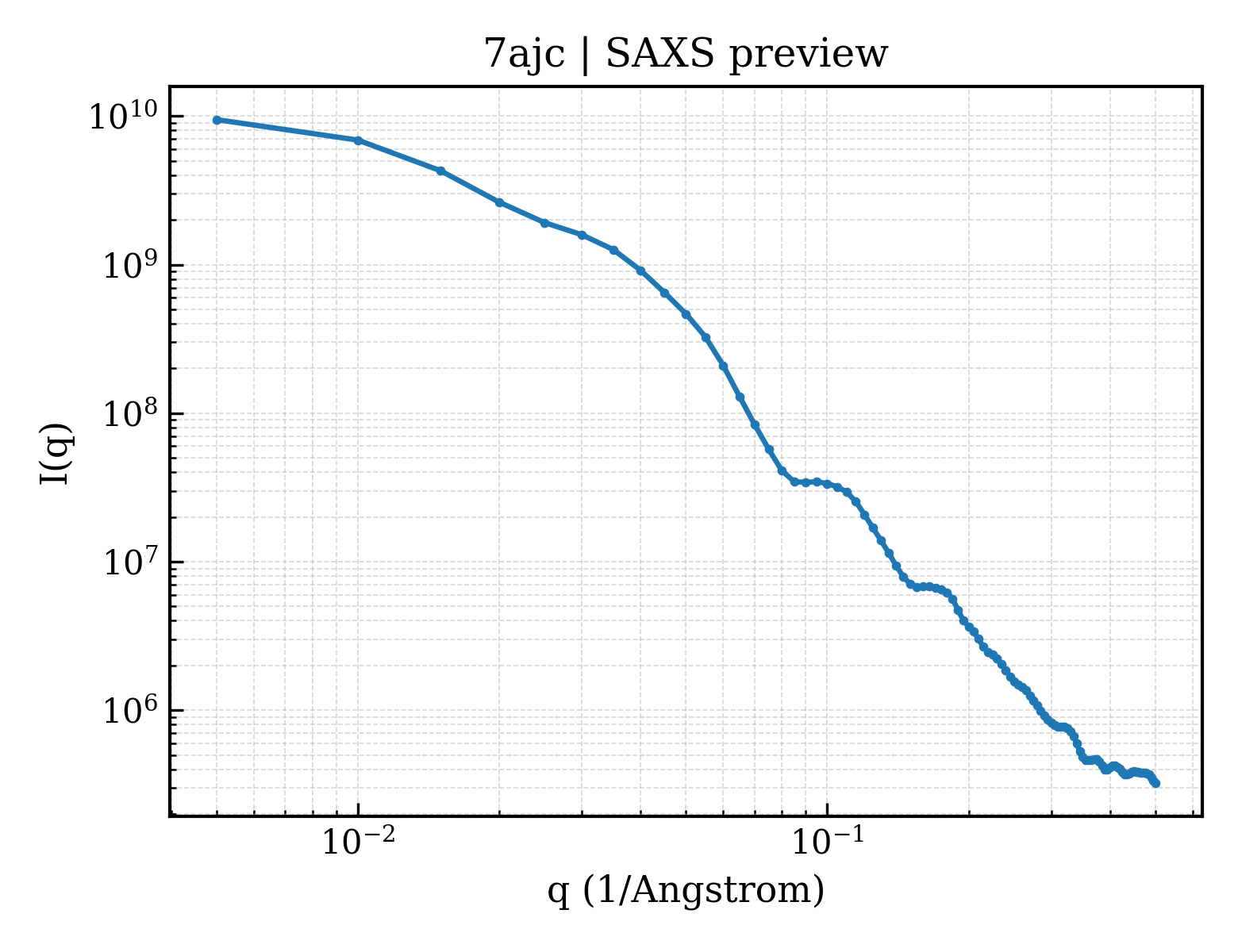
1BMF
BOVINE MITOCHONDRIAL F1-ATPASE
Deposited 1996-03-13
|
Different construct
Different mutation/modification
Different oligomeric state
Different ligand/ion
Different experimental method
Different experimental conditions
Different structure-quality metrics
|
Assembly 1
Protein heterocomplex
Heteromer;Protein × 7
PDB declaration: heptameric
|
Chain A
44–553(510 aa)
Chain B
44–553(510 aa)
Chain C
44–553(510 aa)
|
Not recorded
|
MG MAGNESIUM ION × 5
ANP PHOSPHOAMINOPHOSPHONIC ACID-ADENYLATE ESTER × 4
ADP ADENOSINE-5'-DIPHOSPHATE × 1
|
X-RAY DIFFRACTION
X-ray crystallization conditions
pH 8.2;pH 8.2
|
Resolution 2.85 Å
|
|
1COW
BOVINE MITOCHONDRIAL F1-ATPASE COMPLEXED WITH AUROVERTIN B
Deposited 1996-05-08
|
Different construct
Different mutation/modification
Different oligomeric state
Different ligand/ion
Different experimental method
Different experimental conditions
Different structure-quality metrics
|
Assembly 1
Protein heterocomplex
Heteromer;Protein × 7
PDB declaration: heptameric
|
Chain A
45–553(509 aa)
Chain B
45–553(509 aa)
Chain C
45–553(509 aa)
|
Not recorded
|
MG MAGNESIUM ION × 5
ANP PHOSPHOAMINOPHOSPHONIC ACID-ADENYLATE ESTER × 4
ADP ADENOSINE-5'-DIPHOSPHATE × 1
AUR AUROVERTIN B × 2
|
X-RAY DIFFRACTION
mmCIF provides none of the parsed conditions
|
Resolution 3.10 Å
R-free 0.280
|
|
1E1Q
BOVINE MITOCHONDRIAL F1-ATPASE AT 100K
Deposited 2000-05-10
|
Different construct
Different mutation/modification
Different oligomeric state
Different ligand/ion
Different experimental method
Different experimental conditions
Different structure-quality metrics
|
Assembly 1
Protein heterocomplex
Heteromer;Protein × 7
PDB declaration: heptameric
|
Chain A
44–553(510 aa)
Chain B
44–553(510 aa)
Chain C
44–553(510 aa)
|
Not recorded
|
ANP PHOSPHOAMINOPHOSPHONIC ACID-ADENYLATE ESTER × 4
MG MAGNESIUM ION × 5
ADP ADENOSINE-5'-DIPHOSPHATE × 1
PO4 PHOSPHATE ION × 1
|
X-RAY DIFFRACTION
X-ray crystallization conditions
pH 8;pH 8.00
|
Resolution 2.61 Å
R-free 0.280
|
|
1E1R
BOVINE MITOCHONDRIAL F1-ATPASE INHIBITED BY MG2+ADP AND ALUMINIUM FLUORIDE
Deposited 2000-05-10
|
Different construct
Different mutation/modification
Different oligomeric state
Different ligand/ion
Different experimental method
Different experimental conditions
Different structure-quality metrics
|
Assembly 1
Protein heterocomplex
Heteromer;Protein × 7
PDB declaration: heptameric
|
Chain A
44–553(510 aa)
Chain B
44–553(510 aa)
Chain C
44–553(510 aa)
|
Not recorded
|
ANP PHOSPHOAMINOPHOSPHONIC ACID-ADENYLATE ESTER × 4
MG MAGNESIUM ION × 5
ADP ADENOSINE-5'-DIPHOSPHATE × 1
AF3 ALUMINUM FLUORIDE × 1
PO4 PHOSPHATE ION × 1
|
X-RAY DIFFRACTION
X-ray crystallization conditions
pH 8;pH 8.00
|
Resolution 2.50 Å
R-free 0.282
|
|
1E79
Bovine F1-ATPase inhibited by DCCD (dicyclohexylcarbodiimide)
Deposited 2000-08-25
|
Different construct
Different mutation/modification
Different oligomeric state
Different ligand/ion
Different experimental method
Different experimental conditions
Different structure-quality metrics
|
Assembly 1
Protein heterocomplex
Heteromer;Protein × 9
PDB declaration: nonameric
|
Chain A
44–553(510 aa)
Chain B
44–553(510 aa)
Chain C
44–553(510 aa)
|
Not recorded
|
ATP ADENOSINE-5'-TRIPHOSPHATE × 2
MG MAGNESIUM ION × 5
ADP ADENOSINE-5'-DIPHOSPHATE × 3
GOL GLYCEROL × 1
DCW DICYCLOHEXYLUREA × 1
SO4 SULFATE ION × 1
|
X-RAY DIFFRACTION
X-ray crystallization conditions
pH 7;pH 7.00
|
Resolution 2.40 Å
R-free 0.281
|
|
1EFR
BOVINE MITOCHONDRIAL F1-ATPASE COMPLEXED WITH THE PEPTIDE ANTIBIOTIC EFRAPEPTIN
Deposited 1996-05-24
|
Different construct
Different mutation/modification
Different oligomeric state
Different ligand/ion
Different experimental method
Different experimental conditions
Different structure-quality metrics
|
Assembly 1
Protein heterocomplex
Heteromer;Protein × 8
PDB declaration: octameric
|
Chain A
45–553(509 aa)
Chain B
45–553(509 aa)
Chain C
45–553(509 aa)
|
Not recorded
|
MG MAGNESIUM ION × 5
ANP PHOSPHOAMINOPHOSPHONIC ACID-ADENYLATE ESTER × 4
ADP ADENOSINE-5'-DIPHOSPHATE × 1
|
X-RAY DIFFRACTION
X-ray crystallization conditions
pH 8.2;PH 8.2
|
Resolution 3.10 Å
R-free 0.220
|
|
1H8E
(ADP.AlF4)2(ADP.SO4) bovine F1-ATPase (all three catalytic sites occupied)
Deposited 2001-02-02
|
Different construct
Different mutation/modification
Different oligomeric state
Different ligand/ion
Different experimental method
Different experimental conditions
Different structure-quality metrics
|
Assembly 1
Protein heterocomplex
Heteromer;Protein × 9
PDB declaration: nonameric
|
Chain A
44–553(510 aa)
Chain B
44–553(510 aa)
Chain C
44–553(510 aa)
|
Not recorded
|
ADP ADENOSINE-5'-DIPHOSPHATE × 6
MG MAGNESIUM ION × 6
GOL GLYCEROL × 4
ALF TETRAFLUOROALUMINATE ION × 2
SO4 SULFATE ION × 1
|
X-RAY DIFFRACTION
X-ray crystallization conditions
pH 8;CRYSTALS WERE GROWN IN THE PRESENCE OF AZIDE, A KNOWN INHIBITOR, BUT THIS HAS NOT BEEN LOCATED IN THE STRUCTURE., pH 8.00
|
Resolution 2.00 Å
R-free 0.264
|
|
1H8H
Bovine mitochondrial F1-ATPase crystallised in the presence of 5mm AMPPNP
Deposited 2001-02-06
|
Different construct
Different mutation/modification
Different oligomeric state
Different ligand/ion
Different experimental method
Different experimental conditions
Different structure-quality metrics
|
Assembly 1
Protein heterocomplex
Heteromer;Protein × 7
PDB declaration: heptameric
|
Chain A
44–553(510 aa)
Chain B
44–553(510 aa)
Chain C
44–553(510 aa)
|
Not recorded
|
ATP ADENOSINE-5'-TRIPHOSPHATE × 4
MG MAGNESIUM ION × 5
GOL GLYCEROL × 1
ADP ADENOSINE-5'-DIPHOSPHATE × 1
PO4 PHOSPHATE ION × 1
|
X-RAY DIFFRACTION
X-ray crystallization conditions
pH 8;CRYSTALS WERE GROWN IN THE PRESENCE OF AZIDE, A KNOWN INHIBITOR, BUT THIS HAS NOT BEEN LOCATED IN THE STRUCTURE., pH 8.00
|
Resolution 2.90 Å
R-free 0.292
|
|
1NBM
THE STRUCTURE OF BOVINE F1-ATPASE COVALENTLY INHIBITED WITH 4-CHLORO-7-NITROBENZOFURAZAN
Deposited 1998-04-30
|
Different construct
Different mutation/modification
Different oligomeric state
Different ligand/ion
Different experimental method
Different experimental conditions
Different structure-quality metrics
|
Assembly 1
Protein heterocomplex
Heteromer;Protein × 7
PDB declaration: heptameric
|
Chain A
44–553(510 aa)
Chain B
44–553(510 aa)
Chain C
44–553(510 aa)
|
Not recorded
|
MG MAGNESIUM ION × 5
ATP ADENOSINE-5'-TRIPHOSPHATE × 4
ADP ADENOSINE-5'-DIPHOSPHATE × 1
PO4 PHOSPHATE ION × 1
|
X-RAY DIFFRACTION
X-ray crystallization conditions
pH 7.5;50MM TRIS-HCL, PH7.5, 200MM SODIUM CHLORIDE, 20MM MAGNESIUM SULPHATE, 1MM EDTA, 0.002% (W/V) PHENYL METHYLSULPHONYL FLUORIDE, 0.02%(W/V) SODIUM AZIDE, 10.5% (W/V) PEG MME 5000, 250UM AMP-PNP AND 5UM ADP.
|
Resolution 3.00 Å
R-free 0.297
|
|
1OHH
BOVINE MITOCHONDRIAL F1-ATPASE complexed with the inhibitor protein IF1
Deposited 2003-05-27
|
Different construct
Different mutation/modification
Different oligomeric state
Different ligand/ion
Different experimental method
Different experimental conditions
Different structure-quality metrics
|
Assembly 1
Protein heterocomplex
Heteromer;Protein × 8
PDB declaration: octameric
|
Chain A
44–553(510 aa)
Chain B
44–553(510 aa)
Chain C
44–553(510 aa)
|
Not recorded
|
ANP PHOSPHOAMINOPHOSPHONIC ACID-ADENYLATE ESTER × 5
MG MAGNESIUM ION × 5
|
X-RAY DIFFRACTION
X-ray crystallization conditions
pH 6.6;PROTEIN 20MG/ML IN 100MM PIPES-NAOH PH6.6, 40MM MGSO4, 0.04% NA AZIDE, 10% GLYCEROL, 0.002% PMSF. DROPS EQUAL VOLUME OF PROTEIN AND 10MM AMP-PNP, 300MM NACL, 16% PEG 4000, 5MM SPERMIDINE. BATCH METHOD., pH 6.60
|
Resolution 2.80 Å
R-free 0.280
|
|
1QO1
Molecular Architecture of the Rotary Motor in ATP Synthase from Yeast Mitochondria
Deposited 1999-11-01
|
Different construct
Different mutation/modification
Different oligomeric state
Different ligand/ion
Different experimental method
Different experimental conditions
Different structure-quality metrics
|
Assembly 1
Protein heterocomplex
Heteromer;Protein × 18
PDB declaration: octadecameric
|
Chain A
67–553(487 aa)
Chain B
67–553(487 aa)
Chain C
62–553(492 aa)
|
Not recorded
|
No recorded non-water small molecule
|
X-RAY DIFFRACTION
X-ray crystallization conditions
MICROBATCH;pH 8;0.1 M TRIS/CL PH8.0, 12% PEG 6000, 150 MM NACL, 1 MM AMP-PNP, 40 MICROM ADP, 1 MM DTT, 0.02% NAN3, MIXED 1:1 WITH PROTEIN SOLUTION UNDER PARAFFIN OIL IN MICROBATCH PLATE., pH 8.00
|
Resolution 3.90 Å
|
|
1W0J
Beryllium fluoride inhibited bovine F1-ATPase
Deposited 2004-06-08
|
Different construct
Different mutation/modification
Different oligomeric state
Different ligand/ion
Different experimental method
Different experimental conditions
Different structure-quality metrics
|
Assembly 1
Protein heterocomplex
Heteromer;Protein × 7
PDB declaration: heptameric
|
Chain A
44–553(510 aa)
Chain B
44–553(510 aa)
Chain C
44–553(510 aa)
|
Not recorded
|
ADP ADENOSINE-5'-DIPHOSPHATE × 5
MG MAGNESIUM ION × 5
GOL GLYCEROL × 4
BEF BERYLLIUM TRIFLUORIDE ION × 2
PO4 PHOSPHATE ION × 1
|
X-RAY DIFFRACTION
X-ray crystallization conditions
pH 8.2;CRYSTALS WERE GROWN IN THE PRESENCE OF AZIDE, A KNOWN INHIBITOR, BUT THIS HAS NOT BEEN LOCATED IN THE STRUCTURE., pH 8.20
|
Resolution 2.20 Å
R-free 0.236
|
|
1W0K
ADP inhibited bovine F1-ATPase
Deposited 2004-06-08
|
Different construct
Different mutation/modification
Different oligomeric state
Different ligand/ion
Different experimental method
Different experimental conditions
Different structure-quality metrics
|
Assembly 1
Protein heterocomplex
Heteromer;Protein × 7
PDB declaration: heptameric
|
Chain A
44–553(510 aa)
Chain B
44–553(510 aa)
Chain C
44–553(510 aa)
|
Not recorded
|
ADP ADENOSINE-5'-DIPHOSPHATE × 5
MG MAGNESIUM ION × 5
GOL GLYCEROL × 3
PO4 PHOSPHATE ION × 1
|
X-RAY DIFFRACTION
X-ray crystallization conditions
pH 8.2;CRYSTALS WERE GROWN IN THE PRESENCE OF AZIDE, A KNOWN INHIBITOR, BUT THIS HAS NOT BEEN LOCATED IN THE STRUCTURE., pH 8.20
|
Resolution 2.85 Å
R-free 0.278
|
|
2CK3
Azide inhibited bovine F1-ATPase
Deposited 2006-04-10
|
Different construct
Different mutation/modification
Different oligomeric state
Different ligand/ion
Different experimental method
Different experimental conditions
Different structure-quality metrics
|
Assembly 1
Protein heterocomplex
Heteromer;Protein × 9
PDB declaration: nonameric
|
Chain A
44–553(510 aa)
Chain B
44–553(510 aa)
Chain C
44–553(510 aa)
|
Not recorded
|
ANP PHOSPHOAMINOPHOSPHONIC ACID-ADENYLATE ESTER × 4
MG MAGNESIUM ION × 5
ADP ADENOSINE-5'-DIPHOSPHATE × 1
AZI AZIDE ION × 1
PO4 PHOSPHATE ION × 1
|
X-RAY DIFFRACTION
X-ray crystallization conditions
pH 8.2;50 MM TRIS-HCL PH 8.2, 200 MM NACL, 20 MM MGSO4, 250 UM AMP-PNP, 5 UM ADP, 3 MM NAN3, 0.004% (W/V) PHENYLMETHYLSULFONYL FLUORIDE AND 9% (W/V) POLYETHYLENE GLYCOL 6000.
|
Resolution 1.95 Å
R-free 0.226
|
|
2JDI
Ground state structure of F1-ATPase from bovine heart mitochondria (Bovine F1-ATPase crystallised in the absence of azide)
Deposited 2007-01-09
|
Different construct
Different mutation/modification
Different oligomeric state
Different ligand/ion
Different experimental method
Different experimental conditions
Different structure-quality metrics
|
Assembly 1
Protein heterocomplex
Heteromer;Protein × 9
PDB declaration: nonameric
|
Chain A
44–553(510 aa)
Fragment:RESIDUES 44-553
Chain B
44–553(510 aa)
Fragment:RESIDUES 44-553
Chain C
44–553(510 aa)
Fragment:RESIDUES 44-553
|
Not recorded
|
ANP PHOSPHOAMINOPHOSPHONIC ACID-ADENYLATE ESTER × 5
MG MAGNESIUM ION × 5
|
X-RAY DIFFRACTION
X-ray crystallization conditions
pH 8.2;50 MM TRIS-HCL PH 8.2, 200 MM NACL, 20 MM MGSO4, 250 UM AMP-PNP, 5 UM ADP, 0.004% (W/V) PHENYLMETHYLSULFONYL FLUORIDE AND 12% (W/V) POLYETHYLENE GLYCOL 6000
|
Resolution 1.90 Å
R-free 0.220
|
|
2JIZ
The Structure of F1-ATPase inhibited by resveratrol.
Deposited 2007-07-03
|
Different construct
Different mutation/modification
Different oligomeric state
Different ligand/ion
Different experimental method
Different experimental conditions
Different structure-quality metrics
|
Assembly 1
Protein heterocomplex
Heteromer;Protein × 7
PDB declaration: heptameric
|
Chain A
45–553(509 aa)
Fragment:RESIDUES 44-553
Chain B
45–553(509 aa)
Fragment:RESIDUES 44-553
Chain C
45–553(509 aa)
Fragment:RESIDUES 44-553
|
Not recorded
|
ANP PHOSPHOAMINOPHOSPHONIC ACID-ADENYLATE ESTER × 4
MG MAGNESIUM ION × 5
GOL GLYCEROL × 6
ADP ADENOSINE-5'-DIPHOSPHATE × 1
AZI AZIDE ION × 1
PO4 PHOSPHATE ION × 1
STL RESVERATROL × 1
|
X-RAY DIFFRACTION
X-ray crystallization conditions
pH 8.2;pH 8.2
|
Resolution 2.30 Å
R-free 0.217
|
|
2JIZ
The Structure of F1-ATPase inhibited by resveratrol.
Deposited 2007-07-03
|
Different construct
Different mutation/modification
Different oligomeric state
Different ligand/ion
Different experimental method
Different experimental conditions
Different structure-quality metrics
|
Assembly 2
Protein heterocomplex
Heteromer;Protein × 7
PDB declaration: heptameric
|
Chain H
45–553(509 aa)
Fragment:RESIDUES 44-553
Chain I
45–553(509 aa)
Fragment:RESIDUES 44-553
Chain J
45–553(509 aa)
Fragment:RESIDUES 44-553
|
Not recorded
|
ANP PHOSPHOAMINOPHOSPHONIC ACID-ADENYLATE ESTER × 4
MG MAGNESIUM ION × 5
GOL GLYCEROL × 6
ADP ADENOSINE-5'-DIPHOSPHATE × 1
AZI AZIDE ION × 1
PO4 PHOSPHATE ION × 1
STL RESVERATROL × 1
|
X-RAY DIFFRACTION
X-ray crystallization conditions
pH 8.2;pH 8.2
|
Resolution 2.30 Å
R-free 0.217
|
|
2JJ1
The Structure of F1-ATPase inhibited by piceatannol.
Deposited 2007-07-03
|
Different construct
Different mutation/modification
Different oligomeric state
Different ligand/ion
Different experimental method
Different experimental conditions
Different structure-quality metrics
|
Assembly 1
Protein heterocomplex
Heteromer;Protein × 7
PDB declaration: heptameric
|
Chain A
45–553(509 aa)
Fragment:RESIDUES 44-553
Chain B
45–553(509 aa)
Fragment:RESIDUES 44-553
Chain C
45–553(509 aa)
Fragment:RESIDUES 44-553
|
Not recorded
|
ANP PHOSPHOAMINOPHOSPHONIC ACID-ADENYLATE ESTER × 4
MG MAGNESIUM ION × 5
GOL GLYCEROL × 6
ADP ADENOSINE-5'-DIPHOSPHATE × 1
AZI AZIDE ION × 1
PO4 PHOSPHATE ION × 1
PIT PICEATANNOL × 1
|
X-RAY DIFFRACTION
X-ray crystallization conditions
pH 8.2;pH 8.2
|
Resolution 2.70 Å
R-free 0.269
|
|
2JJ1
The Structure of F1-ATPase inhibited by piceatannol.
Deposited 2007-07-03
|
Different construct
Different mutation/modification
Different oligomeric state
Different ligand/ion
Different experimental method
Different experimental conditions
Different structure-quality metrics
|
Assembly 2
Protein heterocomplex
Heteromer;Protein × 7
PDB declaration: heptameric
|
Chain H
45–553(509 aa)
Fragment:RESIDUES 44-553
Chain I
45–553(509 aa)
Fragment:RESIDUES 44-553
Chain J
45–553(509 aa)
Fragment:RESIDUES 44-553
|
Not recorded
|
ANP PHOSPHOAMINOPHOSPHONIC ACID-ADENYLATE ESTER × 4
MG MAGNESIUM ION × 5
GOL GLYCEROL × 6
ADP ADENOSINE-5'-DIPHOSPHATE × 1
AZI AZIDE ION × 1
PO4 PHOSPHATE ION × 1
PIT PICEATANNOL × 1
|
X-RAY DIFFRACTION
X-ray crystallization conditions
pH 8.2;pH 8.2
|
Resolution 2.70 Å
R-free 0.269
|
|
2JJ2
The Structure of F1-ATPase inhibited by quercetin.
Deposited 2007-07-03
|
Different construct
Different mutation/modification
Different oligomeric state
Different ligand/ion
Different experimental method
Different experimental conditions
Different structure-quality metrics
|
Assembly 1
Protein heterocomplex
Heteromer;Protein × 7
PDB declaration: heptameric
|
Chain A
45–553(509 aa)
Fragment:RESIDUES 44-553
Chain B
45–553(509 aa)
Fragment:RESIDUES 44-553
Chain C
45–553(509 aa)
Fragment:RESIDUES 44-553
|
Not recorded
|
ANP PHOSPHOAMINOPHOSPHONIC ACID-ADENYLATE ESTER × 4
MG MAGNESIUM ION × 5
GOL GLYCEROL × 6
ADP ADENOSINE-5'-DIPHOSPHATE × 1
AZI AZIDE ION × 1
PO4 PHOSPHATE ION × 1
QUE 3,5,7,3',4'-PENTAHYDROXYFLAVONE × 1
|
X-RAY DIFFRACTION
X-ray crystallization conditions
pH 8.2;pH 8.2
|
Resolution 2.40 Å
R-free 0.238
|
|
2JJ2
The Structure of F1-ATPase inhibited by quercetin.
Deposited 2007-07-03
|
Different construct
Different mutation/modification
Different oligomeric state
Different ligand/ion
Different experimental method
Different experimental conditions
Different structure-quality metrics
|
Assembly 2
Protein heterocomplex
Heteromer;Protein × 7
PDB declaration: heptameric
|
Chain H
45–553(509 aa)
Fragment:RESIDUES 44-553
Chain I
45–553(509 aa)
Fragment:RESIDUES 44-553
Chain J
45–553(509 aa)
Fragment:RESIDUES 44-553
|
Not recorded
|
ANP PHOSPHOAMINOPHOSPHONIC ACID-ADENYLATE ESTER × 4
MG MAGNESIUM ION × 5
GOL GLYCEROL × 6
ADP ADENOSINE-5'-DIPHOSPHATE × 1
AZI AZIDE ION × 1
PO4 PHOSPHATE ION × 1
QUE 3,5,7,3',4'-PENTAHYDROXYFLAVONE × 1
|
X-RAY DIFFRACTION
X-ray crystallization conditions
pH 8.2;pH 8.2
|
Resolution 2.40 Å
R-free 0.238
|
|
2JMX
OSCP-NT (1-120) in complex with N-terminal (1-25) alpha subunit from F1-ATPase
Deposited 2006-12-12
|
Different construct
Different mutation/modification
Different oligomeric state
Different ligand/ion
Different experimental method
Different experimental conditions
Different structure-quality metrics
|
Assembly 1
Protein heterocomplex
Heteromer;Protein × 2
PDB declaration: dimeric
|
Chain B
44–68(25 aa)
Fragment:ATP synthase subunit alpha heart isoform, residues 1-25
|
Not recorded
|
No recorded non-water small molecule
|
SOLUTION NMR
NMR measurement conditions
pH 6.5;300 K;Ionic strength (raw mmCIF value) 0.5;Pressure ambient
NMR sample composition
0.5 mM [U-99% 13C, U-99% 15N] oscp-nt, 1.5 mM alpha-nt, 20 mM sodium phosphate, pH 6.5, 0.5 M NaCl, 0.001% PMSF 95% H2O, 5% D2O | 95% H2O/5% D2O
|
Resolution not provided
|
|
2W6E
Low resolution structures of bovine mitochondrial F1-ATPase during controlled dehydration:hydration state 1.
Deposited 2008-12-18
|
Different construct
Different mutation/modification
Different oligomeric state
Different ligand/ion
Different experimental method
Different experimental conditions
Different structure-quality metrics
|
Assembly 1
Protein heterocomplex
Heteromer;Protein × 7
PDB declaration: heptameric
|
Chain A
1–553(553 aa)
Chain B
1–553(553 aa)
Chain C
1–553(553 aa)
|
Not recorded
|
ADP ADENOSINE-5'-DIPHOSPHATE × 1
|
X-RAY DIFFRACTION
X-ray crystallization conditions
pH 8.5;50 MM TRIS-HCL PH 8.2, 200 MM NACL, 20 MM MGSO4, 1 MM ADP, 1 MM ALCL3, 6 MM NAF 0.004% (W/V)PHENYLMETHYLSULFONYL FLUORIDE AND 12% (W/V) POLYETHYLENE GLYCOL 6000
|
Resolution 6.50 Å
R-free 0.285
|
|
2W6F
Low resolution structures of bovine mitochondrial F1-ATPase during controlled dehydration: Hydration State 2.
Deposited 2008-12-18
|
Different construct
Different mutation/modification
Different oligomeric state
Different ligand/ion
Different experimental method
Different experimental conditions
Different structure-quality metrics
|
Assembly 1
Protein heterocomplex
Heteromer;Protein × 7
PDB declaration: heptameric
|
Chain A
1–553(553 aa)
Chain B
1–553(553 aa)
Chain C
1–553(553 aa)
|
Not recorded
|
No recorded non-water small molecule
|
X-RAY DIFFRACTION
X-ray crystallization conditions
pH 8.5;50 MM TRIS-HCL PH 8.2, 200 MM NACL, 20 MM MGSO4, 1 MM ADP, 1 MM ALCL3, 6 MM NAF 0.004% (W/V)PHENYLMETHYLSULFONYL FLUORIDE AND 12% (W/V) POLYETHYLENE GLYCOL 6000
|
Resolution 6.00 Å
R-free 0.343
|
|
2W6G
Low resolution structures of bovine mitochondrial F1-ATPase during controlled dehydration: Hydration State 3.
Deposited 2008-12-18
|
Different construct
Different mutation/modification
Different oligomeric state
Different ligand/ion
Different experimental method
Different experimental conditions
Different structure-quality metrics
|
Assembly 1
Protein heterocomplex
Heteromer;Protein × 7
PDB declaration: heptameric
|
Chain A
1–553(553 aa)
Chain B
1–553(553 aa)
Chain C
1–553(553 aa)
|
Not recorded
|
No recorded non-water small molecule
|
X-RAY DIFFRACTION
X-ray crystallization conditions
pH 8.5;50 MM TRIS-HCL PH 8.2, 200 MM NACL, 20 MM MGSO4, 1 MM ADP, 1 MM ALCL3, 6 MM NAF 0.004% (W/V)PHENYLMETHYLSULFONYL FLUORIDE AND 12% (W/V) POLYETHYLENE GLYCOL 6000
|
Resolution 6.00 Å
|
|
2W6H
Low resolution structures of bovine mitochondrial F1-ATPase during controlled dehydration: Hydration State 4A.
Deposited 2008-12-18
|
Different construct
Different mutation/modification
Different oligomeric state
Different ligand/ion
Different experimental method
Different experimental conditions
Different structure-quality metrics
|
Assembly 1
Protein heterocomplex
Heteromer;Protein × 9
PDB declaration: nonameric
|
Chain A
1–553(553 aa)
Chain B
1–553(553 aa)
Chain C
1–553(553 aa)
|
Not recorded
|
No recorded non-water small molecule
|
X-RAY DIFFRACTION
X-ray crystallization conditions
pH 8.5;50 MM TRIS-HCL PH 8.2, 200 MM NACL, 20 MM MGSO4, 1 MM ADP, 1 MM ALCL3, 6 MM NAF 0.004% (W/V)PHENYLMETHYLSULFONYL FLUORIDE AND 12% (W/V) POLYETHYLENE GLYCOL 6000
|
Resolution 5.00 Å
|
|
2W6I
Low resolution structures of bovine mitochondrial F1-ATPase during controlled dehydration: Hydration State 4B.
Deposited 2008-12-18
|
Different construct
Different mutation/modification
Different oligomeric state
Different ligand/ion
Different experimental method
Different experimental conditions
Different structure-quality metrics
|
Assembly 1
Protein heterocomplex
Heteromer;Protein × 9
PDB declaration: nonameric
|
Chain A
1–553(553 aa)
Chain B
1–553(553 aa)
Chain C
1–553(553 aa)
|
Not recorded
|
No recorded non-water small molecule
|
X-RAY DIFFRACTION
X-ray crystallization conditions
pH 8.5;50 MM TRIS-HCL PH 8.2, 200 MM NACL, 20 MM MGSO4, 1 MM ADP, 1 MM ALCL3, 6 MM NAF 0.004% (W/V)PHENYLMETHYLSULFONYL FLUORIDE AND 12% (W/V) POLYETHYLENE GLYCOL 6000
|
Resolution 4.00 Å
R-free 0.300
|
|
2W6J
Low resolution structures of bovine mitochondrial F1-ATPase during controlled dehydration: Hydration State 5.
Deposited 2008-12-18
|
Different construct
Different mutation/modification
Different oligomeric state
Different ligand/ion
Different experimental method
Different experimental conditions
Different structure-quality metrics
|
Assembly 1
Protein heterocomplex
Heteromer;Protein × 9
PDB declaration: nonameric
|
Chain A
1–553(553 aa)
Chain B
1–553(553 aa)
Chain C
1–553(553 aa)
|
Not recorded
|
No recorded non-water small molecule
|
X-RAY DIFFRACTION
X-ray crystallization conditions
pH 8.5;50 MM TRIS-HCL PH 8.2, 200 MM NACL, 20 MM MGSO4, 1 MM ADP, 1 MM ALCL3, 6 MM NAF 0.004% (W/V)PHENYLMETHYLSULFONYL FLUORIDE AND 12% (W/V) POLYETHYLENE GLYCOL 6000
|
Resolution 3.84 Å
|
|
2WSS
The structure of the membrane extrinsic region of bovine ATP synthase
Deposited 2009-09-09
|
Different construct
Different mutation/modification
Different oligomeric state
Different ligand/ion
Different experimental method
Different experimental conditions
Different structure-quality metrics
|
Assembly 1
Protein heterocomplex
Heteromer;Protein × 13
PDB declaration: tridecameric
|
Chain A
44–553(510 aa)
Chain B
44–553(510 aa)
Chain C
44–553(510 aa)
|
Not recorded
|
ANP PHOSPHOAMINOPHOSPHONIC ACID-ADENYLATE ESTER × 4
MG MAGNESIUM ION × 5
ADP ADENOSINE-5'-DIPHOSPHATE × 1
|
X-RAY DIFFRACTION
X-ray crystallization conditions
MICROBATCH;MICROBATCH UNDER OIL
|
Resolution 3.20 Å
R-free 0.271
|
|
2WSS
The structure of the membrane extrinsic region of bovine ATP synthase
Deposited 2009-09-09
|
Different construct
Different mutation/modification
Different oligomeric state
Different ligand/ion
Different experimental method
Different experimental conditions
Different structure-quality metrics
|
Assembly 2
Protein heterocomplex
Heteromer;Protein × 12
PDB declaration: dodecameric
|
Chain J
44–553(510 aa)
Chain K
44–553(510 aa)
Chain L
44–553(510 aa)
|
Not recorded
|
ANP PHOSPHOAMINOPHOSPHONIC ACID-ADENYLATE ESTER × 4
MG MAGNESIUM ION × 5
ADP ADENOSINE-5'-DIPHOSPHATE × 1
|
X-RAY DIFFRACTION
X-ray crystallization conditions
MICROBATCH;MICROBATCH UNDER OIL
|
Resolution 3.20 Å
R-free 0.271
|
|
2XND
Crystal structure of bovine F1-c8 sub-complex of ATP Synthase
Deposited 2010-08-02
|
Different construct
Different mutation/modification
Different oligomeric state
Different ligand/ion
Different experimental method
Different experimental conditions
Different structure-quality metrics
|
Assembly 1
Protein heterocomplex
Heteromer;Protein × 17
PDB declaration: heptadecameric
|
Chain A
62–553(492 aa)
Fragment:RESIDUES 62-553
Chain B
62–553(492 aa)
Fragment:RESIDUES 62-553
Chain C
62–553(492 aa)
Fragment:RESIDUES 62-553
|
Not recorded
|
ANP PHOSPHOAMINOPHOSPHONIC ACID-ADENYLATE ESTER × 5
MG MAGNESIUM ION × 5
GOL GLYCEROL × 1
SO4 SULFATE ION × 1
|
X-RAY DIFFRACTION
X-ray crystallization conditions
pH 7;CRYSTALS WERE GROWN UNDER OIL BY MIXING EQUAL VOLUMES OF PROTEIN (10MG/ML IN 20MM TRIS PH 8.0, 10% GLYCEROL, 1MM ADP, 1MM AMP-PNP, 2MM MGSO4, 0.02% NAN3, 5.7MM TDM) AND PRECIPITANT SOLUTION (50MM HEPES PH 7.0, 14% PEG4600, 50MM K2HPO4)
|
Resolution 3.50 Å
R-free 0.304
|
|
4ASU
F1-ATPase in which all three catalytic sites contain bound nucleotide, with magnesium ion released in the Empty site
Deposited 2012-05-03
|
Different construct
Different mutation/modification
Different oligomeric state
Different ligand/ion
Different experimental method
Different experimental conditions
Different structure-quality metrics
|
Assembly 1
Protein heterocomplex
Heteromer;Protein × 9
PDB declaration: nonameric
|
Chain A
44–553(510 aa)
Chain B
44–553(510 aa)
Chain C
44–553(510 aa)
|
Not recorded
|
ADP ADENOSINE-5'-DIPHOSPHATE × 6
MG MAGNESIUM ION × 5
|
X-RAY DIFFRACTION
mmCIF provides none of the parsed conditions
|
Resolution 2.60 Å
R-free 0.289
|
|
4TSF
The Pathway of Binding of the Intrinsically Disordered Mitochondrial Inhibitor Protein to F1-ATPase
Deposited 2014-06-18
|
Different construct
Different mutation/modification
Different oligomeric state
Different ligand/ion
Different experimental method
Different experimental conditions
Different structure-quality metrics
|
Assembly 1
Protein heterocomplex
Heteromer;Protein × 9
PDB declaration: Nonameric
|
Chain A
44–553(510 aa)
Chain B
44–553(510 aa)
Chain C
44–553(510 aa)
|
Not recorded
|
ATP ADENOSINE-5'-TRIPHOSPHATE × 3
MG MAGNESIUM ION × 5
ADP ADENOSINE-5'-DIPHOSPHATE × 2
|
X-RAY DIFFRACTION
X-ray crystallization conditions
MICROBATCH;pH 8.2;295 K;Tris-HCl, PEG 4000, magnesium chloride, EDTA, NaCl, spermidine
|
Resolution 3.20 Å
R-free 0.272
|
|
4TT3
The Pathway of Binding of the Intrinsically Disordered Mitochondrial Inhibitor Protein to F1-ATPase
Deposited 2014-06-19
|
Different construct
Different mutation/modification
Different oligomeric state
Different ligand/ion
Different experimental method
Different experimental conditions
Different structure-quality metrics
|
Assembly 1
Protein heterocomplex
Heteromer;Protein × 10
PDB declaration: Decameric
|
Chain A
44–553(510 aa)
Chain B
44–553(510 aa)
Chain C
44–553(510 aa)
|
Not recorded
|
ATP ADENOSINE-5'-TRIPHOSPHATE × 3
MG MAGNESIUM ION × 5
GOL GLYCEROL × 2
ADP ADENOSINE-5'-DIPHOSPHATE × 2
|
X-RAY DIFFRACTION
X-ray crystallization conditions
MICROBATCH;pH 8.2;295 K;PEG 4000, Tris-HCl, magnesium chloride, EDTA, ATP, NaCl, spermidine
|
Resolution 3.21 Å
R-free 0.283
|
|
4YXW
Bovine heart mitochondrial F1-ATPase inhibited by AMP-PNP and ADP in the presence of thiophosphate.
Deposited 2015-03-23
|
Different construct
Different mutation/modification
Different oligomeric state
Different ligand/ion
Different experimental method
Different experimental conditions
Different structure-quality metrics
|
Assembly 1
Protein heterocomplex
Heteromer;Protein × 9
PDB declaration: nonameric
|
Chain A
44–553(510 aa)
Chain B
44–553(510 aa)
Chain C
44–553(510 aa)
|
Not recorded
|
ANP PHOSPHOAMINOPHOSPHONIC ACID-ADENYLATE ESTER × 5
MG MAGNESIUM ION × 5
CL CHLORIDE ION × 1
TS6 Monothiophosphate × 1
NA SODIUM ION × 1
|
X-RAY DIFFRACTION
X-ray crystallization conditions
MICRODIALYSIS;pH 8.2;295 K;PEG 6000, sodium chloride, magnesium chloride, Tris-HCl, AMP-PNP, ADP, sodium monothiophosphate
|
Resolution 3.10 Å
R-free 0.273
|
|
4Z1M
Bovine F1-ATPase inhibited by three copies of the inhibitor protein IF1 crystallised in the presence of thiophosphate.
Deposited 2015-03-27
|
Different construct
Different mutation/modification
Different oligomeric state
Different ligand/ion
Different experimental method
Different experimental conditions
Different structure-quality metrics
|
Assembly 1
Protein heterocomplex
Heteromer;Protein × 10
PDB declaration: decameric
|
Chain A
44–553(510 aa)
Chain B
44–553(510 aa)
Chain C
44–553(510 aa)
|
Not recorded
|
ATP ADENOSINE-5'-TRIPHOSPHATE × 3
MG MAGNESIUM ION × 5
GOL GLYCEROL × 2
CL CHLORIDE ION × 11
ADP ADENOSINE-5'-DIPHOSPHATE × 2
|
X-RAY DIFFRACTION
X-ray crystallization conditions
MICROBATCH;295 K;Tris.HCl, MgCl2, EDTA, sodium monothiophosphate, ATP NaCl, spermidine, PEG 4000
|
Resolution 3.30 Å
R-free 0.275
|
|
5ARA
Bovine mitochondrial ATP synthase state 1a
Deposited 2015-09-24
|
Different construct
Different mutation/modification
Different oligomeric state
Different ligand/ion
Different experimental conditions
Different structure-quality metrics
|
Assembly 1
Protein heterocomplex
Heteromer;Protein × 22
PDB declaration: 22-meric
|
Chain A
44–553(510 aa)
Fragment:UNP RESIDUES 44-553
Chain B
44–553(510 aa)
Fragment:UNP RESIDUES 44-553
Chain C
44–553(510 aa)
Fragment:UNP RESIDUES 44-553
|
Not recorded
|
No recorded non-water small molecule
|
ELECTRON MICROSCOPY
cryo-EM buffer
20 MM TRIS-HCL, 100 MM NACL, DODECYLMALTOSIDE, 2 MM ATP, 0.02% (WT/V) NAN3;pH 7.2;20 MM TRIS-HCL, 100 MM NACL, DODECYLMALTOSIDE, 2 MM ATP, 0.02% (WT/V) NAN3
cryo-EM vitrification conditions
Cryogen ETHANE-PROPANE;VITRIFICATION 1 -- CRYOGEN- ETHANE-PROPANE MIXTURE, HUMIDITY- 100, INSTRUMENT- FEI VITROBOT MARK III, METHOD- BLOT FOR 27 SECONDS BEFORE PLUNGING,
|
Resolution 7.40 Å
|
|
5ARE
Bovine mitochondrial ATP synthase state 1b
Deposited 2015-09-24
|
Different construct
Different mutation/modification
Different oligomeric state
Different ligand/ion
Different experimental conditions
Different structure-quality metrics
|
Assembly 1
Protein heterocomplex
Heteromer;Protein × 22
PDB declaration: 22-meric
|
Chain A
44–553(510 aa)
Fragment:UNP RESIDUES 44-553
Chain B
44–553(510 aa)
Fragment:UNP RESIDUES 44-553
Chain C
44–553(510 aa)
Fragment:UNP RESIDUES 44-553
|
Not recorded
|
No recorded non-water small molecule
|
ELECTRON MICROSCOPY
cryo-EM buffer
20 MM TRIS-HCL, 100 MM NACL, 10% (V/V) DODECYLMALTOSIDE, 2 MM ATP, 0.02% (WT/V) NAN3;pH 7.2;20 MM TRIS-HCL, 100 MM NACL, 10% (V/V) DODECYLMALTOSIDE, 2 MM ATP, 0.02% (WT/V) NAN3
cryo-EM vitrification conditions
Cryogen ETHANE-PROPANE;VITRIFICATION 1 -- CRYOGEN- ETHANE-PROPANE MIXTURE, HUMIDITY- 100, INSTRUMENT- FEI VITROBOT MARK III, METHOD- BLOT FOR 27 SECONDS BEFORE PLUNGING,
|
Resolution 6.70 Å
|
|
5ARH
Bovine mitochondrial ATP synthase state 2a
Deposited 2015-09-24
|
Different construct
Different mutation/modification
Different oligomeric state
Different ligand/ion
Different experimental conditions
Different structure-quality metrics
|
Assembly 1
Protein heterocomplex
Heteromer;Protein × 22
PDB declaration: 22-meric
|
Chain A
44–553(510 aa)
Fragment:UNP RESIDUES 44-553
Chain B
44–553(510 aa)
Fragment:UNP RESIDUES 44-553
Chain C
44–553(510 aa)
Fragment:UNP RESIDUES 44-553
|
Not recorded
|
No recorded non-water small molecule
|
ELECTRON MICROSCOPY
cryo-EM buffer
20 MM TRIS-HCL, 100 MM NACL, 10% (V/V) DODECYLMALTOSIDE, 2 MM ATP, 0.02% (WT/V) NAN3;pH 7.2;20 MM TRIS-HCL, 100 MM NACL, 10% (V/V) DODECYLMALTOSIDE, 2 MM ATP, 0.02% (WT/V) NAN3
cryo-EM vitrification conditions
Cryogen ETHANE-PROPANE;VITRIFICATION 1 -- CRYOGEN- ETHANE-PROPANE MIXTURE, HUMIDITY- 100, INSTRUMENT- FEI VITROBOT MARK III, METHOD- BLOT FOR 27 SECONDS BEFORE PLUNGING,
|
Resolution 7.20 Å
|
|
5ARI
Bovine mitochondrial ATP synthase state 2b
Deposited 2015-09-24
|
Different construct
Different mutation/modification
Different oligomeric state
Different ligand/ion
Different experimental conditions
Different structure-quality metrics
|
Assembly 1
Protein heterocomplex
Heteromer;Protein × 22
PDB declaration: 22-meric
|
Chain A
44–553(510 aa)
Fragment:UNP RESIDUES 44-553
Chain B
44–553(510 aa)
Fragment:UNP RESIDUES 44-553
Chain C
44–553(510 aa)
Fragment:UNP RESIDUES 44-553
|
Not recorded
|
No recorded non-water small molecule
|
ELECTRON MICROSCOPY
cryo-EM buffer
20 MM TRIS-HCL, 100 MM NACL, 10% (V/V) DODECYLMALTOSIDE, 2 MM ATP, 0.02% (WT/V) NAN3;pH 7.2;20 MM TRIS-HCL, 100 MM NACL, 10% (V/V) DODECYLMALTOSIDE, 2 MM ATP, 0.02% (WT/V) NAN3
cryo-EM vitrification conditions
Cryogen ETHANE-PROPANE;VITRIFICATION 1 -- CRYOGEN- ETHANE-PROPANE MIXTURE, HUMIDITY- 100, INSTRUMENT- FEI VITROBOT MARK III, METHOD- BLOT FOR 27 SECONDS BEFORE PLUNGING,
|
Resolution 7.40 Å
|
|
5FIJ
Bovine mitochondrial ATP synthase state 2c
Deposited 2015-09-28
|
Different construct
Different mutation/modification
Different oligomeric state
Different ligand/ion
Different experimental conditions
Different structure-quality metrics
|
Assembly 1
Protein heterocomplex
Heteromer;Protein × 22
PDB declaration: 22-meric
|
Chain A
44–553(510 aa)
Fragment:UNP RESIDUES 44-553
Chain B
44–553(510 aa)
Fragment:UNP RESIDUES 44-553
Chain C
44–553(510 aa)
Fragment:UNP RESIDUES 44-553
|
Not recorded
|
No recorded non-water small molecule
|
ELECTRON MICROSCOPY
cryo-EM buffer
20 MM TRIS-HCL, 100 MM NACL, 10% (V/V) DODECYLMALTOSIDE, 2 MM ATP, 0.02% (WT/V) NAN3;pH 7.2;20 MM TRIS-HCL, 100 MM NACL, 10% (V/V) DODECYLMALTOSIDE, 2 MM ATP, 0.02% (WT/V) NAN3
cryo-EM vitrification conditions
Cryogen ETHANE-PROPANE;VITRIFICATION 1 -- CRYOGEN- ETHANE-PROPANE MIXTURE, HUMIDITY- 100, INSTRUMENT- FEI VITROBOT MARK III, METHOD- BLOT FOR 27 SECONDS BEFORE PLUNGING,
|
Resolution 7.40 Å
|
|
5FIK
Bovine mitochondrial ATP synthase state 3a
Deposited 2015-09-28
|
Different construct
Different mutation/modification
Different oligomeric state
Different ligand/ion
Different experimental conditions
Different structure-quality metrics
|
Assembly 1
Protein heterocomplex
Heteromer;Protein × 22
PDB declaration: 22-meric
|
Chain A
44–553(510 aa)
Fragment:UNP RESIDUES 44-553
Chain B
44–553(510 aa)
Fragment:UNP RESIDUES 44-553
Chain C
44–553(510 aa)
Fragment:UNP RESIDUES 44-553
|
Not recorded
|
No recorded non-water small molecule
|
ELECTRON MICROSCOPY
cryo-EM buffer
20 MM TRIS-HCL, 100 MM NACL, 10% (V/V) DODECYLMALTOSIDE, 2 MM ATP, 0.02% (WT/V) NAN3;pH 7.2;20 MM TRIS-HCL, 100 MM NACL, 10% (V/V) DODECYLMALTOSIDE, 2 MM ATP, 0.02% (WT/V) NAN3
cryo-EM vitrification conditions
Cryogen ETHANE-PROPANE;VITRIFICATION 1 -- CRYOGEN- ETHANE-PROPANE MIXTURE, HUMIDITY- 100, INSTRUMENT- FEI VITROBOT MARK III, METHOD- BLOT FOR 27 SECONDS BEFORE PLUNGING,
|
Resolution 6.40 Å
|
|
5FIL
Bovine mitochondrial ATP synthase state 3b
Deposited 2015-09-28
|
Different construct
Different mutation/modification
Different oligomeric state
Different ligand/ion
Different experimental conditions
Different structure-quality metrics
|
Assembly 1
Protein heterocomplex
Heteromer;Protein × 22
PDB declaration: 22-meric
|
Chain A
44–553(510 aa)
Fragment:UNP RESIDUES 44-553
Chain B
44–553(510 aa)
Fragment:UNP RESIDUES 44-553
Chain C
44–553(510 aa)
Fragment:UNP RESIDUES 44-553
|
Not recorded
|
No recorded non-water small molecule
|
ELECTRON MICROSCOPY
cryo-EM buffer
20 MM TRIS-HCL, 100 MM NACL, 10% (V/V) GLYCEROL, 0.05% (WT/V) DODECYLMALTOSIDE, 2 MM ATP, 0.02% (WT/V) NAN3;pH 7.2;20 MM TRIS-HCL, 100 MM NACL, 10% (V/V) GLYCEROL, 0.05% (WT/V) DODECYLMALTOSIDE, 2 MM ATP, 0.02% (WT/V) NAN3
cryo-EM vitrification conditions
Cryogen ETHANE-PROPANE;VITRIFICATION 1 -- CRYOGEN- ETHANE-PROPANE MIXTURE, HUMIDITY- 100, INSTRUMENT- FEI VITROBOT MARK III, METHOD- BLOT FOR 27 SECONDS BEFORE PLUNGING,
|
Resolution 7.10 Å
|
|
6YY0
bovine ATP synthase F1-peripheral stalk domain, state 1
Deposited 2020-05-04
|
Different construct
Different mutation/modification
Different oligomeric state
Different ligand/ion
Different experimental conditions
Different structure-quality metrics
|
Assembly 1
Protein heterocomplex
Heteromer;Protein × 14
PDB declaration: tetradecameric
|
Chain A
44–553(510 aa)
Chain B
44–553(510 aa)
Chain C
44–553(510 aa)
|
Not recorded
|
ATP ADENOSINE-5'-TRIPHOSPHATE × 3
MG MAGNESIUM ION × 5
ADP ADENOSINE-5'-DIPHOSPHATE × 3
|
ELECTRON MICROSCOPY
cryo-EM buffer
pH 7.4
cryo-EM vitrification conditions
Cryogen ETHANE;The sample was allowed to penetrate through the holey support and to distribute to both sides of the grid surface for ca. 15 sec. Then the grids were blotted with filter paper for 8-10 sec before blotting.
|
Resolution 3.23 Å
|
|
6Z1R
bovine ATP synthase F1-peripheral stalk domain, state 2
Deposited 2020-05-14
|
Different construct
Different mutation/modification
Different oligomeric state
Different ligand/ion
Different experimental conditions
Different structure-quality metrics
|
Assembly 1
Protein heterocomplex
Heteromer;Protein × 21
PDB declaration: 21-meric
|
Chain A
44–553(510 aa)
Chain B
44–553(510 aa)
Chain C
44–553(510 aa)
|
Not recorded
|
ATP ADENOSINE-5'-TRIPHOSPHATE × 3
MG MAGNESIUM ION × 5
ADP ADENOSINE-5'-DIPHOSPHATE × 3
|
ELECTRON MICROSCOPY
cryo-EM buffer
pH 7.4
cryo-EM vitrification conditions
Cryogen ETHANE;The sample was allowed to penetrate through the holey support and to distribute to both sides of the grid surface for ca. 15 sec. Then the grids were blotted with filter paper for 8-10 sec before blotting.
|
Resolution 3.29 Å
|
|
6Z1U
bovine ATP synthase F1c8-peripheral stalk domain, state 3
Deposited 2020-05-14
|
Different construct
Different mutation/modification
Different oligomeric state
Different ligand/ion
Different experimental conditions
Different structure-quality metrics
|
Assembly 1
Protein heterocomplex
Heteromer;Protein × 21
PDB declaration: 21-meric
|
Chain A
44–553(510 aa)
Chain B
44–553(510 aa)
Chain C
44–553(510 aa)
|
Not recorded
|
ATP ADENOSINE-5'-TRIPHOSPHATE × 3
MG MAGNESIUM ION × 5
ADP ADENOSINE-5'-DIPHOSPHATE × 3
|
ELECTRON MICROSCOPY
cryo-EM buffer
pH 7.4
cryo-EM vitrification conditions
Cryogen ETHANE;The sample was allowed to penetrate through the holey support and to distribute to both sides of the grid surface for ca. 15 sec. Then the grids were blotted with filter paper for 8-10 sec before blotting.
|
Resolution 3.47 Å
|
|
6ZIQ
bovine ATP synthase stator domain, state 1
Deposited 2020-06-26
|
Different construct
Different mutation/modification
Different oligomeric state
Different ligand/ion
Different experimental conditions
Different structure-quality metrics
|
Assembly 1
Protein heterocomplex
Heteromer;Protein × 15
PDB declaration: pentadecameric
|
Chain C
44–553(510 aa)
|
Not recorded
|
No recorded non-water small molecule
|
ELECTRON MICROSCOPY
cryo-EM buffer
pH 7.4
cryo-EM vitrification conditions
Cryogen ETHANE;The sample was allowed to penetrate through the holey support and to distribute to both sides of the grid surface for ca. 15 sec. Then the grids were blotted with filter paper for 8-10 sec before blotting.
|
Resolution 4.33 Å
|
|
6ZIT
bovine ATP synthase Stator domain, state 2
Deposited 2020-06-26
|
Different construct
Different mutation/modification
Different oligomeric state
Different ligand/ion
Different experimental conditions
Different structure-quality metrics
|
Assembly 1
Protein heterocomplex
Heteromer;Protein × 15
PDB declaration: pentadecameric
|
Chain C
44–553(510 aa)
|
Not recorded
|
No recorded non-water small molecule
|
ELECTRON MICROSCOPY
cryo-EM buffer
pH 7.4
cryo-EM vitrification conditions
Cryogen ETHANE;The sample was allowed to penetrate through the holey support and to distribute to both sides of the grid surface for ca. 15 sec. Then the grids were blotted with filter paper for 8-10 sec before blotting.
|
Resolution 3.49 Å
|
|
6ZIU
bovine ATP synthase stator domain, state 3
Deposited 2020-06-26
|
Different construct
Different mutation/modification
Different oligomeric state
Different ligand/ion
Different experimental conditions
Different structure-quality metrics
|
Assembly 1
Protein heterocomplex
Heteromer;Protein × 11
PDB declaration: undecameric
|
Chain C
44–553(510 aa)
|
Not recorded
|
No recorded non-water small molecule
|
ELECTRON MICROSCOPY
cryo-EM buffer
pH 7.4
cryo-EM vitrification conditions
Cryogen ETHANE;Th sample was allowed to penetrate through the holey grid support and to distribute to both sides of the grid surface for ~15sec. Then the grids were blotted with filter paper for 8-10 sec before blotting.
|
Resolution 6.02 Å
|
|
6ZPO
bovine ATP synthase monomer state 1 (combined)
Deposited 2020-07-09
|
Different construct
Different mutation/modification
Different oligomeric state
Different ligand/ion
Different experimental conditions
Different structure-quality metrics
|
Assembly 1
Protein heterocomplex
Heteromer;Protein × 29
PDB declaration: 29-meric
|
Chain A
44–553(510 aa)
Chain B
44–553(510 aa)
Chain C
44–553(510 aa)
|
Not recorded
|
ATP ADENOSINE-5'-TRIPHOSPHATE × 3
MG MAGNESIUM ION × 5
ADP ADENOSINE-5'-DIPHOSPHATE × 3
CDL CARDIOLIPIN × 3
LHG 1,2-DIPALMITOYL-PHOSPHATIDYL-GLYCEROLE × 2
|
ELECTRON MICROSCOPY
cryo-EM buffer
pH 7.4
cryo-EM vitrification conditions
Cryogen ETHANE;The sample was allowed to penetrate through the holey support and to distribute to both sides of the grid surface for ca. 15 sec. Then the grids were blotted with filter paper for 8-10 sec before blotting.
|
Resolution 4.00 Å
|
|
6ZQM
bovine ATP synthase monomer state 2 (combined)
Deposited 2020-07-10
|
Different construct
Different mutation/modification
Different oligomeric state
Different ligand/ion
Different experimental conditions
Different structure-quality metrics
|
Assembly 1
Protein heterocomplex
Heteromer;Protein × 29
PDB declaration: 29-meric
|
Chain A
44–553(510 aa)
Chain B
44–553(510 aa)
Chain C
44–553(510 aa)
|
Not recorded
|
ATP ADENOSINE-5'-TRIPHOSPHATE × 3
MG MAGNESIUM ION × 5
ADP ADENOSINE-5'-DIPHOSPHATE × 3
CDL CARDIOLIPIN × 3
LHG 1,2-DIPALMITOYL-PHOSPHATIDYL-GLYCEROLE × 2
|
ELECTRON MICROSCOPY
cryo-EM buffer
pH 7.4
cryo-EM vitrification conditions
Cryogen ETHANE;The sample was allowed to penetrate through the holey support and to distribute to both sides of the grid surface for ca. 15 sec. Then the grids were blotted with filter paper for 8-10 sec before blotting.
|
Resolution 3.29 Å
|
|
6ZQN
bovine ATP synthase monomer state 3 (combined)
Deposited 2020-07-10
|
Different construct
Different mutation/modification
Different oligomeric state
Different ligand/ion
Different experimental conditions
Different structure-quality metrics
|
Assembly 1
Protein heterocomplex
Heteromer;Protein × 29
PDB declaration: 29-meric
|
Chain A
44–553(510 aa)
Chain B
44–553(510 aa)
Chain C
44–553(510 aa)
|
Not recorded
|
ATP ADENOSINE-5'-TRIPHOSPHATE × 3
MG MAGNESIUM ION × 5
ADP ADENOSINE-5'-DIPHOSPHATE × 3
CDL CARDIOLIPIN × 3
LHG 1,2-DIPALMITOYL-PHOSPHATIDYL-GLYCEROLE × 2
|
ELECTRON MICROSCOPY
cryo-EM buffer
pH 7.4
cryo-EM vitrification conditions
Cryogen ETHANE;The sample was allowed to penetrate through the holey support and to distribute to both sides of the grid surface for ca. 15 sec. Then the grids were blotted with filter paper for 8-10 sec before blotting.
|
Resolution 4.00 Å
|
|
7AJB
bovine ATP synthase dimer state1:state1
Deposited 2020-09-29
|
Different structure-quality metrics
|
Assembly 1
Protein heterocomplex
Heteromer;Protein × 58
PDB declaration: 58-meric
|
Chain A
44–553(510 aa)
Chain AA
44–553(510 aa)
Chain AB
44–553(510 aa)
Chain AC
44–553(510 aa)
Chain B
44–553(510 aa)
Chain C
44–553(510 aa)
|
Not recorded
|
CDL CARDIOLIPIN × 6
LHG 1,2-DIPALMITOYL-PHOSPHATIDYL-GLYCEROLE × 4
|
ELECTRON MICROSCOPY
cryo-EM buffer
pH 7.4
cryo-EM vitrification conditions
Cryogen ETHANE
|
Resolution 9.20 Å
|
|
7AJD
bovine ATP synthase dimer state1:state3
Deposited 2020-09-29
|
Different structure-quality metrics
|
Assembly 1
Protein heterocomplex
Heteromer;Protein × 58
PDB declaration: 58-meric
|
Chain A
44–553(510 aa)
Chain AA
44–553(510 aa)
Chain AB
44–553(510 aa)
Chain AC
44–553(510 aa)
Chain B
44–553(510 aa)
Chain C
44–553(510 aa)
|
Not recorded
|
CDL CARDIOLIPIN × 6
LHG 1,2-DIPALMITOYL-PHOSPHATIDYL-GLYCEROLE × 4
|
ELECTRON MICROSCOPY
cryo-EM buffer
pH 7.4
cryo-EM vitrification conditions
Cryogen ETHANE
|
Resolution 9.00 Å
|
|
7AJE
bovine ATP synthase dimer state2:state1
Deposited 2020-09-29
|
Different structure-quality metrics
|
Assembly 1
Protein heterocomplex
Heteromer;Protein × 58
PDB declaration: 58-meric
|
Chain A
44–553(510 aa)
Chain AA
44–553(510 aa)
Chain AB
44–553(510 aa)
Chain AC
44–553(510 aa)
Chain B
44–553(510 aa)
Chain C
44–553(510 aa)
|
Not recorded
|
CDL CARDIOLIPIN × 6
LHG 1,2-DIPALMITOYL-PHOSPHATIDYL-GLYCEROLE × 4
|
ELECTRON MICROSCOPY
cryo-EM buffer
pH 7.4
cryo-EM vitrification conditions
Cryogen ETHANE
|
Resolution 9.40 Å
|
|
7AJF
bovine ATP synthase dimer state2:state2
Deposited 2020-09-29
|
Different structure-quality metrics
|
Assembly 1
Protein heterocomplex
Heteromer;Protein × 58
PDB declaration: 58-meric
|
Chain A
44–553(510 aa)
Chain AA
44–553(510 aa)
Chain AB
44–553(510 aa)
Chain AC
44–553(510 aa)
Chain B
44–553(510 aa)
Chain C
44–553(510 aa)
|
Not recorded
|
CDL CARDIOLIPIN × 6
LHG 1,2-DIPALMITOYL-PHOSPHATIDYL-GLYCEROLE × 4
|
ELECTRON MICROSCOPY
cryo-EM buffer
pH 7.4
cryo-EM vitrification conditions
Cryogen ETHANE
|
Resolution 8.45 Å
|
|
7AJG
bovine ATP synthase dimer state2:state3
Deposited 2020-09-29
|
Different structure-quality metrics
|
Assembly 1
Protein heterocomplex
Heteromer;Protein × 58
PDB declaration: 58-meric
|
Chain A
44–553(510 aa)
Chain AA
44–553(510 aa)
Chain AB
44–553(510 aa)
Chain AC
44–553(510 aa)
Chain B
44–553(510 aa)
Chain C
44–553(510 aa)
|
Not recorded
|
CDL CARDIOLIPIN × 6
LHG 1,2-DIPALMITOYL-PHOSPHATIDYL-GLYCEROLE × 4
|
ELECTRON MICROSCOPY
cryo-EM buffer
pH 7.4
cryo-EM vitrification conditions
Cryogen ETHANE
|
Resolution 10.70 Å
|
|
7AJH
bovine ATP synthase dimer state3:state1
Deposited 2020-09-29
|
Different structure-quality metrics
|
Assembly 1
Protein heterocomplex
Heteromer;Protein × 58
PDB declaration: 58-meric
|
Chain A
44–553(510 aa)
Chain AA
44–553(510 aa)
Chain AB
44–553(510 aa)
Chain AC
44–553(510 aa)
Chain B
44–553(510 aa)
Chain C
44–553(510 aa)
|
Not recorded
|
CDL CARDIOLIPIN × 6
LHG 1,2-DIPALMITOYL-PHOSPHATIDYL-GLYCEROLE × 4
|
ELECTRON MICROSCOPY
cryo-EM buffer
pH 7.4
cryo-EM vitrification conditions
Cryogen ETHANE
|
Resolution 9.70 Å
|
|
7AJI
bovine ATP synthase dimer state3:state2
Deposited 2020-09-29
|
Different structure-quality metrics
|
Assembly 1
Protein heterocomplex
Heteromer;Protein × 58
PDB declaration: 58-meric
|
Chain A
44–553(510 aa)
Chain AA
44–553(510 aa)
Chain AB
44–553(510 aa)
Chain AC
44–553(510 aa)
Chain B
44–553(510 aa)
Chain C
44–553(510 aa)
|
Not recorded
|
CDL CARDIOLIPIN × 6
LHG 1,2-DIPALMITOYL-PHOSPHATIDYL-GLYCEROLE × 4
|
ELECTRON MICROSCOPY
cryo-EM buffer
pH 7.4
cryo-EM vitrification conditions
Cryogen ETHANE
|
Resolution 11.40 Å
|
|
7AJJ
bovine ATP synthase dimer state3:state3
Deposited 2020-09-29
|
Different structure-quality metrics
|
Assembly 1
Protein heterocomplex
Heteromer;Protein × 58
PDB declaration: 58-meric
|
Chain A
44–553(510 aa)
Chain AA
44–553(510 aa)
Chain AB
44–553(510 aa)
Chain AC
44–553(510 aa)
Chain B
44–553(510 aa)
Chain C
44–553(510 aa)
|
Not recorded
|
CDL CARDIOLIPIN × 6
LHG 1,2-DIPALMITOYL-PHOSPHATIDYL-GLYCEROLE × 4
|
ELECTRON MICROSCOPY
cryo-EM buffer
pH 7.4
cryo-EM vitrification conditions
Cryogen ETHANE
|
Resolution 13.10 Å
|
|
9W2R
Cryo-EM structure of FoF1-ATPase monomer state 1 on the bovine heart submitochondrial particles (FoF1-1)
Deposited 2025-07-28
|
Different construct
Different mutation/modification
Different oligomeric state
Different ligand/ion
Different experimental conditions
Different structure-quality metrics
|
Assembly 1
Protein heterocomplex
Heteromer;Protein × 29
PDB declaration: 29-meric
|
Chain A
44–553(510 aa)
Chain B
44–553(510 aa)
Chain C
44–553(510 aa)
|
Not recorded
|
ATP ADENOSINE-5'-TRIPHOSPHATE × 4
MG MAGNESIUM ION × 5
ADP ADENOSINE-5'-DIPHOSPHATE × 1
|
ELECTRON MICROSCOPY
cryo-EM buffer
pH 7.5
cryo-EM vitrification conditions
Cryogen ETHANE
|
Resolution 3.40 Å
|
|
9W2S
Cryo-EM structure of FoF1-ATPase monomer state 3 on the bovine heart submitochondrial particles (FoF1-2)
Deposited 2025-07-28
|
Different construct
Different mutation/modification
Different oligomeric state
Different ligand/ion
Different experimental conditions
Different structure-quality metrics
|
Assembly 1
Protein heterocomplex
Heteromer;Protein × 29
PDB declaration: 29-meric
|
Chain A
44–553(510 aa)
Chain B
44–553(510 aa)
Chain C
44–553(510 aa)
|
Not recorded
|
No recorded non-water small molecule
|
ELECTRON MICROSCOPY
cryo-EM buffer
pH 7.5
cryo-EM vitrification conditions
Cryogen ETHANE
|
Resolution 4.00 Å
|