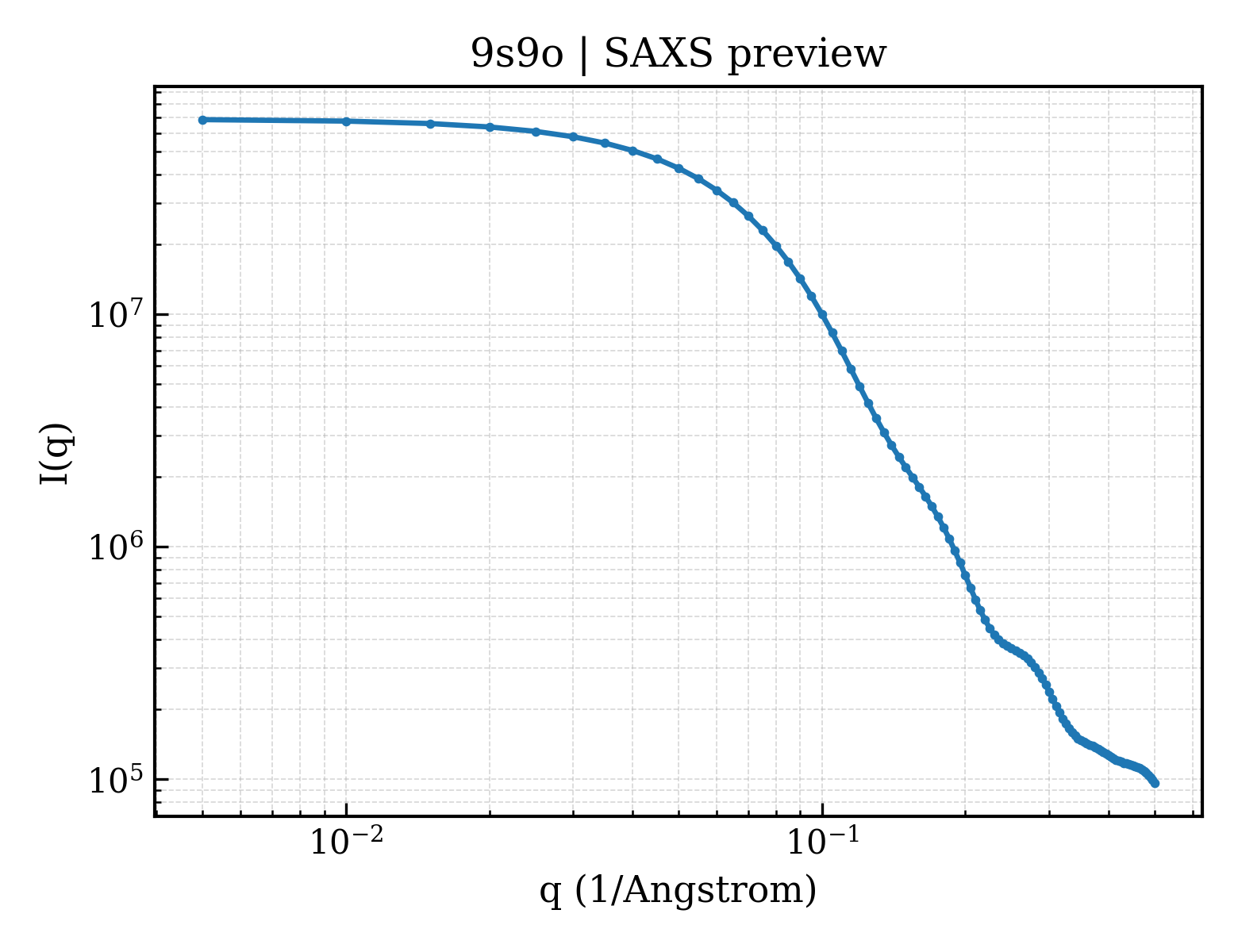
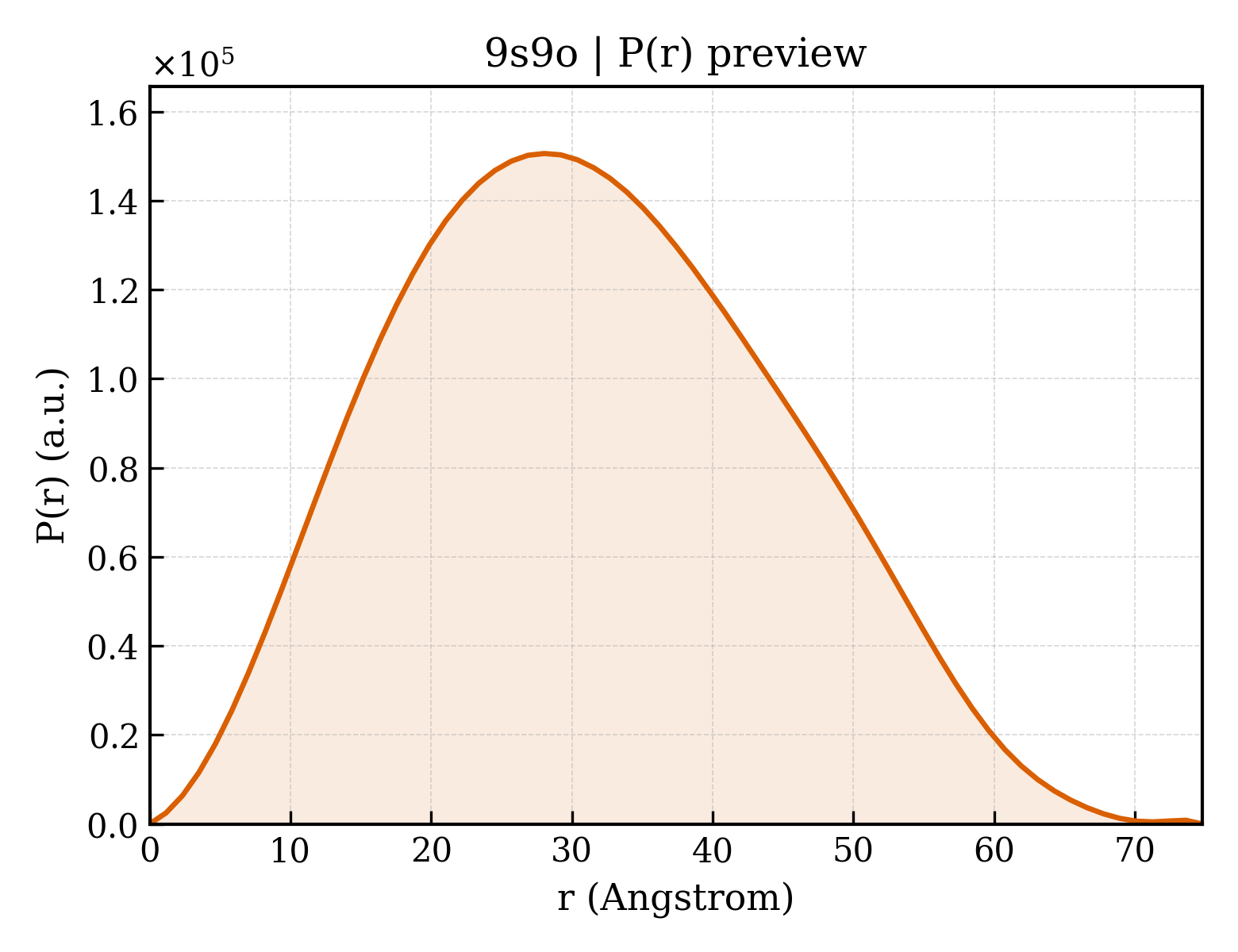
Cellular tumor antigen p53
Homo sapiens
State in the Current Structure
| Assembly | Oligomeric State | Construct | Mutations and Modifications | Ligands, Ions and Associated Components | Method and Experimental Conditions | Structure Quality |
|---|---|---|---|---|---|---|
| 1 | Protein monomer Monomer Protein × 1 PDB declaration: monomeric(1) Consistent with protein copy count | Chain A; UniProt 94–312 | Not recorded | ZN ZINC ION × 1 A1JMR 4-[3-[4-[[(3~{S},4~{R})-3-fluoranyl-1-methyl-piperidin-4-yl]amino]-1-[2,2,2-tris(fluoranyl)ethyl]indol-2-yl]prop-2-ynylamino]-3-methoxy-~{N}-methyl-benzamide × 1 EDO 1,2-ETHANEDIOL × 2 | X-RAY DIFFRACTION X-ray crystallization conditions:VAPOR DIFFUSION, SITTING DROP;293 K;Protein solution: 6 mg/ml protein in 25 mM Hepes, pH 7.5, 150 mM NaCl, 0.5 mM TCEP. Reservoir buffer: 100 mM Hepes, pH 7.0, 19% (w/v) polyethylene glycol 4000. Soaking buffer: 18 mM compound in 100 mM Hepes, pH 7.2, 10 mM sodium phosphate, pH 7.2, 19% (w/v) polyethylene glycol 4000, 20 % (v/v) glycerol, 150 mM KCl | Resolution 1.49 Å R-free 0.186 |
| 2 | Protein monomer Monomer Protein × 1 PDB declaration: monomeric(1) Consistent with protein copy count | Chain B; UniProt 94–312 | Not recorded | ZN ZINC ION × 1 A1JMR 4-[3-[4-[[(3~{S},4~{R})-3-fluoranyl-1-methyl-piperidin-4-yl]amino]-1-[2,2,2-tris(fluoranyl)ethyl]indol-2-yl]prop-2-ynylamino]-3-methoxy-~{N}-methyl-benzamide × 1 EDO 1,2-ETHANEDIOL × 2 | X-RAY DIFFRACTION X-ray crystallization conditions:VAPOR DIFFUSION, SITTING DROP;293 K;Protein solution: 6 mg/ml protein in 25 mM Hepes, pH 7.5, 150 mM NaCl, 0.5 mM TCEP. Reservoir buffer: 100 mM Hepes, pH 7.0, 19% (w/v) polyethylene glycol 4000. Soaking buffer: 18 mM compound in 100 mM Hepes, pH 7.2, 10 mM sodium phosphate, pH 7.2, 19% (w/v) polyethylene glycol 4000, 20 % (v/v) glycerol, 150 mM KCl | Resolution 1.49 Å R-free 0.186 |
Other States of the Same Protein in the Database
Each row is a biological assembly of the same UniProt protein in another PDB entry. The “Difference from current entry” column identifies evidence-level differences; no tag means the currently parsed fields agree.
| Other PDB | Difference from Current Entry 9S9O | Assembly / Oligomeric State | Construct | Mutations and Modifications | Ligands, Ions and Non-polymers | Method and Experimental Conditions | Structure Quality |
|---|---|---|---|---|---|---|---|
| 1A1U SOLUTION STRUCTURE DETERMINATION OF A P53 MUTANT DIMERIZATION DOMAIN, NMR, MINIMIZED AVERAGE STRUCTURE Deposited 1997-12-16 | Different construct Different mutation/modification Different oligomeric state Different ligand/ion Different experimental method Different experimental conditions Different structure-quality metrics | Assembly 1 Protein homooligomer Homooligomer;Protein × 2 PDB declaration: dimeric |
Chain A
324–358(35 aa)
Fragment:OLIGOMERIZATION
Chain C
324–358(35 aa)
Fragment:OLIGOMERIZATION
|
Mutation:M40K, F41I, L44Y Mutation:M40K, F41I, L44Y | No recorded non-water small molecule |
SOLUTION NMR
NMR measurement conditions
pH 7;313 K
|
Resolution not provided |
| 1AIE P53 TETRAMERIZATION DOMAIN CRYSTAL STRUCTURE Deposited 1997-04-17 | Different construct Different oligomeric state Different ligand/ion Different experimental conditions Different structure-quality metrics | Assembly 1 Protein homooligomer Homooligomer;Protein × 4 PDB declaration: tetrameric |
Chain A
326–356(31 aa)
Fragment:TETRAMERIZATION DOMAIN
|
Not recorded | No recorded non-water small molecule |
X-RAY DIFFRACTION
X-ray crystallization conditions
pH 8;1.4 M SODIUM CITRATE, 100 MM HEPES, PH 8.0
|
Resolution 1.50 Å R-free 0.252 |
| 1C26 CRYSTAL STRUCTURE OF P53 TETRAMERIZATION DOMAIN Deposited 1999-07-22 | Different construct Different oligomeric state Different ligand/ion Different experimental conditions Different structure-quality metrics | Assembly 1 Protein homooligomer Homooligomer;Protein × 4 PDB declaration: tetrameric |
Chain A
325–356(32 aa)
Fragment:TETRAMERIZATION DOMAIN
|
Not recorded | No recorded non-water small molecule |
X-RAY DIFFRACTION
X-ray crystallization conditions
VAPOR DIFFUSION, HANGING DROP;pH 8.8;298 K;3.0 M SODIUM FORMATE 0.5 M AMMONIUM SULFATE 50 MM TRIS-HCL PH 8.8, VAPOR DIFFUSION, HANGING DROP at 298 K
|
Resolution 1.70 Å |
| 1DT7 SOLUTION STRUCTURE OF THE C-TERMINAL NEGATIVE REGULATORY DOMAIN OF P53 IN A COMPLEX WITH CA2+-BOUND S100B(BB) Deposited 2000-01-11 | Different construct Different mutation/modification Different oligomeric state Different ligand/ion Different experimental method Different experimental conditions Different structure-quality metrics | Assembly 1 Protein heterocomplex Heteromer;Protein × 4 PDB declaration: tetrameric |
Chain X
367–388(22 aa)
Fragment:C-TERMINAL PEPTIDE
Chain Y
367–388(22 aa)
Fragment:C-TERMINAL PEPTIDE
|
Not recorded | CA CALCIUM ION × 4 |
SOLUTION NMR
NMR measurement conditions
pH 6.5;310 K;Ionic strength (raw mmCIF value) 25 mM;Pressure ambient
NMR sample composition
3-6 mM S100 (subunit concentration), 0.1 mM EDTA, 0.34 mM NaN3, 5 mM DTT, 17 mM NaCl, 10 mM d11-tris-HCl, 7% D2O, 6-13 mM CaCl2, 4.8-10 mM p53 peptide | 7% D2O, 93% H2O
|
Resolution not provided |
| 1GZH Crystal structure of the BRCT domains of human 53BP1 bound to the p53 tumor supressor Deposited 2002-05-22 | Different construct Different oligomeric state Different ligand/ion Different experimental conditions Different structure-quality metrics | Assembly 1 Protein heterocomplex Heteromer;Protein × 2 PDB declaration: dimeric |
Chain A
95–292(198 aa)
Fragment:DNA BINDING REGION, RESIDUES 95-292
|
Not recorded | ZN ZINC ION × 1 SO4 SULFATE ION × 1 |
X-RAY DIFFRACTION
X-ray crystallization conditions
pH 7.4;50 MM TRIS PH 7.4, 250 MM AMMONIUM SULFATE, 25% POLYETHYLENE GLYCOL 4000
|
Resolution 2.60 Å R-free 0.288 |
| 1GZH Crystal structure of the BRCT domains of human 53BP1 bound to the p53 tumor supressor Deposited 2002-05-22 | Different construct Different oligomeric state Different ligand/ion Different experimental conditions Different structure-quality metrics | Assembly 2 Protein heterocomplex Heteromer;Protein × 2 PDB declaration: dimeric |
Chain C
95–292(198 aa)
Fragment:DNA BINDING REGION, RESIDUES 95-292
|
Not recorded | ZN ZINC ION × 1 SO4 SULFATE ION × 1 |
X-RAY DIFFRACTION
X-ray crystallization conditions
pH 7.4;50 MM TRIS PH 7.4, 250 MM AMMONIUM SULFATE, 25% POLYETHYLENE GLYCOL 4000
|
Resolution 2.60 Å R-free 0.288 |
| 1H26 CDK2/CyclinA in complex with an 11-residue recruitment peptide from p53 Deposited 2002-07-31 | Different construct Different oligomeric state Different ligand/ion Different experimental conditions Different structure-quality metrics | Assembly 1 Protein heterocomplex Heteromer;Protein × 3 PDB declaration: trimeric |
Chain E
376–386(11 aa)
Fragment:RESIDUES 376-386
|
Not recorded | No recorded non-water small molecule |
X-RAY DIFFRACTION
X-ray crystallization conditions
pH 7;0.8M KCL, 1.2M (NH4)2SO4, 40MM HEPES PH 7.0. PROTEIN CONCENTRATION = 10MG/ML
|
Resolution 2.24 Å R-free 0.268 |
| 1HS5 NMR SOLUTION STRUCTURE OF DESIGNED P53 DIMER Deposited 2000-12-22 | Different construct Different mutation/modification Different oligomeric state Different ligand/ion Different experimental method Different experimental conditions Different structure-quality metrics | Assembly 1 Protein homooligomer Homooligomer;Protein × 2 PDB declaration: dimeric |
Chain A
324–357(34 aa)
Fragment:RESIDUES 324-357
Chain B
324–357(34 aa)
Fragment:RESIDUES 324-357
|
Mutation:M340Q, L344R Mutation:M340Q, L344R | No recorded non-water small molecule |
SOLUTION NMR
NMR measurement conditions
pH 7;300 K;Ionic strength (raw mmCIF value) 25mM;Pressure ambient
NMR sample composition
2mM U-15N,13C; 25mM sodium phosphate; 150mM sodium chloride | 90% H2O/10% D2O
|
Resolution not provided |
| 1JSP NMR Structure of CBP Bromodomain in complex with p53 peptide Deposited 2001-08-17 | Different construct Different mutation/modification Different oligomeric state Different ligand/ion Different experimental method Different experimental conditions Different structure-quality metrics | Assembly 1 Protein heterocomplex Heteromer;Protein × 2 PDB declaration: dimeric |
Chain A
367–386(20 aa)
Fragment:C-terminal fragment
|
Non-standard monomer:Yes (specific site not provided by mmCIF) | No recorded non-water small molecule |
SOLUTION NMR
NMR measurement conditions
pH 6.5;298 K;Pressure ambient
NMR measurement conditions
pH 6.5;298 K;Pressure ambient
NMR measurement conditions
pH 6.5;298 K;Pressure ambient
NMR measurement conditions
pH 6.5;298 K;Pressure ambient
NMR sample composition
0.5mM CBP bromodomain U-15N; 0.5mM P53 peptide; 100mM phosphate buffer; pH 6.5 | 90% H2O/10% D2O
NMR sample composition
0.5mM CBP bromodomain U-15N,13C; 0.5mM P53 peptide;100mM phosphate buffer; pH 6.5 | 90% H2O/10% D2O
NMR sample composition
0.5mM CBP bromodomain U-15N,13C; 0.5mM P53 peptide;100mM phosphate buffer; pH 6.5 | 99.9%D2O
NMR sample composition
0.5mM CBP bromodomain U-15N,13C,75% 2H; 0.5mM P53 peptide;100mM phosphate buffer; pH 6.5 | 90% H2O/10% D2O
|
Resolution not provided |
| 1KZY Crystal Structure of the 53bp1 BRCT Region Complexed to Tumor Suppressor P53 Deposited 2002-02-08 | Different construct Different mutation/modification Different oligomeric state Different ligand/ion Different experimental conditions Different structure-quality metrics | Assembly 1 Protein heterocomplex Heteromer;Protein × 4 PDB declaration: tetrameric |
Chain A
95–289(195 aa)
Fragment:DNA-BINDING CORE DOMAIN
Chain B
95–289(195 aa)
Fragment:DNA-BINDING CORE DOMAIN
|
Not recorded | ZN ZINC ION × 2 |
X-RAY DIFFRACTION
X-ray crystallization conditions
VAPOR DIFFUSION, HANGING DROP;pH 6;277 K;PEG4000, sodium citrate, ammonium acetate, pH 6.0, VAPOR DIFFUSION, HANGING DROP, temperature 277K
|
Resolution 2.50 Å R-free 0.256 |
| 1MA3 Structure of a Sir2 enzyme bound to an acetylated p53 peptide Deposited 2002-07-31 | Different construct Different mutation/modification Different oligomeric state Different ligand/ion Different experimental conditions Different structure-quality metrics | Assembly 1 Protein heterocomplex Heteromer;Protein × 2 PDB declaration: dimeric |
Chain B
372–389(18 aa)
Fragment:Regulatory C-terminal tail (residues 372-389)
|
Non-standard monomer:Yes (specific site not provided by mmCIF) | ZN ZINC ION × 1 MES 2-(N-MORPHOLINO)-ETHANESULFONIC ACID × 1 |
X-RAY DIFFRACTION
X-ray crystallization conditions
VAPOR DIFFUSION, HANGING DROP;pH 5.5;291 K;PEG 8000, PEG 1000 and Sodium Chloride, pH 5.5, VAPOR DIFFUSION, HANGING DROP, temperature 291K
|
Resolution 2.00 Å R-free 0.254 |
| 1OLG HIGH-RESOLUTION SOLUTION STRUCTURE OF THE OLIGOMERIZATION DOMAIN OF P53 BY MULTI-DIMENSIONAL NMR Deposited 1994-06-13 | Different construct Different mutation/modification Different oligomeric state Different ligand/ion Different experimental method Different experimental conditions Different structure-quality metrics | Assembly 1 Protein homooligomer Homooligomer;Protein × 4 PDB declaration: tetrameric |
Chain A
319–360(42 aa)
Chain B
319–360(42 aa)
Chain C
319–360(42 aa)
Chain D
319–360(42 aa)
|
Not recorded | No recorded non-water small molecule | SOLUTION NMR mmCIF provides none of the parsed conditions | Resolution not provided |
| 1OLH HIGH-RESOLUTION SOLUTION STRUCTURE OF THE OLIGOMERIZATION DOMAIN OF P53 BY MULTI-DIMENSIONAL NMR Deposited 1994-06-13 | Different construct Different mutation/modification Different oligomeric state Different ligand/ion Different experimental method Different experimental conditions Different structure-quality metrics | Assembly 1 Protein homooligomer Homooligomer;Protein × 4 PDB declaration: tetrameric |
Chain A
319–360(42 aa)
Chain B
319–360(42 aa)
Chain C
319–360(42 aa)
Chain D
319–360(42 aa)
|
Not recorded | No recorded non-water small molecule | SOLUTION NMR mmCIF provides none of the parsed conditions | Resolution not provided |
| 1PES NMR SOLUTION STRUCTURE OF THE TETRAMERIC MINIMUM TRANSFORMING DOMAIN OF P53 Deposited 1994-11-24 | Different construct Different mutation/modification Different oligomeric state Different ligand/ion Different experimental method Different experimental conditions Different structure-quality metrics | Assembly 1 Protein homooligomer Homooligomer;Protein × 4 PDB declaration: tetrameric |
Chain A
325–355(31 aa)
Chain B
325–355(31 aa)
Chain C
325–355(31 aa)
Chain D
325–355(31 aa)
|
Not recorded | No recorded non-water small molecule | SOLUTION NMR mmCIF provides none of the parsed conditions | Resolution not provided |
| 1PET NMR SOLUTION STRUCTURE OF THE TETRAMERIC MINIMUM TRANSFORMING DOMAIN OF P53 Deposited 1994-11-24 | Different construct Different mutation/modification Different oligomeric state Different ligand/ion Different experimental method Different experimental conditions Different structure-quality metrics | Assembly 1 Protein homooligomer Homooligomer;Protein × 4 PDB declaration: tetrameric |
Chain A
325–355(31 aa)
Chain B
325–355(31 aa)
Chain C
325–355(31 aa)
Chain D
325–355(31 aa)
|
Not recorded | No recorded non-water small molecule | SOLUTION NMR mmCIF provides none of the parsed conditions | Resolution not provided |
| 1SAE HIGH RESOLUTION SOLUTION NMR STRUCTURE OF THE OLIGOMERIZATION DOMAIN OF P53 BY MULTI-DIMENSIONAL NMR (SAC STRUCTURES) Deposited 1995-03-12 | Different construct Different mutation/modification Different oligomeric state Different ligand/ion Different experimental method Different experimental conditions Different structure-quality metrics | Assembly 1 Protein homooligomer Homooligomer;Protein × 4 PDB declaration: tetrameric |
Chain A
319–360(42 aa)
Chain B
319–360(42 aa)
Chain C
319–360(42 aa)
Chain D
319–360(42 aa)
|
Not recorded | No recorded non-water small molecule | SOLUTION NMR mmCIF provides none of the parsed conditions | Resolution not provided |
| 1SAF HIGH RESOLUTION SOLUTION NMR STRUCTURE OF THE OLIGOMERIZATION DOMAIN OF P53 BY MULTI-DIMENSIONAL NMR (SAD STRUCTURES) Deposited 1995-03-12 | Different construct Different mutation/modification Different oligomeric state Different ligand/ion Different experimental method Different experimental conditions Different structure-quality metrics | Assembly 1 Protein homooligomer Homooligomer;Protein × 4 PDB declaration: tetrameric |
Chain A
319–360(42 aa)
Chain B
319–360(42 aa)
Chain C
319–360(42 aa)
Chain D
319–360(42 aa)
|
Not recorded | No recorded non-water small molecule | SOLUTION NMR mmCIF provides none of the parsed conditions | Resolution not provided |
| 1SAK HIGH RESOLUTION SOLUTION NMR STRUCTURE OF THE OLIGOMERIZATION DOMAIN OF P53 BY MULTI-DIMENSIONAL NMR (SAC STRUCTURES) Deposited 1995-03-12 | Different construct Different mutation/modification Different oligomeric state Different ligand/ion Different experimental method Different experimental conditions Different structure-quality metrics | Assembly 1 Protein homooligomer Homooligomer;Protein × 4 PDB declaration: tetrameric |
Chain A
319–360(42 aa)
Chain B
319–360(42 aa)
Chain C
319–360(42 aa)
Chain D
319–360(42 aa)
|
Not recorded | No recorded non-water small molecule | SOLUTION NMR mmCIF provides none of the parsed conditions | Resolution not provided |
| 1SAL HIGH RESOLUTION SOLUTION NMR STRUCTURE OF THE OLIGOMERIZATION DOMAIN OF P53 BY MULTI-DIMENSIONAL NMR (SAD STRUCTURES) Deposited 1995-03-12 | Different construct Different mutation/modification Different oligomeric state Different ligand/ion Different experimental method Different experimental conditions Different structure-quality metrics | Assembly 1 Protein homooligomer Homooligomer;Protein × 4 PDB declaration: tetrameric |
Chain A
319–360(42 aa)
Chain B
319–360(42 aa)
Chain C
319–360(42 aa)
Chain D
319–360(42 aa)
|
Not recorded | No recorded non-water small molecule | SOLUTION NMR mmCIF provides none of the parsed conditions | Resolution not provided |
| 1TSR P53 CORE DOMAIN IN COMPLEX WITH DNA Deposited 1995-07-28 | Different construct Different mutation/modification Different oligomeric state Different ligand/ion Different experimental conditions Different structure-quality metrics | Assembly 1 Protein–DNA Homooligomer;Protein × 3 PDB declaration: pentameric |
Chain A
94–312(219 aa)
Chain B
94–312(219 aa)
Chain C
94–312(219 aa)
|
Not recorded | ZN ZINC ION × 3 | X-RAY DIFFRACTION mmCIF provides none of the parsed conditions | Resolution 2.20 Å |
| 1TUP TUMOR SUPPRESSOR P53 COMPLEXED WITH DNA Deposited 1995-07-11 | Different construct Different mutation/modification Different oligomeric state Different ligand/ion Different experimental conditions Different structure-quality metrics | Assembly 1 Protein–DNA Homooligomer;Protein × 3 PDB declaration: pentameric |
Chain A
94–312(219 aa)
Chain B
94–312(219 aa)
Chain C
94–312(219 aa)
|
Not recorded | ZN ZINC ION × 3 | X-RAY DIFFRACTION mmCIF provides none of the parsed conditions | Resolution 2.20 Å |
| 1UOL Crystal structure of the human p53 core domain mutant M133L/V203A/N239Y/N268D at 1.9 A resolution. Deposited 2003-09-19 | Different construct Different mutation/modification Different ligand/ion Different experimental conditions Different structure-quality metrics | Assembly 1 Protein monomer Monomer;Protein × 1 PDB declaration: monomeric |
Chain A
94–312(219 aa)
Fragment:DNA BINDING (CORE) DOMAIN, RESIDUES 94-312
|
Mutation:YES | ZN ZINC ION × 1 |
X-RAY DIFFRACTION
X-ray crystallization conditions
pH 7.2;pH 7.20
|
Resolution 1.90 Å R-free 0.230 |
| 1UOL Crystal structure of the human p53 core domain mutant M133L/V203A/N239Y/N268D at 1.9 A resolution. Deposited 2003-09-19 | Different construct Different mutation/modification Different ligand/ion Different experimental conditions Different structure-quality metrics | Assembly 2 Protein monomer Monomer;Protein × 1 PDB declaration: monomeric |
Chain B
94–312(219 aa)
Fragment:DNA BINDING (CORE) DOMAIN, RESIDUES 94-312
|
Mutation:YES | ZN ZINC ION × 1 |
X-RAY DIFFRACTION
X-ray crystallization conditions
pH 7.2;pH 7.20
|
Resolution 1.90 Å R-free 0.230 |
| 1XQH Crystal structure of a ternary complex of the methyltransferase SET9 (also known as SET7/9) with a P53 peptide and SAH Deposited 2004-10-12 | Different construct Different mutation/modification Different oligomeric state Different ligand/ion Different experimental conditions Different structure-quality metrics | Assembly 1 Protein heterocomplex Heteromer;Protein × 2 PDB declaration: dimeric |
Chain B
369–377(9 aa)
Fragment:mono-methylated p53 peptide
|
Non-standard monomer:Yes (specific site not provided by mmCIF) | SAH S-ADENOSYL-L-HOMOCYSTEINE × 1 |
X-RAY DIFFRACTION
X-ray crystallization conditions
VAPOR DIFFUSION;pH 7.8;291 K;PEG3350, Tris-HCL, pH 7.8, VAPOR DIFFUSION, temperature 291K
|
Resolution 1.75 Å R-free 0.223 |
| 1XQH Crystal structure of a ternary complex of the methyltransferase SET9 (also known as SET7/9) with a P53 peptide and SAH Deposited 2004-10-12 | Different construct Different mutation/modification Different oligomeric state Different ligand/ion Different experimental conditions Different structure-quality metrics | Assembly 2 Protein heterocomplex Heteromer;Protein × 2 PDB declaration: dimeric |
Chain F
369–377(9 aa)
Fragment:mono-methylated p53 peptide
|
Non-standard monomer:Yes (specific site not provided by mmCIF) | SAH S-ADENOSYL-L-HOMOCYSTEINE × 1 |
X-RAY DIFFRACTION
X-ray crystallization conditions
VAPOR DIFFUSION;pH 7.8;291 K;PEG3350, Tris-HCL, pH 7.8, VAPOR DIFFUSION, temperature 291K
|
Resolution 1.75 Å R-free 0.223 |
| 1YCQ XENOPUS LAEVIS MDM2 BOUND TO THE TRANSACTIVATION DOMAIN OF HUMAN P53 Deposited 1996-09-30 | Different construct Different oligomeric state Different ligand/ion Different experimental conditions Different structure-quality metrics | Assembly 1 Protein heterocomplex Heteromer;Protein × 4 PDB declaration: tetrameric |
Chain B
13–29(17 aa)
Fragment:RESIDUES 13 - 29
|
Not recorded | No recorded non-water small molecule | X-RAY DIFFRACTION mmCIF provides none of the parsed conditions | Resolution 2.30 Å R-free 0.253 |
| 1YCR MDM2 BOUND TO THE TRANSACTIVATION DOMAIN OF P53 Deposited 1996-09-30 | Different construct Different oligomeric state Different ligand/ion Different experimental conditions Different structure-quality metrics | Assembly 1 Protein heterocomplex Heteromer;Protein × 2 PDB declaration: dimeric |
Chain B
15–29(15 aa)
Fragment:RESIDUES 15 - 29
|
Not recorded | No recorded non-water small molecule | X-RAY DIFFRACTION mmCIF provides none of the parsed conditions | Resolution 2.60 Å R-free 0.276 |
| 1YCS P53-53BP2 COMPLEX Deposited 1996-09-30 | Different construct Different oligomeric state Different ligand/ion Different experimental conditions Different structure-quality metrics | Assembly 1 Protein heterocomplex Heteromer;Protein × 2 PDB declaration: dimeric |
Chain A
94–292(199 aa)
Fragment:RESIDUES 97 - 287
|
Not recorded | ZN ZINC ION × 1 | X-RAY DIFFRACTION mmCIF provides none of the parsed conditions | Resolution 2.20 Å R-free 0.286 |
| 2AC0 Structural Basis of DNA Recognition by p53 Tetramers (complex I) Deposited 2005-07-18 | Different construct Different mutation/modification Different oligomeric state Different ligand/ion Different experimental conditions Different structure-quality metrics | Assembly 1 Protein–DNA Homooligomer;Protein × 4 PDB declaration: octameric |
Chain A
94–293(200 aa)
Fragment:residues 94-293
Chain B
94–293(200 aa)
Fragment:residues 94-293
Chain C
94–293(200 aa)
Fragment:residues 94-293
Chain D
94–293(200 aa)
Fragment:residues 94-293
|
Not recorded | ZN ZINC ION × 4 |
X-RAY DIFFRACTION
X-ray crystallization conditions
VAPOR DIFFUSION, HANGING DROP;pH 6.6;293 K;Ammonium Formate, PEG 3350, pH 6.6, VAPOR DIFFUSION, HANGING DROP, temperature 293K
|
Resolution 1.80 Å R-free 0.217 |
| 2ADY Structural Basis of DNA Recognition by p53 Tetramers (complex IV) Deposited 2005-07-21 | Different construct Different mutation/modification Different oligomeric state Different ligand/ion Different experimental conditions Different structure-quality metrics | Assembly 1 Protein–DNA Homooligomer;Protein × 4 PDB declaration: octameric |
Chain A
94–293(200 aa)
Fragment:residues 94-293
Chain B
94–293(200 aa)
Fragment:residues 94-293
|
Not recorded | ZN ZINC ION × 4 |
X-RAY DIFFRACTION
X-ray crystallization conditions
VAPOR DIFFUSION, HANGING DROP;pH 6.2;293 K;Ammonium Fluoride, PEG 3350, pH 6.2, VAPOR DIFFUSION, HANGING DROP, temperature 293K
|
Resolution 2.50 Å R-free 0.251 |
| 2AHI Structural Basis of DNA Recognition by p53 Tetramers (complex III) Deposited 2005-07-28 | Different construct Different mutation/modification Different oligomeric state Different ligand/ion Different experimental conditions Different structure-quality metrics | Assembly 1 Protein–DNA Homooligomer;Protein × 4 PDB declaration: octameric |
Chain A
94–293(200 aa)
Fragment:residues 94-293
Chain B
94–293(200 aa)
Fragment:residues 94-293
Chain C
94–293(200 aa)
Fragment:residues 94-293
Chain D
94–293(200 aa)
Fragment:residues 94-293
|
Not recorded | ZN ZINC ION × 4 |
X-RAY DIFFRACTION
X-ray crystallization conditions
VAPOR DIFFUSION, HANGING DROP;pH 6.3;293 K;Ammonium Chloride, PEG 3350, pH 6.3, VAPOR DIFFUSION, HANGING DROP, temperature 293K
|
Resolution 1.85 Å R-free 0.225 |
| 2ATA Structural Basis of DNA Recognition by p53 Tetramers (complex II) Deposited 2005-08-24 | Different construct Different mutation/modification Different oligomeric state Different ligand/ion Different experimental conditions Different structure-quality metrics | Assembly 1 Protein–DNA Homooligomer;Protein × 4 PDB declaration: octameric |
Chain A
94–293(200 aa)
Fragment:residues 94-293
Chain B
94–293(200 aa)
Fragment:residues 94-293
Chain C
94–293(200 aa)
Fragment:residues 94-293
Chain D
94–293(200 aa)
Fragment:residues 94-293
|
Not recorded | ZN ZINC ION × 4 |
X-RAY DIFFRACTION
X-ray crystallization conditions
VAPOR DIFFUSION, HANGING DROP;pH 6.7;293 K;Lithium Chloride, PEG 3350, pH 6.7, VAPOR DIFFUSION, HANGING DROP, temperature 293K
|
Resolution 2.20 Å R-free 0.215 |
| 2B3G p53N (fragment 33-60) bound to RPA70N Deposited 2005-09-20 | Different construct Different oligomeric state Different ligand/ion Different experimental conditions Different structure-quality metrics | Assembly 1 Protein heterocomplex Heteromer;Protein × 6 PDB declaration: hexameric |
Chain B
33–60(28 aa)
Fragment:residues 33-60
|
Not recorded | No recorded non-water small molecule |
X-RAY DIFFRACTION
X-ray crystallization conditions
pH 7.9;298 K;Na3 Citrate, Hepes, pH 7.9, VAPOR DIFFUSION, SITTING DROP, temperature 298K, pH 7.90
|
Resolution 1.60 Å R-free 0.239 |
| 2BIM human p53 core domain mutant M133L-V203A-N239Y-N268D-R273H Deposited 2005-01-25 | Different construct Different mutation/modification Different ligand/ion Different experimental conditions Different structure-quality metrics | Assembly 1 Protein monomer Monomer;Protein × 1 PDB declaration: monomeric |
Chain A
94–312(219 aa)
Fragment:DNA-BINDING (CORE) DOMAIN, RESIDUES 94-312
|
Mutation:YES | ZN ZINC ION × 1 SO4 SULFATE ION × 1 | X-RAY DIFFRACTION mmCIF provides none of the parsed conditions | Resolution 1.98 Å R-free 0.223 |
| 2BIM human p53 core domain mutant M133L-V203A-N239Y-N268D-R273H Deposited 2005-01-25 | Different construct Different mutation/modification Different ligand/ion Different experimental conditions Different structure-quality metrics | Assembly 2 Protein monomer Monomer;Protein × 1 PDB declaration: monomeric |
Chain B
94–312(219 aa)
Fragment:DNA-BINDING (CORE) DOMAIN, RESIDUES 94-312
|
Mutation:YES | ZN ZINC ION × 1 SO4 SULFATE ION × 1 | X-RAY DIFFRACTION mmCIF provides none of the parsed conditions | Resolution 1.98 Å R-free 0.223 |
| 2BIN human p53 core domain mutant M133L-H168R-V203A-N239Y-N268D Deposited 2005-01-25 | Different construct Different mutation/modification Different ligand/ion Different experimental conditions Different structure-quality metrics | Assembly 1 Protein monomer Monomer;Protein × 1 PDB declaration: monomeric |
Chain A
94–312(219 aa)
Fragment:DNA-BINDING (CORE) DOMAIN, RESIDUES 94-312
|
Mutation:YES | ZN ZINC ION × 1 | X-RAY DIFFRACTION mmCIF provides none of the parsed conditions | Resolution 1.90 Å R-free 0.236 |
| 2BIO human p53 core domain mutant M133L-V203A-N239Y-R249S-N268D Deposited 2005-01-25 | Different construct Different mutation/modification Different ligand/ion Different experimental conditions Different structure-quality metrics | Assembly 1 Protein monomer Monomer;Protein × 1 PDB declaration: monomeric |
Chain A
94–312(219 aa)
Fragment:DNA-BINDING (CORE) DOMAIN, RESIDUES 94-312
|
Mutation:YES | ZN ZINC ION × 1 | X-RAY DIFFRACTION mmCIF provides none of the parsed conditions | Resolution 1.90 Å R-free 0.234 |
| 2BIP human p53 core domain mutant M133L-H168R-V203A-N239Y-R249S-N268D Deposited 2005-01-25 | Different construct Different mutation/modification Different ligand/ion Different experimental conditions Different structure-quality metrics | Assembly 1 Protein monomer Monomer;Protein × 1 PDB declaration: monomeric |
Chain A
94–312(219 aa)
Fragment:DNA-BINDING (CORE) DOMAIN, RESIDUES 94-312
|
Mutation:YES | ZN ZINC ION × 1 | X-RAY DIFFRACTION mmCIF provides none of the parsed conditions | Resolution 1.80 Å R-free 0.235 |
| 2BIQ human p53 core domain mutant T123A-M133L-H168R-V203A-N239Y-R249S- N268D Deposited 2005-01-25 | Different construct Different mutation/modification Different ligand/ion Different experimental conditions Different structure-quality metrics | Assembly 1 Protein monomer Monomer;Protein × 1 PDB declaration: monomeric |
Chain A
94–312(219 aa)
Fragment:DNA-BINDING (CORE) DOMAIN, RESIDUES 94-312
|
Mutation:YES | ZN ZINC ION × 1 | X-RAY DIFFRACTION mmCIF provides none of the parsed conditions | Resolution 1.80 Å R-free 0.255 |
| 2F1X Crystal structure of the TRAF-like domain of HAUSP/USP7 bound to a p53 peptide Deposited 2005-11-15 | Different construct Different oligomeric state Different ligand/ion Different experimental conditions Different structure-quality metrics | Assembly 1 Insufficient information Monomer;Protein × 1 PDB declaration: monomeric |
Chain A
360–368(9 aa)
Fragment:p53 peptide fusion with HAUSP N terminal
|
Not recorded | No recorded non-water small molecule |
X-RAY DIFFRACTION
X-ray crystallization conditions
VAPOR DIFFUSION, HANGING DROP;pH 7.5;293 K;PEG2000 monomethylether, calcium chloride, pH 7.5, VAPOR DIFFUSION, HANGING DROP, temperature 293K
|
Resolution 2.30 Å R-free 0.263 |
| 2F1X Crystal structure of the TRAF-like domain of HAUSP/USP7 bound to a p53 peptide Deposited 2005-11-15 | Different construct Different oligomeric state Different ligand/ion Different experimental conditions Different structure-quality metrics | Assembly 2 Insufficient information Monomer;Protein × 1 PDB declaration: monomeric |
Chain B
360–368(9 aa)
Fragment:p53 peptide fusion with HAUSP N terminal
|
Not recorded | No recorded non-water small molecule |
X-RAY DIFFRACTION
X-ray crystallization conditions
VAPOR DIFFUSION, HANGING DROP;pH 7.5;293 K;PEG2000 monomethylether, calcium chloride, pH 7.5, VAPOR DIFFUSION, HANGING DROP, temperature 293K
|
Resolution 2.30 Å R-free 0.263 |
| 2GS0 NMR structure of the complex between the PH domain of the Tfb1 subunit from TFIIH and the activation domain of p53 Deposited 2006-04-25 | Different construct Different oligomeric state Different ligand/ion Different experimental method Different experimental conditions Different structure-quality metrics | Assembly 1 Protein heterocomplex Heteromer;Protein × 2 PDB declaration: dimeric |
Chain B
20–73(54 aa)
|
Not recorded | No recorded non-water small molecule |
SOLUTION NMR
NMR measurement conditions
pH 6.5;300 K;Ionic strength (raw mmCIF value) 20 mM sodium phosphate;Pressure ambient
NMR sample composition
1.0 mM Tfb1 U-15N,
1.0 mM p53 unlabeled,
1.00 mM EDTA,
20 mM phosphate buffer;
90% H2O, 10% D2O | 90% H2O/10% D2O
NMR sample composition
1.0 mM Tfb1 U-15N,13C,
1.0 mM p53 unlabeled,
1.00 mM EDTA,
20 mM phosphate buffer;
100% D20 | 100% D20
NMR sample composition
1.0 mM p53 U-15N,
1.0 mM Tfb1 unlabeled,
1.00 mM EDTA,
20 mM phosphate buffer;
90% H2O, 10% D2O | 90% H2O/10% D2O
NMR sample composition
1.0 mM p53 U-15N,
1.0 mM Tfb1 unlabeled,
1.00 mM EDTA,
20 mM phosphate buffer;
100% D20 | 100% D20
|
Resolution not provided |
| 2H2D The Structural Basis for Sirtuin Substrate Affinity Deposited 2006-05-18 | Different construct Different mutation/modification Different oligomeric state Different ligand/ion Different experimental conditions Different structure-quality metrics | Assembly 1 Protein heterocomplex Heteromer;Protein × 2 PDB declaration: dimeric |
Chain B
372–389(18 aa)
|
Non-standard monomer:Yes (specific site not provided by mmCIF) | ZN ZINC ION × 1 |
X-RAY DIFFRACTION
X-ray crystallization conditions
pH 9.6;298 K;20% PEG 3350, 0.1 M CHES pH 9.6, VAPOR DIFFUSION, HANGING DROP, temperature 298K, pH 9.60
|
Resolution 1.70 Å R-free 0.237 |
| 2H2F The Structural basis for Sirtuin Substrate affinity Deposited 2006-05-18 | Different construct Different oligomeric state Different ligand/ion Different experimental conditions Different structure-quality metrics | Assembly 1 Protein heterocomplex Heteromer;Protein × 2 PDB declaration: dimeric |
Chain B
372–389(18 aa)
|
Not recorded | ZN ZINC ION × 1 |
X-RAY DIFFRACTION
X-ray crystallization conditions
pH 9.6;298 K;20% PEG, pH 9.6, VAPOR DIFFUSION, HANGING DROP, temperature 298K, pH 9.60
|
Resolution 2.20 Å R-free 0.246 |
| 2H4J Sir2-deacetylated peptide (from enzymatic turnover in crystal) Deposited 2006-05-24 | Different construct Different oligomeric state Different ligand/ion Different experimental conditions Different structure-quality metrics | Assembly 1 Protein heterocomplex Heteromer;Protein × 2 PDB declaration: dimeric |
Chain D
372–389(18 aa)
|
Not recorded | ZN ZINC ION × 1 NCA NICOTINAMIDE × 1 OAD 2'-O-ACETYL ADENOSINE-5-DIPHOSPHORIBOSE × 1 |
X-RAY DIFFRACTION
X-ray crystallization conditions
VAPOR DIFFUSION, HANGING DROP;pH 9.6;293 K;CHES, PEG3350, PH9.6, acetylated peptide. Crystals were soaked in cryo + 5mM NAD, VAPOR DIFFUSION, HANGING DROP, temperature 293K
|
Resolution 2.10 Å R-free 0.246 |
| 2J0Z p53 tetramerization domain wild type Deposited 2006-08-08 | Different construct Different mutation/modification Different oligomeric state Different ligand/ion Different experimental method Different experimental conditions Different structure-quality metrics | Assembly 1 Protein homooligomer Homooligomer;Protein × 4 PDB declaration: tetrameric |
Chain A
326–356(31 aa)
Fragment:TETRAMERIZATION DOMAIN, RESIDUES 326-356
Chain B
326–356(31 aa)
Fragment:TETRAMERIZATION DOMAIN, RESIDUES 326-356
Chain C
326–356(31 aa)
Fragment:TETRAMERIZATION DOMAIN, RESIDUES 326-356
Chain D
326–356(31 aa)
Fragment:TETRAMERIZATION DOMAIN, RESIDUES 326-356
|
Not recorded | No recorded non-water small molecule |
SOLUTION NMR
NMR measurement conditions
pH 7.2;300 K;Pressure 1.0
NMR sample composition
5% D2O/95% WATER
|
Resolution not provided |
| 2J10 p53 tetramerization domain mutant T329F Q331K Deposited 2006-08-08 | Different construct Different mutation/modification Different oligomeric state Different ligand/ion Different experimental method Different experimental conditions Different structure-quality metrics | Assembly 1 Protein homooligomer Homooligomer;Protein × 4 PDB declaration: tetrameric |
Chain A
326–356(31 aa)
Fragment:TETRAMERIZATION DOMAIN, RESIDUES 326-356
Chain B
326–356(31 aa)
Fragment:TETRAMERIZATION DOMAIN, RESIDUES 326-356
Chain C
326–356(31 aa)
Fragment:TETRAMERIZATION DOMAIN, RESIDUES 326-356
Chain D
326–356(31 aa)
Fragment:TETRAMERIZATION DOMAIN, RESIDUES 326-356
|
Mutation:YES Mutation:YES Mutation:YES Mutation:YES | No recorded non-water small molecule |
SOLUTION NMR
NMR measurement conditions
pH 7.2;300 K;Pressure 1.0
NMR sample composition
5% D2O/95% WATER
|
Resolution not provided |
| 2J11 p53 tetramerization domain mutant Y327S T329G Q331G Deposited 2006-08-08 | Different construct Different mutation/modification Different oligomeric state Different ligand/ion Different experimental method Different experimental conditions Different structure-quality metrics | Assembly 1 Protein homooligomer Homooligomer;Protein × 4 PDB declaration: tetrameric |
Chain A
326–356(31 aa)
Fragment:TETRAMERIZATION DOMAIN, RESIDUES 326-356
Chain B
326–356(31 aa)
Fragment:TETRAMERIZATION DOMAIN, RESIDUES 326-356
Chain C
326–356(31 aa)
Fragment:TETRAMERIZATION DOMAIN, RESIDUES 326-356
Chain D
326–356(31 aa)
Fragment:TETRAMERIZATION DOMAIN, RESIDUES 326-356
|
Mutation:YES Mutation:YES Mutation:YES Mutation:YES | No recorded non-water small molecule |
SOLUTION NMR
NMR measurement conditions
pH 7.2;300 K;Pressure 1.0
NMR sample composition
5% D2O/95% WATER
|
Resolution not provided |
| 2J1W Human p53 core domain mutant M133L-V143A-V203A-N239Y-N268D Deposited 2006-08-15 | Different construct Different mutation/modification Different ligand/ion Different experimental conditions Different structure-quality metrics | Assembly 1 Protein monomer Monomer;Protein × 1 PDB declaration: monomeric |
Chain A
94–312(219 aa)
Fragment:DNA-BINDING CORE DOMAIN, RESIDUES 94-312
|
Mutation:YES | ZN ZINC ION × 1 | X-RAY DIFFRACTION mmCIF provides none of the parsed conditions | Resolution 1.80 Å R-free 0.206 |
| 2J1W Human p53 core domain mutant M133L-V143A-V203A-N239Y-N268D Deposited 2006-08-15 | Different construct Different mutation/modification Different ligand/ion Different experimental conditions Different structure-quality metrics | Assembly 2 Protein monomer Monomer;Protein × 1 PDB declaration: monomeric |
Chain B
94–312(219 aa)
Fragment:DNA-BINDING CORE DOMAIN, RESIDUES 94-312
|
Mutation:YES | ZN ZINC ION × 1 | X-RAY DIFFRACTION mmCIF provides none of the parsed conditions | Resolution 1.80 Å R-free 0.206 |
| 2J1X Human p53 core domain mutant M133L-V203A-Y220C-N239Y-N268D Deposited 2006-08-15 | Different construct Different mutation/modification Different ligand/ion Different experimental conditions Different structure-quality metrics | Assembly 1 Protein monomer Monomer;Protein × 1 PDB declaration: monomeric |
Chain A
94–312(219 aa)
Fragment:DNA-BINDING CORE DOMAIN, RESIDUES 94-312
|
Mutation:YES | ZN ZINC ION × 1 | X-RAY DIFFRACTION mmCIF provides none of the parsed conditions | Resolution 1.65 Å R-free 0.206 |
| 2J1X Human p53 core domain mutant M133L-V203A-Y220C-N239Y-N268D Deposited 2006-08-15 | Different construct Different mutation/modification Different ligand/ion Different experimental conditions Different structure-quality metrics | Assembly 2 Protein monomer Monomer;Protein × 1 PDB declaration: monomeric |
Chain B
94–312(219 aa)
Fragment:DNA-BINDING CORE DOMAIN, RESIDUES 94-312
|
Mutation:YES | ZN ZINC ION × 1 | X-RAY DIFFRACTION mmCIF provides none of the parsed conditions | Resolution 1.65 Å R-free 0.206 |
| 2J1Y Human p53 core domain mutant M133L-V203A-N239Y-G245S-N268D Deposited 2006-08-15 | Different construct Different mutation/modification Different ligand/ion Different experimental conditions Different structure-quality metrics | Assembly 1 Protein monomer Monomer;Protein × 1 PDB declaration: monomeric |
Chain A
94–293(200 aa)
Fragment:DNA-BINDING CORE DOMAIN, RESIDUES 94-293
|
Mutation:YES | ZN ZINC ION × 1 CA CALCIUM ION × 1 | X-RAY DIFFRACTION mmCIF provides none of the parsed conditions | Resolution 1.69 Å R-free 0.218 |
| 2J1Y Human p53 core domain mutant M133L-V203A-N239Y-G245S-N268D Deposited 2006-08-15 | Different construct Different mutation/modification Different ligand/ion Different experimental conditions Different structure-quality metrics | Assembly 2 Protein monomer Monomer;Protein × 1 PDB declaration: monomeric |
Chain B
94–293(200 aa)
Fragment:DNA-BINDING CORE DOMAIN, RESIDUES 94-293
|
Mutation:YES | ZN ZINC ION × 1 CA CALCIUM ION × 1 | X-RAY DIFFRACTION mmCIF provides none of the parsed conditions | Resolution 1.69 Å R-free 0.218 |
| 2J1Y Human p53 core domain mutant M133L-V203A-N239Y-G245S-N268D Deposited 2006-08-15 | Different construct Different mutation/modification Different ligand/ion Different experimental conditions Different structure-quality metrics | Assembly 3 Protein monomer Monomer;Protein × 1 PDB declaration: monomeric |
Chain C
94–293(200 aa)
Fragment:DNA-BINDING CORE DOMAIN, RESIDUES 94-293
|
Mutation:YES | ZN ZINC ION × 1 CA CALCIUM ION × 1 | X-RAY DIFFRACTION mmCIF provides none of the parsed conditions | Resolution 1.69 Å R-free 0.218 |
| 2J1Y Human p53 core domain mutant M133L-V203A-N239Y-G245S-N268D Deposited 2006-08-15 | Different construct Different mutation/modification Different ligand/ion Different experimental conditions Different structure-quality metrics | Assembly 4 Protein monomer Monomer;Protein × 1 PDB declaration: monomeric |
Chain D
94–293(200 aa)
Fragment:DNA-BINDING CORE DOMAIN, RESIDUES 94-293
|
Mutation:YES | ZN ZINC ION × 1 CA CALCIUM ION × 1 | X-RAY DIFFRACTION mmCIF provides none of the parsed conditions | Resolution 1.69 Å R-free 0.218 |
| 2J1Z Human p53 core domain mutant M133L-V203A-N239Y-N268D-F270L Deposited 2006-08-15 | Different construct Different mutation/modification Different ligand/ion Different experimental conditions Different structure-quality metrics | Assembly 1 Protein monomer Monomer;Protein × 1 PDB declaration: monomeric |
Chain A
94–312(219 aa)
Fragment:DNA-BINDING CORE DOMAIN, RESIDUES 94-312
|
Mutation:YES | ZN ZINC ION × 1 | X-RAY DIFFRACTION mmCIF provides none of the parsed conditions | Resolution 1.80 Å R-free 0.213 |
| 2J1Z Human p53 core domain mutant M133L-V203A-N239Y-N268D-F270L Deposited 2006-08-15 | Different construct Different mutation/modification Different ligand/ion Different experimental conditions Different structure-quality metrics | Assembly 2 Protein monomer Monomer;Protein × 1 PDB declaration: monomeric |
Chain B
94–312(219 aa)
Fragment:DNA-BINDING CORE DOMAIN, RESIDUES 94-312
|
Mutation:YES | ZN ZINC ION × 1 | X-RAY DIFFRACTION mmCIF provides none of the parsed conditions | Resolution 1.80 Å R-free 0.213 |
| 2J20 Human p53 core domain mutant M133L-V203A-N239Y-N268D-R273C Deposited 2006-08-15 | Different construct Different mutation/modification Different ligand/ion Different experimental conditions Different structure-quality metrics | Assembly 1 Protein monomer Monomer;Protein × 1 PDB declaration: monomeric |
Chain A
94–312(219 aa)
Fragment:DNA-BINDING CORE DOMAIN, RESIDUES 94-312
|
Mutation:YES | ZN ZINC ION × 1 SO4 SULFATE ION × 1 | X-RAY DIFFRACTION mmCIF provides none of the parsed conditions | Resolution 1.80 Å R-free 0.206 |
| 2J20 Human p53 core domain mutant M133L-V203A-N239Y-N268D-R273C Deposited 2006-08-15 | Different construct Different mutation/modification Different ligand/ion Different experimental conditions Different structure-quality metrics | Assembly 2 Protein monomer Monomer;Protein × 1 PDB declaration: monomeric |
Chain B
94–312(219 aa)
Fragment:DNA-BINDING CORE DOMAIN, RESIDUES 94-312
|
Mutation:YES | ZN ZINC ION × 1 SO4 SULFATE ION × 1 | X-RAY DIFFRACTION mmCIF provides none of the parsed conditions | Resolution 1.80 Å R-free 0.206 |
| 2J21 Human p53 core domain mutant M133L-V203A-N239Y-N268D-R282W Deposited 2006-08-15 | Different construct Different mutation/modification Different ligand/ion Different experimental conditions Different structure-quality metrics | Assembly 1 Protein monomer Monomer;Protein × 1 PDB declaration: monomeric |
Chain A
94–312(219 aa)
Fragment:DNA-BINDING CORE DOMAIN, RESIDUES 94-312
|
Mutation:YES | ZN ZINC ION × 1 | X-RAY DIFFRACTION mmCIF provides none of the parsed conditions | Resolution 1.60 Å R-free 0.224 |
| 2J21 Human p53 core domain mutant M133L-V203A-N239Y-N268D-R282W Deposited 2006-08-15 | Different construct Different mutation/modification Different ligand/ion Different experimental conditions Different structure-quality metrics | Assembly 2 Protein monomer Monomer;Protein × 1 PDB declaration: monomeric |
Chain B
94–312(219 aa)
Fragment:DNA-BINDING CORE DOMAIN, RESIDUES 94-312
|
Mutation:YES | ZN ZINC ION × 1 | X-RAY DIFFRACTION mmCIF provides none of the parsed conditions | Resolution 1.60 Å R-free 0.224 |
| 2K8F Structural Basis for the Regulation of p53 Function by p300 Deposited 2008-09-08 | Different construct Different oligomeric state Different ligand/ion Different experimental method Different experimental conditions Different structure-quality metrics | Assembly 1 Protein heterocomplex Heteromer;Protein × 2 PDB declaration: dimeric |
Chain B
1–39(39 aa)
Fragment:UNP residues 1-39
|
Not recorded | No recorded non-water small molecule |
SOLUTION NMR
NMR measurement conditions
pH 6.3;308 K;Ionic strength (raw mmCIF value) 200;Pressure ambient
NMR sample composition
1.1 mM TAZ2, 1.0 mM [U-100% 15N] TAD(1-39), 1.0 mM [U-100% 13C; U-100% 15N] TAD(1-39), 90% H2O/10% D2O | 90% H2O/10% D2O
NMR sample composition
1.0 mM [U-100% 15N] TAZ2, 1.1 mM TAD(1-39), 90% H2O/10% D2O | 90% H2O/10% D2O
NMR sample composition
1.0 mM [U-100% 13C; U-100% 15N] TAZ2, 1.1 mM TAD(1-39), 90% H2O/10% D2O | 90% H2O/10% D2O
NMR sample composition
1.0 mM TAZ2, 1.1 mM TAD(1-39), 100% D2O | 100% D2O
|
Resolution not provided |
| 2L14 Structure of CBP nuclear coactivator binding domain in complex with p53 TAD Deposited 2010-07-22 | Different construct Different oligomeric state Different ligand/ion Different experimental method Different experimental conditions Different structure-quality metrics | Assembly 1 Protein heterocomplex Heteromer;Protein × 2 PDB declaration: dimeric |
Chain B
13–61(49 aa)
Fragment:p53 TAD
|
Not recorded | No recorded non-water small molecule |
SOLUTION NMR
NMR measurement conditions
pH 6.5;298 K;Ionic strength (raw mmCIF value) 0.05;Pressure ambient
NMR sample composition
0.5 mM [U-99% 15N] CBP nuclear coactivator binding domain, 0.5 mM [U-99% 15N] p53 TAD, 0.5 mM [U-99% 13C; U-99% 15N] CBP nuclear coactivator binding domain, 0.5 mM [U-99% 13C; U-99% 15N] p53 TAD, 90% H2O/10% D2O | 90% H2O/10% D2O
|
Resolution not provided |
| 2LY4 HMGB1-facilitated p53 DNA binding occurs via HMG-box/p53 transactivation domain interaction and is regulated by the acidic tail Deposited 2012-09-12 | Different construct Different oligomeric state Different ligand/ion Different experimental method Different experimental conditions Different structure-quality metrics | Assembly 1 Protein heterocomplex Heteromer;Protein × 2 PDB declaration: dimeric |
Chain B
1–93(93 aa)
|
Not recorded | No recorded non-water small molecule |
SOLUTION NMR
NMR measurement conditions
pH 7;298 K;Ionic strength (raw mmCIF value) 0.01;Pressure ambient
NMR sample composition
10 mM sodium phosphate, 1 mM EDTA, 1 mM DTT, 0.5 mM PMSF, 90% H2O/10% D2O | 90% H2O/10% D2O
|
Resolution not provided |
| 2MEJ Solution Structure of the Complex Between BCL-xL and the p53 Core Domain determined with PRE restraints Deposited 2013-09-25 | Different construct Different oligomeric state Different ligand/ion Different experimental method Different experimental conditions Different structure-quality metrics | Assembly 1 Protein heterocomplex Heteromer;Protein × 2 PDB declaration: dimeric |
Chain B
96–312(217 aa)
Fragment:UNP residues 96-312
|
Not recorded | ZN ZINC ION × 1 |
SOLUTION NMR
NMR measurement conditions
pH 7;298 K;Ionic strength (raw mmCIF value) 0.05;Pressure ambient
NMR sample composition
0.6 mM [U-98% 13C; U-98% 15N; U-98% 2H] p53, 93% H2O/7% D2O | 93% H2O/7% D2O
NMR sample composition
0.1 mM [U-98% 15N; U-98% 2H] BCL-xL, 0.1 mM p53, 93% H2O/7% D2O | 93% H2O/7% D2O
NMR sample composition
0.6 mM [U-98% 13C; U-98% 15N; U-98% 2H] p53, 0.65 mM BCL-xL, 93% H2O/7% D2O | 93% H2O/7% D2O
NMR sample composition
0.1 mM [U-98% 15N; U-98% 2H] p53, 0.1 mM BCL-xL_C151MTSL, 93% H2O/7% D2O | 93% H2O/7% D2O
NMR sample composition
0.1 mM [U-98% 15N; U-98% 2H] p53, 0.1 mM BCL-xL_C151MTSL, 93% H2O/7% D2O | 93% H2O/7% D2O
NMR sample composition
0.1 mM [U-98% 15N; U-98% 2H] p53, 0.1 mM BCL-xL_C151S_S122C MTSL, 93% H2O/7% D2O | 93% H2O/7% D2O
NMR sample composition
0.1 mM [U-98% 15N; U-98% 2H] p53, 0.1 mM BCL-xL_C151S_S122C MTSL, 93% H2O/7% D2O | 93% H2O/7% D2O
NMR sample composition
0.1 mM [U-98% 15N; U-98% 2H] p53, 0.1 mM BCL-xL_C151S_S2C MTSL, 93% H2O/7% D2O | 93% H2O/7% D2O
NMR sample composition
0.1 mM [U-98% 15N; U-98% 2H] p53, 0.1 mM BCL-xL_C151S_S2C MTSL, 93% H2O/7% D2O | 93% H2O/7% D2O
NMR sample composition
0.1 mM [U-98% 15N; U-98% 2H] BCL-xL, 0.1 mM Co_p53, 93% H2O/7% D2O | 93% H2O/7% D2O
|
Resolution not provided |
| 2MWO Solution structure of 53BP1 tandem Tudor domains in complex with a p53K370me2 peptide Deposited 2014-11-15 | Different construct Different mutation/modification Different oligomeric state Different ligand/ion Different experimental method Different experimental conditions Different structure-quality metrics | Assembly 1 Protein heterocomplex Heteromer;Protein × 2 PDB declaration: dimeric |
Chain B
363–377(15 aa)
Fragment:DNA-binding repression region residues 363-377
|
Non-standard monomer:Yes (specific site not provided by mmCIF) | No recorded non-water small molecule |
SOLUTION NMR
NMR measurement conditions
pH 7.5;298 K;Ionic strength (raw mmCIF value) 25;Pressure ambient
NMR sample composition
25 mM sodium phosphate, 1.5 mM sodium azide, 2.0 mM [U-100% 13C; U-100% 15N] 53BP1-Tudor, 6.0 mM p53K370me2, 90% H2O/10% D2O | 90% H2O/10% D2O
NMR sample composition
25 mM sodium phosphate, 1.5 mM sodium azide, 2.0 mM [U-100% 13C; U-100% 15N] 53BP1-Tudor, 6.0 mM p53K370me2, 100% D2O | 100% D2O
NMR sample composition
25 mM sodium phosphate, 1.5 mM sodium azide, 5.0 mM 53BP1-Tudor, 2.0 mM [U-100% 13C; U-100% 15N] p53Kc370me2, 90% H2O/10% D2O | 90% H2O/10% D2O
NMR sample composition
25 mM sodium phosphate, 1.5 mM sodium azide, 5.0 mM 53BP1-Tudor, 2.0 mM [U-100% 13C; U-100% 15N] p53Kc370me2, 100% D2O | 100% D2O
NMR sample composition
25 mM sodium phosphate, 1.5 mM sodium azide, 2.0 mM [U-100% 15N] 53BP1-Tudor, 6.0 mM p53K370me2, 90% H2O/10% D2O | 90% H2O/10% D2O
NMR sample composition
35 mM sodium phosphate, 1.5 mM sodium azide, 2.0 mM p53K370me2, 100% D2O | 100% D2O
NMR sample composition
25 mM sodium phosphate, 1.5 mM sodium azide, 2.0 mM [U-100% 13C; U-100% 15N] p53Kc370me2, 100% D2O | 100% D2O
|
Resolution not provided |
| 2MWP Solution structure of 53BP1 tandem Tudor domains in complex with a p53K382me2 peptide Deposited 2014-11-15 | Different construct Different mutation/modification Different oligomeric state Different ligand/ion Different experimental method Different experimental conditions Different structure-quality metrics | Assembly 1 Protein heterocomplex Heteromer;Protein × 2 PDB declaration: dimeric |
Chain B
376–387(12 aa)
Fragment:DNA-binding repression region residues 376-387
|
Non-standard monomer:Yes (specific site not provided by mmCIF) | No recorded non-water small molecule |
SOLUTION NMR
NMR measurement conditions
pH 7.5;298 K;Ionic strength (raw mmCIF value) 25;Pressure ambient
NMR sample composition
25 mM sodium phosphate, 1.5 mM sodium azide, 2.0 mM [U-100% 13C; U-100% 15N] 53BP1-Tudor, 6.0 mM p53K382me2, 90% H2O/10% D2O | 90% H2O/10% D2O
NMR sample composition
25 mM sodium phosphate, 1.5 mM sodium azide, 2.0 mM [U-100% 13C; U-100% 15N] 53BP1-Tudor, 6.0 mM p53K382me2, 100% D2O | 100% D2O
NMR sample composition
25 mM sodium phosphate, 1.5 mM sodium azide, 2.0 mM 53BP1-Tudor, 0.5 mM [U-100% 13C; U-100% 15N] p53Kc382me2, 100% D2O | 100% D2O
NMR sample composition
25 mM sodium phosphate, 1.5 mM sodium azide, 2.0 mM [U-100% 15N] 53BP1-Tudor, 6.0 mM p53K382me2, 90% H2O/10% D2O | 90% H2O/10% D2O
NMR sample composition
25 mM sodium phosphate, 1.5 mM sodium azide, 2.0 mM p53K382me2, 100% D2O | 100% D2O
NMR sample composition
25 mM sodium phosphate, 1.5 mM sodium azide, 0.5 mM [U-100% 13C; U-100% 15N] p53Kc382me2, 100% D2O | 100% D2O
|
Resolution not provided |
| 2MWY Mdmx-p53 Deposited 2014-12-03 | Different construct Different oligomeric state Different ligand/ion Different experimental method Different experimental conditions Different structure-quality metrics | Assembly 1 Protein heterocomplex Heteromer;Protein × 2 PDB declaration: dimeric |
Chain B
15–29(15 aa)
Fragment:residues 15-23
|
Not recorded | No recorded non-water small molecule |
SOLUTION NMR
NMR measurement conditions
pH 7;298 K;Ionic strength (raw mmCIF value) 200;Pressure ambient
NMR sample composition
1.0 mM [U-100% 13C; U-100% 15N] protein 1, 1.2 mM protein 2, 90% H2O/10% D2O | 90% H2O/10% D2O
|
Resolution not provided |
| 2MZD Characterization of the p300 Taz2-p53 TAD2 Complex and Comparison with the p300 Taz2-p53 TAD1 Complex Deposited 2015-02-11 | Different construct Different oligomeric state Different ligand/ion Different experimental method Different experimental conditions Different structure-quality metrics | Assembly 1 Protein heterocomplex Heteromer;Protein × 2 PDB declaration: dimeric |
Chain B
35–59(25 aa)
Fragment:UNP residues 35-59
|
Not recorded | No recorded non-water small molecule |
SOLUTION NMR
NMR measurement conditions
pH 6.3;308 K;Ionic strength (raw mmCIF value) 200;Pressure ambient
NMR sample composition
1.1 mM Taz2 domain of Histone Acetyltransferase p300, 1.0 mM [U-100% 13C; U-100% 15N] TAD2 sub-domain of Cellular Tumor Antigen p53, 90% H2O/10% D2O | 90% H2O/10% D2O
NMR sample composition
1.0 mM [U-100% 15N] Taz2 domain of Histone Acetyltransferase p300, 1.1 mM TAD2 sub-domain of Cellular Tumor Antigen p53, 90% H2O/10% D2O | 90% H2O/10% D2O
NMR sample composition
1.0 mM [U-100% 13C; U-100% 15N] Taz2 domain of Histone Acetyltransferase p300, 1.1 mM TAD2 sub-domain of Cellular Tumor Antigen p53, 90% H2O/10% D2O | 90% H2O/10% D2O
NMR sample composition
1.0 mM Taz2 domain of Histone Acetyltransferase p300, 1.1 mM TAD2 sub-domain of Cellular Tumor Antigen p53, 100% D2O | 100% D2O
|
Resolution not provided |
| 2OCJ Human p53 core domain in the absence of DNA Deposited 2006-12-20 | Different construct Different ligand/ion Different experimental conditions Different structure-quality metrics | Assembly 1 Protein monomer Monomer;Protein × 1 PDB declaration: monomeric |
Chain A
94–312(219 aa)
Fragment:core domain (residues 94-312)
|
Not recorded | ZN ZINC ION × 1 |
X-RAY DIFFRACTION
X-ray crystallization conditions
VAPOR DIFFUSION, SITTING DROP;pH 7.6;293 K;2 ul protein solution (5 mg/ml protein in 20 mM Tris, pH 7.6, 150 mM NaCl, 10 mM DTT) were mixed with 2 ul reservoir buffer consisting of 100 mM Hepes, pH 7.6, 10 mM DTT, and 14-21% (w/v) polyethylene glycol (PEG) 4000 above 500 ul reservoir buffer. Colorless plate-shaped crystals were obtained within a few days. VAPOR DIFFUSION, SITTING DROP, temperature 293K
|
Resolution 2.05 Å R-free 0.246 |
| 2OCJ Human p53 core domain in the absence of DNA Deposited 2006-12-20 | Different construct Different ligand/ion Different experimental conditions Different structure-quality metrics | Assembly 2 Protein monomer Monomer;Protein × 1 PDB declaration: monomeric |
Chain B
94–312(219 aa)
Fragment:core domain (residues 94-312)
|
Not recorded | ZN ZINC ION × 1 |
X-RAY DIFFRACTION
X-ray crystallization conditions
VAPOR DIFFUSION, SITTING DROP;pH 7.6;293 K;2 ul protein solution (5 mg/ml protein in 20 mM Tris, pH 7.6, 150 mM NaCl, 10 mM DTT) were mixed with 2 ul reservoir buffer consisting of 100 mM Hepes, pH 7.6, 10 mM DTT, and 14-21% (w/v) polyethylene glycol (PEG) 4000 above 500 ul reservoir buffer. Colorless plate-shaped crystals were obtained within a few days. VAPOR DIFFUSION, SITTING DROP, temperature 293K
|
Resolution 2.05 Å R-free 0.246 |
| 2OCJ Human p53 core domain in the absence of DNA Deposited 2006-12-20 | Different construct Different ligand/ion Different experimental conditions Different structure-quality metrics | Assembly 3 Protein monomer Monomer;Protein × 1 PDB declaration: monomeric |
Chain C
94–312(219 aa)
Fragment:core domain (residues 94-312)
|
Not recorded | ZN ZINC ION × 1 |
X-RAY DIFFRACTION
X-ray crystallization conditions
VAPOR DIFFUSION, SITTING DROP;pH 7.6;293 K;2 ul protein solution (5 mg/ml protein in 20 mM Tris, pH 7.6, 150 mM NaCl, 10 mM DTT) were mixed with 2 ul reservoir buffer consisting of 100 mM Hepes, pH 7.6, 10 mM DTT, and 14-21% (w/v) polyethylene glycol (PEG) 4000 above 500 ul reservoir buffer. Colorless plate-shaped crystals were obtained within a few days. VAPOR DIFFUSION, SITTING DROP, temperature 293K
|
Resolution 2.05 Å R-free 0.246 |
| 2OCJ Human p53 core domain in the absence of DNA Deposited 2006-12-20 | Different construct Different ligand/ion Different experimental conditions Different structure-quality metrics | Assembly 4 Protein monomer Monomer;Protein × 1 PDB declaration: monomeric |
Chain D
94–312(219 aa)
Fragment:core domain (residues 94-312)
|
Not recorded | ZN ZINC ION × 1 |
X-RAY DIFFRACTION
X-ray crystallization conditions
VAPOR DIFFUSION, SITTING DROP;pH 7.6;293 K;2 ul protein solution (5 mg/ml protein in 20 mM Tris, pH 7.6, 150 mM NaCl, 10 mM DTT) were mixed with 2 ul reservoir buffer consisting of 100 mM Hepes, pH 7.6, 10 mM DTT, and 14-21% (w/v) polyethylene glycol (PEG) 4000 above 500 ul reservoir buffer. Colorless plate-shaped crystals were obtained within a few days. VAPOR DIFFUSION, SITTING DROP, temperature 293K
|
Resolution 2.05 Å R-free 0.246 |
| 2PCX Crystal structure of p53DBD(R282Q) at 1.54-angstrom Resolution Deposited 2007-03-30 | Different construct Different mutation/modification Different ligand/ion Different experimental conditions Different structure-quality metrics | Assembly 1 Protein monomer Monomer;Protein × 1 PDB declaration: monomeric |
Chain A
94–292(199 aa)
Fragment:p53 DBD DOMAIN
|
Mutation:R282Q | ZN ZINC ION × 1 |
X-RAY DIFFRACTION
X-ray crystallization conditions
VAPOR DIFFUSION, SITTING DROP;pH 7.5;300 K;0.1M ammonium acetate, 25% PEG 3350, 0.1M HEPES, pH 7.5, VAPOR DIFFUSION, SITTING DROP, temperature 300K
|
Resolution 1.54 Å R-free 0.232 |
| 2RUK Solution structure of the complex between p53 transactivation domain 2 and TFIIH p62 PH domain Deposited 2014-09-24 | Different construct Different mutation/modification Different oligomeric state Different ligand/ion Different experimental method Different experimental conditions Different structure-quality metrics | Assembly 1 Protein heterocomplex Heteromer;Protein × 2 PDB declaration: dimeric |
Chain A
41–62(22 aa)
Fragment:p53 transactivation domain, UNP residues 41-62
|
Non-standard monomer:Yes (specific site not provided by mmCIF) | No recorded non-water small molecule |
SOLUTION NMR
NMR measurement conditions
pH 6.8;305 K;Ionic strength (raw mmCIF value) 0.02;Pressure ambient
NMR sample composition
0.4 mM [U-99% 13C; U-99% 15N] TFIIH p62 PH domain-1, 0.48 mM p53 TAD2-2, 20 mM potassium phosphate-3, 5 mM [U-2H] DTT-4, 90% H2O/10% D2O | 90% H2O/10% D2O
NMR sample composition
0.4 mM [U-99% 13C; U-99% 15N] TFIIH p62 PH domain-5, 0.48 mM p53 TAD2-6, 20 mM potassium phosphate-7, 5 mM [U-2H] DTT-8, 100% D2O | 100% D2O
|
Resolution not provided |
| 2VUK Structure of the p53 core domain mutant Y220C bound to the stabilizing small-molecule drug PhiKan083 Deposited 2008-05-26 | Different construct Different mutation/modification Different ligand/ion Different experimental conditions Different structure-quality metrics | Assembly 1 Protein monomer Monomer;Protein × 1 PDB declaration: monomeric |
Chain A
94–312(219 aa)
Fragment:DNA-BINDING DOMAIN, RESIDUES 94-312
|
Mutation:YES | ZN ZINC ION × 1 |
X-RAY DIFFRACTION
X-ray crystallization conditions
VAPOR DIFFUSION, SITTING DROP;pH 7.2;294 K;SITTING DROP VAPOR DIFFUSION AT 21 DEGREE C. PROTEIN SOLUTION: 6 MG/ML PROTEIN IN 25 MM SODIUM PHOSPHATE PH 7.2, 150 MM KCL, 5 MM DTT. RESERVOIR BUFFER: 100 MM HEPES PH 7.2, 19 % PEG 4000, 5 MM DTT. CRYSTALS WERE SOAKED WITH 10 MM PHIKAN083 IN CRYO BUFFER CONTAINING 100 MM HEPES PH 7.2, 10 MM SODIUM PHOSPHATE PH 7.2, 19 % PEG 4000, 20 % GLYCEROL, 150 MM KCL.
|
Resolution 1.50 Å R-free 0.208 |
| 2VUK Structure of the p53 core domain mutant Y220C bound to the stabilizing small-molecule drug PhiKan083 Deposited 2008-05-26 | Different construct Different mutation/modification Different ligand/ion Different experimental conditions Different structure-quality metrics | Assembly 2 Protein monomer Monomer;Protein × 1 PDB declaration: monomeric |
Chain B
94–312(219 aa)
Fragment:DNA-BINDING DOMAIN, RESIDUES 94-312
|
Mutation:YES | ZN ZINC ION × 1 P83 1-(9-ethyl-9H-carbazol-3-yl)-N-methylmethanamine × 1 |
X-RAY DIFFRACTION
X-ray crystallization conditions
VAPOR DIFFUSION, SITTING DROP;pH 7.2;294 K;SITTING DROP VAPOR DIFFUSION AT 21 DEGREE C. PROTEIN SOLUTION: 6 MG/ML PROTEIN IN 25 MM SODIUM PHOSPHATE PH 7.2, 150 MM KCL, 5 MM DTT. RESERVOIR BUFFER: 100 MM HEPES PH 7.2, 19 % PEG 4000, 5 MM DTT. CRYSTALS WERE SOAKED WITH 10 MM PHIKAN083 IN CRYO BUFFER CONTAINING 100 MM HEPES PH 7.2, 10 MM SODIUM PHOSPHATE PH 7.2, 19 % PEG 4000, 20 % GLYCEROL, 150 MM KCL.
|
Resolution 1.50 Å R-free 0.208 |
| 2WGX HUMAN P53 CORE DOMAIN MUTANT M133L-V203A-Y236F-N239Y-T253I-N268D Deposited 2009-04-27 | Different construct Different mutation/modification Different ligand/ion Different experimental conditions Different structure-quality metrics | Assembly 1 Protein monomer Monomer;Protein × 1 PDB declaration: monomeric |
Chain A
94–312(219 aa)
Fragment:DNA-BINDING DOMAIN, RESIDUES 94-312
|
Mutation:YES | ZN ZINC ION × 1 |
X-RAY DIFFRACTION
X-ray crystallization conditions
VAPOR DIFFUSION, SITTING DROP;pH 7.2;294 K;SITTING DROP VAPOR DIFFUSION AT 21 DEGREE C. PROTEIN SOLUTION: 6 MG/ML IN 25 MM SODIUM PHOSPHATE PH 7.2, 150 MM KCL, 5 MM DTT. RESERVOIR BUFFER: 100 MM HEPES PH 7.2, 19 % PEG 4000, 5 MM DTT.
|
Resolution 1.75 Å R-free 0.199 |
| 2WGX HUMAN P53 CORE DOMAIN MUTANT M133L-V203A-Y236F-N239Y-T253I-N268D Deposited 2009-04-27 | Different construct Different mutation/modification Different ligand/ion Different experimental conditions Different structure-quality metrics | Assembly 2 Protein monomer Monomer;Protein × 1 PDB declaration: monomeric |
Chain B
94–312(219 aa)
Fragment:DNA-BINDING DOMAIN, RESIDUES 94-312
|
Mutation:YES | ZN ZINC ION × 1 |
X-RAY DIFFRACTION
X-ray crystallization conditions
VAPOR DIFFUSION, SITTING DROP;pH 7.2;294 K;SITTING DROP VAPOR DIFFUSION AT 21 DEGREE C. PROTEIN SOLUTION: 6 MG/ML IN 25 MM SODIUM PHOSPHATE PH 7.2, 150 MM KCL, 5 MM DTT. RESERVOIR BUFFER: 100 MM HEPES PH 7.2, 19 % PEG 4000, 5 MM DTT.
|
Resolution 1.75 Å R-free 0.199 |
| 2X0U STRUCTURE OF THE P53 CORE DOMAIN MUTANT Y220C BOUND TO A 2-amino substituted benzothiazole scaffold Deposited 2009-12-17 | Different construct Different mutation/modification Different ligand/ion Different experimental conditions Different structure-quality metrics | Assembly 1 Protein monomer Monomer;Protein × 1 PDB declaration: monomeric |
Chain A
94–312(219 aa)
Fragment:DNA-BINDING DOMAIN, RESIDUES 94-312
|
Mutation:YES | ZN ZINC ION × 1 |
X-RAY DIFFRACTION
X-ray crystallization conditions
VAPOR DIFFUSION, SITTING DROP;pH 7.2;294 K;SITTING DROP VAPOR DIFFUSION AT 21 DEGREE C. PROTEIN SOLUTION: 6 MG/ML PROTEIN IN 25 MM SODIUM PHOSPHATE PH 7.2, 150 MM KCL, 5 MM DTT. RESERVOIR BUFFER: 100 MM HEPES PH 7.2, 19% PEG 4000, 5 MM DTT. SOAKING BUFFER: SATURATED SOLUTION OF SMALL MOLECULE LIGAND IN 100 MM HEPES PH 7.2, 10 MM SODIUM PHOSPHATE PH 7.2, 19% PEG 4000, 20% GLYCEROL, 150 MM KCL.
|
Resolution 1.60 Å R-free 0.193 |
| 2X0U STRUCTURE OF THE P53 CORE DOMAIN MUTANT Y220C BOUND TO A 2-amino substituted benzothiazole scaffold Deposited 2009-12-17 | Different construct Different mutation/modification Different ligand/ion Different experimental conditions Different structure-quality metrics | Assembly 2 Protein monomer Monomer;Protein × 1 PDB declaration: monomeric |
Chain B
94–312(219 aa)
Fragment:DNA-BINDING DOMAIN, RESIDUES 94-312
|
Mutation:YES | ZN ZINC ION × 1 X0U 6,7-DIHYDRO[1,4]DIOXINO[2,3-F][1,3]BENZOTHIAZOL-2-AMINE × 1 |
X-RAY DIFFRACTION
X-ray crystallization conditions
VAPOR DIFFUSION, SITTING DROP;pH 7.2;294 K;SITTING DROP VAPOR DIFFUSION AT 21 DEGREE C. PROTEIN SOLUTION: 6 MG/ML PROTEIN IN 25 MM SODIUM PHOSPHATE PH 7.2, 150 MM KCL, 5 MM DTT. RESERVOIR BUFFER: 100 MM HEPES PH 7.2, 19% PEG 4000, 5 MM DTT. SOAKING BUFFER: SATURATED SOLUTION OF SMALL MOLECULE LIGAND IN 100 MM HEPES PH 7.2, 10 MM SODIUM PHOSPHATE PH 7.2, 19% PEG 4000, 20% GLYCEROL, 150 MM KCL.
|
Resolution 1.60 Å R-free 0.193 |
| 2X0V STRUCTURE OF THE P53 CORE DOMAIN MUTANT Y220C BOUND TO 4-(trifluoromethyl)benzene-1,2-diamine Deposited 2009-12-17 | Different construct Different mutation/modification Different ligand/ion Different experimental conditions Different structure-quality metrics | Assembly 1 Protein monomer Monomer;Protein × 1 PDB declaration: monomeric |
Chain A
94–312(219 aa)
Fragment:DNA-BINDING DOMAIN, RESIDUES 94-312
|
Mutation:YES | ZN ZINC ION × 1 |
X-RAY DIFFRACTION
X-ray crystallization conditions
VAPOR DIFFUSION, SITTING DROP;pH 7.2;294 K;SITTING DROP VAPOR DIFFUSION AT 21 DEGREE C. PROTEIN SOLUTION: 6 MG/ML PROTEIN IN 25 MM SODIUM PHOSPHATE PH 7.2, 150 MM KCL, 5 MM DTT. RESERVOIR BUFFER: 100 MM HEPES PH 7.2, 19 % PEG 4000, 5 MM DTT. SOAKING BUFFER: SATURATED SOLUTION OF SMALL MOLECULE LIGAND IN 100 MM HEPES PH 7.2, 10 MM SODIUM PHOSPHATE PH 7.2, 19 % PEG 4000, 20 % GLYCEROL, 150 MM KCL.
|
Resolution 1.80 Å R-free 0.189 |
| 2X0V STRUCTURE OF THE P53 CORE DOMAIN MUTANT Y220C BOUND TO 4-(trifluoromethyl)benzene-1,2-diamine Deposited 2009-12-17 | Different construct Different mutation/modification Different ligand/ion Different experimental conditions Different structure-quality metrics | Assembly 2 Protein monomer Monomer;Protein × 1 PDB declaration: monomeric |
Chain B
94–312(219 aa)
Fragment:DNA-BINDING DOMAIN, RESIDUES 94-312
|
Mutation:YES | ZN ZINC ION × 1 X0V 4-(TRIFLUOROMETHYL)BENZENE-1,2-DIAMINE × 2 |
X-RAY DIFFRACTION
X-ray crystallization conditions
VAPOR DIFFUSION, SITTING DROP;pH 7.2;294 K;SITTING DROP VAPOR DIFFUSION AT 21 DEGREE C. PROTEIN SOLUTION: 6 MG/ML PROTEIN IN 25 MM SODIUM PHOSPHATE PH 7.2, 150 MM KCL, 5 MM DTT. RESERVOIR BUFFER: 100 MM HEPES PH 7.2, 19 % PEG 4000, 5 MM DTT. SOAKING BUFFER: SATURATED SOLUTION OF SMALL MOLECULE LIGAND IN 100 MM HEPES PH 7.2, 10 MM SODIUM PHOSPHATE PH 7.2, 19 % PEG 4000, 20 % GLYCEROL, 150 MM KCL.
|
Resolution 1.80 Å R-free 0.189 |
| 2X0W STRUCTURE OF THE P53 CORE DOMAIN MUTANT Y220C BOUND TO 5,6-dimethoxy- 2-methylbenzothiazole Deposited 2009-12-17 | Different construct Different mutation/modification Different ligand/ion Different experimental conditions Different structure-quality metrics | Assembly 1 Protein monomer Monomer;Protein × 1 PDB declaration: monomeric |
Chain A
94–312(219 aa)
Fragment:DNA-BINDING DOMAIN, RESIDUES 94-312
|
Mutation:YES | ZN ZINC ION × 1 |
X-RAY DIFFRACTION
X-ray crystallization conditions
VAPOR DIFFUSION, SITTING DROP;294 K;SITTING DROP VAPOR DIFFUSION AT 21 DEGREE C. PROTEIN SOLUTION: 6 MG/ML PROTEIN IN 25 MM SODIUM PHOSPHATE PH 7.2, 150 MM KCL, 5 MM DTT. RESERVOIR BUFFER: 100 MM HEPES PH 7.2, 19% PEG 4000, 5 MM DTT. SOAKING BUFFER: SATURATED SOLUTION OF SMALL MOLECULE LIGAND IN 100 MM HEPES PH 7.2, 10 MM SODIUM PHOSPHATE PH 7.2, 19% PEG 4000, 20% GLYCEROL, 150 MM KCL.
|
Resolution 2.10 Å R-free 0.228 |
| 2X0W STRUCTURE OF THE P53 CORE DOMAIN MUTANT Y220C BOUND TO 5,6-dimethoxy- 2-methylbenzothiazole Deposited 2009-12-17 | Different construct Different mutation/modification Different ligand/ion Different experimental conditions Different structure-quality metrics | Assembly 2 Protein monomer Monomer;Protein × 1 PDB declaration: monomeric |
Chain B
94–312(219 aa)
Fragment:DNA-BINDING DOMAIN, RESIDUES 94-312
|
Mutation:YES | ZN ZINC ION × 1 X0W 5,6-DIMETHOXY-2-METHYL-1,3-BENZOTHIAZOLE × 1 |
X-RAY DIFFRACTION
X-ray crystallization conditions
VAPOR DIFFUSION, SITTING DROP;294 K;SITTING DROP VAPOR DIFFUSION AT 21 DEGREE C. PROTEIN SOLUTION: 6 MG/ML PROTEIN IN 25 MM SODIUM PHOSPHATE PH 7.2, 150 MM KCL, 5 MM DTT. RESERVOIR BUFFER: 100 MM HEPES PH 7.2, 19% PEG 4000, 5 MM DTT. SOAKING BUFFER: SATURATED SOLUTION OF SMALL MOLECULE LIGAND IN 100 MM HEPES PH 7.2, 10 MM SODIUM PHOSPHATE PH 7.2, 19% PEG 4000, 20% GLYCEROL, 150 MM KCL.
|
Resolution 2.10 Å R-free 0.228 |
| 2XWR Crystal structure of the DNA-binding domain of human p53 with extended N terminus Deposited 2010-11-04 | Different construct Different ligand/ion Different experimental conditions Different structure-quality metrics | Assembly 1 Protein monomer Monomer;Protein × 1 PDB declaration: monomeric |
Chain A
89–293(205 aa)
Fragment:DNA-BINDING DOMAIN, RESIDUES 89-293
|
Not recorded | ZN ZINC ION × 1 |
X-RAY DIFFRACTION
X-ray crystallization conditions
VAPOR DIFFUSION, SITTING DROP;290 K;SITTING DROP VAPOR DIFFUSION AT 17 DEGREE C. PROTEIN SOLUTION: 5.4 MG/ML IN 25 MM SODIUM PHOSPHATE, PH 7.2, 220 MM NACL, AND 5 MM DTT. RESERVOIR SOLUTION: 100 MM BIS-TRIS, PH 5.5, 23% PEG 3350, 200 MM AMMONIUM SULFATE.
|
Resolution 1.68 Å R-free 0.209 |
| 2XWR Crystal structure of the DNA-binding domain of human p53 with extended N terminus Deposited 2010-11-04 | Different construct Different ligand/ion Different experimental conditions Different structure-quality metrics | Assembly 2 Protein monomer Monomer;Protein × 1 PDB declaration: monomeric |
Chain B
89–293(205 aa)
Fragment:DNA-BINDING DOMAIN, RESIDUES 89-293
|
Not recorded | ZN ZINC ION × 1 |
X-RAY DIFFRACTION
X-ray crystallization conditions
VAPOR DIFFUSION, SITTING DROP;290 K;SITTING DROP VAPOR DIFFUSION AT 17 DEGREE C. PROTEIN SOLUTION: 5.4 MG/ML IN 25 MM SODIUM PHOSPHATE, PH 7.2, 220 MM NACL, AND 5 MM DTT. RESERVOIR SOLUTION: 100 MM BIS-TRIS, PH 5.5, 23% PEG 3350, 200 MM AMMONIUM SULFATE.
|
Resolution 1.68 Å R-free 0.209 |
| 2YBG Structure of Lys120-acetylated p53 core domain Deposited 2011-03-08 | Different construct Different mutation/modification Different ligand/ion Different experimental conditions Different structure-quality metrics | Assembly 1 Protein monomer Monomer;Protein × 1 PDB declaration: monomeric |
Chain A
94–293(200 aa)
Fragment:DNA-BINDING DOMAIN, RESIDUES 94-293
|
Non-standard monomer:Yes (specific site not provided by mmCIF) | ZN ZINC ION × 1 |
X-RAY DIFFRACTION
X-ray crystallization conditions
PROTEIN SOLUTION: 5 MG/ML PROTEIN IN 20 MM CITRATE BUFFER PH 6.1, 150 MM NACL, 10 MM DTT. CRYSTALLIZATION BUFFER: 26% (W/V) PEG 3350, 43 MM SODIUM ACETATE AND 100 MM HEPES, PH 7.5
|
Resolution 1.90 Å R-free 0.226 |
| 2YBG Structure of Lys120-acetylated p53 core domain Deposited 2011-03-08 | Different construct Different mutation/modification Different ligand/ion Different experimental conditions Different structure-quality metrics | Assembly 2 Protein monomer Monomer;Protein × 1 PDB declaration: monomeric |
Chain B
94–293(200 aa)
Fragment:DNA-BINDING DOMAIN, RESIDUES 94-293
|
Non-standard monomer:Yes (specific site not provided by mmCIF) | ZN ZINC ION × 1 |
X-RAY DIFFRACTION
X-ray crystallization conditions
PROTEIN SOLUTION: 5 MG/ML PROTEIN IN 20 MM CITRATE BUFFER PH 6.1, 150 MM NACL, 10 MM DTT. CRYSTALLIZATION BUFFER: 26% (W/V) PEG 3350, 43 MM SODIUM ACETATE AND 100 MM HEPES, PH 7.5
|
Resolution 1.90 Å R-free 0.226 |
| 2YBG Structure of Lys120-acetylated p53 core domain Deposited 2011-03-08 | Different construct Different mutation/modification Different ligand/ion Different experimental conditions Different structure-quality metrics | Assembly 3 Protein monomer Monomer;Protein × 1 PDB declaration: monomeric |
Chain C
94–293(200 aa)
Fragment:DNA-BINDING DOMAIN, RESIDUES 94-293
|
Non-standard monomer:Yes (specific site not provided by mmCIF) | ZN ZINC ION × 1 |
X-RAY DIFFRACTION
X-ray crystallization conditions
PROTEIN SOLUTION: 5 MG/ML PROTEIN IN 20 MM CITRATE BUFFER PH 6.1, 150 MM NACL, 10 MM DTT. CRYSTALLIZATION BUFFER: 26% (W/V) PEG 3350, 43 MM SODIUM ACETATE AND 100 MM HEPES, PH 7.5
|
Resolution 1.90 Å R-free 0.226 |
| 2YBG Structure of Lys120-acetylated p53 core domain Deposited 2011-03-08 | Different construct Different mutation/modification Different ligand/ion Different experimental conditions Different structure-quality metrics | Assembly 4 Protein monomer Monomer;Protein × 1 PDB declaration: monomeric |
Chain D
94–293(200 aa)
Fragment:DNA-BINDING DOMAIN, RESIDUES 94-293
|
Non-standard monomer:Yes (specific site not provided by mmCIF) | ZN ZINC ION × 1 |
X-RAY DIFFRACTION
X-ray crystallization conditions
PROTEIN SOLUTION: 5 MG/ML PROTEIN IN 20 MM CITRATE BUFFER PH 6.1, 150 MM NACL, 10 MM DTT. CRYSTALLIZATION BUFFER: 26% (W/V) PEG 3350, 43 MM SODIUM ACETATE AND 100 MM HEPES, PH 7.5
|
Resolution 1.90 Å R-free 0.226 |
| 2YDR CpOGA D298N in complex with p53-derived O-GlcNAc peptide Deposited 2011-03-24 | Different construct Different oligomeric state Different ligand/ion Different experimental conditions Different structure-quality metrics | Assembly 1 Protein heterocomplex Heteromer;Protein × 2 PDB declaration: dimeric |
Chain P
144–154(11 aa)
Fragment:RESIDUES 144-154
|
Not recorded | CD CADMIUM ION × 18 NAG 2-acetamido-2-deoxy-beta-D-glucopyranose × 1 |
X-RAY DIFFRACTION
X-ray crystallization conditions
pH 8.5;0.6 M NAAC, 0.175 M CDSO4, 0.1 M HEPES PH 7.5
|
Resolution 2.75 Å R-free 0.219 |
| 2Z5S Molecular basis for the inhibition of p53 by Mdmx Deposited 2007-07-17 | Different construct Different oligomeric state Different ligand/ion Different experimental conditions Different structure-quality metrics | Assembly 1 Protein heterocomplex Heteromer;Protein × 2 PDB declaration: dimeric |
Chain P
15–29(15 aa)
Fragment:TRANSACTIVATION DOMAIN
|
Not recorded | No recorded non-water small molecule |
X-RAY DIFFRACTION
X-ray crystallization conditions
VAPOR DIFFUSION, SITTING DROP;pH 6.5;280 K;30% PEG 300, 0.1M MES pH6.5, VAPOR DIFFUSION, SITTING DROP, temperature 280K
|
Resolution 2.30 Å R-free 0.310 |
| 2Z5S Molecular basis for the inhibition of p53 by Mdmx Deposited 2007-07-17 | Different construct Different oligomeric state Different ligand/ion Different experimental conditions Different structure-quality metrics | Assembly 2 Protein heterocomplex Heteromer;Protein × 2 PDB declaration: dimeric |
Chain Q
15–29(15 aa)
Fragment:TRANSACTIVATION DOMAIN
|
Not recorded | No recorded non-water small molecule |
X-RAY DIFFRACTION
X-ray crystallization conditions
VAPOR DIFFUSION, SITTING DROP;pH 6.5;280 K;30% PEG 300, 0.1M MES pH6.5, VAPOR DIFFUSION, SITTING DROP, temperature 280K
|
Resolution 2.30 Å R-free 0.310 |
| 2Z5S Molecular basis for the inhibition of p53 by Mdmx Deposited 2007-07-17 | Different construct Different oligomeric state Different ligand/ion Different experimental conditions Different structure-quality metrics | Assembly 3 Protein heterocomplex Heteromer;Protein × 2 PDB declaration: dimeric |
Chain R
15–29(15 aa)
Fragment:TRANSACTIVATION DOMAIN
|
Not recorded | No recorded non-water small molecule |
X-RAY DIFFRACTION
X-ray crystallization conditions
VAPOR DIFFUSION, SITTING DROP;pH 6.5;280 K;30% PEG 300, 0.1M MES pH6.5, VAPOR DIFFUSION, SITTING DROP, temperature 280K
|
Resolution 2.30 Å R-free 0.310 |
| 2Z5T Molecular basis for the inhibition of p53 by Mdmx Deposited 2007-07-17 | Different construct Different oligomeric state Different ligand/ion Different experimental conditions Different structure-quality metrics | Assembly 1 Protein heterocomplex Heteromer;Protein × 2 PDB declaration: dimeric |
Chain P
15–29(15 aa)
Fragment:TRANSACTIVATION DOMAIN
|
Not recorded | No recorded non-water small molecule |
X-RAY DIFFRACTION
X-ray crystallization conditions
VAPOR DIFFUSION, SITTING DROP;pH 6.5;280 K;30% PEG 300, 0.1M MES, pH6.5, VAPOR DIFFUSION, SITTING DROP, temperature 280K
|
Resolution 2.30 Å R-free 0.264 |
| 2Z5T Molecular basis for the inhibition of p53 by Mdmx Deposited 2007-07-17 | Different construct Different oligomeric state Different ligand/ion Different experimental conditions Different structure-quality metrics | Assembly 2 Protein heterocomplex Heteromer;Protein × 2 PDB declaration: dimeric |
Chain Q
15–29(15 aa)
Fragment:TRANSACTIVATION DOMAIN
|
Not recorded | No recorded non-water small molecule |
X-RAY DIFFRACTION
X-ray crystallization conditions
VAPOR DIFFUSION, SITTING DROP;pH 6.5;280 K;30% PEG 300, 0.1M MES, pH6.5, VAPOR DIFFUSION, SITTING DROP, temperature 280K
|
Resolution 2.30 Å R-free 0.264 |
| 2Z5T Molecular basis for the inhibition of p53 by Mdmx Deposited 2007-07-17 | Different construct Different oligomeric state Different ligand/ion Different experimental conditions Different structure-quality metrics | Assembly 3 Protein heterocomplex Heteromer;Protein × 2 PDB declaration: dimeric |
Chain R
15–29(15 aa)
Fragment:TRANSACTIVATION DOMAIN
|
Not recorded | No recorded non-water small molecule |
X-RAY DIFFRACTION
X-ray crystallization conditions
VAPOR DIFFUSION, SITTING DROP;pH 6.5;280 K;30% PEG 300, 0.1M MES, pH6.5, VAPOR DIFFUSION, SITTING DROP, temperature 280K
|
Resolution 2.30 Å R-free 0.264 |
| 3D05 Human p53 core domain with hot spot mutation R249S (II) Deposited 2008-05-01 | Different construct Different mutation/modification Different ligand/ion Different experimental conditions Different structure-quality metrics | Assembly 1 Protein monomer Monomer;Protein × 1 PDB declaration: monomeric |
Chain A
94–293(200 aa)
Fragment:p53 Core Domain, UNP residues 94-293
|
Mutation:R249S | ZN ZINC ION × 1 |
X-RAY DIFFRACTION
X-ray crystallization conditions
VAPOR DIFFUSION, HANGING DROP;pH 6.1;293 K;0.2M Sodium Acetate, 20% PEG 3350, pH 6.1, VAPOR DIFFUSION, HANGING DROP, temperature 293K
|
Resolution 1.70 Å R-free 0.264 |
| 3D06 Human p53 core domain with hot spot mutation R249S (I) Deposited 2008-05-01 | Different construct Different mutation/modification Different ligand/ion Different experimental conditions Different structure-quality metrics | Assembly 1 Protein monomer Monomer;Protein × 1 PDB declaration: monomeric |
Chain A
94–293(200 aa)
Fragment:p53 Core Domain, UNP residues 94-293
|
Mutation:R249S | ZN ZINC ION × 1 |
X-RAY DIFFRACTION
X-ray crystallization conditions
VAPOR DIFFUSION, HANGING DROP;pH 6.1;293 K;0.15M Sodium acetate, 20% PEG 3350, pH 6.1, VAPOR DIFFUSION, HANGING DROP, temperature 293K
|
Resolution 1.20 Å R-free 0.202 |
| 3D07 Human p53 core domain with hot spot mutation R249S (III) Deposited 2008-05-01 | Different construct Different mutation/modification Different ligand/ion Different experimental conditions Different structure-quality metrics | Assembly 1 Protein monomer Monomer;Protein × 1 PDB declaration: monomeric |
Chain A
94–293(200 aa)
Fragment:p53 Core Domain, UNP residues 94-293
|
Mutation:R249S | ZN ZINC ION × 1 |
X-RAY DIFFRACTION
X-ray crystallization conditions
VAPOR DIFFUSION, HANGING DROP;pH 6.1;293 K;0.2M Sodium Acetate, 20% PEG 3350, pH 6.1, VAPOR DIFFUSION, HANGING DROP, temperature 293K
|
Resolution 2.20 Å R-free 0.256 |
| 3D07 Human p53 core domain with hot spot mutation R249S (III) Deposited 2008-05-01 | Different construct Different mutation/modification Different ligand/ion Different experimental conditions Different structure-quality metrics | Assembly 2 Protein monomer Monomer;Protein × 1 PDB declaration: monomeric |
Chain B
94–293(200 aa)
Fragment:p53 Core Domain, UNP residues 94-293
|
Mutation:R249S | ZN ZINC ION × 1 |
X-RAY DIFFRACTION
X-ray crystallization conditions
VAPOR DIFFUSION, HANGING DROP;pH 6.1;293 K;0.2M Sodium Acetate, 20% PEG 3350, pH 6.1, VAPOR DIFFUSION, HANGING DROP, temperature 293K
|
Resolution 2.20 Å R-free 0.256 |
| 3D08 Human p53 core domain with hot spot mutation R249S and second-site suppressor mutation H168R Deposited 2008-05-01 | Different construct Different mutation/modification Different ligand/ion Different experimental conditions Different structure-quality metrics | Assembly 1 Protein monomer Monomer;Protein × 1 PDB declaration: monomeric |
Chain A
94–293(200 aa)
Fragment:p53 Core Domain, UNP residues 94-293
|
Mutation:R249S, H168R | ZN ZINC ION × 1 |
X-RAY DIFFRACTION
X-ray crystallization conditions
VAPOR DIFFUSION, HANGING DROP;pH 6.1;293 K;0.2M Sodium acetate, 20% PEG3350, pH 6.1, VAPOR DIFFUSION, HANGING DROP, temperature 293K
|
Resolution 1.40 Å R-free 0.212 |
| 3D09 Human p53 core domain with hot spot mutation R249S and second-site suppressor mutations H168R and T123A Deposited 2008-05-01 | Different construct Different mutation/modification Different ligand/ion Different experimental conditions Different structure-quality metrics | Assembly 1 Protein monomer Monomer;Protein × 1 PDB declaration: monomeric |
Chain A
94–293(200 aa)
Fragment:p53 Core Domain, UNP residues 94-293
|
Mutation:R249S, H168R, T123A | ZN ZINC ION × 1 |
X-RAY DIFFRACTION
X-ray crystallization conditions
VAPOR DIFFUSION, HANGING DROP;pH 6.1;293 K;0.2M Sodium Acetate, 20% PEG 3350, pH 6.1, VAPOR DIFFUSION, HANGING DROP, temperature 293K
|
Resolution 1.90 Å R-free 0.262 |
| 3D0A Human p53 core domain with hot spot mutation R249S and second site suppressor mutation H168R in sequence-specific complex with DNA Deposited 2008-05-01 | Different construct Different mutation/modification Different oligomeric state Different ligand/ion Different experimental conditions Different structure-quality metrics | Assembly 1 Protein–DNA Homooligomer;Protein × 2 PDB declaration: tetrameric |
Chain A
94–293(200 aa)
Fragment:P53 core domain, UNP residues 94-293
Chain B
94–293(200 aa)
Fragment:P53 core domain, UNP residues 94-293
|
Mutation:R249S, H168R Mutation:R249S, H168R | ZN ZINC ION × 2 |
X-RAY DIFFRACTION
X-ray crystallization conditions
VAPOR DIFFUSION, HANGING DROP;pH 6.1;293 K;0.2 M ammonium formate, 20% PEG 3350, pH 6.1, VAPOR DIFFUSION, HANGING DROP, temperature 293K
|
Resolution 1.80 Å R-free 0.241 |
| 3D0A Human p53 core domain with hot spot mutation R249S and second site suppressor mutation H168R in sequence-specific complex with DNA Deposited 2008-05-01 | Different construct Different mutation/modification Different oligomeric state Different ligand/ion Different experimental conditions Different structure-quality metrics | Assembly 2 Protein–DNA Homooligomer;Protein × 2 PDB declaration: tetrameric |
Chain C
94–293(200 aa)
Fragment:P53 core domain, UNP residues 94-293
Chain D
94–293(200 aa)
Fragment:P53 core domain, UNP residues 94-293
|
Mutation:R249S, H168R Mutation:R249S, H168R | ZN ZINC ION × 2 |
X-RAY DIFFRACTION
X-ray crystallization conditions
VAPOR DIFFUSION, HANGING DROP;pH 6.1;293 K;0.2 M ammonium formate, 20% PEG 3350, pH 6.1, VAPOR DIFFUSION, HANGING DROP, temperature 293K
|
Resolution 1.80 Å R-free 0.241 |
| 3DAB Structure of the human Mdmx protein bound to the p53 tumor suppressor transactivation domain Deposited 2008-05-29 | Different construct Different oligomeric state Different ligand/ion Different experimental conditions Different structure-quality metrics | Assembly 1 Protein heterocomplex Heteromer;Protein × 2 PDB declaration: dimeric |
Chain B
15–29(15 aa)
Fragment:UNP residues 15-29
|
Not recorded | No recorded non-water small molecule |
X-RAY DIFFRACTION
X-ray crystallization conditions
VAPOR DIFFUSION, SITTING DROP;pH 6.5;300 K;25% PEG3350, 0.1M MES, pH6.5, VAPOR DIFFUSION, SITTING DROP, temperature 300K
|
Resolution 1.90 Å R-free 0.257 |
| 3DAB Structure of the human Mdmx protein bound to the p53 tumor suppressor transactivation domain Deposited 2008-05-29 | Different construct Different oligomeric state Different ligand/ion Different experimental conditions Different structure-quality metrics | Assembly 2 Protein heterocomplex Heteromer;Protein × 2 PDB declaration: dimeric |
Chain D
15–29(15 aa)
Fragment:UNP residues 15-29
|
Not recorded | No recorded non-water small molecule |
X-RAY DIFFRACTION
X-ray crystallization conditions
VAPOR DIFFUSION, SITTING DROP;pH 6.5;300 K;25% PEG3350, 0.1M MES, pH6.5, VAPOR DIFFUSION, SITTING DROP, temperature 300K
|
Resolution 1.90 Å R-free 0.257 |
| 3DAB Structure of the human Mdmx protein bound to the p53 tumor suppressor transactivation domain Deposited 2008-05-29 | Different construct Different oligomeric state Different ligand/ion Different experimental conditions Different structure-quality metrics | Assembly 3 Protein heterocomplex Heteromer;Protein × 2 PDB declaration: dimeric |
Chain F
15–29(15 aa)
Fragment:UNP residues 15-29
|
Not recorded | No recorded non-water small molecule |
X-RAY DIFFRACTION
X-ray crystallization conditions
VAPOR DIFFUSION, SITTING DROP;pH 6.5;300 K;25% PEG3350, 0.1M MES, pH6.5, VAPOR DIFFUSION, SITTING DROP, temperature 300K
|
Resolution 1.90 Å R-free 0.257 |
| 3DAB Structure of the human Mdmx protein bound to the p53 tumor suppressor transactivation domain Deposited 2008-05-29 | Different construct Different oligomeric state Different ligand/ion Different experimental conditions Different structure-quality metrics | Assembly 4 Protein heterocomplex Heteromer;Protein × 2 PDB declaration: dimeric |
Chain H
15–29(15 aa)
Fragment:UNP residues 15-29
|
Not recorded | No recorded non-water small molecule |
X-RAY DIFFRACTION
X-ray crystallization conditions
VAPOR DIFFUSION, SITTING DROP;pH 6.5;300 K;25% PEG3350, 0.1M MES, pH6.5, VAPOR DIFFUSION, SITTING DROP, temperature 300K
|
Resolution 1.90 Å R-free 0.257 |
| 3DAC Structure of the human Mdmx protein bound to the p53 tumor suppressor transactivation domain Deposited 2008-05-29 | Different construct Different oligomeric state Different ligand/ion Different experimental conditions Different structure-quality metrics | Assembly 1 Protein heterocomplex Heteromer;Protein × 2 PDB declaration: dimeric |
Chain P
17–37(21 aa)
Fragment:UNP residues 17-37
|
Not recorded | No recorded non-water small molecule |
X-RAY DIFFRACTION
X-ray crystallization conditions
VAPOR DIFFUSION, SITTING DROP;pH 6.5;300 K;30% PEG300, 0.1M MES, pH6.5, VAPOR DIFFUSION, SITTING DROP
|
Resolution 1.80 Å R-free 0.243 |
| 3DAC Structure of the human Mdmx protein bound to the p53 tumor suppressor transactivation domain Deposited 2008-05-29 | Different construct Different oligomeric state Different ligand/ion Different experimental conditions Different structure-quality metrics | Assembly 2 Protein heterocomplex Heteromer;Protein × 2 PDB declaration: dimeric |
Chain B
17–37(21 aa)
Fragment:UNP residues 17-37
|
Not recorded | No recorded non-water small molecule |
X-RAY DIFFRACTION
X-ray crystallization conditions
VAPOR DIFFUSION, SITTING DROP;pH 6.5;300 K;30% PEG300, 0.1M MES, pH6.5, VAPOR DIFFUSION, SITTING DROP
|
Resolution 1.80 Å R-free 0.243 |
| 3IGK Diversity in DNA recognition by p53 revealed by crystal structures with Hoogsteen base pairs (p53-DNA complex 2) Deposited 2009-07-28 | Different construct Different mutation/modification Different oligomeric state Different ligand/ion Different experimental conditions Different structure-quality metrics | Assembly 1 Protein–DNA Homooligomer;Protein × 2 PDB declaration: tetrameric |
Chain A
94–293(200 aa)
Fragment:P53 core domain, unp residues 94-293
|
Mutation:H168R, R249S | ZN ZINC ION × 2 |
X-RAY DIFFRACTION
X-ray crystallization conditions
VAPOR DIFFUSION, HANGING DROP;pH 6.1;293 K;0.16M SODIUM ACETATE, 16% PEG 3350, pH 6.1, VAPOR DIFFUSION, HANGING DROP, temperature 293K
|
Resolution 1.70 Å R-free 0.214 |
| 3IGL Diversity in DNA recognition by p53 revealed by crystal structures with Hoogsteen base pairs (p53-DNA complex 1) Deposited 2009-07-28 | Different construct Different oligomeric state Different ligand/ion Different experimental conditions Different structure-quality metrics | Assembly 1 Protein–DNA Homooligomer;Protein × 2 PDB declaration: tetrameric |
Chain A
94–293(200 aa)
Fragment:P53 core domain, unp residues 94-293
|
Not recorded | ZN ZINC ION × 2 EDO 1,2-ETHANEDIOL × 2 |
X-RAY DIFFRACTION
X-ray crystallization conditions
VAPOR DIFFUSION, HANGING DROP;pH 6.1;293 K;0.16M SODIUM ACETATE, 16% PEG 3350, pH 6.1, VAPOR DIFFUSION, HANGING DROP, temperature 293K
|
Resolution 1.80 Å R-free 0.212 |
| 3KMD Crystal structure of the p53 core domain bound to a full consensus site as a self-assembled tetramer Deposited 2009-11-10 | Different construct Different mutation/modification Different oligomeric state Different ligand/ion Different experimental conditions Different structure-quality metrics | Assembly 1 Protein–DNA Homooligomer;Protein × 4 PDB declaration: hexameric |
Chain A
92–291(200 aa)
Fragment:UNP residues 92-291, DNA binding domain
Chain B
92–291(200 aa)
Fragment:UNP residues 92-291, DNA binding domain
Chain C
92–291(200 aa)
Fragment:UNP residues 92-291, DNA binding domain
Chain D
92–291(200 aa)
Fragment:UNP residues 92-291, DNA binding domain
|
Not recorded | ZN ZINC ION × 4 |
X-RAY DIFFRACTION
X-ray crystallization conditions
hanging drop;pH 6.68;291 K;16% PEG 4000, 142mM Nacl, pH 6.68, hanging drop, temperature 291K
|
Resolution 2.15 Å R-free 0.239 |
| 3KZ8 Diversity in DNA recognition by p53 revealed by crystal structures with Hoogsteen base pairs (p53-DNA complex 3) Deposited 2009-12-08 | Different construct Different mutation/modification Different oligomeric state Different ligand/ion Different experimental conditions Different structure-quality metrics | Assembly 1 Protein–DNA Homooligomer;Protein × 4 PDB declaration: hexameric |
Chain A
94–293(200 aa)
Fragment:P53 core domain, UNP residues 94-293
Chain B
94–293(200 aa)
Fragment:P53 core domain, UNP residues 94-293
|
Not recorded | ZN ZINC ION × 4 IOD IODIDE ION × 4 |
X-RAY DIFFRACTION
X-ray crystallization conditions
VAPOR DIFFUSION, HANGING DROP;pH 6.1;293 K;0.20M AMMONIUM IODIDE, 20% PEG 3350, pH 6.1, VAPOR DIFFUSION, HANGING DROP, temperature 293K
|
Resolution 1.91 Å R-free 0.266 |
| 3LW1 Binary complex of 14-3-3 sigma and p53 pT387-peptide Deposited 2010-02-23 | Different construct Different mutation/modification Different oligomeric state Different ligand/ion Different experimental conditions Different structure-quality metrics | Assembly 1 Protein heterocomplex Heteromer;Protein × 4 PDB declaration: tetrameric |
Chain P
385–393(9 aa)
Fragment:UNP residues 385-393
|
Non-standard monomer:Yes (specific site not provided by mmCIF) | MG MAGNESIUM ION × 6 CL CHLORIDE ION × 2 GOL GLYCEROL × 2 |
X-RAY DIFFRACTION
X-ray crystallization conditions
VAPOR DIFFUSION, HANGING DROP;pH 7.5;277 K;0.1M HEPES, 0.2M calcium chloride, 28% PEG 400, 5% glycerol, 2mM DTT, pH 7.5, VAPOR DIFFUSION, HANGING DROP, temperature 277K
|
Resolution 1.28 Å R-free 0.151 |
| 3PDH Structure of Sir2Tm bound to a propionylated peptide Deposited 2010-10-22 | Different construct Different mutation/modification Different oligomeric state Different ligand/ion Different experimental conditions Different structure-quality metrics | Assembly 1 Protein heterocomplex Heteromer;Protein × 2 PDB declaration: dimeric |
Chain D
372–389(18 aa)
|
Non-standard monomer:Yes (specific site not provided by mmCIF) | ZN ZINC ION × 1 |
X-RAY DIFFRACTION
X-ray crystallization conditions
VAPOR DIFFUSION, HANGING DROP;pH 8.5;293 K;Crystals were obtained from 1:1 mix of protein and the well solution (9.5% (w/v) PEG3350, 100 mM CHES buffer), pH 8.5, VAPOR DIFFUSION, HANGING DROP, temperature 293K
|
Resolution 1.80 Å R-free 0.210 |
| 3Q01 An induced fit mechanism regulates p53 DNA binding kinetics to confer sequence specificity Deposited 2010-12-15 | Different construct Different mutation/modification Different oligomeric state Different ligand/ion Different experimental conditions Different structure-quality metrics | Assembly 1 Protein homooligomer Homooligomer;Protein × 2 PDB declaration: dimeric |
Chain A
94–291(198 aa)
Fragment:p53 DNA-binding (Res 94-291) and Oligomerization (Res 322-356) domains
Chain A
322–356(35 aa)
Fragment:p53 DNA-binding (Res 94-291) and Oligomerization (Res 322-356) domains
Chain B
94–291(198 aa)
Fragment:p53 DNA-binding (Res 94-291) and Oligomerization (Res 322-356) domains
Chain B
322–356(35 aa)
Fragment:p53 DNA-binding (Res 94-291) and Oligomerization (Res 322-356) domains
|
Mutation:C135V, C141V, W146Y, C182S, V203A, R209P, C229Y, H233Y, Y234F, N235K, Y236F, T253V, N268D, P322T, L323M, M340Q, L344R, G356T Mutation:C135V, C141V, W146Y, C182S, V203A, R209P, C229Y, H233Y, Y234F, N235K, Y236F, T253V, N268D, P322T, L323M, M340Q, L344R, G356T Mutation:C135V, C141V, W146Y, C182S, V203A, R209P, C229Y, H233Y, Y234F, N235K, Y236F, T253V, N268D, P322T, L323M, M340Q, L344R, G356T Mutation:C135V, C141V, W146Y, C182S, V203A, R209P, C229Y, H233Y, Y234F, N235K, Y236F, T253V, N268D, P322T, L323M, M340Q, L344R, G356T | ZN ZINC ION × 2 |
X-RAY DIFFRACTION
X-ray crystallization conditions
VAPOR DIFFUSION, HANGING DROP;pH 7.5;277 K;0.2M ammonium acetate, 0.1M HEPES, pH 7.5, 45% v/v 2-Methyl-2,4-pentanediol, VAPOR DIFFUSION, HANGING DROP, temperature 277K
|
Resolution 2.10 Å R-free 0.282 |
| 3Q05 An induced fit mechanism regulates p53 DNA binding kinetics to confer sequence specificity Deposited 2010-12-15 | Different construct Different mutation/modification Different oligomeric state Different ligand/ion Different experimental conditions Different structure-quality metrics | Assembly 1 Protein–DNA Homooligomer;Protein × 4 PDB declaration: hexameric |
Chain A
94–291(198 aa)
Fragment:p53 DNA-binding (Res 94-291) and Oligomerization (Res 321-356) domains
Chain A
321–356(36 aa)
Fragment:p53 DNA-binding (Res 94-291) and Oligomerization (Res 321-356) domains
Chain B
94–291(198 aa)
Fragment:p53 DNA-binding (Res 94-291) and Oligomerization (Res 321-356) domains
Chain B
321–356(36 aa)
Fragment:p53 DNA-binding (Res 94-291) and Oligomerization (Res 321-356) domains
Chain C
94–291(198 aa)
Fragment:p53 DNA-binding (Res 94-291) and Oligomerization (Res 321-356) domains
Chain C
321–356(36 aa)
Fragment:p53 DNA-binding (Res 94-291) and Oligomerization (Res 321-356) domains
Chain D
94–291(198 aa)
Fragment:p53 DNA-binding (Res 94-291) and Oligomerization (Res 321-356) domains
Chain D
321–356(36 aa)
Fragment:p53 DNA-binding (Res 94-291) and Oligomerization (Res 321-356) domains
|
Mutation:C135V, C141V, W146Y, C182S, V203A, R209P, C229Y, H233Y, Y234F, N235K, Y236F, T253V, N268D, P322A, L323M, M340Q, L344R, G356T Mutation:C135V, C141V, W146Y, C182S, V203A, R209P, C229Y, H233Y, Y234F, N235K, Y236F, T253V, N268D, P322A, L323M, M340Q, L344R, G356T Mutation:C135V, C141V, W146Y, C182S, V203A, R209P, C229Y, H233Y, Y234F, N235K, Y236F, T253V, N268D, P322A, L323M, M340Q, L344R, G356T Mutation:C135V, C141V, W146Y, C182S, V203A, R209P, C229Y, H233Y, Y234F, N235K, Y236F, T253V, N268D, P322A, L323M, M340Q, L344R, G356T Mutation:C135V, C141V, W146Y, C182S, V203A, R209P, C229Y, H233Y, Y234F, N235K, Y236F, T253V, N268D, P322A, L323M, M340Q, L344R, G356T Mutation:C135V, C141V, W146Y, C182S, V203A, R209P, C229Y, H233Y, Y234F, N235K, Y236F, T253V, N268D, P322A, L323M, M340Q, L344R, G356T Mutation:C135V, C141V, W146Y, C182S, V203A, R209P, C229Y, H233Y, Y234F, N235K, Y236F, T253V, N268D, P322A, L323M, M340Q, L344R, G356T Mutation:C135V, C141V, W146Y, C182S, V203A, R209P, C229Y, H233Y, Y234F, N235K, Y236F, T253V, N268D, P322A, L323M, M340Q, L344R, G356T | ZN ZINC ION × 4 |
X-RAY DIFFRACTION
X-ray crystallization conditions
VAPOR DIFFUSION, HANGING DROP;pH 7;277 K;0.15M DL-Malic Acid [pH 7.0], 20% Polyethylene Glycol 3350, VAPOR DIFFUSION, HANGING DROP, temperature 277K
|
Resolution 2.40 Å R-free 0.267 |
| 3Q06 An induced fit mechanism regulates p53 DNA binding kinetics to confer sequence specificity Deposited 2010-12-15 | Different construct Different mutation/modification Different oligomeric state Different ligand/ion Different experimental conditions Different structure-quality metrics | Assembly 1 Protein–DNA Homooligomer;Protein × 4 PDB declaration: hexameric |
Chain A
96–292(197 aa)
Fragment:p53 DNA-binding (Res 96-292) and Oligomerization (Res 321-356) domains
Chain A
321–354(34 aa)
Fragment:p53 DNA-binding (Res 96-292) and Oligomerization (Res 321-356) domains
Chain B
96–292(197 aa)
Fragment:p53 DNA-binding (Res 96-292) and Oligomerization (Res 321-356) domains
Chain B
321–354(34 aa)
Fragment:p53 DNA-binding (Res 96-292) and Oligomerization (Res 321-356) domains
Chain C
96–292(197 aa)
Fragment:p53 DNA-binding (Res 96-292) and Oligomerization (Res 321-356) domains
Chain C
321–354(34 aa)
Fragment:p53 DNA-binding (Res 96-292) and Oligomerization (Res 321-356) domains
Chain D
96–292(197 aa)
Fragment:p53 DNA-binding (Res 96-292) and Oligomerization (Res 321-356) domains
Chain D
321–354(34 aa)
Fragment:p53 DNA-binding (Res 96-292) and Oligomerization (Res 321-356) domains
|
Mutation:C135V, C141V, W146Y, C182S, V203A, R209P, C229Y, H233Y, Y234F, N235K, Y236F, T253V, N268D, M340Q, L344R, G356T Mutation:C135V, C141V, W146Y, C182S, V203A, R209P, C229Y, H233Y, Y234F, N235K, Y236F, T253V, N268D, M340Q, L344R, G356T Mutation:C135V, C141V, W146Y, C182S, V203A, R209P, C229Y, H233Y, Y234F, N235K, Y236F, T253V, N268D, M340Q, L344R, G356T Mutation:C135V, C141V, W146Y, C182S, V203A, R209P, C229Y, H233Y, Y234F, N235K, Y236F, T253V, N268D, M340Q, L344R, G356T Mutation:C135V, C141V, W146Y, C182S, V203A, R209P, C229Y, H233Y, Y234F, N235K, Y236F, T253V, N268D, M340Q, L344R, G356T Mutation:C135V, C141V, W146Y, C182S, V203A, R209P, C229Y, H233Y, Y234F, N235K, Y236F, T253V, N268D, M340Q, L344R, G356T Mutation:C135V, C141V, W146Y, C182S, V203A, R209P, C229Y, H233Y, Y234F, N235K, Y236F, T253V, N268D, M340Q, L344R, G356T Mutation:C135V, C141V, W146Y, C182S, V203A, R209P, C229Y, H233Y, Y234F, N235K, Y236F, T253V, N268D, M340Q, L344R, G356T | ZN ZINC ION × 4 |
X-RAY DIFFRACTION
X-ray crystallization conditions
VAPOR DIFFUSION, HANGING DROP;pH 7;277 K;0.15M DL-Malic Acid pH 7.0, 20% Polyethylene Glycol 3350, VAPOR DIFFUSION, HANGING DROP, temperature 277K
|
Resolution 3.20 Å R-free 0.314 |
| 3SAK HIGH RESOLUTION SOLUTION NMR STRUCTURE OF THE OLIGOMERIZATION DOMAIN OF P53 BY MULTI-DIMENSIONAL NMR (SAC STRUCTURES) Deposited 1999-04-30 | Different construct Different mutation/modification Different oligomeric state Different ligand/ion Different experimental method Different experimental conditions Different structure-quality metrics | Assembly 1 Protein homooligomer Homooligomer;Protein × 4 PDB declaration: tetrameric |
Chain A
319–360(42 aa)
Fragment:OLIGOMERIZATION DOMAIN, RESIDUES 319 - 360
Chain B
319–360(42 aa)
Fragment:OLIGOMERIZATION DOMAIN, RESIDUES 319 - 360
Chain C
319–360(42 aa)
Fragment:OLIGOMERIZATION DOMAIN, RESIDUES 319 - 360
Chain D
319–360(42 aa)
Fragment:OLIGOMERIZATION DOMAIN, RESIDUES 319 - 360
|
Not recorded | No recorded non-water small molecule |
SOLUTION NMR
NMR measurement conditions
308 K
|
Resolution not provided |
| 3TG5 Structure of SMYD2 in complex with p53 and SAH Deposited 2011-08-17 | Different construct Different oligomeric state Different ligand/ion Different experimental conditions Different structure-quality metrics | Assembly 1 Protein heterocomplex Heteromer;Protein × 2 PDB declaration: dimeric |
Chain B
365–375(11 aa)
Fragment:UNP residues 365-375
|
Not recorded | GOL GLYCEROL × 1 SAH S-ADENOSYL-L-HOMOCYSTEINE × 1 ZN ZINC ION × 3 |
X-RAY DIFFRACTION
X-ray crystallization conditions
VAPOR DIFFUSION;pH 7.5;293 K;0.1M Hepes (pH 7.5), 25% PEG 3350, VAPOR DIFFUSION, temperature 293K
|
Resolution 2.30 Å R-free 0.263 |
| 3TS8 Crystal structure of a multidomain human p53 tetramer bound to the natural CDKN1A(p21) p53-response element Deposited 2011-09-12 | Different construct Different mutation/modification Different oligomeric state Different ligand/ion Different experimental conditions Different structure-quality metrics | Assembly 1 Protein–DNA Homooligomer;Protein × 4 PDB declaration: hexameric |
Chain A
94–291(198 aa)
Fragment:p53 DNA-binding (UNP residues 94-291) and Oligomerization (UNP residues 321-356) domains
Chain A
321–356(36 aa)
Fragment:p53 DNA-binding (UNP residues 94-291) and Oligomerization (UNP residues 321-356) domains
Chain B
94–291(198 aa)
Fragment:p53 DNA-binding (UNP residues 94-291) and Oligomerization (UNP residues 321-356) domains
Chain B
321–356(36 aa)
Fragment:p53 DNA-binding (UNP residues 94-291) and Oligomerization (UNP residues 321-356) domains
Chain C
94–291(198 aa)
Fragment:p53 DNA-binding (UNP residues 94-291) and Oligomerization (UNP residues 321-356) domains
Chain C
321–356(36 aa)
Fragment:p53 DNA-binding (UNP residues 94-291) and Oligomerization (UNP residues 321-356) domains
Chain D
94–291(198 aa)
Fragment:p53 DNA-binding (UNP residues 94-291) and Oligomerization (UNP residues 321-356) domains
Chain D
321–356(36 aa)
Fragment:p53 DNA-binding (UNP residues 94-291) and Oligomerization (UNP residues 321-356) domains
|
Mutation:C135V, C141V, W146Y, C182S, V203A, R209P, C229Y, H233Y, Y234F, N235K, Y236F, T253V, N268D, P322t, L323M, M340Q, L344R, G356T Mutation:C135V, C141V, W146Y, C182S, V203A, R209P, C229Y, H233Y, Y234F, N235K, Y236F, T253V, N268D, P322t, L323M, M340Q, L344R, G356T Mutation:C135V, C141V, W146Y, C182S, V203A, R209P, C229Y, H233Y, Y234F, N235K, Y236F, T253V, N268D, P322t, L323M, M340Q, L344R, G356T Mutation:C135V, C141V, W146Y, C182S, V203A, R209P, C229Y, H233Y, Y234F, N235K, Y236F, T253V, N268D, P322t, L323M, M340Q, L344R, G356T Mutation:C135V, C141V, W146Y, C182S, V203A, R209P, C229Y, H233Y, Y234F, N235K, Y236F, T253V, N268D, P322t, L323M, M340Q, L344R, G356T Mutation:C135V, C141V, W146Y, C182S, V203A, R209P, C229Y, H233Y, Y234F, N235K, Y236F, T253V, N268D, P322t, L323M, M340Q, L344R, G356T Mutation:C135V, C141V, W146Y, C182S, V203A, R209P, C229Y, H233Y, Y234F, N235K, Y236F, T253V, N268D, P322t, L323M, M340Q, L344R, G356T Mutation:C135V, C141V, W146Y, C182S, V203A, R209P, C229Y, H233Y, Y234F, N235K, Y236F, T253V, N268D, P322t, L323M, M340Q, L344R, G356T | ZN ZINC ION × 4 |
X-RAY DIFFRACTION
X-ray crystallization conditions
VAPOR DIFFUSION, HANGING DROP;pH 7;277 K;0.2 M potassium tartrate tetrahydrate, 20% Polyethylene Glycol 3350, pH 7.0, VAPOR DIFFUSION, HANGING DROP, temperature 277K
|
Resolution 2.80 Å R-free 0.283 |
| 3ZME Structure of the p53 core domain mutant Y220C bound to the small molecule PhiKan7242 Deposited 2013-02-07 | Different construct Different mutation/modification Different ligand/ion Different experimental conditions Different structure-quality metrics | Assembly 1 Protein monomer Monomer;Protein × 1 PDB declaration: monomeric |
Chain A
94–312(219 aa)
Fragment:DNA-BINDING DOMAIN, RESIDUES 94-312
|
Mutation:Y220C | ZN ZINC ION × 1 QC5 2-(4-(4-fluorophenyl)-5-(1H-pyrrol-1-yl)-1H-pyrazol-1-yl)-N,N-dimethylethanamine × 1 |
X-RAY DIFFRACTION
X-ray crystallization conditions
VAPOR DIFFUSION, SITTING DROP;pH 7.2;294 K;CRYSTALLIZATION CONDITIONS: SITTING-DROP VAPOR DIFFUSION AT 21 DEGREE C. PROTEIN SOLUTION: 6 MG/ML PROTEIN IN 25 MM SODIUM PHOSPHATE, PH 7.2, 150 MM KCL, 5 MM DTT. RESERVOIR BUFFER: 100 MM HEPES, PH 7.2, 19% (W/V) POLYETHYLENE GLYCOL 4000, 5 MM DTT. SOAKING BUFFER: 40 MM COMPOUND (PK7204) IN 100 MM HEPES, PH 7.2, 10 MM SODIUM PHOSPHATE, PH 7.2, 19% (W/V) POLYETHYLENE GLYCOL 4000, 20 % (V/V) GLYCEROL, 150 MM KCL. SOAKING TIME: 3 HOURS.
|
Resolution 1.35 Å R-free 0.175 |
| 3ZME Structure of the p53 core domain mutant Y220C bound to the small molecule PhiKan7242 Deposited 2013-02-07 | Different construct Different mutation/modification Different ligand/ion Different experimental conditions Different structure-quality metrics | Assembly 2 Protein monomer Monomer;Protein × 1 PDB declaration: monomeric |
Chain B
94–312(219 aa)
Fragment:DNA-BINDING DOMAIN, RESIDUES 94-312
|
Mutation:Y220C | ZN ZINC ION × 1 QC5 2-(4-(4-fluorophenyl)-5-(1H-pyrrol-1-yl)-1H-pyrazol-1-yl)-N,N-dimethylethanamine × 1 |
X-RAY DIFFRACTION
X-ray crystallization conditions
VAPOR DIFFUSION, SITTING DROP;pH 7.2;294 K;CRYSTALLIZATION CONDITIONS: SITTING-DROP VAPOR DIFFUSION AT 21 DEGREE C. PROTEIN SOLUTION: 6 MG/ML PROTEIN IN 25 MM SODIUM PHOSPHATE, PH 7.2, 150 MM KCL, 5 MM DTT. RESERVOIR BUFFER: 100 MM HEPES, PH 7.2, 19% (W/V) POLYETHYLENE GLYCOL 4000, 5 MM DTT. SOAKING BUFFER: 40 MM COMPOUND (PK7204) IN 100 MM HEPES, PH 7.2, 10 MM SODIUM PHOSPHATE, PH 7.2, 19% (W/V) POLYETHYLENE GLYCOL 4000, 20 % (V/V) GLYCEROL, 150 MM KCL. SOAKING TIME: 3 HOURS.
|
Resolution 1.35 Å R-free 0.175 |
| 4AGL Structure of the p53 core domain mutant Y220C bound to the stabilizing small molecule PhiKan784 Deposited 2012-01-30 | Different construct Different mutation/modification Different ligand/ion Different experimental conditions Different structure-quality metrics | Assembly 1 Protein monomer Monomer;Protein × 1 PDB declaration: monomeric |
Chain A
94–312(219 aa)
Fragment:DNA-BINDING DOMAIN, RESIDUES 94-312
|
Mutation:YES | P84 2,4-BIS(IODANYL)-6-[[METHYL-(1-METHYLPIPERIDIN-4-YL)AMINO]METHYL]PHENOL × 1 ZN ZINC ION × 1 |
X-RAY DIFFRACTION
X-ray crystallization conditions
VAPOR DIFFUSION, SITTING DROP;294 K;SITTING-DROP VAPOR DIFFUSION AT 21 DEGREE C. PROTEIN SOLUTION: 6 MG/ML PROTEIN IN 25 MM SODIUM PHOSPHATE, PH 7.2, 150 MM KCL, 5 MM DTT. RESERVOIR BUFFER: 100 MM HEPES, PH 7.2, 19% (W/V) POLYETHYLENE GLYCOL 4000, 5 MM DTT. SOAKING BUFFER: 30 MM COMPOUND IN 100 MM HEPES, PH 7.2, 10 MM SODIUM PHOSPHATE, PH 7.2, 19% (W/V) POLYETHYLENE GLYCOL 4000, 20 % (V/V) GLYCEROL, 150 MM KCL.
|
Resolution 1.70 Å R-free 0.193 |
| 4AGL Structure of the p53 core domain mutant Y220C bound to the stabilizing small molecule PhiKan784 Deposited 2012-01-30 | Different construct Different mutation/modification Different ligand/ion Different experimental conditions Different structure-quality metrics | Assembly 2 Protein monomer Monomer;Protein × 1 PDB declaration: monomeric |
Chain B
94–312(219 aa)
Fragment:DNA-BINDING DOMAIN, RESIDUES 94-312
|
Mutation:YES | P84 2,4-BIS(IODANYL)-6-[[METHYL-(1-METHYLPIPERIDIN-4-YL)AMINO]METHYL]PHENOL × 1 ZN ZINC ION × 1 |
X-RAY DIFFRACTION
X-ray crystallization conditions
VAPOR DIFFUSION, SITTING DROP;294 K;SITTING-DROP VAPOR DIFFUSION AT 21 DEGREE C. PROTEIN SOLUTION: 6 MG/ML PROTEIN IN 25 MM SODIUM PHOSPHATE, PH 7.2, 150 MM KCL, 5 MM DTT. RESERVOIR BUFFER: 100 MM HEPES, PH 7.2, 19% (W/V) POLYETHYLENE GLYCOL 4000, 5 MM DTT. SOAKING BUFFER: 30 MM COMPOUND IN 100 MM HEPES, PH 7.2, 10 MM SODIUM PHOSPHATE, PH 7.2, 19% (W/V) POLYETHYLENE GLYCOL 4000, 20 % (V/V) GLYCEROL, 150 MM KCL.
|
Resolution 1.70 Å R-free 0.193 |
| 4AGM Structure of the p53 core domain mutant Y220C bound to the stabilizing small molecule PhiKan5086 Deposited 2012-01-30 | Different construct Different mutation/modification Different ligand/ion Different experimental conditions Different structure-quality metrics | Assembly 1 Protein monomer Monomer;Protein × 1 PDB declaration: monomeric |
Chain A
94–312(219 aa)
Fragment:DNA-BINDING DOMAIN, RESIDUES 94-312
|
Mutation:YES | P86 2-{[4-(DIETHYLAMINO)PIPERIDIN-1-YL]METHYL}-4,6-DIIODOPHENOL × 1 ZN ZINC ION × 1 |
X-RAY DIFFRACTION
X-ray crystallization conditions
VAPOR DIFFUSION, SITTING DROP;294 K;SITTING-DROP VAPOR DIFFUSION AT 21 DEGREE C. PROTEIN SOLUTION: 6 MG/ML PROTEIN IN 25 MM SODIUM PHOSPHATE, PH 7.2, 150 MM KCL, 5 MM DTT. RESERVOIR BUFFER: 100 MM HEPES, PH 7.2, 19% (W/V) POLYETHYLENE GLYCOL 4000, 5 MM DTT. SOAKING BUFFER: 30 MM COMPOUND IN 100 MM HEPES, PH 7.2, 10 MM SODIUM PHOSPHATE, PH 7.2, 19% (W/V) POLYETHYLENE GLYCOL 4000, 20 % (V/V) GLYCEROL, 150 MM KCL.
|
Resolution 1.52 Å R-free 0.197 |
| 4AGM Structure of the p53 core domain mutant Y220C bound to the stabilizing small molecule PhiKan5086 Deposited 2012-01-30 | Different construct Different mutation/modification Different ligand/ion Different experimental conditions Different structure-quality metrics | Assembly 2 Protein monomer Monomer;Protein × 1 PDB declaration: monomeric |
Chain B
94–312(219 aa)
Fragment:DNA-BINDING DOMAIN, RESIDUES 94-312
|
Mutation:YES | P86 2-{[4-(DIETHYLAMINO)PIPERIDIN-1-YL]METHYL}-4,6-DIIODOPHENOL × 1 ZN ZINC ION × 1 |
X-RAY DIFFRACTION
X-ray crystallization conditions
VAPOR DIFFUSION, SITTING DROP;294 K;SITTING-DROP VAPOR DIFFUSION AT 21 DEGREE C. PROTEIN SOLUTION: 6 MG/ML PROTEIN IN 25 MM SODIUM PHOSPHATE, PH 7.2, 150 MM KCL, 5 MM DTT. RESERVOIR BUFFER: 100 MM HEPES, PH 7.2, 19% (W/V) POLYETHYLENE GLYCOL 4000, 5 MM DTT. SOAKING BUFFER: 30 MM COMPOUND IN 100 MM HEPES, PH 7.2, 10 MM SODIUM PHOSPHATE, PH 7.2, 19% (W/V) POLYETHYLENE GLYCOL 4000, 20 % (V/V) GLYCEROL, 150 MM KCL.
|
Resolution 1.52 Å R-free 0.197 |
| 4AGN Structure of the p53 core domain mutant Y220C bound to the stabilizing small molecule PhiKan5116 Deposited 2012-01-30 | Different construct Different mutation/modification Different ligand/ion Different experimental conditions Different structure-quality metrics | Assembly 1 Protein monomer Monomer;Protein × 1 PDB declaration: monomeric |
Chain A
94–312(219 aa)
Fragment:DNA-BINDING DOMAIN, RESIDUES 94-312
|
Mutation:YES | ZN ZINC ION × 1 NXG 2-{[4-(DIETHYLAMINO)PIPERIDIN-1-YL]METHYL}-4-(3-HYDROXYPROP-1-YN-1-YL)-6-IODOPHENOL × 1 |
X-RAY DIFFRACTION
X-ray crystallization conditions
VAPOR DIFFUSION, SITTING DROP;294 K;SITTING-DROP VAPOR DIFFUSION AT 21 DEGREE C. PROTEIN SOLUTION: 6 MG/ML PROTEIN IN 25 MM SODIUM PHOSPHATE, PH 7.2, 150 MM KCL, 5 MM DTT. RESERVOIR BUFFER: 100 MM HEPES, PH 7.2, 19% (W/V) POLYETHYLENE GLYCOL 4000, 5 MM DTT. SOAKING BUFFER: 30 MM COMPOUND IN 100 MM HEPES, PH 7.2, 10 MM SODIUM PHOSPHATE, PH 7.2, 19% (W/V) POLYETHYLENE GLYCOL 4000, 20 % (V/V) GLYCEROL, 150 MM KCL.
|
Resolution 1.60 Å R-free 0.194 |
| 4AGN Structure of the p53 core domain mutant Y220C bound to the stabilizing small molecule PhiKan5116 Deposited 2012-01-30 | Different construct Different mutation/modification Different ligand/ion Different experimental conditions Different structure-quality metrics | Assembly 2 Protein monomer Monomer;Protein × 1 PDB declaration: monomeric |
Chain B
94–312(219 aa)
Fragment:DNA-BINDING DOMAIN, RESIDUES 94-312
|
Mutation:YES | ZN ZINC ION × 1 NXG 2-{[4-(DIETHYLAMINO)PIPERIDIN-1-YL]METHYL}-4-(3-HYDROXYPROP-1-YN-1-YL)-6-IODOPHENOL × 1 |
X-RAY DIFFRACTION
X-ray crystallization conditions
VAPOR DIFFUSION, SITTING DROP;294 K;SITTING-DROP VAPOR DIFFUSION AT 21 DEGREE C. PROTEIN SOLUTION: 6 MG/ML PROTEIN IN 25 MM SODIUM PHOSPHATE, PH 7.2, 150 MM KCL, 5 MM DTT. RESERVOIR BUFFER: 100 MM HEPES, PH 7.2, 19% (W/V) POLYETHYLENE GLYCOL 4000, 5 MM DTT. SOAKING BUFFER: 30 MM COMPOUND IN 100 MM HEPES, PH 7.2, 10 MM SODIUM PHOSPHATE, PH 7.2, 19% (W/V) POLYETHYLENE GLYCOL 4000, 20 % (V/V) GLYCEROL, 150 MM KCL.
|
Resolution 1.60 Å R-free 0.194 |
| 4AGO Structure of the p53 core domain mutant Y220C bound to the stabilizing small molecule PhiKan5174 Deposited 2012-01-30 | Different construct Different mutation/modification Different ligand/ion Different experimental conditions Different structure-quality metrics | Assembly 1 Protein monomer Monomer;Protein × 1 PDB declaration: monomeric |
Chain A
94–312(219 aa)
Fragment:DNA-BINDING DOMAIN, RESIDUES 94-312
|
Mutation:YES | P74 TERT-BUTYL [3-(3-{[4-(DIETHYLAMINO)PIPERIDIN-1-YL]METHYL}-4-HYDROXY-5-IODOPHENYL)PROP-2-YN-1-YL]CARBAMATE × 1 ZN ZINC ION × 1 |
X-RAY DIFFRACTION
X-ray crystallization conditions
VAPOR DIFFUSION, SITTING DROP;294 K;SITTING-DROP VAPOR DIFFUSION AT 21 DEGREE C. PROTEIN SOLUTION: 6 MG/ML PROTEIN IN 25 MM SODIUM PHOSPHATE, PH 7.2, 150 MM KCL, 5 MM DTT. RESERVOIR BUFFER: 100 MM HEPES, PH 7.2, 19% (W/V) POLYETHYLENE GLYCOL 4000, 5 MM DTT. SOAKING BUFFER: 30 MM COMPOUND IN 100 MM HEPES, PH 7.2, 10 MM SODIUM PHOSPHATE, PH 7.2, 19% (W/V) POLYETHYLENE GLYCOL 4000, 20 % (V/V) GLYCEROL, 150 MM KCL.
|
Resolution 1.45 Å R-free 0.217 |
| 4AGO Structure of the p53 core domain mutant Y220C bound to the stabilizing small molecule PhiKan5174 Deposited 2012-01-30 | Different construct Different mutation/modification Different ligand/ion Different experimental conditions Different structure-quality metrics | Assembly 2 Protein monomer Monomer;Protein × 1 PDB declaration: monomeric |
Chain B
94–312(219 aa)
Fragment:DNA-BINDING DOMAIN, RESIDUES 94-312
|
Mutation:YES | P74 TERT-BUTYL [3-(3-{[4-(DIETHYLAMINO)PIPERIDIN-1-YL]METHYL}-4-HYDROXY-5-IODOPHENYL)PROP-2-YN-1-YL]CARBAMATE × 1 ZN ZINC ION × 1 |
X-RAY DIFFRACTION
X-ray crystallization conditions
VAPOR DIFFUSION, SITTING DROP;294 K;SITTING-DROP VAPOR DIFFUSION AT 21 DEGREE C. PROTEIN SOLUTION: 6 MG/ML PROTEIN IN 25 MM SODIUM PHOSPHATE, PH 7.2, 150 MM KCL, 5 MM DTT. RESERVOIR BUFFER: 100 MM HEPES, PH 7.2, 19% (W/V) POLYETHYLENE GLYCOL 4000, 5 MM DTT. SOAKING BUFFER: 30 MM COMPOUND IN 100 MM HEPES, PH 7.2, 10 MM SODIUM PHOSPHATE, PH 7.2, 19% (W/V) POLYETHYLENE GLYCOL 4000, 20 % (V/V) GLYCEROL, 150 MM KCL.
|
Resolution 1.45 Å R-free 0.217 |
| 4AGP Structure of the p53 core domain mutant Y220C bound to the stabilizing small molecule PhiKan5176 Deposited 2012-01-30 | Different construct Different mutation/modification Different ligand/ion Different experimental conditions Different structure-quality metrics | Assembly 1 Protein monomer Monomer;Protein × 1 PDB declaration: monomeric |
Chain A
94–312(219 aa)
Fragment:DNA-BINDING DOMAIN, RESIDUES 94-312
|
Mutation:YES | P51 2-{[4-(diethylamino)piperidin-1-yl]methyl}-6-iodo-4-(3-phenoxyprop-1-yn-1-yl)phenol × 1 ZN ZINC ION × 1 |
X-RAY DIFFRACTION
X-ray crystallization conditions
VAPOR DIFFUSION, SITTING DROP;294 K;SITTING-DROP VAPOR DIFFUSION AT 21 DEGREE C. PROTEIN SOLUTION: 6 MG/ML PROTEIN IN 25 MM SODIUM PHOSPHATE, PH 7.2, 150 MM KCL, 5 MM DTT. RESERVOIR BUFFER: 100 MM HEPES, PH 7.2, 19% (W/V) POLYETHYLENE GLYCOL 4000, 5 MM DTT. SOAKING BUFFER: 30 MM COMPOUND IN 100 MM HEPES, PH 7.2, 10 MM SODIUM PHOSPHATE, PH 7.2, 19% (W/V) POLYETHYLENE GLYCOL 4000, 20 % (V/V) GLYCEROL, 150 MM KCL.
|
Resolution 1.50 Å R-free 0.187 |
| 4AGP Structure of the p53 core domain mutant Y220C bound to the stabilizing small molecule PhiKan5176 Deposited 2012-01-30 | Different construct Different mutation/modification Different ligand/ion Different experimental conditions Different structure-quality metrics | Assembly 2 Protein monomer Monomer;Protein × 1 PDB declaration: monomeric |
Chain B
94–312(219 aa)
Fragment:DNA-BINDING DOMAIN, RESIDUES 94-312
|
Mutation:YES | P51 2-{[4-(diethylamino)piperidin-1-yl]methyl}-6-iodo-4-(3-phenoxyprop-1-yn-1-yl)phenol × 1 ZN ZINC ION × 1 |
X-RAY DIFFRACTION
X-ray crystallization conditions
VAPOR DIFFUSION, SITTING DROP;294 K;SITTING-DROP VAPOR DIFFUSION AT 21 DEGREE C. PROTEIN SOLUTION: 6 MG/ML PROTEIN IN 25 MM SODIUM PHOSPHATE, PH 7.2, 150 MM KCL, 5 MM DTT. RESERVOIR BUFFER: 100 MM HEPES, PH 7.2, 19% (W/V) POLYETHYLENE GLYCOL 4000, 5 MM DTT. SOAKING BUFFER: 30 MM COMPOUND IN 100 MM HEPES, PH 7.2, 10 MM SODIUM PHOSPHATE, PH 7.2, 19% (W/V) POLYETHYLENE GLYCOL 4000, 20 % (V/V) GLYCEROL, 150 MM KCL.
|
Resolution 1.50 Å R-free 0.187 |
| 4AGQ Structure of the p53 core domain mutant Y220C bound to the stabilizing small molecule PhiKan5196 Deposited 2012-01-30 | Different construct Different mutation/modification Different ligand/ion Different experimental conditions Different structure-quality metrics | Assembly 1 Protein monomer Monomer;Protein × 1 PDB declaration: monomeric |
Chain A
94–312(219 aa)
Fragment:DNA-BINDING DOMAIN, RESIDUES 94-312
|
Mutation:YES | P96 2-{[4-(diethylamino)piperidin-1-yl]methyl}-6-iodo-4-[3-(phenylamino)prop-1-yn-1-yl]phenol × 1 ZN ZINC ION × 1 |
X-RAY DIFFRACTION
X-ray crystallization conditions
VAPOR DIFFUSION, SITTING DROP;294 K;SITTING-DROP VAPOR DIFFUSION AT 21 DEGREE C. PROTEIN SOLUTION: 6 MG/ML PROTEIN IN 25 MM SODIUM PHOSPHATE, PH 7.2, 150 MM KCL, 5 MM DTT. RESERVOIR BUFFER: 100 MM HEPES, PH 7.2, 19% (W/V) POLYETHYLENE GLYCOL 4000, 5 MM DTT. SOAKING BUFFER: 13 MM COMPOUND IN 100 MM HEPES, PH 7.2, 10 MM SODIUM PHOSPHATE, PH 7.2, 19% (W/V) POLYETHYLENE GLYCOL 4000, 20 % (V/V) GLYCEROL, 150 MM KCL.
|
Resolution 1.42 Å R-free 0.192 |
| 4AGQ Structure of the p53 core domain mutant Y220C bound to the stabilizing small molecule PhiKan5196 Deposited 2012-01-30 | Different construct Different mutation/modification Different ligand/ion Different experimental conditions Different structure-quality metrics | Assembly 2 Protein monomer Monomer;Protein × 1 PDB declaration: monomeric |
Chain B
94–312(219 aa)
Fragment:DNA-BINDING DOMAIN, RESIDUES 94-312
|
Mutation:YES | P96 2-{[4-(diethylamino)piperidin-1-yl]methyl}-6-iodo-4-[3-(phenylamino)prop-1-yn-1-yl]phenol × 1 ZN ZINC ION × 1 |
X-RAY DIFFRACTION
X-ray crystallization conditions
VAPOR DIFFUSION, SITTING DROP;294 K;SITTING-DROP VAPOR DIFFUSION AT 21 DEGREE C. PROTEIN SOLUTION: 6 MG/ML PROTEIN IN 25 MM SODIUM PHOSPHATE, PH 7.2, 150 MM KCL, 5 MM DTT. RESERVOIR BUFFER: 100 MM HEPES, PH 7.2, 19% (W/V) POLYETHYLENE GLYCOL 4000, 5 MM DTT. SOAKING BUFFER: 13 MM COMPOUND IN 100 MM HEPES, PH 7.2, 10 MM SODIUM PHOSPHATE, PH 7.2, 19% (W/V) POLYETHYLENE GLYCOL 4000, 20 % (V/V) GLYCEROL, 150 MM KCL.
|
Resolution 1.42 Å R-free 0.192 |
| 4BUZ SIR2 COMPLEX STRUCTURE MIXTURE OF EX-527 INHIBITOR AND REACTION PRODUCTS OR OF REACTION SUBSTRATES P53 PEPTIDE AND NAD Deposited 2013-06-24 | Different construct Different mutation/modification Different oligomeric state Different ligand/ion Different experimental conditions Different structure-quality metrics | Assembly 1 Protein heterocomplex Heteromer;Protein × 2 PDB declaration: dimeric |
Chain P
379–386(8 aa)
Fragment:RESIDUES 379-386
|
Non-standard monomer:Yes (specific site not provided by mmCIF) | OCZ (1S)-6-chloro-2,3,4,9-tetrahydro-1H-carbazole-1- carboxamide × 1 NAD NICOTINAMIDE-ADENINE-DINUCLEOTIDE × 1 OAD 2'-O-ACETYL ADENOSINE-5-DIPHOSPHORIBOSE × 1 ZN ZINC ION × 1 EDO 1,2-ETHANEDIOL × 1 |
X-RAY DIFFRACTION
X-ray crystallization conditions
pH 8.5;8.5
|
Resolution 1.90 Å R-free 0.194 |
| 4BV2 CRYSTAL STRUCTURE OF SIR2 IN COMPLEX WITH THE INHIBITOR EX-527, 2'-O-ACETYL-ADP-RIBOSE AND DEACETYLATED P53-PEPTIDE Deposited 2013-06-24 | Different construct Different ligand/ion Different experimental conditions Different structure-quality metrics | Assembly 3 Protein monomer Monomer;Protein × 1 PDB declaration: monomeric |
Chain H
376–388(13 aa)
Fragment:RESIDUES 380-384
|
Not recorded | No recorded non-water small molecule |
X-RAY DIFFRACTION
X-ray crystallization conditions
pH 5.9;20% PEG 6000, 0.1 M BIS-TRIS PH5.9
|
Resolution 3.30 Å R-free 0.318 |
| 4BV2 CRYSTAL STRUCTURE OF SIR2 IN COMPLEX WITH THE INHIBITOR EX-527, 2'-O-ACETYL-ADP-RIBOSE AND DEACETYLATED P53-PEPTIDE Deposited 2013-06-24 | Different construct Different ligand/ion Different experimental conditions Different structure-quality metrics | Assembly 4 Protein monomer Monomer;Protein × 1 PDB declaration: monomeric |
Chain E
376–388(13 aa)
Fragment:RESIDUES 380-384
|
Not recorded | No recorded non-water small molecule |
X-RAY DIFFRACTION
X-ray crystallization conditions
pH 5.9;20% PEG 6000, 0.1 M BIS-TRIS PH5.9
|
Resolution 3.30 Å R-free 0.318 |
| 4BV2 CRYSTAL STRUCTURE OF SIR2 IN COMPLEX WITH THE INHIBITOR EX-527, 2'-O-ACETYL-ADP-RIBOSE AND DEACETYLATED P53-PEPTIDE Deposited 2013-06-24 | Different construct Different oligomeric state Different ligand/ion Different experimental conditions Different structure-quality metrics | Assembly 5 Protein heterocomplex Heteromer;Protein × 2 PDB declaration: dimeric |
Chain H
376–388(13 aa)
Fragment:RESIDUES 380-384
|
Not recorded | ZN ZINC ION × 1 OCZ (1S)-6-chloro-2,3,4,9-tetrahydro-1H-carbazole-1- carboxamide × 1 OAD 2'-O-ACETYL ADENOSINE-5-DIPHOSPHORIBOSE × 1 |
X-RAY DIFFRACTION
X-ray crystallization conditions
pH 5.9;20% PEG 6000, 0.1 M BIS-TRIS PH5.9
|
Resolution 3.30 Å R-free 0.318 |
| 4BV2 CRYSTAL STRUCTURE OF SIR2 IN COMPLEX WITH THE INHIBITOR EX-527, 2'-O-ACETYL-ADP-RIBOSE AND DEACETYLATED P53-PEPTIDE Deposited 2013-06-24 | Different construct Different oligomeric state Different ligand/ion Different experimental conditions Different structure-quality metrics | Assembly 6 Protein heterocomplex Heteromer;Protein × 2 PDB declaration: dimeric |
Chain E
376–388(13 aa)
Fragment:RESIDUES 380-384
|
Not recorded | ZN ZINC ION × 1 OCZ (1S)-6-chloro-2,3,4,9-tetrahydro-1H-carbazole-1- carboxamide × 1 OAD 2'-O-ACETYL ADENOSINE-5-DIPHOSPHORIBOSE × 1 |
X-RAY DIFFRACTION
X-ray crystallization conditions
pH 5.9;20% PEG 6000, 0.1 M BIS-TRIS PH5.9
|
Resolution 3.30 Å R-free 0.318 |
| 4FZ3 Crystal structure of SIRT3 in complex with acetyl p53 peptide coupled with 4-amino-7-methylcoumarin Deposited 2012-07-06 | Different construct Different mutation/modification Different oligomeric state Different ligand/ion Different experimental conditions Different structure-quality metrics | Assembly 1 Protein heterocomplex Heteromer;Protein × 2 PDB declaration: dimeric |
Chain B
379–382(4 aa)
|
Non-standard monomer:Yes (specific site not provided by mmCIF) | ZN ZINC ION × 1 |
X-RAY DIFFRACTION
X-ray crystallization conditions
VAPOR DIFFUSION, HANGING DROP;pH 7.5;288 K;0.85M HEPES-NA, 8.5%(V/V) ISO-PROPANOL, 17%(W/V) PEG4000, 15%(V/V) GLYCEROL, pH 7.5, VAPOR DIFFUSION, HANGING DROP, temperature 288K
|
Resolution 2.10 Å R-free 0.282 |
| 4HFZ Crystal Structure of an MDM2/P53 Peptide Complex Deposited 2012-10-05 | Different construct Different oligomeric state Different ligand/ion Different experimental conditions Different structure-quality metrics | Assembly 1 Protein heterocomplex Heteromer;Protein × 2 PDB declaration: dimeric |
Chain B
15–29(15 aa)
Fragment:residues 15-29
|
Not recorded | SO4 SULFATE ION × 1 |
X-RAY DIFFRACTION
X-ray crystallization conditions
VAPOR DIFFUSION, SITTING DROP;pH 7.4;277 K;0.2 M (NH4)2SO4, pH 4.6 and 30% w/v PEG 8000, VAPOR DIFFUSION, SITTING DROP, temperature 277K
|
Resolution 2.69 Å R-free 0.247 |
| 4HFZ Crystal Structure of an MDM2/P53 Peptide Complex Deposited 2012-10-05 | Different construct Different oligomeric state Different ligand/ion Different experimental conditions Different structure-quality metrics | Assembly 2 Protein heterocomplex Heteromer;Protein × 2 PDB declaration: dimeric |
Chain D
15–29(15 aa)
Fragment:residues 15-29
|
Not recorded | No recorded non-water small molecule |
X-RAY DIFFRACTION
X-ray crystallization conditions
VAPOR DIFFUSION, SITTING DROP;pH 7.4;277 K;0.2 M (NH4)2SO4, pH 4.6 and 30% w/v PEG 8000, VAPOR DIFFUSION, SITTING DROP, temperature 277K
|
Resolution 2.69 Å R-free 0.247 |
| 4HJE Crystal structure of p53 core domain in complex with DNA Deposited 2012-10-12 | Different construct Different mutation/modification Different oligomeric state Different ligand/ion Different experimental conditions Different structure-quality metrics | Assembly 1 Protein–DNA Homooligomer;Protein × 4 PDB declaration: hexameric |
Chain A
92–291(200 aa)
Fragment:UNP residues 92-291
Chain B
92–291(200 aa)
Fragment:UNP residues 92-291
Chain C
92–291(200 aa)
Fragment:UNP residues 92-291
Chain D
92–291(200 aa)
Fragment:UNP residues 92-291
|
Not recorded | ZN ZINC ION × 4 | X-RAY DIFFRACTION mmCIF provides none of the parsed conditions | Resolution 1.91 Å R-free 0.228 |
| 4IBQ Human p53 core domain with hot spot mutation R273C Deposited 2012-12-09 | Different construct Different mutation/modification Different ligand/ion Different experimental conditions Different structure-quality metrics | Assembly 1 Protein monomer Monomer;Protein × 1 PDB declaration: monomeric |
Chain A
94–293(200 aa)
Fragment:DNA binding domain
|
Mutation:R273C | ZN ZINC ION × 1 ACT ACETATE ION × 1 |
X-RAY DIFFRACTION
X-ray crystallization conditions
VAPOR DIFFUSION, HANGING DROP;pH 6.1;293 K;20% PEG 3350, 0.1M K-Acetate, 0.1M KF, pH 6.1, Vapor diffusion, hanging drop, temperature 293K
|
Resolution 1.80 Å R-free 0.163 |
| 4IBQ Human p53 core domain with hot spot mutation R273C Deposited 2012-12-09 | Different construct Different mutation/modification Different ligand/ion Different experimental conditions Different structure-quality metrics | Assembly 2 Protein monomer Monomer;Protein × 1 PDB declaration: monomeric |
Chain B
94–293(200 aa)
Fragment:DNA binding domain
|
Mutation:R273C | ZN ZINC ION × 1 |
X-RAY DIFFRACTION
X-ray crystallization conditions
VAPOR DIFFUSION, HANGING DROP;pH 6.1;293 K;20% PEG 3350, 0.1M K-Acetate, 0.1M KF, pH 6.1, Vapor diffusion, hanging drop, temperature 293K
|
Resolution 1.80 Å R-free 0.163 |
| 4IBQ Human p53 core domain with hot spot mutation R273C Deposited 2012-12-09 | Different construct Different mutation/modification Different ligand/ion Different experimental conditions Different structure-quality metrics | Assembly 3 Protein monomer Monomer;Protein × 1 PDB declaration: monomeric |
Chain C
94–293(200 aa)
Fragment:DNA binding domain
|
Mutation:R273C | ZN ZINC ION × 1 |
X-RAY DIFFRACTION
X-ray crystallization conditions
VAPOR DIFFUSION, HANGING DROP;pH 6.1;293 K;20% PEG 3350, 0.1M K-Acetate, 0.1M KF, pH 6.1, Vapor diffusion, hanging drop, temperature 293K
|
Resolution 1.80 Å R-free 0.163 |
| 4IBQ Human p53 core domain with hot spot mutation R273C Deposited 2012-12-09 | Different construct Different mutation/modification Different ligand/ion Different experimental conditions Different structure-quality metrics | Assembly 4 Protein monomer Monomer;Protein × 1 PDB declaration: monomeric |
Chain D
94–293(200 aa)
Fragment:DNA binding domain
|
Mutation:R273C | ZN ZINC ION × 1 EDO 1,2-ETHANEDIOL × 2 |
X-RAY DIFFRACTION
X-ray crystallization conditions
VAPOR DIFFUSION, HANGING DROP;pH 6.1;293 K;20% PEG 3350, 0.1M K-Acetate, 0.1M KF, pH 6.1, Vapor diffusion, hanging drop, temperature 293K
|
Resolution 1.80 Å R-free 0.163 |
| 4IBS Human p53 core domain with hot spot mutation R273H (form I) Deposited 2012-12-09 | Different construct Different mutation/modification Different ligand/ion Different experimental conditions Different structure-quality metrics | Assembly 1 Protein monomer Monomer;Protein × 1 PDB declaration: monomeric |
Chain A
94–293(200 aa)
Fragment:DNA binding domain
|
Mutation:R273H | ZN ZINC ION × 1 |
X-RAY DIFFRACTION
X-ray crystallization conditions
VAPOR DIFFUSION, HANGING DROP;pH 6.1;293 K;12% PEG 3350, 0.12M Mg-Formate, pH 6.1, Vapor diffusion, hanging drop, temperature 293K
|
Resolution 1.78 Å R-free 0.181 |
| 4IBS Human p53 core domain with hot spot mutation R273H (form I) Deposited 2012-12-09 | Different construct Different mutation/modification Different ligand/ion Different experimental conditions Different structure-quality metrics | Assembly 2 Protein monomer Monomer;Protein × 1 PDB declaration: monomeric |
Chain B
94–293(200 aa)
Fragment:DNA binding domain
|
Mutation:R273H | ZN ZINC ION × 1 |
X-RAY DIFFRACTION
X-ray crystallization conditions
VAPOR DIFFUSION, HANGING DROP;pH 6.1;293 K;12% PEG 3350, 0.12M Mg-Formate, pH 6.1, Vapor diffusion, hanging drop, temperature 293K
|
Resolution 1.78 Å R-free 0.181 |
| 4IBS Human p53 core domain with hot spot mutation R273H (form I) Deposited 2012-12-09 | Different construct Different mutation/modification Different ligand/ion Different experimental conditions Different structure-quality metrics | Assembly 3 Protein monomer Monomer;Protein × 1 PDB declaration: monomeric |
Chain C
94–293(200 aa)
Fragment:DNA binding domain
|
Mutation:R273H | ZN ZINC ION × 1 |
X-RAY DIFFRACTION
X-ray crystallization conditions
VAPOR DIFFUSION, HANGING DROP;pH 6.1;293 K;12% PEG 3350, 0.12M Mg-Formate, pH 6.1, Vapor diffusion, hanging drop, temperature 293K
|
Resolution 1.78 Å R-free 0.181 |
| 4IBS Human p53 core domain with hot spot mutation R273H (form I) Deposited 2012-12-09 | Different construct Different mutation/modification Different ligand/ion Different experimental conditions Different structure-quality metrics | Assembly 4 Protein monomer Monomer;Protein × 1 PDB declaration: monomeric |
Chain D
94–293(200 aa)
Fragment:DNA binding domain
|
Mutation:R273H | ZN ZINC ION × 1 EDO 1,2-ETHANEDIOL × 1 |
X-RAY DIFFRACTION
X-ray crystallization conditions
VAPOR DIFFUSION, HANGING DROP;pH 6.1;293 K;12% PEG 3350, 0.12M Mg-Formate, pH 6.1, Vapor diffusion, hanging drop, temperature 293K
|
Resolution 1.78 Å R-free 0.181 |
| 4IBT Human p53 core domain with hot spot mutation R273H and second-site suppressor mutation T284R Deposited 2012-12-09 | Different construct Different mutation/modification Different ligand/ion Different experimental conditions Different structure-quality metrics | Assembly 1 Protein monomer Monomer;Protein × 1 PDB declaration: monomeric |
Chain A
94–293(200 aa)
Fragment:DNA binding domain
|
Mutation:R273H, T284R | ZN ZINC ION × 1 |
X-RAY DIFFRACTION
X-ray crystallization conditions
VAPOR DIFFUSION, HANGING DROP;pH 6.1;293 K;20% PEG 3350, 0.2M NaF, pH 6.1, Vapor diffusion, hanging drop, temperature 293K
|
Resolution 1.70 Å R-free 0.182 |
| 4IBT Human p53 core domain with hot spot mutation R273H and second-site suppressor mutation T284R Deposited 2012-12-09 | Different construct Different mutation/modification Different ligand/ion Different experimental conditions Different structure-quality metrics | Assembly 2 Protein monomer Monomer;Protein × 1 PDB declaration: monomeric |
Chain B
94–293(200 aa)
Fragment:DNA binding domain
|
Mutation:R273H, T284R | ZN ZINC ION × 1 |
X-RAY DIFFRACTION
X-ray crystallization conditions
VAPOR DIFFUSION, HANGING DROP;pH 6.1;293 K;20% PEG 3350, 0.2M NaF, pH 6.1, Vapor diffusion, hanging drop, temperature 293K
|
Resolution 1.70 Å R-free 0.182 |
| 4IBT Human p53 core domain with hot spot mutation R273H and second-site suppressor mutation T284R Deposited 2012-12-09 | Different construct Different mutation/modification Different ligand/ion Different experimental conditions Different structure-quality metrics | Assembly 3 Protein monomer Monomer;Protein × 1 PDB declaration: monomeric |
Chain C
94–293(200 aa)
Fragment:DNA binding domain
|
Mutation:R273H, T284R | ZN ZINC ION × 1 |
X-RAY DIFFRACTION
X-ray crystallization conditions
VAPOR DIFFUSION, HANGING DROP;pH 6.1;293 K;20% PEG 3350, 0.2M NaF, pH 6.1, Vapor diffusion, hanging drop, temperature 293K
|
Resolution 1.70 Å R-free 0.182 |
| 4IBT Human p53 core domain with hot spot mutation R273H and second-site suppressor mutation T284R Deposited 2012-12-09 | Different construct Different mutation/modification Different ligand/ion Different experimental conditions Different structure-quality metrics | Assembly 4 Protein monomer Monomer;Protein × 1 PDB declaration: monomeric |
Chain D
94–293(200 aa)
Fragment:DNA binding domain
|
Mutation:R273H, T284R | ZN ZINC ION × 1 |
X-RAY DIFFRACTION
X-ray crystallization conditions
VAPOR DIFFUSION, HANGING DROP;pH 6.1;293 K;20% PEG 3350, 0.2M NaF, pH 6.1, Vapor diffusion, hanging drop, temperature 293K
|
Resolution 1.70 Å R-free 0.182 |
| 4IBU Human p53 core domain with hot spot mutation R273C and second-site suppressor mutation T284R in sequence-specific complex with DNA Deposited 2012-12-09 | Different construct Different mutation/modification Different oligomeric state Different ligand/ion Different experimental conditions Different structure-quality metrics | Assembly 1 Protein–DNA Homooligomer;Protein × 4 PDB declaration: octameric |
Chain A
94–293(200 aa)
Fragment:DNA binding domain
Chain B
94–293(200 aa)
Fragment:DNA binding domain
Chain C
94–293(200 aa)
Fragment:DNA binding domain
Chain D
94–293(200 aa)
Fragment:DNA binding domain
|
Mutation:R273C, T284R Mutation:R273C, T284R Mutation:R273C, T284R Mutation:R273C, T284R | ZN ZINC ION × 4 EDO 1,2-ETHANEDIOL × 16 ACT ACETATE ION × 2 |
X-RAY DIFFRACTION
X-ray crystallization conditions
VAPOR DIFFUSION, HANGING DROP;pH 6.1;293 K;1:2 PROTEIN/DNA RATIO, 14% PEG 3350, 0.14M Li-Acetate, pH 6.1, VAPOR DIFFUSION, HANGING DROP, temperature 293K
|
Resolution 1.70 Å R-free 0.197 |
| 4IBV Human p53 core domain with hot spot mutation R273C and second-site suppressor mutation S240R in sequence-specific complex with DNA Deposited 2012-12-09 | Different construct Different mutation/modification Different oligomeric state Different ligand/ion Different experimental conditions Different structure-quality metrics | Assembly 1 Protein–DNA Homooligomer;Protein × 2 PDB declaration: tetrameric |
Chain A
94–293(200 aa)
Fragment:DNA binding domain
|
Mutation:R273C, S240R | EDO 1,2-ETHANEDIOL × 14 ZN ZINC ION × 2 |
X-RAY DIFFRACTION
X-ray crystallization conditions
VAPOR DIFFUSION, HANGING DROP;pH 6.1;293 K;1:2.4 PROTEIN/DNA RATIO, 16% PEG 3350, 0.18M NH4F, pH 6.1, VAPOR DIFFUSION, HANGING DROP, temperature 293K
|
Resolution 2.10 Å R-free 0.229 |
| 4IBW Human p53 core domain with hot spot mutation R273H and second-site suppressor mutation T284R in sequence-specific complex with DNA Deposited 2012-12-09 | Different construct Different mutation/modification Different oligomeric state Different ligand/ion Different experimental conditions Different structure-quality metrics | Assembly 1 Protein–DNA Homooligomer;Protein × 2 PDB declaration: tetrameric |
Chain A
94–293(200 aa)
Fragment:DNA binding domain
|
Mutation:R273H, T284R | ZN ZINC ION × 2 EDO 1,2-ETHANEDIOL × 34 |
X-RAY DIFFRACTION
X-ray crystallization conditions
VAPOR DIFFUSION, HANGING DROP;pH 6.1;293 K;1:2.4 PROTEIN/DNA RATIO, 14% PEG 3350, 0.14M NH4F, pH 6.1, Vapor diffusion, hanging drop, temperature 293K
|
Resolution 1.79 Å R-free 0.188 |
| 4IBY Human p53 core domain with hot spot mutation R273H and second-site suppressor mutation S240R Deposited 2012-12-09 | Different construct Different mutation/modification Different ligand/ion Different experimental conditions Different structure-quality metrics | Assembly 1 Protein monomer Monomer;Protein × 1 PDB declaration: monomeric |
Chain A
94–293(200 aa)
Fragment:DNA binding domain
|
Mutation:R273H, S240R | ZN ZINC ION × 1 EDO 1,2-ETHANEDIOL × 2 |
X-RAY DIFFRACTION
X-ray crystallization conditions
VAPOR DIFFUSION, HANGING DROP;pH 6.1;293 K;20% PEG 3350, 0.2M Na2HPO4, pH 6.1, Vapor diffusion, hanging drop, temperature 293K
|
Resolution 1.45 Å R-free 0.200 |
| 4IBY Human p53 core domain with hot spot mutation R273H and second-site suppressor mutation S240R Deposited 2012-12-09 | Different construct Different mutation/modification Different ligand/ion Different experimental conditions Different structure-quality metrics | Assembly 2 Protein monomer Monomer;Protein × 1 PDB declaration: monomeric |
Chain B
94–293(200 aa)
Fragment:DNA binding domain
|
Mutation:R273H, S240R | ZN ZINC ION × 1 EDO 1,2-ETHANEDIOL × 5 |
X-RAY DIFFRACTION
X-ray crystallization conditions
VAPOR DIFFUSION, HANGING DROP;pH 6.1;293 K;20% PEG 3350, 0.2M Na2HPO4, pH 6.1, Vapor diffusion, hanging drop, temperature 293K
|
Resolution 1.45 Å R-free 0.200 |
| 4IBZ Human p53 core domain with hot spot mutation R273C and second-site suppressor mutation T284R Deposited 2012-12-09 | Different construct Different mutation/modification Different ligand/ion Different experimental conditions Different structure-quality metrics | Assembly 1 Protein monomer Monomer;Protein × 1 PDB declaration: monomeric |
Chain A
94–293(200 aa)
Fragment:DNA binding domain
|
Mutation:R273C, T284R | ZN ZINC ION × 1 EDO 1,2-ETHANEDIOL × 4 |
X-RAY DIFFRACTION
X-ray crystallization conditions
VAPOR DIFFUSION, HANGING DROP;pH 6.1;293 K;12% PEG 3350, 0.12M NH4-Acetate, pH 6.1, Vapor diffusion, hanging drop, temperature 293K
|
Resolution 1.92 Å R-free 0.233 |
| 4IBZ Human p53 core domain with hot spot mutation R273C and second-site suppressor mutation T284R Deposited 2012-12-09 | Different construct Different mutation/modification Different ligand/ion Different experimental conditions Different structure-quality metrics | Assembly 2 Protein monomer Monomer;Protein × 1 PDB declaration: monomeric |
Chain B
94–293(200 aa)
Fragment:DNA binding domain
|
Mutation:R273C, T284R | ZN ZINC ION × 1 EDO 1,2-ETHANEDIOL × 5 ACT ACETATE ION × 1 |
X-RAY DIFFRACTION
X-ray crystallization conditions
VAPOR DIFFUSION, HANGING DROP;pH 6.1;293 K;12% PEG 3350, 0.12M NH4-Acetate, pH 6.1, Vapor diffusion, hanging drop, temperature 293K
|
Resolution 1.92 Å R-free 0.233 |
| 4IBZ Human p53 core domain with hot spot mutation R273C and second-site suppressor mutation T284R Deposited 2012-12-09 | Different construct Different mutation/modification Different ligand/ion Different experimental conditions Different structure-quality metrics | Assembly 3 Protein monomer Monomer;Protein × 1 PDB declaration: monomeric |
Chain C
94–293(200 aa)
Fragment:DNA binding domain
|
Mutation:R273C, T284R | ZN ZINC ION × 1 EDO 1,2-ETHANEDIOL × 4 |
X-RAY DIFFRACTION
X-ray crystallization conditions
VAPOR DIFFUSION, HANGING DROP;pH 6.1;293 K;12% PEG 3350, 0.12M NH4-Acetate, pH 6.1, Vapor diffusion, hanging drop, temperature 293K
|
Resolution 1.92 Å R-free 0.233 |
| 4IBZ Human p53 core domain with hot spot mutation R273C and second-site suppressor mutation T284R Deposited 2012-12-09 | Different construct Different mutation/modification Different ligand/ion Different experimental conditions Different structure-quality metrics | Assembly 4 Protein monomer Monomer;Protein × 1 PDB declaration: monomeric |
Chain D
94–293(200 aa)
Fragment:DNA binding domain
|
Mutation:R273C, T284R | ZN ZINC ION × 1 EDO 1,2-ETHANEDIOL × 6 ACT ACETATE ION × 1 |
X-RAY DIFFRACTION
X-ray crystallization conditions
VAPOR DIFFUSION, HANGING DROP;pH 6.1;293 K;12% PEG 3350, 0.12M NH4-Acetate, pH 6.1, Vapor diffusion, hanging drop, temperature 293K
|
Resolution 1.92 Å R-free 0.233 |
| 4IJT Human p53 core domain with hot spot mutation R273H (form II) Deposited 2012-12-23 | Different construct Different mutation/modification Different ligand/ion Different experimental conditions Different structure-quality metrics | Assembly 1 Protein monomer Monomer;Protein × 1 PDB declaration: monomeric |
Chain A
94–293(200 aa)
Fragment:DNA binding domain
|
Mutation:R273H | ZN ZINC ION × 2 DTT 2,3-DIHYDROXY-1,4-DITHIOBUTANE × 1 EDO 1,2-ETHANEDIOL × 3 |
X-RAY DIFFRACTION
X-ray crystallization conditions
VAPOR DIFFUSION, HANGING DROP;pH 6.1;293 K;1:1.5 PROTEIN/DNA RATIO, 20% PEG 3350, 0.2M KCl, pH 6.1, Vapor diffusion, hanging drop, temperature 293K
|
Resolution 1.78 Å R-free 0.194 |
| 4KVP Human p53 Core Domain Mutant V157F Deposited 2013-05-22 | Different construct Different mutation/modification Different ligand/ion Different experimental conditions Different structure-quality metrics | Assembly 1 Protein monomer Monomer;Protein × 1 PDB declaration: monomeric |
Chain A
94–312(219 aa)
Fragment:p53 Core Domain
|
Mutation:V157F | ZN ZINC ION × 1 |
X-RAY DIFFRACTION
X-ray crystallization conditions
VAPOR DIFFUSION, HANGING DROP;pH 7.6;277 K;2 microliter protein solution (around 5.0-7.0 mg/ml protein in 20 mM Tris, 150 mM NaCl, 10 mM DTT) were mixed with 2 microliter reservoir buffer (25% (w/v) polyethylene glycol (PEG) 2000 monomethyl ether (MME)), pH 7.6, VAPOR DIFFUSION, HANGING DROP, temperature 277K
|
Resolution 1.50 Å R-free 0.210 |
| 4KVP Human p53 Core Domain Mutant V157F Deposited 2013-05-22 | Different construct Different mutation/modification Different ligand/ion Different experimental conditions Different structure-quality metrics | Assembly 2 Protein monomer Monomer;Protein × 1 PDB declaration: monomeric |
Chain B
94–312(219 aa)
Fragment:p53 Core Domain
|
Mutation:V157F | ZN ZINC ION × 1 |
X-RAY DIFFRACTION
X-ray crystallization conditions
VAPOR DIFFUSION, HANGING DROP;pH 7.6;277 K;2 microliter protein solution (around 5.0-7.0 mg/ml protein in 20 mM Tris, 150 mM NaCl, 10 mM DTT) were mixed with 2 microliter reservoir buffer (25% (w/v) polyethylene glycol (PEG) 2000 monomethyl ether (MME)), pH 7.6, VAPOR DIFFUSION, HANGING DROP, temperature 277K
|
Resolution 1.50 Å R-free 0.210 |
| 4KVP Human p53 Core Domain Mutant V157F Deposited 2013-05-22 | Different construct Different mutation/modification Different ligand/ion Different experimental conditions Different structure-quality metrics | Assembly 3 Protein monomer Monomer;Protein × 1 PDB declaration: monomeric |
Chain C
94–312(219 aa)
Fragment:p53 Core Domain
|
Mutation:V157F | ZN ZINC ION × 1 |
X-RAY DIFFRACTION
X-ray crystallization conditions
VAPOR DIFFUSION, HANGING DROP;pH 7.6;277 K;2 microliter protein solution (around 5.0-7.0 mg/ml protein in 20 mM Tris, 150 mM NaCl, 10 mM DTT) were mixed with 2 microliter reservoir buffer (25% (w/v) polyethylene glycol (PEG) 2000 monomethyl ether (MME)), pH 7.6, VAPOR DIFFUSION, HANGING DROP, temperature 277K
|
Resolution 1.50 Å R-free 0.210 |
| 4KVP Human p53 Core Domain Mutant V157F Deposited 2013-05-22 | Different construct Different mutation/modification Different ligand/ion Different experimental conditions Different structure-quality metrics | Assembly 4 Protein monomer Monomer;Protein × 1 PDB declaration: monomeric |
Chain D
94–312(219 aa)
Fragment:p53 Core Domain
|
Mutation:V157F | ZN ZINC ION × 1 |
X-RAY DIFFRACTION
X-ray crystallization conditions
VAPOR DIFFUSION, HANGING DROP;pH 7.6;277 K;2 microliter protein solution (around 5.0-7.0 mg/ml protein in 20 mM Tris, 150 mM NaCl, 10 mM DTT) were mixed with 2 microliter reservoir buffer (25% (w/v) polyethylene glycol (PEG) 2000 monomethyl ether (MME)), pH 7.6, VAPOR DIFFUSION, HANGING DROP, temperature 277K
|
Resolution 1.50 Å R-free 0.210 |
| 4LO9 Human p53 Core Domain Mutant N235K Deposited 2013-07-12 | Different construct Different mutation/modification Different ligand/ion Different experimental conditions Different structure-quality metrics | Assembly 1 Protein monomer Monomer;Protein × 1 PDB declaration: monomeric |
Chain A
94–312(219 aa)
Fragment:p53 Core Domain (UNP residues 94-312)
|
Mutation:N235K | ZN ZINC ION × 1 |
X-RAY DIFFRACTION
X-ray crystallization conditions
VAPOR DIFFUSION, HANGING DROP;pH 7;277 K;2 microliter protein solution (around 5.0-7.0 mg/ml protein in 20 mM Tris pH 7.6, 150 mM NaCl, 10 mM DTT) were mixed with 2 microliter reservoir buffer(100 mM HEPES, 30 % (w/v) polyethylene glycol (PEG) 6000, pH 7.0), VAPOR DIFFUSION, HANGING DROP, temperature 277K
|
Resolution 2.50 Å R-free 0.247 |
| 4LO9 Human p53 Core Domain Mutant N235K Deposited 2013-07-12 | Different construct Different mutation/modification Different ligand/ion Different experimental conditions Different structure-quality metrics | Assembly 2 Protein monomer Monomer;Protein × 1 PDB declaration: monomeric |
Chain B
94–312(219 aa)
Fragment:p53 Core Domain (UNP residues 94-312)
|
Mutation:N235K | ZN ZINC ION × 1 |
X-RAY DIFFRACTION
X-ray crystallization conditions
VAPOR DIFFUSION, HANGING DROP;pH 7;277 K;2 microliter protein solution (around 5.0-7.0 mg/ml protein in 20 mM Tris pH 7.6, 150 mM NaCl, 10 mM DTT) were mixed with 2 microliter reservoir buffer(100 mM HEPES, 30 % (w/v) polyethylene glycol (PEG) 6000, pH 7.0), VAPOR DIFFUSION, HANGING DROP, temperature 277K
|
Resolution 2.50 Å R-free 0.247 |
| 4LO9 Human p53 Core Domain Mutant N235K Deposited 2013-07-12 | Different construct Different mutation/modification Different ligand/ion Different experimental conditions Different structure-quality metrics | Assembly 3 Protein monomer Monomer;Protein × 1 PDB declaration: monomeric |
Chain C
94–312(219 aa)
Fragment:p53 Core Domain (UNP residues 94-312)
|
Mutation:N235K | ZN ZINC ION × 1 |
X-RAY DIFFRACTION
X-ray crystallization conditions
VAPOR DIFFUSION, HANGING DROP;pH 7;277 K;2 microliter protein solution (around 5.0-7.0 mg/ml protein in 20 mM Tris pH 7.6, 150 mM NaCl, 10 mM DTT) were mixed with 2 microliter reservoir buffer(100 mM HEPES, 30 % (w/v) polyethylene glycol (PEG) 6000, pH 7.0), VAPOR DIFFUSION, HANGING DROP, temperature 277K
|
Resolution 2.50 Å R-free 0.247 |
| 4LO9 Human p53 Core Domain Mutant N235K Deposited 2013-07-12 | Different construct Different mutation/modification Different ligand/ion Different experimental conditions Different structure-quality metrics | Assembly 4 Protein monomer Monomer;Protein × 1 PDB declaration: monomeric |
Chain D
94–312(219 aa)
Fragment:p53 Core Domain (UNP residues 94-312)
|
Mutation:N235K | ZN ZINC ION × 1 |
X-RAY DIFFRACTION
X-ray crystallization conditions
VAPOR DIFFUSION, HANGING DROP;pH 7;277 K;2 microliter protein solution (around 5.0-7.0 mg/ml protein in 20 mM Tris pH 7.6, 150 mM NaCl, 10 mM DTT) were mixed with 2 microliter reservoir buffer(100 mM HEPES, 30 % (w/v) polyethylene glycol (PEG) 6000, pH 7.0), VAPOR DIFFUSION, HANGING DROP, temperature 277K
|
Resolution 2.50 Å R-free 0.247 |
| 4LOE Human p53 Core Domain Mutant N239Y Deposited 2013-07-12 | Different construct Different mutation/modification Different ligand/ion Different experimental conditions Different structure-quality metrics | Assembly 1 Protein monomer Monomer;Protein × 1 PDB declaration: monomeric |
Chain A
94–312(219 aa)
Fragment:p53 Core Domain (UNP residues 94-312)
|
Mutation:N239Y | ZN ZINC ION × 1 |
X-RAY DIFFRACTION
X-ray crystallization conditions
VAPOR DIFFUSION, SITTING DROP;pH 7.6;277 K;2 microliter protein solution (around 5.0-7.0 mg/ml protein in 20 mM Tris pH 7.6, 150 mM NaCl, 10 mM DTT) were mixed with 2 microliter reservoir buffer(200 mM lithium acetate, 20% (w/v) PEG 3350), VAPOR DIFFUSION, SITTING DROP, temperature 277K
|
Resolution 1.85 Å R-free 0.244 |
| 4LOE Human p53 Core Domain Mutant N239Y Deposited 2013-07-12 | Different construct Different mutation/modification Different ligand/ion Different experimental conditions Different structure-quality metrics | Assembly 2 Protein monomer Monomer;Protein × 1 PDB declaration: monomeric |
Chain B
94–312(219 aa)
Fragment:p53 Core Domain (UNP residues 94-312)
|
Mutation:N239Y | ZN ZINC ION × 1 |
X-RAY DIFFRACTION
X-ray crystallization conditions
VAPOR DIFFUSION, SITTING DROP;pH 7.6;277 K;2 microliter protein solution (around 5.0-7.0 mg/ml protein in 20 mM Tris pH 7.6, 150 mM NaCl, 10 mM DTT) were mixed with 2 microliter reservoir buffer(200 mM lithium acetate, 20% (w/v) PEG 3350), VAPOR DIFFUSION, SITTING DROP, temperature 277K
|
Resolution 1.85 Å R-free 0.244 |
| 4LOE Human p53 Core Domain Mutant N239Y Deposited 2013-07-12 | Different construct Different mutation/modification Different ligand/ion Different experimental conditions Different structure-quality metrics | Assembly 3 Protein monomer Monomer;Protein × 1 PDB declaration: monomeric |
Chain C
94–312(219 aa)
Fragment:p53 Core Domain (UNP residues 94-312)
|
Mutation:N239Y | ZN ZINC ION × 1 |
X-RAY DIFFRACTION
X-ray crystallization conditions
VAPOR DIFFUSION, SITTING DROP;pH 7.6;277 K;2 microliter protein solution (around 5.0-7.0 mg/ml protein in 20 mM Tris pH 7.6, 150 mM NaCl, 10 mM DTT) were mixed with 2 microliter reservoir buffer(200 mM lithium acetate, 20% (w/v) PEG 3350), VAPOR DIFFUSION, SITTING DROP, temperature 277K
|
Resolution 1.85 Å R-free 0.244 |
| 4LOE Human p53 Core Domain Mutant N239Y Deposited 2013-07-12 | Different construct Different mutation/modification Different ligand/ion Different experimental conditions Different structure-quality metrics | Assembly 4 Protein monomer Monomer;Protein × 1 PDB declaration: monomeric |
Chain D
94–312(219 aa)
Fragment:p53 Core Domain (UNP residues 94-312)
|
Mutation:N239Y | ZN ZINC ION × 1 |
X-RAY DIFFRACTION
X-ray crystallization conditions
VAPOR DIFFUSION, SITTING DROP;pH 7.6;277 K;2 microliter protein solution (around 5.0-7.0 mg/ml protein in 20 mM Tris pH 7.6, 150 mM NaCl, 10 mM DTT) were mixed with 2 microliter reservoir buffer(200 mM lithium acetate, 20% (w/v) PEG 3350), VAPOR DIFFUSION, SITTING DROP, temperature 277K
|
Resolution 1.85 Å R-free 0.244 |
| 4LOF Human p53 Core Domain Mutant V157F/N235K/N239Y Deposited 2013-07-12 | Different construct Different mutation/modification Different ligand/ion Different experimental conditions Different structure-quality metrics | Assembly 1 Protein monomer Monomer;Protein × 1 PDB declaration: monomeric |
Chain A
94–312(219 aa)
Fragment:p53 Core Domain (UNP residues 94-312)
|
Mutation:V157F, N235K, N239Y | ZN ZINC ION × 1 |
X-RAY DIFFRACTION
X-ray crystallization conditions
VAPOR DIFFUSION;pH 7.6;2 microliters protein solution (around 5.0-7.0 mg/ml protein in 20 mM Tris pH 7.6, 150 mM NaCl, 10 mM DTT) were mixed with 2 microliters reservoir buffer with 200 mM di-sodium hydrogen phosphate dehydrate (Na2HPO4), 20% (w/v) PEG 3350, VAPOR DIFFUSION
|
Resolution 2.00 Å R-free 0.256 |
| 4MZI Crystal structure of a human mutant p53 Deposited 2013-09-30 | Different construct Different mutation/modification Different ligand/ion Different experimental conditions Different structure-quality metrics | Assembly 1 Protein monomer Monomer;Protein × 1 PDB declaration: monomeric |
Chain A
94–292(199 aa)
Fragment:UNP residues 94-292
|
Mutation:S121F, V122G | ZN ZINC ION × 1 |
X-RAY DIFFRACTION
X-ray crystallization conditions
VAPOR DIFFUSION, HANGING DROP;pH 6.8;278 K;pH 6.8, VAPOR DIFFUSION, HANGING DROP, temperature 278K
|
Resolution 1.25 Å R-free 0.201 |
| 4MZR Crystal structure of a polypeptide p53 mutant bound to DNA Deposited 2013-09-30 | Different construct Different mutation/modification Different oligomeric state Different ligand/ion Different experimental conditions Different structure-quality metrics | Assembly 1 Protein–DNA Homooligomer;Protein × 4 PDB declaration: hexameric |
Chain A
94–358(265 aa)
Fragment:UNP residues 94-358
Chain B
94–358(265 aa)
Fragment:UNP residues 94-358
Chain C
94–358(265 aa)
Fragment:UNP residues 94-358
Chain D
94–358(265 aa)
Fragment:UNP residues 94-358
|
Mutation:S121F, V122G Mutation:S121F, V122G Mutation:S121F, V122G Mutation:S121F, V122G | ZN ZINC ION × 4 |
X-RAY DIFFRACTION
X-ray crystallization conditions
VAPOR DIFFUSION, HANGING DROP;pH 7;278 K;0.2M potassium sodium tartrate tetrahydrate, 20% PEG 3350, pH 7.0, VAPOR DIFFUSION, HANGING DROP, temperature 278K
|
Resolution 2.90 Å R-free 0.281 |
| 4QO1 p53 DNA binding domain in complex with Nb139 Deposited 2014-06-19 | Different construct Different oligomeric state Different ligand/ion Different experimental conditions Different structure-quality metrics | Assembly 1 Protein heterocomplex Heteromer;Protein × 2 PDB declaration: dimeric |
Chain B
92–312(221 aa)
Fragment:p53DBD, unp residues 92-312
|
Not recorded | ZN ZINC ION × 1 |
X-RAY DIFFRACTION
X-ray crystallization conditions
VAPOR DIFFUSION, HANGING DROP;pH 6.5;293 K;Crystals of the complex were obtained at 293K using the hanging-drop vapour diffusion method after mixing 1:l protein solution with 1:l reservoir solution equilibrated against 125 ul 0.2 M potassium formate 20% w/v PEG 3350 reservoir solution, pH 6.5, VAPOR DIFFUSION, HANGING DROP
|
Resolution 1.92 Å R-free 0.205 |
| 4RP6 Structure of the amyloid-forming segment LTIITLE from p53 (residues 252-258) Deposited 2014-10-29 | Different construct Different oligomeric state Different ligand/ion Different experimental conditions Different structure-quality metrics | Assembly 1 Protein homooligomer Homooligomer;Protein × 6 PDB declaration: hexameric |
Chain Z
252–258(7 aa)
Fragment:UNP residues 252-258
|
Not recorded | No recorded non-water small molecule |
X-RAY DIFFRACTION
X-ray crystallization conditions
VAPOR DIFFUSION, HANGING DROP;pH 8.5;291 K;reservoir contained 0.1 M Tris buffer pH 8.5 and 20% ethanol, VAPOR DIFFUSION, HANGING DROP, temperature 291K
|
Resolution 1.70 Å R-free 0.192 |
| 4RP7 Structure of the amyloid-forming segment TIITLE from p53 (residues 253-258) Deposited 2014-10-29 | Different construct Different oligomeric state Different ligand/ion Different experimental conditions Different structure-quality metrics | Assembly 1 Protein homooligomer Homooligomer;Protein × 6 PDB declaration: hexameric |
Chain Z
253–258(6 aa)
Fragment:UNP residues 253-258
|
Not recorded | ZN ZINC ION × 6 |
X-RAY DIFFRACTION
X-ray crystallization conditions
VAPOR DIFFUSION, HANGING DROP;pH 6;291 K;reservoir contained 0.01 M zinc
chloride, 0.1 M MES buffer pH 6, and 20% PEG 6000, VAPOR DIFFUSION, HANGING DROP, temperature 291K
|
Resolution 1.58 Å R-free 0.193 |
| 4XR8 Crystal structure of the HPV16 E6/E6AP/p53 ternary complex at 2.25 A resolution Deposited 2015-01-20 | Different construct Different oligomeric state Different ligand/ion Different experimental conditions Different structure-quality metrics | Assembly 1 Other combination Heteromer;Protein × 3 PDB declaration: trimeric |
Chain C
55–253(199 aa)
|
Not recorded | PEG DI(HYDROXYETHYL)ETHER × 2 ZN ZINC ION × 3 EDO 1,2-ETHANEDIOL × 1 |
X-RAY DIFFRACTION
X-ray crystallization conditions
VAPOR DIFFUSION, SITTING DROP;pH 6.5;290 K;7.5 % PEG 20K, 0.05 M MES pH 6.5
|
Resolution 2.25 Å R-free 0.246 |
| 4XR8 Crystal structure of the HPV16 E6/E6AP/p53 ternary complex at 2.25 A resolution Deposited 2015-01-20 | Different construct Different oligomeric state Different ligand/ion Different experimental conditions Different structure-quality metrics | Assembly 2 Other combination Heteromer;Protein × 3 PDB declaration: trimeric |
Chain D
55–253(199 aa)
|
Not recorded | PEG DI(HYDROXYETHYL)ETHER × 2 ZN ZINC ION × 3 |
X-RAY DIFFRACTION
X-ray crystallization conditions
VAPOR DIFFUSION, SITTING DROP;pH 6.5;290 K;7.5 % PEG 20K, 0.05 M MES pH 6.5
|
Resolution 2.25 Å R-free 0.246 |
| 5A7B Structure of the p53 cancer Y220C bound to the stabilizing small molecule PhiKan5211 Deposited 2015-07-03 | Different construct Different mutation/modification Different ligand/ion Different experimental conditions Different structure-quality metrics | Assembly 1 Protein monomer Monomer;Protein × 1 PDB declaration: monomeric |
Chain A
94–312(219 aa)
Fragment:DNA-BINDING DOMAIN, UNP RESIDUES 94-312
|
Mutation:YES | ZN ZINC ION × 1 KMN 2-[[4-(diethylamino)piperidin-1-yl]methyl]-6-ethynyl-4-(3-phenoxyprop-1-ynyl)phenol × 1 |
X-RAY DIFFRACTION
X-ray crystallization conditions
VAPOR DIFFUSION, SITTING DROP;294 K;CRYSTALLIZATION CONDITIONS: SITTING-DROP VAPOR DIFFUSION AT 21 DEGREE C. PROTEIN SOLUTION: 6 MG/ML PROTEIN IN 25 MM SODIUM PHOSPHATE, PH 7.2, 150 MM KCL, 5 MM DTT. RESERVOIR BUFFER: 100 MM HEPES, PH 7.2, 19% (W/V) POLYETHYLENE GLYCOL 4000, 5 MM DTT. SOAKING BUFFER: 30 MM COMPOUND IN 100 MM HEPES, PH 7.2, 10 MM SODIUM PHOSPHATE, PH 7.2, 19% (W/V) POLYETHYLENE GLYCOL 4000, 20 % (V/V) GLYCEROL, 150 MM KCL.
|
Resolution 1.40 Å R-free 0.192 |
| 5A7B Structure of the p53 cancer Y220C bound to the stabilizing small molecule PhiKan5211 Deposited 2015-07-03 | Different construct Different mutation/modification Different ligand/ion Different experimental conditions Different structure-quality metrics | Assembly 2 Protein monomer Monomer;Protein × 1 PDB declaration: monomeric |
Chain B
94–312(219 aa)
Fragment:DNA-BINDING DOMAIN, UNP RESIDUES 94-312
|
Mutation:YES | ZN ZINC ION × 1 KMN 2-[[4-(diethylamino)piperidin-1-yl]methyl]-6-ethynyl-4-(3-phenoxyprop-1-ynyl)phenol × 1 |
X-RAY DIFFRACTION
X-ray crystallization conditions
VAPOR DIFFUSION, SITTING DROP;294 K;CRYSTALLIZATION CONDITIONS: SITTING-DROP VAPOR DIFFUSION AT 21 DEGREE C. PROTEIN SOLUTION: 6 MG/ML PROTEIN IN 25 MM SODIUM PHOSPHATE, PH 7.2, 150 MM KCL, 5 MM DTT. RESERVOIR BUFFER: 100 MM HEPES, PH 7.2, 19% (W/V) POLYETHYLENE GLYCOL 4000, 5 MM DTT. SOAKING BUFFER: 30 MM COMPOUND IN 100 MM HEPES, PH 7.2, 10 MM SODIUM PHOSPHATE, PH 7.2, 19% (W/V) POLYETHYLENE GLYCOL 4000, 20 % (V/V) GLYCEROL, 150 MM KCL.
|
Resolution 1.40 Å R-free 0.192 |
| 5AB9 Structure of the p53 cancer mutant Y220C with bound small molecule 7- ethyl-3-(piperidin-4-yl)-1H-indole Deposited 2015-08-04 | Different construct Different mutation/modification Different ligand/ion Different experimental conditions Different structure-quality metrics | Assembly 1 Protein monomer Monomer;Protein × 1 PDB declaration: monomeric |
Chain A
94–312(219 aa)
Fragment:DNA-BINDING DOMAIN, UNP RESIDUES 94-312
|
Mutation:YES | ZN ZINC ION × 1 PEG DI(HYDROXYETHYL)ETHER × 1 92O 7-ethyl-3-(piperidin-4-yl)-1H-indole × 2 |
X-RAY DIFFRACTION
X-ray crystallization conditions
VAPOR DIFFUSION, SITTING DROP;294 K;SITTING-DROP VAPOR DIFFUSION AT 21 DEGREE C. PROTEIN SOLUTION: 6 MG/ML PROTEIN IN 25 MM SODIUM PHOSPHATE, PH 7.2, 150 MM KCL, 5 MM DTT. RESERVOIR BUFFER: 100 MM HEPES, PH 7.2, 19% (W/V) POLYETHYLENE GLYCOL 4000, 5 MM DTT. SOAKING BUFFER: 40 MM COMPOUND IN 100 MM HEPES, PH 7.2, 10 MM SODIUM PHOSPHATE, PH 7.2, 19% (W/V) POLYETHYLENE GLYCOL 4000, 20 % (V/V) GLYCEROL, 150 MM KCL.
|
Resolution 1.36 Å R-free 0.166 |
| 5AB9 Structure of the p53 cancer mutant Y220C with bound small molecule 7- ethyl-3-(piperidin-4-yl)-1H-indole Deposited 2015-08-04 | Different construct Different mutation/modification Different ligand/ion Different experimental conditions Different structure-quality metrics | Assembly 2 Protein monomer Monomer;Protein × 1 PDB declaration: monomeric |
Chain B
94–312(219 aa)
Fragment:DNA-BINDING DOMAIN, UNP RESIDUES 94-312
|
Mutation:YES | ZN ZINC ION × 1 92O 7-ethyl-3-(piperidin-4-yl)-1H-indole × 2 |
X-RAY DIFFRACTION
X-ray crystallization conditions
VAPOR DIFFUSION, SITTING DROP;294 K;SITTING-DROP VAPOR DIFFUSION AT 21 DEGREE C. PROTEIN SOLUTION: 6 MG/ML PROTEIN IN 25 MM SODIUM PHOSPHATE, PH 7.2, 150 MM KCL, 5 MM DTT. RESERVOIR BUFFER: 100 MM HEPES, PH 7.2, 19% (W/V) POLYETHYLENE GLYCOL 4000, 5 MM DTT. SOAKING BUFFER: 40 MM COMPOUND IN 100 MM HEPES, PH 7.2, 10 MM SODIUM PHOSPHATE, PH 7.2, 19% (W/V) POLYETHYLENE GLYCOL 4000, 20 % (V/V) GLYCEROL, 150 MM KCL.
|
Resolution 1.36 Å R-free 0.166 |
| 5ABA Structure of the p53 cancer mutant Y220C with bound small-molecule stabilizer PhiKan5149 Deposited 2015-08-04 | Different construct Different mutation/modification Different ligand/ion Different experimental conditions Different structure-quality metrics | Assembly 1 Protein monomer Monomer;Protein × 1 PDB declaration: monomeric |
Chain A
94–312(219 aa)
Fragment:DNA-BINDING DOMAIN, UNP RESIDUES 94-312
|
Mutation:YES | ZN ZINC ION × 1 UL7 1-{1-[(3-bromo-5-chloro-2-hydroxyphenyl)methyl piperidin-4-yl}piperidin-4-ol] × 1 |
X-RAY DIFFRACTION
X-ray crystallization conditions
VAPOR DIFFUSION, SITTING DROP;294 K;SITTING-DROP VAPOR DIFFUSION AT 21 DEGREE C. PROTEIN SOLUTION: 6 MG/ML PROTEIN IN 25 MM SODIUM PHOSPHATE, PH 7.2, 150 MM KCL, 5 MM DTT. RESERVOIR BUFFER: 100 MM HEPES, PH 7.2, 19% (W/V) POLYETHYLENE GLYCOL 4000, 5 MM DTT. SOAKING BUFFER: 30 MM COMPOUND IN 100 MM HEPES, PH 7.2, 10 MM SODIUM PHOSPHATE, PH 7.2, 19% (W/V) POLYETHYLENE GLYCOL 4000, 20 % (V/V) GLYCEROL, 150 MM KCL.
|
Resolution 1.62 Å R-free 0.203 |
| 5ABA Structure of the p53 cancer mutant Y220C with bound small-molecule stabilizer PhiKan5149 Deposited 2015-08-04 | Different construct Different mutation/modification Different ligand/ion Different experimental conditions Different structure-quality metrics | Assembly 2 Protein monomer Monomer;Protein × 1 PDB declaration: monomeric |
Chain B
94–312(219 aa)
Fragment:DNA-BINDING DOMAIN, UNP RESIDUES 94-312
|
Mutation:YES | ZN ZINC ION × 1 UL7 1-{1-[(3-bromo-5-chloro-2-hydroxyphenyl)methyl piperidin-4-yl}piperidin-4-ol] × 1 |
X-RAY DIFFRACTION
X-ray crystallization conditions
VAPOR DIFFUSION, SITTING DROP;294 K;SITTING-DROP VAPOR DIFFUSION AT 21 DEGREE C. PROTEIN SOLUTION: 6 MG/ML PROTEIN IN 25 MM SODIUM PHOSPHATE, PH 7.2, 150 MM KCL, 5 MM DTT. RESERVOIR BUFFER: 100 MM HEPES, PH 7.2, 19% (W/V) POLYETHYLENE GLYCOL 4000, 5 MM DTT. SOAKING BUFFER: 30 MM COMPOUND IN 100 MM HEPES, PH 7.2, 10 MM SODIUM PHOSPHATE, PH 7.2, 19% (W/V) POLYETHYLENE GLYCOL 4000, 20 % (V/V) GLYCEROL, 150 MM KCL.
|
Resolution 1.62 Å R-free 0.203 |
| 5AOI Structure of the p53 cancer mutant Y220C in complex with an indole- based small molecule Deposited 2015-09-10 | Different construct Different mutation/modification Different ligand/ion Different experimental conditions Different structure-quality metrics | Assembly 1 Protein monomer Monomer;Protein × 1 PDB declaration: monomeric |
Chain A
94–312(219 aa)
Fragment:DNA-BINDING DOMAIN, RESIDUES 94-312
|
Mutation:YES | ZN ZINC ION × 1 |
X-RAY DIFFRACTION
X-ray crystallization conditions
VAPOR DIFFUSION, SITTING DROP;294 K;SITTING-DROP VAPOR DIFFUSION AT 21 DEGREE C. PROTEIN SOLUTION: 6 MG/ML PROTEIN IN 25 MM SODIUM PHOSPHATE, PH 7.2, 150 MM KCL, 5 MM DTT. RESERVOIR BUFFER: 100 MM HEPES, PH 7.2, 19% (W/V) POLYETHYLENE GLYCOL 4000, 5 MM DTT. SOAKING BUFFER: 30 MM COMPOUND IN 100 MM HEPES, PH 7.2, 10 MM SODIUM PHOSPHATE, PH 7.2, 19% (W/V) POLYETHYLENE GLYCOL 4000, 20 % (V/V) GLYCEROL, 150 MM KCL.
|
Resolution 1.78 Å R-free 0.206 |
| 5AOI Structure of the p53 cancer mutant Y220C in complex with an indole- based small molecule Deposited 2015-09-10 | Different construct Different mutation/modification Different ligand/ion Different experimental conditions Different structure-quality metrics | Assembly 2 Protein monomer Monomer;Protein × 1 PDB declaration: monomeric |
Chain B
94–312(219 aa)
Fragment:DNA-BINDING DOMAIN, RESIDUES 94-312
|
Mutation:YES | ZN ZINC ION × 1 PEG DI(HYDROXYETHYL)ETHER × 1 RZH 2-(5-BROMO-7-ETHYL-2-METHYL-1H-INDOLE-3-YL)ETHAN-1-AMIN × 1 |
X-RAY DIFFRACTION
X-ray crystallization conditions
VAPOR DIFFUSION, SITTING DROP;294 K;SITTING-DROP VAPOR DIFFUSION AT 21 DEGREE C. PROTEIN SOLUTION: 6 MG/ML PROTEIN IN 25 MM SODIUM PHOSPHATE, PH 7.2, 150 MM KCL, 5 MM DTT. RESERVOIR BUFFER: 100 MM HEPES, PH 7.2, 19% (W/V) POLYETHYLENE GLYCOL 4000, 5 MM DTT. SOAKING BUFFER: 30 MM COMPOUND IN 100 MM HEPES, PH 7.2, 10 MM SODIUM PHOSPHATE, PH 7.2, 19% (W/V) POLYETHYLENE GLYCOL 4000, 20 % (V/V) GLYCEROL, 150 MM KCL.
|
Resolution 1.78 Å R-free 0.206 |
| 5AOJ Structure of the p53 cancer mutant Y220C in complex with 2-hydroxy-3, 5-diiodo-4-(1H-pyrrol-1-yl)benzoic acid Deposited 2015-09-10 | Different construct Different mutation/modification Different ligand/ion Different experimental conditions Different structure-quality metrics | Assembly 1 Protein monomer Monomer;Protein × 1 PDB declaration: monomeric |
Chain A
94–312(219 aa)
Fragment:DNA-BINDING DOMAIN, UNP RESIDUES 94-312
|
Mutation:YES | Y0V 2-hydroxy-3,5-diiodo-4-(1H-pyrrol-1-yl)benzoic acid × 1 ZN ZINC ION × 1 |
X-RAY DIFFRACTION
X-ray crystallization conditions
VAPOR DIFFUSION, SITTING DROP;294 K;SITTING-DROP VAPOR DIFFUSION AT 21 DEGREE C. PROTEIN SOLUTION: 6 MG/ML PROTEIN IN 25 MM SODIUM PHOSPHATE, PH 7.2, 150 MM KCL, 5 MM DTT. RESERVOIR BUFFER: 100 MM HEPES, PH 7.2, 19% (W/V) POLYETHYLENE GLYCOL 4000, 5 MM DTT. SOAKING BUFFER: 23 MM COMPOUND IN 100 MM HEPES, PH 7.2, 10 MM SODIUM PHOSPHATE, PH 7.2, 19% (W/V) POLYETHYLENE GLYCOL 4000, 20 % (V/V) GLYCEROL, 150 MM KCL.
|
Resolution 1.47 Å R-free 0.178 |
| 5AOJ Structure of the p53 cancer mutant Y220C in complex with 2-hydroxy-3, 5-diiodo-4-(1H-pyrrol-1-yl)benzoic acid Deposited 2015-09-10 | Different construct Different mutation/modification Different ligand/ion Different experimental conditions Different structure-quality metrics | Assembly 2 Protein monomer Monomer;Protein × 1 PDB declaration: monomeric |
Chain B
94–312(219 aa)
Fragment:DNA-BINDING DOMAIN, UNP RESIDUES 94-312
|
Mutation:YES | Y0V 2-hydroxy-3,5-diiodo-4-(1H-pyrrol-1-yl)benzoic acid × 1 ZN ZINC ION × 1 PEG DI(HYDROXYETHYL)ETHER × 1 |
X-RAY DIFFRACTION
X-ray crystallization conditions
VAPOR DIFFUSION, SITTING DROP;294 K;SITTING-DROP VAPOR DIFFUSION AT 21 DEGREE C. PROTEIN SOLUTION: 6 MG/ML PROTEIN IN 25 MM SODIUM PHOSPHATE, PH 7.2, 150 MM KCL, 5 MM DTT. RESERVOIR BUFFER: 100 MM HEPES, PH 7.2, 19% (W/V) POLYETHYLENE GLYCOL 4000, 5 MM DTT. SOAKING BUFFER: 23 MM COMPOUND IN 100 MM HEPES, PH 7.2, 10 MM SODIUM PHOSPHATE, PH 7.2, 19% (W/V) POLYETHYLENE GLYCOL 4000, 20 % (V/V) GLYCEROL, 150 MM KCL.
|
Resolution 1.47 Å R-free 0.178 |
| 5AOK Structure of the p53 cancer mutant Y220C with bound small molecule PhiKan7099 Deposited 2015-09-10 | Different construct Different mutation/modification Different ligand/ion Different experimental conditions Different structure-quality metrics | Assembly 1 Protein monomer Monomer;Protein × 1 PDB declaration: monomeric |
Chain A
94–312(219 aa)
Fragment:DNA-BINDING DOMAIN, UNP RESIDUES 94-312
|
Mutation:YES | ZN ZINC ION × 1 GOL GLYCEROL × 1 GOH 5-[2-cyclopropyl-5-(1H-pyrrol-1-yl)-1,3-oxazol-4-yl]-1H-1,2,3,4-tetrazole × 1 |
X-RAY DIFFRACTION
X-ray crystallization conditions
VAPOR DIFFUSION, SITTING DROP;294 K;SITTING-DROP VAPOR DIFFUSION AT 21 DEGREE C. PROTEIN SOLUTION: 6 MG/ML PROTEIN IN 25 MM SODIUM PHOSPHATE, PH 7.2, 150 MM KCL, 5 MM DTT. RESERVOIR BUFFER: 100 MM HEPES, PH 7.2, 19% (W/V) POLYETHYLENE GLYCOL 4000, 5 MM DTT. SOAKING BUFFER: 38 MM COMPOUND IN 100 MM HEPES, PH 7.2, 10 MM SODIUM PHOSPHATE, PH 7.2, 19% (W/V) POLYETHYLENE GLYCOL 4000, 20 % (V/V) GLYCEROL, 150 MM KCL.
|
Resolution 1.35 Å R-free 0.169 |
| 5AOK Structure of the p53 cancer mutant Y220C with bound small molecule PhiKan7099 Deposited 2015-09-10 | Different construct Different mutation/modification Different ligand/ion Different experimental conditions Different structure-quality metrics | Assembly 2 Protein monomer Monomer;Protein × 1 PDB declaration: monomeric |
Chain B
94–312(219 aa)
Fragment:DNA-BINDING DOMAIN, UNP RESIDUES 94-312
|
Mutation:YES | ZN ZINC ION × 1 GOL GLYCEROL × 1 GOH 5-[2-cyclopropyl-5-(1H-pyrrol-1-yl)-1,3-oxazol-4-yl]-1H-1,2,3,4-tetrazole × 1 PEG DI(HYDROXYETHYL)ETHER × 1 |
X-RAY DIFFRACTION
X-ray crystallization conditions
VAPOR DIFFUSION, SITTING DROP;294 K;SITTING-DROP VAPOR DIFFUSION AT 21 DEGREE C. PROTEIN SOLUTION: 6 MG/ML PROTEIN IN 25 MM SODIUM PHOSPHATE, PH 7.2, 150 MM KCL, 5 MM DTT. RESERVOIR BUFFER: 100 MM HEPES, PH 7.2, 19% (W/V) POLYETHYLENE GLYCOL 4000, 5 MM DTT. SOAKING BUFFER: 38 MM COMPOUND IN 100 MM HEPES, PH 7.2, 10 MM SODIUM PHOSPHATE, PH 7.2, 19% (W/V) POLYETHYLENE GLYCOL 4000, 20 % (V/V) GLYCEROL, 150 MM KCL.
|
Resolution 1.35 Å R-free 0.169 |
| 5AOL Structure of the p53 cancer mutant Y220C with bound 3-bromo-5-(trifluoromethyl)benzene-1,2-diamine Deposited 2015-09-10 | Different construct Different mutation/modification Different ligand/ion Different experimental conditions Different structure-quality metrics | Assembly 1 Protein monomer Monomer;Protein × 1 PDB declaration: monomeric |
Chain A
94–312(219 aa)
Fragment:DNA-BINDING DOMAIN, UNP RESIDUES 94-312
|
Mutation:YES | UFV 3-BROMO-5-(TRIFLUOROMETHYL)BENZENE-1,2-DIAMINE × 1 ZN ZINC ION × 1 |
X-RAY DIFFRACTION
X-ray crystallization conditions
VAPOR DIFFUSION, SITTING DROP;294 K;SITTING-DROP VAPOR DIFFUSION AT 21 DEGREE C. PROTEIN SOLUTION: 6 MG/ML PROTEIN IN 25 MM SODIUM PHOSPHATE, PH 7.2, 150 MM KCL, 5 MM DTT. RESERVOIR BUFFER: 100 MM HEPES, PH 7.2, 19% (W/V) POLYETHYLENE GLYCOL 4000, 5 MM DTT. SOAKING BUFFER: 30 MM COMPOUND IN 100 MM HEPES, PH 7.2, 10 MM SODIUM PHOSPHATE, PH 7.2, 19% (W/V) POLYETHYLENE GLYCOL 4000, 20 % (V/V) GLYCEROL, 150 MM KCL.
|
Resolution 1.50 Å R-free 0.169 |
| 5AOL Structure of the p53 cancer mutant Y220C with bound 3-bromo-5-(trifluoromethyl)benzene-1,2-diamine Deposited 2015-09-10 | Different construct Different mutation/modification Different ligand/ion Different experimental conditions Different structure-quality metrics | Assembly 2 Protein monomer Monomer;Protein × 1 PDB declaration: monomeric |
Chain B
94–312(219 aa)
Fragment:DNA-BINDING DOMAIN, UNP RESIDUES 94-312
|
Mutation:YES | ZN ZINC ION × 1 |
X-RAY DIFFRACTION
X-ray crystallization conditions
VAPOR DIFFUSION, SITTING DROP;294 K;SITTING-DROP VAPOR DIFFUSION AT 21 DEGREE C. PROTEIN SOLUTION: 6 MG/ML PROTEIN IN 25 MM SODIUM PHOSPHATE, PH 7.2, 150 MM KCL, 5 MM DTT. RESERVOIR BUFFER: 100 MM HEPES, PH 7.2, 19% (W/V) POLYETHYLENE GLYCOL 4000, 5 MM DTT. SOAKING BUFFER: 30 MM COMPOUND IN 100 MM HEPES, PH 7.2, 10 MM SODIUM PHOSPHATE, PH 7.2, 19% (W/V) POLYETHYLENE GLYCOL 4000, 20 % (V/V) GLYCEROL, 150 MM KCL.
|
Resolution 1.50 Å R-free 0.169 |
| 5AOM Structure of the p53 cancer mutant Y220C with bound small molecule PhiKan883 Deposited 2015-09-10 | Different construct Different mutation/modification Different ligand/ion Different experimental conditions Different structure-quality metrics | Assembly 1 Protein monomer Monomer;Protein × 1 PDB declaration: monomeric |
Chain A
94–312(219 aa)
Fragment:DNA-BINDING DOMAIN, UNP RESIDUES 94-312
|
Mutation:YES | FY8 N-(5-chloranyl-2-oxidanyl-phenyl)piperidine-4-carboxamide × 2 GOL GLYCEROL × 1 ZN ZINC ION × 1 |
X-RAY DIFFRACTION
X-ray crystallization conditions
VAPOR DIFFUSION, SITTING DROP;294 K;SITTING-DROP VAPOR DIFFUSION AT 21 DEGREE C. PROTEIN SOLUTION: 6 MG/ML PROTEIN IN 25 MM SODIUM PHOSPHATE, PH 7.2, 150 MM KCL, 5 MM DTT. RESERVOIR BUFFER: 100 MM HEPES, PH 7.2, 19% (W/V) POLYETHYLENE GLYCOL 4000, 5 MM DTT. SOAKING BUFFER: SATURATED SOLUTION OF COMPOUND IN 100 MM HEPES, PH 7.2, 10 MM SODIUM PHOSPHATE, PH 7.2, 19% (W/V) POLYETHYLENE GLYCOL 4000, 20 % (V/V) GLYCEROL, 150 MM KCL.
|
Resolution 1.74 Å R-free 0.191 |
| 5AOM Structure of the p53 cancer mutant Y220C with bound small molecule PhiKan883 Deposited 2015-09-10 | Different construct Different mutation/modification Different ligand/ion Different experimental conditions Different structure-quality metrics | Assembly 2 Protein monomer Monomer;Protein × 1 PDB declaration: monomeric |
Chain B
94–312(219 aa)
Fragment:DNA-BINDING DOMAIN, UNP RESIDUES 94-312
|
Mutation:YES | FY8 N-(5-chloranyl-2-oxidanyl-phenyl)piperidine-4-carboxamide × 2 ZN ZINC ION × 1 |
X-RAY DIFFRACTION
X-ray crystallization conditions
VAPOR DIFFUSION, SITTING DROP;294 K;SITTING-DROP VAPOR DIFFUSION AT 21 DEGREE C. PROTEIN SOLUTION: 6 MG/ML PROTEIN IN 25 MM SODIUM PHOSPHATE, PH 7.2, 150 MM KCL, 5 MM DTT. RESERVOIR BUFFER: 100 MM HEPES, PH 7.2, 19% (W/V) POLYETHYLENE GLYCOL 4000, 5 MM DTT. SOAKING BUFFER: SATURATED SOLUTION OF COMPOUND IN 100 MM HEPES, PH 7.2, 10 MM SODIUM PHOSPHATE, PH 7.2, 19% (W/V) POLYETHYLENE GLYCOL 4000, 20 % (V/V) GLYCEROL, 150 MM KCL.
|
Resolution 1.74 Å R-free 0.191 |
| 5BUA Lysine 120-acetylated P53 DNA binding domain in a complex with DNA. Deposited 2015-06-03 | Different construct Different mutation/modification Different oligomeric state Different ligand/ion Different experimental conditions Different structure-quality metrics | Assembly 1 Protein–DNA Homooligomer;Protein × 2 PDB declaration: tetrameric |
Chain A
94–293(200 aa)
Fragment:P53 DNA binding domain, UNP residues 94-293
|
Non-standard monomer:Yes (specific site not provided by mmCIF) | ZN ZINC ION × 2 |
X-RAY DIFFRACTION
X-ray crystallization conditions
VAPOR DIFFUSION, SITTING DROP;pH 6.1;286 K;0.2M Ammonium fluoride; 20% PEG 3350
|
Resolution 1.81 Å R-free 0.213 |
| 5ECG Crystal structure of the BRCT domains of 53BP1 in complex with p53 and H2AX-pSer139 (gammaH2AX) Deposited 2015-10-20 | Different construct Different oligomeric state Different ligand/ion Different experimental conditions Different structure-quality metrics | Assembly 1 Protein heterocomplex Heteromer;Protein × 3 PDB declaration: trimeric |
Chain A
95–312(218 aa)
|
Not recorded | ZN ZINC ION × 1 |
X-RAY DIFFRACTION
X-ray crystallization conditions
VAPOR DIFFUSION, SITTING DROP;pH 6.5;293 K;200mM NaF, 100mM Bis-Tris Propane pH 6.5, 20% (w/v) PEG 3,350
|
Resolution 3.00 Å R-free 0.264 |
| 5ECG Crystal structure of the BRCT domains of 53BP1 in complex with p53 and H2AX-pSer139 (gammaH2AX) Deposited 2015-10-20 | Different construct Different oligomeric state Different ligand/ion Different experimental conditions Different structure-quality metrics | Assembly 2 Protein heterocomplex Heteromer;Protein × 3 PDB declaration: trimeric |
Chain B
95–312(218 aa)
|
Not recorded | ZN ZINC ION × 1 |
X-RAY DIFFRACTION
X-ray crystallization conditions
VAPOR DIFFUSION, SITTING DROP;pH 6.5;293 K;200mM NaF, 100mM Bis-Tris Propane pH 6.5, 20% (w/v) PEG 3,350
|
Resolution 3.00 Å R-free 0.264 |
| 5G4M Crystal structure of the p53 cancer mutant Y220C in complex with a monofluorinated derivative of the small molecule stabilizer Phikan083 Deposited 2016-05-13 | Different construct Different mutation/modification Different ligand/ion Different experimental conditions Different structure-quality metrics | Assembly 1 Protein monomer Monomer;Protein × 1 PDB declaration: monomeric |
Chain A
94–312(219 aa)
Fragment:DNA-BINDING DOMAIN, UNP RESIDUES 94-312
|
Mutation:YES | ZN ZINC ION × 1 O82 1-[9-(2-fluoroethyl)-9H-carbazol-3-yl]-N-methylmethanamine × 1 |
X-RAY DIFFRACTION
X-ray crystallization conditions
VAPOR DIFFUSION, SITTING DROP;294 K;SITTING-DROP VAPOR DIFFUSION AT 21 DEGREE C. PROTEIN SOLUTION: 6 MG/ML PROTEIN IN 25 MM SODIUM PHOSPHATE, PH 7.2, 150 MM KCL, 5 MM DTT. RESERVOIR BUFFER: 100 MM HEPES, PH 7.2, 19% (W/V) POLYETHYLENE GLYCOL 4000, 5 MM DTT. SOAKING BUFFER: SATURATED SOLUTION OF COMPOUND IN 100 MM HEPES, PH 7.2, 10 MM SODIUM PHOSPHATE, PH 7.2, 19% (W/V) POLYETHYLENE GLYCOL 4000, 20 % (V/V) GLYCEROL, 150 MM KCL.
|
Resolution 1.38 Å R-free 0.170 |
| 5G4M Crystal structure of the p53 cancer mutant Y220C in complex with a monofluorinated derivative of the small molecule stabilizer Phikan083 Deposited 2016-05-13 | Different construct Different mutation/modification Different ligand/ion Different experimental conditions Different structure-quality metrics | Assembly 2 Protein monomer Monomer;Protein × 1 PDB declaration: monomeric |
Chain B
94–312(219 aa)
Fragment:DNA-BINDING DOMAIN, UNP RESIDUES 94-312
|
Mutation:YES | ZN ZINC ION × 1 O82 1-[9-(2-fluoroethyl)-9H-carbazol-3-yl]-N-methylmethanamine × 1 GOL GLYCEROL × 1 EDO 1,2-ETHANEDIOL × 1 |
X-RAY DIFFRACTION
X-ray crystallization conditions
VAPOR DIFFUSION, SITTING DROP;294 K;SITTING-DROP VAPOR DIFFUSION AT 21 DEGREE C. PROTEIN SOLUTION: 6 MG/ML PROTEIN IN 25 MM SODIUM PHOSPHATE, PH 7.2, 150 MM KCL, 5 MM DTT. RESERVOIR BUFFER: 100 MM HEPES, PH 7.2, 19% (W/V) POLYETHYLENE GLYCOL 4000, 5 MM DTT. SOAKING BUFFER: SATURATED SOLUTION OF COMPOUND IN 100 MM HEPES, PH 7.2, 10 MM SODIUM PHOSPHATE, PH 7.2, 19% (W/V) POLYETHYLENE GLYCOL 4000, 20 % (V/V) GLYCEROL, 150 MM KCL.
|
Resolution 1.38 Å R-free 0.170 |
| 5G4N Crystal structure of the p53 cancer mutant Y220C in complex with a difluorinated derivative of the small molecule stabilizer Phikan083 Deposited 2016-05-13 | Different construct Different mutation/modification Different ligand/ion Different experimental conditions Different structure-quality metrics | Assembly 1 Protein monomer Monomer;Protein × 1 PDB declaration: monomeric |
Chain A
94–312(219 aa)
Fragment:DNA-BINDING DOMAIN, RESIDUES 94-312
|
Mutation:YES | ZN ZINC ION × 1 O83 1-[9-(2,2-difluoroethyl)-9H-carbazol-3-yl]-N-methylmethanamine × 1 |
X-RAY DIFFRACTION
X-ray crystallization conditions
VAPOR DIFFUSION, SITTING DROP;294 K;SITTING-DROP VAPOR DIFFUSION AT 21 DEGREE C. PROTEIN SOLUTION: 6 MG/ML PROTEIN IN 25 MM SODIUM PHOSPHATE, PH 7.2, 150 MM KCL, 5 MM DTT. RESERVOIR BUFFER: 100 MM HEPES, PH 7.2, 19% (W/V) POLYETHYLENE GLYCOL 4000, 5 MM DTT. SOAKING BUFFER: SATURATED SOLUTION OF COMPOUND IN 100 MM HEPES, PH 7.2, 10 MM SODIUM PHOSPHATE, PH 7.2, 19% (W/V) POLYETHYLENE GLYCOL 4000, 20 % (V/V) GLYCEROL, 150 MM KCL
|
Resolution 1.35 Å R-free 0.168 |
| 5G4N Crystal structure of the p53 cancer mutant Y220C in complex with a difluorinated derivative of the small molecule stabilizer Phikan083 Deposited 2016-05-13 | Different construct Different mutation/modification Different ligand/ion Different experimental conditions Different structure-quality metrics | Assembly 2 Protein monomer Monomer;Protein × 1 PDB declaration: monomeric |
Chain B
94–312(219 aa)
Fragment:DNA-BINDING DOMAIN, RESIDUES 94-312
|
Mutation:YES | ZN ZINC ION × 1 O83 1-[9-(2,2-difluoroethyl)-9H-carbazol-3-yl]-N-methylmethanamine × 1 GOL GLYCEROL × 1 |
X-RAY DIFFRACTION
X-ray crystallization conditions
VAPOR DIFFUSION, SITTING DROP;294 K;SITTING-DROP VAPOR DIFFUSION AT 21 DEGREE C. PROTEIN SOLUTION: 6 MG/ML PROTEIN IN 25 MM SODIUM PHOSPHATE, PH 7.2, 150 MM KCL, 5 MM DTT. RESERVOIR BUFFER: 100 MM HEPES, PH 7.2, 19% (W/V) POLYETHYLENE GLYCOL 4000, 5 MM DTT. SOAKING BUFFER: SATURATED SOLUTION OF COMPOUND IN 100 MM HEPES, PH 7.2, 10 MM SODIUM PHOSPHATE, PH 7.2, 19% (W/V) POLYETHYLENE GLYCOL 4000, 20 % (V/V) GLYCEROL, 150 MM KCL
|
Resolution 1.35 Å R-free 0.168 |
| 5G4O Crystal structure of the p53 cancer mutant Y220C in complex with a trifluorinated derivative of the small molecule stabilizer Phikan083 Deposited 2016-05-13 | Different construct Different mutation/modification Different ligand/ion Different experimental conditions Different structure-quality metrics | Assembly 1 Protein monomer Monomer;Protein × 1 PDB declaration: monomeric |
Chain A
94–312(219 aa)
Fragment:DNA-BINDING DOMAIN, RESIDUES 94-312
|
Mutation:YES | ZN ZINC ION × 1 O80 N,N-dimethyl-1-[9-(2,2,2-trifluoroethyl)-9H-carbazol-3-yl]methanamine × 1 |
X-RAY DIFFRACTION
X-ray crystallization conditions
VAPOR DIFFUSION, SITTING DROP;294 K;SITTING-DROP VAPOR DIFFUSION AT 21 DEGREE C. PROTEIN SOLUTION: 6 MG/ML PROTEIN IN 25 MM SODIUM PHOSPHATE, PH 7.2, 150 MM KCL, 5 MM DTT. RESERVOIR BUFFER: 100 MM HEPES, PH 7.2, 19% (W/V) POLYETHYLENE GLYCOL 4000, 5 MM DTT. SOAKING BUFFER: SATURATED SOLUTION OF COMPOUND IN 100 MM HEPES, PH 7.2, 10 MM SODIUM PHOSPHATE, PH 7.2, 19% (W/V) POLYETHYLENE GLYCOL 4000, 20 % (V/V) GLYCEROL, 150 MM KCL.
|
Resolution 1.48 Å R-free 0.171 |
| 5G4O Crystal structure of the p53 cancer mutant Y220C in complex with a trifluorinated derivative of the small molecule stabilizer Phikan083 Deposited 2016-05-13 | Different construct Different mutation/modification Different ligand/ion Different experimental conditions Different structure-quality metrics | Assembly 2 Protein monomer Monomer;Protein × 1 PDB declaration: monomeric |
Chain B
94–312(219 aa)
Fragment:DNA-BINDING DOMAIN, RESIDUES 94-312
|
Mutation:YES | ZN ZINC ION × 1 O80 N,N-dimethyl-1-[9-(2,2,2-trifluoroethyl)-9H-carbazol-3-yl]methanamine × 1 |
X-RAY DIFFRACTION
X-ray crystallization conditions
VAPOR DIFFUSION, SITTING DROP;294 K;SITTING-DROP VAPOR DIFFUSION AT 21 DEGREE C. PROTEIN SOLUTION: 6 MG/ML PROTEIN IN 25 MM SODIUM PHOSPHATE, PH 7.2, 150 MM KCL, 5 MM DTT. RESERVOIR BUFFER: 100 MM HEPES, PH 7.2, 19% (W/V) POLYETHYLENE GLYCOL 4000, 5 MM DTT. SOAKING BUFFER: SATURATED SOLUTION OF COMPOUND IN 100 MM HEPES, PH 7.2, 10 MM SODIUM PHOSPHATE, PH 7.2, 19% (W/V) POLYETHYLENE GLYCOL 4000, 20 % (V/V) GLYCEROL, 150 MM KCL.
|
Resolution 1.48 Å R-free 0.171 |
| 5HOU Solution Structure of p53TAD-TAZ1 Deposited 2016-01-19 | Different construct Different oligomeric state Different ligand/ion Different experimental method Different experimental conditions Different structure-quality metrics | Assembly 1 Insufficient information Monomer;Protein × 1 PDB declaration: monomeric |
Chain A
1–61(61 aa)
Fragment:p53TAD, TAZ1 domain of CBP
|
Not recorded | ZN ZINC ION × 3 |
SOLUTION NMR
NMR measurement conditions
pH 6.8;308 K;Ionic strength (raw mmCIF value) 50;Pressure 1
NMR measurement conditions
pH 6.4;308 K;Ionic strength (raw mmCIF value) 50;Pressure 1
NMR sample composition
1.0 mM [U-15N] p53TAD - [NA] TAZ1 p53TAD-TAZ1 fusion protein, 20 mM TRIS, 50 mM sodium chloride, 2 mM DTT, 5 % [U-2H] D2O, 95% H2O, 5% D2O | 95% H2O/5% D2O
NMR sample composition
1.0 mM [U-15N][U-13C] p53TAD - [NA] TAZ1 p53TAD-TAZ1 fusion protein, 20 mM [U-2H] TRIS, 50 mM sodium chloride, 2 mM [U-2H] DTT, 99 % [U-2H] D2O, 99% D2O, 1% H2O | 99% D2O, 1% H2O
NMR sample composition
1.0 mM [NA] p53TAD - [U-15N] TAZ1 p53TAD-TAZ1 fusion protein, 20 mM TRIS, 50 mM sodium chloride, 2 mM DTT, 5 % [U-2H] D2O, 95% H2O/5% D2O | 95% H2O/5% D2O
NMR sample composition
1.0 mM [NA] p53TAD - [U-15N][U-13C] TAZ1 p53TAD-TAZ1 fusion protein, 20 mM [U-2H] TRIS, 50 mM sodium chloride, 2 mM [U-2H] DTT, 99 % [U-2H] D2O, 99% D2O, 1% H2O | 99% D2O, 1% H2O
NMR sample composition
1.0 mM [U-15N][U-13C] p53TAD - [NA] TAZ1 p53TAD-TAZ1 fusion protein, 20 mM TRIS, 50 mM sodium chloride, 2 mM DTT, 5 % [U-2H] D2O, 95% H2O/5% D2O | 95% H2O/5% D2O
NMR sample composition
1 mM [NA] p53TAD - [U-15N][U-13C] TAZ1 p53TAD-TAZ1 fusion protein, 20 mM TRIS, 50 mM sodium chloride, 2 mM DTT, 5 % [U-2H] D2O, 95% H2O/5% D2O | 95% H2O/5% D2O
|
Resolution not provided |
| 5HP0 Solution Structure of TAZ2-p53AD2 Deposited 2016-01-19 | Different construct Different oligomeric state Different ligand/ion Different experimental method Different experimental conditions Different structure-quality metrics | Assembly 1 Insufficient information Monomer;Protein × 1 PDB declaration: monomeric |
Chain A
37–61(25 aa)
Fragment:p53AD2, TAZ2 domain of CBP
|
Not recorded | ZN ZINC ION × 3 |
SOLUTION NMR
NMR measurement conditions
pH 6.8;305 K;Ionic strength (raw mmCIF value) 50;Pressure 1
NMR measurement conditions
pH 6.4;305 K;Ionic strength (raw mmCIF value) 50;Pressure 1
NMR sample composition
1 mM [U-13C; U-15N] TAZ2-p53AD2, 20 mM TRIS, 50 mM sodium chloride, 2 mM DTT, 10 % [U-2H] D2O, 90% H2O/10% D2O | 90% H2O/10% D2O
NMR sample composition
1 mM [U-13C; U-15N] TAZ2-p53AD2, 20 mM [U-2H] TRIS, 50 mM sodium chloride, 2 mM [U-2H] DTT, 99 % [U-2H] D2O, 99% D2O, 1% H2O | 99% D2O, 1% H2O
NMR sample composition
1 mM [U-13C; U-15N] TAZ2-p53AD2, 20 mM TRIS, 50 mM sodium chloride, 2 mM DTT, 5 % [U-2H] D2O, 90% H2O/10% D2O | 90% H2O/10% D2O
NMR sample composition
1 mM [U-13C] TAZ2, 1 mM p53AD2(38-61), 20 mM [U-2H] TRIS, 50 mM sodium chloride, 2 mM [U-2H] DTT, 5 % [U-2H] D2O, 99% D2O, 1% H2O | 99% D2O, 1% H2O
NMR sample composition
1 mM [U-13C] p53AD2, 1 mM TAZ2, 20 mM [U-2H] TRIS, 50 mM sodium chloride, 2 mM [U-2H] DTT, 99 % [U-2H] D2O, 99% D2O, 1% H2O | 99% D2O, 1% H2O
|
Resolution not provided |
| 5HPD Solution Structure of TAZ2-p53TAD Deposited 2016-01-20 | Different construct Different oligomeric state Different ligand/ion Different experimental method Different experimental conditions Different structure-quality metrics | Assembly 1 Insufficient information Monomer;Protein × 1 PDB declaration: monomeric |
Chain A
2–61(60 aa)
Fragment:p53TAD, TAZ2 domain of CBP
|
Not recorded | ZN ZINC ION × 3 |
SOLUTION NMR
NMR measurement conditions
pH 6.8;305 K;Ionic strength (raw mmCIF value) 50;Pressure 1
NMR measurement conditions
pH 6.4;305 K;Ionic strength (raw mmCIF value) 50;Pressure 1
NMR sample composition
1.75 mM [U-13C; U-15N] TAZ2-p53TAD, 20 mM TRIS, 50 mM sodium chloride, 1 mM DTT, 10 % [U-2H] D2O, 90% H2O/10% D2O | 90% H2O/10% D2O
NMR sample composition
1.75 mM [U-13C; U-15N] TAZ2-p53TAD, 20 mM [U-2H] TRIS, 50 mM sodium chloride, 1 mM [U-2H] DTT, 99 % [U-2H] D2O, 99% D2O, 1% H2O | 99% D2O, 1% H2O
NMR sample composition
1 mM [U-13C] TAZ2, 1 mM p53(13-37, 1 mM p53(38-61), 50 mM sodium chloride, 2 mM [U-2H] DTT, 99 % [U-2H] D2O, 20 mM [U-2H] TRIS, 99% D2O, 1% H2O | 99% D2O, 1% H2O
NMR sample composition
1 mM TAZ2, 1 mM [U-13C] p53(13-37), 1 mM [U-13C] p53(38-61), 20 mM [U-2H] TRIS, 50 mM sodium chloride, 2 mM [U-2H] DTT, 99 % [U-2H] D2O, 99% D2O, 1% H2O | 99% D2O, 1% H2O
NMR sample composition
1 mM TAZ2, 1 mM [U-15N] p53(13-61), 20 mM TRIS, 50 mM sodium chloride, 2 mM DTT, 10 % [U-2H] D2O, 90% H2O/10% D2O | 90% H2O/10% D2O
NMR sample composition
1 mM [U-15N] TAZ2, 1 mM p53(13-61), 20 mM TRIS, 50 mM sodium chloride, 2 mM DTT, 10 % [U-2H] D2O, 90% H2O/10% D2O | 90% H2O/10% D2O
NMR sample composition
1 mM TAZ2, 1 mM [U-13C] p53(13-61), 20 mM [U-2H] TRIS, 50 mM sodium chloride, 2 mM [U-2H] DTT, 99 % [U-2H] D2O, 99% D2O, 1% H2O | 99% D2O, 1% H2O
NMR sample composition
1 mM [U-13C] TAZ2, 1 mM p53(13-61), 20 mM [U-2H] TRIS, 50 mM sodium chloride, 2 mM [U-2H] DTT, 99 % [U-2H] D2O, 99% D2O, 1% H2O | 99% D2O, 1% H2O
|
Resolution not provided |
| 5LAP p53 cancer mutant Y220C with Cys182 alkylation Deposited 2016-06-14 | Different mutation/modification Different ligand/ion Different experimental conditions Different structure-quality metrics | Assembly 1 Protein monomer Monomer;Protein × 1 PDB declaration: monomeric |
Chain A
94–312(219 aa)
|
Mutation:M133L, V203A, Y220C, N239Y, N268D | ZN ZINC ION × 1 6SM 5-chloranylpyrimidine-4-carboxylic acid × 1 |
X-RAY DIFFRACTION
X-ray crystallization conditions
VAPOR DIFFUSION, SITTING DROP;293 K;PROTEIN SOLUTION: 6 MG/ML PROTEIN IN 25 MM SODIUM PHOSPHATE, PH 7.2, 150 MM KCL, 5 MM DTT. RESERVOIR BUFFER: 100 MM HEPES, PH 7.2, 19% (W/V) POLYETHYLENE GLYCOL 4000, 5 MM DTT. SOAKING BUFFER: 30 mM Phikan11000 IN 100 MM HEPES, PH 7.2, 10 MM SODIUM PHOSPHATE, PH 7.2, 19% (W/V) POLYETHYLENE GLYCOL 4000, 20 % (V/V) GLYCEROL, 150 MM KCL.
|
Resolution 1.42 Å R-free 0.193 |
| 5LAP p53 cancer mutant Y220C with Cys182 alkylation Deposited 2016-06-14 | Different mutation/modification Different ligand/ion Different experimental conditions Different structure-quality metrics | Assembly 2 Protein monomer Monomer;Protein × 1 PDB declaration: monomeric |
Chain B
94–312(219 aa)
|
Mutation:M133L, V203A, Y220C, N239Y, N268D | ZN ZINC ION × 1 6SM 5-chloranylpyrimidine-4-carboxylic acid × 1 |
X-RAY DIFFRACTION
X-ray crystallization conditions
VAPOR DIFFUSION, SITTING DROP;293 K;PROTEIN SOLUTION: 6 MG/ML PROTEIN IN 25 MM SODIUM PHOSPHATE, PH 7.2, 150 MM KCL, 5 MM DTT. RESERVOIR BUFFER: 100 MM HEPES, PH 7.2, 19% (W/V) POLYETHYLENE GLYCOL 4000, 5 MM DTT. SOAKING BUFFER: 30 mM Phikan11000 IN 100 MM HEPES, PH 7.2, 10 MM SODIUM PHOSPHATE, PH 7.2, 19% (W/V) POLYETHYLENE GLYCOL 4000, 20 % (V/V) GLYCEROL, 150 MM KCL.
|
Resolution 1.42 Å R-free 0.193 |
| 5LGY Lysine 120-acetylated P53 DNA binding domain in a complex with the BAX response element. Deposited 2016-07-08 | Different construct Different mutation/modification Different oligomeric state Different ligand/ion Different experimental conditions Different structure-quality metrics | Assembly 1 Protein–DNA Homooligomer;Protein × 4 PDB declaration: hexameric |
Chain A
94–291(198 aa)
Fragment:UNP residues 94-291
Chain B
94–291(198 aa)
Fragment:UNP residues 94-291
Chain C
94–291(198 aa)
Fragment:UNP residues 94-291
Chain D
94–291(198 aa)
Fragment:UNP residues 94-291
|
Non-standard monomer:Yes (specific site not provided by mmCIF) Non-standard monomer:Yes (specific site not provided by mmCIF) Non-standard monomer:Yes (specific site not provided by mmCIF) Non-standard monomer:Yes (specific site not provided by mmCIF) | ZN ZINC ION × 4 |
X-RAY DIFFRACTION
X-ray crystallization conditions
VAPOR DIFFUSION, SITTING DROP;pH 6.1;286 K;0.22 M Ammonium acetate, 10% w/v PEG 3350, tri-Sodium citrate pH 6.1
|
Resolution 2.92 Å R-free 0.277 |
| 5MCT New Insights into the Role of DNA Shape on Its Recognition by p53 Proteins (complex p53DBD-LHG1) Deposited 2016-11-10 | Different construct Different mutation/modification Different oligomeric state Different ligand/ion Different experimental conditions Different structure-quality metrics | Assembly 1 Protein–DNA Homooligomer;Protein × 4 PDB declaration: hexameric |
Chain A
94–293(200 aa)
Fragment:P53 DNA BINDING DOMAIN
Chain B
94–293(200 aa)
Fragment:P53 DNA BINDING DOMAIN
|
Not recorded | ZN ZINC ION × 4 EDO 1,2-ETHANEDIOL × 12 FOR FORMYL GROUP × 4 |
X-RAY DIFFRACTION
X-ray crystallization conditions
EVAPORATION;pH 7;292 K;0.1 M Ammonium citrate, 12% w/v Polyethylene glycol 3,350
|
Resolution 1.45 Å R-free 0.189 |
| 5MCU New Insights into the Role of DNA Shape on Its Recognition by p53 Proteins (complex p53DBD-LHG2) Deposited 2016-11-10 | Different construct Different mutation/modification Different oligomeric state Different ligand/ion Different experimental conditions Different structure-quality metrics | Assembly 1 Protein–DNA Homooligomer;Protein × 4 PDB declaration: hexameric |
Chain A
94–293(200 aa)
Fragment:P53 DNA BINDING DOMAIN
Chain B
94–293(200 aa)
Fragment:P53 DNA BINDING DOMAIN
|
Not recorded | ZN ZINC ION × 4 EDO 1,2-ETHANEDIOL × 12 |
X-RAY DIFFRACTION
X-ray crystallization conditions
EVAPORATION;pH 7;292 K;0.05 M HEPES sodium salt, 12% w/v Polyethylene glycol 3,350
|
Resolution 1.70 Å R-free 0.201 |
| 5MCV New Insights into the Role of DNA Shape on Its Recognition by p53 Proteins (complex p53DBD-LWC1) Deposited 2016-11-10 | Different construct Different mutation/modification Different oligomeric state Different ligand/ion Different experimental conditions Different structure-quality metrics | Assembly 1 Protein–DNA Homooligomer;Protein × 4 PDB declaration: hexameric |
Chain A
55–254(200 aa)
Fragment:P53 DNA BINDING DOMAIN
Chain B
55–254(200 aa)
Fragment:P53 DNA BINDING DOMAIN
|
Not recorded | ZN ZINC ION × 4 EDO 1,2-ETHANEDIOL × 20 ACT ACETATE ION × 4 |
X-RAY DIFFRACTION
X-ray crystallization conditions
EVAPORATION;pH 8.8;292 K;0.02 M Citric acid, 0.08 M BIS-TRIS propane, 16% w/v Polyethylene glycol 3,350
|
Resolution 1.60 Å R-free 0.188 |
| 5MCW New Insights into the Role of DNA Shape on Its Recognition by p53 Proteins (complex p53DBD-LWC2) Deposited 2016-11-10 | Different construct Different mutation/modification Different oligomeric state Different ligand/ion Different experimental conditions Different structure-quality metrics | Assembly 1 Protein–DNA Homooligomer;Protein × 4 PDB declaration: hexameric |
Chain A
94–293(200 aa)
Fragment:P53 DNA BINDING DOMAIN
Chain B
94–293(200 aa)
Fragment:P53 DNA BINDING DOMAIN
|
Not recorded | ZN ZINC ION × 4 FOR FORMYL GROUP × 4 |
X-RAY DIFFRACTION
X-ray crystallization conditions
EVAPORATION;pH 8.8;292 K;0.02 M Citric acid, 0.08 M BIS-TRIS propane, 18% w/v Polyethylene glycol 3,350
|
Resolution 1.90 Å R-free 0.235 |
| 5MF7 New Insights into the Role of DNA Shape on Its Recognition by p53 Proteins (complex p53DBD-GADD45) Deposited 2016-11-17 | Different construct Different mutation/modification Different oligomeric state Different ligand/ion Different experimental conditions Different structure-quality metrics | Assembly 1 Protein–DNA Homooligomer;Protein × 4 PDB declaration: hexameric |
Chain A
55–254(200 aa)
Fragment:P53 DNA BINDING DOMAIN
Chain B
55–254(200 aa)
Fragment:P53 DNA BINDING DOMAIN
|
Not recorded | ZN ZINC ION × 4 PEG DI(HYDROXYETHYL)ETHER × 2 |
X-RAY DIFFRACTION
X-ray crystallization conditions
EVAPORATION;pH 8;292 K;4% Tacsimate, 12% w/v Polyethylene glycol 3,350
|
Resolution 1.59 Å R-free 0.233 |
| 5MG7 New Insights into the Role of DNA Shape on Its Recognition by p53 Proteins (complex p53DBD-p53R2) Deposited 2016-11-21 | Different construct Different mutation/modification Different oligomeric state Different ligand/ion Different experimental conditions Different structure-quality metrics | Assembly 1 Protein–DNA Homooligomer;Protein × 4 PDB declaration: hexameric |
Chain A
94–293(200 aa)
Fragment:P53 DNA Binding Domain, UNP Residues 94-293
Chain B
94–293(200 aa)
Fragment:P53 DNA Binding Domain, UNP Residues 94-293
|
Not recorded | ZN ZINC ION × 4 |
X-RAY DIFFRACTION
X-ray crystallization conditions
EVAPORATION;pH 7;292 K;0.07 M ammonium tartrate dibasic, 8.4% Polyethylene glycol 3,350
|
Resolution 1.45 Å R-free 0.202 |
| 5O1A p53 cancer mutant Y220C in complex with compound MB240 Deposited 2017-05-18 | Different construct Different mutation/modification Different ligand/ion Different experimental conditions Different structure-quality metrics | Assembly 1 Protein monomer Monomer;Protein × 1 PDB declaration: monomeric |
Chain A
94–312(219 aa)
Fragment:UNP residues 94-312
|
Mutation:M133L, V203A, Y220C, N239Y, N268D | ZN ZINC ION × 1 9H5 [3,5-bis(iodanyl)-2-oxidanyl-4-pyrrol-1-yl-phenyl]-(4-methylpiperazin-1-yl)methanone × 1 |
X-RAY DIFFRACTION
X-ray crystallization conditions
VAPOR DIFFUSION, SITTING DROP;293 K;Protein solution: 6 mg/ml protein in 25 mM sodium phosphate, ph 7.2, 150 mm KCl, 5 mm DTT. Reservoir buffer: 100 mm HEPES, pH 7.2, 19% (w/v) polyethylene glycol 4000, 5 mm DTT. Soaking buffer: 30 mM compound in 100 mm HEPES, ph 7.2, 10 mM sodium phosphate, ph 7.2, 19% (w/v) polyethylene glycol 4000, 20 % (v/v) glycerol, 150 mm KCl.
|
Resolution 1.44 Å R-free 0.178 |
| 5O1A p53 cancer mutant Y220C in complex with compound MB240 Deposited 2017-05-18 | Different construct Different mutation/modification Different ligand/ion Different experimental conditions Different structure-quality metrics | Assembly 2 Protein monomer Monomer;Protein × 1 PDB declaration: monomeric |
Chain B
94–312(219 aa)
Fragment:UNP residues 94-312
|
Mutation:M133L, V203A, Y220C, N239Y, N268D | ZN ZINC ION × 1 9H5 [3,5-bis(iodanyl)-2-oxidanyl-4-pyrrol-1-yl-phenyl]-(4-methylpiperazin-1-yl)methanone × 1 GOL GLYCEROL × 1 |
X-RAY DIFFRACTION
X-ray crystallization conditions
VAPOR DIFFUSION, SITTING DROP;293 K;Protein solution: 6 mg/ml protein in 25 mM sodium phosphate, ph 7.2, 150 mm KCl, 5 mm DTT. Reservoir buffer: 100 mm HEPES, pH 7.2, 19% (w/v) polyethylene glycol 4000, 5 mm DTT. Soaking buffer: 30 mM compound in 100 mm HEPES, ph 7.2, 10 mM sodium phosphate, ph 7.2, 19% (w/v) polyethylene glycol 4000, 20 % (v/v) glycerol, 150 mm KCl.
|
Resolution 1.44 Å R-free 0.178 |
| 5O1B p53 cancer mutant Y220C in complex with compound MB84 Deposited 2017-05-18 | Different construct Different mutation/modification Different ligand/ion Different experimental conditions Different structure-quality metrics | Assembly 1 Protein monomer Monomer;Protein × 1 PDB declaration: monomeric |
Chain A
94–312(219 aa)
Fragment:UNP residues 94-312
|
Mutation:M133L, V203A, Y220C, N239Y, N268D | ZN ZINC ION × 1 9H2 6-(hydroxymethyl)-2,4-bis(iodanyl)-3-pyrrol-1-yl-phenol × 1 GOL GLYCEROL × 2 |
X-RAY DIFFRACTION
X-ray crystallization conditions
VAPOR DIFFUSION, SITTING DROP;293 K;Protein solution: 6 mg/ml protein in 25 mM sodium phosphate, ph 7.2, 150 mm KCl, 5 mm DTT. Reservoir buffer: 100 mm HEPES, pH 7.2, 19% (w/v) polyethylene glycol 4000, 5 mm DTT. Soaking buffer: 30 mM compound in 100 mm HEPES, ph 7.2, 10 mM sodium phosphate, ph 7.2, 19% (w/v) polyethylene glycol 4000, 20 % (v/v) glycerol, 150 mm KCl.
|
Resolution 1.43 Å R-free 0.174 |
| 5O1B p53 cancer mutant Y220C in complex with compound MB84 Deposited 2017-05-18 | Different construct Different mutation/modification Different ligand/ion Different experimental conditions Different structure-quality metrics | Assembly 2 Protein monomer Monomer;Protein × 1 PDB declaration: monomeric |
Chain B
94–312(219 aa)
Fragment:UNP residues 94-312
|
Mutation:M133L, V203A, Y220C, N239Y, N268D | ZN ZINC ION × 1 9H2 6-(hydroxymethyl)-2,4-bis(iodanyl)-3-pyrrol-1-yl-phenol × 1 GOL GLYCEROL × 2 |
X-RAY DIFFRACTION
X-ray crystallization conditions
VAPOR DIFFUSION, SITTING DROP;293 K;Protein solution: 6 mg/ml protein in 25 mM sodium phosphate, ph 7.2, 150 mm KCl, 5 mm DTT. Reservoir buffer: 100 mm HEPES, pH 7.2, 19% (w/v) polyethylene glycol 4000, 5 mm DTT. Soaking buffer: 30 mM compound in 100 mm HEPES, ph 7.2, 10 mM sodium phosphate, ph 7.2, 19% (w/v) polyethylene glycol 4000, 20 % (v/v) glycerol, 150 mm KCl.
|
Resolution 1.43 Å R-free 0.174 |
| 5O1C p53 cancer mutant Y220C in complex with compound MB184 Deposited 2017-05-18 | Different construct Different mutation/modification Different ligand/ion Different experimental conditions Different structure-quality metrics | Assembly 1 Protein monomer Monomer;Protein × 1 PDB declaration: monomeric |
Chain A
94–312(219 aa)
Fragment:UNP residues 94-312
|
Mutation:M133L, V203A, Y220C, N239Y, N268D | ZN ZINC ION × 1 9GZ 5-(4-fluorophenyl)-3-iodanyl-2-oxidanyl-4-pyrrol-1-yl-benzoic acid × 1 |
X-RAY DIFFRACTION
X-ray crystallization conditions
VAPOR DIFFUSION, SITTING DROP;293 K;Protein solution: 6 mg/ml protein in 25 mM sodium phosphate, ph 7.2, 150 mm KCl, 5 mm DTT. Reservoir buffer: 100 mm HEPES, pH 7.2, 19% (w/v) polyethylene glycol 4000, 5 mm DTT. Soaking buffer: 30 mM compound in 100 mm HEPES, ph 7.2, 10 mM sodium phosphate, ph 7.2, 19% (w/v) polyethylene glycol 4000, 20 % (v/v) glycerol, 150 mm KCl.
|
Resolution 1.32 Å R-free 0.172 |
| 5O1C p53 cancer mutant Y220C in complex with compound MB184 Deposited 2017-05-18 | Different construct Different mutation/modification Different ligand/ion Different experimental conditions Different structure-quality metrics | Assembly 2 Protein monomer Monomer;Protein × 1 PDB declaration: monomeric |
Chain B
94–312(219 aa)
Fragment:UNP residues 94-312
|
Mutation:M133L, V203A, Y220C, N239Y, N268D | ZN ZINC ION × 1 9GZ 5-(4-fluorophenyl)-3-iodanyl-2-oxidanyl-4-pyrrol-1-yl-benzoic acid × 1 |
X-RAY DIFFRACTION
X-ray crystallization conditions
VAPOR DIFFUSION, SITTING DROP;293 K;Protein solution: 6 mg/ml protein in 25 mM sodium phosphate, ph 7.2, 150 mm KCl, 5 mm DTT. Reservoir buffer: 100 mm HEPES, pH 7.2, 19% (w/v) polyethylene glycol 4000, 5 mm DTT. Soaking buffer: 30 mM compound in 100 mm HEPES, ph 7.2, 10 mM sodium phosphate, ph 7.2, 19% (w/v) polyethylene glycol 4000, 20 % (v/v) glycerol, 150 mm KCl.
|
Resolution 1.32 Å R-free 0.172 |
| 5O1D p53 cancer mutant Y220C in complex with compound MB481 Deposited 2017-05-18 | Different construct Different mutation/modification Different ligand/ion Different experimental conditions Different structure-quality metrics | Assembly 1 Protein monomer Monomer;Protein × 1 PDB declaration: monomeric |
Chain A
94–312(219 aa)
Fragment:UNP residues 94-312
|
Mutation:M133L, V203A, Y220C, N239Y, N268D | ZN ZINC ION × 1 9GW 3-iodanyl-2-oxidanyl-5-propoxy-4-pyrrol-1-yl-benzoic acid × 1 |
X-RAY DIFFRACTION
X-ray crystallization conditions
VAPOR DIFFUSION, SITTING DROP;293 K;Protein solution: 6 mg/ml protein in 25 mM sodium phosphate, ph 7.2, 150 mm KCl, 5 mm DTT. Reservoir buffer: 100 mm HEPES, pH 7.2, 19% (w/v) polyethylene glycol 4000, 5 mm DTT. Soaking buffer: 30 mM compound in 100 mm HEPES, ph 7.2, 10 mM sodium phosphate, ph 7.2, 19% (w/v) polyethylene glycol 4000, 20 % (v/v) glycerol, 150 mm KCl.
|
Resolution 1.36 Å R-free 0.164 |
| 5O1D p53 cancer mutant Y220C in complex with compound MB481 Deposited 2017-05-18 | Different construct Different mutation/modification Different ligand/ion Different experimental conditions Different structure-quality metrics | Assembly 2 Protein monomer Monomer;Protein × 1 PDB declaration: monomeric |
Chain B
94–312(219 aa)
Fragment:UNP residues 94-312
|
Mutation:M133L, V203A, Y220C, N239Y, N268D | ZN ZINC ION × 1 9GW 3-iodanyl-2-oxidanyl-5-propoxy-4-pyrrol-1-yl-benzoic acid × 1 GOL GLYCEROL × 2 |
X-RAY DIFFRACTION
X-ray crystallization conditions
VAPOR DIFFUSION, SITTING DROP;293 K;Protein solution: 6 mg/ml protein in 25 mM sodium phosphate, ph 7.2, 150 mm KCl, 5 mm DTT. Reservoir buffer: 100 mm HEPES, pH 7.2, 19% (w/v) polyethylene glycol 4000, 5 mm DTT. Soaking buffer: 30 mM compound in 100 mm HEPES, ph 7.2, 10 mM sodium phosphate, ph 7.2, 19% (w/v) polyethylene glycol 4000, 20 % (v/v) glycerol, 150 mm KCl.
|
Resolution 1.36 Å R-free 0.164 |
| 5O1E p53 cancer mutant Y220C im complex with compound MB577 Deposited 2017-05-18 | Different construct Different mutation/modification Different ligand/ion Different experimental conditions Different structure-quality metrics | Assembly 1 Protein monomer Monomer;Protein × 1 PDB declaration: monomeric |
Chain A
94–312(219 aa)
Fragment:UNP residues 94-312
|
Mutation:M133L, V203A, Y220C, N239Y, N268D | ZN ZINC ION × 1 9GT 3-iodanyl-2-oxidanyl-5-prop-2-enoxy-4-pyrrol-1-yl-benzoic acid × 1 |
X-RAY DIFFRACTION
X-ray crystallization conditions
VAPOR DIFFUSION, SITTING DROP;293 K;Protein solution: 6 mg/ml protein in 25 mM sodium phosphate, ph 7.2, 150 mm KCl, 5 mm DTT. Reservoir buffer: 100 mm HEPES, pH 7.2, 19% (w/v) polyethylene glycol 4000, 5 mm DTT. Soaking buffer: 30 mM compound in 100 mm HEPES, ph 7.2, 10 mM sodium phosphate, ph 7.2, 19% (w/v) polyethylene glycol 4000, 20 % (v/v) glycerol, 150 mm KCl.
|
Resolution 1.30 Å R-free 0.165 |
| 5O1E p53 cancer mutant Y220C im complex with compound MB577 Deposited 2017-05-18 | Different construct Different mutation/modification Different ligand/ion Different experimental conditions Different structure-quality metrics | Assembly 2 Protein monomer Monomer;Protein × 1 PDB declaration: monomeric |
Chain B
94–312(219 aa)
Fragment:UNP residues 94-312
|
Mutation:M133L, V203A, Y220C, N239Y, N268D | ZN ZINC ION × 1 9GT 3-iodanyl-2-oxidanyl-5-prop-2-enoxy-4-pyrrol-1-yl-benzoic acid × 1 GOL GLYCEROL × 2 |
X-RAY DIFFRACTION
X-ray crystallization conditions
VAPOR DIFFUSION, SITTING DROP;293 K;Protein solution: 6 mg/ml protein in 25 mM sodium phosphate, ph 7.2, 150 mm KCl, 5 mm DTT. Reservoir buffer: 100 mm HEPES, pH 7.2, 19% (w/v) polyethylene glycol 4000, 5 mm DTT. Soaking buffer: 30 mM compound in 100 mm HEPES, ph 7.2, 10 mM sodium phosphate, ph 7.2, 19% (w/v) polyethylene glycol 4000, 20 % (v/v) glycerol, 150 mm KCl.
|
Resolution 1.30 Å R-free 0.165 |
| 5O1F p53 cancer mutant Y220C in complex with compound MB582 Deposited 2017-05-18 | Different construct Different mutation/modification Different ligand/ion Different experimental conditions Different structure-quality metrics | Assembly 1 Protein monomer Monomer;Protein × 1 PDB declaration: monomeric |
Chain A
94–312(219 aa)
Fragment:UNP residues 94-312
|
Mutation:M133L, V203A, Y220C, N239Y, N268D | ZN ZINC ION × 1 9GQ 5-butoxy-3-iodanyl-2-oxidanyl-4-pyrrol-1-yl-benzoic acid × 1 |
X-RAY DIFFRACTION
X-ray crystallization conditions
VAPOR DIFFUSION, SITTING DROP;293 K;Protein solution: 6 mg/ml protein in 25 mM sodium phosphate, ph 7.2, 150 mm KCl, 5 mm DTT. Reservoir buffer: 100 mm HEPES, pH 7.2, 19% (w/v) polyethylene glycol 4000, 5 mm DTT. Soaking buffer: 30 mM compound in 100 mm HEPES, ph 7.2, 10 mM sodium phosphate, ph 7.2, 19% (w/v) polyethylene glycol 4000, 20 % (v/v) glycerol, 150 mm KCl.
|
Resolution 1.38 Å R-free 0.167 |
| 5O1F p53 cancer mutant Y220C in complex with compound MB582 Deposited 2017-05-18 | Different construct Different mutation/modification Different ligand/ion Different experimental conditions Different structure-quality metrics | Assembly 2 Protein monomer Monomer;Protein × 1 PDB declaration: monomeric |
Chain B
94–312(219 aa)
Fragment:UNP residues 94-312
|
Mutation:M133L, V203A, Y220C, N239Y, N268D | ZN ZINC ION × 1 9GQ 5-butoxy-3-iodanyl-2-oxidanyl-4-pyrrol-1-yl-benzoic acid × 1 |
X-RAY DIFFRACTION
X-ray crystallization conditions
VAPOR DIFFUSION, SITTING DROP;293 K;Protein solution: 6 mg/ml protein in 25 mM sodium phosphate, ph 7.2, 150 mm KCl, 5 mm DTT. Reservoir buffer: 100 mm HEPES, pH 7.2, 19% (w/v) polyethylene glycol 4000, 5 mm DTT. Soaking buffer: 30 mM compound in 100 mm HEPES, ph 7.2, 10 mM sodium phosphate, ph 7.2, 19% (w/v) polyethylene glycol 4000, 20 % (v/v) glycerol, 150 mm KCl.
|
Resolution 1.38 Å R-free 0.167 |
| 5O1G p53 cancer mutant Y220C in complex with compound MB487 Deposited 2017-05-18 | Different construct Different mutation/modification Different ligand/ion Different experimental conditions Different structure-quality metrics | Assembly 1 Protein monomer Monomer;Protein × 1 PDB declaration: monomeric |
Chain A
94–312(219 aa)
Fragment:UNP residues 94-312
|
Mutation:M133L, V203A, Y220C, N239Y, N268D | ZN ZINC ION × 1 9GK 3-iodanyl-2-oxidanyl-5-(2-phenylethoxy)-4-pyrrol-1-yl-benzoic acid × 1 |
X-RAY DIFFRACTION
X-ray crystallization conditions
VAPOR DIFFUSION, SITTING DROP;293 K;Protein solution: 6 mg/ml protein in 25 mM sodium phosphate, ph 7.2, 150 mm KCl, 5 mm DTT. Reservoir buffer: 100 mm HEPES, pH 7.2, 19% (w/v) polyethylene glycol 4000, 5 mm DTT. Soaking buffer: 30 mM compound in 100 mm HEPES, ph 7.2, 10 mM sodium phosphate, ph 7.2, 19% (w/v) polyethylene glycol 4000, 20 % (v/v) glycerol, 150 mm KCl.
|
Resolution 1.35 Å R-free 0.166 |
| 5O1G p53 cancer mutant Y220C in complex with compound MB487 Deposited 2017-05-18 | Different construct Different mutation/modification Different ligand/ion Different experimental conditions Different structure-quality metrics | Assembly 2 Protein monomer Monomer;Protein × 1 PDB declaration: monomeric |
Chain B
94–312(219 aa)
Fragment:UNP residues 94-312
|
Mutation:M133L, V203A, Y220C, N239Y, N268D | ZN ZINC ION × 1 9GK 3-iodanyl-2-oxidanyl-5-(2-phenylethoxy)-4-pyrrol-1-yl-benzoic acid × 1 GOL GLYCEROL × 1 |
X-RAY DIFFRACTION
X-ray crystallization conditions
VAPOR DIFFUSION, SITTING DROP;293 K;Protein solution: 6 mg/ml protein in 25 mM sodium phosphate, ph 7.2, 150 mm KCl, 5 mm DTT. Reservoir buffer: 100 mm HEPES, pH 7.2, 19% (w/v) polyethylene glycol 4000, 5 mm DTT. Soaking buffer: 30 mM compound in 100 mm HEPES, ph 7.2, 10 mM sodium phosphate, ph 7.2, 19% (w/v) polyethylene glycol 4000, 20 % (v/v) glycerol, 150 mm KCl.
|
Resolution 1.35 Å R-free 0.166 |
| 5O1H p53 cancer mutant Y220C in complex with compound MB539 Deposited 2017-05-18 | Different construct Different mutation/modification Different ligand/ion Different experimental conditions Different structure-quality metrics | Assembly 1 Protein monomer Monomer;Protein × 1 PDB declaration: monomeric |
Chain A
94–312(219 aa)
Fragment:UNP residues 94-312
|
Mutation:M133L, V203A, Y220C, N239Y, N268D | ZN ZINC ION × 1 9GN 3-iodanyl-2-oxidanyl-5-propylsulfanyl-4-pyrrol-1-yl-benzoic acid × 1 |
X-RAY DIFFRACTION
X-ray crystallization conditions
VAPOR DIFFUSION, SITTING DROP;293 K;Protein solution: 6 mg/ml protein in 25 mM sodium phosphate, ph 7.2, 150 mm KCl, 5 mm DTT. Reservoir buffer: 100 mm HEPES, pH 7.2, 19% (w/v) polyethylene glycol 4000, 5 mm DTT. Soaking buffer: 30 mM compound in 100 mm HEPES, ph 7.2, 10 mM sodium phosphate, ph 7.2, 19% (w/v) polyethylene glycol 4000, 20 % (v/v) glycerol, 150 mm KCl.
|
Resolution 1.32 Å R-free 0.160 |
| 5O1H p53 cancer mutant Y220C in complex with compound MB539 Deposited 2017-05-18 | Different construct Different mutation/modification Different ligand/ion Different experimental conditions Different structure-quality metrics | Assembly 2 Protein monomer Monomer;Protein × 1 PDB declaration: monomeric |
Chain B
94–312(219 aa)
Fragment:UNP residues 94-312
|
Mutation:M133L, V203A, Y220C, N239Y, N268D | ZN ZINC ION × 1 9GN 3-iodanyl-2-oxidanyl-5-propylsulfanyl-4-pyrrol-1-yl-benzoic acid × 1 GOL GLYCEROL × 1 |
X-RAY DIFFRACTION
X-ray crystallization conditions
VAPOR DIFFUSION, SITTING DROP;293 K;Protein solution: 6 mg/ml protein in 25 mM sodium phosphate, ph 7.2, 150 mm KCl, 5 mm DTT. Reservoir buffer: 100 mm HEPES, pH 7.2, 19% (w/v) polyethylene glycol 4000, 5 mm DTT. Soaking buffer: 30 mM compound in 100 mm HEPES, ph 7.2, 10 mM sodium phosphate, ph 7.2, 19% (w/v) polyethylene glycol 4000, 20 % (v/v) glycerol, 150 mm KCl.
|
Resolution 1.32 Å R-free 0.160 |
| 5O1I p53 cancer mutant Y220C in complex with compound MB710 Deposited 2017-05-18 | Different construct Different mutation/modification Different ligand/ion Different experimental conditions Different structure-quality metrics | Assembly 1 Protein monomer Monomer;Protein × 1 PDB declaration: monomeric |
Chain A
94–312(219 aa)
Fragment:UNP residues 94-312
|
Mutation:M133L, V203A, Y220C, N239Y, N268D | ZN ZINC ION × 1 9GH 2-(diethylamino)-6-iodanyl-5-oxidanyl-7-pyrrol-1-yl-1,3-benzothiazole-4-carboxylic acid × 1 EDO 1,2-ETHANEDIOL × 1 |
X-RAY DIFFRACTION
X-ray crystallization conditions
VAPOR DIFFUSION, SITTING DROP;293 K;Protein solution: 6 mg/ml protein in 25 mM sodium phosphate, ph 7.2, 150 mm KCl, 5 mm DTT. Reservoir buffer: 100 mm HEPES, pH 7.2, 19% (w/v) polyethylene glycol 4000, 5 mm DTT. Soaking buffer: Saturated solution of compound in 100 mm HEPES, ph 7.2, 10 mM sodium phosphate, ph 7.2, 19% (w/v) polyethylene glycol 4000, 20 % (v/v) glycerol, 150 mm KCl.
|
Resolution 1.40 Å R-free 0.187 |
| 5O1I p53 cancer mutant Y220C in complex with compound MB710 Deposited 2017-05-18 | Different construct Different mutation/modification Different ligand/ion Different experimental conditions Different structure-quality metrics | Assembly 2 Protein monomer Monomer;Protein × 1 PDB declaration: monomeric |
Chain B
94–312(219 aa)
Fragment:UNP residues 94-312
|
Mutation:M133L, V203A, Y220C, N239Y, N268D | ZN ZINC ION × 1 GOL GLYCEROL × 1 |
X-RAY DIFFRACTION
X-ray crystallization conditions
VAPOR DIFFUSION, SITTING DROP;293 K;Protein solution: 6 mg/ml protein in 25 mM sodium phosphate, ph 7.2, 150 mm KCl, 5 mm DTT. Reservoir buffer: 100 mm HEPES, pH 7.2, 19% (w/v) polyethylene glycol 4000, 5 mm DTT. Soaking buffer: Saturated solution of compound in 100 mm HEPES, ph 7.2, 10 mM sodium phosphate, ph 7.2, 19% (w/v) polyethylene glycol 4000, 20 % (v/v) glycerol, 150 mm KCl.
|
Resolution 1.40 Å R-free 0.187 |
| 5OL0 Structure of Leishmania infantum Silent Information Regulator 2 related protein 1 (LiSIR2rp1) in complex with acetylated p53 peptide Deposited 2017-07-26 | Different construct Different mutation/modification Different oligomeric state Different ligand/ion Different experimental conditions Different structure-quality metrics | Assembly 1 Protein heterocomplex Heteromer;Protein × 2 PDB declaration: dimeric |
Chain C
372–389(18 aa)
Fragment:UNP residues 372-389
|
Non-standard monomer:Yes (specific site not provided by mmCIF) | ZN ZINC ION × 1 |
X-RAY DIFFRACTION
X-ray crystallization conditions
VAPOR DIFFUSION, SITTING DROP;pH 7.5;295 K;100mM Na Hepes pH7.5; 25% PEG2000 MME
|
Resolution 1.99 Å R-free 0.226 |
| 5OL0 Structure of Leishmania infantum Silent Information Regulator 2 related protein 1 (LiSIR2rp1) in complex with acetylated p53 peptide Deposited 2017-07-26 | Different construct Different mutation/modification Different oligomeric state Different ligand/ion Different experimental conditions Different structure-quality metrics | Assembly 2 Protein heterocomplex Heteromer;Protein × 2 PDB declaration: dimeric |
Chain D
372–389(18 aa)
Fragment:UNP residues 372-389
|
Non-standard monomer:Yes (specific site not provided by mmCIF) | ZN ZINC ION × 1 |
X-RAY DIFFRACTION
X-ray crystallization conditions
VAPOR DIFFUSION, SITTING DROP;pH 7.5;295 K;100mM Na Hepes pH7.5; 25% PEG2000 MME
|
Resolution 1.99 Å R-free 0.226 |
| 5XZC Cryo-EM structure of p300-p53 protein complex Deposited 2017-07-12 | Different construct Different mutation/modification Different oligomeric state Different ligand/ion Different experimental method Different experimental conditions Different structure-quality metrics | Assembly 2 Protein homooligomer Homooligomer;Protein × 4 PDB declaration: tetrameric |
Chain B
92–356(265 aa)
Chain C
92–356(265 aa)
Chain D
92–356(265 aa)
Chain E
92–356(265 aa)
|
Not recorded | No recorded non-water small molecule |
ELECTRON MICROSCOPY
cryo-EM buffer
pH 7.5
cryo-EM vitrification conditions
Cryogen ETHANE
|
Resolution 10.70 Å |
| 6FF9 Mutant R280K of human P53 Deposited 2018-01-04 | Different construct Different mutation/modification Different ligand/ion Different experimental conditions Different structure-quality metrics | Assembly 1 Protein monomer Monomer;Protein × 1 PDB declaration: monomeric |
Chain A
58–250(193 aa)
|
Mutation:R280K | ZN ZINC ION × 1 |
X-RAY DIFFRACTION
X-ray crystallization conditions
VAPOR DIFFUSION, SITTING DROP;293 K;35% PEG 3350
|
Resolution 2.00 Å R-free 0.226 |
| 6FF9 Mutant R280K of human P53 Deposited 2018-01-04 | Different construct Different mutation/modification Different ligand/ion Different experimental conditions Different structure-quality metrics | Assembly 2 Protein monomer Monomer;Protein × 1 PDB declaration: monomeric |
Chain B
58–250(193 aa)
|
Mutation:R280K | ZN ZINC ION × 1 |
X-RAY DIFFRACTION
X-ray crystallization conditions
VAPOR DIFFUSION, SITTING DROP;293 K;35% PEG 3350
|
Resolution 2.00 Å R-free 0.226 |
| 6FF9 Mutant R280K of human P53 Deposited 2018-01-04 | Different construct Different mutation/modification Different ligand/ion Different experimental conditions Different structure-quality metrics | Assembly 3 Protein monomer Monomer;Protein × 1 PDB declaration: monomeric |
Chain C
58–250(193 aa)
|
Mutation:R280K | ZN ZINC ION × 1 |
X-RAY DIFFRACTION
X-ray crystallization conditions
VAPOR DIFFUSION, SITTING DROP;293 K;35% PEG 3350
|
Resolution 2.00 Å R-free 0.226 |
| 6FF9 Mutant R280K of human P53 Deposited 2018-01-04 | Different construct Different mutation/modification Different ligand/ion Different experimental conditions Different structure-quality metrics | Assembly 4 Protein monomer Monomer;Protein × 1 PDB declaration: monomeric |
Chain D
58–250(193 aa)
|
Mutation:R280K | ZN ZINC ION × 1 |
X-RAY DIFFRACTION
X-ray crystallization conditions
VAPOR DIFFUSION, SITTING DROP;293 K;35% PEG 3350
|
Resolution 2.00 Å R-free 0.226 |
| 6FJ5 New Insights into the Role of DNA Shape on Its Recognition by p53 Proteins (complex p53DBD-AGG-HG) Deposited 2018-01-20 | Different construct Different mutation/modification Different oligomeric state Different ligand/ion Different experimental conditions Different structure-quality metrics | Assembly 1 Protein–DNA Homooligomer;Protein × 4 PDB declaration: hexameric |
Chain A
94–293(200 aa)
Fragment:P53 DNA BINDING DOMAIN, UNP residues 94-293
Chain B
94–293(200 aa)
Fragment:P53 DNA BINDING DOMAIN, UNP residues 94-293
Chain C
94–293(200 aa)
Fragment:P53 DNA BINDING DOMAIN, UNP residues 94-293
Chain D
94–293(200 aa)
Fragment:P53 DNA BINDING DOMAIN, UNP residues 94-293
|
Not recorded | ZN ZINC ION × 4 EDO 1,2-ETHANEDIOL × 14 |
X-RAY DIFFRACTION
X-ray crystallization conditions
EVAPORATION;pH 7;292 K;0.1 M Sodium formate pH 7.0, 12% w/v Polyethylene glycol 3,350
|
Resolution 2.05 Å R-free 0.244 |
| 6GGA p53 cancer mutant Y220C in complex with small-molecule stabilizer PK9284 Deposited 2018-05-03 | Different mutation/modification Different ligand/ion Different experimental conditions Different structure-quality metrics | Assembly 1 Protein monomer Monomer;Protein × 1 PDB declaration: monomeric |
Chain A
94–312(219 aa)
|
Mutation:M133L, V203A, Y220C, N239Y, N268D | EY2 (7-bromanyl-9-ethyl-carbazol-3-yl)methyl-methyl-azanium × 1 ZN ZINC ION × 1 |
X-RAY DIFFRACTION
X-ray crystallization conditions
VAPOR DIFFUSION, SITTING DROP;293 K;Protein solution: 6 mg/ml protein in 25 mM sodium phosphate, ph 7.2, 150 mm KCl, 5 mm DTT. Reservoir buffer: 100 mm HEPES, pH 7.2, 19% (w/v) polyethylene glycol 4000, 5 mm DTT. Soaking buffer: Saturated solution of compound in 100 mm HEPES, ph 7.2, 10 mM sodium phosphate, ph 7.2, 19% (w/v) polyethylene glycol 4000, 20 % (v/v) glycerol, 150 mm KCl.
|
Resolution 1.55 Å R-free 0.200 |
| 6GGA p53 cancer mutant Y220C in complex with small-molecule stabilizer PK9284 Deposited 2018-05-03 | Different mutation/modification Different ligand/ion Different experimental conditions Different structure-quality metrics | Assembly 2 Protein monomer Monomer;Protein × 1 PDB declaration: monomeric |
Chain B
94–312(219 aa)
|
Mutation:M133L, V203A, Y220C, N239Y, N268D | EY2 (7-bromanyl-9-ethyl-carbazol-3-yl)methyl-methyl-azanium × 1 ZN ZINC ION × 1 |
X-RAY DIFFRACTION
X-ray crystallization conditions
VAPOR DIFFUSION, SITTING DROP;293 K;Protein solution: 6 mg/ml protein in 25 mM sodium phosphate, ph 7.2, 150 mm KCl, 5 mm DTT. Reservoir buffer: 100 mm HEPES, pH 7.2, 19% (w/v) polyethylene glycol 4000, 5 mm DTT. Soaking buffer: Saturated solution of compound in 100 mm HEPES, ph 7.2, 10 mM sodium phosphate, ph 7.2, 19% (w/v) polyethylene glycol 4000, 20 % (v/v) glycerol, 150 mm KCl.
|
Resolution 1.55 Å R-free 0.200 |
| 6GGB p53 cancer mutant Y220C in complex with small-molecule stabilizer PK9318 Deposited 2018-05-03 | Different ligand/ion Different experimental conditions Different structure-quality metrics | Assembly 1 Protein monomer Monomer;Protein × 1 PDB declaration: monomeric |
Chain A
94–312(219 aa)
|
Not recorded | ZN ZINC ION × 1 EXQ [9-ethyl-7-(4-methylthiophen-2-yl)carbazol-3-yl]methyl-methyl-azanium × 1 EDO 1,2-ETHANEDIOL × 1 |
X-RAY DIFFRACTION
X-ray crystallization conditions
VAPOR DIFFUSION, SITTING DROP;293 K;Protein solution: 6 mg/ml protein in 25 mM sodium phosphate, ph 7.2, 150 mm KCl, 5 mm DTT. Reservoir buffer: 100 mm HEPES, pH 7.2, 19% (w/v) polyethylene glycol 4000, 5 mm DTT. Soaking buffer: Saturated solution of compound in 100 mm HEPES, ph 7.2, 10 mM sodium phosphate, ph 7.2, 19% (w/v) polyethylene glycol 4000, 20 % (v/v) glycerol, 150 mm KCl.
|
Resolution 1.32 Å R-free 0.161 |
| 6GGB p53 cancer mutant Y220C in complex with small-molecule stabilizer PK9318 Deposited 2018-05-03 | Different ligand/ion Different experimental conditions Different structure-quality metrics | Assembly 2 Protein monomer Monomer;Protein × 1 PDB declaration: monomeric |
Chain B
94–312(219 aa)
|
Not recorded | ZN ZINC ION × 1 EXQ [9-ethyl-7-(4-methylthiophen-2-yl)carbazol-3-yl]methyl-methyl-azanium × 1 GOL GLYCEROL × 1 EPE 4-(2-HYDROXYETHYL)-1-PIPERAZINE ETHANESULFONIC ACID × 1 |
X-RAY DIFFRACTION
X-ray crystallization conditions
VAPOR DIFFUSION, SITTING DROP;293 K;Protein solution: 6 mg/ml protein in 25 mM sodium phosphate, ph 7.2, 150 mm KCl, 5 mm DTT. Reservoir buffer: 100 mm HEPES, pH 7.2, 19% (w/v) polyethylene glycol 4000, 5 mm DTT. Soaking buffer: Saturated solution of compound in 100 mm HEPES, ph 7.2, 10 mM sodium phosphate, ph 7.2, 19% (w/v) polyethylene glycol 4000, 20 % (v/v) glycerol, 150 mm KCl.
|
Resolution 1.32 Å R-free 0.161 |
| 6GGC p53 cancer mutant Y220C in complex with small-molecule stabilizer PK9320 Deposited 2018-05-03 | Different mutation/modification Different ligand/ion Different experimental conditions Different structure-quality metrics | Assembly 1 Protein monomer Monomer;Protein × 1 PDB declaration: monomeric |
Chain A
94–312(219 aa)
|
Mutation:M133L, V203A, Y220C, N239Y, N268D | ZN ZINC ION × 1 EXN [9-ethyl-7-(furan-2-yl)carbazol-3-yl]methyl-methyl-azanium × 1 EDO 1,2-ETHANEDIOL × 2 |
X-RAY DIFFRACTION
X-ray crystallization conditions
VAPOR DIFFUSION, SITTING DROP;293 K;Protein solution: 6 mg/ml protein in 25 mM sodium phosphate, ph 7.2, 150 mm KCl, 5 mm DTT. Reservoir buffer: 100 mm HEPES, pH 7.2, 19% (w/v) polyethylene glycol 4000, 5 mm DTT. Soaking buffer: Saturated solution of compound in 100 mm HEPES, ph 7.2, 10 mM sodium phosphate, ph 7.2, 19% (w/v) polyethylene glycol 4000, 20 % (v/v) glycerol, 150 mm KCl.
|
Resolution 1.24 Å R-free 0.166 |
| 6GGC p53 cancer mutant Y220C in complex with small-molecule stabilizer PK9320 Deposited 2018-05-03 | Different mutation/modification Different ligand/ion Different experimental conditions Different structure-quality metrics | Assembly 2 Protein monomer Monomer;Protein × 1 PDB declaration: monomeric |
Chain B
94–312(219 aa)
|
Mutation:M133L, V203A, Y220C, N239Y, N268D | ZN ZINC ION × 1 EXN [9-ethyl-7-(furan-2-yl)carbazol-3-yl]methyl-methyl-azanium × 1 GOL GLYCEROL × 1 |
X-RAY DIFFRACTION
X-ray crystallization conditions
VAPOR DIFFUSION, SITTING DROP;293 K;Protein solution: 6 mg/ml protein in 25 mM sodium phosphate, ph 7.2, 150 mm KCl, 5 mm DTT. Reservoir buffer: 100 mm HEPES, pH 7.2, 19% (w/v) polyethylene glycol 4000, 5 mm DTT. Soaking buffer: Saturated solution of compound in 100 mm HEPES, ph 7.2, 10 mM sodium phosphate, ph 7.2, 19% (w/v) polyethylene glycol 4000, 20 % (v/v) glycerol, 150 mm KCl.
|
Resolution 1.24 Å R-free 0.166 |
| 6GGD p53 cancer mutant Y220C in complex with small-molecule stabilizer PK9324 Deposited 2018-05-03 | Different mutation/modification Different ligand/ion Different experimental conditions Different structure-quality metrics | Assembly 1 Protein monomer Monomer;Protein × 1 PDB declaration: monomeric |
Chain A
94–312(219 aa)
|
Mutation:M133L, V203A, Y220C, N239Y, N268D, | ZN ZINC ION × 1 EYB [9-ethyl-7-(1,2-oxazol-4-yl)carbazol-3-yl]methyl-methyl-azanium × 1 |
X-RAY DIFFRACTION
X-ray crystallization conditions
VAPOR DIFFUSION, SITTING DROP;293 K;Protein solution: 6 mg/ml protein in 25 mM sodium phosphate, ph 7.2, 150 mm KCl, 5 mm DTT. Reservoir buffer: 100 mm HEPES, pH 7.2, 19% (w/v) polyethylene glycol 4000, 5 mm DTT. Soaking buffer: Saturated solution of compound in 100 mm HEPES, ph 7.2, 10 mM sodium phosphate, ph 7.2, 19% (w/v) polyethylene glycol 4000, 20 % (v/v) glycerol, 150 mm KCl.
|
Resolution 1.40 Å R-free 0.170 |
| 6GGD p53 cancer mutant Y220C in complex with small-molecule stabilizer PK9324 Deposited 2018-05-03 | Different mutation/modification Different ligand/ion Different experimental conditions Different structure-quality metrics | Assembly 2 Protein monomer Monomer;Protein × 1 PDB declaration: monomeric |
Chain B
94–312(219 aa)
|
Mutation:M133L, V203A, Y220C, N239Y, N268D, | ZN ZINC ION × 1 EYB [9-ethyl-7-(1,2-oxazol-4-yl)carbazol-3-yl]methyl-methyl-azanium × 1 GOL GLYCEROL × 1 |
X-RAY DIFFRACTION
X-ray crystallization conditions
VAPOR DIFFUSION, SITTING DROP;293 K;Protein solution: 6 mg/ml protein in 25 mM sodium phosphate, ph 7.2, 150 mm KCl, 5 mm DTT. Reservoir buffer: 100 mm HEPES, pH 7.2, 19% (w/v) polyethylene glycol 4000, 5 mm DTT. Soaking buffer: Saturated solution of compound in 100 mm HEPES, ph 7.2, 10 mM sodium phosphate, ph 7.2, 19% (w/v) polyethylene glycol 4000, 20 % (v/v) glycerol, 150 mm KCl.
|
Resolution 1.40 Å R-free 0.170 |
| 6GGE p53 cancer mutant Y220C in complex with small-molecule stabilizer PK9327 Deposited 2018-05-03 | Different mutation/modification Different ligand/ion Different experimental conditions Different structure-quality metrics | Assembly 1 Protein monomer Monomer;Protein × 1 PDB declaration: monomeric |
Chain A
94–312(219 aa)
|
Mutation:M133L, V203A, Y220S,N239Y, N268D | ZN ZINC ION × 1 EYE [9-ethyl-7-(5-methylthiophen-2-yl)carbazol-3-yl]methyl-methyl-azanium × 1 |
X-RAY DIFFRACTION
X-ray crystallization conditions
VAPOR DIFFUSION, SITTING DROP;293 K;Protein solution: 6 mg/ml protein in 25 mM sodium phosphate, ph 7.2, 150 mm KCl, 5 mm DTT. Reservoir buffer: 100 mm HEPES, pH 7.2, 19% (w/v) polyethylene glycol 4000, 5 mm DTT. Soaking buffer: Saturated solution of compound in 100 mm HEPES, ph 7.2, 10 mM sodium phosphate, ph 7.2, 19% (w/v) polyethylene glycol 4000, 20 % (v/v) glycerol, 150 mm KCl.
|
Resolution 1.25 Å R-free 0.171 |
| 6GGE p53 cancer mutant Y220C in complex with small-molecule stabilizer PK9327 Deposited 2018-05-03 | Different mutation/modification Different ligand/ion Different experimental conditions Different structure-quality metrics | Assembly 2 Protein monomer Monomer;Protein × 1 PDB declaration: monomeric |
Chain B
94–312(219 aa)
|
Mutation:M133L, V203A, Y220S,N239Y, N268D | ZN ZINC ION × 1 EYE [9-ethyl-7-(5-methylthiophen-2-yl)carbazol-3-yl]methyl-methyl-azanium × 1 |
X-RAY DIFFRACTION
X-ray crystallization conditions
VAPOR DIFFUSION, SITTING DROP;293 K;Protein solution: 6 mg/ml protein in 25 mM sodium phosphate, ph 7.2, 150 mm KCl, 5 mm DTT. Reservoir buffer: 100 mm HEPES, pH 7.2, 19% (w/v) polyethylene glycol 4000, 5 mm DTT. Soaking buffer: Saturated solution of compound in 100 mm HEPES, ph 7.2, 10 mM sodium phosphate, ph 7.2, 19% (w/v) polyethylene glycol 4000, 20 % (v/v) glycerol, 150 mm KCl.
|
Resolution 1.25 Å R-free 0.171 |
| 6GGF Structure of the p53 cancer mutant Y220C in complex with small-molecule stabilizer PK9328 Deposited 2018-05-03 | Different mutation/modification Different ligand/ion Different experimental conditions Different structure-quality metrics | Assembly 1 Protein monomer Monomer;Protein × 1 PDB declaration: monomeric |
Chain A
94–312(219 aa)
|
Mutation:M133L, V203A, Y220C, N239Y, N268D | ZN ZINC ION × 1 EXQ [9-ethyl-7-(4-methylthiophen-2-yl)carbazol-3-yl]methyl-methyl-azanium × 1 |
X-RAY DIFFRACTION
X-ray crystallization conditions
VAPOR DIFFUSION, SITTING DROP;293 K;Protein solution: 6 mg/ml protein in 25 mM sodium phosphate, ph 7.2, 150 mm NaCl, 5 mm DTT. Reservoir buffer: 100 mm HEPES, pH 7.2, 19% (w/v) polyethylene glycol 4000, 5 mm DTT. Soaking buffer: Saturated solution of compound in 100 mm HEPES, ph 7.2, 10 mM sodium phosphate, ph 7.2, 19% (w/v) polyethylene glycol 4000, 20 % (v/v) glycerol, 150 mm NaCl.
|
Resolution 1.32 Å R-free 0.171 |
| 6GGF Structure of the p53 cancer mutant Y220C in complex with small-molecule stabilizer PK9328 Deposited 2018-05-03 | Different mutation/modification Different ligand/ion Different experimental conditions Different structure-quality metrics | Assembly 2 Protein monomer Monomer;Protein × 1 PDB declaration: monomeric |
Chain B
94–312(219 aa)
|
Mutation:M133L, V203A, Y220C, N239Y, N268D | ZN ZINC ION × 1 EXQ [9-ethyl-7-(4-methylthiophen-2-yl)carbazol-3-yl]methyl-methyl-azanium × 1 GOL GLYCEROL × 1 |
X-RAY DIFFRACTION
X-ray crystallization conditions
VAPOR DIFFUSION, SITTING DROP;293 K;Protein solution: 6 mg/ml protein in 25 mM sodium phosphate, ph 7.2, 150 mm NaCl, 5 mm DTT. Reservoir buffer: 100 mm HEPES, pH 7.2, 19% (w/v) polyethylene glycol 4000, 5 mm DTT. Soaking buffer: Saturated solution of compound in 100 mm HEPES, ph 7.2, 10 mM sodium phosphate, ph 7.2, 19% (w/v) polyethylene glycol 4000, 20 % (v/v) glycerol, 150 mm NaCl.
|
Resolution 1.32 Å R-free 0.171 |
| 6LHD Crystal structure of p53/BCL-xL fusion complex Deposited 2019-12-07 | Different construct Different oligomeric state Different ligand/ion Different experimental conditions Different structure-quality metrics | Assembly 1 Insufficient information Monomer;Protein × 1 PDB declaration: monomeric |
Chain A
57–253(197 aa)
|
Not recorded | ZN ZINC ION × 1 |
X-RAY DIFFRACTION
X-ray crystallization conditions
VAPOR DIFFUSION, HANGING DROP;pH 7;291.15 K;0.2M sodium malonate pH 7.0, 20%(w/v) polyethylene glycol 3,350
|
Resolution 2.50 Å R-free 0.260 |
| 6LHD Crystal structure of p53/BCL-xL fusion complex Deposited 2019-12-07 | Different construct Different oligomeric state Different ligand/ion Different experimental conditions Different structure-quality metrics | Assembly 2 Insufficient information Monomer;Protein × 1 PDB declaration: monomeric |
Chain B
57–253(197 aa)
|
Not recorded | ZN ZINC ION × 1 |
X-RAY DIFFRACTION
X-ray crystallization conditions
VAPOR DIFFUSION, HANGING DROP;pH 7;291.15 K;0.2M sodium malonate pH 7.0, 20%(w/v) polyethylene glycol 3,350
|
Resolution 2.50 Å R-free 0.260 |
| 6RJZ Fragment AZ-015 binding at the p53pT387/14-3-3 sigma interface Deposited 2019-04-29 | Different construct Different mutation/modification Different oligomeric state Different ligand/ion Different experimental conditions Different structure-quality metrics | Assembly 1 Protein heterocomplex Heteromer;Protein × 4 PDB declaration: tetrameric |
Chain P
382–393(12 aa)
|
Non-standard monomer:Yes (specific site not provided by mmCIF) | MG MAGNESIUM ION × 4 CL CHLORIDE ION × 2 GOL GLYCEROL × 2 K6B 7-(6-azanylpyridin-2-yl)-1-benzothiophene-2-carboximidamide × 2 |
X-RAY DIFFRACTION
X-ray crystallization conditions
VAPOR DIFFUSION, HANGING DROP;pH 7.5;277 K;0.1M Hepes, pH7.5, 27%PEG, 5% Glycerol, 0.2M Calcium Chloride, 2mM DTT.
|
Resolution 1.58 Å R-free 0.218 |
| 6RK8 Fragment AZ-014 binding at the p53pT387/14-3-3 sigma interface Deposited 2019-04-30 | Different construct Different mutation/modification Different oligomeric state Different ligand/ion Different experimental conditions Different structure-quality metrics | Assembly 1 Protein heterocomplex Heteromer;Protein × 4 PDB declaration: tetrameric |
Chain P
382–393(12 aa)
|
Non-standard monomer:Yes (specific site not provided by mmCIF) | MG MAGNESIUM ION × 4 CL CHLORIDE ION × 2 K6N 7-(3-azanyl-4-methyl-pyrazol-1-yl)-1-benzothiophene-2-carboximidamide × 2 |
X-RAY DIFFRACTION
X-ray crystallization conditions
VAPOR DIFFUSION, HANGING DROP;pH 7.5;277 K;0.1M Hepes, pH7.5, 27%PEG, 5% Glycerol, 0.2M Calcium Chloride, 2mM DTT.
|
Resolution 1.60 Å R-free 0.229 |
| 6RKI Fragment AZ-023 binding at the p53pT387/14-3-3 sigma interface Deposited 2019-04-30 | Different construct Different mutation/modification Different oligomeric state Different ligand/ion Different experimental conditions Different structure-quality metrics | Assembly 1 Protein heterocomplex Heteromer;Protein × 4 PDB declaration: tetrameric |
Chain P
382–393(12 aa)
|
Non-standard monomer:Yes (specific site not provided by mmCIF) | MG MAGNESIUM ION × 4 CL CHLORIDE ION × 2 K6T 5-[(3-aminophenyl)amino]-4-phenyl-thiophene-2-carboximidamide × 2 |
X-RAY DIFFRACTION
X-ray crystallization conditions
VAPOR DIFFUSION, HANGING DROP;pH 7.5;277 K;0.1M Hepes, pH7.5, 27%PEG, 5% Glycerol, 0.2M Calcium Chloride, 2mM DTT.
|
Resolution 1.88 Å R-free 0.206 |
| 6RKK Fragment AZ-021 binding at the p53pT387/14-3-3 sigma interface Deposited 2019-04-30 | Different construct Different mutation/modification Different oligomeric state Different ligand/ion Different experimental conditions Different structure-quality metrics | Assembly 1 Protein heterocomplex Heteromer;Protein × 4 PDB declaration: tetrameric |
Chain P
382–393(12 aa)
|
Non-standard monomer:Yes (specific site not provided by mmCIF) | MG MAGNESIUM ION × 4 CL CHLORIDE ION × 2 GOL GLYCEROL × 2 K6W 4-phenyl-5-(phenylmethyl)thiophene-2-carboximidamide × 2 |
X-RAY DIFFRACTION
X-ray crystallization conditions
VAPOR DIFFUSION, HANGING DROP;pH 7.5;277 K;0.1M Hepes, pH7.5, 27%PEG, 5% Glycerol, 0.2M Calcium Chloride, 2mM DTT.
|
Resolution 1.88 Å R-free 0.272 |
| 6RKM Fragment AZ-022 binding at the p53pT387/14-3-3 sigma interface Deposited 2019-04-30 | Different construct Different mutation/modification Different oligomeric state Different ligand/ion Different experimental conditions Different structure-quality metrics | Assembly 1 Protein heterocomplex Heteromer;Protein × 4 PDB declaration: tetrameric |
Chain P
382–393(12 aa)
|
Non-standard monomer:Yes (specific site not provided by mmCIF) | MG MAGNESIUM ION × 2 CL CHLORIDE ION × 2 GOL GLYCEROL × 2 K6Z 4-phenyl-5-phenylazanyl-thiophene-2-carboximidamide × 4 |
X-RAY DIFFRACTION
X-ray crystallization conditions
VAPOR DIFFUSION, HANGING DROP;pH 7.5;277 K;0.1M Hepes, pH7.5, 27%PEG, 5% Glycerol, 0.2M Calcium Chloride, 2mM DTT.
|
Resolution 1.88 Å R-free 0.230 |
| 6RL3 Fragment AZ-003 binding at the p53pT387/14-3-3 sigma interface Deposited 2019-05-01 | Different construct Different mutation/modification Different oligomeric state Different ligand/ion Different experimental conditions Different structure-quality metrics | Assembly 1 Protein heterocomplex Heteromer;Protein × 4 PDB declaration: tetrameric |
Chain P
382–393(12 aa)
|
Non-standard monomer:Yes (specific site not provided by mmCIF) | MG MAGNESIUM ION × 4 CL CHLORIDE ION × 2 GOL GLYCEROL × 2 K48 5-azanyl-4-phenyl-thiophene-2-carboximidamide × 2 |
X-RAY DIFFRACTION
X-ray crystallization conditions
VAPOR DIFFUSION, HANGING DROP;pH 7.5;277 K;0.1M Hepes, pH7.5, 27%PEG, 5% Glycerol, 0.2M Calcium Chloride, 2mM DTT.
|
Resolution 1.30 Å R-free 0.192 |
| 6RL4 Fragment AZ-025 binding at the p53pT387/14-3-3 sigma interface Deposited 2019-05-01 | Different construct Different mutation/modification Different oligomeric state Different ligand/ion Different experimental conditions Different structure-quality metrics | Assembly 1 Protein heterocomplex Heteromer;Protein × 4 PDB declaration: tetrameric |
Chain P
382–393(12 aa)
|
Non-standard monomer:Yes (specific site not provided by mmCIF) | MG MAGNESIUM ION × 4 CL CHLORIDE ION × 2 K7N 5-[1-(2-azanylethyl)imidazol-4-yl]-4-phenyl-thiophene-2-carboximidamide × 2 |
X-RAY DIFFRACTION
X-ray crystallization conditions
VAPOR DIFFUSION, HANGING DROP;pH 7.5;277 K;0.1M Hepes, pH7.5, 27%PEG, 5% Glycerol, 0.2M Calcium Chloride, 2mM DTT.
|
Resolution 1.60 Å R-free 0.180 |
| 6RL6 Fragment AZ-024 binding at the p53pT387/14-3-3 sigma interface Deposited 2019-05-01 | Different construct Different mutation/modification Different oligomeric state Different ligand/ion Different experimental conditions Different structure-quality metrics | Assembly 1 Protein heterocomplex Heteromer;Protein × 4 PDB declaration: tetrameric |
Chain P
382–393(12 aa)
|
Non-standard monomer:Yes (specific site not provided by mmCIF) | MG MAGNESIUM ION × 2 CL CHLORIDE ION × 2 K7Q ~{N}-(2-azanylethyl)-2-carbamimidoyl-7-methoxy-1-benzothiophene-4-carboxamide × 2 |
X-RAY DIFFRACTION
X-ray crystallization conditions
VAPOR DIFFUSION, HANGING DROP;pH 7.5;277 K;0.1M Hepes, pH7.5, 27%PEG, 5% Glycerol, 0.2M Calcium Chloride, 2mM DTT.
|
Resolution 1.60 Å R-free 0.183 |
| 6RM5 Fragment AZ-016 binding at the p53pT387/14-3-3 sigma interface Deposited 2019-05-05 | Different construct Different mutation/modification Different oligomeric state Different ligand/ion Different experimental conditions Different structure-quality metrics | Assembly 1 Protein heterocomplex Heteromer;Protein × 4 PDB declaration: tetrameric |
Chain P
382–393(12 aa)
|
Non-standard monomer:Yes (specific site not provided by mmCIF) | MG MAGNESIUM ION × 6 CL CHLORIDE ION × 2 K8W 7-(6-azanyl-5-methyl-pyridin-2-yl)-1-benzothiophene-2-carboximidamide × 4 |
X-RAY DIFFRACTION
X-ray crystallization conditions
VAPOR DIFFUSION, HANGING DROP;pH 7.5;277 K;0.1M Hepes, pH7.5, 27%PEG, 5% Glycerol, 0.2M Calcium Chloride, 2mM DTT.
|
Resolution 1.88 Å R-free 0.207 |
| 6RM7 Fragment AZ-026 binding at the p53pT387/14-3-3 sigma interface Deposited 2019-05-05 | Different construct Different mutation/modification Different oligomeric state Different ligand/ion Different experimental conditions Different structure-quality metrics | Assembly 1 Protein heterocomplex Heteromer;Protein × 4 PDB declaration: tetrameric |
Chain P
382–393(12 aa)
|
Non-standard monomer:Yes (specific site not provided by mmCIF) | MG MAGNESIUM ION × 4 CL CHLORIDE ION × 2 K92 5-[3-(2-azanylethyl)imidazol-4-yl]-4-phenyl-thiophene-2-carboximidamide × 2 |
X-RAY DIFFRACTION
X-ray crystallization conditions
VAPOR DIFFUSION, HANGING DROP;pH 7.5;277 K;0.1M Hepes, pH7.5, 27%PEG, 5% Glycerol, 0.2M Calcium Chloride, 2mM DTT.
|
Resolution 1.60 Å R-free 0.169 |
| 6RWH Fragment AZ-007 binding at a primary and secondary binding site of the the p53pT387/14-3-3 sigma complex Deposited 2019-06-05 | Different construct Different mutation/modification Different oligomeric state Different ligand/ion Different experimental conditions Different structure-quality metrics | Assembly 1 Protein heterocomplex Heteromer;Protein × 4 PDB declaration: tetrameric |
Chain P
382–393(12 aa)
|
Non-standard monomer:Yes (specific site not provided by mmCIF) | KLB 5-(1~{H}-imidazol-5-yl)-4-phenyl-thiophene-2-carboximidamide × 4 CA CALCIUM ION × 2 MG MAGNESIUM ION × 4 |
X-RAY DIFFRACTION
X-ray crystallization conditions
VAPOR DIFFUSION, HANGING DROP;277 K;0.1M Hepes, pH7.5, 27%PEG, 5% Glycerol, 0.2M Calcium Chloride, 2mM DTT.
|
Resolution 1.68 Å R-free 0.227 |
| 6RWI Fragment AZ-002 binding at the p53pT387/14-3-3 sigma interface Deposited 2019-06-05 | Different construct Different mutation/modification Different oligomeric state Different ligand/ion Different experimental conditions Different structure-quality metrics | Assembly 1 Protein heterocomplex Heteromer;Protein × 4 PDB declaration: tetrameric |
Chain P
382–393(12 aa)
|
Non-standard monomer:Yes (specific site not provided by mmCIF) | KLE 5-phenylthiophene-2-carboximidamide × 2 CA CALCIUM ION × 2 MG MAGNESIUM ION × 4 |
X-RAY DIFFRACTION
X-ray crystallization conditions
VAPOR DIFFUSION, HANGING DROP;pH 7.5;277 K;0.1M Hepes, pH7.5, 27%PEG, 5% Glycerol, 0.2M Calcium Chloride, 2mM DTT.
|
Resolution 1.65 Å R-free 0.204 |
| 6RWS Fragment AZ-009 binding at the p53pT387/14-3-3 sigma interface Deposited 2019-06-06 | Different construct Different mutation/modification Different oligomeric state Different ligand/ion Different experimental conditions Different structure-quality metrics | Assembly 1 Protein heterocomplex Heteromer;Protein × 4 PDB declaration: tetrameric |
Chain P
382–393(12 aa)
|
Non-standard monomer:Yes (specific site not provided by mmCIF) | KLZ 4-chloranyl-6-(sulfanylmethyl)-1-benzothiophene-2-carboximidamide × 2 MG MAGNESIUM ION × 2 |
X-RAY DIFFRACTION
X-ray crystallization conditions
VAPOR DIFFUSION, HANGING DROP;pH 7.5;277 K;0.1M Hepes, pH7.5, 27%PEG, 5% Glycerol, 0.2M Calcium Chloride, 2mM DTT.
|
Resolution 1.53 Å R-free 0.200 |
| 6RWU Fragment AZ-010 binding at the p53pT387/14-3-3 sigma interface Deposited 2019-06-06 | Different construct Different mutation/modification Different oligomeric state Different ligand/ion Different experimental conditions Different structure-quality metrics | Assembly 1 Protein heterocomplex Heteromer;Protein × 4 PDB declaration: tetrameric |
Chain P
382–393(12 aa)
|
Non-standard monomer:Yes (specific site not provided by mmCIF) | KM8 ~{N}-[3-(5-carbamimidoylthiophen-3-yl)phenyl]propanamide × 2 |
X-RAY DIFFRACTION
X-ray crystallization conditions
VAPOR DIFFUSION, HANGING DROP;pH 7.5;277 K;0.1M Hepes, pH7.5, 27%PEG, 5% Glycerol, 0.2M Calcium Chloride, 2mM DTT.
|
Resolution 1.46 Å R-free 0.221 |
| 6RX2 Fragment AZ-005 binding at the p53pT387/14-3-3 sigma interface Deposited 2019-06-07 | Different construct Different mutation/modification Different oligomeric state Different ligand/ion Different experimental conditions Different structure-quality metrics | Assembly 1 Protein heterocomplex Heteromer;Protein × 4 PDB declaration: tetrameric |
Chain P
382–393(12 aa)
|
Non-standard monomer:Yes (specific site not provided by mmCIF) | MG MAGNESIUM ION × 6 CA CALCIUM ION × 2 KM2 7-[[(2~{S})-1-azanylpropan-2-yl]amino]-1-benzothiophene-2-carboximidamide × 2 |
X-RAY DIFFRACTION
X-ray crystallization conditions
VAPOR DIFFUSION, HANGING DROP;pH 7.5;277 K;0.1M Hepes, pH7.5, 27%PEG, 5% Glycerol, 0.2M Calcium Chloride, 2mM DTT.
|
Resolution 1.82 Å R-free 0.220 |
| 6RZ3 Crystal structure of a complex between the DNA-binding domain of p53 and the carboxyl-terminal conserved region of iASPP Deposited 2019-06-12 | Different construct Different oligomeric state Different ligand/ion Different experimental conditions Different structure-quality metrics | Assembly 1 Protein heterocomplex Heteromer;Protein × 2 PDB declaration: dimeric |
Chain A
62–292(231 aa)
|
Not recorded | ZN ZINC ION × 1 |
X-RAY DIFFRACTION
X-ray crystallization conditions
VAPOR DIFFUSION, SITTING DROP;294 K;18% (w/v) polyethylene glycol 3350, 0.18 M tri-sodium citrate
|
Resolution 4.23 Å R-free 0.292 |
| 6S39 Fragment AZ-018 binding at the p53pT387/14-3-3 sigma interface Deposited 2019-06-24 | Different construct Different mutation/modification Different oligomeric state Different ligand/ion Different experimental conditions Different structure-quality metrics | Assembly 1 Protein heterocomplex Heteromer;Protein × 4 PDB declaration: tetrameric |
Chain P
382–393(12 aa)
|
Non-standard monomer:Yes (specific site not provided by mmCIF) | K5Z 5-(3-azanylpropyl)-4-phenyl-thiophene-2-carboximidamide × 2 CA CALCIUM ION × 2 |
X-RAY DIFFRACTION
X-ray crystallization conditions
VAPOR DIFFUSION, HANGING DROP;pH 7.5;277 K;0.1M Hepes, pH7.5, 27%PEG, 5% Glycerol, 0.2M Calcium Chloride, 2mM DTT.
|
Resolution 1.88 Å R-free 0.388 |
| 6S9Q Fragment AZ-004 binding at a primary and secondary site in a p53pT387/14-3-3 complex Deposited 2019-07-15 | Different construct Different mutation/modification Different oligomeric state Different ligand/ion Different experimental conditions Different structure-quality metrics | Assembly 1 Protein heterocomplex Heteromer;Protein × 4 PDB declaration: tetrameric |
Chain P
382–393(12 aa)
|
Non-standard monomer:Yes (specific site not provided by mmCIF) | L1T 4-methyl-5-phenyl-thiophene-2-carboximidamide × 6 CA CALCIUM ION × 2 |
X-RAY DIFFRACTION
X-ray crystallization conditions
VAPOR DIFFUSION, HANGING DROP;pH 7.5;277 K;0.1M Hepes, pH7.5, 27%PEG, 5% Glycerol, 0.2M Calcium Chloride, 2mM DTT.
|
Resolution 1.69 Å R-free 0.226 |
| 6SHZ p53 cancer mutant Y220C Deposited 2019-08-08 | Different construct Different mutation/modification Different ligand/ion Different experimental conditions Different structure-quality metrics | Assembly 1 Protein monomer Monomer;Protein × 1 PDB declaration: monomeric |
Chain A
94–311(218 aa)
Fragment:DNA-binding domain
|
Mutation:M133L, V203A, Y220C, N239Y, N268D | EDO 1,2-ETHANEDIOL × 2 ZN ZINC ION × 1 |
X-RAY DIFFRACTION
X-ray crystallization conditions
VAPOR DIFFUSION, SITTING DROP;293 K;Protein solution: 6 mg/ml protein in 25 mM sodium phosphate, ph 7.2, 150 mm KCl, 5 mm DTT. Reservoir buffer: 100 mm HEPES, pH 7.2, 19% (w/v) polyethylene glycol 4000, 5 mm DTT.
|
Resolution 1.24 Å R-free 0.172 |
| 6SHZ p53 cancer mutant Y220C Deposited 2019-08-08 | Different construct Different mutation/modification Different ligand/ion Different experimental conditions Different structure-quality metrics | Assembly 2 Protein monomer Monomer;Protein × 1 PDB declaration: monomeric |
Chain B
94–311(218 aa)
Fragment:DNA-binding domain
|
Mutation:M133L, V203A, Y220C, N239Y, N268D | ZN ZINC ION × 1 GOL GLYCEROL × 1 |
X-RAY DIFFRACTION
X-ray crystallization conditions
VAPOR DIFFUSION, SITTING DROP;293 K;Protein solution: 6 mg/ml protein in 25 mM sodium phosphate, ph 7.2, 150 mm KCl, 5 mm DTT. Reservoir buffer: 100 mm HEPES, pH 7.2, 19% (w/v) polyethylene glycol 4000, 5 mm DTT.
|
Resolution 1.24 Å R-free 0.172 |
| 6SI0 p53 cancer mutant Y220C in complex with small-molecule stabilizer PK9323 Deposited 2019-08-08 | Different construct Different mutation/modification Different ligand/ion Different experimental conditions Different structure-quality metrics | Assembly 1 Protein monomer Monomer;Protein × 1 PDB declaration: monomeric |
Chain A
94–312(219 aa)
Fragment:DNA-binding domain
|
Mutation:M133L, V203A, Y220C, N239Y, N268D | ZN ZINC ION × 1 LEK 1-[9-ethyl-7-(1,3-thiazol-4-yl)carbazol-3-yl]-~{N}-methyl-methanamine × 1 EDO 1,2-ETHANEDIOL × 1 |
X-RAY DIFFRACTION
X-ray crystallization conditions
VAPOR DIFFUSION, SITTING DROP;293 K;Protein solution: 6 mg/ml protein in 25 mM sodium phosphate, ph 7.2, 150 mm KCl, 5 mm DTT. Reservoir buffer: 100 mm HEPES, pH 7.2, 19% (w/v) polyethylene glycol 4000, 5 mm DTT. Soaking buffer: Saturated solution of PK9323 in 100 mm HEPES, ph 7.2, 10 mM sodium phosphate, ph 7.2, 19% (w/v) polyethylene glycol 4000, 20 % (v/v) glycerol, 150 mm KCl.
|
Resolution 1.53 Å R-free 0.171 |
| 6SI0 p53 cancer mutant Y220C in complex with small-molecule stabilizer PK9323 Deposited 2019-08-08 | Different construct Different mutation/modification Different ligand/ion Different experimental conditions Different structure-quality metrics | Assembly 2 Protein monomer Monomer;Protein × 1 PDB declaration: monomeric |
Chain B
94–312(219 aa)
Fragment:DNA-binding domain
|
Mutation:M133L, V203A, Y220C, N239Y, N268D | ZN ZINC ION × 1 LEK 1-[9-ethyl-7-(1,3-thiazol-4-yl)carbazol-3-yl]-~{N}-methyl-methanamine × 1 GOL GLYCEROL × 1 EPE 4-(2-HYDROXYETHYL)-1-PIPERAZINE ETHANESULFONIC ACID × 1 |
X-RAY DIFFRACTION
X-ray crystallization conditions
VAPOR DIFFUSION, SITTING DROP;293 K;Protein solution: 6 mg/ml protein in 25 mM sodium phosphate, ph 7.2, 150 mm KCl, 5 mm DTT. Reservoir buffer: 100 mm HEPES, pH 7.2, 19% (w/v) polyethylene glycol 4000, 5 mm DTT. Soaking buffer: Saturated solution of PK9323 in 100 mm HEPES, ph 7.2, 10 mM sodium phosphate, ph 7.2, 19% (w/v) polyethylene glycol 4000, 20 % (v/v) glycerol, 150 mm KCl.
|
Resolution 1.53 Å R-free 0.171 |
| 6SI1 p53 cancer mutant Y220H Deposited 2019-08-08 | Different construct Different mutation/modification Different ligand/ion Different experimental conditions Different structure-quality metrics | Assembly 1 Protein monomer Monomer;Protein × 1 PDB declaration: monomeric |
Chain A
94–312(219 aa)
Fragment:DNA-binding domain
|
Mutation:M133L, V203A, Y220H, N239Y, N268D | ZN ZINC ION × 1 EDO 1,2-ETHANEDIOL × 1 GOL GLYCEROL × 1 EPE 4-(2-HYDROXYETHYL)-1-PIPERAZINE ETHANESULFONIC ACID × 1 |
X-RAY DIFFRACTION
X-ray crystallization conditions
VAPOR DIFFUSION, SITTING DROP;pH 7.2;293 K;Protein solution: 6 mg/ml protein in 25 mM sodium phosphate, ph 7.2, 150 mm KCl, 5 mm DTT. Reservoir buffer: 100 mm HEPES, pH 7.2, 19% (w/v) polyethylene glycol 4000, 5 mm DTT.
|
Resolution 1.44 Å R-free 0.178 |
| 6SI1 p53 cancer mutant Y220H Deposited 2019-08-08 | Different construct Different mutation/modification Different ligand/ion Different experimental conditions Different structure-quality metrics | Assembly 2 Protein monomer Monomer;Protein × 1 PDB declaration: monomeric |
Chain B
94–312(219 aa)
Fragment:DNA-binding domain
|
Mutation:M133L, V203A, Y220H, N239Y, N268D | ZN ZINC ION × 1 GOL GLYCEROL × 1 |
X-RAY DIFFRACTION
X-ray crystallization conditions
VAPOR DIFFUSION, SITTING DROP;pH 7.2;293 K;Protein solution: 6 mg/ml protein in 25 mM sodium phosphate, ph 7.2, 150 mm KCl, 5 mm DTT. Reservoir buffer: 100 mm HEPES, pH 7.2, 19% (w/v) polyethylene glycol 4000, 5 mm DTT.
|
Resolution 1.44 Å R-free 0.178 |
| 6SI2 p53 cancer mutant Y220S Deposited 2019-08-08 | Different construct Different mutation/modification Different ligand/ion Different experimental conditions Different structure-quality metrics | Assembly 1 Protein monomer Monomer;Protein × 1 PDB declaration: monomeric |
Chain A
94–312(219 aa)
Fragment:DNA-binding domain
|
Mutation:M133L, V203A, Y220S, N239Y, N268D | ZN ZINC ION × 1 EDO 1,2-ETHANEDIOL × 2 |
X-RAY DIFFRACTION
X-ray crystallization conditions
VAPOR DIFFUSION, SITTING DROP;293 K;Protein solution: 6 mg/ml protein in 25 mM sodium phosphate, ph 7.2, 150 mm KCl, 5 mm DTT. Reservoir buffer: 100 mm HEPES, pH 7.2, 19% (w/v) polyethylene glycol 4000, 5 mm DTT.
|
Resolution 1.50 Å R-free 0.178 |
| 6SI2 p53 cancer mutant Y220S Deposited 2019-08-08 | Different construct Different mutation/modification Different ligand/ion Different experimental conditions Different structure-quality metrics | Assembly 2 Protein monomer Monomer;Protein × 1 PDB declaration: monomeric |
Chain B
94–312(219 aa)
Fragment:DNA-binding domain
|
Mutation:M133L, V203A, Y220S, N239Y, N268D | ZN ZINC ION × 1 EDO 1,2-ETHANEDIOL × 1 GOL GLYCEROL × 1 |
X-RAY DIFFRACTION
X-ray crystallization conditions
VAPOR DIFFUSION, SITTING DROP;293 K;Protein solution: 6 mg/ml protein in 25 mM sodium phosphate, ph 7.2, 150 mm KCl, 5 mm DTT. Reservoir buffer: 100 mm HEPES, pH 7.2, 19% (w/v) polyethylene glycol 4000, 5 mm DTT.
|
Resolution 1.50 Å R-free 0.178 |
| 6SI3 p53 cancer mutant Y220S in complex with small-molecule stabilizer PK9301 Deposited 2019-08-08 | Different construct Different mutation/modification Different ligand/ion Different experimental conditions Different structure-quality metrics | Assembly 1 Protein monomer Monomer;Protein × 1 PDB declaration: monomeric |
Chain A
94–312(219 aa)
Fragment:DNA-binding domain
|
Mutation:M133L, V203A, Y220S, N239Y, N268D | ZN ZINC ION × 1 LEB 1-[7-bromanyl-9-[2,2,2-tris(fluoranyl)ethyl]carbazol-3-yl]-~{N}-methyl-methanamine × 1 EDO 1,2-ETHANEDIOL × 2 |
X-RAY DIFFRACTION
X-ray crystallization conditions
VAPOR DIFFUSION, SITTING DROP;293 K;Protein solution: 6 mg/ml protein in 25 mM sodium phosphate, ph 7.2, 150 mm KCl, 5 mm DTT. Reservoir buffer: 100 mm HEPES, pH 7.2, 19% (w/v) polyethylene glycol 4000, 5 mm DTT. Soaking buffer: Saturated solution of PK9301 in 100 mm HEPES, ph 7.2, 10 mM sodium phosphate, ph 7.2, 19% (w/v) polyethylene glycol 4000, 20 % (v/v) glycerol, 150 mm KCl.
|
Resolution 1.40 Å R-free 0.176 |
| 6SI3 p53 cancer mutant Y220S in complex with small-molecule stabilizer PK9301 Deposited 2019-08-08 | Different construct Different mutation/modification Different ligand/ion Different experimental conditions Different structure-quality metrics | Assembly 2 Protein monomer Monomer;Protein × 1 PDB declaration: monomeric |
Chain B
94–312(219 aa)
Fragment:DNA-binding domain
|
Mutation:M133L, V203A, Y220S, N239Y, N268D | ZN ZINC ION × 1 LEB 1-[7-bromanyl-9-[2,2,2-tris(fluoranyl)ethyl]carbazol-3-yl]-~{N}-methyl-methanamine × 1 EDO 1,2-ETHANEDIOL × 1 GOL GLYCEROL × 1 |
X-RAY DIFFRACTION
X-ray crystallization conditions
VAPOR DIFFUSION, SITTING DROP;293 K;Protein solution: 6 mg/ml protein in 25 mM sodium phosphate, ph 7.2, 150 mm KCl, 5 mm DTT. Reservoir buffer: 100 mm HEPES, pH 7.2, 19% (w/v) polyethylene glycol 4000, 5 mm DTT. Soaking buffer: Saturated solution of PK9301 in 100 mm HEPES, ph 7.2, 10 mM sodium phosphate, ph 7.2, 19% (w/v) polyethylene glycol 4000, 20 % (v/v) glycerol, 150 mm KCl.
|
Resolution 1.40 Å R-free 0.176 |
| 6SI4 p53 cancer mutant Y220S in complex with small-molecule stabilizer PK9323 Deposited 2019-08-08 | Different construct Different mutation/modification Different ligand/ion Different experimental conditions Different structure-quality metrics | Assembly 1 Protein monomer Monomer;Protein × 1 PDB declaration: monomeric |
Chain A
94–312(219 aa)
Fragment:DNA-binding domain
|
Mutation:M133L, V203A, Y220S, N239Y, N268D | ZN ZINC ION × 1 LEK 1-[9-ethyl-7-(1,3-thiazol-4-yl)carbazol-3-yl]-~{N}-methyl-methanamine × 1 |
X-RAY DIFFRACTION
X-ray crystallization conditions
VAPOR DIFFUSION, SITTING DROP;293 K;Protein solution: 6 mg/ml protein in 25 mM sodium phosphate, ph 7.2, 150 mm KCl, 5 mm DTT. Reservoir buffer: 100 mm HEPES, pH 7.2, 19% (w/v) polyethylene glycol 4000, 5 mm DTT, 2 mM PK9323.
|
Resolution 1.80 Å R-free 0.225 |
| 6SI4 p53 cancer mutant Y220S in complex with small-molecule stabilizer PK9323 Deposited 2019-08-08 | Different construct Different mutation/modification Different ligand/ion Different experimental conditions Different structure-quality metrics | Assembly 2 Protein monomer Monomer;Protein × 1 PDB declaration: monomeric |
Chain B
94–312(219 aa)
Fragment:DNA-binding domain
|
Mutation:M133L, V203A, Y220S, N239Y, N268D | ZN ZINC ION × 1 LEK 1-[9-ethyl-7-(1,3-thiazol-4-yl)carbazol-3-yl]-~{N}-methyl-methanamine × 1 GOL GLYCEROL × 1 |
X-RAY DIFFRACTION
X-ray crystallization conditions
VAPOR DIFFUSION, SITTING DROP;293 K;Protein solution: 6 mg/ml protein in 25 mM sodium phosphate, ph 7.2, 150 mm KCl, 5 mm DTT. Reservoir buffer: 100 mm HEPES, pH 7.2, 19% (w/v) polyethylene glycol 4000, 5 mm DTT, 2 mM PK9323.
|
Resolution 1.80 Å R-free 0.225 |
| 6SIN Fragment AZ-020 binding at the p53pT387/14-3-3 sigma interface Deposited 2019-08-10 | Different construct Different mutation/modification Different oligomeric state Different ligand/ion Different experimental conditions Different structure-quality metrics | Assembly 1 Protein heterocomplex Heteromer;Protein × 4 PDB declaration: tetrameric |
Chain P
382–393(12 aa)
|
Non-standard monomer:Yes (specific site not provided by mmCIF) | K65 ~{N}-[2-(5-carbamimidoylthiophen-3-yl)phenyl]prop-2-enamide × 2 |
X-RAY DIFFRACTION
X-ray crystallization conditions
VAPOR DIFFUSION, HANGING DROP;pH 7.5;277 K;0.1M Hepes, pH7.5, 27%PEG, 5% Glycerol, 0.2M Calcium Chloride, 2mM DTT
|
Resolution 1.64 Å R-free 0.211 |
| 6SIO Fragment AZ-017 binding at the p53pT387/14-3-3 sigma interface Deposited 2019-08-10 | Different construct Different mutation/modification Different oligomeric state Different ligand/ion Different experimental conditions Different structure-quality metrics | Assembly 1 Protein heterocomplex Heteromer;Protein × 4 PDB declaration: tetrameric |
Chain P
382–393(12 aa)
|
Non-standard monomer:Yes (specific site not provided by mmCIF) | MG MAGNESIUM ION × 4 CL CHLORIDE ION × 2 LFQ 5-methyl-4-phenyl-thiophene-2-carboximidamide × 4 |
X-RAY DIFFRACTION
X-ray crystallization conditions
VAPOR DIFFUSION, HANGING DROP;277 K;0.1M Hepes, pH7.5, 27%PEG, 5% Glycerol, 0.2M Calcium Chloride, 2mM DTT
|
Resolution 1.60 Å R-free 0.195 |
| 6SIP Fragment AZ-011 binding at the p53pT387/14-3-3 sigma interface Deposited 2019-08-10 | Different construct Different mutation/modification Different oligomeric state Different ligand/ion Different experimental conditions Different structure-quality metrics | Assembly 1 Protein heterocomplex Heteromer;Protein × 4 PDB declaration: tetrameric |
Chain P
382–393(12 aa)
|
Non-standard monomer:Yes (specific site not provided by mmCIF) | MG MAGNESIUM ION × 4 CL CHLORIDE ION × 2 LFB 7-methoxy-1-benzothiophene-2-carboximidamide × 2 |
X-RAY DIFFRACTION
X-ray crystallization conditions
VAPOR DIFFUSION, HANGING DROP;277 K;0.1M Hepes, pH7.5, 27%PEG, 5% Glycerol, 0.2M Calcium Chloride, 2mM DTT
|
Resolution 1.60 Å R-free 0.196 |
| 6SIQ Fragment AZ-012 binding at the p53pT387/14-3-3 sigma interface Deposited 2019-08-10 | Different construct Different mutation/modification Different oligomeric state Different ligand/ion Different experimental conditions Different structure-quality metrics | Assembly 1 Protein heterocomplex Heteromer;Protein × 4 PDB declaration: tetrameric |
Chain P
382–393(12 aa)
|
Non-standard monomer:Yes (specific site not provided by mmCIF) | CL CHLORIDE ION × 2 LF5 4-chloranyl-7-propan-2-yloxy-1-benzothiophene-2-carboximidamide × 4 |
X-RAY DIFFRACTION
X-ray crystallization conditions
VAPOR DIFFUSION, HANGING DROP;277 K;0.1M Hepes, pH7.5, 27%PEG, 5% Glycerol, 0.2M Calcium Chloride, 2mM DTT
|
Resolution 1.60 Å R-free 0.217 |
| 6SL6 p53 charged core Deposited 2019-08-18 | Different construct Different mutation/modification Different ligand/ion Different experimental conditions Different structure-quality metrics | Assembly 1 Protein monomer Monomer;Protein × 1 PDB declaration: monomeric |
Chain A
89–311(223 aa)
|
Mutation:A138G, L252K, T256R, R267L, N268D | GOL GLYCEROL × 2 ZN ZINC ION × 1 |
X-RAY DIFFRACTION
X-ray crystallization conditions
VAPOR DIFFUSION, SITTING DROP;pH 7;277 K;0.2 M Ammonium citrate tribasic pH 7.0, 20% w/v Polyethylene glycol 3,350
|
Resolution 1.67 Å R-free 0.180 |
| 6SLV Fragment AZ-013 binding at the p53pT387/14-3-3 sigma interface Deposited 2019-08-20 | Different construct Different mutation/modification Different oligomeric state Different ligand/ion Different experimental conditions Different structure-quality metrics | Assembly 1 Protein heterocomplex Heteromer;Protein × 4 PDB declaration: tetrameric |
Chain P
382–393(12 aa)
|
Non-standard monomer:Yes (specific site not provided by mmCIF) | MG MAGNESIUM ION × 6 CL CHLORIDE ION × 2 LJW 4-(methylamino)-7-propan-2-yloxy-1-benzothiophene-2-carboximidamide × 2 |
X-RAY DIFFRACTION
X-ray crystallization conditions
VAPOR DIFFUSION, HANGING DROP;pH 7.5;277 K;0.1M Hepes, pH7.5, 27%PEG, 5% Glycerol, 0.2M Calcium Chloride, 2mM DTT
|
Resolution 1.90 Å R-free 0.218 |
| 6T58 Structure determination of the transactivation domain of p53 in complex with S100A4 using annexin A2 as a crystallization chaperone Deposited 2019-10-15 | Different construct Different mutation/modification Different oligomeric state Different ligand/ion Different experimental conditions Different structure-quality metrics | Assembly 1 Insufficient information Monomer;Protein × 1 PDB declaration: monomeric |
Chain A
17–56(40 aa)
|
Mutation:A280E,A280E,A280E,A280E | GOL GLYCEROL × 2 CA CALCIUM ION × 10 |
X-RAY DIFFRACTION
X-ray crystallization conditions
VAPOR DIFFUSION, HANGING DROP;291 K;0.12 M Alcohols
0.1 M Sodium HEPES; MOPS (acid) pH7.5
20% v/v Ethylene glycol; 10 % w/v PEG 8000
Morpheus D6
|
Resolution 3.10 Å R-free 0.272 |
| 6T58 Structure determination of the transactivation domain of p53 in complex with S100A4 using annexin A2 as a crystallization chaperone Deposited 2019-10-15 | Different construct Different mutation/modification Different oligomeric state Different ligand/ion Different experimental conditions Different structure-quality metrics | Assembly 2 Insufficient information Monomer;Protein × 1 PDB declaration: monomeric |
Chain B
17–56(40 aa)
|
Mutation:A280E,A280E,A280E,A280E | GOL GLYCEROL × 2 CA CALCIUM ION × 6 |
X-RAY DIFFRACTION
X-ray crystallization conditions
VAPOR DIFFUSION, HANGING DROP;291 K;0.12 M Alcohols
0.1 M Sodium HEPES; MOPS (acid) pH7.5
20% v/v Ethylene glycol; 10 % w/v PEG 8000
Morpheus D6
|
Resolution 3.10 Å R-free 0.272 |
| 6VR1 Complex of HLA-A2, a class I MHC, with a p53 peptide Deposited 2020-02-06 | Different construct Different oligomeric state Different ligand/ion Different experimental conditions Different structure-quality metrics | Assembly 1 Protein heterocomplex Heteromer;Protein × 3 PDB declaration: trimeric |
Chain P
168–176(9 aa)
Fragment:residues 168-176
|
Not recorded | No recorded non-water small molecule |
X-RAY DIFFRACTION
X-ray crystallization conditions
VAPOR DIFFUSION;pH 8;296 K;15% PEG 8000, 0.1 M Tris-HCl, 0.2 M magnesium chloride
|
Resolution 2.37 Å R-free 0.274 |
| 6VR1 Complex of HLA-A2, a class I MHC, with a p53 peptide Deposited 2020-02-06 | Different construct Different oligomeric state Different ligand/ion Different experimental conditions Different structure-quality metrics | Assembly 2 Protein heterocomplex Heteromer;Protein × 3 PDB declaration: trimeric |
Chain Q
168–176(9 aa)
Fragment:residues 168-176
|
Not recorded | No recorded non-water small molecule |
X-RAY DIFFRACTION
X-ray crystallization conditions
VAPOR DIFFUSION;pH 8;296 K;15% PEG 8000, 0.1 M Tris-HCl, 0.2 M magnesium chloride
|
Resolution 2.37 Å R-free 0.274 |
| 6VR5 Complex of HLA-A2, a class I MHC, with a p53 peptide Deposited 2020-02-06 | Different construct Different mutation/modification Different oligomeric state Different ligand/ion Different experimental conditions Different structure-quality metrics | Assembly 1 Protein heterocomplex Heteromer;Protein × 3 PDB declaration: trimeric |
Chain P
168–176(9 aa)
Fragment:residues 168-176
|
Mutation:R175H | No recorded non-water small molecule |
X-RAY DIFFRACTION
X-ray crystallization conditions
VAPOR DIFFUSION;pH 8;295 K;15% PEG 8000, 0.1 M Tris-HCl, 0.2 M magnesium chloride
|
Resolution 2.38 Å R-free 0.280 |
| 6VR5 Complex of HLA-A2, a class I MHC, with a p53 peptide Deposited 2020-02-06 | Different construct Different mutation/modification Different oligomeric state Different ligand/ion Different experimental conditions Different structure-quality metrics | Assembly 2 Protein heterocomplex Heteromer;Protein × 3 PDB declaration: trimeric |
Chain Q
168–176(9 aa)
Fragment:residues 168-176
|
Mutation:R175H | No recorded non-water small molecule |
X-RAY DIFFRACTION
X-ray crystallization conditions
VAPOR DIFFUSION;pH 8;295 K;15% PEG 8000, 0.1 M Tris-HCl, 0.2 M magnesium chloride
|
Resolution 2.38 Å R-free 0.280 |
| 6VRM T cell receptor-p53-HLA-A2 complex Deposited 2020-02-08 | Different construct Different mutation/modification Different oligomeric state Different ligand/ion Different experimental conditions Different structure-quality metrics | Assembly 1 Protein heterocomplex Heteromer;Protein × 5 PDB declaration: pentameric |
Chain P
168–176(9 aa)
|
Mutation:R175H | No recorded non-water small molecule |
X-RAY DIFFRACTION
X-ray crystallization conditions
VAPOR DIFFUSION;pH 7;295 K;20% (w/v) polyacrylic acid 5100, 0.1 M HEPES, 20 mM MgCl2
|
Resolution 2.61 Å R-free 0.275 |
| 6VRN T cell receptor-p53-HLA-A2 complex Deposited 2020-02-08 | Different construct Different mutation/modification Different oligomeric state Different ligand/ion Different experimental conditions Different structure-quality metrics | Assembly 1 Protein heterocomplex Heteromer;Protein × 5 PDB declaration: pentameric |
Chain P
168–176(9 aa)
|
Mutation:R175H | No recorded non-water small molecule |
X-RAY DIFFRACTION
X-ray crystallization conditions
VAPOR DIFFUSION;pH 6.7;295 K;20% (w/v) PEG 3350, 0.2 M ammonium tartrate dibasic
|
Resolution 2.46 Å R-free 0.259 |
| 6W51 Structure of the antibody fragment H2 in complex with HLA-A*02:01/p53R175H Deposited 2020-03-12 | Different construct Different mutation/modification Different oligomeric state Different ligand/ion Different experimental conditions Different structure-quality metrics | Assembly 1 Protein heterocomplex Heteromer;Protein × 5 PDB declaration: pentameric |
Chain C
168–176(9 aa)
|
Mutation:R175H | No recorded non-water small molecule |
X-RAY DIFFRACTION
X-ray crystallization conditions
VAPOR DIFFUSION;277 K;0.2 M Ammonium chloride, 20% w/v PEG 3350
|
Resolution 3.53 Å R-free 0.283 |
| 6W51 Structure of the antibody fragment H2 in complex with HLA-A*02:01/p53R175H Deposited 2020-03-12 | Different construct Different mutation/modification Different oligomeric state Different ligand/ion Different experimental conditions Different structure-quality metrics | Assembly 2 Protein heterocomplex Heteromer;Protein × 5 PDB declaration: pentameric |
Chain F
168–176(9 aa)
|
Mutation:R175H | No recorded non-water small molecule |
X-RAY DIFFRACTION
X-ray crystallization conditions
VAPOR DIFFUSION;277 K;0.2 M Ammonium chloride, 20% w/v PEG 3350
|
Resolution 3.53 Å R-free 0.283 |
| 6W51 Structure of the antibody fragment H2 in complex with HLA-A*02:01/p53R175H Deposited 2020-03-12 | Different construct Different mutation/modification Different oligomeric state Different ligand/ion Different experimental conditions Different structure-quality metrics | Assembly 3 Protein heterocomplex Heteromer;Protein × 5 PDB declaration: pentameric |
Chain I
168–176(9 aa)
|
Mutation:R175H | No recorded non-water small molecule |
X-RAY DIFFRACTION
X-ray crystallization conditions
VAPOR DIFFUSION;277 K;0.2 M Ammonium chloride, 20% w/v PEG 3350
|
Resolution 3.53 Å R-free 0.283 |
| 6W51 Structure of the antibody fragment H2 in complex with HLA-A*02:01/p53R175H Deposited 2020-03-12 | Different construct Different mutation/modification Different oligomeric state Different ligand/ion Different experimental conditions Different structure-quality metrics | Assembly 4 Protein heterocomplex Heteromer;Protein × 5 PDB declaration: pentameric |
Chain L
168–176(9 aa)
|
Mutation:R175H | PO4 PHOSPHATE ION × 1 |
X-RAY DIFFRACTION
X-ray crystallization conditions
VAPOR DIFFUSION;277 K;0.2 M Ammonium chloride, 20% w/v PEG 3350
|
Resolution 3.53 Å R-free 0.283 |
| 6XRE Structure of the p53/RNA polymerase II assembly Deposited 2020-07-12 | Different construct Different oligomeric state Different ligand/ion Different experimental method Different experimental conditions Different structure-quality metrics | Assembly 1 Protein heterocomplex Heteromer;Protein × 13 PDB declaration: tridecameric |
Chain M
1–393(393 aa)
|
Not recorded | MG MAGNESIUM ION × 1 ZN ZINC ION × 8 |
ELECTRON MICROSCOPY
cryo-EM buffer
pH 7.9
cryo-EM vitrification conditions
Cryogen ETHANE;The assembled p53/Pol II co-complex was applied directly on the grid for 10 sec followed by
5.5 sec of blotting. The sample grid was then washed with 3.5% trehalose in 0.1 M KCl/HEM buffer (20 mM HEPES, 0.2 mM EDTA, 2 mM MgCl2 at pH 7.9) for 10 sec, blotted for 5.5 sec, and finally frozen in liquid ethane.
|
Resolution 4.60 Å |
| 6ZNC Structural basis of reactivation of oncogenic p53 mutants by a small molecule: methylene quinuclidinone (MQ). Human wild-type p53DBD bound to DNA and MQ: wt-DNA-MQ (I) Deposited 2020-07-06 | Different construct Different oligomeric state Different ligand/ion Different experimental conditions Different structure-quality metrics | Assembly 1 Protein–DNA Homooligomer;Protein × 2 PDB declaration: tetrameric |
Chain A
94–293(200 aa)
Fragment:p53 human DNA binding domain
|
Not recorded | ZN ZINC ION × 2 QNN (2~{S})-2-methyl-1-azabicyclo[2.2.2]octan-3-one × 6 QN8 (2~{R})-2-methyl-1-azabicyclo[2.2.2]octan-3-one × 2 |
X-RAY DIFFRACTION
X-ray crystallization conditions
EVAPORATION;292 K;Protein/DNA ratio 1:2.4, TRIS pH=8.5 100mM, 2.0% Tacsimate(tm) pH=8.0, 16% w/v PEG 3350
|
Resolution 1.64 Å R-free 0.190 |
| 7B46 Structural basis of reactivation of oncogenic p53 mutants by a small molecule: methylene quinuclidinone (MQ). Human wild-type p53DBD bound to DNA and MQ: wt-DNA-MQ (P1) Deposited 2020-12-02 | Different construct Different mutation/modification Different oligomeric state Different ligand/ion Different experimental conditions Different structure-quality metrics | Assembly 1 Protein–DNA Homooligomer;Protein × 4 PDB declaration: octameric |
Chain A
94–293(200 aa)
Fragment:p53 human DNA binding domain
Chain B
94–293(200 aa)
Fragment:p53 human DNA binding domain
Chain C
94–293(200 aa)
Fragment:p53 human DNA binding domain
Chain D
94–293(200 aa)
Fragment:p53 human DNA binding domain
|
Mutation:' Mutation:' Mutation:' Mutation:' | ZN ZINC ION × 4 QN8 (2~{R})-2-methyl-1-azabicyclo[2.2.2]octan-3-one × 7 ACT ACETATE ION × 1 QNN (2~{S})-2-methyl-1-azabicyclo[2.2.2]octan-3-one × 1 |
X-RAY DIFFRACTION
X-ray crystallization conditions
EVAPORATION;292 K;Protein/DNA ratio 1:2.4, 0.1M DL-Malic acid pH=7.0, 19% w/v PEG 3350
|
Resolution 2.02 Å R-free 0.203 |
| 7B47 Structural basis of reactivation of oncogenic p53 mutants by a small molecule: methylene quinuclidinone (MQ). Human p53DBD-R273H mutant bound to MQ: R273H-MQ (I) Deposited 2020-12-02 | Different construct Different mutation/modification Different oligomeric state Different ligand/ion Different experimental conditions Different structure-quality metrics | Assembly 1 Protein homooligomer Homooligomer;Protein × 4 PDB declaration: tetrameric |
Chain A
94–293(200 aa)
Fragment:p53 human DNA binding domain
Chain B
94–293(200 aa)
Fragment:p53 human DNA binding domain
Chain C
94–293(200 aa)
Fragment:p53 human DNA binding domain
Chain D
94–293(200 aa)
Fragment:p53 human DNA binding domain
|
Mutation:R273H Mutation:R273H Mutation:R273H Mutation:R273H | ZN ZINC ION × 4 QN8 (2~{R})-2-methyl-1-azabicyclo[2.2.2]octan-3-one × 6 QNN (2~{S})-2-methyl-1-azabicyclo[2.2.2]octan-3-one × 4 |
X-RAY DIFFRACTION
X-ray crystallization conditions
EVAPORATION;pH 6.1;292 K;0.1M Li Acetate, 16% w/v PEG 3350
|
Resolution 1.80 Å R-free 0.202 |
| 7B48 Structural basis of reactivation of oncogenic p53 mutants by a small molecule: methylene quinuclidinone (MQ). Human p53DBD-R273H mutant bound to MQ: R273H-MQ (II) Deposited 2020-12-02 | Different construct Different mutation/modification Different oligomeric state Different ligand/ion Different experimental conditions Different structure-quality metrics | Assembly 1 Protein homooligomer Homooligomer;Protein × 4 PDB declaration: tetrameric |
Chain A
94–293(200 aa)
Fragment:p53 human DNA binding domain
Chain B
94–293(200 aa)
Fragment:p53 human DNA binding domain
Chain C
94–293(200 aa)
Fragment:p53 human DNA binding domain
Chain D
94–293(200 aa)
Fragment:p53 human DNA binding domain
|
Mutation:R273H Mutation:R273H Mutation:R273H Mutation:R273H | ZN ZINC ION × 4 QNN (2~{S})-2-methyl-1-azabicyclo[2.2.2]octan-3-one × 2 EDO 1,2-ETHANEDIOL × 6 ACT ACETATE ION × 2 PEG DI(HYDROXYETHYL)ETHER × 3 |
X-RAY DIFFRACTION
X-ray crystallization conditions
EVAPORATION;pH 6.1;292 K;0.2M Li Acetate, 2.0% w/v Agarose, 20% w/v PEG 3350
|
Resolution 2.05 Å R-free 0.228 |
| 7B49 Structural basis of reactivation of oncogenic p53 mutants by a small molecule: methylene quinuclidinone (MQ). Human p53DBD-R273H mutant bound to DNA and MQ: R273H-DNA-MQ Deposited 2020-12-02 | Different construct Different mutation/modification Different oligomeric state Different ligand/ion Different experimental conditions Different structure-quality metrics | Assembly 1 Protein–DNA Homooligomer;Protein × 4 PDB declaration: hexameric |
Chain A
94–293(200 aa)
Fragment:p53 human DNA binding domain
Chain B
94–293(200 aa)
Fragment:p53 human DNA binding domain
|
Mutation:R273H Mutation:R273H | ZN ZINC ION × 4 QN8 (2~{R})-2-methyl-1-azabicyclo[2.2.2]octan-3-one × 4 QNN (2~{S})-2-methyl-1-azabicyclo[2.2.2]octan-3-one × 4 FMT FORMIC ACID × 48 ACT ACETATE ION × 4 EDO 1,2-ETHANEDIOL × 8 |
X-RAY DIFFRACTION
X-ray crystallization conditions
EVAPORATION;pH 6.1;292 K;Protein/DNA ratio 1:1.4, 0.075M Sodium formate, 7.5% w/v PEG 3350
|
Resolution 1.42 Å R-free 0.229 |
| 7B4A Structural basis of reactivation of oncogenic p53 mutants by a small molecule: methylene quinuclidinone (MQ). Human p53DBD-R273H mutant bound to DNA: R273H-DNA Deposited 2020-12-02 | Different construct Different mutation/modification Different oligomeric state Different ligand/ion Different experimental conditions Different structure-quality metrics | Assembly 1 Protein–DNA Homooligomer;Protein × 4 PDB declaration: hexamer |
Chain A
94–293(200 aa)
Fragment:p53 human DNA binding domain
Chain B
94–293(200 aa)
Fragment:p53 human DNA binding domain
|
Mutation:R273H Mutation:R273H | ZN ZINC ION × 4 FMT FORMIC ACID × 4 |
X-RAY DIFFRACTION
X-ray crystallization conditions
EVAPORATION;pH 6.1;292 K;Protein/DNA ratio 1:1.4, 0.075M Sodium formate, 7.5% w/v PEG 3350
|
Resolution 1.90 Å R-free 0.254 |
| 7B4B Structural basis of reactivation of oncogenic p53 mutants by a small molecule: methylene quinuclidinone (MQ). Human p53DBD-R273C mutant bound to MQ: R273C-MQ (I) Deposited 2020-12-02 | Different construct Different mutation/modification Different oligomeric state Different ligand/ion Different experimental conditions Different structure-quality metrics | Assembly 1 Protein homooligomer Homooligomer;Protein × 4 PDB declaration: tetramer |
Chain A
94–293(200 aa)
Fragment:p53 human DNA binding domain
Chain B
94–293(200 aa)
Fragment:p53 human DNA binding domain
Chain C
94–293(200 aa)
Fragment:p53 human DNA binding domain
Chain D
94–293(200 aa)
Fragment:p53 human DNA binding domain
|
Mutation:R273C Mutation:R273C Mutation:R273C Mutation:R273C | ZN ZINC ION × 4 QN8 (2~{R})-2-methyl-1-azabicyclo[2.2.2]octan-3-one × 4 QNN (2~{S})-2-methyl-1-azabicyclo[2.2.2]octan-3-one × 6 EDO 1,2-ETHANEDIOL × 4 PEG DI(HYDROXYETHYL)ETHER × 2 |
X-RAY DIFFRACTION
X-ray crystallization conditions
EVAPORATION;pH 6.1;292 K;PROTEIN/DNA RATIO 1:2.4, 0.2M Sodium Fluoride, 20% w/v PEG 3350
|
Resolution 1.76 Å R-free 0.218 |
| 7B4C Structural basis of reactivation of oncogenic p53 mutants by a small molecule: methylene quinuclidinone (MQ). Human p53DBD-R273C mutant bound to MQ: R273C-MQ (II) Deposited 2020-12-02 | Different construct Different mutation/modification Different oligomeric state Different ligand/ion Different experimental conditions Different structure-quality metrics | Assembly 1 Protein homooligomer Homooligomer;Protein × 4 PDB declaration: tetramer |
Chain A
94–293(200 aa)
Fragment:p53 human DNA binding domain
Chain B
94–293(200 aa)
Fragment:p53 human DNA binding domain
Chain C
94–293(200 aa)
Fragment:p53 human DNA binding domain
Chain D
94–293(200 aa)
Fragment:p53 human DNA binding domain
|
Mutation:R273C Mutation:R273C Mutation:R273C Mutation:R273C | ZN ZINC ION × 4 QNN (2~{S})-2-methyl-1-azabicyclo[2.2.2]octan-3-one × 6 EDO 1,2-ETHANEDIOL × 4 QN8 (2~{R})-2-methyl-1-azabicyclo[2.2.2]octan-3-one × 1 PEG DI(HYDROXYETHYL)ETHER × 2 |
X-RAY DIFFRACTION
X-ray crystallization conditions
EVAPORATION;pH 6.1;292 K;0.2M Sodium Fluoride, 20% w/v PEG 3350
|
Resolution 1.71 Å R-free 0.182 |
| 7B4D Structural basis of reactivation of oncogenic p53 mutants by a small molecule: methylene quinuclidinone (MQ). Human p53DBD-R273C/S240R double mutant bound to DNA and MQ: R273C/S240R-DNA-MQ Deposited 2020-12-02 | Different construct Different mutation/modification Different oligomeric state Different ligand/ion Different experimental conditions Different structure-quality metrics | Assembly 1 Protein–DNA Homooligomer;Protein × 2 PDB declaration: tetramer |
Chain A
94–293(200 aa)
Fragment:p53 human DNA binding domain
|
Mutation:R273C S240R | ZN ZINC ION × 2 QNN (2~{S})-2-methyl-1-azabicyclo[2.2.2]octan-3-one × 6 QN8 (2~{R})-2-methyl-1-azabicyclo[2.2.2]octan-3-one × 2 FMT FORMIC ACID × 6 |
X-RAY DIFFRACTION
X-ray crystallization conditions
EVAPORATION;pH 6.1;292 K;Protein/DNA ratio 1:2.4, 0.2M Sodium formate, 20% w/v PEG 3350
|
Resolution 1.85 Å R-free 0.204 |
| 7B4E Structural basis of reactivation of oncogenic p53 mutants by a small molecule: methylene quinuclidinone (MQ). Human p53DBD-R282W mutant bound to DNA and MQ: R282W-DNA-MQ Deposited 2020-12-02 | Different construct Different mutation/modification Different oligomeric state Different ligand/ion Different experimental conditions Different structure-quality metrics | Assembly 1 Protein–DNA Homooligomer;Protein × 2 PDB declaration: tetramer |
Chain A
94–293(200 aa)
Fragment:p53 human DNA binding domain
|
Mutation:R282W | ZN ZINC ION × 2 QN8 (2~{R})-2-methyl-1-azabicyclo[2.2.2]octan-3-one × 6 QNN (2~{S})-2-methyl-1-azabicyclo[2.2.2]octan-3-one × 2 PG4 TETRAETHYLENE GLYCOL × 2 EDO 1,2-ETHANEDIOL × 4 |
X-RAY DIFFRACTION
X-ray crystallization conditions
EVAPORATION;pH 6.1;292 K;Protein/DNA ratio 1:1.5, 0.1M Sodium Acetate, 20% w/v PEG 3350
|
Resolution 1.58 Å R-free 0.201 |
| 7B4F Structural basis of reactivation of oncogenic p53 mutants by a small molecule: methylene quinuclidinone (MQ). Human p53DBD-R282W mutant bound to DNA: R282W-MQ (I) Deposited 2020-12-02 | Different construct Different mutation/modification Different oligomeric state Different ligand/ion Different experimental conditions Different structure-quality metrics | Assembly 1 Protein–DNA Homooligomer;Protein × 2 PDB declaration: tetramer |
Chain A
94–293(200 aa)
Fragment:p53 human DNA binding domain
|
Mutation:R282W | ZN ZINC ION × 2 FMT FORMIC ACID × 4 |
X-RAY DIFFRACTION
X-ray crystallization conditions
EVAPORATION;292 K;Protein/DNA ratio 1:1.5, 0.03M Citric acid, 0.07M BIS-TRIS propane pH=7.6, 20% w/v PEG 3350
|
Resolution 1.78 Å R-free 0.195 |
| 7B4G Structural basis of reactivation of oncogenic p53 mutants by a small molecule: methylene quinuclidinone (MQ). Human p53DBD-R282W mutant bound to DNA: R282W-MQ (II) Deposited 2020-12-02 | Different construct Different mutation/modification Different oligomeric state Different ligand/ion Different experimental conditions Different structure-quality metrics | Assembly 1 Protein–DNA Homooligomer;Protein × 2 PDB declaration: tetrameric |
Chain A
94–293(200 aa)
Fragment:p53 human DNA binding domain
|
Mutation:R282W | ZN ZINC ION × 2 EDO 1,2-ETHANEDIOL × 8 PEG DI(HYDROXYETHYL)ETHER × 2 |
X-RAY DIFFRACTION
X-ray crystallization conditions
EVAPORATION;292 K;Protein/DNA ratio 1:1.5, 0.2M Sodium Acetate, 20% w/v PEG 3350
|
Resolution 1.86 Å R-free 0.205 |
| 7B4H Structural basis of reactivation of oncogenic p53 mutants by a small molecule: methylene quinuclidinone (MQ). Human wild-type p53DBD bound to DNA and MQ: wt-DNA-MQ (III) Deposited 2020-12-02 | Different construct Different oligomeric state Different ligand/ion Different experimental conditions Different structure-quality metrics | Assembly 1 Protein–DNA Homooligomer;Protein × 2 PDB declaration: tetrameric |
Chain A
94–293(200 aa)
Fragment:p53 human DNA binding domain
|
Not recorded | ZN ZINC ION × 2 ACT ACETATE ION × 2 EDO 1,2-ETHANEDIOL × 2 QN8 (2~{R})-2-methyl-1-azabicyclo[2.2.2]octan-3-one × 4 QNN (2~{S})-2-methyl-1-azabicyclo[2.2.2]octan-3-one × 2 FMT FORMIC ACID × 8 |
X-RAY DIFFRACTION
X-ray crystallization conditions
EVAPORATION;292 K;Protein/DNA ratio 1:2.4, 0.1M TRIS pH=8.5, 2% Tacsimate(TM) pH=8.0, 16% w/v PEG 3350
|
Resolution 1.39 Å R-free 0.200 |
| 7B4N Structural basis of reactivation of oncogenic p53 mutants by a small molecule: methylene quinuclidinone (MQ). Human wild-type p53DBD bound to DNA and MQ: wt-DNA-MQ (II) Deposited 2020-12-02 | Different construct Different oligomeric state Different ligand/ion Different experimental conditions Different structure-quality metrics | Assembly 1 Protein–DNA Homooligomer;Protein × 2 PDB declaration: tetrameric |
Chain A
94–293(200 aa)
Fragment:p53 human DNA binding domain
|
Not recorded | ZN ZINC ION × 2 QN8 (2~{R})-2-methyl-1-azabicyclo[2.2.2]octan-3-one × 4 QNN (2~{S})-2-methyl-1-azabicyclo[2.2.2]octan-3-one × 2 FMT FORMIC ACID × 4 |
X-RAY DIFFRACTION
X-ray crystallization conditions
EVAPORATION;292 K;Protein/DNA ratio 1:2.4, 0.1M TRIS pH=8.5, 2% Tacsimate(TM) pH=8.0, 16% w/v PEG 3350
|
Resolution 1.32 Å R-free 0.166 |
| 7BWN Crystal Structure of a Designed Protein Heterocatenane Deposited 2020-04-15 | Different construct Different oligomeric state Different ligand/ion Different experimental conditions Different structure-quality metrics | Assembly 1 Protein heterocomplex Heteromer;Protein × 2 PDB declaration: dimeric |
Chain L
326–356(31 aa)
|
Not recorded | No recorded non-water small molecule |
X-RAY DIFFRACTION
X-ray crystallization conditions
VAPOR DIFFUSION, SITTING DROP;pH 5.5;298.15 K;Bis-Tris, (NH4)2SO4
|
Resolution 2.40 Å R-free 0.239 |
| 7BWN Crystal Structure of a Designed Protein Heterocatenane Deposited 2020-04-15 | Different construct Different oligomeric state Different ligand/ion Different experimental conditions Different structure-quality metrics | Assembly 2 Protein heterocomplex Heteromer;Protein × 2 PDB declaration: dimeric |
Chain B
326–356(31 aa)
|
Not recorded | No recorded non-water small molecule |
X-RAY DIFFRACTION
X-ray crystallization conditions
VAPOR DIFFUSION, SITTING DROP;pH 5.5;298.15 K;Bis-Tris, (NH4)2SO4
|
Resolution 2.40 Å R-free 0.239 |
| 7BWN Crystal Structure of a Designed Protein Heterocatenane Deposited 2020-04-15 | Different construct Different oligomeric state Different ligand/ion Different experimental conditions Different structure-quality metrics | Assembly 3 Protein heterocomplex Heteromer;Protein × 2 PDB declaration: dimeric |
Chain D
326–356(31 aa)
|
Not recorded | No recorded non-water small molecule |
X-RAY DIFFRACTION
X-ray crystallization conditions
VAPOR DIFFUSION, SITTING DROP;pH 5.5;298.15 K;Bis-Tris, (NH4)2SO4
|
Resolution 2.40 Å R-free 0.239 |
| 7BWN Crystal Structure of a Designed Protein Heterocatenane Deposited 2020-04-15 | Different construct Different oligomeric state Different ligand/ion Different experimental conditions Different structure-quality metrics | Assembly 4 Protein heterocomplex Heteromer;Protein × 2 PDB declaration: dimeric |
Chain G
326–356(31 aa)
|
Not recorded | No recorded non-water small molecule |
X-RAY DIFFRACTION
X-ray crystallization conditions
VAPOR DIFFUSION, SITTING DROP;pH 5.5;298.15 K;Bis-Tris, (NH4)2SO4
|
Resolution 2.40 Å R-free 0.239 |
| 7BWN Crystal Structure of a Designed Protein Heterocatenane Deposited 2020-04-15 | Different construct Different oligomeric state Different ligand/ion Different experimental conditions Different structure-quality metrics | Assembly 5 Protein heterocomplex Heteromer;Protein × 2 PDB declaration: dimeric |
Chain I
326–356(31 aa)
|
Not recorded | No recorded non-water small molecule |
X-RAY DIFFRACTION
X-ray crystallization conditions
VAPOR DIFFUSION, SITTING DROP;pH 5.5;298.15 K;Bis-Tris, (NH4)2SO4
|
Resolution 2.40 Å R-free 0.239 |
| 7BWN Crystal Structure of a Designed Protein Heterocatenane Deposited 2020-04-15 | Different construct Different oligomeric state Different ligand/ion Different experimental conditions Different structure-quality metrics | Assembly 6 Protein heterocomplex Heteromer;Protein × 2 PDB declaration: dimeric |
Chain K
326–356(31 aa)
|
Not recorded | No recorded non-water small molecule |
X-RAY DIFFRACTION
X-ray crystallization conditions
VAPOR DIFFUSION, SITTING DROP;pH 5.5;298.15 K;Bis-Tris, (NH4)2SO4
|
Resolution 2.40 Å R-free 0.239 |
| 7BWN Crystal Structure of a Designed Protein Heterocatenane Deposited 2020-04-15 | Different construct Different oligomeric state Different ligand/ion Different experimental conditions Different structure-quality metrics | Assembly 7 Protein heterocomplex Heteromer;Protein × 2 PDB declaration: dimeric |
Chain N
326–356(31 aa)
|
Not recorded | No recorded non-water small molecule |
X-RAY DIFFRACTION
X-ray crystallization conditions
VAPOR DIFFUSION, SITTING DROP;pH 5.5;298.15 K;Bis-Tris, (NH4)2SO4
|
Resolution 2.40 Å R-free 0.239 |
| 7BWN Crystal Structure of a Designed Protein Heterocatenane Deposited 2020-04-15 | Different construct Different oligomeric state Different ligand/ion Different experimental conditions Different structure-quality metrics | Assembly 8 Protein heterocomplex Heteromer;Protein × 2 PDB declaration: dimeric |
Chain P
326–356(31 aa)
|
Not recorded | No recorded non-water small molecule |
X-RAY DIFFRACTION
X-ray crystallization conditions
VAPOR DIFFUSION, SITTING DROP;pH 5.5;298.15 K;Bis-Tris, (NH4)2SO4
|
Resolution 2.40 Å R-free 0.239 |
| 7DHY Arsenic-bound p53 DNA-binding domain mutant G245S Deposited 2020-11-18 | Different construct Different mutation/modification Different ligand/ion Different experimental conditions Different structure-quality metrics | Assembly 1 Protein monomer Monomer;Protein × 1 PDB declaration: monomeric |
Chain A
94–293(200 aa)
|
Mutation:G245S | ARS ARSENIC × 1 ZN ZINC ION × 1 |
X-RAY DIFFRACTION
X-ray crystallization conditions
VAPOR DIFFUSION, HANGING DROP;291 K;0.1 M Tris (pH 8.0-8.5), 20% (w/v) PEG 8000, 0.2 M Li2SO4, 2 mM EDTA, and 2 mM As2O3
|
Resolution 2.15 Å R-free 0.219 |
| 7DHY Arsenic-bound p53 DNA-binding domain mutant G245S Deposited 2020-11-18 | Different construct Different mutation/modification Different ligand/ion Different experimental conditions Different structure-quality metrics | Assembly 2 Protein monomer Monomer;Protein × 1 PDB declaration: monomeric |
Chain B
94–293(200 aa)
|
Mutation:G245S | ARS ARSENIC × 1 ZN ZINC ION × 1 GOL GLYCEROL × 3 |
X-RAY DIFFRACTION
X-ray crystallization conditions
VAPOR DIFFUSION, HANGING DROP;291 K;0.1 M Tris (pH 8.0-8.5), 20% (w/v) PEG 8000, 0.2 M Li2SO4, 2 mM EDTA, and 2 mM As2O3
|
Resolution 2.15 Å R-free 0.219 |
| 7DHY Arsenic-bound p53 DNA-binding domain mutant G245S Deposited 2020-11-18 | Different construct Different mutation/modification Different ligand/ion Different experimental conditions Different structure-quality metrics | Assembly 3 Protein monomer Monomer;Protein × 1 PDB declaration: monomeric |
Chain C
94–293(200 aa)
|
Mutation:G245S | ARS ARSENIC × 1 ZN ZINC ION × 1 GOL GLYCEROL × 1 |
X-RAY DIFFRACTION
X-ray crystallization conditions
VAPOR DIFFUSION, HANGING DROP;291 K;0.1 M Tris (pH 8.0-8.5), 20% (w/v) PEG 8000, 0.2 M Li2SO4, 2 mM EDTA, and 2 mM As2O3
|
Resolution 2.15 Å R-free 0.219 |
| 7DHY Arsenic-bound p53 DNA-binding domain mutant G245S Deposited 2020-11-18 | Different construct Different mutation/modification Different ligand/ion Different experimental conditions Different structure-quality metrics | Assembly 4 Protein monomer Monomer;Protein × 1 PDB declaration: monomeric |
Chain D
94–293(200 aa)
|
Mutation:G245S | ARS ARSENIC × 1 ZN ZINC ION × 1 GOL GLYCEROL × 3 SO4 SULFATE ION × 1 |
X-RAY DIFFRACTION
X-ray crystallization conditions
VAPOR DIFFUSION, HANGING DROP;291 K;0.1 M Tris (pH 8.0-8.5), 20% (w/v) PEG 8000, 0.2 M Li2SO4, 2 mM EDTA, and 2 mM As2O3
|
Resolution 2.15 Å R-free 0.219 |
| 7DHZ Arsenic-bound p53 DNA-binding domain mutant R249S Deposited 2020-11-18 | Different construct Different mutation/modification Different ligand/ion Different experimental conditions Different structure-quality metrics | Assembly 1 Protein monomer Monomer;Protein × 1 PDB declaration: monomeric |
Chain A
94–293(200 aa)
|
Mutation:R249S | GOL GLYCEROL × 1 ARS ARSENIC × 1 ZN ZINC ION × 1 |
X-RAY DIFFRACTION
X-ray crystallization conditions
VAPOR DIFFUSION, HANGING DROP;pH 7;291 K;0.1 M Bis-Tris (pH 7) and 22% PEG5000-MME
|
Resolution 1.74 Å R-free 0.257 |
| 7DHZ Arsenic-bound p53 DNA-binding domain mutant R249S Deposited 2020-11-18 | Different construct Different mutation/modification Different ligand/ion Different experimental conditions Different structure-quality metrics | Assembly 2 Protein monomer Monomer;Protein × 1 PDB declaration: monomeric |
Chain B
94–293(200 aa)
|
Mutation:R249S | GOL GLYCEROL × 2 ARS ARSENIC × 1 ZN ZINC ION × 1 |
X-RAY DIFFRACTION
X-ray crystallization conditions
VAPOR DIFFUSION, HANGING DROP;pH 7;291 K;0.1 M Bis-Tris (pH 7) and 22% PEG5000-MME
|
Resolution 1.74 Å R-free 0.257 |
| 7DVD The crystal structure of p53 DNA binding domain and PUMA complex Deposited 2021-01-13 | Different construct Different ligand/ion Different experimental conditions Different structure-quality metrics | Assembly 1 Protein monomer Monomer;Protein × 1 PDB declaration: monomeric |
Chain A
92–290(199 aa)
|
Not recorded | ZN ZINC ION × 1 |
X-RAY DIFFRACTION
X-ray crystallization conditions
VAPOR DIFFUSION, HANGING DROP;pH 7;293.15 K;0.1 M Tris (pH 7.0)
20% (w/w) PEG-200 MME
|
Resolution 2.59 Å R-free 0.318 |
| 7DVD The crystal structure of p53 DNA binding domain and PUMA complex Deposited 2021-01-13 | Different construct Different ligand/ion Different experimental conditions Different structure-quality metrics | Assembly 2 Protein monomer Monomer;Protein × 1 PDB declaration: monomeric |
Chain B
92–290(199 aa)
|
Not recorded | ZN ZINC ION × 1 |
X-RAY DIFFRACTION
X-ray crystallization conditions
VAPOR DIFFUSION, HANGING DROP;pH 7;293.15 K;0.1 M Tris (pH 7.0)
20% (w/w) PEG-200 MME
|
Resolution 2.59 Å R-free 0.318 |
| 7DVD The crystal structure of p53 DNA binding domain and PUMA complex Deposited 2021-01-13 | Different construct Different ligand/ion Different experimental conditions Different structure-quality metrics | Assembly 3 Protein monomer Monomer;Protein × 1 PDB declaration: monomeric |
Chain C
92–290(199 aa)
|
Not recorded | ZN ZINC ION × 1 |
X-RAY DIFFRACTION
X-ray crystallization conditions
VAPOR DIFFUSION, HANGING DROP;pH 7;293.15 K;0.1 M Tris (pH 7.0)
20% (w/w) PEG-200 MME
|
Resolution 2.59 Å R-free 0.318 |
| 7DVD The crystal structure of p53 DNA binding domain and PUMA complex Deposited 2021-01-13 | Different construct Different ligand/ion Different experimental conditions Different structure-quality metrics | Assembly 4 Protein monomer Monomer;Protein × 1 PDB declaration: monomeric |
Chain D
92–290(199 aa)
|
Not recorded | ZN ZINC ION × 1 |
X-RAY DIFFRACTION
X-ray crystallization conditions
VAPOR DIFFUSION, HANGING DROP;pH 7;293.15 K;0.1 M Tris (pH 7.0)
20% (w/w) PEG-200 MME
|
Resolution 2.59 Å R-free 0.318 |
| 7EAX Crystal complex of p53-V272M and antimony ion Deposited 2021-03-08 | Different construct Different mutation/modification Different ligand/ion Different experimental conditions Different structure-quality metrics | Assembly 1 Protein monomer Monomer;Protein × 1 PDB declaration: monomeric |
Chain A
96–289(194 aa)
|
Mutation:V272M | ZN ZINC ION × 1 SB ANTIMONY (III) ION × 1 |
X-RAY DIFFRACTION
X-ray crystallization conditions
VAPOR DIFFUSION, HANGING DROP;pH 8.5;277 K;0.1M Tris pH 8.5, 10% w/v PEG3350 and 0.2M Magnesium chloride
|
Resolution 2.55 Å R-free 0.235 |
| 7EAX Crystal complex of p53-V272M and antimony ion Deposited 2021-03-08 | Different construct Different mutation/modification Different ligand/ion Different experimental conditions Different structure-quality metrics | Assembly 2 Protein monomer Monomer;Protein × 1 PDB declaration: monomeric |
Chain B
96–289(194 aa)
|
Mutation:V272M | ZN ZINC ION × 1 SB ANTIMONY (III) ION × 1 |
X-RAY DIFFRACTION
X-ray crystallization conditions
VAPOR DIFFUSION, HANGING DROP;pH 8.5;277 K;0.1M Tris pH 8.5, 10% w/v PEG3350 and 0.2M Magnesium chloride
|
Resolution 2.55 Å R-free 0.235 |
| 7EAX Crystal complex of p53-V272M and antimony ion Deposited 2021-03-08 | Different construct Different mutation/modification Different ligand/ion Different experimental conditions Different structure-quality metrics | Assembly 3 Protein monomer Monomer;Protein × 1 PDB declaration: monomeric |
Chain C
96–289(194 aa)
|
Mutation:V272M | ZN ZINC ION × 1 SB ANTIMONY (III) ION × 1 |
X-RAY DIFFRACTION
X-ray crystallization conditions
VAPOR DIFFUSION, HANGING DROP;pH 8.5;277 K;0.1M Tris pH 8.5, 10% w/v PEG3350 and 0.2M Magnesium chloride
|
Resolution 2.55 Å R-free 0.235 |
| 7EAX Crystal complex of p53-V272M and antimony ion Deposited 2021-03-08 | Different construct Different mutation/modification Different ligand/ion Different experimental conditions Different structure-quality metrics | Assembly 4 Protein monomer Monomer;Protein × 1 PDB declaration: monomeric |
Chain D
96–289(194 aa)
|
Mutation:V272M | ZN ZINC ION × 1 SB ANTIMONY (III) ION × 1 |
X-RAY DIFFRACTION
X-ray crystallization conditions
VAPOR DIFFUSION, HANGING DROP;pH 8.5;277 K;0.1M Tris pH 8.5, 10% w/v PEG3350 and 0.2M Magnesium chloride
|
Resolution 2.55 Å R-free 0.235 |
| 7EDS Human p53 core domain with germline hot spot mutation M133T in complex with the natural PIG3 p53-response element and Arsenic Deposited 2021-03-17 | Different construct Different mutation/modification Different oligomeric state Different ligand/ion Different experimental conditions Different structure-quality metrics | Assembly 1 Protein–DNA Monomer;Protein × 1 PDB declaration: trimeric |
Chain A
94–293(200 aa)
|
Mutation:M133T | ARS ARSENIC × 1 ZN ZINC ION × 1 |
X-RAY DIFFRACTION
X-ray crystallization conditions
VAPOR DIFFUSION, HANGING DROP;291 K;0.1 M Tris pH 8.5-9, 17-19% PEG20000, 0.1 M MgCl2, 2 mM EDTA, 2 mM As2O3
|
Resolution 1.77 Å R-free 0.227 |
| 7EEU Human p53 core domain with hot spot mutation R282W in complex with the natural CDKN1A(p21) p53-response element and Arsenic Deposited 2021-03-19 | Different construct Different mutation/modification Different oligomeric state Different ligand/ion Different experimental conditions Different structure-quality metrics | Assembly 1 Protein–DNA Homooligomer;Protein × 4 PDB declaration: hexameric |
Chain A
94–293(200 aa)
Chain B
94–293(200 aa)
Chain C
94–293(200 aa)
Chain D
94–293(200 aa)
|
Mutation:R282W Mutation:R282W Mutation:R282W Mutation:R282W | ARS ARSENIC × 4 ZN ZINC ION × 4 |
X-RAY DIFFRACTION
X-ray crystallization conditions
VAPOR DIFFUSION, HANGING DROP;291 K;0.1 M Sodium acetate, 0.1 M HEPES pH 8.0, 10% w/v PEG 4000, 2mM EDTA and 2mM As2O3
|
Resolution 2.90 Å R-free 0.264 |
| 7EEU Human p53 core domain with hot spot mutation R282W in complex with the natural CDKN1A(p21) p53-response element and Arsenic Deposited 2021-03-19 | Different construct Different mutation/modification Different oligomeric state Different ligand/ion Different experimental conditions Different structure-quality metrics | Assembly 2 Protein–DNA Homooligomer;Protein × 4 PDB declaration: hexameric |
Chain E
94–293(200 aa)
Chain F
94–293(200 aa)
Chain G
94–293(200 aa)
Chain H
94–293(200 aa)
|
Mutation:R282W Mutation:R282W Mutation:R282W Mutation:R282W | ARS ARSENIC × 4 ZN ZINC ION × 4 PEG DI(HYDROXYETHYL)ETHER × 2 |
X-RAY DIFFRACTION
X-ray crystallization conditions
VAPOR DIFFUSION, HANGING DROP;291 K;0.1 M Sodium acetate, 0.1 M HEPES pH 8.0, 10% w/v PEG 4000, 2mM EDTA and 2mM As2O3
|
Resolution 2.90 Å R-free 0.264 |
| 7EL4 The crystal structure of p53p peptide fragment in complex with the N-terminal domain of MdmX Deposited 2021-04-08 | Different construct Different oligomeric state Different ligand/ion Different experimental conditions Different structure-quality metrics | Assembly 1 Insufficient information Monomer;Protein × 1 PDB declaration: monomeric |
Chain A
23–29(7 aa)
|
Not recorded | O4B 1,4,7,10,13,16-HEXAOXACYCLOOCTADECANE × 2 MG MAGNESIUM ION × 2 |
X-RAY DIFFRACTION
X-ray crystallization conditions
VAPOR DIFFUSION, SITTING DROP;pH 7.16;291 K;0.1 M MES pH 6.5, 2 M MgSO4
|
Resolution 2.11 Å R-free 0.225 |
| 7NMI Transactivation domain of p53 in complex with S100P, using annexin A2 as crystallization chaperone Deposited 2021-02-23 | Different construct Different oligomeric state Different ligand/ion Different experimental conditions Different structure-quality metrics | Assembly 1 Insufficient information Heteromer;Protein × 2 PDB declaration: dimeric |
Chain A
17–56(40 aa)
|
Not recorded | GOL GLYCEROL × 1 CA CALCIUM ION × 9 |
X-RAY DIFFRACTION
X-ray crystallization conditions
VAPOR DIFFUSION, HANGING DROP;293 K;Morpheus screen (MD) H6
0.1 M Amino acids, 0.1 M Buffer System 2, pH 7.5, 30 % v/v Precipitant Mix 2
|
Resolution 2.10 Å R-free 0.228 |
| 7RM4 Neoantigen p53R175H-specific TCR 6-11 binds to p53R175H-HLA-A2 Deposited 2021-07-26 | Different construct Different mutation/modification Different oligomeric state Different ligand/ion Different experimental conditions Different structure-quality metrics | Assembly 1 Protein heterocomplex Heteromer;Protein × 5 PDB declaration: pentameric |
Chain C
168–176(9 aa)
|
Mutation:R175H | No recorded non-water small molecule |
X-RAY DIFFRACTION
X-ray crystallization conditions
VAPOR DIFFUSION, HANGING DROP;pH 8.5;293 K;20% (w/v) PEG 3350, 0.1 M Bis-Tris propane (pH 8.5), and 0.2 M Potassium thiocyanate
|
Resolution 3.33 Å R-free 0.295 |
| 7RM4 Neoantigen p53R175H-specific TCR 6-11 binds to p53R175H-HLA-A2 Deposited 2021-07-26 | Different construct Different mutation/modification Different oligomeric state Different ligand/ion Different experimental conditions Different structure-quality metrics | Assembly 2 Protein heterocomplex Heteromer;Protein × 5 PDB declaration: pentameric |
Chain H
168–176(9 aa)
|
Mutation:R175H | No recorded non-water small molecule |
X-RAY DIFFRACTION
X-ray crystallization conditions
VAPOR DIFFUSION, HANGING DROP;pH 8.5;293 K;20% (w/v) PEG 3350, 0.1 M Bis-Tris propane (pH 8.5), and 0.2 M Potassium thiocyanate
|
Resolution 3.33 Å R-free 0.295 |
| 7RM4 Neoantigen p53R175H-specific TCR 6-11 binds to p53R175H-HLA-A2 Deposited 2021-07-26 | Different construct Different mutation/modification Different oligomeric state Different ligand/ion Different experimental conditions Different structure-quality metrics | Assembly 3 Protein heterocomplex Heteromer;Protein × 5 PDB declaration: pentameric |
Chain M
168–176(9 aa)
|
Mutation:R175H | No recorded non-water small molecule |
X-RAY DIFFRACTION
X-ray crystallization conditions
VAPOR DIFFUSION, HANGING DROP;pH 8.5;293 K;20% (w/v) PEG 3350, 0.1 M Bis-Tris propane (pH 8.5), and 0.2 M Potassium thiocyanate
|
Resolution 3.33 Å R-free 0.295 |
| 7RM4 Neoantigen p53R175H-specific TCR 6-11 binds to p53R175H-HLA-A2 Deposited 2021-07-26 | Different construct Different mutation/modification Different oligomeric state Different ligand/ion Different experimental conditions Different structure-quality metrics | Assembly 4 Protein heterocomplex Heteromer;Protein × 5 PDB declaration: pentameric |
Chain R
168–176(9 aa)
|
Mutation:R175H | No recorded non-water small molecule |
X-RAY DIFFRACTION
X-ray crystallization conditions
VAPOR DIFFUSION, HANGING DROP;pH 8.5;293 K;20% (w/v) PEG 3350, 0.1 M Bis-Tris propane (pH 8.5), and 0.2 M Potassium thiocyanate
|
Resolution 3.33 Å R-free 0.295 |
| 7V97 Arsenic-bound p53 DNA-binding domain mutant V272M Deposited 2021-08-24 | Different construct Different ligand/ion Different experimental conditions Different structure-quality metrics | Assembly 1 Protein monomer Monomer;Protein × 1 PDB declaration: monomeric |
Chain A
94–293(200 aa)
|
Not recorded | ARS ARSENIC × 1 ZN ZINC ION × 1 |
X-RAY DIFFRACTION
X-ray crystallization conditions
VAPOR DIFFUSION, HANGING DROP;291.15 K;0.1 M Tris pH 8.2, 0.2 M Magnesium chloride, 16% w/v PEG 8000
|
Resolution 2.02 Å R-free 0.251 |
| 7V97 Arsenic-bound p53 DNA-binding domain mutant V272M Deposited 2021-08-24 | Different construct Different ligand/ion Different experimental conditions Different structure-quality metrics | Assembly 2 Protein monomer Monomer;Protein × 1 PDB declaration: monomeric |
Chain B
94–293(200 aa)
|
Not recorded | ARS ARSENIC × 1 ZN ZINC ION × 1 |
X-RAY DIFFRACTION
X-ray crystallization conditions
VAPOR DIFFUSION, HANGING DROP;291.15 K;0.1 M Tris pH 8.2, 0.2 M Magnesium chloride, 16% w/v PEG 8000
|
Resolution 2.02 Å R-free 0.251 |
| 7V97 Arsenic-bound p53 DNA-binding domain mutant V272M Deposited 2021-08-24 | Different construct Different ligand/ion Different experimental conditions Different structure-quality metrics | Assembly 3 Protein monomer Monomer;Protein × 1 PDB declaration: monomeric |
Chain C
94–293(200 aa)
|
Not recorded | ARS ARSENIC × 1 ZN ZINC ION × 1 |
X-RAY DIFFRACTION
X-ray crystallization conditions
VAPOR DIFFUSION, HANGING DROP;291.15 K;0.1 M Tris pH 8.2, 0.2 M Magnesium chloride, 16% w/v PEG 8000
|
Resolution 2.02 Å R-free 0.251 |
| 7V97 Arsenic-bound p53 DNA-binding domain mutant V272M Deposited 2021-08-24 | Different construct Different ligand/ion Different experimental conditions Different structure-quality metrics | Assembly 4 Protein monomer Monomer;Protein × 1 PDB declaration: monomeric |
Chain D
94–293(200 aa)
|
Not recorded | ARS ARSENIC × 1 ZN ZINC ION × 1 |
X-RAY DIFFRACTION
X-ray crystallization conditions
VAPOR DIFFUSION, HANGING DROP;291.15 K;0.1 M Tris pH 8.2, 0.2 M Magnesium chloride, 16% w/v PEG 8000
|
Resolution 2.02 Å R-free 0.251 |
| 7XZX Cryo-EM structure of the nucleosome in complex with p53 DNA-binding domain Deposited 2022-06-03 | Different construct Different mutation/modification Different oligomeric state Different ligand/ion Different experimental method Different experimental conditions Different structure-quality metrics | Assembly 1 Protein–DNA Heteromer;Protein × 12 PDB declaration: tetradecameric |
Chain K
94–293(200 aa)
Chain L
94–293(200 aa)
Chain M
94–293(200 aa)
Chain N
94–293(200 aa)
|
Not recorded | No recorded non-water small molecule |
ELECTRON MICROSCOPY
cryo-EM buffer
pH 8
cryo-EM vitrification conditions
Cryogen ETHANE
|
Resolution 4.53 Å |
| 7XZZ Cryo-EM structure of the nucleosome in complex with p53 Deposited 2022-06-03 | Different construct Different mutation/modification Different oligomeric state Different ligand/ion Different experimental method Different experimental conditions Different structure-quality metrics | Assembly 1 Protein–DNA Heteromer;Protein × 12 PDB declaration: tetradecameric |
Chain K
1–393(393 aa)
Chain L
1–393(393 aa)
Chain M
1–393(393 aa)
Chain N
1–393(393 aa)
|
Not recorded | No recorded non-water small molecule |
ELECTRON MICROSCOPY
cryo-EM buffer
pH 8
cryo-EM vitrification conditions
Cryogen ETHANE
|
Resolution 4.07 Å |
| 7YGI Crystal structure of p53 DBD domain in complex with azurin Deposited 2022-07-11 | Different construct Different mutation/modification Different oligomeric state Different ligand/ion Different experimental conditions Different structure-quality metrics | Assembly 1 Protein heterocomplex Heteromer;Protein × 4 PDB declaration: tetrameric |
Chain A
92–289(198 aa)
Chain B
92–289(198 aa)
|
Not recorded | NA SODIUM ION × 2 ZN ZINC ION × 2 K POTASSIUM ION × 2 PO4 PHOSPHATE ION × 1 |
X-RAY DIFFRACTION
X-ray crystallization conditions
BATCH MODE;298 K;0.05M Na2HPO4+19.5% PEG3350
|
Resolution 2.10 Å R-free 0.270 |
| 8A31 p53 cancer mutant Y220C in complex with iodophenol-based small-molecule stabilizer JC694 Deposited 2022-06-06 | Different construct Different ligand/ion Different experimental conditions Different structure-quality metrics | Assembly 1 Protein monomer Monomer;Protein × 1 PDB declaration: monomeric |
Chain A
94–312(219 aa)
Fragment:DNA-binding domain
|
Not recorded | KV0 4-(3-fluoranylpyrrol-1-yl)-3,5-bis(iodanyl)-2-oxidanyl-benzoic acid × 1 ZN ZINC ION × 1 |
X-RAY DIFFRACTION
X-ray crystallization conditions
VAPOR DIFFUSION, SITTING DROP;293 K;Protein solution: 5.7 mg/ml protein in 25 mM sodium phosphate, pH 7.2, 150 mm NaCl, 0.5 mM TCEP. Reservoir buffer: 100 mm HEPES, pH 7.2, 19% (w/v) polyethylene glycol 4000, 5 mm DTT. Soaking buffer: 20 mM compound in 100 mm HEPES, pH 7.2, 10 mM sodium phosphate, pH 7.2, 19% (w/v) polyethylene glycol 4000, 20 % (v/v) glycerol, 150 mm NaCl.
|
Resolution 1.46 Å R-free 0.191 |
| 8A31 p53 cancer mutant Y220C in complex with iodophenol-based small-molecule stabilizer JC694 Deposited 2022-06-06 | Different construct Different ligand/ion Different experimental conditions Different structure-quality metrics | Assembly 2 Protein monomer Monomer;Protein × 1 PDB declaration: monomeric |
Chain B
94–312(219 aa)
Fragment:DNA-binding domain
|
Not recorded | KV0 4-(3-fluoranylpyrrol-1-yl)-3,5-bis(iodanyl)-2-oxidanyl-benzoic acid × 1 ZN ZINC ION × 1 GOL GLYCEROL × 1 |
X-RAY DIFFRACTION
X-ray crystallization conditions
VAPOR DIFFUSION, SITTING DROP;293 K;Protein solution: 5.7 mg/ml protein in 25 mM sodium phosphate, pH 7.2, 150 mm NaCl, 0.5 mM TCEP. Reservoir buffer: 100 mm HEPES, pH 7.2, 19% (w/v) polyethylene glycol 4000, 5 mm DTT. Soaking buffer: 20 mM compound in 100 mm HEPES, pH 7.2, 10 mM sodium phosphate, pH 7.2, 19% (w/v) polyethylene glycol 4000, 20 % (v/v) glycerol, 150 mm NaCl.
|
Resolution 1.46 Å R-free 0.191 |
| 8A32 p53 cancer mutant Y220C in complex with iodophenol-based small-molecule stabilizer JC769 Deposited 2022-06-06 | Different construct Different ligand/ion Different experimental conditions Different structure-quality metrics | Assembly 1 Protein monomer Monomer;Protein × 1 PDB declaration: monomeric |
Chain A
94–312(219 aa)
Fragment:DNA-binding domain
|
Not recorded | KVA 4-[3,4-bis(fluoranyl)pyrrol-1-yl]-3,5-bis(iodanyl)-2-oxidanyl-benzoic acid × 1 EDO 1,2-ETHANEDIOL × 1 ZN ZINC ION × 1 |
X-RAY DIFFRACTION
X-ray crystallization conditions
VAPOR DIFFUSION, SITTING DROP;293 K;Protein solution: 5.7 mg/ml protein in 25 mM sodium phosphate, pH 7.2, 150 mm NaCl, 0.5 mM TCEP. Reservoir buffer: 100 mm HEPES, pH 7.2, 19% (w/v) polyethylene glycol 4000, 5 mm DTT. Soaking buffer: 20 mM compound in 100 mm HEPES, pH 7.2, 10 mM sodium phosphate, pH 7.2, 19% (w/v) polyethylene glycol 4000, 20 % (v/v) glycerol, 150 mm NaCl.
|
Resolution 1.47 Å R-free 0.193 |
| 8A32 p53 cancer mutant Y220C in complex with iodophenol-based small-molecule stabilizer JC769 Deposited 2022-06-06 | Different construct Different ligand/ion Different experimental conditions Different structure-quality metrics | Assembly 2 Protein monomer Monomer;Protein × 1 PDB declaration: monomeric |
Chain B
94–312(219 aa)
Fragment:DNA-binding domain
|
Not recorded | KVA 4-[3,4-bis(fluoranyl)pyrrol-1-yl]-3,5-bis(iodanyl)-2-oxidanyl-benzoic acid × 1 ZN ZINC ION × 1 GOL GLYCEROL × 1 |
X-RAY DIFFRACTION
X-ray crystallization conditions
VAPOR DIFFUSION, SITTING DROP;293 K;Protein solution: 5.7 mg/ml protein in 25 mM sodium phosphate, pH 7.2, 150 mm NaCl, 0.5 mM TCEP. Reservoir buffer: 100 mm HEPES, pH 7.2, 19% (w/v) polyethylene glycol 4000, 5 mm DTT. Soaking buffer: 20 mM compound in 100 mm HEPES, pH 7.2, 10 mM sodium phosphate, pH 7.2, 19% (w/v) polyethylene glycol 4000, 20 % (v/v) glycerol, 150 mm NaCl.
|
Resolution 1.47 Å R-free 0.193 |
| 8A92 p53-Y220C Core Domain in Complex with a Bromo-trifluoro-pyrazole-amine Deposited 2022-06-27 | Different mutation/modification Different ligand/ion Different experimental conditions Different structure-quality metrics | Assembly 1 Protein monomer Monomer;Protein × 1 PDB declaration: monomeric |
Chain A
94–312(219 aa)
|
Mutation:Y220C,M133L,V203A,N239Y,N268D | LE9 4-bromanyl-5-(trifluoromethyl)-1H-pyrazol-3-amine × 1 EPE 4-(2-HYDROXYETHYL)-1-PIPERAZINE ETHANESULFONIC ACID × 1 ZN ZINC ION × 1 PEG DI(HYDROXYETHYL)ETHER × 1 |
X-RAY DIFFRACTION
X-ray crystallization conditions
VAPOR DIFFUSION, SITTING DROP;pH 7.15;293 K;100 mM HEPES (pH = 7.15), 19 % PEG4000 and
10 mM DTT
|
Resolution 1.37 Å R-free 0.183 |
| 8A92 p53-Y220C Core Domain in Complex with a Bromo-trifluoro-pyrazole-amine Deposited 2022-06-27 | Different mutation/modification Different ligand/ion Different experimental conditions Different structure-quality metrics | Assembly 2 Protein monomer Monomer;Protein × 1 PDB declaration: monomeric |
Chain B
94–312(219 aa)
|
Mutation:Y220C,M133L,V203A,N239Y,N268D | LE9 4-bromanyl-5-(trifluoromethyl)-1H-pyrazol-3-amine × 1 EPE 4-(2-HYDROXYETHYL)-1-PIPERAZINE ETHANESULFONIC ACID × 1 ZN ZINC ION × 1 |
X-RAY DIFFRACTION
X-ray crystallization conditions
VAPOR DIFFUSION, SITTING DROP;pH 7.15;293 K;100 mM HEPES (pH = 7.15), 19 % PEG4000 and
10 mM DTT
|
Resolution 1.37 Å R-free 0.183 |
| 8CG7 Structure of p53 cancer mutant Y220C with arylation at Cys182 and Cys277 Deposited 2023-02-03 | Different ligand/ion Different experimental conditions Different structure-quality metrics | Assembly 1 Protein monomer Monomer;Protein × 1 PDB declaration: monomeric |
Chain A
94–312(219 aa)
|
Not recorded | ZN ZINC ION × 1 A1IH3 5-iodanyl-2-methylsulfonyl-pyrimidine × 2 |
X-RAY DIFFRACTION
X-ray crystallization conditions
VAPOR DIFFUSION, SITTING DROP;293 K;Protein solution: 6 mg/ml in 25 mM phosphate buffer, pH 7.2, 0.5 mM TCEP. Crystallization buffer: 19% polyethylene glycol 4000, 100 mM Hepes, pH 7.0. For covalent modification, crystals were soaked for 4 hours in crystallization buffer complemented with 20% glycerol and 30 mM compound before flash freezing in liquid nitrogen.
|
Resolution 1.53 Å R-free 0.187 |
| 8CG7 Structure of p53 cancer mutant Y220C with arylation at Cys182 and Cys277 Deposited 2023-02-03 | Different ligand/ion Different experimental conditions Different structure-quality metrics | Assembly 2 Protein monomer Monomer;Protein × 1 PDB declaration: monomeric |
Chain B
94–312(219 aa)
|
Not recorded | ZN ZINC ION × 1 A1IH3 5-iodanyl-2-methylsulfonyl-pyrimidine × 2 EDO 1,2-ETHANEDIOL × 1 |
X-RAY DIFFRACTION
X-ray crystallization conditions
VAPOR DIFFUSION, SITTING DROP;293 K;Protein solution: 6 mg/ml in 25 mM phosphate buffer, pH 7.2, 0.5 mM TCEP. Crystallization buffer: 19% polyethylene glycol 4000, 100 mM Hepes, pH 7.0. For covalent modification, crystals were soaked for 4 hours in crystallization buffer complemented with 20% glycerol and 30 mM compound before flash freezing in liquid nitrogen.
|
Resolution 1.53 Å R-free 0.187 |
| 8DC4 Crystal structure of p53 Y220C covalently bound to carbazole KG3 Deposited 2022-06-15 | Different mutation/modification Different ligand/ion Different experimental conditions Different structure-quality metrics | Assembly 1 Protein monomer Monomer;Protein × 1 PDB declaration: monomeric |
Chain B
94–312(219 aa)
|
Mutation:Y220C | ZN ZINC ION × 1 R3R 9-propanoyl-9H-carbazole-3-carbaldehyde, bound form × 1 |
X-RAY DIFFRACTION
X-ray crystallization conditions
VAPOR DIFFUSION, HANGING DROP;pH 7;298 K;100 mM HEPES, 2.2M MgSO4
|
Resolution 2.40 Å R-free 0.231 |
| 8DC4 Crystal structure of p53 Y220C covalently bound to carbazole KG3 Deposited 2022-06-15 | Different mutation/modification Different ligand/ion Different experimental conditions Different structure-quality metrics | Assembly 2 Protein monomer Monomer;Protein × 1 PDB declaration: monomeric |
Chain A
94–312(219 aa)
|
Mutation:Y220C | ZN ZINC ION × 1 R3R 9-propanoyl-9H-carbazole-3-carbaldehyde, bound form × 1 |
X-RAY DIFFRACTION
X-ray crystallization conditions
VAPOR DIFFUSION, HANGING DROP;pH 7;298 K;100 mM HEPES, 2.2M MgSO4
|
Resolution 2.40 Å R-free 0.231 |
| 8DC4 Crystal structure of p53 Y220C covalently bound to carbazole KG3 Deposited 2022-06-15 | Different mutation/modification Different ligand/ion Different experimental conditions Different structure-quality metrics | Assembly 3 Protein monomer Monomer;Protein × 1 PDB declaration: monomeric |
Chain C
94–312(219 aa)
|
Mutation:Y220C | ZN ZINC ION × 1 R3R 9-propanoyl-9H-carbazole-3-carbaldehyde, bound form × 1 |
X-RAY DIFFRACTION
X-ray crystallization conditions
VAPOR DIFFUSION, HANGING DROP;pH 7;298 K;100 mM HEPES, 2.2M MgSO4
|
Resolution 2.40 Å R-free 0.231 |
| 8DC4 Crystal structure of p53 Y220C covalently bound to carbazole KG3 Deposited 2022-06-15 | Different mutation/modification Different ligand/ion Different experimental conditions Different structure-quality metrics | Assembly 4 Protein monomer Monomer;Protein × 1 PDB declaration: monomeric |
Chain D
94–312(219 aa)
|
Mutation:Y220C | ZN ZINC ION × 1 R3R 9-propanoyl-9H-carbazole-3-carbaldehyde, bound form × 1 |
X-RAY DIFFRACTION
X-ray crystallization conditions
VAPOR DIFFUSION, HANGING DROP;pH 7;298 K;100 mM HEPES, 2.2M MgSO4
|
Resolution 2.40 Å R-free 0.231 |
| 8DC6 Crystal structure of p53 Y220C covalently bound to indole KG6 Deposited 2022-06-15 | Different mutation/modification Different ligand/ion Different experimental conditions Different structure-quality metrics | Assembly 1 Protein monomer Monomer;Protein × 1 PDB declaration: monomeric |
Chain A
94–312(219 aa)
|
Mutation:Y220C | ZN ZINC ION × 1 SO4 SULFATE ION × 5 R4F 1-(2-methylprop-2-enoyl)-1H-indole-3-carbaldehyde, bound form × 1 |
X-RAY DIFFRACTION
X-ray crystallization conditions
VAPOR DIFFUSION, HANGING DROP;pH 7;298 K;100 mM HEPES, 2.2M MgSO4
|
Resolution 1.60 Å R-free 0.202 |
| 8DC6 Crystal structure of p53 Y220C covalently bound to indole KG6 Deposited 2022-06-15 | Different mutation/modification Different ligand/ion Different experimental conditions Different structure-quality metrics | Assembly 2 Protein monomer Monomer;Protein × 1 PDB declaration: monomeric |
Chain B
94–312(219 aa)
|
Mutation:Y220C | ZN ZINC ION × 1 SO4 SULFATE ION × 3 R4F 1-(2-methylprop-2-enoyl)-1H-indole-3-carbaldehyde, bound form × 1 MG MAGNESIUM ION × 1 |
X-RAY DIFFRACTION
X-ray crystallization conditions
VAPOR DIFFUSION, HANGING DROP;pH 7;298 K;100 mM HEPES, 2.2M MgSO4
|
Resolution 1.60 Å R-free 0.202 |
| 8DC6 Crystal structure of p53 Y220C covalently bound to indole KG6 Deposited 2022-06-15 | Different mutation/modification Different ligand/ion Different experimental conditions Different structure-quality metrics | Assembly 3 Protein monomer Monomer;Protein × 1 PDB declaration: monomeric |
Chain C
94–312(219 aa)
|
Mutation:Y220C | ZN ZINC ION × 1 SO4 SULFATE ION × 2 R4F 1-(2-methylprop-2-enoyl)-1H-indole-3-carbaldehyde, bound form × 1 MG MAGNESIUM ION × 1 |
X-RAY DIFFRACTION
X-ray crystallization conditions
VAPOR DIFFUSION, HANGING DROP;pH 7;298 K;100 mM HEPES, 2.2M MgSO4
|
Resolution 1.60 Å R-free 0.202 |
| 8DC6 Crystal structure of p53 Y220C covalently bound to indole KG6 Deposited 2022-06-15 | Different mutation/modification Different ligand/ion Different experimental conditions Different structure-quality metrics | Assembly 4 Protein monomer Monomer;Protein × 1 PDB declaration: monomeric |
Chain D
94–312(219 aa)
|
Mutation:Y220C | ZN ZINC ION × 1 SO4 SULFATE ION × 3 R4F 1-(2-methylprop-2-enoyl)-1H-indole-3-carbaldehyde, bound form × 1 |
X-RAY DIFFRACTION
X-ray crystallization conditions
VAPOR DIFFUSION, HANGING DROP;pH 7;298 K;100 mM HEPES, 2.2M MgSO4
|
Resolution 1.60 Å R-free 0.202 |
| 8DC7 Crystal structure of p53 Y220C covalently bound to indole KG10 Deposited 2022-06-15 | Different ligand/ion Different experimental conditions Different structure-quality metrics | Assembly 1 Protein monomer Monomer;Protein × 1 PDB declaration: monomeric |
Chain A
94–312(219 aa)
|
Not recorded | SO4 SULFATE ION × 3 ZN ZINC ION × 1 MG MAGNESIUM ION × 2 R4R 4-[4-(4-methylpiperazin-1-yl)phenyl]-1-(2-methylprop-2-enoyl)-1H-indole-3-carbaldehyde, bound form × 1 |
X-RAY DIFFRACTION
X-ray crystallization conditions
VAPOR DIFFUSION, HANGING DROP;pH 7;298 K;100 mM HEPES, 2.2M MgSO4
|
Resolution 1.99 Å R-free 0.193 |
| 8DC8 Crystal structure of p53 Y220C covalently bound to azaindole KG13 Deposited 2022-06-15 | Different ligand/ion Different experimental conditions Different structure-quality metrics | Assembly 1 Protein monomer Monomer;Protein × 1 PDB declaration: monomeric |
Chain A
94–312(219 aa)
|
Not recorded | R50 2-methyl-1-[(4P)-3-methyl-4-(2-methyl-1,2,3,4-tetrahydroisoquinolin-6-yl)-1H-pyrrolo[2,3-c]pyridin-1-yl]prop-2-en-1-one, bound form × 1 SO4 SULFATE ION × 2 ZN ZINC ION × 1 MG MAGNESIUM ION × 1 |
X-RAY DIFFRACTION
X-ray crystallization conditions
VAPOR DIFFUSION, HANGING DROP;pH 7;298 K;100 mM HEPES, 2.2M MgSO4
|
Resolution 1.72 Å R-free 0.204 |
| 8E7A Crystal structure of the p53 (Y107H) core domain orthorhombic P form Deposited 2022-08-23 | Different construct Different mutation/modification Different ligand/ion Different experimental conditions Different structure-quality metrics | Assembly 1 Protein monomer Monomer;Protein × 1 PDB declaration: monomeric |
Chain A
94–312(219 aa)
Fragment:S94-T312
|
Mutation:Y107H | ZN ZINC ION × 1 |
X-RAY DIFFRACTION
X-ray crystallization conditions
VAPOR DIFFUSION, SITTING DROP;291 K;30 % (w/v) PEG 2000 MME, 100 mM potassium thiocyanate
|
Resolution 1.30 Å R-free 0.174 |
| 8E7B Crystal structure of the p53 (Y107H) core domain monoclinic P form Deposited 2022-08-23 | Different construct Different mutation/modification Different ligand/ion Different experimental conditions Different structure-quality metrics | Assembly 1 Protein monomer Monomer;Protein × 1 PDB declaration: monomeric |
Chain A
94–312(219 aa)
Fragment:S94-T312
|
Mutation:Y107H | ZN ZINC ION × 1 |
X-RAY DIFFRACTION
X-ray crystallization conditions
VAPOR DIFFUSION, SITTING DROP;pH 6.5;291 K;28 % (w/v) PEG 2000 MME, 100 mM Bis-Tris
|
Resolution 2.50 Å R-free 0.279 |
| 8E7B Crystal structure of the p53 (Y107H) core domain monoclinic P form Deposited 2022-08-23 | Different construct Different mutation/modification Different ligand/ion Different experimental conditions Different structure-quality metrics | Assembly 2 Protein monomer Monomer;Protein × 1 PDB declaration: monomeric |
Chain B
94–312(219 aa)
Fragment:S94-T312
|
Mutation:Y107H | ZN ZINC ION × 1 |
X-RAY DIFFRACTION
X-ray crystallization conditions
VAPOR DIFFUSION, SITTING DROP;pH 6.5;291 K;28 % (w/v) PEG 2000 MME, 100 mM Bis-Tris
|
Resolution 2.50 Å R-free 0.279 |
| 8GCR HPV16 E6-E6AP-p53 complex Deposited 2023-03-02 | Different oligomeric state Different ligand/ion Different experimental method Different experimental conditions Different structure-quality metrics | Assembly 1 Insufficient information Heteromer;Protein × 3 PDB declaration: trimeric |
Chain B
94–312(219 aa)
|
Not recorded | ZN ZINC ION × 3 |
ELECTRON MICROSCOPY
cryo-EM buffer
pH 7
cryo-EM vitrification conditions
Cryogen ETHANE
|
Resolution 3.38 Å |
| 8HLL Crystal structure of p53/BCL2 fusion complex (complex 1) Deposited 2022-11-30 | Different construct Different oligomeric state Different ligand/ion Different experimental conditions Different structure-quality metrics | Assembly 1 Protein heterocomplex Heteromer;Protein × 2 PDB declaration: dimeric |
Chain A
95–290(196 aa)
|
Not recorded | ZN ZINC ION × 1 |
X-RAY DIFFRACTION
X-ray crystallization conditions
VAPOR DIFFUSION, HANGING DROP;pH 7.8;291 K;PEG 8K 8%-13%, 0.1M HEPES, 0.8% ethylene glycol
|
Resolution 2.62 Å R-free 0.280 |
| 8HLM Crystal structure of p53/BCL2 fusion complex (complex 2) Deposited 2022-11-30 | Different construct Different oligomeric state Different ligand/ion Different experimental conditions Different structure-quality metrics | Assembly 1 Protein heterocomplex Heteromer;Protein × 2 PDB declaration: dimeric |
Chain A
95–290(196 aa)
|
Not recorded | ZN ZINC ION × 1 |
X-RAY DIFFRACTION
X-ray crystallization conditions
VAPOR DIFFUSION, HANGING DROP;pH 7.8;291 K;0.1 M Imidazole, PEG8K 15-21%
|
Resolution 2.52 Å R-free 0.290 |
| 8HLN Crystal structure of p53/BCL2 fusion complex(complex3) Deposited 2022-11-30 | Different construct Different oligomeric state Different ligand/ion Different experimental conditions Different structure-quality metrics | Assembly 1 Protein heterocomplex Heteromer;Protein × 2 PDB declaration: dimeric |
Chain A
95–290(196 aa)
|
Not recorded | ZN ZINC ION × 1 |
X-RAY DIFFRACTION
X-ray crystallization conditions
VAPOR DIFFUSION, HANGING DROP;pH 7.8;291 K;0.1 M HEPES, PEG8000 8%-13%, 0.8% ethylene glycol
|
Resolution 2.35 Å R-free 0.253 |
| 8J8N Structure of p53 DNA-binding domain and ZNF568 KRAB domain complex Deposited 2023-05-02 | Different construct Different mutation/modification Different oligomeric state Different ligand/ion Different experimental method Different experimental conditions Different structure-quality metrics | Assembly 1 Protein heterocomplex Heteromer;Protein × 6 PDB declaration: hexameric |
Chain A
92–292(201 aa)
Fragment:DNA binding domain
Chain B
92–292(201 aa)
Fragment:DNA binding domain
Chain C
92–292(201 aa)
Fragment:DNA binding domain
Chain D
92–292(201 aa)
Fragment:DNA binding domain
|
Not recorded | ZN ZINC ION × 4 |
ELECTRON MICROSCOPY
cryo-EM buffer
pH 7.8
cryo-EM vitrification conditions
Cryogen NITROGEN
|
Resolution 9.02 Å |
| 8OXM ATM(Q2971A) activated by oxidative stress in complex with Mg AMP-PNP and p53 peptide Deposited 2023-05-02 | Different construct Different mutation/modification Different oligomeric state Different ligand/ion Different experimental method Different experimental conditions Different structure-quality metrics | Assembly 1 Protein heterocomplex Heteromer;Protein × 4 PDB declaration: tetrameric |
Chain E
11–22(12 aa)
Chain F
11–22(12 aa)
|
Not recorded | ANP PHOSPHOAMINOPHOSPHONIC ACID-ADENYLATE ESTER × 2 MG MAGNESIUM ION × 2 ZN ZINC ION × 2 |
ELECTRON MICROSCOPY
cryo-EM buffer
pH 7.5
cryo-EM vitrification conditions
Cryogen ETHANE
|
Resolution 3.30 Å |
| 8OXO ATM(Q2971A) dimeric C-terminal region activated by oxidative stress in complex with Mg AMP-PNP and p53 peptide Deposited 2023-05-02 | Different construct Different mutation/modification Different oligomeric state Different ligand/ion Different experimental method Different experimental conditions Different structure-quality metrics | Assembly 1 Protein heterocomplex Heteromer;Protein × 4 PDB declaration: tetrameric |
Chain E
11–22(12 aa)
Chain F
11–22(12 aa)
|
Not recorded | ANP PHOSPHOAMINOPHOSPHONIC ACID-ADENYLATE ESTER × 2 MG MAGNESIUM ION × 2 ZN ZINC ION × 2 |
ELECTRON MICROSCOPY
cryo-EM buffer
pH 7.5
cryo-EM vitrification conditions
Cryogen ETHANE
|
Resolution 3.00 Å |
| 8QWK Structure of p53 cancer mutant Y126C Deposited 2023-10-19 | Different ligand/ion Different experimental conditions Different structure-quality metrics | Assembly 1 Protein monomer Monomer;Protein × 1 PDB declaration: monomeric |
Chain A
94–312(219 aa)
|
Not recorded | ZN ZINC ION × 1 EDO 1,2-ETHANEDIOL × 1 |
X-RAY DIFFRACTION
X-ray crystallization conditions
VAPOR DIFFUSION, SITTING DROP;293 K;Protein solution: 5.5-6.0 mg/ml in 25 mM HEPES (pH 7.5), 300 mM NaCl, 0.5 mM TCEP.
Reservoir buffer: 0.7 M sodium citrate and 0.1 M bis-tris-propane (pH 7.0).
|
Resolution 1.69 Å R-free 0.218 |
| 8QWL Structure of p53 cancer mutant Y163C Deposited 2023-10-19 | Different ligand/ion Different experimental conditions Different structure-quality metrics | Assembly 1 Protein monomer Monomer;Protein × 1 PDB declaration: monomeric |
Chain A
94–312(219 aa)
|
Not recorded | ZN ZINC ION × 1 EDO 1,2-ETHANEDIOL × 3 MLI MALONATE ION × 1 |
X-RAY DIFFRACTION
X-ray crystallization conditions
VAPOR DIFFUSION, SITTING DROP;277 K;Protein solution: 5.5-6.0 mg/ml in 25 mM HEPES (pH 7.5), 200 mM NaCl, 0.5 mM TCEP.
Reservoir buffer: 24% PEG 3350 (w/v), 10% ethylene glycol (v/v), 0.2 M sodium malonate (pH 7.0).
|
Resolution 1.65 Å R-free 0.189 |
| 8QWM Structure of p53 cancer mutant Y205C Deposited 2023-10-19 | Different ligand/ion Different experimental conditions Different structure-quality metrics | Assembly 1 Protein monomer Monomer;Protein × 1 PDB declaration: monomeric |
Chain A
94–312(219 aa)
|
Not recorded | ZN ZINC ION × 1 TLA L(+)-TARTARIC ACID × 1 EDO 1,2-ETHANEDIOL × 3 |
X-RAY DIFFRACTION
X-ray crystallization conditions
VAPOR DIFFUSION, SITTING DROP;277 K;Protein solution: 5.5-6.0 mg/ml in 25 mM HEPES (pH 7.5), 200 mM NaCl, 0.5 mM TCEP
Reservoir buffer: 18% PEG 3350 (w/v), 15% ethylene glycol (v/v), 0.2 M Na/K tartrate.
|
Resolution 1.54 Å R-free 0.221 |
| 8QWM Structure of p53 cancer mutant Y205C Deposited 2023-10-19 | Different ligand/ion Different experimental conditions Different structure-quality metrics | Assembly 2 Protein monomer Monomer;Protein × 1 PDB declaration: monomeric |
Chain B
94–312(219 aa)
|
Not recorded | ZN ZINC ION × 1 TLA L(+)-TARTARIC ACID × 1 EDO 1,2-ETHANEDIOL × 5 |
X-RAY DIFFRACTION
X-ray crystallization conditions
VAPOR DIFFUSION, SITTING DROP;277 K;Protein solution: 5.5-6.0 mg/ml in 25 mM HEPES (pH 7.5), 200 mM NaCl, 0.5 mM TCEP
Reservoir buffer: 18% PEG 3350 (w/v), 15% ethylene glycol (v/v), 0.2 M Na/K tartrate.
|
Resolution 1.54 Å R-free 0.221 |
| 8QWN Structure of p53 cancer mutant Y220C (hexagonal crystal form) Deposited 2023-10-19 | Different ligand/ion Different experimental conditions Different structure-quality metrics | Assembly 1 Protein monomer Monomer;Protein × 1 PDB declaration: monomeric |
Chain A
94–312(219 aa)
|
Not recorded | ZN ZINC ION × 1 EDO 1,2-ETHANEDIOL × 2 MG MAGNESIUM ION × 1 CL CHLORIDE ION × 1 |
X-RAY DIFFRACTION
X-ray crystallization conditions
VAPOR DIFFUSION, SITTING DROP;293 K;Protein solution: 5.5-6.0 mg/ml in 25 mM HEPES (pH 7.5), 200 mM NaCl, 0.5 mM TCEP
Reservoir buffer: 20% w/v PEG 8000 (w/v), 0.1 M Tris pH 8.5, 0.2 M magnesium chloride hexahydrate.
|
Resolution 1.44 Å R-free 0.209 |
| 8QWO Structure of p53 cancer mutant Y234C Deposited 2023-10-19 | Different ligand/ion Different experimental conditions Different structure-quality metrics | Assembly 1 Protein monomer Monomer;Protein × 1 PDB declaration: monomeric |
Chain A
94–312(219 aa)
|
Not recorded | ZN ZINC ION × 1 GOL GLYCEROL × 1 |
X-RAY DIFFRACTION
X-ray crystallization conditions
VAPOR DIFFUSION, SITTING DROP;293 K;Protein solution: 5.5-6.0 mg/ml in 25 mM phosphate (pH 7.5), 150 mM NaCl, 0.5 mM TCEP
Reservoir buffer: 19% PEG 4000 (w/v), 0.1 M HEPES (pH 7.0), 5 mM DTT.
|
Resolution 1.38 Å R-free 0.178 |
| 8QWO Structure of p53 cancer mutant Y234C Deposited 2023-10-19 | Different ligand/ion Different experimental conditions Different structure-quality metrics | Assembly 2 Protein monomer Monomer;Protein × 1 PDB declaration: monomeric |
Chain B
94–312(219 aa)
|
Not recorded | ZN ZINC ION × 1 GOL GLYCEROL × 1 |
X-RAY DIFFRACTION
X-ray crystallization conditions
VAPOR DIFFUSION, SITTING DROP;293 K;Protein solution: 5.5-6.0 mg/ml in 25 mM phosphate (pH 7.5), 150 mM NaCl, 0.5 mM TCEP
Reservoir buffer: 19% PEG 4000 (w/v), 0.1 M HEPES (pH 7.0), 5 mM DTT.
|
Resolution 1.38 Å R-free 0.178 |
| 8QWP Structure of p53 cancer mutant Y236C Deposited 2023-10-19 | Different ligand/ion Different experimental conditions Different structure-quality metrics | Assembly 1 Protein monomer Monomer;Protein × 1 PDB declaration: monomeric |
Chain A
94–312(219 aa)
|
Not recorded | ZN ZINC ION × 1 TLA L(+)-TARTARIC ACID × 1 EDO 1,2-ETHANEDIOL × 1 |
X-RAY DIFFRACTION
X-ray crystallization conditions
VAPOR DIFFUSION, SITTING DROP;277 K;Protein solution: 5.5-6.0 mg/ml in 25 mM HEPES (pH 7.5), 200 mM NaCl, 0.5 mM TCEP.
Reservoir buffer: 19% PEG 3350 (w/v), 5% ethylene glycol (v/v), 0.2 M Na/K tartrate.
|
Resolution 2.10 Å R-free 0.268 |
| 8QWP Structure of p53 cancer mutant Y236C Deposited 2023-10-19 | Different ligand/ion Different experimental conditions Different structure-quality metrics | Assembly 2 Protein monomer Monomer;Protein × 1 PDB declaration: monomeric |
Chain B
94–312(219 aa)
|
Not recorded | ZN ZINC ION × 1 TLA L(+)-TARTARIC ACID × 1 EDO 1,2-ETHANEDIOL × 1 |
X-RAY DIFFRACTION
X-ray crystallization conditions
VAPOR DIFFUSION, SITTING DROP;277 K;Protein solution: 5.5-6.0 mg/ml in 25 mM HEPES (pH 7.5), 200 mM NaCl, 0.5 mM TCEP.
Reservoir buffer: 19% PEG 3350 (w/v), 5% ethylene glycol (v/v), 0.2 M Na/K tartrate.
|
Resolution 2.10 Å R-free 0.268 |
| 8R1F Monomeric E6AP-E6-p53 ternary complex Deposited 2023-11-01 | Different construct Different oligomeric state Different ligand/ion Different experimental method Different experimental conditions Different structure-quality metrics | Assembly 1 Insufficient information Heteromer;Protein × 3 PDB declaration: trimeric |
Chain C
1–393(393 aa)
|
Not recorded | ZN ZINC ION × 3 |
ELECTRON MICROSCOPY
cryo-EM buffer
pH 8
cryo-EM vitrification conditions
Cryogen ETHANE
|
Resolution 3.67 Å |
| 8R1G Dimeric ternary structure of E6AP-E6-p53 Deposited 2023-11-01 | Different construct Different mutation/modification Different oligomeric state Different ligand/ion Different experimental method Different experimental conditions Different structure-quality metrics | Assembly 1 Insufficient information Heteromer;Protein × 6 PDB declaration: hexameric |
Chain C
1–393(393 aa)
Chain F
1–393(393 aa)
|
Not recorded | ZN ZINC ION × 6 |
ELECTRON MICROSCOPY
cryo-EM buffer
pH 8
cryo-EM vitrification conditions
Cryogen ETHANE
|
Resolution 3.99 Å |
| 8RBA p53-Y220C Core Domain Covalently Bound to 2,5,6-trifluoropyridine-3-carbonitrile Soaked at 5 mM Deposited 2023-12-04 | Different mutation/modification Different ligand/ion Different experimental conditions Different structure-quality metrics | Assembly 1 Protein monomer Monomer;Protein × 1 PDB declaration: monomeric |
Chain A
94–312(219 aa)
|
Mutation:M133L, V203A, N239Y, N268D, Y220C | YLW 2,5-bis(fluoranyl)pyridine-3-carbonitrile × 1 PEG DI(HYDROXYETHYL)ETHER × 2 EDO 1,2-ETHANEDIOL × 7 EPE 4-(2-HYDROXYETHYL)-1-PIPERAZINE ETHANESULFONIC ACID × 1 ZN ZINC ION × 1 |
X-RAY DIFFRACTION
X-ray crystallization conditions
VAPOR DIFFUSION, SITTING DROP;pH 7.2;293 K;100 mM HEPES (pH = 7.2), 19 % PEG4000 and 10 mM DTT
|
Resolution 1.57 Å R-free 0.183 |
| 8RBA p53-Y220C Core Domain Covalently Bound to 2,5,6-trifluoropyridine-3-carbonitrile Soaked at 5 mM Deposited 2023-12-04 | Different mutation/modification Different ligand/ion Different experimental conditions Different structure-quality metrics | Assembly 2 Protein monomer Monomer;Protein × 1 PDB declaration: monomeric |
Chain B
94–312(219 aa)
|
Mutation:M133L, V203A, N239Y, N268D, Y220C | YLW 2,5-bis(fluoranyl)pyridine-3-carbonitrile × 1 PEG DI(HYDROXYETHYL)ETHER × 2 EDO 1,2-ETHANEDIOL × 5 EPE 4-(2-HYDROXYETHYL)-1-PIPERAZINE ETHANESULFONIC ACID × 2 ZN ZINC ION × 1 GOL GLYCEROL × 1 |
X-RAY DIFFRACTION
X-ray crystallization conditions
VAPOR DIFFUSION, SITTING DROP;pH 7.2;293 K;100 mM HEPES (pH = 7.2), 19 % PEG4000 and 10 mM DTT
|
Resolution 1.57 Å R-free 0.183 |
| 8RBB p53-Y220C Core Domain Covalently Bound to 2,5,6-trifluoropyridine-3-carbonitrile Soaked at 40 mM Deposited 2023-12-04 | Different ligand/ion Different experimental conditions Different structure-quality metrics | Assembly 1 Protein monomer Monomer;Protein × 1 PDB declaration: monomeric |
Chain A
94–312(219 aa)
|
Not recorded | YLW 2,5-bis(fluoranyl)pyridine-3-carbonitrile × 5 GOL GLYCEROL × 1 EDO 1,2-ETHANEDIOL × 4 EPE 4-(2-HYDROXYETHYL)-1-PIPERAZINE ETHANESULFONIC ACID × 2 ZN ZINC ION × 1 |
X-RAY DIFFRACTION
X-ray crystallization conditions
VAPOR DIFFUSION, SITTING DROP;pH 7.2;293 K;100 mM HEPES (pH = 7.2), 19 % PEG4000 and 10 mM DTT
|
Resolution 1.69 Å R-free 0.207 |
| 8RBB p53-Y220C Core Domain Covalently Bound to 2,5,6-trifluoropyridine-3-carbonitrile Soaked at 40 mM Deposited 2023-12-04 | Different ligand/ion Different experimental conditions Different structure-quality metrics | Assembly 2 Protein monomer Monomer;Protein × 1 PDB declaration: monomeric |
Chain B
94–312(219 aa)
|
Not recorded | YLW 2,5-bis(fluoranyl)pyridine-3-carbonitrile × 4 EDO 1,2-ETHANEDIOL × 3 EPE 4-(2-HYDROXYETHYL)-1-PIPERAZINE ETHANESULFONIC ACID × 2 ZN ZINC ION × 1 |
X-RAY DIFFRACTION
X-ray crystallization conditions
VAPOR DIFFUSION, SITTING DROP;pH 7.2;293 K;100 mM HEPES (pH = 7.2), 19 % PEG4000 and 10 mM DTT
|
Resolution 1.69 Å R-free 0.207 |
| 8RBC p53-Y220C Core Domain Covalently Bound to 3-amino-5-chloropyrazine-2,6-dicarbonitrile Soaked at 5 mM Deposited 2023-12-04 | Different ligand/ion Different experimental conditions Different structure-quality metrics | Assembly 1 Protein monomer Monomer;Protein × 1 PDB declaration: monomeric |
Chain A
94–312(219 aa)
|
Not recorded | YM0 3-azanylpyrazine-2,6-dicarbonitrile × 2 EPE 4-(2-HYDROXYETHYL)-1-PIPERAZINE ETHANESULFONIC ACID × 1 PEG DI(HYDROXYETHYL)ETHER × 1 ZN ZINC ION × 1 |
X-RAY DIFFRACTION
X-ray crystallization conditions
VAPOR DIFFUSION, SITTING DROP;pH 7.2;293 K;100 mM HEPES (pH = 7.2), 19 % PEG4000 and 10 mM DTT
|
Resolution 2.06 Å R-free 0.260 |
| 8RBC p53-Y220C Core Domain Covalently Bound to 3-amino-5-chloropyrazine-2,6-dicarbonitrile Soaked at 5 mM Deposited 2023-12-04 | Different ligand/ion Different experimental conditions Different structure-quality metrics | Assembly 2 Protein monomer Monomer;Protein × 1 PDB declaration: monomeric |
Chain B
94–312(219 aa)
|
Not recorded | YM0 3-azanylpyrazine-2,6-dicarbonitrile × 1 EPE 4-(2-HYDROXYETHYL)-1-PIPERAZINE ETHANESULFONIC ACID × 2 ZN ZINC ION × 1 |
X-RAY DIFFRACTION
X-ray crystallization conditions
VAPOR DIFFUSION, SITTING DROP;pH 7.2;293 K;100 mM HEPES (pH = 7.2), 19 % PEG4000 and 10 mM DTT
|
Resolution 2.06 Å R-free 0.260 |
| 8RCI Human p53 DNA-binding domain bound to DARPin C10 Deposited 2023-12-06 | Different construct Different oligomeric state Different ligand/ion Different experimental conditions Different structure-quality metrics | Assembly 1 Protein heterocomplex Heteromer;Protein × 2 PDB declaration: dimeric |
Chain A
94–294(201 aa)
|
Not recorded | EDO 1,2-ETHANEDIOL × 4 ZN ZINC ION × 1 |
X-RAY DIFFRACTION
X-ray crystallization conditions
VAPOR DIFFUSION, SITTING DROP;293 K;Protein solution: 2.5 mg/ml of p53 DBD-DARPin C10 complex (1:1 ratio) in 50 mM Tris, pH 8, 150 mM NaCl, 0.5 mM TCEP
Crystallization buffer: 25 % w/v PEG 3350
|
Resolution 1.50 Å R-free 0.197 |
| 8UQR Crystal structure of the human p53 tetramerization domain Deposited 2023-10-24 | Different construct Different mutation/modification Different oligomeric state Different ligand/ion Different experimental conditions Different structure-quality metrics | Assembly 1 Protein homooligomer Homooligomer;Protein × 4 PDB declaration: tetrameric |
Chain A
326–356(31 aa)
Chain B
326–356(31 aa)
Chain C
326–356(31 aa)
Chain D
326–356(31 aa)
|
Not recorded | No recorded non-water small molecule |
X-RAY DIFFRACTION
X-ray crystallization conditions
VAPOR DIFFUSION, HANGING DROP;296 K;15% PEG 1500
|
Resolution 1.22 Å R-free 0.177 |
| 8WD2 The Crystal Structure of p53 from Biortus. Deposited 2023-09-14 | Different mutation/modification Different ligand/ion Different experimental conditions Different structure-quality metrics | Assembly 1 Protein monomer Monomer;Protein × 1 PDB declaration: monomeric |
Chain A
94–312(219 aa)
|
Mutation:M133L,V203A,Y220C,N239Y,N268D | PO4 PHOSPHATE ION × 1 ZN ZINC ION × 1 EDO 1,2-ETHANEDIOL × 1 |
X-RAY DIFFRACTION
X-ray crystallization conditions
VAPOR DIFFUSION, SITTING DROP;277 K;0.04M Potassium phosphate monobasic, 16% w/v PEG 8000, 20% v/v Glycerol
|
Resolution 1.85 Å R-free 0.208 |
| 8WD2 The Crystal Structure of p53 from Biortus. Deposited 2023-09-14 | Different mutation/modification Different ligand/ion Different experimental conditions Different structure-quality metrics | Assembly 2 Protein monomer Monomer;Protein × 1 PDB declaration: monomeric |
Chain B
94–312(219 aa)
|
Mutation:M133L,V203A,Y220C,N239Y,N268D | PO4 PHOSPHATE ION × 1 ZN ZINC ION × 1 EDO 1,2-ETHANEDIOL × 1 |
X-RAY DIFFRACTION
X-ray crystallization conditions
VAPOR DIFFUSION, SITTING DROP;277 K;0.04M Potassium phosphate monobasic, 16% w/v PEG 8000, 20% v/v Glycerol
|
Resolution 1.85 Å R-free 0.208 |
| 8XCB Cocrystal structure of p53 in complex of B20 Deposited 2023-12-08 | Different construct Different mutation/modification Different oligomeric state Different ligand/ion Different experimental conditions Different structure-quality metrics | Assembly 1 Protein homooligomer Homooligomer;Protein × 2 PDB declaration: dimeric |
Chain A
94–293(200 aa)
Chain B
94–293(200 aa)
|
Not recorded | ZN ZINC ION × 2 GOL GLYCEROL × 1 A1LUZ 2-[2,4-bis(oxidanylidene)-1,3-thiazolidin-5-ylidene]ethanoic acid × 1 |
X-RAY DIFFRACTION
X-ray crystallization conditions
VAPOR DIFFUSION, HANGING DROP;277 K;0.1M Tris pH 8.5, 10% w/v PEG3350 and 0.2M Magnesium chloride
|
Resolution 1.83 Å R-free 0.245 |
| 8XCB Cocrystal structure of p53 in complex of B20 Deposited 2023-12-08 | Different construct Different mutation/modification Different oligomeric state Different ligand/ion Different experimental conditions Different structure-quality metrics | Assembly 2 Protein homooligomer Homooligomer;Protein × 2 PDB declaration: dimeric |
Chain C
94–293(200 aa)
Chain D
94–293(200 aa)
|
Not recorded | ZN ZINC ION × 2 GOL GLYCEROL × 2 A1LUZ 2-[2,4-bis(oxidanylidene)-1,3-thiazolidin-5-ylidene]ethanoic acid × 1 |
X-RAY DIFFRACTION
X-ray crystallization conditions
VAPOR DIFFUSION, HANGING DROP;277 K;0.1M Tris pH 8.5, 10% w/v PEG3350 and 0.2M Magnesium chloride
|
Resolution 1.83 Å R-free 0.245 |
| 8XCE cocrystal structure of p53 in complex of B23 Deposited 2023-12-08 | Different construct Different mutation/modification Different oligomeric state Different ligand/ion Different experimental conditions Different structure-quality metrics | Assembly 1 Protein homooligomer Homooligomer;Protein × 4 PDB declaration: tetrameric |
Chain A
94–293(200 aa)
Chain B
94–293(200 aa)
Chain C
94–293(200 aa)
Chain D
94–293(200 aa)
|
Not recorded | ZN ZINC ION × 4 A1LU0 2-[(2~{Z})-2-(3-ethynylphenyl)imino-4-oxidanylidene-1,3-thiazolidin-5-ylidene]ethanoic acid × 1 |
X-RAY DIFFRACTION
X-ray crystallization conditions
VAPOR DIFFUSION, HANGING DROP;pH 8.5;277 K;0.1M Tris pH 8.5, 10% w/v PEG3350 and 0.2M Magnesium chloride
|
Resolution 2.00 Å R-free 0.223 |
| 8XP5 The Crystal Structure of p53/BCL-xL fusion complex from Biortus. Deposited 2024-01-03 | Different construct Different oligomeric state Different ligand/ion Different experimental conditions Different structure-quality metrics | Assembly 1 Insufficient information Monomer;Protein × 1 PDB declaration: monomeric |
Chain A
95–292(198 aa)
|
Not recorded | ZN ZINC ION × 1 |
X-RAY DIFFRACTION
X-ray crystallization conditions
VAPOR DIFFUSION, SITTING DROP;293 K;0.05M MgCl2, 0.1M MES pH6.5, 5% PEG 4000, 10% 2-Propanol, 30% w/v D-Sorbito
|
Resolution 2.55 Å R-free 0.328 |
| 8XP5 The Crystal Structure of p53/BCL-xL fusion complex from Biortus. Deposited 2024-01-03 | Different construct Different oligomeric state Different ligand/ion Different experimental conditions Different structure-quality metrics | Assembly 2 Insufficient information Monomer;Protein × 1 PDB declaration: monomeric |
Chain B
95–292(198 aa)
|
Not recorded | ZN ZINC ION × 1 |
X-RAY DIFFRACTION
X-ray crystallization conditions
VAPOR DIFFUSION, SITTING DROP;293 K;0.05M MgCl2, 0.1M MES pH6.5, 5% PEG 4000, 10% 2-Propanol, 30% w/v D-Sorbito
|
Resolution 2.55 Å R-free 0.328 |
| 9BR3 Crystal structure of p53 Y220C mutant in complex with PC-10709 Deposited 2024-05-10 | Different mutation/modification Different ligand/ion Different experimental conditions Different structure-quality metrics | Assembly 1 Protein monomer Monomer;Protein × 1 PDB declaration: monomeric |
Chain A
94–312(219 aa)
|
Mutation:Y220C | A1ARO 2-{3-[4-(methanesulfonyl)-2-methoxyanilino]prop-1-yn-1-yl}-N-(1-methylpiperidin-4-yl)-1-(2,2,2-trifluoroethyl)-1H-indol-4-amine × 1 ZN ZINC ION × 1 |
X-RAY DIFFRACTION
X-ray crystallization conditions
VAPOR DIFFUSION, SITTING DROP;289 K;p53, CID 3799, at 6.93mg/ml; Optimization screen PMV1-opt1 e3: 16% PEG 8000, 20% glycerol, 20mM K2HPO4; protein + 1mM BSI 107743; cryo: direct; crystal tracking ID 285723e3, puck tai0-7
|
Resolution 1.90 Å R-free 0.191 |
| 9BR3 Crystal structure of p53 Y220C mutant in complex with PC-10709 Deposited 2024-05-10 | Different mutation/modification Different ligand/ion Different experimental conditions Different structure-quality metrics | Assembly 2 Protein monomer Monomer;Protein × 1 PDB declaration: monomeric |
Chain B
94–312(219 aa)
|
Mutation:Y220C | A1ARO 2-{3-[4-(methanesulfonyl)-2-methoxyanilino]prop-1-yn-1-yl}-N-(1-methylpiperidin-4-yl)-1-(2,2,2-trifluoroethyl)-1H-indol-4-amine × 1 ZN ZINC ION × 1 |
X-RAY DIFFRACTION
X-ray crystallization conditions
VAPOR DIFFUSION, SITTING DROP;289 K;p53, CID 3799, at 6.93mg/ml; Optimization screen PMV1-opt1 e3: 16% PEG 8000, 20% glycerol, 20mM K2HPO4; protein + 1mM BSI 107743; cryo: direct; crystal tracking ID 285723e3, puck tai0-7
|
Resolution 1.90 Å R-free 0.191 |
| 9BR4 Crystal structure of p53 Y220C mutant in complex with PC-9859 Deposited 2024-05-10 | Different construct Different ligand/ion Different experimental conditions Different structure-quality metrics | Assembly 1 Protein monomer Monomer;Protein × 1 PDB declaration: monomeric |
Chain A
94–312(219 aa)
Fragment:p53
|
Not recorded | A1ARZ 2-methyl-2-{5-[(3-{4-[(1-methylpiperidin-4-yl)amino]-1-(2,2,2-trifluoroethyl)-1H-indol-2-yl}prop-2-yn-1-yl)amino]pyridin-2-yl}propanenitrile × 1 ZN ZINC ION × 1 CL CHLORIDE ION × 1 |
X-RAY DIFFRACTION
X-ray crystallization conditions
VAPOR DIFFUSION, SITTING DROP;289 K;p53, at 6.93mg/ml; Optimization screen PMV1-opt1 e2: 18% PEG 8000, 20% glycerol, 20mM K2HPO4; protein + 1mM compound PC-8959; cryo: direct; crystal tracking ID 272674 e2, puck mqj6-10
|
Resolution 1.70 Å R-free 0.175 |
| 9BR4 Crystal structure of p53 Y220C mutant in complex with PC-9859 Deposited 2024-05-10 | Different construct Different ligand/ion Different experimental conditions Different structure-quality metrics | Assembly 2 Protein monomer Monomer;Protein × 1 PDB declaration: monomeric |
Chain B
94–312(219 aa)
Fragment:p53
|
Not recorded | A1ARZ 2-methyl-2-{5-[(3-{4-[(1-methylpiperidin-4-yl)amino]-1-(2,2,2-trifluoroethyl)-1H-indol-2-yl}prop-2-yn-1-yl)amino]pyridin-2-yl}propanenitrile × 1 ZN ZINC ION × 1 CL CHLORIDE ION × 1 |
X-RAY DIFFRACTION
X-ray crystallization conditions
VAPOR DIFFUSION, SITTING DROP;289 K;p53, at 6.93mg/ml; Optimization screen PMV1-opt1 e2: 18% PEG 8000, 20% glycerol, 20mM K2HPO4; protein + 1mM compound PC-8959; cryo: direct; crystal tracking ID 272674 e2, puck mqj6-10
|
Resolution 1.70 Å R-free 0.175 |
| 9C5S Disulfide-linked, antiparallel p53-derived peptide dimer (CV1) Deposited 2024-06-06 | Different construct Different mutation/modification Different oligomeric state Different ligand/ion Different experimental conditions Different structure-quality metrics | Assembly 1 Protein homooligomer Homooligomer;Protein × 4 PDB declaration: tetrameric |
Chain A
17–30(14 aa)
Chain B
17–30(14 aa)
Chain C
17–30(14 aa)
Chain D
17–30(14 aa)
|
Mutation:p53-derived Non-standard monomer:Yes (specific site not provided by mmCIF) Mutation:p53-derived Non-standard monomer:Yes (specific site not provided by mmCIF) Mutation:p53-derived Non-standard monomer:Yes (specific site not provided by mmCIF) Mutation:p53-derived Non-standard monomer:Yes (specific site not provided by mmCIF) | SO4 SULFATE ION × 1 |
X-RAY DIFFRACTION
X-ray crystallization conditions
VAPOR DIFFUSION, HANGING DROP;pH 6.5;291 K;30 mM sodium nitrate, 30 mM dibasic sodium phosphate, 30 mM ammonium sulfate, 20% v/v glycerol, 10% w/v PEG4000, 100 mM imidazole/MES monohydrate buffer, pH 6.5
|
Resolution 1.01 Å R-free 0.202 |
| 9CHT Human E3 ligase E6AP in complex with HPV16-E6 and p53 Deposited 2024-07-02 | Different construct Different oligomeric state Different ligand/ion Different experimental method Different experimental conditions Different structure-quality metrics | Assembly 1 Insufficient information Heteromer;Protein × 3 PDB declaration: trimeric |
Chain B
1–312(312 aa)
|
Not recorded | No recorded non-water small molecule |
ELECTRON MICROSCOPY
cryo-EM buffer
pH 7.4
cryo-EM vitrification conditions
Cryogen ETHANE
|
Resolution 3.54 Å |
| 9FZB Human p53 DNA-binding domain bound to DARPin C10-H82R Deposited 2024-07-05 | Different construct Different oligomeric state Different ligand/ion Different experimental conditions Different structure-quality metrics | Assembly 1 Protein heterocomplex Heteromer;Protein × 2 PDB declaration: dimeric |
Chain A
94–294(201 aa)
|
Not recorded | EDO 1,2-ETHANEDIOL × 3 ZN ZINC ION × 1 |
X-RAY DIFFRACTION
X-ray crystallization conditions
VAPOR DIFFUSION, SITTING DROP;293 K;Protein solution: 2.6 mg/ml p53 DBD-DARPin C10-H82R complex in
50 mM HEPES, pH 7.5, 300 mM NaCl, 0.5 mM TCEP.
Reservoir solution: 0.1 M Bis-Tris propane, pH 7.5, 0.02 M sodium potassium phosphate, pH 7.5, 20% (w/v) PEG 3350, 10% (v/v) ethylene glycol.
Drop volume ratio: 2:1
|
Resolution 1.44 Å R-free 0.184 |
| 9G5H p53-Y220C Core Domain Covalently Bound to 2-chloro-5-cyanopyrazine Soaked at 5 mM Deposited 2024-07-17 | Different mutation/modification Different ligand/ion Different experimental conditions Different structure-quality metrics | Assembly 1 Protein monomer Monomer;Protein × 1 PDB declaration: monomeric |
Chain A
94–312(219 aa)
|
Mutation:Y220C, M133L, V203A, N239Y, N268D | A1IIO 5-chloranylpyrazine-2-carbonitrile × 2 EPE 4-(2-HYDROXYETHYL)-1-PIPERAZINE ETHANESULFONIC ACID × 2 PEG DI(HYDROXYETHYL)ETHER × 1 EDO 1,2-ETHANEDIOL × 6 ZN ZINC ION × 1 |
X-RAY DIFFRACTION
X-ray crystallization conditions
VAPOR DIFFUSION, SITTING DROP;pH 7.2;293 K;100 mM HEPES (pH = 7.2), 19 % PEG4000 and 10 mM DTT
|
Resolution 1.65 Å R-free 0.206 |
| 9G5H p53-Y220C Core Domain Covalently Bound to 2-chloro-5-cyanopyrazine Soaked at 5 mM Deposited 2024-07-17 | Different mutation/modification Different ligand/ion Different experimental conditions Different structure-quality metrics | Assembly 2 Protein monomer Monomer;Protein × 1 PDB declaration: monomeric |
Chain B
94–312(219 aa)
|
Mutation:Y220C, M133L, V203A, N239Y, N268D | A1IIO 5-chloranylpyrazine-2-carbonitrile × 3 EPE 4-(2-HYDROXYETHYL)-1-PIPERAZINE ETHANESULFONIC ACID × 1 PEG DI(HYDROXYETHYL)ETHER × 1 EDO 1,2-ETHANEDIOL × 3 ZN ZINC ION × 1 GOL GLYCEROL × 2 |
X-RAY DIFFRACTION
X-ray crystallization conditions
VAPOR DIFFUSION, SITTING DROP;pH 7.2;293 K;100 mM HEPES (pH = 7.2), 19 % PEG4000 and 10 mM DTT
|
Resolution 1.65 Å R-free 0.206 |
| 9G6T p53-Y220C Core Domain Covalently Bound to 5-Chloro-6-methylpyrazine-2-carbonitrile Soaked at 5 mM Deposited 2024-07-19 | Different construct Different mutation/modification Different oligomeric state Different ligand/ion Different experimental conditions Different structure-quality metrics | Assembly 1 Protein homooligomer Homooligomer;Protein × 2 PDB declaration: dimeric |
Chain A
94–312(219 aa)
Chain B
94–312(219 aa)
|
Mutation:Y220C, M133L, V203A, N239Y, N268D Mutation:Y220C, M133L, V203A, N239Y, N268D | A1II4 5-chloranyl-6-methyl-pyrazine-2-carbonitrile × 5 EDO 1,2-ETHANEDIOL × 6 EPE 4-(2-HYDROXYETHYL)-1-PIPERAZINE ETHANESULFONIC ACID × 3 PEG DI(HYDROXYETHYL)ETHER × 3 GOL GLYCEROL × 3 ZN ZINC ION × 2 |
X-RAY DIFFRACTION
X-ray crystallization conditions
VAPOR DIFFUSION, SITTING DROP;pH 7.2;293 K;100 mM HEPES (pH = 7.2), 19 % PEG4000 and 10 mM DTT
|
Resolution 1.56 Å R-free 0.199 |
| 9G6U p53-Y220C Core Domain Covalently Bound to 3,5-Dichloro-6-Ethylpyrazine-2-carbonitirle Soaked at 5 mM Deposited 2024-07-19 | Different mutation/modification Different ligand/ion Different experimental conditions Different structure-quality metrics | Assembly 1 Protein monomer Monomer;Protein × 1 PDB declaration: monomeric |
Chain A
94–312(219 aa)
|
Mutation:Y220C, M133L, V203A, N239Y, N268D | A1II1 3,5-bis(chloranyl)-6-ethyl-pyrazine-2-carbonitrile × 2 PEG DI(HYDROXYETHYL)ETHER × 2 EDO 1,2-ETHANEDIOL × 6 EPE 4-(2-HYDROXYETHYL)-1-PIPERAZINE ETHANESULFONIC ACID × 2 ZN ZINC ION × 1 |
X-RAY DIFFRACTION
X-ray crystallization conditions
VAPOR DIFFUSION, SITTING DROP;pH 7.2;293 K;100 mM HEPES (pH = 7.2), 19 % PEG4000 and 10 mM DTT
|
Resolution 1.64 Å R-free 0.229 |
| 9G6U p53-Y220C Core Domain Covalently Bound to 3,5-Dichloro-6-Ethylpyrazine-2-carbonitirle Soaked at 5 mM Deposited 2024-07-19 | Different mutation/modification Different ligand/ion Different experimental conditions Different structure-quality metrics | Assembly 2 Protein monomer Monomer;Protein × 1 PDB declaration: monomeric |
Chain B
94–312(219 aa)
|
Mutation:Y220C, M133L, V203A, N239Y, N268D | A1II1 3,5-bis(chloranyl)-6-ethyl-pyrazine-2-carbonitrile × 3 PEG DI(HYDROXYETHYL)ETHER × 1 EDO 1,2-ETHANEDIOL × 3 EPE 4-(2-HYDROXYETHYL)-1-PIPERAZINE ETHANESULFONIC ACID × 1 ZN ZINC ION × 1 GOL GLYCEROL × 3 PGE TRIETHYLENE GLYCOL × 1 |
X-RAY DIFFRACTION
X-ray crystallization conditions
VAPOR DIFFUSION, SITTING DROP;pH 7.2;293 K;100 mM HEPES (pH = 7.2), 19 % PEG4000 and 10 mM DTT
|
Resolution 1.64 Å R-free 0.229 |
| 9R04 p53 bound to the nucleosome at position SHL-5.7 (crosslinked sample) Deposited 2025-04-24 | Different construct Different mutation/modification Different oligomeric state Different ligand/ion Different experimental method Different experimental conditions Different structure-quality metrics | Assembly 1 Protein–DNA Heteromer;Protein × 12 PDB declaration: 14-meric |
Chain K
1–393(393 aa)
Chain L
1–393(393 aa)
Chain M
1–393(393 aa)
Chain N
1–393(393 aa)
|
Not recorded | No recorded non-water small molecule |
ELECTRON MICROSCOPY
cryo-EM buffer
pH 8
cryo-EM vitrification conditions
Cryogen ETHANE
|
Resolution 4.20 Å |
| 9R2M p53 bound to nucleosome at position SHL+5.9 (non-crosslinked sample, composite map) Deposited 2025-04-30 | Different construct Different mutation/modification Different oligomeric state Different ligand/ion Different experimental method Different experimental conditions Different structure-quality metrics | Assembly 1 Protein–DNA Heteromer;Protein × 12 PDB declaration: 14-meric |
Chain K
1–393(393 aa)
Chain L
1–393(393 aa)
Chain M
1–393(393 aa)
Chain N
1–393(393 aa)
|
Not recorded | No recorded non-water small molecule |
ELECTRON MICROSCOPY
cryo-EM buffer
pH 8
cryo-EM vitrification conditions
Cryogen ETHANE
|
Resolution 3.50 Å |
| 9R2P p53 bound to nucleosome at position SHL+5.9 (crosslinked sample) Deposited 2025-04-30 | Different construct Different mutation/modification Different oligomeric state Different ligand/ion Different experimental method Different experimental conditions Different structure-quality metrics | Assembly 1 Protein–DNA Heteromer;Protein × 12 PDB declaration: 14-meric |
Chain K
1–393(393 aa)
Chain L
1–393(393 aa)
Chain M
1–393(393 aa)
Chain N
1–393(393 aa)
|
Not recorded | No recorded non-water small molecule |
ELECTRON MICROSCOPY
cryo-EM buffer
pH 8
cryo-EM vitrification conditions
Cryogen ETHANE
|
Resolution 4.18 Å |
| 9R2Q p53 bound to nucleosome at position SHL-5.7 (non-crosslinked sample) Deposited 2025-04-30 | Different construct Different mutation/modification Different oligomeric state Different ligand/ion Different experimental method Different experimental conditions Different structure-quality metrics | Assembly 1 Protein–DNA Heteromer;Protein × 12 PDB declaration: 14-meric |
Chain K
1–393(393 aa)
Chain L
1–393(393 aa)
Chain M
1–393(393 aa)
Chain N
1–393(393 aa)
|
Not recorded | No recorded non-water small molecule |
ELECTRON MICROSCOPY
cryo-EM buffer
pH 8
cryo-EM vitrification conditions
Cryogen ETHANE
|
Resolution 3.20 Å |
| 9S9L Crystal structure of p53 cancer mutant Y220C in complex with rezatapopt precursor Deposited 2025-08-06 | Different ligand/ion Different experimental conditions Different structure-quality metrics | Assembly 1 Protein monomer Monomer;Protein × 1 PDB declaration: monomeric |
Chain A
94–312(219 aa)
|
Not recorded | ZN ZINC ION × 1 A1JMS ~{N}-[3-[1-ethyl-5-(methylaminomethyl)indol-2-yl]prop-2-ynyl]aniline × 1 EDO 1,2-ETHANEDIOL × 1 |
X-RAY DIFFRACTION
X-ray crystallization conditions
VAPOR DIFFUSION, SITTING DROP;293 K;Protein solution: 6 mg/ml protein in 25 mm sodium phosphate, pH 7.2, 150 mM KCl, 5 mM DTT. Reservoir buffer: 100 mM Hepes, pH 7.2, 19% (w/v) polyethylene glycol 4000, 5 mM DTT. Soaking buffer: 19 mM compound in 100 mM Hepes, ph 7.2, 10 mM sodium phosphate, pH 7.2, 19% (w/v) polyethylene glycol 4000, 20 % (v/v) glycerol, 150 mM KCl.
|
Resolution 1.38 Å R-free 0.174 |
| 9S9L Crystal structure of p53 cancer mutant Y220C in complex with rezatapopt precursor Deposited 2025-08-06 | Different ligand/ion Different experimental conditions Different structure-quality metrics | Assembly 2 Protein monomer Monomer;Protein × 1 PDB declaration: monomeric |
Chain B
94–312(219 aa)
|
Not recorded | ZN ZINC ION × 1 A1JMS ~{N}-[3-[1-ethyl-5-(methylaminomethyl)indol-2-yl]prop-2-ynyl]aniline × 1 GOL GLYCEROL × 1 |
X-RAY DIFFRACTION
X-ray crystallization conditions
VAPOR DIFFUSION, SITTING DROP;293 K;Protein solution: 6 mg/ml protein in 25 mm sodium phosphate, pH 7.2, 150 mM KCl, 5 mM DTT. Reservoir buffer: 100 mM Hepes, pH 7.2, 19% (w/v) polyethylene glycol 4000, 5 mM DTT. Soaking buffer: 19 mM compound in 100 mM Hepes, ph 7.2, 10 mM sodium phosphate, pH 7.2, 19% (w/v) polyethylene glycol 4000, 20 % (v/v) glycerol, 150 mM KCl.
|
Resolution 1.38 Å R-free 0.174 |
| 9S9M Crystal structure of p53 cancer mutant Y220C in complex with rezatapopt analog 7 Deposited 2025-08-06 | Different ligand/ion Different experimental conditions Different structure-quality metrics | Assembly 1 Protein monomer Monomer;Protein × 1 PDB declaration: monomeric |
Chain A
94–312(219 aa)
|
Not recorded | A1JMO 2-[3-[(4-dimethylphosphorylphenyl)amino]prop-1-ynyl]-~{N}-(1-methylpiperidin-4-yl)-1-[2,2,2-tris(fluoranyl)ethyl]indol-4-amine × 1 EDO 1,2-ETHANEDIOL × 1 ZN ZINC ION × 1 |
X-RAY DIFFRACTION
X-ray crystallization conditions
VAPOR DIFFUSION, SITTING DROP;293 K;mM Hepes, pH 7.0, 19% (w/v) polyethylene glycol 4000. Soaking buffer: 19 mM compound in 100 mM Hepes, pH 7.2, 10 mM sodium phosphate, pH 7.2, 19% (w/v) polyethylene glycol 4000, 20 % (v/v) glycerol, 150 mM KCl.
|
Resolution 1.83 Å R-free 0.226 |
| 9S9M Crystal structure of p53 cancer mutant Y220C in complex with rezatapopt analog 7 Deposited 2025-08-06 | Different ligand/ion Different experimental conditions Different structure-quality metrics | Assembly 2 Protein monomer Monomer;Protein × 1 PDB declaration: monomeric |
Chain B
94–312(219 aa)
|
Not recorded | A1JMO 2-[3-[(4-dimethylphosphorylphenyl)amino]prop-1-ynyl]-~{N}-(1-methylpiperidin-4-yl)-1-[2,2,2-tris(fluoranyl)ethyl]indol-4-amine × 1 EDO 1,2-ETHANEDIOL × 2 ZN ZINC ION × 1 |
X-RAY DIFFRACTION
X-ray crystallization conditions
VAPOR DIFFUSION, SITTING DROP;293 K;mM Hepes, pH 7.0, 19% (w/v) polyethylene glycol 4000. Soaking buffer: 19 mM compound in 100 mM Hepes, pH 7.2, 10 mM sodium phosphate, pH 7.2, 19% (w/v) polyethylene glycol 4000, 20 % (v/v) glycerol, 150 mM KCl.
|
Resolution 1.83 Å R-free 0.226 |
| 9S9N Crystal structure of p53 cancer mutant Y220C in complex with rezatapopt analog 8 Deposited 2025-08-06 | Different ligand/ion Different experimental conditions Different structure-quality metrics | Assembly 1 Protein monomer Monomer;Protein × 1 PDB declaration: monomeric |
Chain A
94–312(219 aa)
|
Not recorded | A1JMP 1-~{tert}-butyl-~{N}-[[3-[4-[[(3~{S},4~{R})-3-fluoranyl-1-methyl-piperidin-4-yl]amino]-1-[2,2,2-tris(fluoranyl)ethyl]indol-2-yl]-1,2,4-oxadiazol-5-yl]methyl]pyrazole-4-carboxamide × 1 EDO 1,2-ETHANEDIOL × 5 ZN ZINC ION × 1 |
X-RAY DIFFRACTION
X-ray crystallization conditions
VAPOR DIFFUSION, SITTING DROP;293 K;Protein solution: 6 mg/ml protein in 25 mM Hepes, pH 7.5, 150 mM NaCl, 0.5 mM TCEP. Reservoir buffer: 100 mM Hepes, pH 7.0, 19% (w/v) polyethylene glycol 4000. Soaking buffer: 17 mM compound in 100 mM Hepes, pH 7.2, 10 mM sodium phosphate, pH 7.2, 19% (w/v) polyethylene glycol 4000, 20 % (v/v) glycerol, 150 mM KCl.
|
Resolution 1.72 Å R-free 0.207 |
| 9S9N Crystal structure of p53 cancer mutant Y220C in complex with rezatapopt analog 8 Deposited 2025-08-06 | Different ligand/ion Different experimental conditions Different structure-quality metrics | Assembly 2 Protein monomer Monomer;Protein × 1 PDB declaration: monomeric |
Chain B
94–312(219 aa)
|
Not recorded | A1JMP 1-~{tert}-butyl-~{N}-[[3-[4-[[(3~{S},4~{R})-3-fluoranyl-1-methyl-piperidin-4-yl]amino]-1-[2,2,2-tris(fluoranyl)ethyl]indol-2-yl]-1,2,4-oxadiazol-5-yl]methyl]pyrazole-4-carboxamide × 1 EDO 1,2-ETHANEDIOL × 2 ZN ZINC ION × 1 |
X-RAY DIFFRACTION
X-ray crystallization conditions
VAPOR DIFFUSION, SITTING DROP;293 K;Protein solution: 6 mg/ml protein in 25 mM Hepes, pH 7.5, 150 mM NaCl, 0.5 mM TCEP. Reservoir buffer: 100 mM Hepes, pH 7.0, 19% (w/v) polyethylene glycol 4000. Soaking buffer: 17 mM compound in 100 mM Hepes, pH 7.2, 10 mM sodium phosphate, pH 7.2, 19% (w/v) polyethylene glycol 4000, 20 % (v/v) glycerol, 150 mM KCl.
|
Resolution 1.72 Å R-free 0.207 |
| 9S9P Crystal structure of p53 cancer mutant Y220S in complex with rezatapopt Deposited 2025-08-06 | Different construct Different ligand/ion Different experimental conditions Different structure-quality metrics | Assembly 1 Protein monomer Monomer;Protein × 1 PDB declaration: monomeric |
Chain A
94–312(219 aa)
Fragment:DNA-binding domain
|
Not recorded | ZN ZINC ION × 1 A1JMR 4-[3-[4-[[(3~{S},4~{R})-3-fluoranyl-1-methyl-piperidin-4-yl]amino]-1-[2,2,2-tris(fluoranyl)ethyl]indol-2-yl]prop-2-ynylamino]-3-methoxy-~{N}-methyl-benzamide × 1 EDO 1,2-ETHANEDIOL × 6 NO3 NITRATE ION × 3 |
X-RAY DIFFRACTION
X-ray crystallization conditions
VAPOR DIFFUSION, SITTING DROP;293 K;Protein solution: 6 mg/ml in 25 mM Hepes, pH 7.5, 150 mM NaCl, 0.5 mM TCEP, 1 mM Rezatapopt, 2% DMSO. Reservoir buffer: 0.2 M ammonium nitrate, 20% PEG 3350 (protein:buffer 2:1). Cryo: reservoir buffer supplemented with 23% ethylene glycol.
|
Resolution 1.40 Å R-free 0.188 |
| 9S9P Crystal structure of p53 cancer mutant Y220S in complex with rezatapopt Deposited 2025-08-06 | Different construct Different ligand/ion Different experimental conditions Different structure-quality metrics | Assembly 2 Protein monomer Monomer;Protein × 1 PDB declaration: monomeric |
Chain B
94–312(219 aa)
Fragment:DNA-binding domain
|
Not recorded | ZN ZINC ION × 1 A1JMR 4-[3-[4-[[(3~{S},4~{R})-3-fluoranyl-1-methyl-piperidin-4-yl]amino]-1-[2,2,2-tris(fluoranyl)ethyl]indol-2-yl]prop-2-ynylamino]-3-methoxy-~{N}-methyl-benzamide × 1 EDO 1,2-ETHANEDIOL × 10 NO3 NITRATE ION × 2 |
X-RAY DIFFRACTION
X-ray crystallization conditions
VAPOR DIFFUSION, SITTING DROP;293 K;Protein solution: 6 mg/ml in 25 mM Hepes, pH 7.5, 150 mM NaCl, 0.5 mM TCEP, 1 mM Rezatapopt, 2% DMSO. Reservoir buffer: 0.2 M ammonium nitrate, 20% PEG 3350 (protein:buffer 2:1). Cryo: reservoir buffer supplemented with 23% ethylene glycol.
|
Resolution 1.40 Å R-free 0.188 |
| 9S9Q Crystal structure of p53 cancer mutant Y220N in complex with rezatapopt Deposited 2025-08-06 | Different ligand/ion Different experimental conditions Different structure-quality metrics | Assembly 1 Protein monomer Monomer;Protein × 1 PDB declaration: monomeric |
Chain A
94–312(219 aa)
|
Not recorded | ZN ZINC ION × 1 A1JMR 4-[3-[4-[[(3~{S},4~{R})-3-fluoranyl-1-methyl-piperidin-4-yl]amino]-1-[2,2,2-tris(fluoranyl)ethyl]indol-2-yl]prop-2-ynylamino]-3-methoxy-~{N}-methyl-benzamide × 1 EDO 1,2-ETHANEDIOL × 3 |
X-RAY DIFFRACTION
X-ray crystallization conditions
VAPOR DIFFUSION, SITTING DROP;293 K;Protein solution: 6 mg/ml in 25 mM Hepes, pH 7.5, 150 mM NaCl, 0.5 mM TCEP, 1 mM Rezatapopt, 2% DMSO. Reservoir buffer: 10% PEG 8000, 8% ethylene glycol, 0.1 M HEPES pH 7.5 (protein:buffer 1:1). Cryo: reservoir buffer supplemented with 23% ethylene glycol.
|
Resolution 1.87 Å R-free 0.243 |
| 9S9Q Crystal structure of p53 cancer mutant Y220N in complex with rezatapopt Deposited 2025-08-06 | Different ligand/ion Different experimental conditions Different structure-quality metrics | Assembly 2 Protein monomer Monomer;Protein × 1 PDB declaration: monomeric |
Chain B
94–312(219 aa)
|
Not recorded | ZN ZINC ION × 1 A1JMR 4-[3-[4-[[(3~{S},4~{R})-3-fluoranyl-1-methyl-piperidin-4-yl]amino]-1-[2,2,2-tris(fluoranyl)ethyl]indol-2-yl]prop-2-ynylamino]-3-methoxy-~{N}-methyl-benzamide × 1 EDO 1,2-ETHANEDIOL × 5 PEG DI(HYDROXYETHYL)ETHER × 1 |
X-RAY DIFFRACTION
X-ray crystallization conditions
VAPOR DIFFUSION, SITTING DROP;293 K;Protein solution: 6 mg/ml in 25 mM Hepes, pH 7.5, 150 mM NaCl, 0.5 mM TCEP, 1 mM Rezatapopt, 2% DMSO. Reservoir buffer: 10% PEG 8000, 8% ethylene glycol, 0.1 M HEPES pH 7.5 (protein:buffer 1:1). Cryo: reservoir buffer supplemented with 23% ethylene glycol.
|
Resolution 1.87 Å R-free 0.243 |
| 9S9R Crystal structure of p53 cancer mutant Y220N Deposited 2025-08-06 | Different ligand/ion Different experimental conditions Different structure-quality metrics | Assembly 1 Protein monomer Monomer;Protein × 1 PDB declaration: monomeric |
Chain A
94–312(219 aa)
|
Not recorded | ZN ZINC ION × 1 EDO 1,2-ETHANEDIOL × 2 |
X-RAY DIFFRACTION
X-ray crystallization conditions
VAPOR DIFFUSION, SITTING DROP;277 K;22.5% (w/v) BMW PEG smear broad, 0.1 M potassium sodium tartrate, 10% (w/v) ethylene glycol, 0.1 M sodium cacodylate, pH 5.3-5.8
|
Resolution 1.70 Å R-free 0.229 |
| 9S9R Crystal structure of p53 cancer mutant Y220N Deposited 2025-08-06 | Different ligand/ion Different experimental conditions Different structure-quality metrics | Assembly 2 Protein monomer Monomer;Protein × 1 PDB declaration: monomeric |
Chain B
94–312(219 aa)
|
Not recorded | ZN ZINC ION × 1 EDO 1,2-ETHANEDIOL × 3 |
X-RAY DIFFRACTION
X-ray crystallization conditions
VAPOR DIFFUSION, SITTING DROP;277 K;22.5% (w/v) BMW PEG smear broad, 0.1 M potassium sodium tartrate, 10% (w/v) ethylene glycol, 0.1 M sodium cacodylate, pH 5.3-5.8
|
Resolution 1.70 Å R-free 0.229 |
| 9SPM p53 cancer mutant V143A in complex with DARPin C10 Deposited 2025-09-17 | Different construct Different oligomeric state Different ligand/ion Different experimental conditions Different structure-quality metrics | Assembly 1 Protein heterocomplex Heteromer;Protein × 2 PDB declaration: dimeric |
Chain A
94–294(201 aa)
|
Not recorded | EDO 1,2-ETHANEDIOL × 7 ZN ZINC ION × 1 |
X-RAY DIFFRACTION
X-ray crystallization conditions
VAPOR DIFFUSION, SITTING DROP;293 K;Protein solution: 0.75 mg/ml p53-DARPin complex in 25 mM HEPES pH 7.5, 150 mM NaCl, 0.5 mM TCEP. Reservoir buffer: 20% PEG smear broad, 0.1 M HEPES pH 7.5, 0.2 M ammonium nitrate. Drop volume ratio 1:1.
|
Resolution 1.71 Å R-free 0.218 |
| 9SPN p53 cancer mutant V157F in complex with DARPin C10 Deposited 2025-09-17 | Different construct Different oligomeric state Different ligand/ion Different experimental conditions Different structure-quality metrics | Assembly 1 Protein heterocomplex Heteromer;Protein × 2 PDB declaration: dimeric |
Chain A
94–294(201 aa)
|
Not recorded | EDO 1,2-ETHANEDIOL × 1 ZN ZINC ION × 1 |
X-RAY DIFFRACTION
X-ray crystallization conditions
VAPOR DIFFUSION, SITTING DROP;293 K;Protein solution: 0.6 mg/ml DARPin-p53 complex in 25 mM HEPES pH 7.5, 150 mM NaCl, 0.5 mM TCEP. Reservoir solution: 20% PEG3350, 10% ethylene glycol, 0.2 M sodium/potassium phosphate. Drop volume ratio 2:1.
|
Resolution 1.93 Å R-free 0.223 |
| 9SPO p53 cancer mutant Y220C in complex with DARPin C10 Deposited 2025-09-17 | Different construct Different oligomeric state Different ligand/ion Different experimental conditions Different structure-quality metrics | Assembly 1 Protein heterocomplex Heteromer;Protein × 2 PDB declaration: dimeric |
Chain A
94–294(201 aa)
|
Not recorded | EDO 1,2-ETHANEDIOL × 9 ZN ZINC ION × 1 |
X-RAY DIFFRACTION
X-ray crystallization conditions
VAPOR DIFFUSION, SITTING DROP;293 K;Protein solution: 2.3 mg/ml DARPin-p53 complex in 25 mM HEPES pH 7.5, 150 mM NaCl, 0.5 mM TCEP. Reservoir solution: 20% PEG3350, 10% ethylene glycol, 0.1 M bis-tris-propane pH 8.5, 0.2 M sodium formate. Drop volume ratio 2:1.
|
Resolution 1.48 Å R-free 0.182 |
| 9SPP p53 cancer mutant Y234C in complex with DARPin C10 Deposited 2025-09-17 | Different construct Different oligomeric state Different ligand/ion Different experimental conditions Different structure-quality metrics | Assembly 1 Protein heterocomplex Heteromer;Protein × 2 PDB declaration: dimeric |
Chain A
94–294(201 aa)
|
Not recorded | EDO 1,2-ETHANEDIOL × 8 ZN ZINC ION × 1 |
X-RAY DIFFRACTION
X-ray crystallization conditions
VAPOR DIFFUSION, SITTING DROP;293 K;Protein solution: 1.5 mg/ml p53-DARPin complex in 25 mM HEPES pH 7.5, 150 mM NaCl, 0.5 mM TCEP. Reservoir buffer: 15% PEG3350, 0.1 M magnesium formate. Drop volume ratio 2:1.
|
Resolution 1.47 Å R-free 0.192 |
| 9SPQ p53 cancer mutant V272M in complex with DARPin C10 Deposited 2025-09-17 | Different construct Different oligomeric state Different ligand/ion Different experimental conditions Different structure-quality metrics | Assembly 1 Protein heterocomplex Heteromer;Protein × 2 PDB declaration: dimeric |
Chain A
94–294(201 aa)
|
Not recorded | ZN ZINC ION × 1 EDO 1,2-ETHANEDIOL × 2 |
X-RAY DIFFRACTION
X-ray crystallization conditions
VAPOR DIFFUSION, SITTING DROP;293 K;Protein solution: 0.9 mg/ml p53-DARPin complex in 25 mM HEPES pH 7.5, 150 mM NaCl, 0.5 mM TCEP. Reservoir buffer: 2.4 M sodium malonate. Drop volume ratio 2:1.
|
Resolution 2.20 Å R-free 0.222 |
| 9SPR p53 cancer mutant R282W in complex with DARPin C10 Deposited 2025-09-17 | Different construct Different oligomeric state Different ligand/ion Different experimental conditions Different structure-quality metrics | Assembly 1 Protein heterocomplex Heteromer;Protein × 2 PDB declaration: dimeric |
Chain A
94–294(201 aa)
|
Not recorded | ZN ZINC ION × 1 EDO 1,2-ETHANEDIOL × 6 |
X-RAY DIFFRACTION
X-ray crystallization conditions
VAPOR DIFFUSION, SITTING DROP;293 K;Protein concentration: 1.8 mg/ml in 25 mM HEPES pH 7.5, 150 mM NaCl, 0.5 mM TCEP. Reservoir buffer: 25% PEG3350, 0.1M HEPES pH 7.5, 0.2M lithium sulfate. Drop volume ratio 1:1.
|
Resolution 1.66 Å R-free 0.201 |
| 9SPS p53 cancer mutant E285K in complex with DARPin C10-H82R Deposited 2025-09-17 | Different construct Different oligomeric state Different ligand/ion Different experimental conditions Different structure-quality metrics | Assembly 1 Protein heterocomplex Heteromer;Protein × 2 PDB declaration: dimeric |
Chain A
94–294(201 aa)
|
Not recorded | EDO 1,2-ETHANEDIOL × 1 SO4 SULFATE ION × 1 ZN ZINC ION × 1 |
X-RAY DIFFRACTION
X-ray crystallization conditions
VAPOR DIFFUSION, SITTING DROP;293 K;Protein concentration p53-DARPin complex: 0.54 mg/ml in 25 mM HEPES pH 7.5, 150 mM NaCl, 0.5 mM TCEP. Reservoir buffer: 25% PEG3350, 0.2 M lithium sulfate monohydrate, 0.1 M Tris pH 8.5. Drop ratio protein vs reservoir buffer: 2:1.
|
Resolution 1.42 Å R-free 0.177 |
| 9SUK p53-R282W Core Domain Covalently Bound to a Vinyl-Sulfone Fragment Deposited 2025-09-29 | Different mutation/modification Different ligand/ion Different experimental conditions Different structure-quality metrics | Assembly 1 Protein monomer Monomer;Protein × 1 PDB declaration: monomeric |
Chain A
94–312(219 aa)
|
Mutation:M133L, V203A, N239Y, N268D, R282W | EPE 4-(2-HYDROXYETHYL)-1-PIPERAZINE ETHANESULFONIC ACID × 2 PEG DI(HYDROXYETHYL)ETHER × 3 A1JQW 3-bromanyl-1-benzothiophene 1,1-dioxide × 1 GOL GLYCEROL × 1 EDO 1,2-ETHANEDIOL × 2 ZN ZINC ION × 1 |
X-RAY DIFFRACTION
X-ray crystallization conditions
VAPOR DIFFUSION, SITTING DROP;pH 7.2;293 K;100 mM HEPES (pH = 7.2), 19 % PEG4000 and 10 mM DTT
|
Resolution 1.56 Å R-free 0.206 |
| 9SUK p53-R282W Core Domain Covalently Bound to a Vinyl-Sulfone Fragment Deposited 2025-09-29 | Different mutation/modification Different ligand/ion Different experimental conditions Different structure-quality metrics | Assembly 2 Protein monomer Monomer;Protein × 1 PDB declaration: monomeric |
Chain B
94–312(219 aa)
|
Mutation:M133L, V203A, N239Y, N268D, R282W | EPE 4-(2-HYDROXYETHYL)-1-PIPERAZINE ETHANESULFONIC ACID × 1 PEG DI(HYDROXYETHYL)ETHER × 1 A1JQW 3-bromanyl-1-benzothiophene 1,1-dioxide × 1 EDO 1,2-ETHANEDIOL × 4 ZN ZINC ION × 1 |
X-RAY DIFFRACTION
X-ray crystallization conditions
VAPOR DIFFUSION, SITTING DROP;pH 7.2;293 K;100 mM HEPES (pH = 7.2), 19 % PEG4000 and 10 mM DTT
|
Resolution 1.56 Å R-free 0.206 |
| 9SZZ p53-Y220C Core Domain Covalently Bound to a Vinyl-Sulfone Fragment Deposited 2025-10-16 | Different ligand/ion Different experimental conditions Different structure-quality metrics | Assembly 1 Protein monomer Monomer;Protein × 1 PDB declaration: monomeric |
Chain A
94–312(219 aa)
|
Not recorded | A1JQW 3-bromanyl-1-benzothiophene 1,1-dioxide × 1 EPE 4-(2-HYDROXYETHYL)-1-PIPERAZINE ETHANESULFONIC ACID × 2 ZN ZINC ION × 1 PGE TRIETHYLENE GLYCOL × 1 |
X-RAY DIFFRACTION
X-ray crystallization conditions
VAPOR DIFFUSION, SITTING DROP;293 K;100 mM HEPES (pH = 7.2), 19 % PEG4000 and 10 mM DTT
|
Resolution 1.70 Å R-free 0.213 |
| 9SZZ p53-Y220C Core Domain Covalently Bound to a Vinyl-Sulfone Fragment Deposited 2025-10-16 | Different ligand/ion Different experimental conditions Different structure-quality metrics | Assembly 2 Protein monomer Monomer;Protein × 1 PDB declaration: monomeric |
Chain B
94–312(219 aa)
|
Not recorded | A1JQW 3-bromanyl-1-benzothiophene 1,1-dioxide × 1 EPE 4-(2-HYDROXYETHYL)-1-PIPERAZINE ETHANESULFONIC ACID × 1 ZN ZINC ION × 1 PEG DI(HYDROXYETHYL)ETHER × 1 EDO 1,2-ETHANEDIOL × 2 |
X-RAY DIFFRACTION
X-ray crystallization conditions
VAPOR DIFFUSION, SITTING DROP;293 K;100 mM HEPES (pH = 7.2), 19 % PEG4000 and 10 mM DTT
|
Resolution 1.70 Å R-free 0.213 |
291 other PDB entries and 461 assemblies. Open the comparison page and filter oligomeric states
View Construct and Data Evidence
| UniProt name | P53_HUMAN |
| Isoform | — |
| PDB entities | 1 |
| Chains and sequence ranges | Author chain A; PDBConstruct 1–219; UniProt 94–312 Author chain B; PDBConstruct 1–219; UniProt 94–312 |