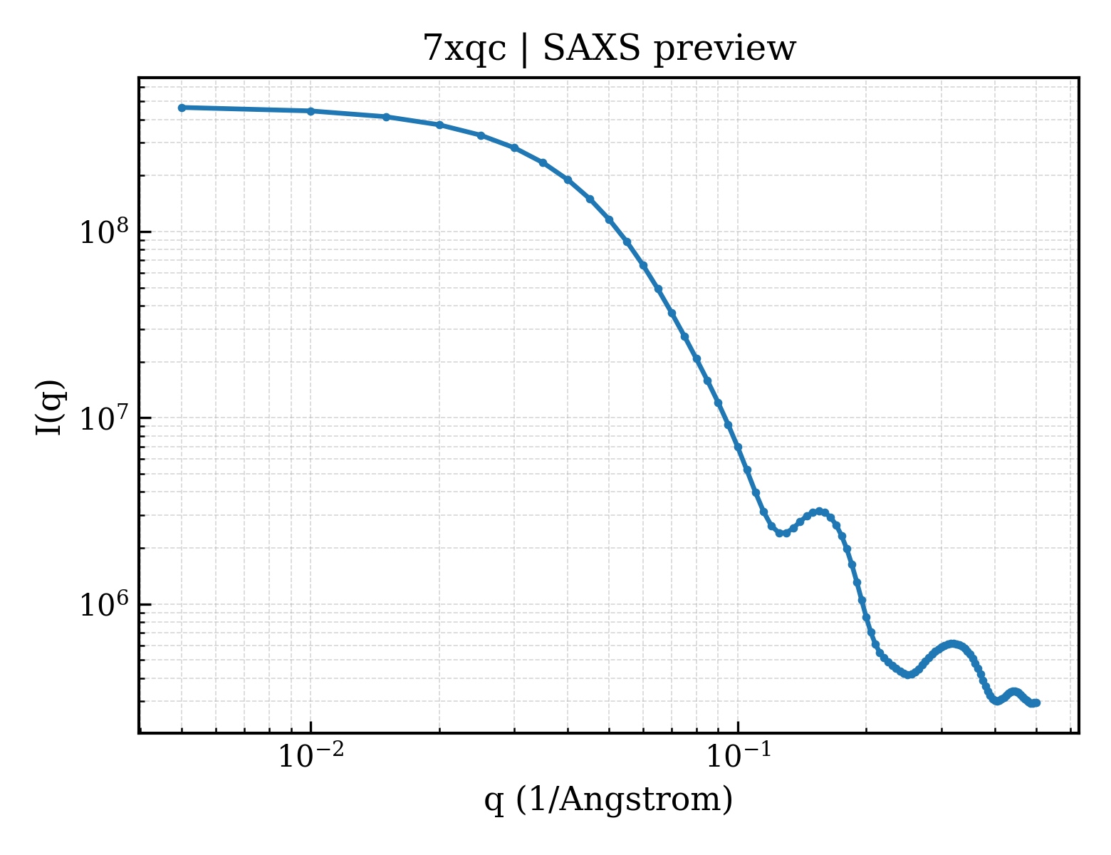
Fusion protein of Maltose-binding periplasmic protein and RNA-binding protein KhpA
Mycobacterium tuberculosis H37Rv
State in the Current Structure
| Assembly | Oligomeric State | Construct | Mutations and Modifications | Ligands, Ions and Associated Components | Method and Experimental Conditions | Structure Quality |
|---|---|---|---|---|---|---|
| 1 | Insufficient information Homooligomer Protein × 2 PDB declaration: dimeric(2) Consistent with protein copy count | Chain A; UniProt 27–392 Chain C; UniProt 27–392 | Not recorded | PEG DI(HYDROXYETHYL)ETHER × 2 | X-RAY DIFFRACTION X-ray crystallization conditions:MICROBATCH;pH 7.5;291.15 K;20 mM Na/K Phosphate, 20% (v/v) PEG 3350 | Resolution 2.80 Å R-free 0.254 |
| 2 | Insufficient information Homooligomer Protein × 2 PDB declaration: dimeric(2) Consistent with protein copy count | Chain B; UniProt 27–392 Chain D; UniProt 27–392 | Not recorded | PEG DI(HYDROXYETHYL)ETHER × 2 | X-RAY DIFFRACTION X-ray crystallization conditions:MICROBATCH;pH 7.5;291.15 K;20 mM Na/K Phosphate, 20% (v/v) PEG 3350 | Resolution 2.80 Å R-free 0.254 |
Other States of the Same Protein in the Database
Each row is a biological assembly of the same UniProt protein in another PDB entry. The “Difference from current entry” column identifies evidence-level differences; no tag means the currently parsed fields agree.
| Other PDB | Difference from Current Entry 7XQC | Assembly / Oligomeric State | Construct | Mutations and Modifications | Ligands, Ions and Non-polymers | Method and Experimental Conditions | Structure Quality |
|---|---|---|---|---|---|---|---|
| 21DH Crystal structure of MBP-fused Monobody P' in complex with HPPU Deposited 2025-12-09 | Different construct Different mutation/modification Different oligomeric state Different ligand/ion Different experimental conditions Different structure-quality metrics | Assembly 1 Other combination Monomer;Protein × 1 PDB declaration: monomeric |
Chain A
31–392(362 aa)
|
Mutation:A324V | A1MC1 1-(4-hydroxyphenyl)-3-phenyl-urea × 1 |
X-RAY DIFFRACTION
X-ray crystallization conditions
VAPOR DIFFUSION, SITTING DROP;293 K;20% (w/v) PEG 4000, 0.1M sodium acetate, 0.28M ammonium sulfate
|
Resolution 2.57 Å R-free 0.247 |
| 21DH Crystal structure of MBP-fused Monobody P' in complex with HPPU Deposited 2025-12-09 | Different construct Different mutation/modification Different oligomeric state Different ligand/ion Different experimental conditions Different structure-quality metrics | Assembly 2 Other combination Monomer;Protein × 1 PDB declaration: monomeric |
Chain B
31–392(362 aa)
|
Mutation:A324V | A1MC1 1-(4-hydroxyphenyl)-3-phenyl-urea × 1 |
X-RAY DIFFRACTION
X-ray crystallization conditions
VAPOR DIFFUSION, SITTING DROP;293 K;20% (w/v) PEG 4000, 0.1M sodium acetate, 0.28M ammonium sulfate
|
Resolution 2.57 Å R-free 0.247 |
| 21DH Crystal structure of MBP-fused Monobody P' in complex with HPPU Deposited 2025-12-09 | Different construct Different mutation/modification Different oligomeric state Different ligand/ion Different experimental conditions Different structure-quality metrics | Assembly 3 Other combination Monomer;Protein × 1 PDB declaration: monomeric |
Chain C
31–392(362 aa)
|
Mutation:A324V | A1MC1 1-(4-hydroxyphenyl)-3-phenyl-urea × 1 |
X-RAY DIFFRACTION
X-ray crystallization conditions
VAPOR DIFFUSION, SITTING DROP;293 K;20% (w/v) PEG 4000, 0.1M sodium acetate, 0.28M ammonium sulfate
|
Resolution 2.57 Å R-free 0.247 |
| 21DH Crystal structure of MBP-fused Monobody P' in complex with HPPU Deposited 2025-12-09 | Different construct Different mutation/modification Different oligomeric state Different ligand/ion Different experimental conditions Different structure-quality metrics | Assembly 4 Other combination Monomer;Protein × 1 PDB declaration: monomeric |
Chain D
31–392(362 aa)
|
Mutation:A324V | A1MC1 1-(4-hydroxyphenyl)-3-phenyl-urea × 1 |
X-RAY DIFFRACTION
X-ray crystallization conditions
VAPOR DIFFUSION, SITTING DROP;293 K;20% (w/v) PEG 4000, 0.1M sodium acetate, 0.28M ammonium sulfate
|
Resolution 2.57 Å R-free 0.247 |
| 21DH Crystal structure of MBP-fused Monobody P' in complex with HPPU Deposited 2025-12-09 | Different construct Different mutation/modification Different oligomeric state Different ligand/ion Different experimental conditions Different structure-quality metrics | Assembly 5 Other combination Monomer;Protein × 1 PDB declaration: monomeric |
Chain E
31–392(362 aa)
|
Mutation:A324V | A1MC1 1-(4-hydroxyphenyl)-3-phenyl-urea × 1 |
X-RAY DIFFRACTION
X-ray crystallization conditions
VAPOR DIFFUSION, SITTING DROP;293 K;20% (w/v) PEG 4000, 0.1M sodium acetate, 0.28M ammonium sulfate
|
Resolution 2.57 Å R-free 0.247 |
| 21DH Crystal structure of MBP-fused Monobody P' in complex with HPPU Deposited 2025-12-09 | Different construct Different mutation/modification Different oligomeric state Different ligand/ion Different experimental conditions Different structure-quality metrics | Assembly 6 Other combination Monomer;Protein × 1 PDB declaration: monomeric |
Chain F
31–392(362 aa)
|
Mutation:A324V | A1MC1 1-(4-hydroxyphenyl)-3-phenyl-urea × 1 |
X-RAY DIFFRACTION
X-ray crystallization conditions
VAPOR DIFFUSION, SITTING DROP;293 K;20% (w/v) PEG 4000, 0.1M sodium acetate, 0.28M ammonium sulfate
|
Resolution 2.57 Å R-free 0.247 |
| 2D21 NMR Structure of stereo-array isotope labelled (SAIL) maltodextrin-binding protein (MBP) Deposited 2005-09-02 | Different construct Different mutation/modification Different oligomeric state Different ligand/ion Different experimental method Different experimental conditions Different structure-quality metrics | Assembly 1 Protein monomer Monomer;Protein × 1 PDB declaration: monomeric |
Chain A
27–396(370 aa)
|
Not recorded | No recorded non-water small molecule |
SOLUTION NMR
NMR measurement conditions
pH 7.2;310 K;Ionic strength (raw mmCIF value) 20mM sodium phosphate;Pressure AMBIENT
NMR sample composition
0.33mM SAIL-MBP, 3.3mM beta-cyclodextrin, 20mM sodium phosphate, 3mM NaN3, CompleteMini protease inhibitor mix | 90% H2O/10% D2O
|
Resolution not provided |
| 2H25 Solution Structure of Maltose Binding Protein complexed with beta-cyclodextrin Deposited 2006-05-18 | Different construct Different mutation/modification Different oligomeric state Different ligand/ion Different experimental method Different experimental conditions Different structure-quality metrics | Assembly 1 Protein monomer Monomer;Protein × 1 PDB declaration: monomeric |
Chain A
27–396(370 aa)
|
Not recorded | No recorded non-water small molecule |
SOLUTION NMR
NMR measurement conditions
pH 7;303 K;Ionic strength (raw mmCIF value) 20mM sodium phosphates;Pressure ambient
NMR sample composition
1.2mM MBP, PH 7; 10% D2O | 10% D2O
|
Resolution not provided |
| 2KLF PERE NMR structure of maltodextrin-binding protein Deposited 2009-07-02 | Different construct Different mutation/modification Different oligomeric state Different ligand/ion Different experimental method Different experimental conditions Different structure-quality metrics | Assembly 1 Protein monomer Monomer;Protein × 1 PDB declaration: monomeric |
Chain A
27–396(370 aa)
|
Mutation:I2T | No recorded non-water small molecule |
SOLUTION NMR
NMR measurement conditions
pH 7.2;310 K;Ionic strength (raw mmCIF value) 0.02;Pressure ambient
NMR sample composition
20mM potassium phosphate-1, 2mM beta-cyclodextrin-2, 3mM sodium azide-3, 100mM EDTA-4, 90% H2O/10% D2O | 90% H2O/10% D2O
|
Resolution not provided |
| 2MV0 Solution NMR Structure of Maltose-binding protein from Escherichia coli, Northeast Structural Genomics Consortium (NESG) Target ER690 Deposited 2014-09-18 | Different construct Different mutation/modification Different oligomeric state Different ligand/ion Different experimental method Different experimental conditions Different structure-quality metrics | Assembly 1 Protein monomer Monomer;Protein × 1 PDB declaration: monomeric |
Chain A
27–396(370 aa)
|
Not recorded | No recorded non-water small molecule |
SOLUTION NMR
NMR measurement conditions
pH 7.2;310 K;Pressure ambient
NMR sample composition
1.05 mM ER690.005, 90% H2O/10% D2O | 90% H2O/10% D2O
|
Resolution not provided |
| 2N44 EC-NMR Structure of Escherichia coli Maltose-binding protein Determined by Combining Evolutionary Couplings (EC) and Sparse NMR Data. Northeast Structural Genomics Consortium target ER690 Deposited 2015-06-16 | Different construct Different mutation/modification Different oligomeric state Different ligand/ion Different experimental method Different experimental conditions Different structure-quality metrics | Assembly 1 Protein monomer Monomer;Protein × 1 PDB declaration: monomeric |
Chain A
27–396(370 aa)
Fragment:UNP residues 27-396
|
Not recorded | No recorded non-water small molecule | SOLUTION NMR mmCIF provides none of the parsed conditions | Resolution not provided |
| 2N45 EC-NMR Structure of Escherichia coli Maltose-binding protein Determined by Combining Evolutionary Couplings (EC) and Sparse NMR Data with a second set of RDC data simulated for an alternative alignment tensor. Northeast Structural Genomics Consortium target ER690 Deposited 2015-06-17 | Different construct Different mutation/modification Different oligomeric state Different ligand/ion Different experimental method Different experimental conditions Different structure-quality metrics | Assembly 1 Protein monomer Monomer;Protein × 1 PDB declaration: monomeric |
Chain A
27–396(370 aa)
Fragment:UNP RESIDUES 27-396
|
Not recorded | No recorded non-water small molecule | SOLUTION NMR mmCIF provides none of the parsed conditions | Resolution not provided |
| 2OBG Crystal Structure of Monobody MBP-74/Maltose Binding Protein Fusion Complex Deposited 2006-12-19 | Different construct Different mutation/modification Different oligomeric state Different ligand/ion Different experimental conditions Different structure-quality metrics | Assembly 1 Protein homooligomer Homooligomer;Protein × 2 PDB declaration: dimeric |
Chain A
31–396(366 aa)
Fragment:MBP (residues 5-370), MBP-74 (residues 1001-1093)
|
Not recorded | No recorded non-water small molecule |
X-RAY DIFFRACTION
X-ray crystallization conditions
VAPOR DIFFUSION, SITTING DROP;pH 6.5;292 K;20% PEG-1000, 0.1 M Na/K phosphate, 0.2 M NaCl, pH 6.5, VAPOR DIFFUSION, SITTING DROP, temperature 292K
|
Resolution 2.35 Å R-free 0.250 |
| 2OK2 MutS C-terminal domain fused to Maltose Binding Protein Deposited 2007-01-15 | Different construct Different oligomeric state Different ligand/ion Different experimental conditions Different structure-quality metrics | Assembly 1 Insufficient information Homooligomer;Protein × 4 PDB declaration: tetrameric |
Chain A
28–392(365 aa)
Fragment:MBP/MutS C-terminal fusion
Chain B
28–392(365 aa)
Fragment:MBP/MutS C-terminal fusion
|
Not recorded | SO4 SULFATE ION × 2 |
X-RAY DIFFRACTION
X-ray crystallization conditions
VAPOR DIFFUSION, HANGING DROP;pH 5.6;298 K;15% PEG 4K, 100 mM sodium citrate, 100 mM lithium acetate, pH 5.6, VAPOR DIFFUSION, HANGING DROP, temperature 298K
|
Resolution 2.00 Å R-free 0.277 |
| 2R6G The Crystal Structure of the E. coli Maltose Transporter Deposited 2007-09-05 | Different construct Different mutation/modification Different oligomeric state Different ligand/ion Different experimental conditions Different structure-quality metrics | Assembly 1 Other combination Heteromer;Protein × 5 PDB declaration: pentameric |
Chain E
27–396(370 aa)
|
Not recorded | ATP ADENOSINE-5'-TRIPHOSPHATE × 2 |
X-RAY DIFFRACTION
X-ray crystallization conditions
VAPOR DIFFUSION, SITTING DROP;pH 7.5;293 K;27% PEG 400, 500mM NaCl, 100mM Sodium Hepes pH 7.5, 10mM betaine hydrochloride, VAPOR DIFFUSION, SITTING DROP, temperature 293K
|
Resolution 2.80 Å R-free 0.271 |
| 2ZXT Crystal structure of Tim40/MIA40, a disulfide relay system in mitochondria, solved as MBP fusion protein Deposited 2009-01-07 | Different construct Different mutation/modification Different oligomeric state Different ligand/ion Different experimental conditions Different structure-quality metrics | Assembly 1 Insufficient information Monomer;Protein × 1 PDB declaration: monomeric |
Chain A
27–392(366 aa)
Fragment:MBPTim40C4
|
Not recorded | No recorded non-water small molecule |
X-RAY DIFFRACTION
X-ray crystallization conditions
VAPOR DIFFUSION, SITTING DROP;pH 5.6;293 K;100mM KOAc, 200 mM NH4OAc, 30 % PEG 4000, pH 5.6, VAPOR DIFFUSION, SITTING DROP, temperature 293K
|
Resolution 3.00 Å R-free 0.278 |
| 3A3C Crystal structure of TIM40/MIA40 fusing MBP, C296S and C298S mutant Deposited 2009-06-11 | Different construct Different mutation/modification Different oligomeric state Different ligand/ion Different experimental conditions Different structure-quality metrics | Assembly 1 Insufficient information Monomer;Protein × 1 PDB declaration: monomeric |
Chain A
29–392(364 aa)
|
Mutation:C296S, C298S | No recorded non-water small molecule |
X-RAY DIFFRACTION
X-ray crystallization conditions
VAPOR DIFFUSION, HANGING DROP;pH 5.6;298 K;100mM K-acetate, 200 mM NH4-acetate, 30% PEG 4000, pH 5.6, VAPOR DIFFUSION, HANGING DROP, temperature 298K
|
Resolution 2.50 Å R-free 0.316 |
| 3C4M Structure of human parathyroid hormone in complex with the extracellular domain of its G-protein-coupled receptor (PTH1R) Deposited 2008-01-30 | Different construct Different mutation/modification Different oligomeric state Different ligand/ion Different experimental conditions Different structure-quality metrics | Assembly 1 Insufficient information Heteromer;Protein × 2 PDB declaration: dimeric |
Chain A
26–392(367 aa)
Fragment:extracellular domain
|
Not recorded | No recorded non-water small molecule |
X-RAY DIFFRACTION
X-ray crystallization conditions
VAPOR DIFFUSION, HANGING DROP;pH 6.5;293 K;30% PPG P400, 0.1 M NaCacodylate, pH 6.5, VAPOR DIFFUSION, HANGING DROP, temperature 293K
|
Resolution 1.95 Å R-free 0.227 |
| 3C4M Structure of human parathyroid hormone in complex with the extracellular domain of its G-protein-coupled receptor (PTH1R) Deposited 2008-01-30 | Different construct Different mutation/modification Different oligomeric state Different ligand/ion Different experimental conditions Different structure-quality metrics | Assembly 2 Insufficient information Heteromer;Protein × 2 PDB declaration: dimeric |
Chain B
26–392(367 aa)
Fragment:extracellular domain
|
Not recorded | No recorded non-water small molecule |
X-RAY DIFFRACTION
X-ray crystallization conditions
VAPOR DIFFUSION, HANGING DROP;pH 6.5;293 K;30% PPG P400, 0.1 M NaCacodylate, pH 6.5, VAPOR DIFFUSION, HANGING DROP, temperature 293K
|
Resolution 1.95 Å R-free 0.227 |
| 3CSB Crystal Structure of Monobody YSX1/Maltose Binding Protein Fusion Complex Deposited 2008-04-09 | Different construct Different mutation/modification Different oligomeric state Different ligand/ion Different experimental conditions Different structure-quality metrics | Assembly 1 Protein monomer Monomer;Protein × 1 PDB declaration: monomeric |
Chain A
31–396(366 aa)
|
Not recorded | MN MANGANESE (II) ION × 4 EDO 1,2-ETHANEDIOL × 2 PGE TRIETHYLENE GLYCOL × 1 PG4 TETRAETHYLENE GLYCOL × 1 1PE PENTAETHYLENE GLYCOL × 1 PEG DI(HYDROXYETHYL)ETHER × 9 |
X-RAY DIFFRACTION
X-ray crystallization conditions
VAPOR DIFFUSION, HANGING DROP;pH 6.5;292 K;41% polyethelyeneglycol-400, 2% 2-methyl-2,4-pentanediol, 50 mM MnCl2, 0.1 M 2-(N-morpholino)ethanesulfonic acid, pH 6.5, VAPOR DIFFUSION, HANGING DROP, temperature 292K
|
Resolution 2.00 Å R-free 0.236 |
| 3CSG Crystal Structure of Monobody YS1(MBP-74)/Maltose Binding Protein Fusion Complex Deposited 2008-04-09 | Different construct Different mutation/modification Different oligomeric state Different ligand/ion Different experimental conditions Different structure-quality metrics | Assembly 1 Protein monomer Monomer;Protein × 1 PDB declaration: monomeric |
Chain A
31–396(366 aa)
|
Not recorded | No recorded non-water small molecule |
X-RAY DIFFRACTION
X-ray crystallization conditions
VAPOR DIFFUSION, HANGING DROP;pH 6.2;292 K;20% polyethyleneglycol-1000, 0.1 M Na/K phosphate, 0.2 M NaCl, pH 6.2, VAPOR DIFFUSION, HANGING DROP, temperature 292K
|
Resolution 1.80 Å R-free 0.235 |
| 3D4C ZP-N domain of mammalian sperm receptor ZP3 (crystal form I) Deposited 2008-05-14 | Different construct Different mutation/modification Different oligomeric state Different ligand/ion Different experimental conditions Different structure-quality metrics | Assembly 1 Insufficient information Monomer;Protein × 1 PDB declaration: monomeric |
Chain A
27–393(367 aa)
Fragment:ZP3 ZP-N domain, UNP residues 27-393, UNP residues 42-143
|
Mutation:I3T, E360A, K363A, D364A, R368N | CD CADMIUM ION × 7 |
X-RAY DIFFRACTION
X-ray crystallization conditions
VAPOR DIFFUSION, HANGING DROP;pH 7.5;293 K;Sample: 15mg/ml protein in 0.05M sodium chloride, 0.01M tris-HCL, PH7.2, 0.001M maltose.
Reservoir: 1.0M sodium acetate, 0,1M sodium HEPES, PH7.5, 0.05M cadmium sulfate.
Sample to reservoir ratio in drop: 1:1, pH7.5, VAPOR DIFFUSION, HANGING DROP, temperature 293.0K
|
Resolution 2.90 Å R-free 0.227 |
| 3D4G ZP-N domain of mammalian sperm receptor ZP3 (crystal form II) Deposited 2008-05-14 | Different construct Different mutation/modification Different oligomeric state Different ligand/ion Different experimental conditions Different structure-quality metrics | Assembly 1 Insufficient information Monomer;Protein × 1 PDB declaration: monomeric |
Chain A
27–393(367 aa)
Fragment:ZP3 ZP-N domain, UNP residues 27-393, UNP residues 42-143
|
Mutation:I3T, E360A, K363A, D364A, R368N | CA CALCIUM ION × 3 |
X-RAY DIFFRACTION
X-ray crystallization conditions
VAPOR DIFFUSION, HANGING DROP;pH 7;293 K;Sample: 15mg/ml protein in 0.05M sodium chloride, 0.01M Tris-HCL, PH7.2, 0.001M maltose.
Reservoir: 10% PEG6000, 0.2M calcium chloride, 0.1M Tris-HCL, PH7.0.
Sample to reservoir ratio in drop: 1:1, pH7.0, VAPOR DIFFUSION, HANGING DROP, temperature 293.0K
|
Resolution 2.30 Å R-free 0.227 |
| 3D4G ZP-N domain of mammalian sperm receptor ZP3 (crystal form II) Deposited 2008-05-14 | Different construct Different mutation/modification Different oligomeric state Different ligand/ion Different experimental conditions Different structure-quality metrics | Assembly 2 Insufficient information Monomer;Protein × 1 PDB declaration: monomeric |
Chain B
27–393(367 aa)
Fragment:ZP3 ZP-N domain, UNP residues 27-393, UNP residues 42-143
|
Mutation:I3T, E360A, K363A, D364A, R368N | CA CALCIUM ION × 3 |
X-RAY DIFFRACTION
X-ray crystallization conditions
VAPOR DIFFUSION, HANGING DROP;pH 7;293 K;Sample: 15mg/ml protein in 0.05M sodium chloride, 0.01M Tris-HCL, PH7.2, 0.001M maltose.
Reservoir: 10% PEG6000, 0.2M calcium chloride, 0.1M Tris-HCL, PH7.0.
Sample to reservoir ratio in drop: 1:1, pH7.0, VAPOR DIFFUSION, HANGING DROP, temperature 293.0K
|
Resolution 2.30 Å R-free 0.227 |
| 3D4G ZP-N domain of mammalian sperm receptor ZP3 (crystal form II) Deposited 2008-05-14 | Different construct Different mutation/modification Different oligomeric state Different ligand/ion Different experimental conditions Different structure-quality metrics | Assembly 3 Insufficient information Monomer;Protein × 1 PDB declaration: monomeric |
Chain C
27–393(367 aa)
Fragment:ZP3 ZP-N domain, UNP residues 27-393, UNP residues 42-143
|
Mutation:I3T, E360A, K363A, D364A, R368N | CA CALCIUM ION × 3 |
X-RAY DIFFRACTION
X-ray crystallization conditions
VAPOR DIFFUSION, HANGING DROP;pH 7;293 K;Sample: 15mg/ml protein in 0.05M sodium chloride, 0.01M Tris-HCL, PH7.2, 0.001M maltose.
Reservoir: 10% PEG6000, 0.2M calcium chloride, 0.1M Tris-HCL, PH7.0.
Sample to reservoir ratio in drop: 1:1, pH7.0, VAPOR DIFFUSION, HANGING DROP, temperature 293.0K
|
Resolution 2.30 Å R-free 0.227 |
| 3D4G ZP-N domain of mammalian sperm receptor ZP3 (crystal form II) Deposited 2008-05-14 | Different construct Different mutation/modification Different oligomeric state Different ligand/ion Different experimental conditions Different structure-quality metrics | Assembly 4 Insufficient information Monomer;Protein × 1 PDB declaration: monomeric |
Chain D
27–393(367 aa)
Fragment:ZP3 ZP-N domain, UNP residues 27-393, UNP residues 42-143
|
Mutation:I3T, E360A, K363A, D364A, R368N | CA CALCIUM ION × 3 |
X-RAY DIFFRACTION
X-ray crystallization conditions
VAPOR DIFFUSION, HANGING DROP;pH 7;293 K;Sample: 15mg/ml protein in 0.05M sodium chloride, 0.01M Tris-HCL, PH7.2, 0.001M maltose.
Reservoir: 10% PEG6000, 0.2M calcium chloride, 0.1M Tris-HCL, PH7.0.
Sample to reservoir ratio in drop: 1:1, pH7.0, VAPOR DIFFUSION, HANGING DROP, temperature 293.0K
|
Resolution 2.30 Å R-free 0.227 |
| 3D4G ZP-N domain of mammalian sperm receptor ZP3 (crystal form II) Deposited 2008-05-14 | Different construct Different mutation/modification Different oligomeric state Different ligand/ion Different experimental conditions Different structure-quality metrics | Assembly 5 Insufficient information Monomer;Protein × 1 PDB declaration: monomeric |
Chain E
27–393(367 aa)
Fragment:ZP3 ZP-N domain, UNP residues 27-393, UNP residues 42-143
|
Mutation:I3T, E360A, K363A, D364A, R368N | CA CALCIUM ION × 3 |
X-RAY DIFFRACTION
X-ray crystallization conditions
VAPOR DIFFUSION, HANGING DROP;pH 7;293 K;Sample: 15mg/ml protein in 0.05M sodium chloride, 0.01M Tris-HCL, PH7.2, 0.001M maltose.
Reservoir: 10% PEG6000, 0.2M calcium chloride, 0.1M Tris-HCL, PH7.0.
Sample to reservoir ratio in drop: 1:1, pH7.0, VAPOR DIFFUSION, HANGING DROP, temperature 293.0K
|
Resolution 2.30 Å R-free 0.227 |
| 3D4G ZP-N domain of mammalian sperm receptor ZP3 (crystal form II) Deposited 2008-05-14 | Different construct Different mutation/modification Different oligomeric state Different ligand/ion Different experimental conditions Different structure-quality metrics | Assembly 6 Insufficient information Monomer;Protein × 1 PDB declaration: monomeric |
Chain F
27–393(367 aa)
Fragment:ZP3 ZP-N domain, UNP residues 27-393, UNP residues 42-143
|
Mutation:I3T, E360A, K363A, D364A, R368N | CA CALCIUM ION × 3 |
X-RAY DIFFRACTION
X-ray crystallization conditions
VAPOR DIFFUSION, HANGING DROP;pH 7;293 K;Sample: 15mg/ml protein in 0.05M sodium chloride, 0.01M Tris-HCL, PH7.2, 0.001M maltose.
Reservoir: 10% PEG6000, 0.2M calcium chloride, 0.1M Tris-HCL, PH7.0.
Sample to reservoir ratio in drop: 1:1, pH7.0, VAPOR DIFFUSION, HANGING DROP, temperature 293.0K
|
Resolution 2.30 Å R-free 0.227 |
| 3D4G ZP-N domain of mammalian sperm receptor ZP3 (crystal form II) Deposited 2008-05-14 | Different construct Different mutation/modification Different oligomeric state Different ligand/ion Different experimental conditions Different structure-quality metrics | Assembly 7 Insufficient information Monomer;Protein × 1 PDB declaration: monomeric |
Chain G
27–393(367 aa)
Fragment:ZP3 ZP-N domain, UNP residues 27-393, UNP residues 42-143
|
Mutation:I3T, E360A, K363A, D364A, R368N | CA CALCIUM ION × 3 |
X-RAY DIFFRACTION
X-ray crystallization conditions
VAPOR DIFFUSION, HANGING DROP;pH 7;293 K;Sample: 15mg/ml protein in 0.05M sodium chloride, 0.01M Tris-HCL, PH7.2, 0.001M maltose.
Reservoir: 10% PEG6000, 0.2M calcium chloride, 0.1M Tris-HCL, PH7.0.
Sample to reservoir ratio in drop: 1:1, pH7.0, VAPOR DIFFUSION, HANGING DROP, temperature 293.0K
|
Resolution 2.30 Å R-free 0.227 |
| 3D4G ZP-N domain of mammalian sperm receptor ZP3 (crystal form II) Deposited 2008-05-14 | Different construct Different mutation/modification Different oligomeric state Different ligand/ion Different experimental conditions Different structure-quality metrics | Assembly 8 Insufficient information Monomer;Protein × 1 PDB declaration: monomeric |
Chain H
27–393(367 aa)
Fragment:ZP3 ZP-N domain, UNP residues 27-393, UNP residues 42-143
|
Mutation:I3T, E360A, K363A, D364A, R368N | CA CALCIUM ION × 3 |
X-RAY DIFFRACTION
X-ray crystallization conditions
VAPOR DIFFUSION, HANGING DROP;pH 7;293 K;Sample: 15mg/ml protein in 0.05M sodium chloride, 0.01M Tris-HCL, PH7.2, 0.001M maltose.
Reservoir: 10% PEG6000, 0.2M calcium chloride, 0.1M Tris-HCL, PH7.0.
Sample to reservoir ratio in drop: 1:1, pH7.0, VAPOR DIFFUSION, HANGING DROP, temperature 293.0K
|
Resolution 2.30 Å R-free 0.227 |
| 3DM0 Maltose Binding Protein fusion with RACK1 from A. thaliana Deposited 2008-06-30 | Different construct Different mutation/modification Different oligomeric state Different ligand/ion Different experimental conditions Different structure-quality metrics | Assembly 1 Insufficient information Monomer;Protein × 1 PDB declaration: monomeric |
Chain A
27–387(361 aa)
Fragment:Fusion protein of MBP (UNP residues 27 to 387 ) and RACK1 (UNP residues 4 to 327)
|
Mutation:D82A, K83A, K239A, E359A | EDO 1,2-ETHANEDIOL × 1 |
X-RAY DIFFRACTION
X-ray crystallization conditions
VAPOR DIFFUSION, SITTING DROP;pH 7.5;278 K;100mM HEPES pH 7.5
20% w/v PEG 10,000, VAPOR DIFFUSION, SITTING DROP, temperature 278K
|
Resolution 2.40 Å R-free 0.250 |
| 3EF7 ZP-N domain of mammalian sperm receptor ZP3 (crystal form III) Deposited 2008-09-08 | Different construct Different mutation/modification Different oligomeric state Different ligand/ion Different experimental conditions Different structure-quality metrics | Assembly 1 Insufficient information Monomer;Protein × 1 PDB declaration: monomeric |
Chain A
27–393(367 aa)
Fragment:ZP3 ZP-N domain, UNP residues 27-393, UNP residues 42-143
|
Mutation:I3T, E360A, K363A, D364A, R368N | ZN ZINC ION × 5 CA CALCIUM ION × 2 |
X-RAY DIFFRACTION
X-ray crystallization conditions
VAPOR DIFFUSION, HANGING DROP;pH 7.8;293 K;Sample: 15mg/ml protein in 0.05M sodium chloride, 0.01M Tris-HCL, PH7.2, 0.001M Maltose.
Reservoir: 12% PEG6000, 0.15M calcium chloride, 0.0025M zinc chloride, 0.1M Tris-HCL, PH8.2.
Sample to reservoir ratio in drop: 1:1, PH8.2, VAPOR DIFFUSION, HANGING DROP, TEMPERATURE 293.0K, pH7.8
|
Resolution 3.10 Å R-free 0.256 |
| 3EF7 ZP-N domain of mammalian sperm receptor ZP3 (crystal form III) Deposited 2008-09-08 | Different construct Different mutation/modification Different oligomeric state Different ligand/ion Different experimental conditions Different structure-quality metrics | Assembly 2 Insufficient information Monomer;Protein × 1 PDB declaration: monomeric |
Chain B
27–393(367 aa)
Fragment:ZP3 ZP-N domain, UNP residues 27-393, UNP residues 42-143
|
Mutation:I3T, E360A, K363A, D364A, R368N | ZN ZINC ION × 8 CA CALCIUM ION × 2 |
X-RAY DIFFRACTION
X-ray crystallization conditions
VAPOR DIFFUSION, HANGING DROP;pH 7.8;293 K;Sample: 15mg/ml protein in 0.05M sodium chloride, 0.01M Tris-HCL, PH7.2, 0.001M Maltose.
Reservoir: 12% PEG6000, 0.15M calcium chloride, 0.0025M zinc chloride, 0.1M Tris-HCL, PH8.2.
Sample to reservoir ratio in drop: 1:1, PH8.2, VAPOR DIFFUSION, HANGING DROP, TEMPERATURE 293.0K, pH7.8
|
Resolution 3.10 Å R-free 0.256 |
| 3EF7 ZP-N domain of mammalian sperm receptor ZP3 (crystal form III) Deposited 2008-09-08 | Different construct Different mutation/modification Different oligomeric state Different ligand/ion Different experimental conditions Different structure-quality metrics | Assembly 3 Insufficient information Homooligomer;Protein × 4 PDB declaration: tetrameric |
Chain A
27–393(367 aa)
Fragment:ZP3 ZP-N domain, UNP residues 27-393, UNP residues 42-143
Chain B
27–393(367 aa)
Fragment:ZP3 ZP-N domain, UNP residues 27-393, UNP residues 42-143
|
Mutation:I3T, E360A, K363A, D364A, R368N Mutation:I3T, E360A, K363A, D364A, R368N | ZN ZINC ION × 26 CA CALCIUM ION × 8 |
X-RAY DIFFRACTION
X-ray crystallization conditions
VAPOR DIFFUSION, HANGING DROP;pH 7.8;293 K;Sample: 15mg/ml protein in 0.05M sodium chloride, 0.01M Tris-HCL, PH7.2, 0.001M Maltose.
Reservoir: 12% PEG6000, 0.15M calcium chloride, 0.0025M zinc chloride, 0.1M Tris-HCL, PH8.2.
Sample to reservoir ratio in drop: 1:1, PH8.2, VAPOR DIFFUSION, HANGING DROP, TEMPERATURE 293.0K, pH7.8
|
Resolution 3.10 Å R-free 0.256 |
| 3EHS Crystal structure of the extracellular domain of human corticotropin releasing factor receptor type 1 (CRFR1) Deposited 2008-09-14 | Different construct Different mutation/modification Different oligomeric state Different ligand/ion Different experimental conditions Different structure-quality metrics | Assembly 1 Insufficient information Monomer;Protein × 1 PDB declaration: monomeric |
Chain A
26–392(367 aa)
|
Not recorded | No recorded non-water small molecule |
X-RAY DIFFRACTION
X-ray crystallization conditions
VAPOR DIFFUSION;pH 4.7;293 K;NaCl, sucrose, sodium acetate, pH 4.7, vapor diffusion, temperature 293K
|
Resolution 2.76 Å R-free 0.240 |
| 3EHT Crystal structure of the extracellular domain of human corticotropin releasing factor receptor type 1 (CRFR1) in complex with CRF Deposited 2008-09-14 | Different construct Different mutation/modification Different oligomeric state Different ligand/ion Different experimental conditions Different structure-quality metrics | Assembly 1 Insufficient information Heteromer;Protein × 2 PDB declaration: dimeric |
Chain A
26–392(367 aa)
|
Mutation:F(-257)E | No recorded non-water small molecule |
X-RAY DIFFRACTION
X-ray crystallization conditions
VAPOR DIFFUSION;pH 6.25;293 K;PEG 3350, Lithium sulfate, Bis-Tris, pH 6.25, VAPOR DIFFUSION, temperature 293K
|
Resolution 3.40 Å R-free 0.252 |
| 3EHU Crystal structure of the extracellular domain of human corticotropin releasing factor receptor type 1 (CRFR1) in complex with CRF Deposited 2008-09-14 | Different construct Different mutation/modification Different oligomeric state Different ligand/ion Different experimental conditions Different structure-quality metrics | Assembly 1 Insufficient information Heteromer;Protein × 2 PDB declaration: dimeric |
Chain A
26–392(367 aa)
|
Mutation:A(-25)E | CA CALCIUM ION × 1 BTB 2-[BIS-(2-HYDROXY-ETHYL)-AMINO]-2-HYDROXYMETHYL-PROPANE-1,3-DIOL × 1 PEG DI(HYDROXYETHYL)ETHER × 1 |
X-RAY DIFFRACTION
X-ray crystallization conditions
VAPOR DIFFUSION;pH 6.75;293 K;PEG MME 550, calcium chloride, tert-butanol, Bis-Tris, pH 6.75, VAPOR DIFFUSION, temperature 293K
|
Resolution 1.96 Å R-free 0.256 |
| 3EHU Crystal structure of the extracellular domain of human corticotropin releasing factor receptor type 1 (CRFR1) in complex with CRF Deposited 2008-09-14 | Different construct Different mutation/modification Different oligomeric state Different ligand/ion Different experimental conditions Different structure-quality metrics | Assembly 2 Insufficient information Heteromer;Protein × 2 PDB declaration: dimeric |
Chain B
26–392(367 aa)
|
Mutation:A(-25)E | CA CALCIUM ION × 1 BTB 2-[BIS-(2-HYDROXY-ETHYL)-AMINO]-2-HYDROXYMETHYL-PROPANE-1,3-DIOL × 1 PEG DI(HYDROXYETHYL)ETHER × 1 |
X-RAY DIFFRACTION
X-ray crystallization conditions
VAPOR DIFFUSION;pH 6.75;293 K;PEG MME 550, calcium chloride, tert-butanol, Bis-Tris, pH 6.75, VAPOR DIFFUSION, temperature 293K
|
Resolution 1.96 Å R-free 0.256 |
| 3F5F Crystal structure of heparan sulfate 2-O-sulfotransferase from gallus gallus as a maltose binding protein fusion. Deposited 2008-11-03 | Different construct Different mutation/modification Different oligomeric state Different ligand/ion Different experimental conditions Different structure-quality metrics | Assembly 1 Insufficient information Homooligomer;Protein × 3 PDB declaration: trimeric |
Chain A
27–387(361 aa)
Fragment:P0AEX9 residues 27-392, Q76KB1 residues 69-356
|
Mutation:E359A | A3P ADENOSINE-3'-5'-DIPHOSPHATE × 3 |
X-RAY DIFFRACTION
X-ray crystallization conditions
VAPOR DIFFUSION, SITTING DROP;pH 6;277 K;Ammonium Citrate, Bis-tris-propane, Phenol, pH 6.0, VAPOR DIFFUSION, SITTING DROP, temperature 277K
|
Resolution 2.65 Å R-free 0.243 |
| 3G7V Islet Amyloid Polypeptide (IAPP or Amylin) fused to Maltose Binding Protein Deposited 2009-02-10 | Different oligomeric state Different ligand/ion Different experimental conditions Different structure-quality metrics | Assembly 1 Insufficient information Homooligomer;Protein × 2 PDB declaration: dimeric |
Chain A
27–392(366 aa)
Chain B
27–392(366 aa)
|
Not recorded | SO4 SULFATE ION × 7 GOL GLYCEROL × 3 |
X-RAY DIFFRACTION
X-ray crystallization conditions
VAPOR DIFFUSION, HANGING DROP;pH 4.6;298 K;2.0 M Ammonium Sulfate, 0.1M Sodium Acetate pH 4.6, vapor diffusion, hanging drop, temperature 298K
|
Resolution 1.86 Å R-free 0.205 |
| 3G7V Islet Amyloid Polypeptide (IAPP or Amylin) fused to Maltose Binding Protein Deposited 2009-02-10 | Different oligomeric state Different ligand/ion Different experimental conditions Different structure-quality metrics | Assembly 2 Insufficient information Homooligomer;Protein × 2 PDB declaration: dimeric |
Chain C
27–392(366 aa)
Chain D
27–392(366 aa)
|
Not recorded | SO4 SULFATE ION × 8 GOL GLYCEROL × 2 |
X-RAY DIFFRACTION
X-ray crystallization conditions
VAPOR DIFFUSION, HANGING DROP;pH 4.6;298 K;2.0 M Ammonium Sulfate, 0.1M Sodium Acetate pH 4.6, vapor diffusion, hanging drop, temperature 298K
|
Resolution 1.86 Å R-free 0.205 |
| 3G7W Islet Amyloid Polypeptide (IAPP or Amylin) Residues 1 to 22 fused to Maltose Binding Protein Deposited 2009-02-11 | Different construct Different mutation/modification Different oligomeric state Different ligand/ion Different experimental conditions Different structure-quality metrics | Assembly 1 Insufficient information Monomer;Protein × 1 PDB declaration: monomeric |
Chain A
27–392(366 aa)
|
Not recorded | SO4 SULFATE ION × 20 GOL GLYCEROL × 2 |
X-RAY DIFFRACTION
X-ray crystallization conditions
VAPOR DIFFUSION, HANGING DROP;pH 4.6;298 K;2.0 M Ammonium Sulfate, 0.1M Sodium Acetate pH 4.6, vapor diffusion, hanging drop, temperature 298K
|
Resolution 1.75 Å R-free 0.194 |
| 3G7W Islet Amyloid Polypeptide (IAPP or Amylin) Residues 1 to 22 fused to Maltose Binding Protein Deposited 2009-02-11 | Different construct Different mutation/modification Different oligomeric state Different ligand/ion Different experimental conditions Different structure-quality metrics | Assembly 2 Insufficient information Homooligomer;Protein × 2 PDB declaration: dimeric |
Chain A
27–392(366 aa)
|
Not recorded | SO4 SULFATE ION × 40 GOL GLYCEROL × 4 |
X-RAY DIFFRACTION
X-ray crystallization conditions
VAPOR DIFFUSION, HANGING DROP;pH 4.6;298 K;2.0 M Ammonium Sulfate, 0.1M Sodium Acetate pH 4.6, vapor diffusion, hanging drop, temperature 298K
|
Resolution 1.75 Å R-free 0.194 |
| 3H3G Crystal structure of the extracellular domain of the human parathyroid hormone receptor (PTH1R) in complex with parathyroid hormone-related protein (PTHrP) Deposited 2009-04-16 | Different construct Different mutation/modification Different oligomeric state Different ligand/ion Different experimental conditions Different structure-quality metrics | Assembly 1 Insufficient information Heteromer;Protein × 2 PDB declaration: dimeric |
Chain A
26–392(367 aa)
Fragment:extracellular domain
|
Not recorded | No recorded non-water small molecule |
X-RAY DIFFRACTION
X-ray crystallization conditions
VAPOR DIFFUSION, SITTING DROP;pH 7.5;298 K;7.5% PEG 2000, 13% PEG 400, VAPOR DIFFUSION, SITTING DROP, temperature 298K
|
Resolution 1.94 Å R-free 0.233 |
| 3H4Z Crystal Structure of an MBP-Der p 7 fusion protein Deposited 2009-04-21 | Different construct Different mutation/modification Different oligomeric state Different ligand/ion Different experimental conditions Different structure-quality metrics | Assembly 1 Insufficient information Monomer;Protein × 1 PDB declaration: monomeric |
Chain A
27–387(361 aa)
|
Not recorded | NA SODIUM ION × 1 |
X-RAY DIFFRACTION
X-ray crystallization conditions
VAPOR DIFFUSION, SITTING DROP;pH 8.5;277 K;85mM Lithium sulfate, 42mM Tris pH 8.5, 12.75% PEG4K, 7.5% glycerol, VAPOR DIFFUSION, SITTING DROP, temperature 277K
|
Resolution 2.35 Å R-free 0.293 |
| 3H4Z Crystal Structure of an MBP-Der p 7 fusion protein Deposited 2009-04-21 | Different construct Different mutation/modification Different oligomeric state Different ligand/ion Different experimental conditions Different structure-quality metrics | Assembly 2 Insufficient information Monomer;Protein × 1 PDB declaration: monomeric |
Chain B
27–387(361 aa)
|
Not recorded | No recorded non-water small molecule |
X-RAY DIFFRACTION
X-ray crystallization conditions
VAPOR DIFFUSION, SITTING DROP;pH 8.5;277 K;85mM Lithium sulfate, 42mM Tris pH 8.5, 12.75% PEG4K, 7.5% glycerol, VAPOR DIFFUSION, SITTING DROP, temperature 277K
|
Resolution 2.35 Å R-free 0.293 |
| 3H4Z Crystal Structure of an MBP-Der p 7 fusion protein Deposited 2009-04-21 | Different construct Different mutation/modification Different oligomeric state Different ligand/ion Different experimental conditions Different structure-quality metrics | Assembly 3 Insufficient information Monomer;Protein × 1 PDB declaration: monomeric |
Chain C
27–387(361 aa)
|
Not recorded | No recorded non-water small molecule |
X-RAY DIFFRACTION
X-ray crystallization conditions
VAPOR DIFFUSION, SITTING DROP;pH 8.5;277 K;85mM Lithium sulfate, 42mM Tris pH 8.5, 12.75% PEG4K, 7.5% glycerol, VAPOR DIFFUSION, SITTING DROP, temperature 277K
|
Resolution 2.35 Å R-free 0.293 |
| 3HPI Crystal structure of maltose-binding protein mutant with bound sucrose Deposited 2009-06-04 | Different construct Different mutation/modification Different oligomeric state Different ligand/ion Different experimental conditions Different structure-quality metrics | Assembly 1 Other combination Monomer;Protein × 1 PDB declaration: monomeric |
Chain A
27–396(370 aa)
|
Mutation:D14L, K15F, W62Y, E111Y | ZN ZINC ION × 2 ACT ACETATE ION × 1 |
X-RAY DIFFRACTION
X-ray crystallization conditions
VAPOR DIFFUSION;pH 6.2;289 K;PEG MME 5000, Sodium acetate, Sucrose, Magnesium chloride, Zinc chloride, pH 6.2, VAPOR DIFFUSION, temperature 289K
|
Resolution 2.00 Å R-free 0.284 |
| 3HPI Crystal structure of maltose-binding protein mutant with bound sucrose Deposited 2009-06-04 | Different construct Different mutation/modification Different oligomeric state Different ligand/ion Different experimental conditions Different structure-quality metrics | Assembly 2 Other combination Monomer;Protein × 1 PDB declaration: monomeric |
Chain B
27–396(370 aa)
|
Mutation:D14L, K15F, W62Y, E111Y | ZN ZINC ION × 4 ACT ACETATE ION × 1 |
X-RAY DIFFRACTION
X-ray crystallization conditions
VAPOR DIFFUSION;pH 6.2;289 K;PEG MME 5000, Sodium acetate, Sucrose, Magnesium chloride, Zinc chloride, pH 6.2, VAPOR DIFFUSION, temperature 289K
|
Resolution 2.00 Å R-free 0.284 |
| 3HST N-Terminal RNASE H domain of rv2228c from mycobacterium tuberculosis as a fusion protein with maltose binding protein Deposited 2009-06-10 | Different construct Different mutation/modification Different oligomeric state Different ligand/ion Different experimental conditions Different structure-quality metrics | Assembly 1 Other combination Monomer;Protein × 1 PDB declaration: monomeric |
Chain A
27–392(366 aa)
|
Not recorded | EDO 1,2-ETHANEDIOL × 1 |
X-RAY DIFFRACTION
X-ray crystallization conditions
VAPOR DIFFUSION, SITTING DROP;291 K;20% PEG2000, 0.2M Ammounium tartrate, pH pH7.5, VAPOR DIFFUSION, SITTING DROP, temperature 291K
|
Resolution 2.25 Å R-free 0.239 |
| 3HST N-Terminal RNASE H domain of rv2228c from mycobacterium tuberculosis as a fusion protein with maltose binding protein Deposited 2009-06-10 | Different construct Different mutation/modification Different oligomeric state Different ligand/ion Different experimental conditions Different structure-quality metrics | Assembly 3 Other combination Monomer;Protein × 1 PDB declaration: monomeric |
Chain C
27–392(366 aa)
|
Not recorded | EDO 1,2-ETHANEDIOL × 2 TAR D(-)-TARTARIC ACID × 1 |
X-RAY DIFFRACTION
X-ray crystallization conditions
VAPOR DIFFUSION, SITTING DROP;291 K;20% PEG2000, 0.2M Ammounium tartrate, pH pH7.5, VAPOR DIFFUSION, SITTING DROP, temperature 291K
|
Resolution 2.25 Å R-free 0.239 |
| 3IOR Huntingtin amino-terminal region with 17 Gln residues - crystal C95 Deposited 2009-08-14 | Different construct Different mutation/modification Different oligomeric state Different ligand/ion Different experimental conditions Different structure-quality metrics | Assembly 1 Insufficient information Homooligomer;Protein × 3 PDB declaration: trimeric |
Chain A
27–384(358 aa)
Fragment:Fusion protein, see remark 999
Chain B
27–384(358 aa)
Fragment:Fusion protein, see remark 999
Chain C
27–384(358 aa)
Fragment:Fusion protein, see remark 999
|
Not recorded | ZN ZINC ION × 6 CA CALCIUM ION × 6 |
X-RAY DIFFRACTION
X-ray crystallization conditions
VAPOR DIFFUSION, HANGING DROP;278 K;12% PEG 4000, 200 mM Zn acetate, 200 mM Sodium acetate, 100 mM Sodium Cacodylate pH 6.5-7.4 , VAPOR DIFFUSION, HANGING DROP, temperature 278K
|
Resolution 3.60 Å R-free 0.267 |
| 3IOT Huntingtin amino-terminal region with 17 Gln residues - crystal C92-b Deposited 2009-08-14 | Different construct Different mutation/modification Different oligomeric state Different ligand/ion Different experimental conditions Different structure-quality metrics | Assembly 1 Insufficient information Homooligomer;Protein × 3 PDB declaration: trimeric |
Chain A
27–384(358 aa)
Fragment:Fusion protein, see remark 999,Fusion protein, see remark 999
Chain B
27–384(358 aa)
Fragment:Fusion protein, see remark 999,Fusion protein, see remark 999
Chain C
27–384(358 aa)
Fragment:Fusion protein, see remark 999,Fusion protein, see remark 999
|
Not recorded | ZN ZINC ION × 9 CA CALCIUM ION × 9 |
X-RAY DIFFRACTION
X-ray crystallization conditions
VAPOR DIFFUSION, HANGING DROP;278 K;12% PEG 4000, 200 mM Zn acetate, 200 mM Sodium acetate, 100 mM Sodium Cacodylate pH 6.5-7.4, VAPOR DIFFUSION, HANGING DROP, temperature 278K
|
Resolution 3.50 Å R-free 0.295 |
| 3IOU Huntingtin amino-terminal region with 17 Gln residues - crystal C94 Deposited 2009-08-14 | Different construct Different mutation/modification Different oligomeric state Different ligand/ion Different experimental conditions Different structure-quality metrics | Assembly 1 Insufficient information Homooligomer;Protein × 3 PDB declaration: trimeric |
Chain A
27–384(358 aa)
Fragment:Fusion protein, see remark 999,Fusion protein, see remark 999
Chain B
27–384(358 aa)
Fragment:Fusion protein, see remark 999,Fusion protein, see remark 999
Chain C
27–384(358 aa)
Fragment:Fusion protein, see remark 999,Fusion protein, see remark 999
|
Not recorded | ZN ZINC ION × 8 CA CALCIUM ION × 5 |
X-RAY DIFFRACTION
X-ray crystallization conditions
VAPOR DIFFUSION, HANGING DROP;278 K;12% PEG 4000, 200 mM Zn acetate, 200 mM Sodium acetate, 100 mM Sodium Cacodylate pH 6.5-7.4, VAPOR DIFFUSION, HANGING DROP, temperature 278K
|
Resolution 3.70 Å R-free 0.299 |
| 3IOV Huntingtin amino-terminal region with 17 Gln residues - crystal C99 Deposited 2009-08-14 | Different construct Different mutation/modification Different oligomeric state Different ligand/ion Different experimental conditions Different structure-quality metrics | Assembly 1 Insufficient information Homooligomer;Protein × 3 PDB declaration: trimeric |
Chain A
27–384(358 aa)
Fragment:Fusion protein, see remark 999,Fusion protein, see remark 999
Chain B
27–384(358 aa)
Fragment:Fusion protein, see remark 999,Fusion protein, see remark 999
Chain C
27–384(358 aa)
Fragment:Fusion protein, see remark 999,Fusion protein, see remark 999
|
Not recorded | ZN ZINC ION × 6 CA CALCIUM ION × 7 |
X-RAY DIFFRACTION
X-ray crystallization conditions
VAPOR DIFFUSION, HANGING DROP;278 K;12% PEG 4000, 200 mM Zn acetate, 200 mM Sodium acetate, 100 mM Sodium Cacodylate pH 6.5-7.4, VAPOR DIFFUSION, HANGING DROP, temperature 278K
|
Resolution 3.70 Å R-free 0.280 |
| 3IOW Huntingtin amino-terminal region with 17 Gln residues - crystal C99-Hg Deposited 2009-08-14 | Different construct Different mutation/modification Different oligomeric state Different ligand/ion Different experimental conditions Different structure-quality metrics | Assembly 1 Insufficient information Homooligomer;Protein × 3 PDB declaration: trimeric |
Chain A
27–384(358 aa)
Fragment:Fusion protein, see remark 999,Fusion protein, see remark 999
Chain B
27–384(358 aa)
Fragment:Fusion protein, see remark 999,Fusion protein, see remark 999
Chain C
27–384(358 aa)
Fragment:Fusion protein, see remark 999,Fusion protein, see remark 999
|
Not recorded | ZN ZINC ION × 7 CA CALCIUM ION × 10 |
X-RAY DIFFRACTION
X-ray crystallization conditions
VAPOR DIFFUSION, HANGING DROP;278 K;12% PEG 4000, 200 mM Zn acetate, 200 mM Sodium acetate, 100 mM Sodium Cacodylate pH 6.5-7.4, VAPOR DIFFUSION, HANGING DROP, temperature 278K
|
Resolution 3.50 Å R-free 0.281 |
| 3J9P Structure of the TRPA1 ion channel determined by electron cryo-microscopy Deposited 2015-02-14 | Different construct Different mutation/modification Different oligomeric state Different ligand/ion Different experimental method Different experimental conditions Different structure-quality metrics | Assembly 1 Insufficient information Homooligomer;Protein × 4 PDB declaration: tetrameric |
Chain A
27–392(366 aa)
Fragment:SEE REMARK 999
Chain B
27–392(366 aa)
Fragment:SEE REMARK 999
Chain C
27–392(366 aa)
Fragment:SEE REMARK 999
Chain D
27–392(366 aa)
Fragment:SEE REMARK 999
|
Not recorded | No recorded non-water small molecule |
ELECTRON MICROSCOPY
cryo-EM buffer
20 mM HEPES, 150 mM NaCl, 1 mM DTT, 1 mM IP6;pH 8;20 mM HEPES, 150 mM NaCl, 1 mM DTT, 1 mM IP6
cryo-EM vitrification conditions
Blot for 7 seconds before plunging.;120 K;Cryogen ETHANE;Blot for 7 seconds before plunging into liquid ethane (FEI VITROBOT MARK I).
|
Resolution 4.24 Å |
| 3KJT Stimulation of the maltose transporter by a mutant sucrose binding protein gives insights into ABC transporter coupling Deposited 2009-11-03 | Different construct Different mutation/modification Different oligomeric state Different ligand/ion Different experimental conditions Different structure-quality metrics | Assembly 1 Protein monomer Monomer;Protein × 1 PDB declaration: monomeric |
Chain A
27–396(370 aa)
Fragment:UNP residues 27-396
|
Mutation:D14L, K15F, W62Y, E111Y | No recorded non-water small molecule |
X-RAY DIFFRACTION
X-ray crystallization conditions
VAPOR DIFFUSION, SITTING DROP;pH 6.5;289 K;PEG MME 5000, 0.1M Sodium acetate, 60mM MgCl2, 10mM ZnCl2, pH 6.5, VAPOR DIFFUSION, SITTING DROP, temperature 289K
|
Resolution 1.50 Å R-free 0.242 |
| 3L2J Dimeric structure of the ligand-free extracellular domain of the human parathyroid hormone receptor (PTH1R) Deposited 2009-12-15 | Different construct Different oligomeric state Different ligand/ion Different experimental conditions Different structure-quality metrics | Assembly 1 Insufficient information Homooligomer;Protein × 2 PDB declaration: dimeric |
Chain A
29–392(364 aa)
Chain B
29–392(364 aa)
|
Not recorded | No recorded non-water small molecule |
X-RAY DIFFRACTION
X-ray crystallization conditions
VAPOR DIFFUSION, HANGING DROP;pH 7.5;293 K;ammonium sulfate, HEPES, sodium acetate, PEG400, cadaverine dihydrochloride, pH 7.5, VAPOR DIFFUSION, HANGING DROP, temperature 293K
|
Resolution 3.24 Å R-free 0.258 |
| 3LBS Crystal structure of the cytoplasmic tail of (pro)renin receptor as a MBP fusion (Maltose-bound form) Deposited 2010-01-08 | Different construct Different oligomeric state Different ligand/ion Different experimental conditions Different structure-quality metrics | Assembly 1 Insufficient information Homooligomer;Protein × 2 PDB declaration: dimeric |
Chain A
29–390(362 aa)
Fragment:Maltose-binding periplasmic protein, residues 29-390, Renin receptor, residues 332-350
Chain B
29–390(362 aa)
Fragment:Maltose-binding periplasmic protein, residues 29-390, Renin receptor, residues 332-350
|
Not recorded | No recorded non-water small molecule |
X-RAY DIFFRACTION
X-ray crystallization conditions
EVAPORATION;pH 6.5;293 K;28% PEG4000, 0.2M Magnesium Chloride, 0.1 M Cacodylate, pH 6.5, EVAPORATION, temperature 293K
|
Resolution 2.15 Å R-free 0.278 |
| 3LC8 Crystal structure of the cytoplasmic tail of (pro)renin receptor as a MBP fusion (Maltose-free form) Deposited 2010-01-10 | Different construct Different ligand/ion Different experimental conditions Different structure-quality metrics | Assembly 1 Insufficient information Homooligomer;Protein × 2 PDB declaration: dimeric |
Chain A
29–390(362 aa)
Fragment:Maltose-binding periplasmic protein, residues 29-390, Renin receptor, residues 332-350
Chain B
29–390(362 aa)
Fragment:Maltose-binding periplasmic protein, residues 29-390, Renin receptor, residues 332-350
|
Not recorded | PG4 TETRAETHYLENE GLYCOL × 2 GOL GLYCEROL × 3 MG MAGNESIUM ION × 1 |
X-RAY DIFFRACTION
X-ray crystallization conditions
EVAPORATION;pH 8.5;293 K;20% PEG 4000, 0.2M Magnesium Chloride, 0.1M Tris, pH 8.5, EVAPORATION, temperature 293K
|
Resolution 2.00 Å R-free 0.257 |
| 3MP1 Complex structure of Sgf29 and trimethylated H3K4 Deposited 2010-04-24 | Different construct Different mutation/modification Different oligomeric state Different ligand/ion Different experimental conditions Different structure-quality metrics | Assembly 1 Insufficient information Heteromer;Protein × 2 PDB declaration: dimeric |
Chain A
27–387(361 aa)
|
Not recorded | ACT ACETATE ION × 1 |
X-RAY DIFFRACTION
X-ray crystallization conditions
VAPOR DIFFUSION, HANGING DROP;pH 4.5;281 K;25% PEG3350, 0.1M sodium acetate, pH 4.5, VAPOR DIFFUSION, HANGING DROP, temperature 281K
|
Resolution 2.60 Å R-free 0.260 |
| 3MP6 Complex Structure of Sgf29 and dimethylated H3K4 Deposited 2010-04-25 | Different construct Different mutation/modification Different oligomeric state Different ligand/ion Different experimental conditions Different structure-quality metrics | Assembly 1 Insufficient information Heteromer;Protein × 2 PDB declaration: dimeric |
Chain A
27–387(361 aa)
|
Not recorded | No recorded non-water small molecule |
X-RAY DIFFRACTION
X-ray crystallization conditions
VAPOR DIFFUSION, HANGING DROP;pH 4.5;281 K;25% PEG3350, 0.1M sodium acetate, pH 4.5, vapor diffusion, hanging drop, temperature 281K
|
Resolution 1.48 Å R-free 0.205 |
| 3MP8 Crystal structure of Sgf29 tudor domain Deposited 2010-04-26 | Different construct Different mutation/modification Different oligomeric state Different ligand/ion Different experimental conditions Different structure-quality metrics | Assembly 1 Insufficient information Monomer;Protein × 1 PDB declaration: monomeric |
Chain A
27–387(361 aa)
|
Not recorded | GOL GLYCEROL × 9 SO4 SULFATE ION × 6 NA SODIUM ION × 5 ACY ACETIC ACID × 3 4BZ 4-(HYDROXYMETHYL)BENZAMIDINE × 1 |
X-RAY DIFFRACTION
X-ray crystallization conditions
VAPOR DIFFUSION, HANGING DROP;pH 4;281 K;2.0M ammonium sulfate, 0.1M sodium acetate, pH 4.0, VAPOR DIFFUSION, HANGING DROP, temperature 281K
|
Resolution 1.92 Å R-free 0.216 |
| 3MQ9 Crystal Structure of Ectodomain Mutant of BST-2/Tetherin/CD317 Fused to MBP Deposited 2010-04-27 | Different construct Different mutation/modification Different oligomeric state Different ligand/ion Different experimental conditions Different structure-quality metrics | Assembly 1 Insufficient information Homooligomer;Protein × 4 PDB declaration: tetrameric |
Chain A
27–395(369 aa)
Fragment:MBP residues 27-395 fused to BST-2 residues 66-139
Chain B
27–395(369 aa)
Fragment:MBP residues 27-395 fused to BST-2 residues 66-139
Chain C
27–395(369 aa)
Fragment:MBP residues 27-395 fused to BST-2 residues 66-139
Chain D
27–395(369 aa)
Fragment:MBP residues 27-395 fused to BST-2 residues 66-139
|
Non-standard monomer:Yes (specific site not provided by mmCIF) Non-standard monomer:Yes (specific site not provided by mmCIF) Non-standard monomer:Yes (specific site not provided by mmCIF) Non-standard monomer:Yes (specific site not provided by mmCIF) | No recorded non-water small molecule |
X-RAY DIFFRACTION
X-ray crystallization conditions
EVAPORATION;pH 5;298 K;Crystals were grown at 25C by using microbatch under oil by mixing protein with crystallization buffer containing 100 mM sodium acetate (pH 5.0), 200 mM
NaCl, 20% PEG 6000. Micro batch under oil, pH 5.0, EVAPORATION, temperature 298.0K
|
Resolution 2.80 Å R-free 0.279 |
| 3MQ9 Crystal Structure of Ectodomain Mutant of BST-2/Tetherin/CD317 Fused to MBP Deposited 2010-04-27 | Different construct Different mutation/modification Different oligomeric state Different ligand/ion Different experimental conditions Different structure-quality metrics | Assembly 2 Insufficient information Homooligomer;Protein × 4 PDB declaration: tetrameric |
Chain E
27–395(369 aa)
Fragment:MBP residues 27-395 fused to BST-2 residues 66-139
Chain F
27–395(369 aa)
Fragment:MBP residues 27-395 fused to BST-2 residues 66-139
Chain G
27–395(369 aa)
Fragment:MBP residues 27-395 fused to BST-2 residues 66-139
Chain H
27–395(369 aa)
Fragment:MBP residues 27-395 fused to BST-2 residues 66-139
|
Non-standard monomer:Yes (specific site not provided by mmCIF) Non-standard monomer:Yes (specific site not provided by mmCIF) Non-standard monomer:Yes (specific site not provided by mmCIF) Non-standard monomer:Yes (specific site not provided by mmCIF) | No recorded non-water small molecule |
X-RAY DIFFRACTION
X-ray crystallization conditions
EVAPORATION;pH 5;298 K;Crystals were grown at 25C by using microbatch under oil by mixing protein with crystallization buffer containing 100 mM sodium acetate (pH 5.0), 200 mM
NaCl, 20% PEG 6000. Micro batch under oil, pH 5.0, EVAPORATION, temperature 298.0K
|
Resolution 2.80 Å R-free 0.279 |
| 3N94 Crystal structure of human pituitary adenylate cyclase 1 Receptor-short N-terminal extracellular domain Deposited 2010-05-28 | Different construct Different mutation/modification Different oligomeric state Different ligand/ion Different experimental conditions Different structure-quality metrics | Assembly 1 Insufficient information Monomer;Protein × 1 PDB declaration: monomeric |
Chain A
26–392(367 aa)
Fragment:UNP 26-119
|
Not recorded | SO4 SULFATE ION × 1 |
X-RAY DIFFRACTION
X-ray crystallization conditions
VAPOR DIFFUSION, HANGING DROP;pH 7.5;293.15 K;25.5% PEG 4000, 15% Glycerol, 170mM Ammonium sulphate, pH 7.5, VAPOR DIFFUSION, HANGING DROP, temperature 293.15K
|
Resolution 1.80 Å R-free 0.216 |
| 3O3U Crystal Structure of Human Receptor for Advanced Glycation Endproducts (RAGE) Deposited 2010-07-26 | Different construct Different mutation/modification Different oligomeric state Different ligand/ion Different experimental conditions Different structure-quality metrics | Assembly 1 Insufficient information Monomer;Protein × 1 PDB declaration: monomeric |
Chain N
28–384(357 aa)
Fragment:MBP: UNP residues 28-384, RAGE: UNP residues 23-231
|
Not recorded | SO4 SULFATE ION × 1 |
X-RAY DIFFRACTION
X-ray crystallization conditions
VAPOR DIFFUSION, HANGING DROP;pH 7.5;277 K;200 mM Li sulfate, 100 mM Tris-HCl, pH 7.5 and 10% (w/v) polyethylene glycol 4,000 , VAPOR DIFFUSION, HANGING DROP, temperature 277K
|
Resolution 1.50 Å R-free 0.184 |
| 3PGF Crystal structure of maltose bound MBP with a conformationally specific synthetic antigen binder (sAB) Deposited 2010-11-01 | Different construct Different mutation/modification Different oligomeric state Different ligand/ion Different experimental conditions Different structure-quality metrics | Assembly 1 Other combination Heteromer;Protein × 3 PDB declaration: trimeric |
Chain A
27–393(367 aa)
|
Mutation:r367n delta (368-370) | IMD IMIDAZOLE × 2 GOL GLYCEROL × 2 |
X-RAY DIFFRACTION
X-ray crystallization conditions
VAPOR DIFFUSION, HANGING DROP;pH 6;292 K;19% PEG 3400, 8% Tacsimate, pH 6.0, VAPOR DIFFUSION, HANGING DROP, temperature 292K
|
Resolution 2.10 Å R-free 0.227 |
| 3PUV Crystal Structure of an outward-facing MBP-Maltose transporter complex bound to ADP-VO4 Deposited 2010-12-06 | Different construct Different mutation/modification Different oligomeric state Different ligand/ion Different experimental conditions Different structure-quality metrics | Assembly 1 Other combination Heteromer;Protein × 5 PDB declaration: pentameric |
Chain E
27–396(370 aa)
Fragment:unp residues 27-396
|
Not recorded | PGV (1R)-2-{[{[(2S)-2,3-DIHYDROXYPROPYL]OXY}(HYDROXY)PHOSPHORYL]OXY}-1-[(PALMITOYLOXY)METHYL]ETHYL (11E)-OCTADEC-11-ENOATE × 10 UMQ UNDECYL-MALTOSIDE × 1 MG MAGNESIUM ION × 2 ADP ADENOSINE-5'-DIPHOSPHATE × 2 VO4 VANADATE ION × 2 |
X-RAY DIFFRACTION
X-ray crystallization conditions
VAPOR DIFFUSION, SITTING DROP;pH 7.5;293 K;27% PEG 400, 0.1M HEPES, 10 mM magnesium chloride, 50 mM sodium chloride, VAPOR DIFFUSION, SITTING DROP, temperature 293K, pH 7.5
|
Resolution 2.40 Å R-free 0.253 |
| 3PUW Crystal Structure of an outward-facing MBP-Maltose transporter complex bound to ADP-AlF4 Deposited 2010-12-06 | Different construct Different mutation/modification Different oligomeric state Different ligand/ion Different experimental conditions Different structure-quality metrics | Assembly 1 Other combination Heteromer;Protein × 5 PDB declaration: pentameric |
Chain E
27–396(370 aa)
Fragment:unp residues 27-396
|
Not recorded | PGV (1R)-2-{[{[(2S)-2,3-DIHYDROXYPROPYL]OXY}(HYDROXY)PHOSPHORYL]OXY}-1-[(PALMITOYLOXY)METHYL]ETHYL (11E)-OCTADEC-11-ENOATE × 10 UMQ UNDECYL-MALTOSIDE × 1 MG MAGNESIUM ION × 2 ADP ADENOSINE-5'-DIPHOSPHATE × 2 ALF TETRAFLUOROALUMINATE ION × 2 |
X-RAY DIFFRACTION
X-ray crystallization conditions
VAPOR DIFFUSION, SITTING DROP;pH 7.5;293 K;27 % PEG 400, 0.1 M HEPES, 10 mM magnesium chloride, 50 mM sodium chloride, VAPOR DIFFUSION, SITTING DROP, temperature 293K, pH 7.5
|
Resolution 2.30 Å R-free 0.255 |
| 3PUX Crystal Structure of an outward-facing MBP-Maltose transporter complex bound to ADP-BeF3 Deposited 2010-12-06 | Different construct Different mutation/modification Different oligomeric state Different ligand/ion Different experimental conditions Different structure-quality metrics | Assembly 1 Other combination Heteromer;Protein × 5 PDB declaration: pentameric |
Chain E
27–396(370 aa)
Fragment:unp residues 27-396
|
Not recorded | UMQ UNDECYL-MALTOSIDE × 1 PGV (1R)-2-{[{[(2S)-2,3-DIHYDROXYPROPYL]OXY}(HYDROXY)PHOSPHORYL]OXY}-1-[(PALMITOYLOXY)METHYL]ETHYL (11E)-OCTADEC-11-ENOATE × 10 MG MAGNESIUM ION × 2 ADP ADENOSINE-5'-DIPHOSPHATE × 2 BEF BERYLLIUM TRIFLUORIDE ION × 2 |
X-RAY DIFFRACTION
X-ray crystallization conditions
pH 7.5;293 K;27% PEG 400, 0.1 M HEPES, 10 mM magnesium chloride, 50 mM sodium chloride, VAPOR DIFFUSION, SITTING DROP, temperature 293K, pH 7.5
|
Resolution 2.30 Å R-free 0.265 |
| 3PY7 Crystal structure of full-length Bovine Papillomavirus oncoprotein E6 in complex with LD1 motif of paxillin at 2.3A resolution Deposited 2010-12-12 | Different construct Different mutation/modification Different oligomeric state Different ligand/ion Different experimental conditions Different structure-quality metrics | Assembly 1 Insufficient information Monomer;Protein × 1 PDB declaration: monomeric |
Chain A
27–392(366 aa)
|
Mutation:D108A,K109A,K265A,E385A,K388A,D389A (maltose-binding periplasmic protein) | ZN ZINC ION × 2 |
X-RAY DIFFRACTION
X-ray crystallization conditions
VAPOR DIFFUSION, SITTING DROP;pH 7.5;290 K;1.6 M ammonium sulfate, 2% PEG2000 MME 0.1 M HEPES sodium, pH 7.5, VAPOR DIFFUSION, SITTING DROP, temperature 290K
|
Resolution 2.29 Å R-free 0.225 |
| 3Q25 Crystal structure of human alpha-synuclein (1-19) fused to maltose binding protein (MBP) Deposited 2010-12-19 | Different construct Different mutation/modification Different oligomeric state Different ligand/ion Different experimental conditions Different structure-quality metrics | Assembly 1 Insufficient information Monomer;Protein × 1 PDB declaration: monomeric |
Chain A
27–392(366 aa)
|
Not recorded | SO4 SULFATE ION × 10 GOL GLYCEROL × 6 |
X-RAY DIFFRACTION
X-ray crystallization conditions
VAPOR DIFFUSION, HANGING DROP;pH 8;290 K;2.2 M AMMONIUM SULFATE, 20% (w/v) GLYCEROL, pH 8.0, vapor diffusion, hanging drop, temperature 290K
|
Resolution 1.90 Å R-free 0.196 |
| 3Q25 Crystal structure of human alpha-synuclein (1-19) fused to maltose binding protein (MBP) Deposited 2010-12-19 | Different construct Different mutation/modification Different oligomeric state Different ligand/ion Different experimental conditions Different structure-quality metrics | Assembly 2 Insufficient information Homooligomer;Protein × 2 PDB declaration: dimeric |
Chain A
27–392(366 aa)
|
Not recorded | SO4 SULFATE ION × 20 GOL GLYCEROL × 12 |
X-RAY DIFFRACTION
X-ray crystallization conditions
VAPOR DIFFUSION, HANGING DROP;pH 8;290 K;2.2 M AMMONIUM SULFATE, 20% (w/v) GLYCEROL, pH 8.0, vapor diffusion, hanging drop, temperature 290K
|
Resolution 1.90 Å R-free 0.196 |
| 3Q26 Cyrstal structure of human alpha-synuclein (10-42) fused to maltose binding protein (MBP) Deposited 2010-12-19 | Different construct Different mutation/modification Different oligomeric state Different ligand/ion Different experimental conditions Different structure-quality metrics | Assembly 1 Insufficient information Monomer;Protein × 1 PDB declaration: monomeric |
Chain A
27–392(366 aa)
|
Not recorded | GOL GLYCEROL × 6 SO4 SULFATE ION × 3 |
X-RAY DIFFRACTION
X-ray crystallization conditions
VAPOR DIFFUSION, HANGING DROP;pH 8;290 K;0.2M tri-lithium citrate, 2.2 M ammonium sulfate, pH 8.0, vapor diffusion, hanging drop, temperature 290K
|
Resolution 1.54 Å R-free 0.183 |
| 3Q27 Cyrstal structure of human alpha-synuclein (32-57) fused to maltose binding protein (MBP) Deposited 2010-12-19 | Different construct Different mutation/modification Different oligomeric state Different ligand/ion Different experimental conditions Different structure-quality metrics | Assembly 1 Insufficient information Monomer;Protein × 1 PDB declaration: monomeric |
Chain A
27–392(366 aa)
|
Not recorded | SO4 SULFATE ION × 3 GOL GLYCEROL × 1 |
X-RAY DIFFRACTION
X-ray crystallization conditions
VAPOR DIFFUSION, HANGING DROP;pH 9;290 K;0.1M bicine pH 9.0, 2.4 M ammonium sulfate, vapor diffusion, hanging drop, temperature 290K
|
Resolution 1.30 Å R-free 0.156 |
| 3Q28 Cyrstal structure of human alpha-synuclein (58-79) fused to maltose binding protein (MBP) Deposited 2010-12-19 | Different construct Different mutation/modification Different oligomeric state Different ligand/ion Different experimental conditions Different structure-quality metrics | Assembly 1 Insufficient information Monomer;Protein × 1 PDB declaration: monomeric |
Chain A
27–392(366 aa)
|
Not recorded | SO4 SULFATE ION × 4 |
X-RAY DIFFRACTION
X-ray crystallization conditions
VAPOR DIFFUSION, HANGING DROP;pH 9;290 K;0.1M BICINE pH 9.0, 3.2 M AMMONIUM SULFATE, vapor diffusion, hanging drop, temperature 290K
|
Resolution 1.60 Å R-free 0.173 |
| 3Q29 Cyrstal structure of human alpha-synuclein (1-19) fused to maltose binding protein (MBP) Deposited 2010-12-19 | Different oligomeric state Different ligand/ion Different experimental conditions Different structure-quality metrics | Assembly 1 Insufficient information Homooligomer;Protein × 2 PDB declaration: dimeric |
Chain A
27–392(366 aa)
Chain C
27–392(366 aa)
|
Not recorded | GOL GLYCEROL × 4 SO4 SULFATE ION × 11 |
X-RAY DIFFRACTION
X-ray crystallization conditions
VAPOR DIFFUSION, HANGING DROP;pH 8;290 K;0.1M TRIS pH 8.0, 2.4 M AMMONIUM SULFATE, vapor diffusion, hanging drop, temperature 290K
|
Resolution 2.30 Å R-free 0.232 |
| 3Q29 Cyrstal structure of human alpha-synuclein (1-19) fused to maltose binding protein (MBP) Deposited 2010-12-19 | Different oligomeric state Different ligand/ion Different experimental conditions Different structure-quality metrics | Assembly 2 Insufficient information Homooligomer;Protein × 2 PDB declaration: dimeric |
Chain A
27–392(366 aa)
Chain C
27–392(366 aa)
|
Not recorded | GOL GLYCEROL × 4 SO4 SULFATE ION × 11 |
X-RAY DIFFRACTION
X-ray crystallization conditions
VAPOR DIFFUSION, HANGING DROP;pH 8;290 K;0.1M TRIS pH 8.0, 2.4 M AMMONIUM SULFATE, vapor diffusion, hanging drop, temperature 290K
|
Resolution 2.30 Å R-free 0.232 |
| 3RLF Crystal structure of the maltose-binding protein/maltose transporter complex in an outward-facing conformation bound to MgAMPPNP Deposited 2011-04-19 | Different construct Different mutation/modification Different oligomeric state Different ligand/ion Different experimental conditions Different structure-quality metrics | Assembly 1 Other combination Heteromer;Protein × 5 PDB declaration: pentameric |
Chain E
27–396(370 aa)
|
Not recorded | UMQ UNDECYL-MALTOSIDE × 1 PGV (1R)-2-{[{[(2S)-2,3-DIHYDROXYPROPYL]OXY}(HYDROXY)PHOSPHORYL]OXY}-1-[(PALMITOYLOXY)METHYL]ETHYL (11E)-OCTADEC-11-ENOATE × 4 MG MAGNESIUM ION × 2 ANP PHOSPHOAMINOPHOSPHONIC ACID-ADENYLATE ESTER × 2 |
X-RAY DIFFRACTION
X-ray crystallization conditions
VAPOR DIFFUSION, SITTING DROP;pH 7.5;293 K;28% PEG 4000, 0.1M sodium hepes pH 7.5, 0.2M sodium chloride, 0.05M magnesium chloride, VAPOR DIFFUSION, SITTING DROP, temperature 293K
|
Resolution 2.20 Å R-free 0.254 |
| 3RUM New strategy to analyze structures of glycopeptide antibiotic-target complexes Deposited 2011-05-05 | Different construct Different mutation/modification Different oligomeric state Different ligand/ion Different experimental conditions Different structure-quality metrics | Assembly 1 Other combination Heteromer;Protein × 3 PDB declaration: trimeric |
Chain A
27–392(366 aa)
Fragment:UNP RESIDUES 27-392
|
Non-standard monomer:Yes (specific site not provided by mmCIF) | IPA ISOPROPYL ALCOHOL × 3 SO4 SULFATE ION × 4 RST 3-amino-2,3,6-trideoxy-alpha-L-ribo-hexopyranose × 2 MAN alpha-D-mannopyranose × 2 |
X-RAY DIFFRACTION
X-ray crystallization conditions
VAPOR DIFFUSION, SITTING DROP;291 K;5% isopropanol, 2M ammonium sulfate, vapor diffusion, sitting drop, temperature 291K
|
Resolution 1.85 Å R-free 0.219 |
| 3SER Zn-mediated Polymer of Maltose-binding Protein K26H/K30H by Synthetic Symmetrization Deposited 2011-06-11 | Different construct Different mutation/modification Different oligomeric state Different ligand/ion Different experimental conditions Different structure-quality metrics | Assembly 1 Other combination Monomer;Protein × 1 PDB declaration: monomeric |
Chain A
27–384(358 aa)
|
Mutation:K26H, K30H | CL CHLORIDE ION × 1 CA CALCIUM ION × 1 |
X-RAY DIFFRACTION
X-ray crystallization conditions
VAPOR DIFFUSION, HANGING DROP;pH 8;290 K;0.1M MES, 0.2M CALCIUM ACETATE, 20% (W/V) PEG 8000, pH 8.0, vapor diffusion, hanging drop, temperature 290K
|
Resolution 2.35 Å R-free 0.222 |
| 3SER Zn-mediated Polymer of Maltose-binding Protein K26H/K30H by Synthetic Symmetrization Deposited 2011-06-11 | Different construct Different mutation/modification Different oligomeric state Different ligand/ion Different experimental conditions Different structure-quality metrics | Assembly 2 Other combination Monomer;Protein × 1 PDB declaration: monomeric |
Chain C
27–384(358 aa)
|
Mutation:K26H, K30H | CL CHLORIDE ION × 2 ZN ZINC ION × 2 |
X-RAY DIFFRACTION
X-ray crystallization conditions
VAPOR DIFFUSION, HANGING DROP;pH 8;290 K;0.1M MES, 0.2M CALCIUM ACETATE, 20% (W/V) PEG 8000, pH 8.0, vapor diffusion, hanging drop, temperature 290K
|
Resolution 2.35 Å R-free 0.222 |
| 3SES Cu-mediated Dimer of Maltose-binding Protein A216H/K220H by Synthetic Symmetrization Deposited 2011-06-11 | Different construct Different mutation/modification Different oligomeric state Different ligand/ion Different experimental conditions Different structure-quality metrics | Assembly 1 Other combination Homooligomer;Protein × 2 PDB declaration: dimeric |
Chain A
27–384(358 aa)
Chain C
27–384(358 aa)
|
Mutation:A216H, K220H Mutation:A216H, K220H | CU COPPER (II) ION × 2 CL CHLORIDE ION × 1 |
X-RAY DIFFRACTION
X-ray crystallization conditions
VAPOR DIFFUSION, HANGING DROP;pH 8;290 K;0.1M TRIS, 0.2M MAGNESIUM CHLORIDE, 20% (W/V) PEG 8000, pH 8.0, vapor diffusion, hanging drop, temperature 290K
|
Resolution 1.90 Å R-free 0.205 |
| 3SES Cu-mediated Dimer of Maltose-binding Protein A216H/K220H by Synthetic Symmetrization Deposited 2011-06-11 | Different construct Different mutation/modification Different oligomeric state Different ligand/ion Different experimental conditions Different structure-quality metrics | Assembly 2 Other combination Monomer;Protein × 1 PDB declaration: monomeric |
Chain A
27–384(358 aa)
|
Mutation:A216H, K220H | CU COPPER (II) ION × 2 CL CHLORIDE ION × 1 |
X-RAY DIFFRACTION
X-ray crystallization conditions
VAPOR DIFFUSION, HANGING DROP;pH 8;290 K;0.1M TRIS, 0.2M MAGNESIUM CHLORIDE, 20% (W/V) PEG 8000, pH 8.0, vapor diffusion, hanging drop, temperature 290K
|
Resolution 1.90 Å R-free 0.205 |
| 3SES Cu-mediated Dimer of Maltose-binding Protein A216H/K220H by Synthetic Symmetrization Deposited 2011-06-11 | Different construct Different mutation/modification Different oligomeric state Different ligand/ion Different experimental conditions Different structure-quality metrics | Assembly 3 Other combination Monomer;Protein × 1 PDB declaration: monomeric |
Chain C
27–384(358 aa)
|
Mutation:A216H, K220H | No recorded non-water small molecule |
X-RAY DIFFRACTION
X-ray crystallization conditions
VAPOR DIFFUSION, HANGING DROP;pH 8;290 K;0.1M TRIS, 0.2M MAGNESIUM CHLORIDE, 20% (W/V) PEG 8000, pH 8.0, vapor diffusion, hanging drop, temperature 290K
|
Resolution 1.90 Å R-free 0.205 |
| 3SET Ni-mediated Dimer of Maltose-binding Protein A216H/K220H by Synthetic Symmetrization (Form I) Deposited 2011-06-11 | Different construct Different mutation/modification Different oligomeric state Different ligand/ion Different experimental conditions Different structure-quality metrics | Assembly 1 Other combination Homooligomer;Protein × 2 PDB declaration: dimeric |
Chain A
27–384(358 aa)
Chain C
27–384(358 aa)
|
Mutation:A216H, K220H Mutation:A216H, K220H | NI NICKEL (II) ION × 2 |
X-RAY DIFFRACTION
X-ray crystallization conditions
VAPOR DIFFUSION, HANGING DROP;pH 8;290 K;0.1M TRIS, 25% (W/V) PEG 3350, pH 8.0, vapor diffusion, hanging drop, temperature 290K
|
Resolution 1.90 Å R-free 0.282 |
| 3SET Ni-mediated Dimer of Maltose-binding Protein A216H/K220H by Synthetic Symmetrization (Form I) Deposited 2011-06-11 | Different construct Different mutation/modification Different oligomeric state Different ligand/ion Different experimental conditions Different structure-quality metrics | Assembly 2 Other combination Monomer;Protein × 1 PDB declaration: monomeric |
Chain A
27–384(358 aa)
|
Mutation:A216H, K220H | NI NICKEL (II) ION × 1 |
X-RAY DIFFRACTION
X-ray crystallization conditions
VAPOR DIFFUSION, HANGING DROP;pH 8;290 K;0.1M TRIS, 25% (W/V) PEG 3350, pH 8.0, vapor diffusion, hanging drop, temperature 290K
|
Resolution 1.90 Å R-free 0.282 |
| 3SET Ni-mediated Dimer of Maltose-binding Protein A216H/K220H by Synthetic Symmetrization (Form I) Deposited 2011-06-11 | Different construct Different mutation/modification Different oligomeric state Different ligand/ion Different experimental conditions Different structure-quality metrics | Assembly 3 Other combination Monomer;Protein × 1 PDB declaration: monomeric |
Chain C
27–384(358 aa)
|
Mutation:A216H, K220H | NI NICKEL (II) ION × 1 |
X-RAY DIFFRACTION
X-ray crystallization conditions
VAPOR DIFFUSION, HANGING DROP;pH 8;290 K;0.1M TRIS, 25% (W/V) PEG 3350, pH 8.0, vapor diffusion, hanging drop, temperature 290K
|
Resolution 1.90 Å R-free 0.282 |
| 3SEU Zn-mediated Polymer of Maltose-binding Protein A216H/K220H by Synthetic Symmetrization (Form III) Deposited 2011-06-11 | Different construct Different mutation/modification Different oligomeric state Different ligand/ion Different experimental conditions Different structure-quality metrics | Assembly 1 Other combination Monomer;Protein × 1 PDB declaration: monomeric |
Chain A
27–384(358 aa)
|
Mutation:A216H, K220H | ZN ZINC ION × 8 ACT ACETATE ION × 6 GOL GLYCEROL × 1 |
X-RAY DIFFRACTION
X-ray crystallization conditions
VAPOR DIFFUSION, HANGING DROP;pH 8;290 K;0.1M IMIDAZOLE, 0.2M ZINC ACETATE, 20% (W/V) PEG 3000, pH 8.0, vapor diffusion, hanging drop, temperature 290K
|
Resolution 1.85 Å R-free 0.191 |
| 3SEV Zn-mediated Trimer of Maltose-binding Protein E310H/K314H by Synthetic Symmetrization Deposited 2011-06-11 | Different construct Different mutation/modification Different oligomeric state Different ligand/ion Different experimental conditions Different structure-quality metrics | Assembly 1 Other combination Homooligomer;Protein × 3 PDB declaration: trimeric |
Chain A
27–384(358 aa)
Chain C
27–384(358 aa)
Chain E
27–384(358 aa)
|
Mutation:E310H, K314H Mutation:E310H, K314H Mutation:E310H, K314H | ZN ZINC ION × 3 CL CHLORIDE ION × 1 |
X-RAY DIFFRACTION
X-ray crystallization conditions
VAPOR DIFFUSION, HANGING DROP;pH 8;290 K;0.1M HEPES, 2.4M AMMONIUM SULFATE, pH 8.0, vapor diffusion, hanging drop, temperature 290K
|
Resolution 3.05 Å R-free 0.263 |
| 3SEW Zn-mediated Polymer of Maltose-binding Protein A216H/K220H by Synthetic Symmetrization (Form I) Deposited 2011-06-11 | Different construct Different mutation/modification Different oligomeric state Different ligand/ion Different experimental conditions Different structure-quality metrics | Assembly 1 Other combination Monomer;Protein × 1 PDB declaration: monomeric |
Chain A
27–384(358 aa)
|
Mutation:A216H, K220H | ZN ZINC ION × 1 CL CHLORIDE ION × 4 GOL GLYCEROL × 5 |
X-RAY DIFFRACTION
X-ray crystallization conditions
VAPOR DIFFUSION, HANGING DROP;pH 8;290 K;0.1M TRIS, 0.2M SODIUM CHLORIDE, 30% (W/V) PEG 3000, pH 8.0, vapor diffusion, hanging drop, temperature 290K
|
Resolution 1.55 Å R-free 0.188 |
| 3SEX Ni-mediated Dimer of Maltose-binding Protein A216H/K220H by Synthetic Symmetrization (Form II) Deposited 2011-06-11 | Different construct Different mutation/modification Different oligomeric state Different ligand/ion Different experimental conditions Different structure-quality metrics | Assembly 1 Other combination Homooligomer;Protein × 2 PDB declaration: dimeric |
Chain A
27–384(358 aa)
Chain C
27–384(358 aa)
|
Mutation:A216H, K220H Mutation:A216H, K220H | IOD IODIDE ION × 26 NI NICKEL (II) ION × 2 |
X-RAY DIFFRACTION
X-ray crystallization conditions
VAPOR DIFFUSION, HANGING DROP;pH 8;290 K;0.2M SODIUM IODIDE, 2.2M AMMONIUM SULFATE, pH 8.0, vapor diffusion, hanging drop, temperature 290K
|
Resolution 1.95 Å R-free 0.238 |
| 3SEY Zn-mediated Polymer of Maltose-binding Protein A216H/K220H by Synthetic Symmetrization (Form II) Deposited 2011-06-11 | Different construct Different mutation/modification Different oligomeric state Different ligand/ion Different experimental conditions Different structure-quality metrics | Assembly 1 Other combination Homooligomer;Protein × 2 PDB declaration: dimeric |
Chain A
27–384(358 aa)
|
Mutation:A216H, K220H | ZN ZINC ION × 20 ACT ACETATE ION × 6 GOL GLYCEROL × 6 |
X-RAY DIFFRACTION
X-ray crystallization conditions
VAPOR DIFFUSION, HANGING DROP;pH 8;290 K;0.1M MES, 0.2M ZINC ACETATE, 10% (W/V) PEG 8000, pH 8.0, vapor diffusion, hanging drop, temperature 290K
|
Resolution 1.85 Å R-free 0.247 |
| 3SEY Zn-mediated Polymer of Maltose-binding Protein A216H/K220H by Synthetic Symmetrization (Form II) Deposited 2011-06-11 | Different construct Different mutation/modification Different oligomeric state Different ligand/ion Different experimental conditions Different structure-quality metrics | Assembly 2 Other combination Homooligomer;Protein × 2 PDB declaration: dimeric |
Chain C
27–384(358 aa)
Chain E
27–384(358 aa)
|
Mutation:A216H, K220H Mutation:A216H, K220H | ZN ZINC ION × 13 ACT ACETATE ION × 5 GOL GLYCEROL × 2 |
X-RAY DIFFRACTION
X-ray crystallization conditions
VAPOR DIFFUSION, HANGING DROP;pH 8;290 K;0.1M MES, 0.2M ZINC ACETATE, 10% (W/V) PEG 8000, pH 8.0, vapor diffusion, hanging drop, temperature 290K
|
Resolution 1.85 Å R-free 0.247 |
| 3VFJ The structure of monodechloro-teicoplanin in complex with its ligand, using MBP as a ligand carrier Deposited 2012-01-09 | Different construct Different mutation/modification Different oligomeric state Different ligand/ion Different experimental conditions Different structure-quality metrics | Assembly 1 Protein heterocomplex Heteromer;Protein × 2 PDB declaration: dimeric |
Chain A
27–392(366 aa)
|
Non-standard monomer:Yes (specific site not provided by mmCIF) | ZN ZINC ION × 8 ACT ACETATE ION × 2 CAC CACODYLATE ION × 2 GCS 2-amino-2-deoxy-beta-D-glucopyranose × 1 T55 8-METHYLNONANOIC ACID × 1 NAG 2-acetamido-2-deoxy-beta-D-glucopyranose × 1 MAN alpha-D-mannopyranose × 1 |
X-RAY DIFFRACTION
X-ray crystallization conditions
VAPOR DIFFUSION, HANGING DROP;pH 6.5;291 K;0.2 M zinc acetate, 0.1 M sodium cacodylate 6.5, 16% PEG8000, VAPOR DIFFUSION, HANGING DROP, temperature 291K
|
Resolution 2.05 Å R-free 0.231 |
| 3W15 Structure of peroxisomal targeting signal 2 (PTS2) of Saccharomyces cerevisiae 3-ketoacyl-CoA thiolase in complex with Pex7p and Pex21p Deposited 2012-11-06 | Different construct Different mutation/modification Different oligomeric state Different ligand/ion Different experimental conditions Different structure-quality metrics | Assembly 1 Insufficient information Heteromer;Protein × 3 PDB declaration: trimeric |
Chain C
27–396(370 aa)
Fragment:UNP residue 1-15, UNP residues 27-396
|
Not recorded | NO3 NITRATE ION × 9 MG MAGNESIUM ION × 6 |
X-RAY DIFFRACTION
X-ray crystallization conditions
VAPOR DIFFUSION, HANGING DROP;pH 7.7;293 K;25% PEG2000, 0.3M Magnesium nitrate, 0.1M Tris-HCl, pH 7.7, VAPOR DIFFUSION, HANGING DROP, temperature 293K
|
Resolution 1.80 Å R-free 0.224 |
| 3WAI Crystal structure of the C-terminal globular domain of oligosaccharyltransferase (AfAglB-L, O29867_ARCFU) from Archaeoglobus fulgidus as a MBP fusion Deposited 2013-05-03 | Different construct Different mutation/modification Different oligomeric state Different ligand/ion Different experimental conditions Different structure-quality metrics | Assembly 1 Insufficient information Monomer;Protein × 1 PDB declaration: monomeric |
Chain A
27–392(366 aa)
Fragment:UNP residues of MBP 27-392, C-terminal globular domain residues 500-868
|
Not recorded | No recorded non-water small molecule |
X-RAY DIFFRACTION
X-ray crystallization conditions
VAPOR DIFFUSION, HANGING DROP;pH 9.4;293 K;0.1M CAPSO, 33% PEG 3350, pH 9.4, VAPOR DIFFUSION, HANGING DROP, temperature 293K
|
Resolution 1.90 Å R-free 0.202 |
| 3WOA Crystal structure of lambda repressor (1-45) fused with maltose-binding protein Deposited 2013-12-25 | Different construct Different mutation/modification Different oligomeric state Different ligand/ion Different experimental conditions Different structure-quality metrics | Assembly 1 Insufficient information Monomer;Protein × 1 PDB declaration: monomeric |
Chain A
27–393(367 aa)
|
Mutation:N416R | No recorded non-water small molecule |
X-RAY DIFFRACTION
X-ray crystallization conditions
VAPOR DIFFUSION, SITTING DROP;pH 7;293 K;1.6M DL-Malic acid, pH 7.0, VAPOR DIFFUSION, SITTING DROP, temperature 293K
|
Resolution 2.00 Å R-free 0.194 |
| 4B3N Crystal structure of rhesus TRIM5alpha PRY/SPRY domain Deposited 2012-07-25 | Different construct Different mutation/modification Different oligomeric state Different ligand/ion Different experimental conditions Different structure-quality metrics | Assembly 1 Insufficient information Monomer;Protein × 1 PDB declaration: monomeric |
Chain A
27–395(369 aa)
Fragment:MBP RESIDUES 27-395, TRIM5ALPHA PRY/SPRY DOMAIN RESIDUES 275-493
|
Not recorded | MES 2-(N-MORPHOLINO)-ETHANESULFONIC ACID × 1 |
X-RAY DIFFRACTION
X-ray crystallization conditions
pH 6.2;6% (W/V) GLUCOSE, 6% (W/V) TREHALOSE, 100 MM MES PH 6.2 AND 25% PEG 3350
|
Resolution 3.30 Å R-free 0.248 |
| 4B3N Crystal structure of rhesus TRIM5alpha PRY/SPRY domain Deposited 2012-07-25 | Different construct Different mutation/modification Different oligomeric state Different ligand/ion Different experimental conditions Different structure-quality metrics | Assembly 2 Insufficient information Monomer;Protein × 1 PDB declaration: monomeric |
Chain B
27–395(369 aa)
Fragment:MBP RESIDUES 27-395, TRIM5ALPHA PRY/SPRY DOMAIN RESIDUES 275-493
|
Not recorded | MES 2-(N-MORPHOLINO)-ETHANESULFONIC ACID × 1 |
X-RAY DIFFRACTION
X-ray crystallization conditions
pH 6.2;6% (W/V) GLUCOSE, 6% (W/V) TREHALOSE, 100 MM MES PH 6.2 AND 25% PEG 3350
|
Resolution 3.30 Å R-free 0.248 |
| 4BL8 Crystal structure of full-length human Suppressor of fused (SUFU) Deposited 2013-05-02 | Different construct Different mutation/modification Different oligomeric state Different ligand/ion Different experimental conditions Different structure-quality metrics | Assembly 1 Insufficient information Monomer;Protein × 1 PDB declaration: monomeric |
Chain A
27–393(367 aa)
Fragment:MBPP RESIDUES 29-387,SUFUH RESIDUES 32-278,361-483
|
Mutation:YES | No recorded non-water small molecule |
X-RAY DIFFRACTION
X-ray crystallization conditions
VAPOR DIFFUSION, HANGING DROP;pH 6.6;PROTEIN (12 MG/ML IN 10 MM TRIS-HCL PH 7.5, 50 MM NACL, 1 MM DTT, 1 MM MALTOSE) WAS CRYSTALLISED AT 4OC BY HANGING DROP VAPOUR DIFFUSION WITH 0.2 M K/NA TARTRATE, 0.1 M BIS-TRIS PROPANE PH 8.5 AND 16% (V/V) PEG 3350 (AT A PROTEIN:MOTHER LIQUOR RATIO OF 2:1)
|
Resolution 3.04 Å R-free 0.246 |
| 4BL8 Crystal structure of full-length human Suppressor of fused (SUFU) Deposited 2013-05-02 | Different construct Different mutation/modification Different oligomeric state Different ligand/ion Different experimental conditions Different structure-quality metrics | Assembly 2 Insufficient information Monomer;Protein × 1 PDB declaration: monomeric |
Chain B
27–393(367 aa)
Fragment:MBPP RESIDUES 29-387,SUFUH RESIDUES 32-278,361-483
|
Mutation:YES | No recorded non-water small molecule |
X-RAY DIFFRACTION
X-ray crystallization conditions
VAPOR DIFFUSION, HANGING DROP;pH 6.6;PROTEIN (12 MG/ML IN 10 MM TRIS-HCL PH 7.5, 50 MM NACL, 1 MM DTT, 1 MM MALTOSE) WAS CRYSTALLISED AT 4OC BY HANGING DROP VAPOUR DIFFUSION WITH 0.2 M K/NA TARTRATE, 0.1 M BIS-TRIS PROPANE PH 8.5 AND 16% (V/V) PEG 3350 (AT A PROTEIN:MOTHER LIQUOR RATIO OF 2:1)
|
Resolution 3.04 Å R-free 0.246 |
| 4BL9 Crystal structure of full-length human Suppressor of fused (SUFU) mutant lacking a regulatory subdomain (crystal form I) Deposited 2013-05-02 | Different construct Different mutation/modification Different oligomeric state Different ligand/ion Different experimental conditions Different structure-quality metrics | Assembly 1 Insufficient information Monomer;Protein × 1 PDB declaration: monomeric |
Chain A
27–393(367 aa)
Fragment:MBPP RESIDUES 29-387,SUFUH RESIDUES 32-278,361-483
|
Mutation:YES | No recorded non-water small molecule |
X-RAY DIFFRACTION
X-ray crystallization conditions
VAPOR DIFFUSION, HANGING DROP;pH 6.6;277 K;PROTEIN (6.5 MG/ML IN 10 MM TRIS-HCL PH 7.5, 50 MM NACL, 1 MM DTT, 1 MM MALTOSE) WAS CRYSTALLISED AT 4C BY HANGING DROP VAPOUR DIFFUSION WITH 0.08 M NA-CACODYLATE (PH 6.6), 20% (V/V) GLYCEROL, 160 MM CA(OAC)2 AND 9% (V/V) PEG 8000
|
Resolution 2.80 Å R-free 0.234 |
| 4BL9 Crystal structure of full-length human Suppressor of fused (SUFU) mutant lacking a regulatory subdomain (crystal form I) Deposited 2013-05-02 | Different construct Different mutation/modification Different oligomeric state Different ligand/ion Different experimental conditions Different structure-quality metrics | Assembly 2 Insufficient information Monomer;Protein × 1 PDB declaration: monomeric |
Chain B
27–393(367 aa)
Fragment:MBPP RESIDUES 29-387,SUFUH RESIDUES 32-278,361-483
|
Mutation:YES | No recorded non-water small molecule |
X-RAY DIFFRACTION
X-ray crystallization conditions
VAPOR DIFFUSION, HANGING DROP;pH 6.6;277 K;PROTEIN (6.5 MG/ML IN 10 MM TRIS-HCL PH 7.5, 50 MM NACL, 1 MM DTT, 1 MM MALTOSE) WAS CRYSTALLISED AT 4C BY HANGING DROP VAPOUR DIFFUSION WITH 0.08 M NA-CACODYLATE (PH 6.6), 20% (V/V) GLYCEROL, 160 MM CA(OAC)2 AND 9% (V/V) PEG 8000
|
Resolution 2.80 Å R-free 0.234 |
| 4BL9 Crystal structure of full-length human Suppressor of fused (SUFU) mutant lacking a regulatory subdomain (crystal form I) Deposited 2013-05-02 | Different construct Different mutation/modification Different oligomeric state Different ligand/ion Different experimental conditions Different structure-quality metrics | Assembly 3 Insufficient information Monomer;Protein × 1 PDB declaration: monomeric |
Chain C
27–393(367 aa)
Fragment:MBPP RESIDUES 29-387,SUFUH RESIDUES 32-278,361-483
|
Mutation:YES | No recorded non-water small molecule |
X-RAY DIFFRACTION
X-ray crystallization conditions
VAPOR DIFFUSION, HANGING DROP;pH 6.6;277 K;PROTEIN (6.5 MG/ML IN 10 MM TRIS-HCL PH 7.5, 50 MM NACL, 1 MM DTT, 1 MM MALTOSE) WAS CRYSTALLISED AT 4C BY HANGING DROP VAPOUR DIFFUSION WITH 0.08 M NA-CACODYLATE (PH 6.6), 20% (V/V) GLYCEROL, 160 MM CA(OAC)2 AND 9% (V/V) PEG 8000
|
Resolution 2.80 Å R-free 0.234 |
| 4BL9 Crystal structure of full-length human Suppressor of fused (SUFU) mutant lacking a regulatory subdomain (crystal form I) Deposited 2013-05-02 | Different construct Different mutation/modification Different oligomeric state Different ligand/ion Different experimental conditions Different structure-quality metrics | Assembly 4 Insufficient information Monomer;Protein × 1 PDB declaration: monomeric |
Chain D
27–393(367 aa)
Fragment:MBPP RESIDUES 29-387,SUFUH RESIDUES 32-278,361-483
|
Mutation:YES | No recorded non-water small molecule |
X-RAY DIFFRACTION
X-ray crystallization conditions
VAPOR DIFFUSION, HANGING DROP;pH 6.6;277 K;PROTEIN (6.5 MG/ML IN 10 MM TRIS-HCL PH 7.5, 50 MM NACL, 1 MM DTT, 1 MM MALTOSE) WAS CRYSTALLISED AT 4C BY HANGING DROP VAPOUR DIFFUSION WITH 0.08 M NA-CACODYLATE (PH 6.6), 20% (V/V) GLYCEROL, 160 MM CA(OAC)2 AND 9% (V/V) PEG 8000
|
Resolution 2.80 Å R-free 0.234 |
| 4BLA Crystal structure of full-length human Suppressor of fused (SUFU) mutant lacking a regulatory subdomain (crystal form II) Deposited 2013-05-02 | Different construct Different mutation/modification Different oligomeric state Different ligand/ion Different experimental conditions Different structure-quality metrics | Assembly 1 Insufficient information Monomer;Protein × 1 PDB declaration: monomeric |
Chain A
27–393(367 aa)
Fragment:MBPP RESIDUES 29-387,SUFUH RESIDUES 32-278,361-483
|
Mutation:YES | No recorded non-water small molecule |
X-RAY DIFFRACTION
X-ray crystallization conditions
VAPOR DIFFUSION, HANGING DROP;pH 7.5;277 K;PROTEIN (11.6 MG/ML IN 10 MM TRIS-HCL PH 7.5, 50 MM NACL, 1 MM DTT) WAS CRYSTALLISED BY HANGING DROP VAPOUR DIFFUSION AT 4C WITH 0.1 M NA-HEPES PH 7.5, 17% (V/V) PEG 3350, 0.2 M NACL.
|
Resolution 3.50 Å R-free 0.293 |
| 4BLA Crystal structure of full-length human Suppressor of fused (SUFU) mutant lacking a regulatory subdomain (crystal form II) Deposited 2013-05-02 | Different construct Different mutation/modification Different oligomeric state Different ligand/ion Different experimental conditions Different structure-quality metrics | Assembly 2 Insufficient information Monomer;Protein × 1 PDB declaration: monomeric |
Chain B
27–393(367 aa)
Fragment:MBPP RESIDUES 29-387,SUFUH RESIDUES 32-278,361-483
|
Mutation:YES | No recorded non-water small molecule |
X-RAY DIFFRACTION
X-ray crystallization conditions
VAPOR DIFFUSION, HANGING DROP;pH 7.5;277 K;PROTEIN (11.6 MG/ML IN 10 MM TRIS-HCL PH 7.5, 50 MM NACL, 1 MM DTT) WAS CRYSTALLISED BY HANGING DROP VAPOUR DIFFUSION AT 4C WITH 0.1 M NA-HEPES PH 7.5, 17% (V/V) PEG 3350, 0.2 M NACL.
|
Resolution 3.50 Å R-free 0.293 |
| 4BLA Crystal structure of full-length human Suppressor of fused (SUFU) mutant lacking a regulatory subdomain (crystal form II) Deposited 2013-05-02 | Different construct Different mutation/modification Different oligomeric state Different ligand/ion Different experimental conditions Different structure-quality metrics | Assembly 3 Insufficient information Monomer;Protein × 1 PDB declaration: monomeric |
Chain C
27–393(367 aa)
Fragment:MBPP RESIDUES 29-387,SUFUH RESIDUES 32-278,361-483
|
Mutation:YES | No recorded non-water small molecule |
X-RAY DIFFRACTION
X-ray crystallization conditions
VAPOR DIFFUSION, HANGING DROP;pH 7.5;277 K;PROTEIN (11.6 MG/ML IN 10 MM TRIS-HCL PH 7.5, 50 MM NACL, 1 MM DTT) WAS CRYSTALLISED BY HANGING DROP VAPOUR DIFFUSION AT 4C WITH 0.1 M NA-HEPES PH 7.5, 17% (V/V) PEG 3350, 0.2 M NACL.
|
Resolution 3.50 Å R-free 0.293 |
| 4BLA Crystal structure of full-length human Suppressor of fused (SUFU) mutant lacking a regulatory subdomain (crystal form II) Deposited 2013-05-02 | Different construct Different mutation/modification Different oligomeric state Different ligand/ion Different experimental conditions Different structure-quality metrics | Assembly 4 Insufficient information Monomer;Protein × 1 PDB declaration: monomeric |
Chain not uniquely mapped
Reference range not declared
Fragment:MBPP RESIDUES 29-387,SUFUH RESIDUES 32-278,361-483
|
Mutation:YES | No recorded non-water small molecule |
X-RAY DIFFRACTION
X-ray crystallization conditions
VAPOR DIFFUSION, HANGING DROP;pH 7.5;277 K;PROTEIN (11.6 MG/ML IN 10 MM TRIS-HCL PH 7.5, 50 MM NACL, 1 MM DTT) WAS CRYSTALLISED BY HANGING DROP VAPOUR DIFFUSION AT 4C WITH 0.1 M NA-HEPES PH 7.5, 17% (V/V) PEG 3350, 0.2 M NACL.
|
Resolution 3.50 Å R-free 0.293 |
| 4BLB Crystal structure of a human Suppressor of fused (SUFU)-GLI1p complex Deposited 2013-05-02 | Different construct Different mutation/modification Different oligomeric state Different ligand/ion Different experimental conditions Different structure-quality metrics | Assembly 1 Insufficient information Heteromer;Protein × 2 PDB declaration: dimeric |
Chain C
27–393(367 aa)
Fragment:MBPP RESIDUES 29-392,SUFUH RESIDUES 32-278,361-483
|
Mutation:YES | ZN ZINC ION × 1 |
X-RAY DIFFRACTION
X-ray crystallization conditions
VAPOR DIFFUSION, HANGING DROP;pH 7.5;277 K;PROTEIN (10 MG/ML IN 10 MM TRIS-HCL PH 7.5, 50 MM NACL, 1 MM DTT, 1 MM MALTOSE) WAS MIXED IN A 1:1 MOLAR RATIO WITH ZN(OAC)2 AND A 1:4 MOLAR RATIO WITH GLI1 PEPTIDE. THE COMPLEX WAS CRYSTALLISED BY HANGING DROP VAPOUR DIFFUSION AT 4C WITH 1:1 OR 2:1 DROPS OF PROTEIN:WELL SOLUTION (14-18% (V/V) PEG 3350, AND 0.2 M NA FORMATE)
|
Resolution 2.80 Å R-free 0.234 |
| 4BLB Crystal structure of a human Suppressor of fused (SUFU)-GLI1p complex Deposited 2013-05-02 | Different construct Different mutation/modification Different oligomeric state Different ligand/ion Different experimental conditions Different structure-quality metrics | Assembly 2 Insufficient information Heteromer;Protein × 2 PDB declaration: dimeric |
Chain A
27–393(367 aa)
Fragment:MBPP RESIDUES 29-392,SUFUH RESIDUES 32-278,361-483
|
Mutation:YES | ZN ZINC ION × 1 |
X-RAY DIFFRACTION
X-ray crystallization conditions
VAPOR DIFFUSION, HANGING DROP;pH 7.5;277 K;PROTEIN (10 MG/ML IN 10 MM TRIS-HCL PH 7.5, 50 MM NACL, 1 MM DTT, 1 MM MALTOSE) WAS MIXED IN A 1:1 MOLAR RATIO WITH ZN(OAC)2 AND A 1:4 MOLAR RATIO WITH GLI1 PEPTIDE. THE COMPLEX WAS CRYSTALLISED BY HANGING DROP VAPOUR DIFFUSION AT 4C WITH 1:1 OR 2:1 DROPS OF PROTEIN:WELL SOLUTION (14-18% (V/V) PEG 3350, AND 0.2 M NA FORMATE)
|
Resolution 2.80 Å R-free 0.234 |
| 4BLB Crystal structure of a human Suppressor of fused (SUFU)-GLI1p complex Deposited 2013-05-02 | Different construct Different mutation/modification Different oligomeric state Different ligand/ion Different experimental conditions Different structure-quality metrics | Assembly 3 Insufficient information Heteromer;Protein × 2 PDB declaration: dimeric |
Chain D
27–393(367 aa)
Fragment:MBPP RESIDUES 29-392,SUFUH RESIDUES 32-278,361-483
|
Mutation:YES | ZN ZINC ION × 1 |
X-RAY DIFFRACTION
X-ray crystallization conditions
VAPOR DIFFUSION, HANGING DROP;pH 7.5;277 K;PROTEIN (10 MG/ML IN 10 MM TRIS-HCL PH 7.5, 50 MM NACL, 1 MM DTT, 1 MM MALTOSE) WAS MIXED IN A 1:1 MOLAR RATIO WITH ZN(OAC)2 AND A 1:4 MOLAR RATIO WITH GLI1 PEPTIDE. THE COMPLEX WAS CRYSTALLISED BY HANGING DROP VAPOUR DIFFUSION AT 4C WITH 1:1 OR 2:1 DROPS OF PROTEIN:WELL SOLUTION (14-18% (V/V) PEG 3350, AND 0.2 M NA FORMATE)
|
Resolution 2.80 Å R-free 0.234 |
| 4BLB Crystal structure of a human Suppressor of fused (SUFU)-GLI1p complex Deposited 2013-05-02 | Different construct Different mutation/modification Different oligomeric state Different ligand/ion Different experimental conditions Different structure-quality metrics | Assembly 4 Insufficient information Heteromer;Protein × 2 PDB declaration: dimeric |
Chain B
27–396(370 aa)
Fragment:MBPP RESIDUES 29-392,SUFUH RESIDUES 32-278,361-483
|
Mutation:YES | ZN ZINC ION × 1 |
X-RAY DIFFRACTION
X-ray crystallization conditions
VAPOR DIFFUSION, HANGING DROP;pH 7.5;277 K;PROTEIN (10 MG/ML IN 10 MM TRIS-HCL PH 7.5, 50 MM NACL, 1 MM DTT, 1 MM MALTOSE) WAS MIXED IN A 1:1 MOLAR RATIO WITH ZN(OAC)2 AND A 1:4 MOLAR RATIO WITH GLI1 PEPTIDE. THE COMPLEX WAS CRYSTALLISED BY HANGING DROP VAPOUR DIFFUSION AT 4C WITH 1:1 OR 2:1 DROPS OF PROTEIN:WELL SOLUTION (14-18% (V/V) PEG 3350, AND 0.2 M NA FORMATE)
|
Resolution 2.80 Å R-free 0.234 |
| 4BLD Crystal structure of a human Suppressor of fused (SUFU)-GLI3p complex Deposited 2013-05-02 | Different construct Different mutation/modification Different oligomeric state Different ligand/ion Different experimental conditions Different structure-quality metrics | Assembly 1 Insufficient information Heteromer;Protein × 2 PDB declaration: dimeric |
Chain A
27–393(367 aa)
Fragment:MBPP RESIDUES 29-392,SUFUH RESIDUES 32-278,361-483
|
Mutation:YES | ZN ZINC ION × 1 |
X-RAY DIFFRACTION
X-ray crystallization conditions
VAPOR DIFFUSION, HANGING DROP;pH 7.5;277 K;PROTEIN (10 MG/ML IN 10 MM TRIS-HCL PH 7.5, 50 MM NACL, 1 MM DTT, 1 MM MALTOSE) WAS MIXED IN A 1:1 MOLAR RATIO WITH ZN(OAC)2 AND A 1:4 MOLAR RATIO WITH GLI3 PEPTIDE. THE COMPLEX WAS CRYSTALLISED BY HANGING DROP VAPOUR DIFFUSION AT 4C WITH 1:1 OR 2:1 DROPS OF PROTEIN:WELL SOLUTION (14-18% (V/V) PEG 3350 AND 0.2 M NA FORMATE)
|
Resolution 2.80 Å R-free 0.234 |
| 4BLD Crystal structure of a human Suppressor of fused (SUFU)-GLI3p complex Deposited 2013-05-02 | Different construct Different mutation/modification Different oligomeric state Different ligand/ion Different experimental conditions Different structure-quality metrics | Assembly 2 Insufficient information Heteromer;Protein × 2 PDB declaration: dimeric |
Chain C
27–393(367 aa)
Fragment:MBPP RESIDUES 29-392,SUFUH RESIDUES 32-278,361-483
|
Mutation:YES | ZN ZINC ION × 1 |
X-RAY DIFFRACTION
X-ray crystallization conditions
VAPOR DIFFUSION, HANGING DROP;pH 7.5;277 K;PROTEIN (10 MG/ML IN 10 MM TRIS-HCL PH 7.5, 50 MM NACL, 1 MM DTT, 1 MM MALTOSE) WAS MIXED IN A 1:1 MOLAR RATIO WITH ZN(OAC)2 AND A 1:4 MOLAR RATIO WITH GLI3 PEPTIDE. THE COMPLEX WAS CRYSTALLISED BY HANGING DROP VAPOUR DIFFUSION AT 4C WITH 1:1 OR 2:1 DROPS OF PROTEIN:WELL SOLUTION (14-18% (V/V) PEG 3350 AND 0.2 M NA FORMATE)
|
Resolution 2.80 Å R-free 0.234 |
| 4BLD Crystal structure of a human Suppressor of fused (SUFU)-GLI3p complex Deposited 2013-05-02 | Different construct Different mutation/modification Different oligomeric state Different ligand/ion Different experimental conditions Different structure-quality metrics | Assembly 3 Insufficient information Heteromer;Protein × 2 PDB declaration: dimeric |
Chain B
27–393(367 aa)
Fragment:MBPP RESIDUES 29-392,SUFUH RESIDUES 32-278,361-483
|
Mutation:YES | ZN ZINC ION × 1 |
X-RAY DIFFRACTION
X-ray crystallization conditions
VAPOR DIFFUSION, HANGING DROP;pH 7.5;277 K;PROTEIN (10 MG/ML IN 10 MM TRIS-HCL PH 7.5, 50 MM NACL, 1 MM DTT, 1 MM MALTOSE) WAS MIXED IN A 1:1 MOLAR RATIO WITH ZN(OAC)2 AND A 1:4 MOLAR RATIO WITH GLI3 PEPTIDE. THE COMPLEX WAS CRYSTALLISED BY HANGING DROP VAPOUR DIFFUSION AT 4C WITH 1:1 OR 2:1 DROPS OF PROTEIN:WELL SOLUTION (14-18% (V/V) PEG 3350 AND 0.2 M NA FORMATE)
|
Resolution 2.80 Å R-free 0.234 |
| 4BLD Crystal structure of a human Suppressor of fused (SUFU)-GLI3p complex Deposited 2013-05-02 | Different construct Different mutation/modification Different oligomeric state Different ligand/ion Different experimental conditions Different structure-quality metrics | Assembly 4 Insufficient information Heteromer;Protein × 2 PDB declaration: dimeric |
Chain D
27–393(367 aa)
Fragment:MBPP RESIDUES 29-392,SUFUH RESIDUES 32-278,361-483
|
Mutation:YES | ZN ZINC ION × 1 |
X-RAY DIFFRACTION
X-ray crystallization conditions
VAPOR DIFFUSION, HANGING DROP;pH 7.5;277 K;PROTEIN (10 MG/ML IN 10 MM TRIS-HCL PH 7.5, 50 MM NACL, 1 MM DTT, 1 MM MALTOSE) WAS MIXED IN A 1:1 MOLAR RATIO WITH ZN(OAC)2 AND A 1:4 MOLAR RATIO WITH GLI3 PEPTIDE. THE COMPLEX WAS CRYSTALLISED BY HANGING DROP VAPOUR DIFFUSION AT 4C WITH 1:1 OR 2:1 DROPS OF PROTEIN:WELL SOLUTION (14-18% (V/V) PEG 3350 AND 0.2 M NA FORMATE)
|
Resolution 2.80 Å R-free 0.234 |
| 4DXB 2.29A structure of the engineered MBP TEM-1 fusion protein RG13 in complex with zinc, P1 space group Deposited 2012-02-27 | Different construct Different oligomeric state Different ligand/ion Different experimental conditions Different structure-quality metrics | Assembly 1 Insufficient information Monomer;Protein × 1 PDB declaration: monomeric |
Chain A
27–342(316 aa)
Fragment:SEE REMARK 999
Chain A
345–396(52 aa)
Fragment:SEE REMARK 999
|
Not recorded | ZN ZINC ION × 2 |
X-RAY DIFFRACTION
X-ray crystallization conditions
VAPOR DIFFUSION, SITTING DROP;293 K;A 1.0 uL drop was prepared using 0.5 uL protein mixture (13.8 mg/mL RG13, 2.5 mM zinc chloride) and 0.5 uL reservoir solution (0.2 M ammonium acetate, 0.1 M Tris, pH 8.5-9.5, 15-30% PEG3350) and equilibrated over a 1 ml reservoir solution, VAPOR DIFFUSION, SITTING DROP, temperature 293.0K
|
Resolution 2.29 Å R-free 0.292 |
| 4DXB 2.29A structure of the engineered MBP TEM-1 fusion protein RG13 in complex with zinc, P1 space group Deposited 2012-02-27 | Different construct Different oligomeric state Different ligand/ion Different experimental conditions Different structure-quality metrics | Assembly 2 Insufficient information Monomer;Protein × 1 PDB declaration: monomeric |
Chain B
27–342(316 aa)
Fragment:SEE REMARK 999
Chain B
345–396(52 aa)
Fragment:SEE REMARK 999
|
Not recorded | ZN ZINC ION × 2 |
X-RAY DIFFRACTION
X-ray crystallization conditions
VAPOR DIFFUSION, SITTING DROP;293 K;A 1.0 uL drop was prepared using 0.5 uL protein mixture (13.8 mg/mL RG13, 2.5 mM zinc chloride) and 0.5 uL reservoir solution (0.2 M ammonium acetate, 0.1 M Tris, pH 8.5-9.5, 15-30% PEG3350) and equilibrated over a 1 ml reservoir solution, VAPOR DIFFUSION, SITTING DROP, temperature 293.0K
|
Resolution 2.29 Å R-free 0.292 |
| 4DXC Crystal structure of the engineered MBP TEM-1 fusion protein RG13, C2 space group Deposited 2012-02-27 | Different construct Different oligomeric state Different ligand/ion Different experimental conditions Different structure-quality metrics | Assembly 1 Insufficient information Monomer;Protein × 1 PDB declaration: monomeric |
Chain A
27–342(316 aa)
Fragment:SEE REMARK 999
Chain A
345–396(52 aa)
Fragment:SEE REMARK 999
|
Not recorded | ZN ZINC ION × 2 |
X-RAY DIFFRACTION
X-ray crystallization conditions
VAPOR DIFFUSION, SITTING DROP;293 K;A 1.0 uL drop was prepared using 0.5 uL protein mixture (13.8 mg/mL RG13, 2.5 mM zinc chloride) and 0.5 uL reservoir solution (0.2 M ammonium acetate, 0.1 M Tris, pH 8.5-9.5, 15-30% PEG3350) and equilibrated over a 1 ml reservoir solution, VAPOR DIFFUSION, SITTING DROP, temperature 293.0K
|
Resolution 2.30 Å R-free 0.291 |
| 4EDQ MBP-fusion protein of myosin-binding protein c residues 149-269 Deposited 2012-03-27 | Different construct Different mutation/modification Different oligomeric state Different ligand/ion Different experimental conditions Different structure-quality metrics | Assembly 1 Insufficient information Monomer;Protein × 1 PDB declaration: monomeric |
Chain A
27–384(358 aa)
Fragment:UNP P0AEX9 residues 27-384 and UNP O70468 residues 149-269
|
Not recorded | No recorded non-water small molecule |
X-RAY DIFFRACTION
X-ray crystallization conditions
VAPOR DIFFUSION, HANGING DROP;pH 9;290 K;20% PEG 5K MME
0.1 M Bicine pH 9.0
2.9mM 1-s-Nonyl- -D-thioglucoside, VAPOR DIFFUSION, HANGING DROP, temperature 290K
|
Resolution 1.64 Å R-free 0.214 |
| 4EDQ MBP-fusion protein of myosin-binding protein c residues 149-269 Deposited 2012-03-27 | Different construct Different mutation/modification Different oligomeric state Different ligand/ion Different experimental conditions Different structure-quality metrics | Assembly 2 Insufficient information Monomer;Protein × 1 PDB declaration: monomeric |
Chain B
27–384(358 aa)
Fragment:UNP P0AEX9 residues 27-384 and UNP O70468 residues 149-269
|
Not recorded | No recorded non-water small molecule |
X-RAY DIFFRACTION
X-ray crystallization conditions
VAPOR DIFFUSION, HANGING DROP;pH 9;290 K;20% PEG 5K MME
0.1 M Bicine pH 9.0
2.9mM 1-s-Nonyl- -D-thioglucoside, VAPOR DIFFUSION, HANGING DROP, temperature 290K
|
Resolution 1.64 Å R-free 0.214 |
| 4EGC Crystal Structure of MBP-fused Human Six1 Bound to Human Eya2 Eya Domain Deposited 2012-03-30 | Different construct Different mutation/modification Different oligomeric state Different ligand/ion Different experimental conditions Different structure-quality metrics | Assembly 1 Insufficient information Heteromer;Protein × 2 PDB declaration: dimeric |
Chain A
27–392(366 aa)
Fragment:SEE REMARK 999
|
Mutation:E172A,N173A,E359A,K362A,D363A | MG MAGNESIUM ION × 1 |
X-RAY DIFFRACTION
X-ray crystallization conditions
VAPOR DIFFUSION, HANGING DROP;pH 5.1;291 K;13.75% PEG8000, 0.01 M magnesium chloride, 0.05 M MES, pH 5.1, VAPOR DIFFUSION, HANGING DROP, temperature 291K
|
Resolution 1.99 Å R-free 0.224 |
| 4EXK A chimera protein containing MBP fused to the C-terminal domain of the uncharacterized protein STM14_2015 from Salmonella enterica Deposited 2012-04-30 | Different construct Different mutation/modification Different oligomeric state Different ligand/ion Different experimental conditions Different structure-quality metrics | Assembly 1 Insufficient information Homooligomer;Protein × 2 PDB declaration: dimeric |
Chain A
26–392(367 aa)
|
Not recorded | PGE TRIETHYLENE GLYCOL × 2 |
X-RAY DIFFRACTION
X-ray crystallization conditions
VAPOR DIFFUSION, SITTING DROP;pH 5.5;291 K;0.2 M Ammonium Acetate, 25 % peg 3320, pH 5.5, VAPOR DIFFUSION, SITTING DROP, temperature 291K
|
Resolution 1.28 Å R-free 0.178 |
| 4FE8 Crystal Structure of Htt36Q3H-EX1-X1-C1(Alpha) Deposited 2012-05-29 | Different construct Different mutation/modification Different oligomeric state Different ligand/ion Different experimental conditions Different structure-quality metrics | Assembly 1 Insufficient information Homooligomer;Protein × 3 PDB declaration: trimeric |
Chain A
27–384(358 aa)
Fragment:Huntingtin protein exon1 domain (UNP residues 1-164),Huntingtin protein exon1 domain (UNP residues 1-164)
Chain B
27–384(358 aa)
Fragment:Huntingtin protein exon1 domain (UNP residues 1-164),Huntingtin protein exon1 domain (UNP residues 1-164)
Chain C
27–384(358 aa)
Fragment:Huntingtin protein exon1 domain (UNP residues 1-164),Huntingtin protein exon1 domain (UNP residues 1-164)
|
Mutation:HQHQH,HQHQH Mutation:HQHQH,HQHQH Mutation:HQHQH,HQHQH | ZN ZINC ION × 31 |
X-RAY DIFFRACTION
X-ray crystallization conditions
VAPOR DIFFUSION, HANGING DROP;pH 6.5;278 K;PEG12K, Na Acetate, Zn Acetate, Na Cacodylate, pH 6.5, VAPOR DIFFUSION, HANGING DROP, temperature 278K
|
Resolution 3.00 Å R-free 0.275 |
| 4FEC Crystal Structure of Htt36Q3H Deposited 2012-05-30 | Different construct Different mutation/modification Different oligomeric state Different ligand/ion Different experimental conditions Different structure-quality metrics | Assembly 1 Insufficient information Homooligomer;Protein × 3 PDB declaration: trimeric |
Chain A
27–384(358 aa)
Fragment:Huntingtin protein exon1 domain
Chain B
27–384(358 aa)
Fragment:Huntingtin protein exon1 domain
Chain C
27–384(358 aa)
Fragment:Huntingtin protein exon1 domain
|
Mutation:HQHQH,HQHQH Mutation:HQHQH,HQHQH Mutation:HQHQH,HQHQH | ZN ZINC ION × 31 |
X-RAY DIFFRACTION
X-ray crystallization conditions
VAPOR DIFFUSION, HANGING DROP;pH 6.5;278 K;PEG12K, Na Acetate, Zn Acetate, Na Cacodylate, pH 6.5, VAPOR DIFFUSION, HANGING DROP, temperature 278K
|
Resolution 3.00 Å R-free 0.275 |
| 4FED Crystal Structure of Htt36Q3H Deposited 2012-05-30 | Different construct Different mutation/modification Different oligomeric state Different ligand/ion Different experimental conditions Different structure-quality metrics | Assembly 1 Insufficient information Homooligomer;Protein × 3 PDB declaration: trimeric |
Chain A
27–384(358 aa)
Fragment:Huntingtin protein exon1 domain,Huntingtin protein exon1 domain
Chain B
27–384(358 aa)
Fragment:Huntingtin protein exon1 domain,Huntingtin protein exon1 domain
Chain C
27–384(358 aa)
Fragment:Huntingtin protein exon1 domain,Huntingtin protein exon1 domain
|
Mutation:HQHQH Mutation:HQHQH Mutation:HQHQH | ZN ZINC ION × 35 CA CALCIUM ION × 1 |
X-RAY DIFFRACTION
X-ray crystallization conditions
VAPOR DIFFUSION, HANGING DROP;pH 6.5;278 K;PEG12K, Na Acetate, Zn Acetate, Na Cacodylate, pH 6.5, VAPOR DIFFUSION, HANGING DROP, temperature 278K
|
Resolution 2.81 Å R-free 0.269 |
| 4GIZ Crystal structure of full-length human papillomavirus oncoprotein E6 in complex with LXXLL peptide of ubiquitin ligase E6AP at 2.55 A resolution Deposited 2012-08-09 | Different construct Different mutation/modification Different oligomeric state Different ligand/ion Different experimental conditions Different structure-quality metrics | Assembly 1 Insufficient information Heteromer;Protein × 2 PDB declaration: dimeric |
Chain A
27–392(366 aa)
Fragment:unp residues 27-392/403-414
|
Mutation:D83A,K84A,K240A,E360A,D364A,K363A | ZN ZINC ION × 2 |
X-RAY DIFFRACTION
X-ray crystallization conditions
VAPOR DIFFUSION, SITTING DROP;pH 9.5;290 K;10% peg8000, pH 9.5, VAPOR DIFFUSION, SITTING DROP, temperature 290K
|
Resolution 2.55 Å R-free 0.196 |
| 4GIZ Crystal structure of full-length human papillomavirus oncoprotein E6 in complex with LXXLL peptide of ubiquitin ligase E6AP at 2.55 A resolution Deposited 2012-08-09 | Different construct Different mutation/modification Different oligomeric state Different ligand/ion Different experimental conditions Different structure-quality metrics | Assembly 2 Insufficient information Heteromer;Protein × 2 PDB declaration: dimeric |
Chain B
27–392(366 aa)
Fragment:unp residues 27-392/403-414
|
Mutation:D83A,K84A,K240A,E360A,D364A,K363A | ZN ZINC ION × 2 |
X-RAY DIFFRACTION
X-ray crystallization conditions
VAPOR DIFFUSION, SITTING DROP;pH 9.5;290 K;10% peg8000, pH 9.5, VAPOR DIFFUSION, SITTING DROP, temperature 290K
|
Resolution 2.55 Å R-free 0.196 |
| 4GLI Crystal Structure of Human SMN YG-Dimer Deposited 2012-08-14 | Different construct Different mutation/modification Different ligand/ion Different experimental conditions Different structure-quality metrics | Assembly 1 Insufficient information Homooligomer;Protein × 2 PDB declaration: dimeric |
Chain A
27–395(369 aa)
Fragment:SEE REMARK 999
|
Not recorded | No recorded non-water small molecule |
X-RAY DIFFRACTION
X-ray crystallization conditions
VAPOR DIFFUSION, HANGING DROP;pH 8;294.15 K;0.1 M Tris-HCl, pH 8.0, 14% w/v PEG3350, 10 mM calcium chloride, 0.1 M potassium chloride, 0.1 M ammonium sulfate, 18% ethylene glycol, VAPOR DIFFUSION, HANGING DROP, temperature 294.15K
|
Resolution 1.90 Å R-free 0.247 |
| 4IFP X-ray Crystal Structure of Human NLRP1 CARD Domain Deposited 2012-12-14 | Different construct Different mutation/modification Different oligomeric state Different ligand/ion Different experimental conditions Different structure-quality metrics | Assembly 1 Insufficient information Monomer;Protein × 1 PDB declaration: monomeric |
Chain A
27–392(366 aa)
Fragment:NLRP1-CARD
|
Not recorded | MLI MALONATE ION × 1 |
X-RAY DIFFRACTION
X-ray crystallization conditions
VAPOR DIFFUSION, HANGING DROP;pH 7.4;298 K;1.4 M Malonate, 100 mM HEPES 7.4, VAPOR DIFFUSION, HANGING DROP, temperature 298K
|
Resolution 1.99 Å R-free 0.208 |
| 4IFP X-ray Crystal Structure of Human NLRP1 CARD Domain Deposited 2012-12-14 | Different construct Different mutation/modification Different oligomeric state Different ligand/ion Different experimental conditions Different structure-quality metrics | Assembly 2 Insufficient information Monomer;Protein × 1 PDB declaration: monomeric |
Chain B
27–392(366 aa)
Fragment:NLRP1-CARD
|
Not recorded | MLI MALONATE ION × 1 |
X-RAY DIFFRACTION
X-ray crystallization conditions
VAPOR DIFFUSION, HANGING DROP;pH 7.4;298 K;1.4 M Malonate, 100 mM HEPES 7.4, VAPOR DIFFUSION, HANGING DROP, temperature 298K
|
Resolution 1.99 Å R-free 0.208 |
| 4IFP X-ray Crystal Structure of Human NLRP1 CARD Domain Deposited 2012-12-14 | Different construct Different mutation/modification Different oligomeric state Different ligand/ion Different experimental conditions Different structure-quality metrics | Assembly 3 Insufficient information Monomer;Protein × 1 PDB declaration: monomeric |
Chain C
27–392(366 aa)
Fragment:NLRP1-CARD
|
Not recorded | MLI MALONATE ION × 1 |
X-RAY DIFFRACTION
X-ray crystallization conditions
VAPOR DIFFUSION, HANGING DROP;pH 7.4;298 K;1.4 M Malonate, 100 mM HEPES 7.4, VAPOR DIFFUSION, HANGING DROP, temperature 298K
|
Resolution 1.99 Å R-free 0.208 |
| 4IKM X-ray structure of CARD8 CARD domain Deposited 2012-12-26 | Different construct Different mutation/modification Different oligomeric state Different ligand/ion Different experimental conditions Different structure-quality metrics | Assembly 1 Insufficient information Monomer;Protein × 1 PDB declaration: monomeric |
Chain A
27–392(366 aa)
Fragment:MBP tagged human CARD8 CARD domain
|
Non-standard monomer:Yes (specific site not provided by mmCIF) | IOD IODIDE ION × 2 EDO 1,2-ETHANEDIOL × 2 |
X-RAY DIFFRACTION
X-ray crystallization conditions
VAPOR DIFFUSION, HANGING DROP;pH 5.2;298 K;16% PEG8000, 0.1 M NaI, 0.1 M NaAc, pH 5.2, VAPOR DIFFUSION, HANGING DROP, temperature 298K
|
Resolution 2.46 Å R-free 0.253 |
| 4IRL X-ray structure of the CARD domain of zebrafish GBP-NLRP1 like protein Deposited 2013-01-15 | Different construct Different mutation/modification Different oligomeric state Different ligand/ion Different experimental conditions Different structure-quality metrics | Assembly 1 Insufficient information Monomer;Protein × 1 PDB declaration: monomeric |
Chain A
27–384(358 aa)
Fragment:Zebrafish GBP-NLRP1 CARD domain
|
Not recorded | MLI MALONATE ION × 1 EDO 1,2-ETHANEDIOL × 1 ACT ACETATE ION × 1 NA SODIUM ION × 1 |
X-RAY DIFFRACTION
X-ray crystallization conditions
VAPOR DIFFUSION, HANGING DROP;pH 6;298 K;20% PEG1000, 0.2 M Sodium Malonate, 0.1 M MES 6.0, VAPOR DIFFUSION, HANGING DROP, temperature 298K
|
Resolution 1.47 Å R-free 0.224 |
| 4IRL X-ray structure of the CARD domain of zebrafish GBP-NLRP1 like protein Deposited 2013-01-15 | Different construct Different mutation/modification Different oligomeric state Different ligand/ion Different experimental conditions Different structure-quality metrics | Assembly 2 Insufficient information Monomer;Protein × 1 PDB declaration: monomeric |
Chain B
27–384(358 aa)
Fragment:Zebrafish GBP-NLRP1 CARD domain
|
Not recorded | MLI MALONATE ION × 1 EDO 1,2-ETHANEDIOL × 3 ACT ACETATE ION × 1 NA SODIUM ION × 2 |
X-RAY DIFFRACTION
X-ray crystallization conditions
VAPOR DIFFUSION, HANGING DROP;pH 6;298 K;20% PEG1000, 0.2 M Sodium Malonate, 0.1 M MES 6.0, VAPOR DIFFUSION, HANGING DROP, temperature 298K
|
Resolution 1.47 Å R-free 0.224 |
| 4IRL X-ray structure of the CARD domain of zebrafish GBP-NLRP1 like protein Deposited 2013-01-15 | Different construct Different mutation/modification Different oligomeric state Different ligand/ion Different experimental conditions Different structure-quality metrics | Assembly 3 Insufficient information Monomer;Protein × 1 PDB declaration: monomeric |
Chain C
27–384(358 aa)
Fragment:Zebrafish GBP-NLRP1 CARD domain
|
Not recorded | MLI MALONATE ION × 1 EDO 1,2-ETHANEDIOL × 1 ACT ACETATE ION × 1 NA SODIUM ION × 1 PEG DI(HYDROXYETHYL)ETHER × 1 |
X-RAY DIFFRACTION
X-ray crystallization conditions
VAPOR DIFFUSION, HANGING DROP;pH 6;298 K;20% PEG1000, 0.2 M Sodium Malonate, 0.1 M MES 6.0, VAPOR DIFFUSION, HANGING DROP, temperature 298K
|
Resolution 1.47 Å R-free 0.224 |
| 4JBZ Structure of Mcm10 coiled-coil region Deposited 2013-02-20 | Different construct Different mutation/modification Different oligomeric state Different ligand/ion Different experimental conditions Different structure-quality metrics | Assembly 1 Insufficient information Homooligomer;Protein × 3 PDB declaration: trimeric |
Chain A
27–392(366 aa)
Fragment:MALTOSE-BINDING PERIPLASMIC PROTEIN: unp residues 27-392, XENOPUS LAEVIS MCM10 COILED-COIL REGION (RESIDUES 95-124)
Chain B
27–392(366 aa)
Fragment:MALTOSE-BINDING PERIPLASMIC PROTEIN: unp residues 27-392, XENOPUS LAEVIS MCM10 COILED-COIL REGION (RESIDUES 95-124)
Chain C
27–392(366 aa)
Fragment:MALTOSE-BINDING PERIPLASMIC PROTEIN: unp residues 27-392, XENOPUS LAEVIS MCM10 COILED-COIL REGION (RESIDUES 95-124)
|
Mutation:D82A, K83A, E172A, N173A, K239A Mutation:D82A, K83A, E172A, N173A, K239A Mutation:D82A, K83A, E172A, N173A, K239A | No recorded non-water small molecule |
X-RAY DIFFRACTION
X-ray crystallization conditions
VAPOR DIFFUSION, SITTING DROP;pH 4.8;289.15 K;0.1M sodium acetate, 0.1M NaCl, 0.1M CaCl2, 15% PEG 2K,5% (w/v) N-dodecyl-beta-D-maltoside, pH 4.8, VAPOR DIFFUSION, SITTING DROP, temperature 289.15K
|
Resolution 2.40 Å R-free 0.205 |
| 4JKM Crystal Structure of Clostridium perfringens beta-glucuronidase Deposited 2013-03-09 | Different oligomeric state Different ligand/ion Different experimental conditions Different structure-quality metrics | Assembly 1 Protein heterocomplex Heteromer;Protein × 6 PDB declaration: hexameric |
Chain C
27–392(366 aa)
Chain D
27–392(366 aa)
|
Not recorded | No recorded non-water small molecule |
X-RAY DIFFRACTION
X-ray crystallization conditions
VAPOR DIFFUSION, HANGING DROP;289.15 K;0.1 M MES pH 6.0-6.5, 28-36% PEG 400, 0.02% sodium azide, vapor diffusion, hanging drop, temperature 289.15K
|
Resolution 2.26 Å R-free 0.234 |
| 4KEG Crystal Structure of MBP Fused Human SPLUNC1 Deposited 2013-04-25 | Different construct Different mutation/modification Different oligomeric state Different ligand/ion Different experimental conditions Different structure-quality metrics | Assembly 1 Insufficient information Monomer;Protein × 1 PDB declaration: monomeric |
Chain A
27–387(361 aa)
Fragment:UNP P0AEX9 residues 27-387, UNP Q9NP55 residues 43-256
|
Not recorded | BOG octyl beta-D-glucopyranoside × 1 MG MAGNESIUM ION × 1 |
X-RAY DIFFRACTION
X-ray crystallization conditions
VAPOR DIFFUSION, HANGING DROP;pH 7.5;292 K;25% PEG 550 MME, 0.05M HEPES, 0.02M magnesium chloride, pH 7.5, VAPOR DIFFUSION, HANGING DROP, temperature 292K
|
Resolution 2.50 Å R-free 0.259 |
| 4KEG Crystal Structure of MBP Fused Human SPLUNC1 Deposited 2013-04-25 | Different construct Different mutation/modification Different ligand/ion Different experimental conditions Different structure-quality metrics | Assembly 2 Insufficient information Homooligomer;Protein × 2 PDB declaration: dimeric |
Chain A
27–387(361 aa)
Fragment:UNP P0AEX9 residues 27-387, UNP Q9NP55 residues 43-256
|
Not recorded | BOG octyl beta-D-glucopyranoside × 2 MG MAGNESIUM ION × 2 |
X-RAY DIFFRACTION
X-ray crystallization conditions
VAPOR DIFFUSION, HANGING DROP;pH 7.5;292 K;25% PEG 550 MME, 0.05M HEPES, 0.02M magnesium chloride, pH 7.5, VAPOR DIFFUSION, HANGING DROP, temperature 292K
|
Resolution 2.50 Å R-free 0.259 |
| 4KHZ Crystal structure of the maltose-binding protein/maltose transporter complex in an pre-translocation conformation bound to maltoheptaose Deposited 2013-05-01 | Different construct Different mutation/modification Different oligomeric state Different ligand/ion Different experimental conditions Different structure-quality metrics | Assembly 1 Other combination Heteromer;Protein × 5 PDB declaration: pentameric |
Chain E
27–396(370 aa)
|
Not recorded | PGV (1R)-2-{[{[(2S)-2,3-DIHYDROXYPROPYL]OXY}(HYDROXY)PHOSPHORYL]OXY}-1-[(PALMITOYLOXY)METHYL]ETHYL (11E)-OCTADEC-11-ENOATE × 1 |
X-RAY DIFFRACTION
X-ray crystallization conditions
VAPOR DIFFUSION, SITTING DROP;pH 7.5;295 K;30% poly-ethylene glycol 400, 100 mM NaCl, 10 mM MgCl2, 100 mM sodium HEPES pH 7.5, VAPOR DIFFUSION, SITTING DROP, temperature 295K
|
Resolution 2.90 Å R-free 0.283 |
| 4KI0 Crystal structure of the maltose-binding protein/maltose transporter complex in an outward-facing conformation bound to maltohexaose Deposited 2013-05-01 | Different construct Different mutation/modification Different oligomeric state Different ligand/ion Different experimental conditions Different structure-quality metrics | Assembly 1 Other combination Heteromer;Protein × 5 PDB declaration: pentameric |
Chain E
27–396(370 aa)
Fragment:UNP residues 27-396
|
Not recorded | MG MAGNESIUM ION × 2 ANP PHOSPHOAMINOPHOSPHONIC ACID-ADENYLATE ESTER × 2 UMQ UNDECYL-MALTOSIDE × 11 PGV (1R)-2-{[{[(2S)-2,3-DIHYDROXYPROPYL]OXY}(HYDROXY)PHOSPHORYL]OXY}-1-[(PALMITOYLOXY)METHYL]ETHYL (11E)-OCTADEC-11-ENOATE × 2 |
X-RAY DIFFRACTION
X-ray crystallization conditions
VAPOR DIFFUSION, SITTING DROP;pH 7.5;295 K;27% poly-ethylene glycol 400, 200 mM NaCl, 50 mM MgCl2, 100 mM sodium HEPES pH 7.5, VAPOR DIFFUSION, SITTING DROP, temperature 295K
|
Resolution 2.38 Å R-free 0.228 |
| 4KV3 Ubiquitin-like domain of the Mycobacterium tuberculosis type VII secretion system protein EccD1 as maltose-binding protein fusion Deposited 2013-05-22 | Different oligomeric state Different ligand/ion Different experimental conditions Different structure-quality metrics | Assembly 1 Insufficient information Homooligomer;Protein × 2 PDB declaration: dimeric |
Chain A
27–392(366 aa)
Chain B
27–392(366 aa)
|
Not recorded | No recorded non-water small molecule |
X-RAY DIFFRACTION
X-ray crystallization conditions
VAPOR DIFFUSION, SITTING DROP;pH 7.5;295 K;0.1M HEPES PH 7.5, 1.4M SODIUM CITRATE, vapor diffusion, sitting drop, temperature 295K
|
Resolution 2.20 Å R-free 0.205 |
| 4KYC Structure of the C-terminal domain of the Menangle virus phosphoprotein, fused to MBP. Deposited 2013-05-28 | Different construct Different mutation/modification Different oligomeric state Different ligand/ion Different experimental conditions Different structure-quality metrics | Assembly 1 Insufficient information Monomer;Protein × 1 PDB declaration: monomeric |
Chain A
27–392(366 aa)
Fragment:unprot P0AEX9 residues 27-392, unprot Q91MK1 residues 339-388
|
Mutation:E172A, N173A, E359A, K362A, D363A, C352S | EDO 1,2-ETHANEDIOL × 1 BO3 BORIC ACID × 1 |
X-RAY DIFFRACTION
X-ray crystallization conditions
VAPOR DIFFUSION, SITTING DROP;pH 9.1;291.15 K;24 %(w/v) PEG8000, 0.2 M Boric acid/KOH, pH 9.1, VAPOR DIFFUSION, SITTING DROP, temperature 291.15K
|
Resolution 1.95 Å R-free 0.213 |
| 4KYD Partial Structure of the C-terminal domain of the HPIV4B phosphoprotein, fused to MBP. Deposited 2013-05-28 | Different construct Different mutation/modification Different oligomeric state Different ligand/ion Different experimental conditions Different structure-quality metrics | Assembly 1 Insufficient information Monomer;Protein × 1 PDB declaration: monomeric |
Chain A
27–392(366 aa)
Fragment:unp P0AEX9 residues 27-392, unp P21738 residues 351-399
|
Mutation:D82A,K83A, E359A, K362A, D363A, C368S | MPO 3[N-MORPHOLINO]PROPANE SULFONIC ACID × 4 |
X-RAY DIFFRACTION
X-ray crystallization conditions
VAPOR DIFFUSION, SITTING DROP;pH 7.3;277.15 K;24 %(w/v) PEG 2000
0.2 M MOPS/KOH, pH 7.30, VAPOR DIFFUSION, SITTING DROP, temperature 277.15K
|
Resolution 2.21 Å R-free 0.222 |
| 4KYD Partial Structure of the C-terminal domain of the HPIV4B phosphoprotein, fused to MBP. Deposited 2013-05-28 | Different construct Different mutation/modification Different oligomeric state Different ligand/ion Different experimental conditions Different structure-quality metrics | Assembly 2 Insufficient information Monomer;Protein × 1 PDB declaration: monomeric |
Chain B
27–392(366 aa)
Fragment:unp P0AEX9 residues 27-392, unp P21738 residues 351-399
|
Mutation:D82A,K83A, E359A, K362A, D363A, C368S | MPO 3[N-MORPHOLINO]PROPANE SULFONIC ACID × 2 |
X-RAY DIFFRACTION
X-ray crystallization conditions
VAPOR DIFFUSION, SITTING DROP;pH 7.3;277.15 K;24 %(w/v) PEG 2000
0.2 M MOPS/KOH, pH 7.30, VAPOR DIFFUSION, SITTING DROP, temperature 277.15K
|
Resolution 2.21 Å R-free 0.222 |
| 4KYD Partial Structure of the C-terminal domain of the HPIV4B phosphoprotein, fused to MBP. Deposited 2013-05-28 | Different construct Different mutation/modification Different oligomeric state Different ligand/ion Different experimental conditions Different structure-quality metrics | Assembly 3 Insufficient information Homooligomer;Protein × 2 PDB declaration: dimeric |
Chain A
27–392(366 aa)
Fragment:unp P0AEX9 residues 27-392, unp P21738 residues 351-399
Chain B
27–392(366 aa)
Fragment:unp P0AEX9 residues 27-392, unp P21738 residues 351-399
|
Mutation:D82A,K83A, E359A, K362A, D363A, C368S Mutation:D82A,K83A, E359A, K362A, D363A, C368S | MPO 3[N-MORPHOLINO]PROPANE SULFONIC ACID × 6 |
X-RAY DIFFRACTION
X-ray crystallization conditions
VAPOR DIFFUSION, SITTING DROP;pH 7.3;277.15 K;24 %(w/v) PEG 2000
0.2 M MOPS/KOH, pH 7.30, VAPOR DIFFUSION, SITTING DROP, temperature 277.15K
|
Resolution 2.21 Å R-free 0.222 |
| 4KYE Partial Structure of the C-terminal domain of the HPIV4B phosphoprotein, fused to MBP. Deposited 2013-05-28 | Different construct Different mutation/modification Different oligomeric state Different ligand/ion Different experimental conditions Different structure-quality metrics | Assembly 1 Insufficient information Monomer;Protein × 1 PDB declaration: monomeric |
Chain A
27–392(366 aa)
Fragment:unprot P0AEX9 residues 27-392, unprot P21738 residues 351-399
|
Mutation:D82A,K83A,E359A,K362A,D363A, C368S | No recorded non-water small molecule |
X-RAY DIFFRACTION
X-ray crystallization conditions
VAPOR DIFFUSION, SITTING DROP;pH 6.7;277.15 K;24 %(w/v) PEG 8000,
0.2 M Bis-tris/HCl,
0.5 M Ammonium nitrate , pH 6.7, VAPOR DIFFUSION, SITTING DROP, temperature 277.15K
|
Resolution 2.60 Å R-free 0.246 |
| 4KYE Partial Structure of the C-terminal domain of the HPIV4B phosphoprotein, fused to MBP. Deposited 2013-05-28 | Different construct Different mutation/modification Different oligomeric state Different ligand/ion Different experimental conditions Different structure-quality metrics | Assembly 2 Insufficient information Homooligomer;Protein × 2 PDB declaration: dimeric |
Chain A
27–392(366 aa)
Fragment:unprot P0AEX9 residues 27-392, unprot P21738 residues 351-399
|
Mutation:D82A,K83A,E359A,K362A,D363A, C368S | No recorded non-water small molecule |
X-RAY DIFFRACTION
X-ray crystallization conditions
VAPOR DIFFUSION, SITTING DROP;pH 6.7;277.15 K;24 %(w/v) PEG 8000,
0.2 M Bis-tris/HCl,
0.5 M Ammonium nitrate , pH 6.7, VAPOR DIFFUSION, SITTING DROP, temperature 277.15K
|
Resolution 2.60 Å R-free 0.246 |
| 4N4X Crystal Structure of the MBP fused human SPLUNC1 (native form) Deposited 2013-10-08 | Different construct Different mutation/modification Different oligomeric state Different ligand/ion Different experimental conditions Different structure-quality metrics | Assembly 1 Insufficient information Monomer;Protein × 1 PDB declaration: monomeric |
Chain A
27–387(361 aa)
Fragment:UNP P0AEX9 residues 27-387, UNP Q9NP55 residues 43-256
|
Not recorded | PEG DI(HYDROXYETHYL)ETHER × 1 MG MAGNESIUM ION × 1 |
X-RAY DIFFRACTION
X-ray crystallization conditions
EVAPORATION;pH 7.5;292 K;50mM HEPES pH 7.5, 20mM MgCl2, 25% PEG 550MME, EVAPORATION, temperature 292K
|
Resolution 2.50 Å R-free 0.268 |
| 4N4X Crystal Structure of the MBP fused human SPLUNC1 (native form) Deposited 2013-10-08 | Different construct Different mutation/modification Different ligand/ion Different experimental conditions Different structure-quality metrics | Assembly 2 Insufficient information Homooligomer;Protein × 2 PDB declaration: dimeric |
Chain A
27–387(361 aa)
Fragment:UNP P0AEX9 residues 27-387, UNP Q9NP55 residues 43-256
|
Not recorded | PEG DI(HYDROXYETHYL)ETHER × 2 MG MAGNESIUM ION × 2 |
X-RAY DIFFRACTION
X-ray crystallization conditions
EVAPORATION;pH 7.5;292 K;50mM HEPES pH 7.5, 20mM MgCl2, 25% PEG 550MME, EVAPORATION, temperature 292K
|
Resolution 2.50 Å R-free 0.268 |
| 4NDZ Structure of Maltose Binding Protein fusion to 2-O-Sulfotransferase with bound heptasaccharide and PAP Deposited 2013-10-28 | Different construct Different mutation/modification Different oligomeric state Different ligand/ion Different experimental conditions Different structure-quality metrics | Assembly 1 Insufficient information Homooligomer;Protein × 3 PDB declaration: trimeric |
Chain A
27–393(367 aa)
Chain B
27–393(367 aa)
Chain C
27–393(367 aa)
|
Mutation:E359A, K362A, D363A, R367N Mutation:E359A, K362A, D363A, R367N Mutation:E359A, K362A, D363A, R367N | A3P ADENOSINE-3'-5'-DIPHOSPHATE × 3 NPO P-NITROPHENOL × 2 |
X-RAY DIFFRACTION
X-ray crystallization conditions
VAPOR DIFFUSION, HANGING DROP;pH 5.6;295 K;17.9% PEG 4000, 60mM sodium citrate, 120mM ammonium acetate, 10.5% glycerol, 10mM hexamine cobalt chloride, pH 5.6, VAPOR DIFFUSION, HANGING DROP, temperature 295K
|
Resolution 3.45 Å R-free 0.228 |
| 4NDZ Structure of Maltose Binding Protein fusion to 2-O-Sulfotransferase with bound heptasaccharide and PAP Deposited 2013-10-28 | Different construct Different mutation/modification Different oligomeric state Different ligand/ion Different experimental conditions Different structure-quality metrics | Assembly 2 Insufficient information Homooligomer;Protein × 3 PDB declaration: trimeric |
Chain D
27–393(367 aa)
Chain E
27–393(367 aa)
Chain F
27–393(367 aa)
|
Mutation:E359A, K362A, D363A, R367N Mutation:E359A, K362A, D363A, R367N Mutation:E359A, K362A, D363A, R367N | A3P ADENOSINE-3'-5'-DIPHOSPHATE × 3 NPO P-NITROPHENOL × 2 |
X-RAY DIFFRACTION
X-ray crystallization conditions
VAPOR DIFFUSION, HANGING DROP;pH 5.6;295 K;17.9% PEG 4000, 60mM sodium citrate, 120mM ammonium acetate, 10.5% glycerol, 10mM hexamine cobalt chloride, pH 5.6, VAPOR DIFFUSION, HANGING DROP, temperature 295K
|
Resolution 3.45 Å R-free 0.228 |
| 4O2X Structure of a malarial protein Deposited 2013-12-17 | Different construct Different mutation/modification Different oligomeric state Different ligand/ion Different experimental conditions Different structure-quality metrics | Assembly 1 Insufficient information Monomer;Protein × 1 PDB declaration: monomeric |
Chain A
1–371(371 aa)
Fragment:MBP residues, malarial ClpS residues 73-192
|
Mutation:A83D, A84K, A1733, A174N, A240K, A360E, A363K, A364D | No recorded non-water small molecule |
X-RAY DIFFRACTION
X-ray crystallization conditions
pH 5;273 K;Ammonium Sulfate 2 M, NaCl 1.8 M no buffer, pH 5, VAPOR DIFFUSION, HANGING DROP, temperature 273K
|
Resolution 2.70 Å R-free 0.239 |
| 4O2X Structure of a malarial protein Deposited 2013-12-17 | Different construct Different mutation/modification Different oligomeric state Different ligand/ion Different experimental conditions Different structure-quality metrics | Assembly 2 Insufficient information Monomer;Protein × 1 PDB declaration: monomeric |
Chain B
1–371(371 aa)
Fragment:MBP residues, malarial ClpS residues 73-192
|
Mutation:A83D, A84K, A1733, A174N, A240K, A360E, A363K, A364D | No recorded non-water small molecule |
X-RAY DIFFRACTION
X-ray crystallization conditions
pH 5;273 K;Ammonium Sulfate 2 M, NaCl 1.8 M no buffer, pH 5, VAPOR DIFFUSION, HANGING DROP, temperature 273K
|
Resolution 2.70 Å R-free 0.239 |
| 4QSZ Crystal structure of mouse JMJd7 fused with maltose-binding protein Deposited 2014-07-06 | Different construct Different mutation/modification Different oligomeric state Different ligand/ion Different experimental conditions Different structure-quality metrics | Assembly 1 Insufficient information Monomer;Protein × 1 PDB declaration: monomeric |
Chain A
27–387(361 aa)
Fragment:SEE REMARK 999
|
Not recorded | GLC alpha-D-glucopyranose × 2 FLC CITRATE ANION × 1 |
X-RAY DIFFRACTION
X-ray crystallization conditions
EVAPORATION;pH 8;277 K;Hampton Research PEG/ION HT (89): 20 mM sodium citrate, 80 mM Bis-Tris propane, 16% PEG3350, pH 8.0, EVAPORATION, temperature 277K
|
Resolution 2.86 Å R-free 0.264 |
| 4QSZ Crystal structure of mouse JMJd7 fused with maltose-binding protein Deposited 2014-07-06 | Different construct Different mutation/modification Different oligomeric state Different ligand/ion Different experimental conditions Different structure-quality metrics | Assembly 2 Insufficient information Monomer;Protein × 1 PDB declaration: monomeric |
Chain B
27–387(361 aa)
Fragment:SEE REMARK 999
|
Not recorded | GLC alpha-D-glucopyranose × 2 FLC CITRATE ANION × 1 |
X-RAY DIFFRACTION
X-ray crystallization conditions
EVAPORATION;pH 8;277 K;Hampton Research PEG/ION HT (89): 20 mM sodium citrate, 80 mM Bis-Tris propane, 16% PEG3350, pH 8.0, EVAPORATION, temperature 277K
|
Resolution 2.86 Å R-free 0.264 |
| 4QSZ Crystal structure of mouse JMJd7 fused with maltose-binding protein Deposited 2014-07-06 | Different construct Different ligand/ion Different experimental conditions Different structure-quality metrics | Assembly 3 Insufficient information Homooligomer;Protein × 2 PDB declaration: dimeric |
Chain A
27–387(361 aa)
Fragment:SEE REMARK 999
Chain B
27–387(361 aa)
Fragment:SEE REMARK 999
|
Not recorded | GLC alpha-D-glucopyranose × 4 FLC CITRATE ANION × 2 |
X-RAY DIFFRACTION
X-ray crystallization conditions
EVAPORATION;pH 8;277 K;Hampton Research PEG/ION HT (89): 20 mM sodium citrate, 80 mM Bis-Tris propane, 16% PEG3350, pH 8.0, EVAPORATION, temperature 277K
|
Resolution 2.86 Å R-free 0.264 |
| 4QVH Crystal structure of the essential Mycobacterium tuberculosis phosphopantetheinyl transferase PptT, solved as a fusion protein with maltose binding protein Deposited 2014-07-15 | Different oligomeric state Different ligand/ion Different experimental conditions Different structure-quality metrics | Assembly 1 Insufficient information Homooligomer;Protein × 2 PDB declaration: dimeric |
Chain A
27–392(366 aa)
Chain B
27–392(366 aa)
|
Not recorded | COA COENZYME A × 2 MG MAGNESIUM ION × 2 GOL GLYCEROL × 7 FLC CITRATE ANION × 2 |
X-RAY DIFFRACTION
X-ray crystallization conditions
VAPOR DIFFUSION, SITTING DROP;pH 6.5;291 K;1.6M Na citrate, pH 6.5, VAPOR DIFFUSION, SITTING DROP, temperature 291K
|
Resolution 1.75 Å R-free 0.191 |
| 4R0Y Structure of Maltose-binding Protein Fusion with the C-terminal GH1 domain of Guanylate Kinase-associated Protein from Rattus norvegicus Deposited 2014-08-03 | Different construct Different mutation/modification Different oligomeric state Different ligand/ion Different experimental conditions Different structure-quality metrics | Assembly 1 Insufficient information Monomer;Protein × 1 PDB declaration: monomeric |
Chain A
27–387(361 aa)
|
Not recorded | No recorded non-water small molecule |
X-RAY DIFFRACTION
X-ray crystallization conditions
VAPOR DIFFUSION, HANGING DROP;pH 5;298 K;0.1M Sodium Citrate pH 5.0, 15% PEG 1500, 0.1M NaCl, VAPOR DIFFUSION, HANGING DROP, temperature 298K
|
Resolution 2.00 Å R-free 0.281 |
| 4R0Y Structure of Maltose-binding Protein Fusion with the C-terminal GH1 domain of Guanylate Kinase-associated Protein from Rattus norvegicus Deposited 2014-08-03 | Different construct Different mutation/modification Different oligomeric state Different ligand/ion Different experimental conditions Different structure-quality metrics | Assembly 2 Insufficient information Monomer;Protein × 1 PDB declaration: monomeric |
Chain B
27–387(361 aa)
|
Not recorded | No recorded non-water small molecule |
X-RAY DIFFRACTION
X-ray crystallization conditions
VAPOR DIFFUSION, HANGING DROP;pH 5;298 K;0.1M Sodium Citrate pH 5.0, 15% PEG 1500, 0.1M NaCl, VAPOR DIFFUSION, HANGING DROP, temperature 298K
|
Resolution 2.00 Å R-free 0.281 |
| 4RWF Crystal structure of the CLR:RAMP2 extracellular domain heterodimer with bound adrenomedullin Deposited 2014-12-03 | Different construct Different mutation/modification Different oligomeric state Different ligand/ion Different experimental conditions Different structure-quality metrics | Assembly 1 Insufficient information Heteromer;Protein × 2 PDB declaration: dimeric |
Chain A
26–398(373 aa)
|
Not recorded | EDO 1,2-ETHANEDIOL × 5 |
X-RAY DIFFRACTION
X-ray crystallization conditions
VAPOR DIFFUSION, HANGING DROP;pH 8.3;293 K;19% PEG3350
0.1 M Tris-HCl, pH 8.3
225 mM Sodium Acetate
20% ethylene glycol, VAPOR DIFFUSION, HANGING DROP, temperature 293K
|
Resolution 1.76 Å R-free 0.200 |
| 4RWG Crystal structure of the CLR:RAMP1 extracellular domain heterodimer with bound high affinity CGRP analog Deposited 2014-12-03 | Different construct Different mutation/modification Different oligomeric state Different ligand/ion Different experimental conditions Different structure-quality metrics | Assembly 1 Insufficient information Heteromer;Protein × 2 PDB declaration: dimeric |
Chain A
26–398(373 aa)
|
Not recorded | No recorded non-water small molecule |
X-RAY DIFFRACTION
X-ray crystallization conditions
VAPOR DIFFUSION, HANGING DROP;pH 6;293 K;16% PEG3350, 8% Tacsimate, pH 6.0, VAPOR DIFFUSION, HANGING DROP, temperature 293K
|
Resolution 2.44 Å R-free 0.243 |
| 4RWG Crystal structure of the CLR:RAMP1 extracellular domain heterodimer with bound high affinity CGRP analog Deposited 2014-12-03 | Different construct Different mutation/modification Different oligomeric state Different ligand/ion Different experimental conditions Different structure-quality metrics | Assembly 2 Insufficient information Heteromer;Protein × 2 PDB declaration: dimeric |
Chain B
26–398(373 aa)
|
Not recorded | MG MAGNESIUM ION × 1 |
X-RAY DIFFRACTION
X-ray crystallization conditions
VAPOR DIFFUSION, HANGING DROP;pH 6;293 K;16% PEG3350, 8% Tacsimate, pH 6.0, VAPOR DIFFUSION, HANGING DROP, temperature 293K
|
Resolution 2.44 Å R-free 0.243 |
| 4RWG Crystal structure of the CLR:RAMP1 extracellular domain heterodimer with bound high affinity CGRP analog Deposited 2014-12-03 | Different construct Different mutation/modification Different oligomeric state Different ligand/ion Different experimental conditions Different structure-quality metrics | Assembly 3 Insufficient information Heteromer;Protein × 2 PDB declaration: dimeric |
Chain C
26–398(373 aa)
|
Not recorded | No recorded non-water small molecule |
X-RAY DIFFRACTION
X-ray crystallization conditions
VAPOR DIFFUSION, HANGING DROP;pH 6;293 K;16% PEG3350, 8% Tacsimate, pH 6.0, VAPOR DIFFUSION, HANGING DROP, temperature 293K
|
Resolution 2.44 Å R-free 0.243 |
| 4WJV Crystal structure of Rsa4 in complex with the Nsa2 binding peptide Deposited 2014-10-01 | Different construct Different mutation/modification Different oligomeric state Different ligand/ion Different experimental conditions Different structure-quality metrics | Assembly 5 Other combination Monomer;Protein × 1 PDB declaration: monomeric |
Chain E
27–393(367 aa)
|
Not recorded | No recorded non-water small molecule |
X-RAY DIFFRACTION
X-ray crystallization conditions
VAPOR DIFFUSION, HANGING DROP;291 K;0.2M NH4SO4, 20% PEG 3350
|
Resolution 3.20 Å R-free 0.244 |
| 4WJV Crystal structure of Rsa4 in complex with the Nsa2 binding peptide Deposited 2014-10-01 | Different construct Different mutation/modification Different oligomeric state Different ligand/ion Different experimental conditions Different structure-quality metrics | Assembly 6 Other combination Monomer;Protein × 1 PDB declaration: monomeric |
Chain F
27–393(367 aa)
|
Not recorded | No recorded non-water small molecule |
X-RAY DIFFRACTION
X-ray crystallization conditions
VAPOR DIFFUSION, HANGING DROP;291 K;0.2M NH4SO4, 20% PEG 3350
|
Resolution 3.20 Å R-free 0.244 |
| 4WJV Crystal structure of Rsa4 in complex with the Nsa2 binding peptide Deposited 2014-10-01 | Different construct Different mutation/modification Different oligomeric state Different ligand/ion Different experimental conditions Different structure-quality metrics | Assembly 7 Other combination Monomer;Protein × 1 PDB declaration: monomeric |
Chain G
27–393(367 aa)
|
Not recorded | No recorded non-water small molecule |
X-RAY DIFFRACTION
X-ray crystallization conditions
VAPOR DIFFUSION, HANGING DROP;291 K;0.2M NH4SO4, 20% PEG 3350
|
Resolution 3.20 Å R-free 0.244 |
| 4WJV Crystal structure of Rsa4 in complex with the Nsa2 binding peptide Deposited 2014-10-01 | Different construct Different mutation/modification Different oligomeric state Different ligand/ion Different experimental conditions Different structure-quality metrics | Assembly 8 Other combination Monomer;Protein × 1 PDB declaration: monomeric |
Chain H
27–393(367 aa)
|
Not recorded | No recorded non-water small molecule |
X-RAY DIFFRACTION
X-ray crystallization conditions
VAPOR DIFFUSION, HANGING DROP;291 K;0.2M NH4SO4, 20% PEG 3350
|
Resolution 3.20 Å R-free 0.244 |
| 4WMS STRUCTURE OF APO MBP-MCL1 AT 1.9A Deposited 2014-10-09 | Different construct Different mutation/modification Different oligomeric state Different ligand/ion Different experimental conditions Different structure-quality metrics | Assembly 1 Insufficient information Monomer;Protein × 1 PDB declaration: monomeric |
Chain A
27–392(366 aa)
Fragment:UNP P0AEX9 residues 27-392,UNP Q07820 residues 174-321
|
Mutation:K194A, K197A, R201A | MG MAGNESIUM ION × 1 EDO 1,2-ETHANEDIOL × 2 FMT FORMIC ACID × 10 |
X-RAY DIFFRACTION
X-ray crystallization conditions
VAPOR DIFFUSION, SITTING DROP;pH 7;298 K;10 MG/ML MBP-MCL1 VCID 9272, 200MM MG FORMATE, 20% PEG3350, 1MM MALTOSE, CRYOPROTECTANT 20%
|
Resolution 1.90 Å R-free 0.214 |
| 4WMT STRUCTURE OF MBP-MCL1 BOUND TO ligand 1 AT 2.35A Deposited 2014-10-09 | Different construct Different mutation/modification Different oligomeric state Different ligand/ion Different experimental conditions Different structure-quality metrics | Assembly 1 Insufficient information Monomer;Protein × 1 PDB declaration: monomeric |
Chain A
27–392(366 aa)
Fragment:UNP P0AEX9 residues 27-392,UNP Q07820 residues 174-321
|
Mutation:K194A, K197A, R201A | 865 7-[2-(1H-imidazol-1-yl)-4-methylpyridin-3-yl]-3-[3-(naphthalen-1-yloxy)propyl]-1-[2-oxo-2-(piperazin-1-yl)ethyl]-1H-indole-2-carboxylic acid × 1 EDO 1,2-ETHANEDIOL × 1 FMT FORMIC ACID × 11 |
X-RAY DIFFRACTION
X-ray crystallization conditions
VAPOR DIFFUSION, SITTING DROP;pH 7;298 K;10 MG/ML MBP-MCL1, 200MM MG FORMATE, 20% PEG3350, 1MM MALTOSE, 2MM ligand
|
Resolution 2.35 Å R-free 0.215 |
| 4WMU STRUCTURE OF MBP-MCL1 BOUND TO ligand 2 AT 1.55A Deposited 2014-10-09 | Different construct Different mutation/modification Different oligomeric state Different ligand/ion Different experimental conditions Different structure-quality metrics | Assembly 1 Insufficient information Monomer;Protein × 1 PDB declaration: monomeric |
Chain A
27–392(366 aa)
Fragment:UNP P0AEX9 residues 27-392,UNP Q07820 residues 174-321
|
Mutation:K194A, K197A, R201A | 19H 6-chloro-3-[3-(4-chloro-3,5-dimethylphenoxy)propyl]-1H-indole-2-carboxylic acid × 1 MG MAGNESIUM ION × 2 FMT FORMIC ACID × 13 NA SODIUM ION × 1 EDO 1,2-ETHANEDIOL × 4 |
X-RAY DIFFRACTION
X-ray crystallization conditions
VAPOR DIFFUSION, SITTING DROP;pH 7;298 K;10 MG/ML MBP-MCL1, 200MM MG FORMATE, 20% PEG3350, 1MM MALTOSE, 1MM ligand 2
|
Resolution 1.55 Å R-free 0.190 |
| 4WMV STRUCTURE OF MBP-MCL1 BOUND TO ligand 4 AT 2.4A Deposited 2014-10-09 | Different construct Different mutation/modification Different oligomeric state Different ligand/ion Different experimental conditions Different structure-quality metrics | Assembly 1 Insufficient information Monomer;Protein × 1 PDB declaration: monomeric |
Chain A
27–392(366 aa)
|
Mutation:K194A, K197A, R201A | CL CHLORIDE ION × 1 MG MAGNESIUM ION × 1 3R4 3-chloro-6-fluoro-1-benzothiophene-2-carboxylic acid × 1 |
X-RAY DIFFRACTION
X-ray crystallization conditions
VAPOR DIFFUSION, SITTING DROP;pH 7;298 K;10 MG/ML MBP-MCL1, 200MM MG FORMATE, 20% PEG3350, 1MM MALTOSE, CRYOPROTECTANT 20% ethylene glycol, SOAKED IN 10MM ligand for 2 DAYS
|
Resolution 2.40 Å R-free 0.253 |
| 4WMW The structure of MBP-MCL1 bound to ligand 5 at 1.9A Deposited 2014-10-09 | Different construct Different mutation/modification Different oligomeric state Different ligand/ion Different experimental conditions Different structure-quality metrics | Assembly 1 Insufficient information Monomer;Protein × 1 PDB declaration: monomeric |
Chain A
27–392(366 aa)
|
Mutation:K194A, K197A, R201A | MG MAGNESIUM ION × 1 EDO 1,2-ETHANEDIOL × 2 FMT FORMIC ACID × 7 3R6 2-hydroxy-5-(methylsulfanyl)benzoic acid × 1 |
X-RAY DIFFRACTION
X-ray crystallization conditions
VAPOR DIFFUSION, SITTING DROP;pH 7;298 K;10 MG/ML MBP-MCL1 VCID 9272, 200MM MG FORMATE, 20% PEG3350, 1MM MALTOSE, 2MM ligand 5, CRYOPROTECTANT 20% ETHYLENE GLYCOL, PH 7.0
|
Resolution 1.90 Å R-free 0.214 |
| 4WMX The structure of MBP-MCL1 bound to ligand 6 at 2.0A Deposited 2014-10-09 | Different construct Different mutation/modification Different oligomeric state Different ligand/ion Different experimental conditions Different structure-quality metrics | Assembly 1 Insufficient information Monomer;Protein × 1 PDB declaration: monomeric |
Chain A
27–392(366 aa)
Fragment:UNP P0AEX9 residues 27-392,UNP Q07820 residues 174-321
|
Mutation:K194A, K197A, R201A | 3R7 4-ethenyl-2-[(phenylsulfonyl)amino]benzoic acid × 1 FMT FORMIC ACID × 10 MG MAGNESIUM ION × 1 |
X-RAY DIFFRACTION
X-ray crystallization conditions
VAPOR DIFFUSION, SITTING DROP;pH 7;298 K;10 MG/ML MBP-MCL1, 200MM MG FORMATE, 20% PEG3350, 1MM MALTOSE, 2MM ligand 6, CRYOPROTECTANT 20% ETHYLENE GLYCOL, PH 7.0
|
Resolution 2.00 Å R-free 0.217 |
| 4XR8 Crystal structure of the HPV16 E6/E6AP/p53 ternary complex at 2.25 A resolution Deposited 2015-01-20 | Different construct Different mutation/modification Different oligomeric state Different ligand/ion Different experimental conditions Different structure-quality metrics | Assembly 1 Other combination Heteromer;Protein × 3 PDB declaration: trimeric |
Chain A
27–393(367 aa)
|
Mutation:D83A,K84A,K240A,E360A,D364A,K363A | PEG DI(HYDROXYETHYL)ETHER × 2 ZN ZINC ION × 3 EDO 1,2-ETHANEDIOL × 1 |
X-RAY DIFFRACTION
X-ray crystallization conditions
VAPOR DIFFUSION, SITTING DROP;pH 6.5;290 K;7.5 % PEG 20K, 0.05 M MES pH 6.5
|
Resolution 2.25 Å R-free 0.246 |
| 4XR8 Crystal structure of the HPV16 E6/E6AP/p53 ternary complex at 2.25 A resolution Deposited 2015-01-20 | Different construct Different mutation/modification Different oligomeric state Different ligand/ion Different experimental conditions Different structure-quality metrics | Assembly 2 Other combination Heteromer;Protein × 3 PDB declaration: trimeric |
Chain B
27–393(367 aa)
|
Mutation:D83A,K84A,K240A,E360A,D364A,K363A | PEG DI(HYDROXYETHYL)ETHER × 2 ZN ZINC ION × 3 |
X-RAY DIFFRACTION
X-ray crystallization conditions
VAPOR DIFFUSION, SITTING DROP;pH 6.5;290 K;7.5 % PEG 20K, 0.05 M MES pH 6.5
|
Resolution 2.25 Å R-free 0.246 |
| 4XZS Crystal Structure of TRIAP1-MBP fusion Deposited 2015-02-04 | Different construct Different mutation/modification Different oligomeric state Different ligand/ion Different experimental conditions Different structure-quality metrics | Assembly 1 Insufficient information Monomer;Protein × 1 PDB declaration: monomeric |
Chain A
27–392(366 aa)
|
Not recorded | No recorded non-water small molecule |
X-RAY DIFFRACTION
X-ray crystallization conditions
VAPOR DIFFUSION, SITTING DROP;pH 4.6;293 K;100 mM sodium acetate, 25% (w/v) PEG 4000, 18% (w/v) MPD, 200 mM ammonium sulphate
|
Resolution 2.12 Å R-free 0.242 |
| 4XZS Crystal Structure of TRIAP1-MBP fusion Deposited 2015-02-04 | Different construct Different mutation/modification Different oligomeric state Different ligand/ion Different experimental conditions Different structure-quality metrics | Assembly 2 Insufficient information Monomer;Protein × 1 PDB declaration: monomeric |
Chain B
27–392(366 aa)
|
Not recorded | No recorded non-water small molecule |
X-RAY DIFFRACTION
X-ray crystallization conditions
VAPOR DIFFUSION, SITTING DROP;pH 4.6;293 K;100 mM sodium acetate, 25% (w/v) PEG 4000, 18% (w/v) MPD, 200 mM ammonium sulphate
|
Resolution 2.12 Å R-free 0.242 |
| 4XZV Crystal Structure of SLMO1-TRIAP1 Complex Deposited 2015-02-05 | Different construct Different mutation/modification Different oligomeric state Different ligand/ion Different experimental conditions Different structure-quality metrics | Assembly 1 Insufficient information Heteromer;Protein × 2 PDB declaration: dimeric |
Chain A
27–392(366 aa)
|
Not recorded | No recorded non-water small molecule |
X-RAY DIFFRACTION
X-ray crystallization conditions
VAPOR DIFFUSION, SITTING DROP;pH 7;293 K;100 mM sodium formate, 12% (w/v) PEG 3350
|
Resolution 3.58 Å R-free 0.309 |
| 4XZV Crystal Structure of SLMO1-TRIAP1 Complex Deposited 2015-02-05 | Different construct Different mutation/modification Different oligomeric state Different ligand/ion Different experimental conditions Different structure-quality metrics | Assembly 2 Insufficient information Heteromer;Protein × 2 PDB declaration: dimeric |
Chain C
27–392(366 aa)
|
Not recorded | No recorded non-water small molecule |
X-RAY DIFFRACTION
X-ray crystallization conditions
VAPOR DIFFUSION, SITTING DROP;pH 7;293 K;100 mM sodium formate, 12% (w/v) PEG 3350
|
Resolution 3.58 Å R-free 0.309 |
| 4XZV Crystal Structure of SLMO1-TRIAP1 Complex Deposited 2015-02-05 | Different construct Different mutation/modification Different oligomeric state Different ligand/ion Different experimental conditions Different structure-quality metrics | Assembly 3 Insufficient information Heteromer;Protein × 2 PDB declaration: dimeric |
Chain E
27–392(366 aa)
|
Not recorded | No recorded non-water small molecule |
X-RAY DIFFRACTION
X-ray crystallization conditions
VAPOR DIFFUSION, SITTING DROP;pH 7;293 K;100 mM sodium formate, 12% (w/v) PEG 3350
|
Resolution 3.58 Å R-free 0.309 |
| 4XZV Crystal Structure of SLMO1-TRIAP1 Complex Deposited 2015-02-05 | Different construct Different mutation/modification Different oligomeric state Different ligand/ion Different experimental conditions Different structure-quality metrics | Assembly 4 Insufficient information Heteromer;Protein × 2 PDB declaration: dimeric |
Chain G
27–392(366 aa)
|
Not recorded | No recorded non-water small molecule |
X-RAY DIFFRACTION
X-ray crystallization conditions
VAPOR DIFFUSION, SITTING DROP;pH 7;293 K;100 mM sodium formate, 12% (w/v) PEG 3350
|
Resolution 3.58 Å R-free 0.309 |
| 5AQ9 DARPin-based Crystallization Chaperones exploit Molecular Geometry as a Screening Dimension in Protein Crystallography Deposited 2015-09-21 | Different construct Different mutation/modification Different oligomeric state Different ligand/ion Different experimental conditions Different structure-quality metrics | Assembly 3 Protein monomer Monomer;Protein × 1 PDB declaration: monomeric |
Chain B
33–392(360 aa)
|
Not recorded | No recorded non-water small molecule |
X-RAY DIFFRACTION
X-ray crystallization conditions
pH 7.1;PEG6000 17% W/V, AMMONIUM CHLORIDE 0.2 M, HEPES 0.05 M, PH 7.1
|
Resolution 1.86 Å R-free 0.204 |
| 5AQ9 DARPin-based Crystallization Chaperones exploit Molecular Geometry as a Screening Dimension in Protein Crystallography Deposited 2015-09-21 | Different construct Different mutation/modification Different oligomeric state Different ligand/ion Different experimental conditions Different structure-quality metrics | Assembly 4 Protein monomer Monomer;Protein × 1 PDB declaration: monomeric |
Chain D
33–392(360 aa)
|
Not recorded | No recorded non-water small molecule |
X-RAY DIFFRACTION
X-ray crystallization conditions
pH 7.1;PEG6000 17% W/V, AMMONIUM CHLORIDE 0.2 M, HEPES 0.05 M, PH 7.1
|
Resolution 1.86 Å R-free 0.204 |
| 5AZ6 Crystal structure of MBP-Tom20 fusion protein with a 2-residue spacer in the connector helix Deposited 2015-09-27 | Different construct Different mutation/modification Different oligomeric state Different ligand/ion Different experimental conditions Different structure-quality metrics | Assembly 1 Insufficient information Monomer;Protein × 1 PDB declaration: monomeric |
Chain B
27–394(368 aa)
Fragment:UNP RESIDUES 27-394,UNP RESIDUES 65-126
|
Mutation:A313V | No recorded non-water small molecule |
X-RAY DIFFRACTION
X-ray crystallization conditions
VAPOR DIFFUSION, HANGING DROP;pH 7.3;293 K;16% PEG 3350, 0.2M Disodium malonate, 0.1M HEPES (pH7.3)
|
Resolution 2.56 Å R-free 0.260 |
| 5AZ6 Crystal structure of MBP-Tom20 fusion protein with a 2-residue spacer in the connector helix Deposited 2015-09-27 | Different construct Different mutation/modification Different oligomeric state Different ligand/ion Different experimental conditions Different structure-quality metrics | Assembly 2 Insufficient information Monomer;Protein × 1 PDB declaration: monomeric |
Chain A
27–394(368 aa)
Fragment:UNP RESIDUES 27-394,UNP RESIDUES 65-126
|
Mutation:A313V | No recorded non-water small molecule |
X-RAY DIFFRACTION
X-ray crystallization conditions
VAPOR DIFFUSION, HANGING DROP;pH 7.3;293 K;16% PEG 3350, 0.2M Disodium malonate, 0.1M HEPES (pH7.3)
|
Resolution 2.56 Å R-free 0.260 |
| 5AZ7 Crystal structure of MBP-Tom20 fusion protein with a 4-residue spacer in the connector helix Deposited 2015-09-27 | Different construct Different mutation/modification Different oligomeric state Different ligand/ion Different experimental conditions Different structure-quality metrics | Assembly 1 Insufficient information Monomer;Protein × 1 PDB declaration: monomeric |
Chain A
27–394(368 aa)
Fragment:UNP RESIDUES 27-394,UNP RESIDUES 65-126
|
Mutation:A313V, C409S,A313V, C409S | No recorded non-water small molecule |
X-RAY DIFFRACTION
X-ray crystallization conditions
VAPOR DIFFUSION, HANGING DROP;pH 4.4;293 K;50% PEG 400, 0.1M Phosphate citrate (pH 4.4)
|
Resolution 1.96 Å R-free 0.242 |
| 5AZ8 Crystal structure of MBP-Tom20 fusion protein tethered with ALDH presequence via a disulfide bond Deposited 2015-09-27 | Different construct Different mutation/modification Different oligomeric state Different ligand/ion Different experimental conditions Different structure-quality metrics | Assembly 1 Insufficient information Heteromer;Protein × 2 PDB declaration: dimeric |
Chain A
27–394(368 aa)
Fragment:UNP RESIDUES 27-394,UNP RESIDUES 65-126,UNP RESIDUES 27-394,UNP RESIDUES 65-126
|
Mutation:A314V | AAC ACETYLAMINO-ACETIC ACID × 1 |
X-RAY DIFFRACTION
X-ray crystallization conditions
VAPOR DIFFUSION, HANGING DROP;293 K;24% PEG 8000, 20% Glycerol, 0.04M Potassium thiocyanate
|
Resolution 1.70 Å R-free 0.196 |
| 5AZ9 Crystal structure of (5-residue deleted)MBP-Tom20 fusion protein tethered with ALDH presequence via a disulfide bond Deposited 2015-09-27 | Different construct Different mutation/modification Different oligomeric state Different ligand/ion Different experimental conditions Different structure-quality metrics | Assembly 1 Insufficient information Monomer;Protein × 1 PDB declaration: monomeric |
Chain A
27–394(368 aa)
Fragment:UNP RESIDUES 27-394,UNP RESIDUES 65-126
|
Mutation:A308V | No recorded non-water small molecule |
X-RAY DIFFRACTION
X-ray crystallization conditions
VAPOR DIFFUSION, HANGING DROP;293 K;20% PEG 3350, 0.2M Potassium nitrate
|
Resolution 1.82 Å R-free 0.227 |
| 5AZA Crystal structure of MBP-sAglB fusion protein with a 20-residue spacer in the connector helix Deposited 2015-09-27 | Different construct Different mutation/modification Different oligomeric state Different ligand/ion Different experimental conditions Different structure-quality metrics | Assembly 1 Insufficient information Monomer;Protein × 1 PDB declaration: monomeric |
Chain A
27–394(368 aa)
Fragment:UNP RESIDUES 27-394,UNP RESIDUES 491-967
|
Mutation:A312V | CA CALCIUM ION × 1 |
X-RAY DIFFRACTION
X-ray crystallization conditions
VAPOR DIFFUSION, HANGING DROP;pH 5.5;293 K;17% PEG 10000, 0.1M Ammounium phosphate, 0.1M Bis-Tris pH5.5, 1.0M Lithium chloride
|
Resolution 2.08 Å R-free 0.248 |
| 5B3W Crystal structure of hPin1 WW domain (5-15) fused with maltose-binding protein in C2221 form Deposited 2016-03-17 | Different construct Different mutation/modification Different oligomeric state Different ligand/ion Different experimental conditions Different structure-quality metrics | Assembly 1 Insufficient information Monomer;Protein × 1 PDB declaration: monomeric |
Chain A
27–393(367 aa)
Fragment:UNP(Q13526) residues 5-15,UNP(P0AEX9) residues 27-393
|
Mutation:R382N | No recorded non-water small molecule |
X-RAY DIFFRACTION
X-ray crystallization conditions
VAPOR DIFFUSION, SITTING DROP;293 K;2.2 M DL-malic acid
|
Resolution 2.40 Å R-free 0.214 |
| 5B3W Crystal structure of hPin1 WW domain (5-15) fused with maltose-binding protein in C2221 form Deposited 2016-03-17 | Different construct Different mutation/modification Different oligomeric state Different ligand/ion Different experimental conditions Different structure-quality metrics | Assembly 2 Insufficient information Monomer;Protein × 1 PDB declaration: monomeric |
Chain B
27–393(367 aa)
Fragment:UNP(Q13526) residues 5-15,UNP(P0AEX9) residues 27-393
|
Mutation:R382N | CIT CITRIC ACID × 1 |
X-RAY DIFFRACTION
X-ray crystallization conditions
VAPOR DIFFUSION, SITTING DROP;293 K;2.2 M DL-malic acid
|
Resolution 2.40 Å R-free 0.214 |
| 5B3X Crystal structure of hPin1 WW domain (5-15) fused with maltose-binding protein in P41212 form Deposited 2016-03-17 | Different construct Different mutation/modification Different oligomeric state Different ligand/ion Different experimental conditions Different structure-quality metrics | Assembly 1 Insufficient information Monomer;Protein × 1 PDB declaration: monomeric |
Chain A
27–393(367 aa)
Fragment:UNP(Q13526) residues 5-15,UNP(P0AEX9) residues 27-393
|
Mutation:R382N | No recorded non-water small molecule |
X-RAY DIFFRACTION
X-ray crystallization conditions
VAPOR DIFFUSION, SITTING DROP;293 K;2.2 M DL-malic acid
|
Resolution 2.40 Å R-free 0.260 |
| 5B3Y Crystal structure of hPin1 WW domain (5-23) fused with maltose-binding protein Deposited 2016-03-17 | Different construct Different mutation/modification Different oligomeric state Different ligand/ion Different experimental conditions Different structure-quality metrics | Assembly 1 Insufficient information Monomer;Protein × 1 PDB declaration: monomeric |
Chain A
27–393(367 aa)
Fragment:UNP(Q13526) residues 5-23,UNP(P0AEX9) residues 27-393
|
Mutation:R390N | No recorded non-water small molecule |
X-RAY DIFFRACTION
X-ray crystallization conditions
VAPOR DIFFUSION, SITTING DROP;293 K;1.6M Ammonium citrate
|
Resolution 1.90 Å R-free 0.189 |
| 5B3Z Crystal structure of hPin1 WW domain (5-39) fused with maltose-binding protein Deposited 2016-03-17 | Different construct Different mutation/modification Different oligomeric state Different ligand/ion Different experimental conditions Different structure-quality metrics | Assembly 1 Insufficient information Monomer;Protein × 1 PDB declaration: monomeric |
Chain A
27–393(367 aa)
Fragment:UNP(Q13526) 5-39,UNP(P0AEX9) residues 27-393
|
Mutation:R403N | No recorded non-water small molecule |
X-RAY DIFFRACTION
X-ray crystallization conditions
VAPOR DIFFUSION, SITTING DROP;293 K;1.6M ammonium citrate
|
Resolution 2.30 Å R-free 0.224 |
| 5B3Z Crystal structure of hPin1 WW domain (5-39) fused with maltose-binding protein Deposited 2016-03-17 | Different construct Different mutation/modification Different oligomeric state Different ligand/ion Different experimental conditions Different structure-quality metrics | Assembly 2 Insufficient information Monomer;Protein × 1 PDB declaration: monomeric |
Chain B
27–393(367 aa)
Fragment:UNP(Q13526) 5-39,UNP(P0AEX9) residues 27-393
|
Mutation:R403N | No recorded non-water small molecule |
X-RAY DIFFRACTION
X-ray crystallization conditions
VAPOR DIFFUSION, SITTING DROP;293 K;1.6M ammonium citrate
|
Resolution 2.30 Å R-free 0.224 |
| 5B3Z Crystal structure of hPin1 WW domain (5-39) fused with maltose-binding protein Deposited 2016-03-17 | Different construct Different mutation/modification Different oligomeric state Different ligand/ion Different experimental conditions Different structure-quality metrics | Assembly 3 Insufficient information Monomer;Protein × 1 PDB declaration: monomeric |
Chain C
27–393(367 aa)
Fragment:UNP(Q13526) 5-39,UNP(P0AEX9) residues 27-393
|
Mutation:R403N | No recorded non-water small molecule |
X-RAY DIFFRACTION
X-ray crystallization conditions
VAPOR DIFFUSION, SITTING DROP;293 K;1.6M ammonium citrate
|
Resolution 2.30 Å R-free 0.224 |
| 5B3Z Crystal structure of hPin1 WW domain (5-39) fused with maltose-binding protein Deposited 2016-03-17 | Different construct Different mutation/modification Different oligomeric state Different ligand/ion Different experimental conditions Different structure-quality metrics | Assembly 4 Insufficient information Monomer;Protein × 1 PDB declaration: monomeric |
Chain D
27–393(367 aa)
Fragment:UNP(Q13526) 5-39,UNP(P0AEX9) residues 27-393
|
Mutation:R403N | No recorded non-water small molecule |
X-RAY DIFFRACTION
X-ray crystallization conditions
VAPOR DIFFUSION, SITTING DROP;293 K;1.6M ammonium citrate
|
Resolution 2.30 Å R-free 0.224 |
| 5BK2 Crystal structure of maltose binding protein in complex with a peristeric synthetic antibody Deposited 2017-09-12 | Different construct Different mutation/modification Different oligomeric state Different ligand/ion Different experimental conditions Different structure-quality metrics | Assembly 1 Other combination Heteromer;Protein × 3 PDB declaration: trimeric |
Chain B
27–392(366 aa)
|
Not recorded | CL CHLORIDE ION × 3 GOL GLYCEROL × 7 PO4 PHOSPHATE ION × 2 |
X-RAY DIFFRACTION
X-ray crystallization conditions
VAPOR DIFFUSION, HANGING DROP;293 K;0.2 M KSCN and 18% PEG 3350
|
Resolution 2.60 Å R-free 0.259 |
| 5BK2 Crystal structure of maltose binding protein in complex with a peristeric synthetic antibody Deposited 2017-09-12 | Different construct Different mutation/modification Different oligomeric state Different ligand/ion Different experimental conditions Different structure-quality metrics | Assembly 2 Other combination Heteromer;Protein × 3 PDB declaration: trimeric |
Chain A
27–392(366 aa)
|
Not recorded | CL CHLORIDE ION × 4 GOL GLYCEROL × 4 PO4 PHOSPHATE ION × 2 |
X-RAY DIFFRACTION
X-ray crystallization conditions
VAPOR DIFFUSION, HANGING DROP;293 K;0.2 M KSCN and 18% PEG 3350
|
Resolution 2.60 Å R-free 0.259 |
| 5CBN Fusion protein of mbp3-16 and B4 domain of protein A from staphylococcal aureus with chemical cross-linker EY-CBS Deposited 2015-07-01 | Different construct Different mutation/modification Different oligomeric state Different ligand/ion Different experimental conditions Different structure-quality metrics | Assembly 1 Protein heterocomplex Heteromer;Protein × 2 PDB declaration: dimeric |
Chain B
27–392(366 aa)
Fragment:UNP RESIDUES 31-392
|
Not recorded | EYC 2,2'-ethyne-1,2-diylbis{5-[(chloroacetyl)amino]benzenesulfonic acid} × 1 |
X-RAY DIFFRACTION
X-ray crystallization conditions
EVAPORATION;pH 6.5;277 K;36% w/v PEG 2000, 0.2M magnesium chloride hexahydrate
|
Resolution 2.30 Å R-free 0.260 |
| 5CFV Fusion of Maltose-binding Protein and PilA from Acinetobacter nosocomialis M2 Deposited 2015-07-08 | Different construct Different mutation/modification Different oligomeric state Different ligand/ion Different experimental conditions Different structure-quality metrics | Assembly 1 Other combination Monomer;Protein × 1 PDB declaration: monomeric |
Chain A
27–392(366 aa)
|
Not recorded | MPD (4S)-2-METHYL-2,4-PENTANEDIOL × 1 GOL GLYCEROL × 3 MG MAGNESIUM ION × 2 |
X-RAY DIFFRACTION
X-ray crystallization conditions
VAPOR DIFFUSION, SITTING DROP;pH 8;298 K;Drops were set at a ratio of 2:1 mother-liquor to protein, at 10mg/ml protein concentration. The mother-liquor consisted of 0.1M Bicine/Trizma pH 8.0, 0.06M MgCl2, 0.06M CaCl2, 25% MPD, 25% PEG3350, 25% PEG400
|
Resolution 1.80 Å R-free 0.228 |
| 5DIS Crystal structure of a CRM1-RanGTP-SPN1 export complex bound to a 113 amino acid FG-repeat containing fragment of Nup214 Deposited 2015-09-01 | Different construct Different mutation/modification Different oligomeric state Different ligand/ion Different experimental conditions Different structure-quality metrics | Assembly 1 Insufficient information Heteromer;Protein × 4 PDB declaration: tetrameric |
Chain D
32–387(356 aa)
|
Not recorded | PRO PROLINE × 2 GTP GUANOSINE-5'-TRIPHOSPHATE × 1 MG MAGNESIUM ION × 1 |
X-RAY DIFFRACTION
X-ray crystallization conditions
VAPOR DIFFUSION, SITTING DROP;pH 7.5;293 K;5% PEG 8000, 0.2M L-proline, 0.1M Tris pH 7.5, 4 mM D-maltose, 180 mM LiCl
|
Resolution 2.85 Å R-free 0.249 |
| 5FSG Structure of the hantavirus nucleoprotein provides insights into the mechanism of RNA encapsidation and a template for drug design Deposited 2016-01-05 | Different construct Different mutation/modification Different oligomeric state Different ligand/ion Different experimental conditions Different structure-quality metrics | Assembly 1 Insufficient information Monomer;Protein × 1 PDB declaration: monomeric |
Chain A
26–384(359 aa)
|
Non-standard monomer:Yes (specific site not provided by mmCIF) | No recorded non-water small molecule |
X-RAY DIFFRACTION
X-ray crystallization conditions
pH 5;17% PEG3350 0.2M AMMONIUM CITRATE PH 5.0
|
Resolution 3.21 Å R-free 0.299 |
| 5GRU Structure of mono-specific diabody Deposited 2016-08-12 | Different construct Different mutation/modification Different oligomeric state Different ligand/ion Different experimental conditions Different structure-quality metrics | Assembly 1 Protein heterocomplex Heteromer;Protein × 3 PDB declaration: trimeric |
Chain A
27–392(366 aa)
Fragment:UNP RESIDUES 27-392
|
Not recorded | No recorded non-water small molecule |
X-RAY DIFFRACTION
X-ray crystallization conditions
VAPOR DIFFUSION, SITTING DROP;pH 5.6;296 K;0.1 M Sodium citrate tribasic pH 5.6, 27.5% PEG 12000, 0.1 M Sodium iodide
|
Resolution 1.96 Å R-free 0.237 |
| 5GS2 Crystal structure of diabody complex with repebody and MBP Deposited 2016-08-13 | Different construct Different mutation/modification Different oligomeric state Different ligand/ion Different experimental conditions Different structure-quality metrics | Assembly 1 Protein heterocomplex Heteromer;Protein × 4 PDB declaration: tetrameric |
Chain A
27–393(367 aa)
Fragment:UNP RESIDUES 27-393
|
Mutation:R367N | No recorded non-water small molecule |
X-RAY DIFFRACTION
X-ray crystallization conditions
VAPOR DIFFUSION, SITTING DROP;pH 4.2;296 K;0.1 M Sodium acetate pH 4.2,
2 M Ammonium sulfate
|
Resolution 3.59 Å R-free 0.278 |
| 5GXT Crystal structure of PigG Deposited 2016-09-20 | Different construct Different mutation/modification Different oligomeric state Different ligand/ion Different experimental conditions Different structure-quality metrics | Assembly 1 Protein monomer Monomer;Protein × 1 PDB declaration: monomeric |
Chain A
27–392(366 aa)
|
Not recorded | MG MAGNESIUM ION × 1 |
X-RAY DIFFRACTION
X-ray crystallization conditions
VAPOR DIFFUSION, SITTING DROP;pH 8.5;277 K;MgCl2, Tris-HCl, pH 8.5, PEG 4000
|
Resolution 2.25 Å R-free 0.296 |
| 5GXV Crystal structure of PigG Deposited 2016-09-20 | Different construct Different mutation/modification Different oligomeric state Different ligand/ion Different experimental conditions Different structure-quality metrics | Assembly 1 Protein monomer Monomer;Protein × 1 PDB declaration: monomeric |
Chain A
27–392(366 aa)
|
Not recorded | MG MAGNESIUM ION × 1 |
X-RAY DIFFRACTION
X-ray crystallization conditions
VAPOR DIFFUSION, SITTING DROP;pH 8.5;277 K;MgCl2,Tris-HCl, pH 8.5, PEG 4000
|
Resolution 2.10 Å R-free 0.232 |
| 5HZV Crystal structure of the zona pellucida module of human endoglin/CD105 Deposited 2016-02-03 | Different construct Different mutation/modification Different oligomeric state Different ligand/ion Different experimental conditions Different structure-quality metrics | Assembly 1 Insufficient information Monomer;Protein × 1 PDB declaration: monomeric |
Chain A
27–393(367 aa)
|
Mutation:;D449A, K450A, E539A, N540A, A582H, K586H, K606A, A679V, I684V, E726A, E729A, D730A AND R734N,D449A, K450A, E539A, N540A, A582H, K586H, K606A, A679V, I684V, E726A, E729A, D730A AND R734N,D449A, K450A, E539A, N540A, A582H, K586H, K606A, A679V, I684V, E726A, E729A, D730A AND R734N,D449A, K450A, E539A, N540A, A582H, K586H, K606A, A679V, I684V, E726A, E729A, D730A AND R734N,D449A, K450A, E539A, N540A, A582H, K586H, K606A, A679V, I684V, E726A, E729A, D730A AND R734N,D449A, K450A, E539A, N540A, A582H, K586H, K606A, A679V, I684V, E726A, E729A, D730A AND R734N,D449A, K450A, E539A, N540A, A582H, K586H, K606A, A679V, I684V, E726A, E729A, D730A AND R734N,D449A, K450A, E539A, N540A, A582H, K586H, K606A, A679V, I684V, E726A, E729A, D730A AND R734N,D449A, K450A, E539A, N540A, A582H, K586H, K606A, A679V, I684V, E726A, E729A, D730A AND R734N,D449A, K450A, E539A, N540A, A582H, K586H, K606A, A679V, I684V, E726A, E729A, D730A AND R734N,D449A, K450A, E539A, N540A, A582H, K586H, K606A, A679V, I684V, E726A, E729A, D730A AND R734N,D449A, K450A, E539A, N540A, A582H, K586H, K606A, A679V, I684V, E726A, E729A, D730A AND R734N,D449A, K450A, E539A, N540A, A582H, K586H, K606A, A679V, I684V, E726A, E729A, D730A AND R734N,D449A, K450A, E539A, N540A, A582H, K586H, K606A, A679V, I684V, E726A, E729A, D730A AND R734N,D449A, K450A, E539A, N540A, A582H, K586H, K606A, A679V, I684V, E726A, E729A, D730A AND R734N,D449A, K450A, E539A, N540A, A582H, K586H, K606A, A679V, I684V, E726A, E729A, D730A AND R734N ; | GOL GLYCEROL × 1 |
X-RAY DIFFRACTION
X-ray crystallization conditions
VAPOR DIFFUSION, HANGING DROP;pH 6.5;293 K;11.5% equal mixture of MPD, PEG 1000 and PEG 3350 (1:1:1), MES/imidazole mix
|
Resolution 2.70 Å R-free 0.272 |
| 5HZW Crystal structure of the orphan region of human endoglin/CD105 in complex with BMP9 Deposited 2016-02-03 | Different construct Different mutation/modification Different oligomeric state Different ligand/ion Different experimental conditions Different structure-quality metrics | Assembly 1 Insufficient information Heteromer;Protein × 4 PDB declaration: tetrameric |
Chain A
27–393(367 aa)
Fragment:UNP Residues 27-393,UNP Residues 25-337,UNP Residues 27-393,UNP Residues 25-337
|
Mutation:;I57T, D137A, K138A, E227A, N228A, A270H, K274H, K294A, A367V, I372V, E414A, E417A, D418A, R422N,I57T, D137A, K138A, E227A, N228A, A270H, K274H, K294A, A367V, I372V, E414A, E417A, D418A, R422N ; | NAG 2-acetamido-2-deoxy-beta-D-glucopyranose × 4 |
X-RAY DIFFRACTION
X-ray crystallization conditions
VAPOR DIFFUSION, HANGING DROP;pH 7;293 K;1.1 M AMMONIUM TARTRATE
|
Resolution 4.45 Å R-free 0.318 |
| 5I04 Crystal structure of the orphan region of human endoglin/CD105 Deposited 2016-02-03 | Different construct Different mutation/modification Different oligomeric state Different ligand/ion Different experimental conditions Different structure-quality metrics | Assembly 1 Insufficient information Monomer;Protein × 1 PDB declaration: monomeric |
Chain A
27–393(367 aa)
|
Mutation:;I57T, D137A, K138A, E227A, N228A, A270H, K274H, K294A, A367V, I372V, E414A, E417A, D418A, R422N,I57T, D137A, K138A, E227A, N228A, A270H, K274H, K294A, A367V, I372V, E414A, E417A, D418A, R422N,I57T, D137A, K138A, E227A, N228A, A270H, K274H, K294A, A367V, I372V, E414A, E417A, D418A, R422N,I57T, D137A, K138A, E227A, N228A, A270H, K274H, K294A, A367V, I372V, E414A, E417A, D418A, R422N,I57T, D137A, K138A, E227A, N228A, A270H, K274H, K294A, A367V, I372V, E414A, E417A, D418A, R422N,I57T, D137A, K138A, E227A, N228A, A270H, K274H, K294A, A367V, I372V, E414A, E417A, D418A, R422N,I57T, D137A, K138A, E227A, N228A, A270H, K274H, K294A, A367V, I372V, E414A, E417A, D418A, R422N,I57T, D137A, K138A, E227A, N228A, A270H, K274H, K294A, A367V, I372V, E414A, E417A, D418A, R422N,I57T, D137A, K138A, E227A, N228A, A270H, K274H, K294A, A367V, I372V, E414A, E417A, D418A, R422N,I57T, D137A, K138A, E227A, N228A, A270H, K274H, K294A, A367V, I372V, E414A, E417A, D418A, R422N,I57T, D137A, K138A, E227A, N228A, A270H, K274H, K294A, A367V, I372V, E414A, E417A, D418A, R422N,I57T, D137A, K138A, E227A, N228A, A270H, K274H, K294A, A367V, I372V, E414A, E417A, D418A, R422N,I57T, D137A, K138A, E227A, N228A, A270H, K274H, K294A, A367V, I372V, E414A, E417A, D418A, R422N,I57T, D137A, K138A, E227A, N228A, A270H, K274H, K294A, A367V, I372V, E414A, E417A, D418A, R422N,I57T, D137A, K138A, E227A, N228A, A270H, K274H, K294A, A367V, I372V, E414A, E417A, D418A, R422N,I57T, D137A, K138A, E227A, N228A, A270H, K274H, K294A, A367V, I372V, E414A, E417A, D418A, R422N ; | NAG 2-acetamido-2-deoxy-beta-D-glucopyranose × 1 PGE TRIETHYLENE GLYCOL × 3 |
X-RAY DIFFRACTION
X-ray crystallization conditions
VAPOR DIFFUSION, HANGING DROP;pH 8.5;293 K;30% PEG 1000, 0.1 M TRIS-HCL
|
Resolution 2.42 Å R-free 0.263 |
| 5I69 MBP-MamC magnetite-interaction component mutant-D70A Deposited 2016-02-16 | Different construct Different mutation/modification Different oligomeric state Different ligand/ion Different experimental conditions Different structure-quality metrics | Assembly 1 Insufficient information Monomer;Protein × 1 PDB declaration: monomeric |
Chain A
27–389(363 aa)
Chain A
390–396(7 aa)
|
Mutation:D70A Mutation:D70A | SO4 SULFATE ION × 1 |
X-RAY DIFFRACTION
X-ray crystallization conditions
VAPOR DIFFUSION, SITTING DROP;293 K;2M ammonium sulfate and 0.1M sodium citrate pH 4.5
|
Resolution 2.70 Å R-free 0.293 |
| 5IHJ Fusion of Maltose-binding Protein and PilA from Acinetobacter baumannii BIDMC57 Deposited 2016-02-29 | Different construct Different mutation/modification Different oligomeric state Different ligand/ion Different experimental conditions Different structure-quality metrics | Assembly 1 Insufficient information Monomer;Protein × 1 PDB declaration: monomeric |
Chain A
27–392(366 aa)
|
Not recorded | MPD (4S)-2-METHYL-2,4-PENTANEDIOL × 1 GOL GLYCEROL × 4 CA CALCIUM ION × 3 |
X-RAY DIFFRACTION
X-ray crystallization conditions
VAPOR DIFFUSION, HANGING DROP;pH 8;298 K;0.1M Bicine/Trizma pH 8.0
12.5% w/v PEG 1000,
12.5% w/v PEG 3350,
12.5% v/v MPD
0.06M CaCl
0.06M MgCl
50 mM NaCl
3% EtOH
|
Resolution 2.20 Å R-free 0.244 |
| 5II5 Crystal structure of red abalone VERL repeat 1 at 1.8 A resolution Deposited 2016-03-01 | Different construct Different mutation/modification Different oligomeric state Different ligand/ion Different experimental conditions Different structure-quality metrics | Assembly 1 Insufficient information Monomer;Protein × 1 PDB declaration: monomeric |
Chain A
27–393(367 aa)
|
Mutation:N4115Q, N4122T, N4142Y,N4115Q, N4122T, N4142Y,N4115Q, N4122T, N4142Y,N4115Q, N4122T, N4142Y | PGE TRIETHYLENE GLYCOL × 4 |
X-RAY DIFFRACTION
X-ray crystallization conditions
VAPOR DIFFUSION, HANGING DROP;pH 9.5;293 K;40% PEG 600, 0.1M CHES
|
Resolution 1.80 Å R-free 0.233 |
| 5IIC Crystal structure of red abalone VERL repeat 3 at 2.9 A resolution Deposited 2016-03-01 | Different construct Different oligomeric state Different ligand/ion Different experimental conditions Different structure-quality metrics | Assembly 1 Insufficient information Homooligomer;Protein × 2 PDB declaration: dimeric |
Chain A
27–393(367 aa)
Chain B
27–393(367 aa)
|
Not recorded | NAG 2-acetamido-2-deoxy-beta-D-glucopyranose × 4 |
X-RAY DIFFRACTION
X-ray crystallization conditions
VAPOR DIFFUSION, HANGING DROP;pH 5.5;293 K;20% PEG 4000, 20% isopropanol, 0.1M tri-sodium citrate
|
Resolution 2.90 Å R-free 0.310 |
| 5JST MBP fused MDV1 coiled coil Deposited 2016-05-09 | Different construct Different oligomeric state Different ligand/ion Different experimental conditions Different structure-quality metrics | Assembly 1 Insufficient information Homooligomer;Protein × 2 PDB declaration: dimeric |
Chain A
27–392(366 aa)
Fragment:UNP RESIDUES 27-392,UNP RESIDUES 230-300
Chain B
27–392(366 aa)
Fragment:UNP RESIDUES 27-392,UNP RESIDUES 230-300
|
Not recorded | ACT ACETATE ION × 5 GOL GLYCEROL × 1 |
X-RAY DIFFRACTION
X-ray crystallization conditions
VAPOR DIFFUSION, HANGING DROP;pH 4.5;293 K;1.3~1.7 M Sodium formate, 25% PEG 3350, 0.1 M CaCl2, 0.1 M Sodium acetate/Acetic acid, pH 4.5
|
Resolution 2.20 Å R-free 0.274 |
| 5M13 Synthetic nanobody in complex with MBP Deposited 2016-10-07 | Different construct Different mutation/modification Different oligomeric state Different ligand/ion Different experimental conditions Different structure-quality metrics | Assembly 1 Protein heterocomplex Heteromer;Protein × 2 PDB declaration: dimeric |
Chain A
27–392(366 aa)
|
Not recorded | EDO 1,2-ETHANEDIOL × 7 |
X-RAY DIFFRACTION
X-ray crystallization conditions
VAPOR DIFFUSION, SITTING DROP;293.15 K;100 mM Tris pH 8.5, 25 % PEG3350
|
Resolution 1.37 Å R-free 0.186 |
| 5M14 Synthetic nanobody in complex with MBP Deposited 2016-10-07 | Different construct Different mutation/modification Different oligomeric state Different ligand/ion Different experimental conditions Different structure-quality metrics | Assembly 1 Protein heterocomplex Heteromer;Protein × 2 PDB declaration: dimeric |
Chain A
27–392(366 aa)
|
Not recorded | No recorded non-water small molecule |
X-RAY DIFFRACTION
X-ray crystallization conditions
VAPOR DIFFUSION, SITTING DROP;293.15 K;100 mM Potassium thiocyanate, 30 % PEG2000MME
|
Resolution 1.60 Å R-free 0.216 |
| 5M14 Synthetic nanobody in complex with MBP Deposited 2016-10-07 | Different construct Different mutation/modification Different oligomeric state Different ligand/ion Different experimental conditions Different structure-quality metrics | Assembly 2 Protein heterocomplex Heteromer;Protein × 2 PDB declaration: dimeric |
Chain B
27–392(366 aa)
|
Not recorded | No recorded non-water small molecule |
X-RAY DIFFRACTION
X-ray crystallization conditions
VAPOR DIFFUSION, SITTING DROP;293.15 K;100 mM Potassium thiocyanate, 30 % PEG2000MME
|
Resolution 1.60 Å R-free 0.216 |
| 5M14 Synthetic nanobody in complex with MBP Deposited 2016-10-07 | Different construct Different mutation/modification Different oligomeric state Different ligand/ion Different experimental conditions Different structure-quality metrics | Assembly 3 Protein heterocomplex Heteromer;Protein × 2 PDB declaration: dimeric |
Chain B
27–392(366 aa)
|
Not recorded | No recorded non-water small molecule |
X-RAY DIFFRACTION
X-ray crystallization conditions
VAPOR DIFFUSION, SITTING DROP;293.15 K;100 mM Potassium thiocyanate, 30 % PEG2000MME
|
Resolution 1.60 Å R-free 0.216 |
| 5M15 Synthetic nanobody in complex with MBP Deposited 2016-10-07 | Different construct Different mutation/modification Different oligomeric state Different ligand/ion Different experimental conditions Different structure-quality metrics | Assembly 1 Protein heterocomplex Heteromer;Protein × 2 PDB declaration: dimeric |
Chain B
27–392(366 aa)
|
Not recorded | No recorded non-water small molecule |
X-RAY DIFFRACTION
X-ray crystallization conditions
VAPOR DIFFUSION, SITTING DROP;293.15 K;100 mM Sodium acetate, 200 mM Ammonium acetate, 30 % PEG4000, pH 4.6
|
Resolution 1.90 Å R-free 0.257 |
| 5M15 Synthetic nanobody in complex with MBP Deposited 2016-10-07 | Different construct Different mutation/modification Different oligomeric state Different ligand/ion Different experimental conditions Different structure-quality metrics | Assembly 2 Protein heterocomplex Heteromer;Protein × 2 PDB declaration: dimeric |
Chain A
27–392(366 aa)
|
Not recorded | No recorded non-water small molecule |
X-RAY DIFFRACTION
X-ray crystallization conditions
VAPOR DIFFUSION, SITTING DROP;293.15 K;100 mM Sodium acetate, 200 mM Ammonium acetate, 30 % PEG4000, pH 4.6
|
Resolution 1.90 Å R-free 0.257 |
| 5M15 Synthetic nanobody in complex with MBP Deposited 2016-10-07 | Different construct Different mutation/modification Different oligomeric state Different ligand/ion Different experimental conditions Different structure-quality metrics | Assembly 3 Protein heterocomplex Heteromer;Protein × 2 PDB declaration: dimeric |
Chain B
27–392(366 aa)
|
Not recorded | No recorded non-water small molecule |
X-RAY DIFFRACTION
X-ray crystallization conditions
VAPOR DIFFUSION, SITTING DROP;293.15 K;100 mM Sodium acetate, 200 mM Ammonium acetate, 30 % PEG4000, pH 4.6
|
Resolution 1.90 Å R-free 0.257 |
| 5OSQ ZP-N domain of mammalian sperm receptor ZP3 (crystal form II, processed in P21221) Deposited 2017-08-18 | Different construct Different mutation/modification Different oligomeric state Different ligand/ion Different experimental conditions Different structure-quality metrics | Assembly 1 Insufficient information Monomer;Protein × 1 PDB declaration: monomeric |
Chain A
27–393(367 aa)
Fragment:UNP RESIDUES 27-393,ZP3 ZP-N domain, UNP residues 42-143
|
Mutation:I28T, E385A, K388A, D389A, R393N | CA CALCIUM ION × 3 |
X-RAY DIFFRACTION
X-ray crystallization conditions
VAPOR DIFFUSION, HANGING DROP;pH 7;293 K;SAMPLE: 15MG/ML PROTEIN IN 0.05M SODIUM CHLORIDE, 0.01M TRIS-HCL, PH7.2, 0.001M MALTOSE. RESERVOIR: 10% PEG6000, 0.2M CALCIUM CHLORIDE, 0.1M TRIS-HCL, PH7.0. SAMPLE TO RESERVOIR RATIO IN DROP: 1:1, PH7.0, VAPOR DIFFUSION, HANGING DROP, TEMPERATURE 293.0K
|
Resolution 2.05 Å R-free 0.241 |
| 5OSQ ZP-N domain of mammalian sperm receptor ZP3 (crystal form II, processed in P21221) Deposited 2017-08-18 | Different construct Different mutation/modification Different oligomeric state Different ligand/ion Different experimental conditions Different structure-quality metrics | Assembly 2 Insufficient information Monomer;Protein × 1 PDB declaration: monomeric |
Chain B
27–393(367 aa)
Fragment:UNP RESIDUES 27-393,ZP3 ZP-N domain, UNP residues 42-143
|
Mutation:I28T, E385A, K388A, D389A, R393N | CA CALCIUM ION × 3 PGE TRIETHYLENE GLYCOL × 1 |
X-RAY DIFFRACTION
X-ray crystallization conditions
VAPOR DIFFUSION, HANGING DROP;pH 7;293 K;SAMPLE: 15MG/ML PROTEIN IN 0.05M SODIUM CHLORIDE, 0.01M TRIS-HCL, PH7.2, 0.001M MALTOSE. RESERVOIR: 10% PEG6000, 0.2M CALCIUM CHLORIDE, 0.1M TRIS-HCL, PH7.0. SAMPLE TO RESERVOIR RATIO IN DROP: 1:1, PH7.0, VAPOR DIFFUSION, HANGING DROP, TEMPERATURE 293.0K
|
Resolution 2.05 Å R-free 0.241 |
| 5WVM Crystal structure of baeS cocrystallized with 2 mM indole Deposited 2016-12-26 | Different construct Different mutation/modification Different oligomeric state Different ligand/ion Different experimental conditions Different structure-quality metrics | Assembly 1 Insufficient information Monomer;Protein × 1 PDB declaration: monomeric |
Chain A
27–392(366 aa)
|
Not recorded | SO4 SULFATE ION × 4 |
X-RAY DIFFRACTION
X-ray crystallization conditions
VAPOR DIFFUSION, SITTING DROP;277 K;Citric Acid: NaOH , 2.0M Ammonium Sulfate
|
Resolution 2.90 Å R-free 0.247 |
| 5WVN Crystal structure of MBS-BaeS fusion protein Deposited 2016-12-26 | Different construct Different mutation/modification Different oligomeric state Different ligand/ion Different experimental conditions Different structure-quality metrics | Assembly 1 Insufficient information Monomer;Protein × 1 PDB declaration: monomeric |
Chain A
27–392(366 aa)
|
Not recorded | SO4 SULFATE ION × 4 |
X-RAY DIFFRACTION
X-ray crystallization conditions
VAPOR DIFFUSION, SITTING DROP;277 K;Citric Acid: NaOH , Ammonium Sulfate
|
Resolution 2.80 Å R-free 0.270 |
| 5Y2G Structure of MBP tagged GBS CAMP Deposited 2017-07-25 | Different construct Different mutation/modification Different oligomeric state Different ligand/ion Different experimental conditions Different structure-quality metrics | Assembly 1 Insufficient information Monomer;Protein × 1 PDB declaration: monomeric |
Chain A
27–392(366 aa)
Fragment:UNP RESIDUES 27-392,UNP RESIDUES 1-226
|
Not recorded | SO4 SULFATE ION × 3 |
X-RAY DIFFRACTION
X-ray crystallization conditions
VAPOR DIFFUSION, HANGING DROP;pH 7;291 K;1.5 M Ammonium Sulfate, 0.1 M HEPES, pH 7.0
|
Resolution 3.00 Å R-free 0.315 |
| 5ZCA Crystal structure of lambda repressor (1-20) fused with maltose-binding protein Deposited 2018-02-16 | Different construct Different mutation/modification Different oligomeric state Different ligand/ion Different experimental conditions Different structure-quality metrics | Assembly 1 Insufficient information Monomer;Protein × 1 PDB declaration: monomeric |
Chain A
27–392(366 aa)
|
Not recorded | CIT CITRIC ACID × 1 |
X-RAY DIFFRACTION
X-ray crystallization conditions
VAPOR DIFFUSION, SITTING DROP;293 K;1.6 M triammonium citrate
|
Resolution 1.80 Å R-free 0.215 |
| 5ZNY Structure of mDR3_DD-C363G with MBP tag Deposited 2018-04-11 | Different construct Different mutation/modification Different oligomeric state Different ligand/ion Different experimental conditions Different structure-quality metrics | Assembly 1 Insufficient information Homooligomer;Protein × 4 PDB declaration: tetrameric |
Chain A
27–392(366 aa)
Chain B
27–392(366 aa)
Chain C
27–392(366 aa)
Chain D
27–392(366 aa)
|
Mutation:D108A,K109A,E198A,N199A,K265A,E385A,K388A,D389A,I387V Mutation:D108A,K109A,E198A,N199A,K265A,E385A,K388A,D389A,I387V Mutation:D108A,K109A,E198A,N199A,K265A,E385A,K388A,D389A,I387V Mutation:D108A,K109A,E198A,N199A,K265A,E385A,K388A,D389A,I387V | SO4 SULFATE ION × 27 |
X-RAY DIFFRACTION
X-ray crystallization conditions
VAPOR DIFFUSION, HANGING DROP;pH 5.5;291 K;2.2 M ammonium sulfate, 0.1 M MES pH 5.5
|
Resolution 2.74 Å R-free 0.248 |
| 5ZNZ Structure of mDR3 DD with MBP tag mutant-I387V Deposited 2018-04-12 | Different construct Different mutation/modification Different oligomeric state Different ligand/ion Different experimental conditions Different structure-quality metrics | Assembly 1 Insufficient information Homooligomer;Protein × 4 PDB declaration: tetrameric |
Chain A
27–392(366 aa)
Chain B
27–392(366 aa)
Chain C
27–392(366 aa)
Chain D
27–392(366 aa)
|
Mutation:D108A,K109A,E198A,N199A,K265A,E385A,K388A,D389A,I387V Mutation:D108A,K109A,E198A,N199A,K265A,E385A,K388A,D389A,I387V Mutation:D108A,K109A,E198A,N199A,K265A,E385A,K388A,D389A,I387V Mutation:D108A,K109A,E198A,N199A,K265A,E385A,K388A,D389A,I387V | SO4 SULFATE ION × 26 |
X-RAY DIFFRACTION
X-ray crystallization conditions
VAPOR DIFFUSION, HANGING DROP;291 K;2.5 M ammonium sulfate, 0.1 M MES 5.5
|
Resolution 2.55 Å R-free 0.274 |
| 5ZR0 Solution structure of peptidyl-prolyl cis/trans isomerase domain of Trigger Factor in complex with MBP Deposited 2018-04-21 | Different construct Different mutation/modification Different oligomeric state Different ligand/ion Different experimental method Different experimental conditions Different structure-quality metrics | Assembly 1 Insufficient information Monomer;Protein × 1 PDB declaration: monomeric |
Chain A
238–266(29 aa)
|
Not recorded | No recorded non-water small molecule |
SOLUTION NMR
NMR measurement conditions
pH 7;295 K;Ionic strength (raw mmCIF value) 100;Pressure 1
NMR sample composition
1 mM [U-99% 13C; U-99% 15N] MBP238-266-PPD fusion, 90% H2O/10% D2O | 90% H2O/10% D2O
|
Resolution not provided |
| 6APX Crystal structure of human dual specificity phosphatase 1 catalytic domain (C258S) as a maltose binding protein fusion in complex with the monobody YSX1 Deposited 2017-08-18 | Different construct Different mutation/modification Different oligomeric state Different ligand/ion Different experimental conditions Different structure-quality metrics | Assembly 1 Insufficient information Heteromer;Protein × 2 PDB declaration: dimeric |
Chain A
27–392(366 aa)
|
Mutation:D82A, K83A, E172A, N173A, K239A, K362A, E359A, D363A, C258S | SO4 SULFATE ION × 3 GOL GLYCEROL × 1 |
X-RAY DIFFRACTION
X-ray crystallization conditions
VAPOR DIFFUSION, HANGING DROP;pH 5.9;292 K;75 mM MES pH 5.9
2.4 M ammonium sulfate
|
Resolution 2.49 Å R-free 0.236 |
| 6CXS Crystal Structure of Clostridium perfringens beta-glucuronidase bound with a novel, potent inhibitor 4-(8-(piperazin-1-yl)-1,2,3,4-tetrahydro-[1,2,3]triazino[4',5':4,5]thieno[2,3-c]isoquinolin-5-yl)morpholine Deposited 2018-04-04 | Different construct Different oligomeric state Different ligand/ion Different experimental conditions Different structure-quality metrics | Assembly 1 Protein heterocomplex Heteromer;Protein × 4 PDB declaration: tetrameric |
Chain C
27–392(366 aa)
Fragment:residues 27-392
Chain D
27–392(366 aa)
Fragment:residues 27-392
|
Not recorded | FJV 4-(8-(piperazin-1-yl)-1,2,3,4-tetrahydro-[1,2,3]triazino[4',5':4,5]thieno[2,3-c]isoquinolin-5-yl)morpholine × 2 |
X-RAY DIFFRACTION
X-ray crystallization conditions
VAPOR DIFFUSION, HANGING DROP;289 K;0.1 M MES, 28-36% PEG 400
|
Resolution 2.80 Å R-free 0.236 |
| 6D65 Crystal structure of the human dual specificity phosphatase 1 catalytic domain (C258S) as a maltose binding protein fusion in complex with the designed AR protein off7 Deposited 2018-04-20 | Different construct Different mutation/modification Different oligomeric state Different ligand/ion Different experimental conditions Different structure-quality metrics | Assembly 1 Insufficient information Heteromer;Protein × 2 PDB declaration: dimeric |
Chain A
27–391(365 aa)
|
Mutation:D82A, K83A, E172A, N173A, K239A, E362A, D363A, C258S | GOL GLYCEROL × 4 SO4 SULFATE ION × 11 EOH ETHANOL × 9 |
X-RAY DIFFRACTION
X-ray crystallization conditions
VAPOR DIFFUSION, HANGING DROP;pH 6;292 K;0.15 M NaCl
0.1 M sodium cacodylate
2.0 M ammonium sulfate
|
Resolution 2.35 Å R-free 0.241 |
| 6D65 Crystal structure of the human dual specificity phosphatase 1 catalytic domain (C258S) as a maltose binding protein fusion in complex with the designed AR protein off7 Deposited 2018-04-20 | Different construct Different mutation/modification Different oligomeric state Different ligand/ion Different experimental conditions Different structure-quality metrics | Assembly 2 Insufficient information Heteromer;Protein × 2 PDB declaration: dimeric |
Chain C
27–391(365 aa)
|
Mutation:D82A, K83A, E172A, N173A, K239A, E362A, D363A, C258S | GOL GLYCEROL × 2 SO4 SULFATE ION × 12 EOH ETHANOL × 12 |
X-RAY DIFFRACTION
X-ray crystallization conditions
VAPOR DIFFUSION, HANGING DROP;pH 6;292 K;0.15 M NaCl
0.1 M sodium cacodylate
2.0 M ammonium sulfate
|
Resolution 2.35 Å R-free 0.241 |
| 6D66 Crystal structure of the human dual specificity 1 catalytic domain (C258S) as a maltose binding protein fusion in complex with the designed AR protein mbp3_16 Deposited 2018-04-20 | Different construct Different mutation/modification Different oligomeric state Different ligand/ion Different experimental conditions Different structure-quality metrics | Assembly 1 Insufficient information Heteromer;Protein × 2 PDB declaration: dimeric |
Chain A
27–391(365 aa)
|
Mutation:D82A, K83A, E172A, N173A, K239A, E362A, D363A, C258S,D82A, K83A, E172A, N173A, K239A, E362A, D363A, C258S | PEG DI(HYDROXYETHYL)ETHER × 3 PO4 PHOSPHATE ION × 1 GLY GLYCINE × 3 PGE TRIETHYLENE GLYCOL × 3 EDO 1,2-ETHANEDIOL × 12 PG4 TETRAETHYLENE GLYCOL × 1 DAL D-ALANINE × 1 |
X-RAY DIFFRACTION
X-ray crystallization conditions
VAPOR DIFFUSION, HANGING DROP;pH 8.5;292 K;0.2 M DL-glutamic acid
0.2 M DL-alanine
0.2 M -glycine
0.2 M-DL-lysine
0.2 M DL-serine
0.1 M Tris: Bicine
25% MPD
25% PEG1000
25% PEG3350
|
Resolution 2.23 Å R-free 0.202 |
| 6D67 Crystal structure of the human dual specificity phosphatase 1 catalytic domain (C258S) as a maltose binding protein fusion (maltose bound form) in complex with the designed AR protein mbp3_16 Deposited 2018-04-20 | Different construct Different mutation/modification Different oligomeric state Different ligand/ion Different experimental conditions Different structure-quality metrics | Assembly 1 Insufficient information Heteromer;Protein × 2 PDB declaration: dimeric |
Chain A
27–391(365 aa)
|
Mutation:D82A, K83A, E172A, N173A, K239A, E359A, K362A, D363A,D82A, K83A, E172A, N173A, C258S, K239A, E359A, K362A, D363A | PEG DI(HYDROXYETHYL)ETHER × 1 PO4 PHOSPHATE ION × 1 EDO 1,2-ETHANEDIOL × 3 |
X-RAY DIFFRACTION
X-ray crystallization conditions
VAPOR DIFFUSION, HANGING DROP;pH 8.5;292 K;0.2 M DL-GLUTAMIC ACID
0.2 M DL-ALANINE
0.2 M GLYCINE
0.2 M DL-LYSINE
0.2 M DL-SERINE
0.1 M TRIS; BICINE
25% MPD
25% PEG1000
25% PEG3350
|
Resolution 2.55 Å R-free 0.252 |
| 6DBI Cryo-EM structure of RAG in complex with 12-RSS and 23-RSS nicked DNA intermediates Deposited 2018-05-03 | Different construct Different oligomeric state Different ligand/ion Different experimental method Different experimental conditions Different structure-quality metrics | Assembly 1 Insufficient information Heteromer;Protein × 4 PDB declaration: decameric |
Chain A
29–392(364 aa)
Chain C
29–392(364 aa)
|
Not recorded | ZN ZINC ION × 2 CA CALCIUM ION × 4 |
ELECTRON MICROSCOPY
cryo-EM buffer
pH 7.5;Solutions were made fresh from concentrated to avoid microbial contamination.
cryo-EM vitrification conditions
Cryogen ETHANE
|
Resolution 3.40 Å R-free 0.478 |
| 6DBJ Cryo-EM structure of RAG in complex with 12-RSS and 23-RSS nicked DNA intermediates Deposited 2018-05-03 | Different construct Different oligomeric state Different ligand/ion Different experimental method Different experimental conditions Different structure-quality metrics | Assembly 1 Insufficient information Heteromer;Protein × 4 PDB declaration: decameric |
Chain A
29–392(364 aa)
Chain C
29–392(364 aa)
|
Not recorded | ZN ZINC ION × 2 CA CALCIUM ION × 4 |
ELECTRON MICROSCOPY
cryo-EM buffer
pH 7.5;Solutions were made fresh from concentrated to avoid microbial contamination.
cryo-EM vitrification conditions
Cryogen ETHANE
|
Resolution 3.00 Å R-free 0.384 |
| 6DBL Cryo-EM structure of RAG in complex with 12-RSS and 23-RSS substrate DNAs Deposited 2018-05-03 | Different construct Different oligomeric state Different ligand/ion Different experimental method Different experimental conditions Different structure-quality metrics | Assembly 1 Insufficient information Heteromer;Protein × 4 PDB declaration: octameric |
Chain A
29–392(364 aa)
Chain C
29–392(364 aa)
|
Not recorded | ZN ZINC ION × 2 CA CALCIUM ION × 4 |
ELECTRON MICROSCOPY
cryo-EM buffer
pH 7.5;Solutions were made fresh from concentrated to avoid microbial contamination.
cryo-EM vitrification conditions
Cryogen ETHANE
|
Resolution 5.00 Å R-free 0.410 |
| 6DBO Cryo-EM structure of RAG in complex with 12-RSS and 23-RSS substrate DNAs Deposited 2018-05-03 | Different construct Different oligomeric state Different ligand/ion Different experimental method Different experimental conditions Different structure-quality metrics | Assembly 1 Insufficient information Heteromer;Protein × 4 PDB declaration: octameric |
Chain A
29–392(364 aa)
Chain C
29–392(364 aa)
|
Not recorded | ZN ZINC ION × 2 CA CALCIUM ION × 4 |
ELECTRON MICROSCOPY
cryo-EM buffer
pH 7.5;Solutions were made fresh from concentrated to avoid microbial contamination.
cryo-EM vitrification conditions
Cryogen ETHANE
|
Resolution 4.40 Å R-free 0.452 |
| 6DBQ Cryo-EM structure of RAG in complex with 12-RSS and 23-RSS substrate DNAs Deposited 2018-05-03 | Different construct Different oligomeric state Different ligand/ion Different experimental method Different experimental conditions Different structure-quality metrics | Assembly 1 Insufficient information Heteromer;Protein × 4 PDB declaration: octameric |
Chain A
29–392(364 aa)
Chain C
29–392(364 aa)
|
Not recorded | ZN ZINC ION × 2 CA CALCIUM ION × 4 |
ELECTRON MICROSCOPY
cryo-EM buffer
pH 7.5;Solutions were made fresh from concentrated to avoid microbial contamination.
cryo-EM vitrification conditions
Cryogen ETHANE
|
Resolution 4.22 Å R-free 0.441 |
| 6DBR Cryo-EM structure of RAG in complex with one melted RSS and one unmelted RSS Deposited 2018-05-03 | Different construct Different oligomeric state Different ligand/ion Different experimental method Different experimental conditions Different structure-quality metrics | Assembly 1 Insufficient information Heteromer;Protein × 4 PDB declaration: octameric |
Chain A
29–392(364 aa)
Chain C
29–392(364 aa)
|
Not recorded | ZN ZINC ION × 2 CA CALCIUM ION × 4 |
ELECTRON MICROSCOPY
cryo-EM buffer
pH 7.5;Solutions were made fresh from concentrated to avoid microbial contamination.
cryo-EM vitrification conditions
Cryogen ETHANE
|
Resolution 4.00 Å R-free 0.417 |
| 6DBT Cryo-EM structure of RAG in complex with 12-RSS and 23-RSS substrate DNAs Deposited 2018-05-03 | Different construct Different oligomeric state Different ligand/ion Different experimental method Different experimental conditions Different structure-quality metrics | Assembly 1 Insufficient information Heteromer;Protein × 4 PDB declaration: octameric |
Chain A
29–392(364 aa)
Chain C
29–392(364 aa)
|
Not recorded | ZN ZINC ION × 2 CA CALCIUM ION × 4 |
ELECTRON MICROSCOPY
cryo-EM buffer
pH 7.5;Solutions were made fresh from concentrated to avoid microbial contamination.
cryo-EM vitrification conditions
Cryogen ETHANE
|
Resolution 4.30 Å R-free 0.443 |
| 6DBU Cryo-EM structure of RAG in complex with 12-RSS and 23-RSS substrate DNAs Deposited 2018-05-03 | Different construct Different oligomeric state Different ligand/ion Different experimental method Different experimental conditions Different structure-quality metrics | Assembly 1 Insufficient information Heteromer;Protein × 4 PDB declaration: octameric |
Chain A
29–392(364 aa)
Chain C
29–392(364 aa)
|
Not recorded | ZN ZINC ION × 2 CA CALCIUM ION × 4 |
ELECTRON MICROSCOPY
cryo-EM buffer
pH 7.5;Solutions were made fresh from concentrated to avoid microbial contamination.
cryo-EM vitrification conditions
Cryogen ETHANE
|
Resolution 3.90 Å R-free 0.451 |
| 6DBV Cryo-EM structure of RAG in complex with 12-RSS and 23-RSS substrate DNAs Deposited 2018-05-03 | Different construct Different oligomeric state Different ligand/ion Different experimental method Different experimental conditions Different structure-quality metrics | Assembly 1 Insufficient information Heteromer;Protein × 4 PDB declaration: octameric |
Chain A
29–392(364 aa)
Chain C
29–392(364 aa)
|
Not recorded | ZN ZINC ION × 2 CA CALCIUM ION × 4 |
ELECTRON MICROSCOPY
cryo-EM buffer
pH 7.5;Solutions were made fresh from concentrated to avoid microbial contamination.
cryo-EM vitrification conditions
Cryogen ETHANE
|
Resolution 4.29 Å R-free 0.433 |
| 6DBW Cryo-EM structure of RAG in complex with 12-RSS substrate DNA Deposited 2018-05-03 | Different construct Different oligomeric state Different ligand/ion Different experimental method Different experimental conditions Different structure-quality metrics | Assembly 1 Insufficient information Heteromer;Protein × 4 PDB declaration: hexameric |
Chain A
29–392(364 aa)
Chain C
29–392(364 aa)
|
Not recorded | ZN ZINC ION × 2 CA CALCIUM ION × 4 |
ELECTRON MICROSCOPY
cryo-EM buffer
pH 7.5;Solutions were made fresh from concentrated to avoid microbial contamination.
cryo-EM vitrification conditions
Cryogen ETHANE
|
Resolution 4.70 Å R-free 0.386 |
| 6DBX Cryo-EM structure of RAG in complex with 12-RSS substrate DNA Deposited 2018-05-03 | Different construct Different oligomeric state Different ligand/ion Different experimental method Different experimental conditions Different structure-quality metrics | Assembly 1 Insufficient information Heteromer;Protein × 4 PDB declaration: hexameric |
Chain A
29–392(364 aa)
Chain C
29–392(364 aa)
|
Not recorded | ZN ZINC ION × 2 CA CALCIUM ION × 4 |
ELECTRON MICROSCOPY
cryo-EM buffer
pH 7.5;Solutions were made fresh from concentrated to avoid microbial contamination.
cryo-EM vitrification conditions
Cryogen ETHANE
|
Resolution 4.20 Å R-free 0.421 |
| 6DM8 Understanding the Species Selectivity of Myeloid cell leukemia-1 (Mcl-1) inhibitors Deposited 2018-06-04 | Different construct Different mutation/modification Different oligomeric state Different ligand/ion Different experimental conditions Different structure-quality metrics | Assembly 1 Insufficient information Monomer;Protein × 1 PDB declaration: monomeric |
Chain A
27–392(366 aa)
|
Not recorded | 6AK 4-{8-chloro-11-[3-(4-chloro-3,5-dimethylphenoxy)propyl]-1-oxo-7-(1,3,5-trimethyl-1H-pyrazol-4-yl)-4,5-dihydro-1H-[1,4]diazepino[1,2-a]indol-2(3H)-yl}-1-methyl-1H-indole-6-carboxylic acid × 3 |
X-RAY DIFFRACTION
X-ray crystallization conditions
VAPOR DIFFUSION, SITTING DROP;pH 8.5;291 K;PEG6000, Bicine
|
Resolution 2.70 Å R-free 0.288 |
| 6DM8 Understanding the Species Selectivity of Myeloid cell leukemia-1 (Mcl-1) inhibitors Deposited 2018-06-04 | Different construct Different mutation/modification Different oligomeric state Different ligand/ion Different experimental conditions Different structure-quality metrics | Assembly 2 Insufficient information Monomer;Protein × 1 PDB declaration: monomeric |
Chain B
27–392(366 aa)
|
Not recorded | 6AK 4-{8-chloro-11-[3-(4-chloro-3,5-dimethylphenoxy)propyl]-1-oxo-7-(1,3,5-trimethyl-1H-pyrazol-4-yl)-4,5-dihydro-1H-[1,4]diazepino[1,2-a]indol-2(3H)-yl}-1-methyl-1H-indole-6-carboxylic acid × 2 |
X-RAY DIFFRACTION
X-ray crystallization conditions
VAPOR DIFFUSION, SITTING DROP;pH 8.5;291 K;PEG6000, Bicine
|
Resolution 2.70 Å R-free 0.288 |
| 6DM8 Understanding the Species Selectivity of Myeloid cell leukemia-1 (Mcl-1) inhibitors Deposited 2018-06-04 | Different construct Different mutation/modification Different oligomeric state Different ligand/ion Different experimental conditions Different structure-quality metrics | Assembly 3 Insufficient information Monomer;Protein × 1 PDB declaration: monomeric |
Chain C
27–392(366 aa)
|
Not recorded | 6AK 4-{8-chloro-11-[3-(4-chloro-3,5-dimethylphenoxy)propyl]-1-oxo-7-(1,3,5-trimethyl-1H-pyrazol-4-yl)-4,5-dihydro-1H-[1,4]diazepino[1,2-a]indol-2(3H)-yl}-1-methyl-1H-indole-6-carboxylic acid × 4 |
X-RAY DIFFRACTION
X-ray crystallization conditions
VAPOR DIFFUSION, SITTING DROP;pH 8.5;291 K;PEG6000, Bicine
|
Resolution 2.70 Å R-free 0.288 |
| 6DM8 Understanding the Species Selectivity of Myeloid cell leukemia-1 (Mcl-1) inhibitors Deposited 2018-06-04 | Different construct Different mutation/modification Different oligomeric state Different ligand/ion Different experimental conditions Different structure-quality metrics | Assembly 4 Insufficient information Monomer;Protein × 1 PDB declaration: monomeric |
Chain D
27–392(366 aa)
|
Not recorded | 6AK 4-{8-chloro-11-[3-(4-chloro-3,5-dimethylphenoxy)propyl]-1-oxo-7-(1,3,5-trimethyl-1H-pyrazol-4-yl)-4,5-dihydro-1H-[1,4]diazepino[1,2-a]indol-2(3H)-yl}-1-methyl-1H-indole-6-carboxylic acid × 2 |
X-RAY DIFFRACTION
X-ray crystallization conditions
VAPOR DIFFUSION, SITTING DROP;pH 8.5;291 K;PEG6000, Bicine
|
Resolution 2.70 Å R-free 0.288 |
| 6DM8 Understanding the Species Selectivity of Myeloid cell leukemia-1 (Mcl-1) inhibitors Deposited 2018-06-04 | Different construct Different mutation/modification Different oligomeric state Different ligand/ion Different experimental conditions Different structure-quality metrics | Assembly 5 Insufficient information Monomer;Protein × 1 PDB declaration: monomeric |
Chain E
27–392(366 aa)
|
Not recorded | 6AK 4-{8-chloro-11-[3-(4-chloro-3,5-dimethylphenoxy)propyl]-1-oxo-7-(1,3,5-trimethyl-1H-pyrazol-4-yl)-4,5-dihydro-1H-[1,4]diazepino[1,2-a]indol-2(3H)-yl}-1-methyl-1H-indole-6-carboxylic acid × 2 |
X-RAY DIFFRACTION
X-ray crystallization conditions
VAPOR DIFFUSION, SITTING DROP;pH 8.5;291 K;PEG6000, Bicine
|
Resolution 2.70 Å R-free 0.288 |
| 6DM8 Understanding the Species Selectivity of Myeloid cell leukemia-1 (Mcl-1) inhibitors Deposited 2018-06-04 | Different construct Different mutation/modification Different oligomeric state Different ligand/ion Different experimental conditions Different structure-quality metrics | Assembly 6 Insufficient information Monomer;Protein × 1 PDB declaration: monomeric |
Chain F
27–392(366 aa)
|
Not recorded | 6AK 4-{8-chloro-11-[3-(4-chloro-3,5-dimethylphenoxy)propyl]-1-oxo-7-(1,3,5-trimethyl-1H-pyrazol-4-yl)-4,5-dihydro-1H-[1,4]diazepino[1,2-a]indol-2(3H)-yl}-1-methyl-1H-indole-6-carboxylic acid × 4 |
X-RAY DIFFRACTION
X-ray crystallization conditions
VAPOR DIFFUSION, SITTING DROP;pH 8.5;291 K;PEG6000, Bicine
|
Resolution 2.70 Å R-free 0.288 |
| 6DM8 Understanding the Species Selectivity of Myeloid cell leukemia-1 (Mcl-1) inhibitors Deposited 2018-06-04 | Different construct Different mutation/modification Different oligomeric state Different ligand/ion Different experimental conditions Different structure-quality metrics | Assembly 7 Insufficient information Monomer;Protein × 1 PDB declaration: monomeric |
Chain G
27–392(366 aa)
|
Not recorded | 6AK 4-{8-chloro-11-[3-(4-chloro-3,5-dimethylphenoxy)propyl]-1-oxo-7-(1,3,5-trimethyl-1H-pyrazol-4-yl)-4,5-dihydro-1H-[1,4]diazepino[1,2-a]indol-2(3H)-yl}-1-methyl-1H-indole-6-carboxylic acid × 3 |
X-RAY DIFFRACTION
X-ray crystallization conditions
VAPOR DIFFUSION, SITTING DROP;pH 8.5;291 K;PEG6000, Bicine
|
Resolution 2.70 Å R-free 0.288 |
| 6DM8 Understanding the Species Selectivity of Myeloid cell leukemia-1 (Mcl-1) inhibitors Deposited 2018-06-04 | Different construct Different mutation/modification Different oligomeric state Different ligand/ion Different experimental conditions Different structure-quality metrics | Assembly 8 Insufficient information Monomer;Protein × 1 PDB declaration: monomeric |
Chain H
27–392(366 aa)
|
Not recorded | 6AK 4-{8-chloro-11-[3-(4-chloro-3,5-dimethylphenoxy)propyl]-1-oxo-7-(1,3,5-trimethyl-1H-pyrazol-4-yl)-4,5-dihydro-1H-[1,4]diazepino[1,2-a]indol-2(3H)-yl}-1-methyl-1H-indole-6-carboxylic acid × 4 |
X-RAY DIFFRACTION
X-ray crystallization conditions
VAPOR DIFFUSION, SITTING DROP;pH 8.5;291 K;PEG6000, Bicine
|
Resolution 2.70 Å R-free 0.288 |
| 6HD8 Crystal structure of the potassium channel MtTMEM175 in complex with a Nanobody-MBP fusion protein Deposited 2018-08-17 | Different construct Different mutation/modification Different oligomeric state Different ligand/ion Different experimental conditions Different structure-quality metrics | Assembly 1 Other combination Heteromer;Protein × 8 PDB declaration: octameric |
Chain A
32–392(361 aa)
|
Not recorded | LMT DODECYL-BETA-D-MALTOSIDE × 4 K POTASSIUM ION × 8 |
X-RAY DIFFRACTION
X-ray crystallization conditions
VAPOR DIFFUSION, SITTING DROP;293 K;100 mM Tris-HCl pH 8.5, 150 mM NaCl, 150 mM MgCl2 and 28- 30% PEG400
|
Resolution 2.40 Å R-free 0.253 |
| 6HD9 Crystal structure of the potassium channel MtTMEM175 with rubidium Deposited 2018-08-17 | Different construct Different mutation/modification Different oligomeric state Different ligand/ion Different experimental conditions Different structure-quality metrics | Assembly 1 Other combination Heteromer;Protein × 8 PDB declaration: octameric |
Chain A
32–392(361 aa)
|
Not recorded | LMT DODECYL-BETA-D-MALTOSIDE × 4 RB RUBIDIUM ION × 4 |
X-RAY DIFFRACTION
X-ray crystallization conditions
VAPOR DIFFUSION, SITTING DROP;293 K;100 mM Tris-HCl pH 8.5, 150 mM NaCl, 150 mM MgCl2 and 28- 30% PEG400
|
Resolution 3.50 Å R-free 0.291 |
| 6HDA Crystal structure of the potassium channel MtTMEM175 with cesium Deposited 2018-08-17 | Different construct Different mutation/modification Different oligomeric state Different ligand/ion Different experimental conditions Different structure-quality metrics | Assembly 1 Other combination Heteromer;Protein × 8 PDB declaration: octameric |
Chain A
32–392(361 aa)
|
Not recorded | LMT DODECYL-BETA-D-MALTOSIDE × 4 CS CESIUM ION × 4 |
X-RAY DIFFRACTION
X-ray crystallization conditions
VAPOR DIFFUSION, SITTING DROP;293 K;100 mM Tris-HCl pH 8.5, 150 mM NaCl, 150 mM MgCl2 and 28- 30% PEG400
|
Resolution 3.80 Å R-free 0.337 |
| 6HDB Crystal structure of the potassium channel MtTMEM175 with zinc Deposited 2018-08-17 | Different construct Different mutation/modification Different oligomeric state Different ligand/ion Different experimental conditions Different structure-quality metrics | Assembly 1 Other combination Heteromer;Protein × 8 PDB declaration: octameric |
Chain A
32–392(361 aa)
|
Not recorded | LMT DODECYL-BETA-D-MALTOSIDE × 4 K POTASSIUM ION × 8 ZN ZINC ION × 4 |
X-RAY DIFFRACTION
X-ray crystallization conditions
VAPOR DIFFUSION, SITTING DROP;293 K;100 mM Tris-HCl pH 8.5, 150 mM NaCl, 150 mM MgCl2 and 28- 30% PEG400
|
Resolution 2.90 Å R-free 0.286 |
| 6HDC Crystal structure of the potassium channel MtTMEM175 T38A variant in complex with a Nanobody-MBP fusion protein Deposited 2018-08-17 | Different construct Different mutation/modification Different oligomeric state Different ligand/ion Different experimental conditions Different structure-quality metrics | Assembly 1 Other combination Heteromer;Protein × 8 PDB declaration: octameric |
Chain A
32–392(361 aa)
|
Not recorded | LMT DODECYL-BETA-D-MALTOSIDE × 4 K POTASSIUM ION × 4 |
X-RAY DIFFRACTION
X-ray crystallization conditions
VAPOR DIFFUSION, SITTING DROP;293 K;100 mM Tris-HCl pH 8.5, 150 mM NaCl, 150 mM MgCl2 and 28- 30% PEG400
|
Resolution 3.40 Å R-free 0.292 |
| 6K7D Crystal structure of MBPapo-Tim21 fusion protein with a 16-residue helical linker Deposited 2019-06-07 | Different construct Different mutation/modification Different oligomeric state Different ligand/ion Different experimental conditions Different structure-quality metrics | Assembly 1 Insufficient information Monomer;Protein × 1 PDB declaration: monomeric |
Chain A
27–394(368 aa)
|
Mutation:A313V | No recorded non-water small molecule |
X-RAY DIFFRACTION
X-ray crystallization conditions
VAPOR DIFFUSION, HANGING DROP;293 K;0.1M MMT, pH 6.0, 24% PEG 1500 (w/v),
0.01M Betaine hydrochloride (additive)
|
Resolution 2.00 Å R-free 0.229 |
| 6K7E Crystal structure of MBPapo-Tim21 fusion protein with a 17-residue helical linker Deposited 2019-06-07 | Different construct Different mutation/modification Different oligomeric state Different ligand/ion Different experimental conditions Different structure-quality metrics | Assembly 1 Insufficient information Monomer;Protein × 1 PDB declaration: monomeric |
Chain A
27–394(368 aa)
|
Mutation:A313V | No recorded non-water small molecule |
X-RAY DIFFRACTION
X-ray crystallization conditions
VAPOR DIFFUSION, HANGING DROP;293 K;0.2M CaCl2, 0.1M HEPES, pH 7.0, 20% PEG 6000 (w/v)
|
Resolution 1.53 Å R-free 0.189 |
| 6K7F Crystal structure of MBPholo-Tim21 fusion protein with a 17-residue helical linker Deposited 2019-06-07 | Different construct Different mutation/modification Different oligomeric state Different ligand/ion Different experimental conditions Different structure-quality metrics | Assembly 1 Insufficient information Monomer;Protein × 1 PDB declaration: monomeric |
Chain A
27–394(368 aa)
|
Mutation:A313V | No recorded non-water small molecule |
X-RAY DIFFRACTION
X-ray crystallization conditions
VAPOR DIFFUSION, HANGING DROP;293 K;0.2M K/Na Tartrate, 16% PEG 3350 (w/v), microseeds
|
Resolution 1.80 Å R-free 0.216 |
| 6KEA crystal structure of MBP-tagged REV7-IpaB complex Deposited 2019-07-04 | Different construct Different mutation/modification Different oligomeric state Different ligand/ion Different experimental conditions Different structure-quality metrics | Assembly 1 Insufficient information Monomer;Protein × 1 PDB declaration: monomeric |
Chain A
27–384(358 aa)
|
Mutation:D83A/K84A/E173A/N174A/K240A/R485A | No recorded non-water small molecule |
X-RAY DIFFRACTION
X-ray crystallization conditions
VAPOR DIFFUSION, HANGING DROP;pH 5.6;293 K;100 mM Bistris, pH 5.6, 25% PEG3350
|
Resolution 2.35 Å R-free 0.226 |
| 6KEA crystal structure of MBP-tagged REV7-IpaB complex Deposited 2019-07-04 | Different construct Different mutation/modification Different oligomeric state Different ligand/ion Different experimental conditions Different structure-quality metrics | Assembly 2 Insufficient information Monomer;Protein × 1 PDB declaration: monomeric |
Chain B
27–384(358 aa)
|
Mutation:D83A/K84A/E173A/N174A/K240A/R485A | No recorded non-water small molecule |
X-RAY DIFFRACTION
X-ray crystallization conditions
VAPOR DIFFUSION, HANGING DROP;pH 5.6;293 K;100 mM Bistris, pH 5.6, 25% PEG3350
|
Resolution 2.35 Å R-free 0.226 |
| 6KEA crystal structure of MBP-tagged REV7-IpaB complex Deposited 2019-07-04 | Different construct Different mutation/modification Different oligomeric state Different ligand/ion Different experimental conditions Different structure-quality metrics | Assembly 3 Insufficient information Monomer;Protein × 1 PDB declaration: monomeric |
Chain C
27–384(358 aa)
|
Mutation:D83A/K84A/E173A/N174A/K240A/R485A | No recorded non-water small molecule |
X-RAY DIFFRACTION
X-ray crystallization conditions
VAPOR DIFFUSION, HANGING DROP;pH 5.6;293 K;100 mM Bistris, pH 5.6, 25% PEG3350
|
Resolution 2.35 Å R-free 0.226 |
| 6KEA crystal structure of MBP-tagged REV7-IpaB complex Deposited 2019-07-04 | Different construct Different mutation/modification Different oligomeric state Different ligand/ion Different experimental conditions Different structure-quality metrics | Assembly 4 Insufficient information Monomer;Protein × 1 PDB declaration: monomeric |
Chain D
27–384(358 aa)
|
Mutation:D83A/K84A/E173A/N174A/K240A/R485A | No recorded non-water small molecule |
X-RAY DIFFRACTION
X-ray crystallization conditions
VAPOR DIFFUSION, HANGING DROP;pH 5.6;293 K;100 mM Bistris, pH 5.6, 25% PEG3350
|
Resolution 2.35 Å R-free 0.226 |
| 6KI0 Crystal Structure of Human ASC-CARD Deposited 2019-07-16 | Different construct Different mutation/modification Different oligomeric state Different ligand/ion Different experimental conditions Different structure-quality metrics | Assembly 1 Insufficient information Monomer;Protein × 1 PDB declaration: monomeric |
Chain A
27–384(358 aa)
Fragment:caspase recruitment domain
|
Mutation:D108A,K109A,E198A,N199A,K265A | SO4 SULFATE ION × 2 |
X-RAY DIFFRACTION
X-ray crystallization conditions
VAPOR DIFFUSION, HANGING DROP;pH 7;291.15 K;1.80 M Ammonium Sulfate, 0.1 M HEPES 7.0
|
Resolution 2.00 Å R-free 0.252 |
| 6KI0 Crystal Structure of Human ASC-CARD Deposited 2019-07-16 | Different construct Different mutation/modification Different oligomeric state Different ligand/ion Different experimental conditions Different structure-quality metrics | Assembly 2 Insufficient information Monomer;Protein × 1 PDB declaration: monomeric |
Chain B
27–384(358 aa)
Fragment:caspase recruitment domain
|
Mutation:D108A,K109A,E198A,N199A,K265A | SO4 SULFATE ION × 1 |
X-RAY DIFFRACTION
X-ray crystallization conditions
VAPOR DIFFUSION, HANGING DROP;pH 7;291.15 K;1.80 M Ammonium Sulfate, 0.1 M HEPES 7.0
|
Resolution 2.00 Å R-free 0.252 |
| 6LES 3D domain-swapped dimer of the maltose-binding protein fused to a fragment of the focal adhesion kinase Deposited 2019-11-26 | Different mutation/modification Different ligand/ion Different experimental conditions Different structure-quality metrics | Assembly 1 Insufficient information Homooligomer;Protein × 2 PDB declaration: dimeric |
Chain A
27–392(366 aa)
Chain B
27–392(366 aa)
|
Mutation:surface entropy reduction mutant,D83A,K84A,E173A,N174A,K240A,E360A,K363A,D364A Mutation:surface entropy reduction mutant,D83A,K84A,E173A,N174A,K240A,E360A,K363A,D364A | SO4 SULFATE ION × 1 |
X-RAY DIFFRACTION
X-ray crystallization conditions
VAPOR DIFFUSION, HANGING DROP;pH 10.5;298 K;2000mM Ammonium sulfate, 100mM CAPS/ Sodium hydroxide pH 10.5
|
Resolution 2.00 Å R-free 0.231 |
| 6LES 3D domain-swapped dimer of the maltose-binding protein fused to a fragment of the focal adhesion kinase Deposited 2019-11-26 | Different mutation/modification Different ligand/ion Different experimental conditions Different structure-quality metrics | Assembly 2 Insufficient information Homooligomer;Protein × 2 PDB declaration: dimeric |
Chain X
27–392(366 aa)
Chain Y
27–392(366 aa)
|
Mutation:surface entropy reduction mutant,D83A,K84A,E173A,N174A,K240A,E360A,K363A,D364A Mutation:surface entropy reduction mutant,D83A,K84A,E173A,N174A,K240A,E360A,K363A,D364A | SO4 SULFATE ION × 1 |
X-RAY DIFFRACTION
X-ray crystallization conditions
VAPOR DIFFUSION, HANGING DROP;pH 10.5;298 K;2000mM Ammonium sulfate, 100mM CAPS/ Sodium hydroxide pH 10.5
|
Resolution 2.00 Å R-free 0.231 |
| 6LF3 3D domain-swapped dimer of the maltose-binding protein fused to a fragment of the protein-tyrosine kinase 2-beta Deposited 2019-11-28 | Different mutation/modification Different oligomeric state Different ligand/ion Different experimental conditions Different structure-quality metrics | Assembly 1 Insufficient information Homooligomer;Protein × 2 PDB declaration: dimeric |
Chain A
27–392(366 aa)
Chain B
27–392(366 aa)
|
Mutation:surface entropy reduction mutant, D83A,K84A,E173A,N174A,K240A,E360A,K363A,D364A Mutation:surface entropy reduction mutant, D83A,K84A,E173A,N174A,K240A,E360A,K363A,D364A | No recorded non-water small molecule |
X-RAY DIFFRACTION
X-ray crystallization conditions
VAPOR DIFFUSION, HANGING DROP;pH 10.5;298 K;30% (v/v) PEG 400, 0.1M CAPS/Sodium hydroxide
|
Resolution 3.20 Å R-free 0.284 |
| 6LF3 3D domain-swapped dimer of the maltose-binding protein fused to a fragment of the protein-tyrosine kinase 2-beta Deposited 2019-11-28 | Different mutation/modification Different oligomeric state Different ligand/ion Different experimental conditions Different structure-quality metrics | Assembly 2 Insufficient information Homooligomer;Protein × 2 PDB declaration: dimeric |
Chain C
27–392(366 aa)
Chain D
27–392(366 aa)
|
Mutation:surface entropy reduction mutant, D83A,K84A,E173A,N174A,K240A,E360A,K363A,D364A Mutation:surface entropy reduction mutant, D83A,K84A,E173A,N174A,K240A,E360A,K363A,D364A | No recorded non-water small molecule |
X-RAY DIFFRACTION
X-ray crystallization conditions
VAPOR DIFFUSION, HANGING DROP;pH 10.5;298 K;30% (v/v) PEG 400, 0.1M CAPS/Sodium hydroxide
|
Resolution 3.20 Å R-free 0.284 |
| 6LF3 3D domain-swapped dimer of the maltose-binding protein fused to a fragment of the protein-tyrosine kinase 2-beta Deposited 2019-11-28 | Different mutation/modification Different oligomeric state Different ligand/ion Different experimental conditions Different structure-quality metrics | Assembly 3 Insufficient information Homooligomer;Protein × 2 PDB declaration: dimeric |
Chain E
27–392(366 aa)
Chain F
27–392(366 aa)
|
Mutation:surface entropy reduction mutant, D83A,K84A,E173A,N174A,K240A,E360A,K363A,D364A Mutation:surface entropy reduction mutant, D83A,K84A,E173A,N174A,K240A,E360A,K363A,D364A | No recorded non-water small molecule |
X-RAY DIFFRACTION
X-ray crystallization conditions
VAPOR DIFFUSION, HANGING DROP;pH 10.5;298 K;30% (v/v) PEG 400, 0.1M CAPS/Sodium hydroxide
|
Resolution 3.20 Å R-free 0.284 |
| 6M4V Crystal structure of MBP fused split FKBP in complex with rapamycin Deposited 2020-03-09 | Different construct Different mutation/modification Different oligomeric state Different ligand/ion Different experimental conditions Different structure-quality metrics | Assembly 1 Insufficient information Heteromer;Protein × 2 PDB declaration: dimeric |
Chain A
27–392(366 aa)
|
Mutation:K-131A, N-197A, E-198A, K-287A, D-288A | RAP RAPAMYCIN IMMUNOSUPPRESSANT DRUG × 1 |
X-RAY DIFFRACTION
X-ray crystallization conditions
VAPOR DIFFUSION, SITTING DROP;293 K;100 mM Na-HEPES buffer (pH 7.5), 20% (w/v) PEG 8000
|
Resolution 2.92 Å R-free 0.298 |
| 6M4V Crystal structure of MBP fused split FKBP in complex with rapamycin Deposited 2020-03-09 | Different construct Different mutation/modification Different oligomeric state Different ligand/ion Different experimental conditions Different structure-quality metrics | Assembly 2 Insufficient information Heteromer;Protein × 2 PDB declaration: dimeric |
Chain C
27–392(366 aa)
|
Mutation:K-131A, N-197A, E-198A, K-287A, D-288A | RAP RAPAMYCIN IMMUNOSUPPRESSANT DRUG × 1 GOL GLYCEROL × 1 |
X-RAY DIFFRACTION
X-ray crystallization conditions
VAPOR DIFFUSION, SITTING DROP;293 K;100 mM Na-HEPES buffer (pH 7.5), 20% (w/v) PEG 8000
|
Resolution 2.92 Å R-free 0.298 |
| 6M4W Crystal structure of MBP fused split FKBP-FRB T2098L mutant in complex with rapamycin Deposited 2020-03-09 | Different construct Different mutation/modification Different oligomeric state Different ligand/ion Different experimental conditions Different structure-quality metrics | Assembly 1 Insufficient information Heteromer;Protein × 3 PDB declaration: trimeric |
Chain A
27–392(366 aa)
|
Mutation:D-288A, K-287A, E-198A, N-197A, K-131A | RAP RAPAMYCIN IMMUNOSUPPRESSANT DRUG × 1 GOL GLYCEROL × 1 |
X-RAY DIFFRACTION
X-ray crystallization conditions
VAPOR DIFFUSION, SITTING DROP;293 K;100 mM Tris-HCl buffer (pH 7.0), 200 mM calcium acetate and 20% (w/v) PEG 3000
|
Resolution 3.11 Å R-free 0.278 |
| 6M4W Crystal structure of MBP fused split FKBP-FRB T2098L mutant in complex with rapamycin Deposited 2020-03-09 | Different construct Different mutation/modification Different oligomeric state Different ligand/ion Different experimental conditions Different structure-quality metrics | Assembly 2 Insufficient information Heteromer;Protein × 3 PDB declaration: trimeric |
Chain B
27–392(366 aa)
|
Mutation:D-288A, K-287A, E-198A, N-197A, K-131A | RAP RAPAMYCIN IMMUNOSUPPRESSANT DRUG × 1 |
X-RAY DIFFRACTION
X-ray crystallization conditions
VAPOR DIFFUSION, SITTING DROP;293 K;100 mM Tris-HCl buffer (pH 7.0), 200 mM calcium acetate and 20% (w/v) PEG 3000
|
Resolution 3.11 Å R-free 0.278 |
| 6M4W Crystal structure of MBP fused split FKBP-FRB T2098L mutant in complex with rapamycin Deposited 2020-03-09 | Different construct Different mutation/modification Different oligomeric state Different ligand/ion Different experimental conditions Different structure-quality metrics | Assembly 3 Insufficient information Heteromer;Protein × 3 PDB declaration: trimeric |
Chain C
27–392(366 aa)
|
Mutation:D-288A, K-287A, E-198A, N-197A, K-131A | RAP RAPAMYCIN IMMUNOSUPPRESSANT DRUG × 1 |
X-RAY DIFFRACTION
X-ray crystallization conditions
VAPOR DIFFUSION, SITTING DROP;293 K;100 mM Tris-HCl buffer (pH 7.0), 200 mM calcium acetate and 20% (w/v) PEG 3000
|
Resolution 3.11 Å R-free 0.278 |
| 6NDJ Crystal structure of human NLRP6 PYD domain with MBP fusion Deposited 2018-12-13 | Different construct Different mutation/modification Different oligomeric state Different ligand/ion Different experimental conditions Different structure-quality metrics | Assembly 1 Insufficient information Monomer;Protein × 1 PDB declaration: monomeric |
Chain A
29–387(359 aa)
Fragment:MBP (UNP residues 29-387) + PYD domain (UNP residues 14-106)
|
Not recorded | No recorded non-water small molecule |
X-RAY DIFFRACTION
X-ray crystallization conditions
VAPOR DIFFUSION, HANGING DROP;293 K;0.2 M lithium sulfate, 0.1 M Bis-Tris, pH 6.5, 25% PEG3350
|
Resolution 2.27 Å R-free 0.229 |
| 6NDJ Crystal structure of human NLRP6 PYD domain with MBP fusion Deposited 2018-12-13 | Different construct Different mutation/modification Different oligomeric state Different ligand/ion Different experimental conditions Different structure-quality metrics | Assembly 2 Insufficient information Monomer;Protein × 1 PDB declaration: monomeric |
Chain B
29–387(359 aa)
Fragment:MBP (UNP residues 29-387) + PYD domain (UNP residues 14-106)
|
Not recorded | No recorded non-water small molecule |
X-RAY DIFFRACTION
X-ray crystallization conditions
VAPOR DIFFUSION, HANGING DROP;293 K;0.2 M lithium sulfate, 0.1 M Bis-Tris, pH 6.5, 25% PEG3350
|
Resolution 2.27 Å R-free 0.229 |
| 6SIV Structure of HPV16 E6 oncoprotein in complex with mutant IRF3 LxxLL motif Deposited 2019-08-12 | Different construct Different mutation/modification Different oligomeric state Different ligand/ion Different experimental conditions Different structure-quality metrics | Assembly 1 Insufficient information Heteromer;Protein × 2 PDB declaration: dimeric |
Chain A
26–387(362 aa)
|
Mutation:;K84A,K240A,E360A,K363A,D364A,N2146E,M2147E,V2148R,K84A,K240A,E360A,K363A,D364A,N2146E,M2147E,V2148R,K84A,K240A,E360A,K363A,D364A,N2146E,M2147E,V2148R,K84A,K240A,E360A,K363A,D364A,N2146E,M2147E,V2148R ; | ZN ZINC ION × 2 |
X-RAY DIFFRACTION
X-ray crystallization conditions
EVAPORATION;298 K;PEG 1500 30%
|
Resolution 1.75 Å R-free 0.220 |
| 6SJA Structure of HPV16 E6 oncoprotein in complex with IRF3 LxxLL motif Deposited 2019-08-13 | Different construct Different mutation/modification Different oligomeric state Different ligand/ion Different experimental conditions Different structure-quality metrics | Assembly 1 Insufficient information Heteromer;Protein × 2 PDB declaration: dimeric |
Chain A
27–387(361 aa)
|
Mutation:E360A,K363A,D364A,K84A,K240A | ZN ZINC ION × 2 |
X-RAY DIFFRACTION
X-ray crystallization conditions
EVAPORATION;pH 6.8;298 K;PEG 1500 30%
|
Resolution 1.50 Å R-free 0.191 |
| 6SLM Crystal structure of full-length HPV31 E6 oncoprotein in complex with LXXLL peptide of ubiquitin ligase E6AP Deposited 2019-08-20 | Different construct Different mutation/modification Different oligomeric state Different ligand/ion Different experimental conditions Different structure-quality metrics | Assembly 1 Insufficient information Monomer;Protein × 1 PDB declaration: monomeric |
Chain A
26–396(371 aa)
|
Not recorded | ZN ZINC ION × 2 GOL GLYCEROL × 2 |
X-RAY DIFFRACTION
X-ray crystallization conditions
VAPOR DIFFUSION, HANGING DROP;293 K;200mM tri-lithium citrate, 20% PEG 33350
protein concentration: 30mg/ml
cryo condition: 35% glycerol
|
Resolution 2.80 Å R-free 0.268 |
| 6SMV Structure of HPV49 E6 protein in complex with MAML1 LxxLL motif Deposited 2019-08-22 | Different construct Different mutation/modification Different oligomeric state Different ligand/ion Different experimental conditions Different structure-quality metrics | Assembly 1 Insufficient information Monomer;Protein × 1 PDB declaration: monomeric |
Chain A
26–392(367 aa)
|
Mutation:K84A,K240A,E360A,K363A,D364A,C1008A | ZN ZINC ION × 2 PEG DI(HYDROXYETHYL)ETHER × 1 |
X-RAY DIFFRACTION
X-ray crystallization conditions
EVAPORATION;pH 6.8;277 K;Lithium Acetate 200mM, PEG 3350 22.5%
|
Resolution 2.14 Å R-free 0.255 |
| 6SQC Crystal structure of complex between nuclear coactivator binding domain of CBP and [1040-1086]ACTR containing alpha-methylated Leu1055 and Leu1076 Deposited 2019-09-03 | Different construct Different mutation/modification Different oligomeric state Different ligand/ion Different experimental conditions Different structure-quality metrics | Assembly 1 Insufficient information Heteromer;Protein × 2 PDB declaration: dimeric |
Chain A
27–396(370 aa)
|
Not recorded | EDO 1,2-ETHANEDIOL × 1 ZN ZINC ION × 2 |
X-RAY DIFFRACTION
X-ray crystallization conditions
VAPOR DIFFUSION, SITTING DROP;298 K;20% PEG6000, 100 mM Tris pH 8 and 10 mM ZnCl2
|
Resolution 2.28 Å R-free 0.274 |
| 6SWR Crystal structure of the lysosomal potassium channel MtTMEM175 T38A mutant soaked with zinc Deposited 2019-09-23 | Different construct Different oligomeric state Different ligand/ion Different experimental conditions Different structure-quality metrics | Assembly 1 Other combination Heteromer;Protein × 8 PDB declaration: octameric |
Chain A
1–395(395 aa)
Chain D
1–395(395 aa)
|
Not recorded | LMT DODECYL-BETA-D-MALTOSIDE × 4 K POTASSIUM ION × 2 |
X-RAY DIFFRACTION
X-ray crystallization conditions
VAPOR DIFFUSION, SITTING DROP;293 K;100 mM Tris-HCl pH 8.5, 150 mM NaCl, 150 mM MgCl2 and 28-30 % PEG400
|
Resolution 3.20 Å R-free 0.299 |
| 6TZC Crystal Structure of African Swine Fever Virus A179L with the Autophagy Regulator Beclin Deposited 2019-08-12 | Different construct Different mutation/modification Different oligomeric state Different ligand/ion Different experimental conditions Different structure-quality metrics | Assembly 1 Other combination Heteromer;Protein × 3 PDB declaration: trimeric |
Chain A
27–392(366 aa)
|
Not recorded | No recorded non-water small molecule |
X-RAY DIFFRACTION
X-ray crystallization conditions
VAPOR DIFFUSION, SITTING DROP;pH 5.5;293 K;0.2M Ammonium sulfate, 0.1M Bis-Tris pH 5.5, 25% PEG 3350
|
Resolution 2.41 Å R-free 0.253 |
| 6V2E Crystal structure of the human CLR:RAMP2 extracellular domain heterodimer with bound high-affinity adrenomedullin S45R/K46L/S48G/Q50W variant Deposited 2019-11-22 | Different construct Different mutation/modification Different oligomeric state Different ligand/ion Different experimental conditions Different structure-quality metrics | Assembly 1 Insufficient information Heteromer;Protein × 2 PDB declaration: dimeric |
Chain A
26–392(367 aa)
|
Mutation:L106R,RAMP2 L106R | FMT FORMIC ACID × 3 NH2 AMINO GROUP × 1 |
X-RAY DIFFRACTION
X-ray crystallization conditions
VAPOR DIFFUSION, HANGING DROP;pH 8.3;293 K;20% PEG MME 5000, 0.1 M sodium HEPEs pH 8.2, 150 mM sodium formate, 3% (v/v) dimethyl sulfoxide
|
Resolution 1.83 Å R-free 0.190 |
| 6VLS Structure of C-terminal fragment of Vip3A toxin Deposited 2020-01-25 | Different construct Different mutation/modification Different oligomeric state Different experimental conditions Different structure-quality metrics | Assembly 1 Insufficient information Monomer;Protein × 1 PDB declaration: monomeric |
Chain A
27–392(366 aa)
|
Not recorded | PEG DI(HYDROXYETHYL)ETHER × 2 |
X-RAY DIFFRACTION
X-ray crystallization conditions
VAPOR DIFFUSION, HANGING DROP;pH 4.2;291 K;0.1 M sodium acetate pH 4.2, 0.5 M potassium formate, 0.1 M ammonium sulfate and 11% PEG4000
|
Resolution 3.20 Å R-free 0.239 |
| 6VLS Structure of C-terminal fragment of Vip3A toxin Deposited 2020-01-25 | Different construct Different mutation/modification Different oligomeric state Different ligand/ion Different experimental conditions Different structure-quality metrics | Assembly 2 Insufficient information Monomer;Protein × 1 PDB declaration: monomeric |
Chain B
27–392(366 aa)
|
Not recorded | PEG DI(HYDROXYETHYL)ETHER × 1 |
X-RAY DIFFRACTION
X-ray crystallization conditions
VAPOR DIFFUSION, HANGING DROP;pH 4.2;291 K;0.1 M sodium acetate pH 4.2, 0.5 M potassium formate, 0.1 M ammonium sulfate and 11% PEG4000
|
Resolution 3.20 Å R-free 0.239 |
| 6VLS Structure of C-terminal fragment of Vip3A toxin Deposited 2020-01-25 | Different construct Different mutation/modification Different oligomeric state Different ligand/ion Different experimental conditions Different structure-quality metrics | Assembly 3 Insufficient information Monomer;Protein × 1 PDB declaration: monomeric |
Chain C
27–392(366 aa)
|
Not recorded | PEG DI(HYDROXYETHYL)ETHER × 1 |
X-RAY DIFFRACTION
X-ray crystallization conditions
VAPOR DIFFUSION, HANGING DROP;pH 4.2;291 K;0.1 M sodium acetate pH 4.2, 0.5 M potassium formate, 0.1 M ammonium sulfate and 11% PEG4000
|
Resolution 3.20 Å R-free 0.239 |
| 6VLS Structure of C-terminal fragment of Vip3A toxin Deposited 2020-01-25 | Different construct Different mutation/modification Different oligomeric state Different experimental conditions Different structure-quality metrics | Assembly 4 Insufficient information Monomer;Protein × 1 PDB declaration: monomeric |
Chain D
27–392(366 aa)
|
Not recorded | PEG DI(HYDROXYETHYL)ETHER × 2 |
X-RAY DIFFRACTION
X-ray crystallization conditions
VAPOR DIFFUSION, HANGING DROP;pH 4.2;291 K;0.1 M sodium acetate pH 4.2, 0.5 M potassium formate, 0.1 M ammonium sulfate and 11% PEG4000
|
Resolution 3.20 Å R-free 0.239 |
| 6XRX Crystal structure of the mosquito protein AZ1 as an MBP fusion Deposited 2020-07-14 | Different construct Different mutation/modification Different oligomeric state Different ligand/ion Different experimental conditions Different structure-quality metrics | Assembly 1 Insufficient information Monomer;Protein × 1 PDB declaration: monomeric |
Chain A
27–393(367 aa)
|
Mutation:surface entropy reduction mutations in MBP | NA SODIUM ION × 2 EDO 1,2-ETHANEDIOL × 1 CL CHLORIDE ION × 2 |
X-RAY DIFFRACTION
X-ray crystallization conditions
VAPOR DIFFUSION, SITTING DROP;pH 6.5;293 K;20% PEG 1000, 50mM MES, 100mM NaCl, 200mM MgCl2
|
Resolution 1.95 Å R-free 0.201 |
| 6YSN Human TRPC5 in complex with Pico145 (HC-608) Deposited 2020-04-22 | Different construct Different mutation/modification Different oligomeric state Different ligand/ion Different experimental method Different experimental conditions Different structure-quality metrics | Assembly 1 Insufficient information Homooligomer;Protein × 4 PDB declaration: tetrameric |
Chain A
27–392(366 aa)
Chain B
27–392(366 aa)
Chain C
27–392(366 aa)
Chain D
27–392(366 aa)
|
Not recorded | PJQ 7-[(4-chlorophenyl)methyl]-3-methyl-1-(3-oxidanylpropyl)-8-[3-(trifluoromethyloxy)phenoxy]purine-2,6-dione × 4 |
ELECTRON MICROSCOPY
cryo-EM buffer
pH 7.5
cryo-EM vitrification conditions
Cryogen ETHANE
|
Resolution 3.00 Å |
| 6ZHO Crystal structure of a CGRP receptor ectodomain heterodimer with bound high affinity inhibitor Deposited 2020-06-23 | Different construct Different mutation/modification Different oligomeric state Different ligand/ion Different experimental conditions Different structure-quality metrics | Assembly 1 Insufficient information Monomer;Protein × 1 PDB declaration: monomeric |
Chain A
26–392(367 aa)
|
Not recorded | QLQ ~{N}-[(2~{R})-3-(7-methyl-2~{H}-indazol-5-yl)-1-oxidanylidene-1-[[(2~{S})-1-oxidanylidene-3-piperidin-4-yl-1-(4-pyridin-4-ylpiperazin-1-yl)propan-2-yl]amino]propan-2-yl]-2-oxidanylidene-spiro[1~{H}-pyrido[2,3-d][1,3]oxazine-4,4'-piperidine]-1'-carboxamide × 1 PG4 TETRAETHYLENE GLYCOL × 2 |
X-RAY DIFFRACTION
X-ray crystallization conditions
VAPOR DIFFUSION, SITTING DROP;298 K;0.1 M BIS-TRIS pH 5.5, 0.1 M ammonium acetate, 15 % PEG 10,000
|
Resolution 1.60 Å R-free 0.214 |
| 6ZIS Crystal structure of a CGRP receptor ectodomain heterodimer with bound high affinity inhibitor Deposited 2020-06-26 | Different construct Different mutation/modification Different oligomeric state Different ligand/ion Different experimental conditions Different structure-quality metrics | Assembly 1 Insufficient information Monomer;Protein × 1 PDB declaration: monomeric |
Chain A
26–392(367 aa)
|
Not recorded | PG4 TETRAETHYLENE GLYCOL × 3 3N6 N-{(1S)-5-amino-1-[(4-pyridin-4-ylpiperazin-1-yl)carbonyl]pentyl}-3,5-dibromo-Nalpha-{[4-(2-oxo-1,4-dihydroquinazolin-3 (2H)-yl)piperidin-1-yl]carbonyl}-D-tyrosinamide × 1 |
X-RAY DIFFRACTION
X-ray crystallization conditions
VAPOR DIFFUSION, SITTING DROP;298 K;0.1 M BIS-TRIS pH 5.5, 0.1 M ammonium acetate, 15 % PEG 10,000
|
Resolution 1.73 Å R-free 0.244 |
| 7BG0 Fusion of MBP and the backbone of the long-acting amylin analog AM833. Deposited 2021-01-05 | Different construct Different oligomeric state Different ligand/ion Different experimental conditions Different structure-quality metrics | Assembly 1 Insufficient information Homooligomer;Protein × 2 PDB declaration: dimeric |
Chain A
27–387(361 aa)
Chain B
27–387(361 aa)
|
Not recorded | No recorded non-water small molecule |
X-RAY DIFFRACTION
X-ray crystallization conditions
VAPOR DIFFUSION, SITTING DROP;pH 7.5;291 K;20mM Tris pH 7.4, 50mM NaCl, 0.02 M magnesium chloride, 0.1 M HEPES pH 7.5, 22 %(w/v) polyacrylic acid 5100 sodium salt
|
Resolution 2.89 Å R-free 0.279 |
| 7BG0 Fusion of MBP and the backbone of the long-acting amylin analog AM833. Deposited 2021-01-05 | Different construct Different oligomeric state Different ligand/ion Different experimental conditions Different structure-quality metrics | Assembly 2 Insufficient information Homooligomer;Protein × 2 PDB declaration: dimeric |
Chain D
27–387(361 aa)
Chain E
27–387(361 aa)
|
Not recorded | No recorded non-water small molecule |
X-RAY DIFFRACTION
X-ray crystallization conditions
VAPOR DIFFUSION, SITTING DROP;pH 7.5;291 K;20mM Tris pH 7.4, 50mM NaCl, 0.02 M magnesium chloride, 0.1 M HEPES pH 7.5, 22 %(w/v) polyacrylic acid 5100 sodium salt
|
Resolution 2.89 Å R-free 0.279 |
| 7DD9 Cryo-EM structure of the Ams1 and Nbr1 complex Deposited 2020-10-28 | Different construct Different mutation/modification Different oligomeric state Different ligand/ion Different experimental method Different experimental conditions Different structure-quality metrics | Assembly 1 Insufficient information Homooligomer;Protein × 4 PDB declaration: tetrameric |
Chain A
27–392(366 aa)
Chain C
27–392(366 aa)
Chain E
27–392(366 aa)
Chain G
27–392(366 aa)
|
Not recorded | ZN ZINC ION × 12 |
ELECTRON MICROSCOPY
cryo-EM buffer
pH 7.5
cryo-EM vitrification conditions
Cryogen ETHANE;blot for 5 seconds before plunging
|
Resolution 2.40 Å |
| 7DDE Cryo-EM structure of the Ape4 and Nbr1 complex Deposited 2020-10-28 | Different construct Different mutation/modification Different oligomeric state Different ligand/ion Different experimental method Different experimental conditions Different structure-quality metrics | Assembly 1 Insufficient information Homooligomer;Protein × 12 PDB declaration: dodecameric |
Chain A
27–392(366 aa)
Chain C
27–392(366 aa)
Chain E
27–392(366 aa)
Chain G
27–392(366 aa)
Chain I
27–392(366 aa)
Chain K
27–392(366 aa)
Chain M
27–392(366 aa)
Chain O
27–392(366 aa)
Chain Q
27–392(366 aa)
Chain S
27–392(366 aa)
Chain V
27–392(366 aa)
Chain X
27–392(366 aa)
|
Not recorded | ZN ZINC ION × 48 |
ELECTRON MICROSCOPY
cryo-EM buffer
pH 7.5
cryo-EM vitrification conditions
Cryogen ETHANE;blot for 5 seconds before plunging
|
Resolution 2.26 Å |
| 7E29 Crystal Structure of Saccharomyces cerevisiae Ioc4 PWWP domain fused with MBP Deposited 2021-02-05 | Different construct Different mutation/modification Different oligomeric state Different ligand/ion Different experimental conditions Different structure-quality metrics | Assembly 1 Insufficient information Monomer;Protein × 1 PDB declaration: monomeric |
Chain A
27–392(366 aa)
|
Mutation:E385A, K388A, D389A | No recorded non-water small molecule |
X-RAY DIFFRACTION
X-ray crystallization conditions
VAPOR DIFFUSION, HANGING DROP;289 K;Potassium thiocyanate, Polyethylene glycol monomethyl ether 2000
|
Resolution 2.30 Å R-free 0.229 |
| 7JTR Complex of maltose-binding protein (MBP) with single-chain Fv (scFv) Deposited 2020-08-18 | Different construct Different mutation/modification Different oligomeric state Different ligand/ion Different experimental conditions Different structure-quality metrics | Assembly 1 Other combination Heteromer;Protein × 2 PDB declaration: dimeric |
Chain A
27–395(369 aa)
|
Not recorded | CL CHLORIDE ION × 1 |
X-RAY DIFFRACTION
X-ray crystallization conditions
MICROBATCH;pH 7.5;277 K;0.2 M ammonium sulfate, 20% v/v PEG 4000
|
Resolution 2.50 Å R-free 0.237 |
| 7JTR Complex of maltose-binding protein (MBP) with single-chain Fv (scFv) Deposited 2020-08-18 | Different construct Different mutation/modification Different oligomeric state Different ligand/ion Different experimental conditions Different structure-quality metrics | Assembly 2 Other combination Heteromer;Protein × 2 PDB declaration: dimeric |
Chain C
27–395(369 aa)
|
Not recorded | No recorded non-water small molecule |
X-RAY DIFFRACTION
X-ray crystallization conditions
MICROBATCH;pH 7.5;277 K;0.2 M ammonium sulfate, 20% v/v PEG 4000
|
Resolution 2.50 Å R-free 0.237 |
| 7JTR Complex of maltose-binding protein (MBP) with single-chain Fv (scFv) Deposited 2020-08-18 | Different construct Different mutation/modification Different oligomeric state Different ligand/ion Different experimental conditions Different structure-quality metrics | Assembly 3 Other combination Heteromer;Protein × 2 PDB declaration: dimeric |
Chain E
27–395(369 aa)
|
Not recorded | CL CHLORIDE ION × 1 |
X-RAY DIFFRACTION
X-ray crystallization conditions
MICROBATCH;pH 7.5;277 K;0.2 M ammonium sulfate, 20% v/v PEG 4000
|
Resolution 2.50 Å R-free 0.237 |
| 7JTR Complex of maltose-binding protein (MBP) with single-chain Fv (scFv) Deposited 2020-08-18 | Different construct Different mutation/modification Different oligomeric state Different ligand/ion Different experimental conditions Different structure-quality metrics | Assembly 4 Other combination Heteromer;Protein × 2 PDB declaration: dimeric |
Chain G
27–395(369 aa)
|
Not recorded | CL CHLORIDE ION × 1 |
X-RAY DIFFRACTION
X-ray crystallization conditions
MICROBATCH;pH 7.5;277 K;0.2 M ammonium sulfate, 20% v/v PEG 4000
|
Resolution 2.50 Å R-free 0.237 |
| 7K48 Structure of NavAb/Nav1.7-VS2A chimera trapped in the resting state by tarantula toxin m3-Huwentoxin-IV Deposited 2020-09-15 | Different construct Different mutation/modification Different oligomeric state Different ligand/ion Different experimental method Different experimental conditions Different structure-quality metrics | Assembly 1 Insufficient information Heteromer;Protein × 8 PDB declaration: octameric |
Chain A
27–392(366 aa)
Chain B
27–392(366 aa)
Chain C
27–392(366 aa)
Chain D
27–392(366 aa)
|
Mutation:R398A,L506A,M513V Mutation:R398A,L506A,M513V Mutation:R398A,L506A,M513V Mutation:R398A,L506A,M513V | No recorded non-water small molecule |
ELECTRON MICROSCOPY
cryo-EM buffer
pH 7.5
cryo-EM vitrification conditions
Cryogen ETHANE;Blot for 2.5-4.0 seconds before plunging
|
Resolution 3.60 Å |
| 7MHW Crystal structure of the protease inhibitor U-Omp19 from Brucella abortus fused to Maltose-binding protein Deposited 2021-04-15 | Different construct Different mutation/modification Different oligomeric state Different ligand/ion Different experimental conditions Different structure-quality metrics | Assembly 1 Insufficient information Monomer;Protein × 1 PDB declaration: monomeric |
Chain A
27–392(366 aa)
|
Mutation:D(-233)A,K(-232)A,K(-76)A,E44A,K47A,D48A | SO4 SULFATE ION × 7 |
X-RAY DIFFRACTION
X-ray crystallization conditions
VAPOR DIFFUSION, SITTING DROP;pH 7;294 K;2.4 M ammonium sulfate, 0.1 M sodium citrate
|
Resolution 2.55 Å R-free 0.242 |
| 7MN5 Structure of the HER2/HER3/NRG1b Heterodimer Extracellular Domain Deposited 2021-04-30 | Different construct Different mutation/modification Different oligomeric state Different ligand/ion Different experimental method Different experimental conditions Different structure-quality metrics | Assembly 1 Insufficient information Heteromer;Protein × 3 PDB declaration: trimeric |
Chain B
27–392(366 aa)
Fragment:Extracellular Domain
|
Not recorded | NAG 2-acetamido-2-deoxy-beta-D-glucopyranose × 5 |
ELECTRON MICROSCOPY
cryo-EM buffer
pH 7.4
cryo-EM vitrification conditions
Cryogen ETHANE
|
Resolution 2.93 Å |
| 7MN6 Structure of the HER2 S310F/HER3/NRG1b Heterodimer Extracellular Domain Deposited 2021-04-30 | Different construct Different mutation/modification Different oligomeric state Different ligand/ion Different experimental method Different experimental conditions Different structure-quality metrics | Assembly 1 Insufficient information Heteromer;Protein × 3 PDB declaration: trimeric |
Chain B
27–392(366 aa)
Fragment:Extracellular Domain
|
Mutation:S310F | NAG 2-acetamido-2-deoxy-beta-D-glucopyranose × 5 |
ELECTRON MICROSCOPY
cryo-EM buffer
pH 7.4
cryo-EM vitrification conditions
Cryogen ETHANE
|
Resolution 3.09 Å |
| 7MN8 Structure of the HER2/HER3/NRG1b Heterodimer Extracellular Domain bound to Trastuzumab Fab Deposited 2021-04-30 | Different construct Different mutation/modification Different oligomeric state Different ligand/ion Different experimental method Different experimental conditions Different structure-quality metrics | Assembly 1 Insufficient information Heteromer;Protein × 5 PDB declaration: pentameric |
Chain B
27–392(366 aa)
|
Mutation:S310F | NAG 2-acetamido-2-deoxy-beta-D-glucopyranose × 5 |
ELECTRON MICROSCOPY
cryo-EM buffer
pH 7.4
cryo-EM vitrification conditions
Cryogen ETHANE
|
Resolution 3.45 Å |
| 7NZM Cryo-EM structure of pre-dephosphorylation complex of phosphorylated eIF2alpha with trapped holophosphatase (PP1A_D64A/PPP1R15A/G-actin/DNase I) Deposited 2021-03-24 | Different construct Different mutation/modification Different oligomeric state Different ligand/ion Different experimental method Different experimental conditions Different structure-quality metrics | Assembly 1 Insufficient information Heteromer;Protein × 5 PDB declaration: pentameric |
Chain C
26–396(371 aa)
|
Not recorded | MN MANGANESE (II) ION × 1 ATP ADENOSINE-5'-TRIPHOSPHATE × 1 |
ELECTRON MICROSCOPY
cryo-EM buffer
pH 7.4;0.22mM Triton X-100 was added into the solution before plunging.
cryo-EM vitrification conditions
Cryogen ETHANE
|
Resolution 3.96 Å |
| 7O2W Structure of the C9orf72-SMCR8 complex Deposited 2021-03-31 | Different construct Different mutation/modification Different oligomeric state Different ligand/ion Different experimental method Different experimental conditions Different structure-quality metrics | Assembly 1 Insufficient information Heteromer;Protein × 2 PDB declaration: dimeric |
Chain B
27–392(366 aa)
|
Not recorded | No recorded non-water small molecule |
ELECTRON MICROSCOPY
cryo-EM buffer
pH 8
cryo-EM vitrification conditions
Cryogen ETHANE
|
Resolution 3.80 Å |
| 7P0F Crystal structure of a CGRP receptor ectodomain heterodimer bound to macrocyclic inhibitor HTL0028125 Deposited 2021-06-29 | Different construct Different mutation/modification Different oligomeric state Different ligand/ion Different experimental conditions Different structure-quality metrics | Assembly 1 Insufficient information Monomer;Protein × 1 PDB declaration: monomeric |
Chain A
26–392(367 aa)
|
Not recorded | 7IR (1S,10R,23E)-12-methyl-10-[(7-methyl-1H-indazol-5-yl)methyl]-15,18,21-trioxa-5,9,12,27,29-pentazapentacyclo[23.5.2.11,4.13,7.028,31]tetratriaconta-3(33),4,6,23,25(32),26,28(31)-heptaene-8,11,30-trione × 1 PG4 TETRAETHYLENE GLYCOL × 1 |
X-RAY DIFFRACTION
X-ray crystallization conditions
VAPOR DIFFUSION, SITTING DROP;298 K;0.1 M BIS-TRIS PH 5.5, 0.1 M AMMONIUM ACETATE, 15 % PEG 10,000
|
Resolution 1.85 Å R-free 0.249 |
| 7P0I Crystal structure of a CGRP receptor ectodomain heterodimer bound to macrocyclic inhibitor Compound 13 Deposited 2021-06-29 | Different construct Different mutation/modification Different oligomeric state Different ligand/ion Different experimental conditions Different structure-quality metrics | Assembly 1 Insufficient information Monomer;Protein × 1 PDB declaration: monomeric |
Chain A
26–392(367 aa)
|
Not recorded | PG4 TETRAETHYLENE GLYCOL × 2 7IU (1S,20E)-10-(benzofuran-3-ylmethyl)-12-methyl-15,18-dioxa-5,9,12,24,26-pentazapentacyclo[20.5.2.11,4.13,7.025,28]hentriaconta-3(30),4,6,20,22(29),23,25(28)-heptaene-8,11,27-trione × 1 |
X-RAY DIFFRACTION
X-ray crystallization conditions
VAPOR DIFFUSION, SITTING DROP;298 K;0.1 M BIS-TRIS pH 5.5, 0.1 M ammonium acetate, 15 % PEG 10,000
|
Resolution 2.30 Å R-free 0.251 |
| 7P1G Structure of the P. aeruginosa ExoY-F-actin complex Deposited 2021-07-01 | Different construct Different mutation/modification Different oligomeric state Different ligand/ion Different experimental method Different experimental conditions Different structure-quality metrics | Assembly 1 Insufficient information Heteromer;Protein × 15 PDB declaration: pentadecameric |
Chain K
27–392(366 aa)
Chain L
27–392(366 aa)
Chain M
27–392(366 aa)
Chain N
27–392(366 aa)
Chain O
27–392(366 aa)
|
Not recorded | MG MAGNESIUM ION × 10 GH3 3'-DEOXY-GUANOSINE-5'-TRIPHOSPHATE × 5 ADP ADENOSINE-5'-DIPHOSPHATE × 5 PO4 PHOSPHATE ION × 5 |
ELECTRON MICROSCOPY
cryo-EM buffer
pH 8
cryo-EM vitrification conditions
Cryogen ETHANE
|
Resolution 3.20 Å |
| 7P1H Structure of the V. vulnificus ExoY-G-actin-profilin complex Deposited 2021-07-01 | Different construct Different mutation/modification Different oligomeric state Different ligand/ion Different experimental method Different experimental conditions Different structure-quality metrics | Assembly 1 Insufficient information Heteromer;Protein × 3 PDB declaration: trimeric |
Chain A
27–392(366 aa)
|
Not recorded | CA CALCIUM ION × 1 ATP ADENOSINE-5'-TRIPHOSPHATE × 1 |
ELECTRON MICROSCOPY
cryo-EM buffer
pH 8
cryo-EM vitrification conditions
Cryogen ETHANE
|
Resolution 3.90 Å |
| 7RW6 BORF2-APOBEC3Bctd Complex Deposited 2021-08-19 | Different construct Different mutation/modification Different oligomeric state Different ligand/ion Different experimental method Different experimental conditions Different structure-quality metrics | Assembly 1 Insufficient information Heteromer;Protein × 4 PDB declaration: tetrameric |
Chain A
27–384(358 aa)
Chain C
27–384(358 aa)
|
Mutation:K-131A, N -197A, E -198A, K-287A, D-288A Mutation:K-131A, N -197A, E -198A, K-287A, D-288A | ZN ZINC ION × 2 |
ELECTRON MICROSCOPY
cryo-EM buffer
pH 8
cryo-EM vitrification conditions
Cryogen ETHANE
|
Resolution 2.55 Å |
| 7T31 X-ray Structure of Clostridiodies difficile PilW Deposited 2021-12-06 | Different construct Different mutation/modification Different oligomeric state Different ligand/ion Different experimental conditions Different structure-quality metrics | Assembly 1 Insufficient information Monomer;Protein × 1 PDB declaration: monomeric |
Chain A
27–393(367 aa)
|
Not recorded | No recorded non-water small molecule |
X-RAY DIFFRACTION
X-ray crystallization conditions
VAPOR DIFFUSION, HANGING DROP;pH 7.5;280 K;0.1M Hepes pH 7.5, 25% Peg 3350, 0.0375M NaCl, 0.2M CsCl2, 2% ethanol
|
Resolution 2.30 Å R-free 0.245 |
| 7T31 X-ray Structure of Clostridiodies difficile PilW Deposited 2021-12-06 | Different construct Different mutation/modification Different oligomeric state Different ligand/ion Different experimental conditions Different structure-quality metrics | Assembly 2 Insufficient information Monomer;Protein × 1 PDB declaration: monomeric |
Chain B
27–393(367 aa)
|
Not recorded | No recorded non-water small molecule |
X-RAY DIFFRACTION
X-ray crystallization conditions
VAPOR DIFFUSION, HANGING DROP;pH 7.5;280 K;0.1M Hepes pH 7.5, 25% Peg 3350, 0.0375M NaCl, 0.2M CsCl2, 2% ethanol
|
Resolution 2.30 Å R-free 0.245 |
| 7T31 X-ray Structure of Clostridiodies difficile PilW Deposited 2021-12-06 | Different construct Different mutation/modification Different oligomeric state Different ligand/ion Different experimental conditions Different structure-quality metrics | Assembly 3 Insufficient information Monomer;Protein × 1 PDB declaration: monomeric |
Chain C
27–393(367 aa)
|
Not recorded | No recorded non-water small molecule |
X-RAY DIFFRACTION
X-ray crystallization conditions
VAPOR DIFFUSION, HANGING DROP;pH 7.5;280 K;0.1M Hepes pH 7.5, 25% Peg 3350, 0.0375M NaCl, 0.2M CsCl2, 2% ethanol
|
Resolution 2.30 Å R-free 0.245 |
| 7T31 X-ray Structure of Clostridiodies difficile PilW Deposited 2021-12-06 | Different construct Different mutation/modification Different oligomeric state Different ligand/ion Different experimental conditions Different structure-quality metrics | Assembly 4 Insufficient information Monomer;Protein × 1 PDB declaration: monomeric |
Chain D
27–393(367 aa)
|
Not recorded | No recorded non-water small molecule |
X-RAY DIFFRACTION
X-ray crystallization conditions
VAPOR DIFFUSION, HANGING DROP;pH 7.5;280 K;0.1M Hepes pH 7.5, 25% Peg 3350, 0.0375M NaCl, 0.2M CsCl2, 2% ethanol
|
Resolution 2.30 Å R-free 0.245 |
| 7TSZ BamABCDE bound to substrate EspP class 1 Deposited 2022-01-31 | Different construct Different mutation/modification Different oligomeric state Different ligand/ion Different experimental method Different experimental conditions Different structure-quality metrics | Assembly 1 Insufficient information Heteromer;Protein × 6 PDB declaration: hexameric |
Chain P
26–392(367 aa)
|
Not recorded | No recorded non-water small molecule |
ELECTRON MICROSCOPY
cryo-EM buffer
pH 8
cryo-EM vitrification conditions
Cryogen ETHANE
|
Resolution 4.50 Å |
| 7TT0 BamABCDE bound to substrate EspP class 2 Deposited 2022-01-31 | Different construct Different mutation/modification Different oligomeric state Different ligand/ion Different experimental method Different experimental conditions Different structure-quality metrics | Assembly 1 Insufficient information Heteromer;Protein × 5 PDB declaration: pentameric |
Chain P
26–392(367 aa)
|
Not recorded | No recorded non-water small molecule |
ELECTRON MICROSCOPY
cryo-EM buffer
pH 8
cryo-EM vitrification conditions
Cryogen ETHANE
|
Resolution 4.30 Å |
| 7TT1 BamABCDE bound to substrate EspP class 4 Deposited 2022-01-31 | Different construct Different mutation/modification Different oligomeric state Different ligand/ion Different experimental method Different experimental conditions Different structure-quality metrics | Assembly 1 Insufficient information Heteromer;Protein × 6 PDB declaration: hexameric |
Chain P
26–392(367 aa)
|
Not recorded | No recorded non-water small molecule |
ELECTRON MICROSCOPY
cryo-EM buffer
pH 8
cryo-EM vitrification conditions
Cryogen ETHANE
|
Resolution 4.30 Å |
| 7TT2 BamABCDE bound to substrate EspP class 3 Deposited 2022-01-31 | Different construct Different mutation/modification Different oligomeric state Different ligand/ion Different experimental method Different experimental conditions Different structure-quality metrics | Assembly 1 Insufficient information Heteromer;Protein × 6 PDB declaration: hexameric |
Chain P
26–392(367 aa)
|
Not recorded | No recorded non-water small molecule |
ELECTRON MICROSCOPY
cryo-EM buffer
pH 8
cryo-EM vitrification conditions
Cryogen ETHANE
|
Resolution 4.20 Å |
| 7TT3 BamABCDE bound to substrate EspP class 5 Deposited 2022-01-31 | Different construct Different mutation/modification Different oligomeric state Different ligand/ion Different experimental method Different experimental conditions Different structure-quality metrics | Assembly 1 Insufficient information Heteromer;Protein × 6 PDB declaration: hexameric |
Chain P
26–392(367 aa)
|
Not recorded | No recorded non-water small molecule |
ELECTRON MICROSCOPY
cryo-EM buffer
pH 8
cryo-EM vitrification conditions
Cryogen ETHANE
|
Resolution 4.30 Å |
| 7TT4 BamABCDE bound to substrate EspP class 6 Deposited 2022-01-31 | Different construct Different mutation/modification Different oligomeric state Different ligand/ion Different experimental method Different experimental conditions Different structure-quality metrics | Assembly 1 Insufficient information Heteromer;Protein × 6 PDB declaration: hexameric |
Chain P
26–392(367 aa)
|
Not recorded | No recorded non-water small molecule |
ELECTRON MICROSCOPY
cryo-EM buffer
pH 8
cryo-EM vitrification conditions
Cryogen ETHANE
|
Resolution 4.20 Å |
| 7TT5 BamABCDE bound to substrate EspP in the open-sheet EspP state Deposited 2022-01-31 | Different construct Different mutation/modification Different oligomeric state Different ligand/ion Different experimental method Different experimental conditions Different structure-quality metrics | Assembly 1 Insufficient information Heteromer;Protein × 5 PDB declaration: pentameric |
Chain P
26–392(367 aa)
|
Not recorded | No recorded non-water small molecule |
ELECTRON MICROSCOPY
cryo-EM buffer
pH 8
cryo-EM vitrification conditions
Cryogen ETHANE
|
Resolution 4.30 Å |
| 7TT6 BamABCDE bound to substrate EspP in the intermediate-open EspP state Deposited 2022-01-31 | Different construct Different mutation/modification Different oligomeric state Different ligand/ion Different experimental method Different experimental conditions Different structure-quality metrics | Assembly 1 Insufficient information Heteromer;Protein × 5 PDB declaration: pentameric |
Chain P
26–392(367 aa)
|
Not recorded | No recorded non-water small molecule |
ELECTRON MICROSCOPY
cryo-EM buffer
pH 8
cryo-EM vitrification conditions
Cryogen ETHANE
|
Resolution 4.30 Å |
| 7TT7 BamABCDE bound to substrate EspP in the barrelized EspP/continuous open BamA state Deposited 2022-01-31 | Different construct Different mutation/modification Different oligomeric state Different ligand/ion Different experimental method Different experimental conditions Different structure-quality metrics | Assembly 1 Insufficient information Heteromer;Protein × 5 PDB declaration: pentameric |
Chain P
26–392(367 aa)
|
Not recorded | No recorded non-water small molecule |
ELECTRON MICROSCOPY
cryo-EM buffer
pH 8
cryo-EM vitrification conditions
Cryogen ETHANE
|
Resolution 4.80 Å |
| 7UAJ Crystal structure of apo HPV16 E6 Deposited 2022-03-13 | Different construct Different mutation/modification Different oligomeric state Different ligand/ion Different experimental conditions Different structure-quality metrics | Assembly 1 Insufficient information Homooligomer;Protein × 4 PDB declaration: tetrameric |
Chain A
27–392(366 aa)
Chain B
27–392(366 aa)
Chain C
27–392(366 aa)
Chain D
27–392(366 aa)
|
Not recorded | ZN ZINC ION × 8 |
X-RAY DIFFRACTION
X-ray crystallization conditions
VAPOR DIFFUSION, SITTING DROP;298 K;0.1 M HEPES, 8% ethylene glycol and 10% PEG 8000 at pH 7.5
|
Resolution 3.25 Å R-free 0.282 |
| 7VGQ Cryo-EM structure of Machupo virus polymerase L in complex with matrix protein Z Deposited 2021-09-18 | Different construct Different mutation/modification Different oligomeric state Different ligand/ion Different experimental method Different experimental conditions Different structure-quality metrics | Assembly 1 Insufficient information Heteromer;Protein × 2 PDB declaration: dimeric |
Chain B
27–392(366 aa)
|
Not recorded | ZN ZINC ION × 2 |
ELECTRON MICROSCOPY
cryo-EM buffer
pH 8
cryo-EM vitrification conditions
Cryogen ETHANE
|
Resolution 4.00 Å |
| 7VH1 Cryo-EM structure of Machupo virus dimeric L-Z complex Deposited 2021-09-20 | Different construct Different mutation/modification Different oligomeric state Different ligand/ion Different experimental method Different experimental conditions Different structure-quality metrics | Assembly 1 Insufficient information Heteromer;Protein × 2 PDB declaration: dimeric |
Chain B
27–392(366 aa)
|
Not recorded | ZN ZINC ION × 2 |
ELECTRON MICROSCOPY
cryo-EM buffer
pH 8
cryo-EM vitrification conditions
Cryogen ETHANE
|
Resolution 4.20 Å |
| 7VQO Cryo-EM structure of Ams1 bound to the FW domain of Nbr1 Deposited 2021-10-20 | Different construct Different mutation/modification Different oligomeric state Different ligand/ion Different experimental method Different experimental conditions Different structure-quality metrics | Assembly 1 Insufficient information Homooligomer;Protein × 4 PDB declaration: tetrameric |
Chain A
27–392(366 aa)
Chain B
27–392(366 aa)
Chain C
27–392(366 aa)
Chain D
27–392(366 aa)
|
Not recorded | ZN ZINC ION × 4 |
ELECTRON MICROSCOPY
cryo-EM buffer
pH 7.5
cryo-EM vitrification conditions
Cryogen ETHANE;blot for 3 seconds before plunging
|
Resolution 2.19 Å |
| 7WR3 Crystal structure of MBP-fused OspC3 in complex with calmodulin Deposited 2022-01-26 | Different construct Different mutation/modification Different oligomeric state Different ligand/ion Different experimental conditions Different structure-quality metrics | Assembly 1 Insufficient information Heteromer;Protein × 2 PDB declaration: dimeric |
Chain A
27–384(358 aa)
|
Not recorded | SO4 SULFATE ION × 2 NCA NICOTINAMIDE × 1 |
X-RAY DIFFRACTION
X-ray crystallization conditions
VAPOR DIFFUSION, SITTING DROP;293 K;20% PEG3350, 0.1 M Tris pH 8.2-8.4, 0.2 M Lithium Sulfate, 0.7% 1-Butanol
|
Resolution 1.87 Å R-free 0.228 |
| 7WR3 Crystal structure of MBP-fused OspC3 in complex with calmodulin Deposited 2022-01-26 | Different construct Different mutation/modification Different oligomeric state Different ligand/ion Different experimental conditions Different structure-quality metrics | Assembly 2 Insufficient information Heteromer;Protein × 2 PDB declaration: dimeric |
Chain B
27–384(358 aa)
|
Not recorded | SO4 SULFATE ION × 2 NCA NICOTINAMIDE × 1 |
X-RAY DIFFRACTION
X-ray crystallization conditions
VAPOR DIFFUSION, SITTING DROP;293 K;20% PEG3350, 0.1 M Tris pH 8.2-8.4, 0.2 M Lithium Sulfate, 0.7% 1-Butanol
|
Resolution 1.87 Å R-free 0.228 |
| 7Y5Q Structure of 1:1 PAPP-A.STC2 complex(half map) Deposited 2022-06-17 | Different construct Different oligomeric state Different ligand/ion Different experimental method Different experimental conditions Different structure-quality metrics | Assembly 1 Insufficient information Heteromer;Protein × 3 PDB declaration: trimeric |
Chain A
29–392(364 aa)
Chain B
29–392(364 aa)
|
Not recorded | ZN ZINC ION × 1 |
ELECTRON MICROSCOPY
cryo-EM buffer
pH 7.4
cryo-EM vitrification conditions
Cryogen ETHANE
|
Resolution 3.80 Å |
| 7Z7H Structure of P. luminescens TccC3-F-actin complex Deposited 2022-03-15 | Different construct Different mutation/modification Different oligomeric state Different ligand/ion Different experimental method Different experimental conditions Different structure-quality metrics | Assembly 1 Insufficient information Heteromer;Protein × 6 PDB declaration: hexameric |
Chain F
27–392(366 aa)
|
Not recorded | ADP ADENOSINE-5'-DIPHOSPHATE × 5 MG MAGNESIUM ION × 5 APR ADENOSINE-5-DIPHOSPHORIBOSE × 1 NCA NICOTINAMIDE × 1 |
ELECTRON MICROSCOPY
cryo-EM buffer
pH 8
cryo-EM vitrification conditions
Cryogen ETHANE
|
Resolution 3.80 Å |
| 8AG0 Crystal structure of mutant PRELID3a-TRIAP1 complex - R53E Deposited 2022-07-18 | Different construct Different mutation/modification Different oligomeric state Different ligand/ion Different experimental conditions Different structure-quality metrics | Assembly 1 Insufficient information Heteromer;Protein × 2 PDB declaration: dimeric |
Chain B
27–387(361 aa)
|
Not recorded | No recorded non-water small molecule |
X-RAY DIFFRACTION
X-ray crystallization conditions
VAPOR DIFFUSION, SITTING DROP;pH 4.6;293.15 K;Sodium acetate pH 4.6, 8% (w/v) PEG 4000
|
Resolution 2.70 Å R-free 0.304 |
| 8AX5 Crystal structure of a CGRP receptor ectodomain heterodimer bound to macrocyclic inhibitor HTL0029881 Deposited 2022-08-30 | Different construct Different mutation/modification Different oligomeric state Different ligand/ion Different experimental conditions Different structure-quality metrics | Assembly 1 Insufficient information Monomer;Protein × 1 PDB declaration: monomeric |
Chain A
26–392(367 aa)
|
Not recorded | OKU (1~{R},10~{R},20~{E})-12-methyl-10-[(7-methyl-2~{H}-indazol-5-yl)methyl]-15,18-dioxa-9,12,24,26-tetrazapentacyclo[20.5.2.1^{1,4}.1^{3,7}.0^{25,28}]hentriaconta-3,5,7(30),20,22,24,28-heptaene-8,11,27-trione × 1 PG4 TETRAETHYLENE GLYCOL × 1 |
X-RAY DIFFRACTION
X-ray crystallization conditions
VAPOR DIFFUSION, SITTING DROP;298 K;0.1 M BIS-TRIS pH 5.5, 0.1 M ammonium acetate, 15 % PEG 10,000
|
Resolution 2.75 Å R-free 0.277 |
| 8AX6 Crystal structure of a CGRP receptor ectodomain heterodimer bound to macrocyclic inhibitor HTL0029882 Deposited 2022-08-30 | Different construct Different mutation/modification Different oligomeric state Different ligand/ion Different experimental conditions Different structure-quality metrics | Assembly 1 Insufficient information Monomer;Protein × 1 PDB declaration: monomeric |
Chain A
26–392(367 aa)
|
Not recorded | OP9 (1~{S},10~{R},20~{E})-12-methyl-10-[(7-methyl-2~{H}-indazol-5-yl)methyl]-15,18-dioxa-9,12,24,26-tetrazapentacyclo[20.5.2.1^{1,4}.1^{3,7}.0^{25,28}]hentriaconta-3(30),4,6,20,22,24,28-heptaene-8,11,27-trione × 1 PG4 TETRAETHYLENE GLYCOL × 1 |
X-RAY DIFFRACTION
X-ray crystallization conditions
VAPOR DIFFUSION, SITTING DROP;298 K;0.1 M BIS-TRIS PH 5.5, 0.1 M AMMONIUM ACETATE, 15 % PEG 10,000
|
Resolution 1.90 Å R-free 0.244 |
| 8AX7 Crystal structure of a CGRP receptor ectodomain heterodimer bound to macrocyclic inhibitor HTL0031448 Deposited 2022-08-30 | Different construct Different mutation/modification Different oligomeric state Different ligand/ion Different experimental conditions Different structure-quality metrics | Assembly 1 Insufficient information Monomer;Protein × 1 PDB declaration: monomeric |
Chain A
26–392(367 aa)
|
Not recorded | PG4 TETRAETHYLENE GLYCOL × 1 OL0 (1~{S},10~{R},20~{E})-10-[(1,7-dimethylindazol-5-yl)methyl]-12-methyl-15,18-dioxa-9,12,24,26-tetrazapentacyclo[20.5.2.1^{1,4}.1^{3,7}.0^{25,28}]hentriaconta-3(30),4,6,20,22,24,28-heptaene-8,11,27-trione × 1 ACT ACETATE ION × 3 |
X-RAY DIFFRACTION
X-ray crystallization conditions
VAPOR DIFFUSION, SITTING DROP;298 K;0.1 M BIS-TRIS pH 5.5, 0.1 M ammonium acetate, 15 % PEG 10,000
|
Resolution 1.65 Å R-free 0.251 |
| 8AX8 Human Apolipoprotein E4 (ApoE4) N-terminal domain (space group P3121) Deposited 2022-08-31 | Different construct Different mutation/modification Different oligomeric state Different ligand/ion Different experimental conditions Different structure-quality metrics | Assembly 1 Insufficient information Monomer;Protein × 1 PDB declaration: monomeric |
Chain A
24–392(369 aa)
|
Not recorded | No recorded non-water small molecule |
X-RAY DIFFRACTION
X-ray crystallization conditions
VAPOR DIFFUSION, HANGING DROP;pH 7.5;293.15 K;HEPES, PEG3350
|
Resolution 1.55 Å R-free 0.231 |
| 8AX9 Human Apolipoprotein E4 (ApoE4) N-terminal domain (space group P212121) Deposited 2022-08-31 | Different construct Different mutation/modification Different oligomeric state Different ligand/ion Different experimental conditions Different structure-quality metrics | Assembly 1 Insufficient information Monomer;Protein × 1 PDB declaration: monomeric |
Chain A
24–392(369 aa)
|
Not recorded | No recorded non-water small molecule |
X-RAY DIFFRACTION
X-ray crystallization conditions
VAPOR DIFFUSION, HANGING DROP;pH 7.5;293.15 K;HEPES, PEG3350
|
Resolution 1.55 Å R-free 0.195 |
| 8C5L NR2F6 ligand binding domain in complex with NSD1 peptide Deposited 2023-01-09 | Different mutation/modification Different oligomeric state Different ligand/ion Different experimental conditions Different structure-quality metrics | Assembly 1 Insufficient information Heteromer;Protein × 4 PDB declaration: dimeric |
Chain A
27–392(366 aa)
Chain B
27–392(366 aa)
|
Mutation:D108A,K109A,E198A,N199A,K265A,E385A,K388A,D389A in Maltose/maltodextrin-binding periplasmic protein (Uniprot ID P0AEX9) Mutation:D108A,K109A,E198A,N199A,K265A,E385A,K388A,D389A in Maltose/maltodextrin-binding periplasmic protein (Uniprot ID P0AEX9) | No recorded non-water small molecule |
X-RAY DIFFRACTION
X-ray crystallization conditions
VAPOR DIFFUSION, SITTING DROP;pH 8;298 K;90 mM SPG pH 8.0, 22.5% w/v PEG1500, 100 mM potassium chloride
|
Resolution 2.60 Å R-free 0.233 |
| 8CR9 Cryo-EM structure of PcrV/Fab(30-B8) Deposited 2023-03-08 | Different construct Different mutation/modification Different oligomeric state Different ligand/ion Different experimental method Different experimental conditions Different structure-quality metrics | Assembly 1 Insufficient information Heteromer;Protein × 3 PDB declaration: trimeric |
Chain C
29–384(356 aa)
|
Not recorded | No recorded non-water small molecule |
ELECTRON MICROSCOPY
cryo-EM buffer
pH 7.4;1xPBS
cryo-EM vitrification conditions
Cryogen ETHANE-PROPANE
|
Resolution 4.20 Å |
| 8CRB Cryo-EM structure of PcrV/Fab(11-E5) Deposited 2023-03-08 | Different construct Different mutation/modification Different oligomeric state Different ligand/ion Different experimental method Different experimental conditions Different structure-quality metrics | Assembly 1 Insufficient information Heteromer;Protein × 3 PDB declaration: trimeric |
Chain C
29–384(356 aa)
|
Not recorded | No recorded non-water small molecule |
ELECTRON MICROSCOPY
cryo-EM buffer
pH 7.4;1xPBS
cryo-EM vitrification conditions
Cryogen ETHANE-PROPANE
|
Resolution 4.60 Å |
| 8EKX Structure of MBP-Mcl-1 in complex with MIK665 Deposited 2022-09-22 | Different construct Different mutation/modification Different oligomeric state Different ligand/ion Different experimental conditions Different structure-quality metrics | Assembly 1 Insufficient information Monomer;Protein × 1 PDB declaration: monomeric |
Chain A
27–392(366 aa)
|
Mutation:K194A,K197A,R201A | OK5 (2~{R})-2-[5-[3-chloranyl-2-methyl-4-[2-(4-methylpiperazin-1-yl)ethoxy]phenyl]-6-(4-fluorophenyl)thieno[2,3-d]pyrimidin-4-yl]oxy-3-[2-[[2-(2-methoxyphenyl)pyrimidin-4-yl]methoxy]phenyl]propanoic acid × 1 |
X-RAY DIFFRACTION
X-ray crystallization conditions
VAPOR DIFFUSION, HANGING DROP;pH 7.5;290 K;30% (w/v) PEG 3350, 0.1 M magnesium formate
|
Resolution 1.55 Å R-free 0.214 |
| 8EL0 Structure of MBP-Mcl-1 in complex with a macrocyclic compound Deposited 2022-09-22 | Different construct Different mutation/modification Different oligomeric state Different ligand/ion Different experimental conditions Different structure-quality metrics | Assembly 1 Insufficient information Monomer;Protein × 1 PDB declaration: monomeric |
Chain A
27–392(366 aa)
|
Mutation:K194A,K197A,R201A | WLW (7R,20P)-18-chloro-1-(4-fluorophenyl)-10-{[(2M)-2-(2-methoxyphenyl)pyrimidin-4-yl]methoxy}-19-methyl-15-[2-(4-methylpiperazin-1-yl)ethyl]-7,8,15,16-tetrahydro-14H-17,20-etheno-9,13-(metheno)-6-oxa-2-thia-3,5,15-triazacyclooctadeca[1,2,3-cd]indene-7-carboxylic acid × 1 |
X-RAY DIFFRACTION
X-ray crystallization conditions
VAPOR DIFFUSION, HANGING DROP;pH 7.5;290 K;26% (w/v) PEG 3350, 0.05 M magnesium formate
|
Resolution 1.92 Å R-free 0.224 |
| 8EL1 Structure of MBP-Mcl-1 in complex with ABBV-467 Deposited 2022-09-22 | Different construct Different mutation/modification Different oligomeric state Different ligand/ion Different experimental conditions Different structure-quality metrics | Assembly 1 Insufficient information Monomer;Protein × 1 PDB declaration: monomeric |
Chain A
27–392(366 aa)
|
Mutation:K194A,K197A,R201A | WME (7R,16R)-19,23-dichloro-10-{[2-(4-{[(2R)-1,4-dioxan-2-yl]methoxy}phenyl)pyrimidin-4-yl]methoxy}-1-(4-fluorophenyl)-20,22-dimethyl-16-[(4-methylpiperazin-1-yl)methyl]-7,8,15,16-tetrahydro-18,21-etheno-13,9-(metheno)-6,14,17-trioxa-2-thia-3,5-diazacyclononadeca[1,2,3-cd]indene-7-carboxylic acid × 1 |
X-RAY DIFFRACTION
X-ray crystallization conditions
VAPOR DIFFUSION, SITTING DROP;pH 6;290 K;25% (w/v) PEG 1500, 0.1 M MIB pH 6.0
(MIB is sodium malonate dibasic monohydrate, imidazole, boric acid)
|
Resolution 2.41 Å R-free 0.291 |
| 8EL1 Structure of MBP-Mcl-1 in complex with ABBV-467 Deposited 2022-09-22 | Different construct Different mutation/modification Different oligomeric state Different ligand/ion Different experimental conditions Different structure-quality metrics | Assembly 2 Insufficient information Monomer;Protein × 1 PDB declaration: monomeric |
Chain B
27–392(366 aa)
|
Mutation:K194A,K197A,R201A | WME (7R,16R)-19,23-dichloro-10-{[2-(4-{[(2R)-1,4-dioxan-2-yl]methoxy}phenyl)pyrimidin-4-yl]methoxy}-1-(4-fluorophenyl)-20,22-dimethyl-16-[(4-methylpiperazin-1-yl)methyl]-7,8,15,16-tetrahydro-18,21-etheno-13,9-(metheno)-6,14,17-trioxa-2-thia-3,5-diazacyclononadeca[1,2,3-cd]indene-7-carboxylic acid × 1 |
X-RAY DIFFRACTION
X-ray crystallization conditions
VAPOR DIFFUSION, SITTING DROP;pH 6;290 K;25% (w/v) PEG 1500, 0.1 M MIB pH 6.0
(MIB is sodium malonate dibasic monohydrate, imidazole, boric acid)
|
Resolution 2.41 Å R-free 0.291 |
| 8EL1 Structure of MBP-Mcl-1 in complex with ABBV-467 Deposited 2022-09-22 | Different construct Different mutation/modification Different oligomeric state Different ligand/ion Different experimental conditions Different structure-quality metrics | Assembly 3 Insufficient information Monomer;Protein × 1 PDB declaration: monomeric |
Chain C
27–392(366 aa)
|
Mutation:K194A,K197A,R201A | WME (7R,16R)-19,23-dichloro-10-{[2-(4-{[(2R)-1,4-dioxan-2-yl]methoxy}phenyl)pyrimidin-4-yl]methoxy}-1-(4-fluorophenyl)-20,22-dimethyl-16-[(4-methylpiperazin-1-yl)methyl]-7,8,15,16-tetrahydro-18,21-etheno-13,9-(metheno)-6,14,17-trioxa-2-thia-3,5-diazacyclononadeca[1,2,3-cd]indene-7-carboxylic acid × 1 |
X-RAY DIFFRACTION
X-ray crystallization conditions
VAPOR DIFFUSION, SITTING DROP;pH 6;290 K;25% (w/v) PEG 1500, 0.1 M MIB pH 6.0
(MIB is sodium malonate dibasic monohydrate, imidazole, boric acid)
|
Resolution 2.41 Å R-free 0.291 |
| 8EL1 Structure of MBP-Mcl-1 in complex with ABBV-467 Deposited 2022-09-22 | Different construct Different mutation/modification Different oligomeric state Different ligand/ion Different experimental conditions Different structure-quality metrics | Assembly 4 Insufficient information Monomer;Protein × 1 PDB declaration: monomeric |
Chain D
27–392(366 aa)
|
Mutation:K194A,K197A,R201A | WME (7R,16R)-19,23-dichloro-10-{[2-(4-{[(2R)-1,4-dioxan-2-yl]methoxy}phenyl)pyrimidin-4-yl]methoxy}-1-(4-fluorophenyl)-20,22-dimethyl-16-[(4-methylpiperazin-1-yl)methyl]-7,8,15,16-tetrahydro-18,21-etheno-13,9-(metheno)-6,14,17-trioxa-2-thia-3,5-diazacyclononadeca[1,2,3-cd]indene-7-carboxylic acid × 1 |
X-RAY DIFFRACTION
X-ray crystallization conditions
VAPOR DIFFUSION, SITTING DROP;pH 6;290 K;25% (w/v) PEG 1500, 0.1 M MIB pH 6.0
(MIB is sodium malonate dibasic monohydrate, imidazole, boric acid)
|
Resolution 2.41 Å R-free 0.291 |
| 8FNE phiPA3 PhuN Tetramer, p2 Deposited 2022-12-27 | Different construct Different mutation/modification Different oligomeric state Different ligand/ion Different experimental method Different experimental conditions Different structure-quality metrics | Assembly 1 Insufficient information Homooligomer;Protein × 8 PDB declaration: octameric |
Chain A
27–392(366 aa)
Chain B
27–392(366 aa)
Chain C
27–392(366 aa)
Chain D
27–392(366 aa)
Chain E
27–392(366 aa)
Chain F
27–392(366 aa)
Chain G
27–392(366 aa)
Chain H
27–392(366 aa)
|
Not recorded | No recorded non-water small molecule |
ELECTRON MICROSCOPY
cryo-EM buffer
pH 6.5;0.25 cOmplete Protease Inhibitor Tablet also included
cryo-EM vitrification conditions
Cryogen ETHANE
|
Resolution 3.90 Å |
| 8FV5 Representation of 16-mer phiPA3 PhuN Lattice, p2 Deposited 2023-01-18 | Different construct Different mutation/modification Different oligomeric state Different ligand/ion Different experimental method Different experimental conditions Different structure-quality metrics | Assembly 1 Insufficient information Homooligomer;Protein × 32 PDB declaration: 32-meric |
Chain A
27–392(366 aa)
Chain B
27–392(366 aa)
Chain C
27–392(366 aa)
Chain D
27–392(366 aa)
Chain E
27–392(366 aa)
Chain F
27–392(366 aa)
Chain G
27–392(366 aa)
Chain H
27–392(366 aa)
Chain I
27–392(366 aa)
Chain J
27–392(366 aa)
Chain K
27–392(366 aa)
Chain L
27–392(366 aa)
Chain M
27–392(366 aa)
Chain N
27–392(366 aa)
Chain O
27–392(366 aa)
Chain P
27–392(366 aa)
Chain Q
27–392(366 aa)
Chain R
27–392(366 aa)
Chain S
27–392(366 aa)
Chain T
27–392(366 aa)
Chain U
27–392(366 aa)
Chain V
27–392(366 aa)
Chain W
27–392(366 aa)
Chain X
27–392(366 aa)
Chain Y
27–392(366 aa)
Chain Z
27–392(366 aa)
Chain a
27–392(366 aa)
Chain b
27–392(366 aa)
Chain c
27–392(366 aa)
Chain d
27–392(366 aa)
Chain e
27–392(366 aa)
Chain f
27–392(366 aa)
|
Not recorded | No recorded non-water small molecule |
ELECTRON MICROSCOPY
cryo-EM buffer
pH 6.5;0.25 cOmplete Protease Inhibitor Tablet also included
cryo-EM vitrification conditions
Cryogen ETHANE
|
Resolution 4.21 Å |
| 8G8W Molecular mechanism of nucleotide inhibition of human uncoupling protein 1 Deposited 2023-02-20 | Different construct Different oligomeric state Different ligand/ion Different experimental method Different experimental conditions Different structure-quality metrics | Assembly 1 Other combination Heteromer;Protein × 3 PDB declaration: trimeric |
Chain B
33–392(360 aa)
Chain C
33–392(360 aa)
|
Not recorded | GTP GUANOSINE-5'-TRIPHOSPHATE × 1 CDL CARDIOLIPIN × 3 |
ELECTRON MICROSCOPY
cryo-EM buffer
pH 6
cryo-EM vitrification conditions
Cryogen ETHANE
|
Resolution 3.80 Å |
| 8HGH Structure of 2:2 PAPP-A.STC2 complex Deposited 2022-11-14 | Different oligomeric state Different ligand/ion Different experimental method Different experimental conditions Different structure-quality metrics | Assembly 1 Insufficient information Heteromer;Protein × 4 PDB declaration: tetrameric |
Chain A
27–392(366 aa)
Chain B
27–392(366 aa)
|
Not recorded | ZN ZINC ION × 2 |
ELECTRON MICROSCOPY
cryo-EM buffer
pH 7.4
cryo-EM vitrification conditions
Cryogen ETHANE
|
Resolution 4.16 Å |
| 8HLB Cryo-EM structure of biparatopic antibody Bp109-92 in complex with TNFR2 Deposited 2022-11-29 | Different construct Different mutation/modification Different oligomeric state Different ligand/ion Different experimental method Different experimental conditions Different structure-quality metrics | Assembly 1 Insufficient information Heteromer;Protein × 5 PDB declaration: pentameric |
Chain A
27–391(365 aa)
|
Mutation:A490V | No recorded non-water small molecule |
ELECTRON MICROSCOPY
cryo-EM buffer
pH 7.4;1x PBS
cryo-EM vitrification conditions
Cryogen ETHANE
|
Resolution 3.63 Å |
| 8J25 Crystal structure of PML B-box2 mutant Deposited 2023-04-14 | Different construct Different mutation/modification Different oligomeric state Different ligand/ion Different experimental conditions Different structure-quality metrics | Assembly 1 Insufficient information Monomer;Protein × 1 PDB declaration: monomeric |
Chain A
27–392(366 aa)
|
Mutation:C213A,A216V | ZN ZINC ION × 2 |
X-RAY DIFFRACTION
X-ray crystallization conditions
VAPOR DIFFUSION, HANGING DROP;pH 6.5;291 K;22% PEG 3350
|
Resolution 2.60 Å R-free 0.248 |
| 8J2P Crystal structure of PML B-box2 Deposited 2023-04-15 | Different construct Different mutation/modification Different oligomeric state Different ligand/ion Different experimental conditions Different structure-quality metrics | Assembly 1 Insufficient information Monomer;Protein × 1 PDB declaration: monomeric |
Chain E
27–392(366 aa)
|
Not recorded | ZN ZINC ION × 2 |
X-RAY DIFFRACTION
X-ray crystallization conditions
VAPOR DIFFUSION, HANGING DROP;pH 6.5;291 K;20% PEG 3350
|
Resolution 2.09 Å R-free 0.235 |
| 8J2P Crystal structure of PML B-box2 Deposited 2023-04-15 | Different construct Different mutation/modification Different oligomeric state Different ligand/ion Different experimental conditions Different structure-quality metrics | Assembly 2 Insufficient information Monomer;Protein × 1 PDB declaration: monomeric |
Chain A
27–392(366 aa)
|
Not recorded | ZN ZINC ION × 2 |
X-RAY DIFFRACTION
X-ray crystallization conditions
VAPOR DIFFUSION, HANGING DROP;pH 6.5;291 K;20% PEG 3350
|
Resolution 2.09 Å R-free 0.235 |
| 8JI0 Cryo-EM structure of the TcsH-CROP in complex with TMPRSS2 Deposited 2023-05-25 | Different construct Different mutation/modification Different oligomeric state Different ligand/ion Different experimental method Different experimental conditions Different structure-quality metrics | Assembly 1 Insufficient information Heteromer;Protein × 2 PDB declaration: dimeric |
Chain A
27–392(366 aa)
|
Not recorded | No recorded non-water small molecule |
ELECTRON MICROSCOPY
cryo-EM buffer
pH 7.4
cryo-EM vitrification conditions
Cryogen ETHANE
|
Resolution 3.00 Å |
| 8JXR Structure of nanobody-bound DRD1_LSD complex Deposited 2023-07-01 | Different construct Different mutation/modification Different oligomeric state Different ligand/ion Different experimental method Different experimental conditions Different structure-quality metrics | Assembly 1 Insufficient information Heteromer;Protein × 5 PDB declaration: pentameric |
Chain C
27–394(368 aa)
|
Mutation:E360Q,K363A,D364F,T367I,R368L | 7LD (8alpha)-N,N-diethyl-6-methyl-9,10-didehydroergoline-8-carboxamide × 1 |
ELECTRON MICROSCOPY
cryo-EM buffer
pH 7.5
cryo-EM vitrification conditions
Cryogen ETHANE
|
Resolution 3.57 Å |
| 8JXS Structure of nanobody-bound DRD1_PF-6142 complex Deposited 2023-07-01 | Different construct Different mutation/modification Different oligomeric state Different ligand/ion Different experimental method Different experimental conditions Different structure-quality metrics | Assembly 1 Insufficient information Heteromer;Protein × 5 PDB declaration: pentameric |
Chain C
27–394(368 aa)
|
Mutation:E360Q,K363A,D364F,T367I,R368L,D404E,A405H | V6X 4-[3-methyl-4-(6-methylimidazo[1,2-a]pyrazin-5-yl)phenoxy]furo[3,2-c]pyridine × 1 |
ELECTRON MICROSCOPY
cryo-EM buffer
pH 7.5
cryo-EM vitrification conditions
Cryogen ETHANE
|
Resolution 3.00 Å |
| 8QSO Crystal structure of human Mcl-1 in complex with compound 1 Deposited 2023-10-10 | Different construct Different mutation/modification Different oligomeric state Different ligand/ion Different experimental conditions Different structure-quality metrics | Assembly 1 Insufficient information Monomer;Protein × 1 PDB declaration: monomeric |
Chain A
27–392(366 aa)
|
Not recorded | WXW (13S,16R,19S)-16-benzyl-43-ethoxy-N-methyl-7,11,14,17-tetraoxo-13-phenyl-5-oxa-2,8,12,15,18-pentaaza-1(1,4),4(1,2)-dibenzena-9(1,4)-cyclohexanacycloicosaphane-19-carboxamide × 1 |
X-RAY DIFFRACTION
X-ray crystallization conditions
VAPOR DIFFUSION, SITTING DROP;pH 7;293 K;1.94 M Ammonium Citrate pH 7.0
|
Resolution 2.11 Å R-free 0.250 |
| 8SBU Crystal structure of MBP fusion with HPPK from Methanocaldococcus jannaschii Deposited 2023-04-04 | Different construct Different mutation/modification Different oligomeric state Different ligand/ion Different experimental conditions Different structure-quality metrics | Assembly 1 Insufficient information Monomer;Protein × 1 PDB declaration: monomeric |
Chain A
1–396(396 aa)
|
Mutation:D89A,K90A,E179A,N180A,K246A,E366A,K369A,D370A,R374N,I375A,T376A,K377A,I376A | No recorded non-water small molecule |
X-RAY DIFFRACTION
X-ray crystallization conditions
VAPOR DIFFUSION, SITTING DROP;pH 7.2;293 K;Lithium sulphate, PEG400
|
Resolution 2.20 Å R-free 0.274 |
| 8SBU Crystal structure of MBP fusion with HPPK from Methanocaldococcus jannaschii Deposited 2023-04-04 | Different construct Different mutation/modification Different oligomeric state Different ligand/ion Different experimental conditions Different structure-quality metrics | Assembly 2 Insufficient information Monomer;Protein × 1 PDB declaration: monomeric |
Chain B
1–396(396 aa)
|
Mutation:D89A,K90A,E179A,N180A,K246A,E366A,K369A,D370A,R374N,I375A,T376A,K377A,I376A | No recorded non-water small molecule |
X-RAY DIFFRACTION
X-ray crystallization conditions
VAPOR DIFFUSION, SITTING DROP;pH 7.2;293 K;Lithium sulphate, PEG400
|
Resolution 2.20 Å R-free 0.274 |
| 8T6F Crystal structure of human MBP-Myeloid cell leukemia 1 (Mcl-1) in complex with BRD810 inhibitor Deposited 2023-06-15 | Different construct Different mutation/modification Different oligomeric state Different ligand/ion Different experimental conditions Different structure-quality metrics | Assembly 1 Insufficient information Monomer;Protein × 1 PDB declaration: monomeric |
Chain A
27–392(366 aa)
|
Not recorded | MG MAGNESIUM ION × 1 YI7 (3aM,9S,15R)-4-chloro-3-ethyl-7-{3-[(6-fluoronaphthalen-1-yl)oxy]propyl}-2-methyl-15-[2-(morpholin-4-yl)ethyl]-2,10,11,12,13,15-hexahydropyrazolo[4',3':9,10][1,6]oxazacycloundecino[8,7,6-hi]indole-8-carboxylic acid × 1 DMS DIMETHYL SULFOXIDE × 1 PGE TRIETHYLENE GLYCOL × 1 |
X-RAY DIFFRACTION
X-ray crystallization conditions
VAPOR DIFFUSION, HANGING DROP;pH 7.5;293 K;3.75 mg/mL MBP-MCL1, 17.5 mM HEPES pH 7.5, 8% PEG 3350, 5% MPD, 5% DMSO, 2.5% PEG400, 75mM NaCl, 25mM Magnesium Formate, 0.75mM DTT, 0.75 mM Maltose, 0.5mM ANJ810, 0.375% glycerol, ~10-4 diluted microseeds, equilibrated against 1.5M NaCl in a EasyXtal 15-Well DropGuard Crystallization Tool
|
Resolution 1.56 Å R-free 0.218 |
| 8TLV Crystal structure of MBP and AF9 AHD fusion protein 4AQK in complex with peptidomimetic inhibitor 28 Deposited 2023-07-27 | Different construct Different mutation/modification Different oligomeric state Different ligand/ion Different experimental conditions Different structure-quality metrics | Assembly 1 Insufficient information Heteromer;Protein × 2 PDB declaration: dimeric |
Chain A
27–392(366 aa)
|
Not recorded | GSH Glutathione × 1 |
X-RAY DIFFRACTION
X-ray crystallization conditions
VAPOR DIFFUSION, SITTING DROP;293 K;20-30% PEG3350, 0.1 M HEPES pH 7.5, and 0.2 M ammonium acetate
|
Resolution 2.66 Å R-free 0.246 |
| 8TLW Crystal structure of MBP and AF9 AHD fusion protein 3AQA in complex with peptidomimetic inhibitor 28 Deposited 2023-07-27 | Different construct Different mutation/modification Different oligomeric state Different ligand/ion Different experimental conditions Different structure-quality metrics | Assembly 1 Insufficient information Heteromer;Protein × 2 PDB declaration: dimeric |
Chain A
27–392(366 aa)
|
Not recorded | No recorded non-water small molecule |
X-RAY DIFFRACTION
X-ray crystallization conditions
VAPOR DIFFUSION, SITTING DROP;293 K;20-30% PEG3350, 0.1 M Bis-Tris pH 5.5
|
Resolution 2.11 Å R-free 0.213 |
| 8TLX Crystal structure of MBP and AF9 AHD fusion protein 3AQA in complex with peptidomimetic inhibitor 21a Deposited 2023-07-27 | Different construct Different mutation/modification Different oligomeric state Different ligand/ion Different experimental conditions Different structure-quality metrics | Assembly 1 Insufficient information Heteromer;Protein × 2 PDB declaration: dimeric |
Chain A
27–392(366 aa)
|
Not recorded | GOL GLYCEROL × 3 |
X-RAY DIFFRACTION
X-ray crystallization conditions
VAPOR DIFFUSION, SITTING DROP;293 K;20-30% PEG3350 and 0.1 M Bis-Tris pH 5.5
|
Resolution 2.10 Å R-free 0.227 |
| 8TNP Cryo-EM structure of DDB1dB:CRBN:Pomalidomide:SD40 Deposited 2023-08-02 | Different construct Different mutation/modification Different oligomeric state Different ligand/ion Different experimental method Different experimental conditions Different structure-quality metrics | Assembly 1 Insufficient information Heteromer;Protein × 3 PDB declaration: trimeric |
Chain C
27–396(370 aa)
|
Mutation:engineered to enhance binding of cereblon/DDB1 in the presence of IMiD derivatives | ZN ZINC ION × 2 Y70 S-Pomalidomide × 1 |
ELECTRON MICROSCOPY
cryo-EM buffer
pH 7;20 mM HEPES/NaOH pH 7.0, 150 mM NaCl, and 3 mM TCEP. DMSO concentrations were kept below 2% (v/v)
cryo-EM vitrification conditions
Cryogen ETHANE;Leica EM-GP plunge freezer with chamber conditions of 10 C and 90% relative humidity. Grids were first pre-incubated with 4 uL of 10 uM CRBN-agnostic IKZF1_140-196_Q146A,G151N for 1 minute and then blotted from behind for 4 s. Immediately, 4 uL of mixture 1 diluted 10-fold--with the dilution buffer during the 1-minute incubation time--was applied to the grids before blotting for 4 s and plunging into liquid ethane at -181 C.
|
Resolution 3.30 Å |
| 8TNQ Cryo-EM structure of DDB1dB:CRBN:PT-179:SD40, conformation 1 Deposited 2023-08-02 | Different construct Different mutation/modification Different oligomeric state Different ligand/ion Different experimental method Different experimental conditions Different structure-quality metrics | Assembly 1 Insufficient information Heteromer;Protein × 3 PDB declaration: trimeric |
Chain C
27–396(370 aa)
|
Mutation:engineered to enhance binding of cereblon/DDB1 in the presence of IMiD derivatives | ZN ZINC ION × 2 MIQ 2-[(3S)-2,6-dioxopiperidin-3-yl]-5-(morpholin-4-yl)-1H-isoindole-1,3(2H)-dione × 1 |
ELECTRON MICROSCOPY
cryo-EM buffer
pH 7;20 mM HEPES/NaOH pH 7.0, 150 mM NaCl, and 3 mM TCEP. DMSO concentrations were kept below 2% (v/v)
cryo-EM vitrification conditions
Cryogen ETHANE;Leica EM-GP plunge freezer with chamber conditions of 10 C and 90% relative humidity. Grids were first pre-incubated with 4 uL of 10 uM CRBN-agnostic IKZF1_140-196_Q146A,G151N for 1 minute and then blotted from behind for 4 s. Immediately, 4 uL of mixture 1 diluted 10-fold--with the dilution buffer during the 1-minute incubation time--was applied to the grids before blotting for 4 s and plunging into liquid ethane at -181 C.
|
Resolution 2.41 Å |
| 8TNR Cryo-EM structure of DDB1dB:CRBN:PT-179:SD40, conformation 2 Deposited 2023-08-02 | Different construct Different mutation/modification Different oligomeric state Different ligand/ion Different experimental method Different experimental conditions Different structure-quality metrics | Assembly 1 Insufficient information Heteromer;Protein × 3 PDB declaration: trimeric |
Chain C
27–396(370 aa)
|
Mutation:engineered to enhance binding of cereblon/DDB1 in the presence of IMiD derivatives | ZN ZINC ION × 2 MIQ 2-[(3S)-2,6-dioxopiperidin-3-yl]-5-(morpholin-4-yl)-1H-isoindole-1,3(2H)-dione × 1 |
ELECTRON MICROSCOPY
cryo-EM buffer
pH 7;20 mM HEPES/NaOH pH 7.0, 150 mM NaCl, and 3 mM TCEP. DMSO concentrations were kept below 2% (v/v)
cryo-EM vitrification conditions
Cryogen ETHANE;Leica EM-GP plunge freezer with chamber conditions of 10 C and 90% relative humidity. Grids were first pre-incubated with 4 uL of 10 uM CRBN-agnostic IKZF1_140-196_Q146A,G151N for 1 minute and then blotted from behind for 4 s. Immediately, 4 uL of mixture 1 diluted 10-fold--with the dilution buffer during the 1-minute incubation time--was applied to the grids before blotting for 4 s and plunging into liquid ethane at -181 C.
|
Resolution 2.50 Å |
| 8VG0 Cryo-EM structure of GATA4 in complex with ALBN1 nucleosome Deposited 2023-12-22 | Different construct Different mutation/modification Different oligomeric state Different ligand/ion Different experimental method Different experimental conditions Different structure-quality metrics | Assembly 1 Insufficient information Heteromer;Protein × 9 PDB declaration: undecameric |
Chain T
27–392(366 aa)
|
Not recorded | ZN ZINC ION × 1 |
ELECTRON MICROSCOPY
cryo-EM buffer
pH 7.5
cryo-EM vitrification conditions
Cryogen ETHANE
|
Resolution 3.07 Å |
| 8VG1 Cryo-EM structure of FoxA1 and GATA4 in complex with ALBN1 nucleosome Deposited 2023-12-22 | Different construct Different mutation/modification Different oligomeric state Different ligand/ion Different experimental method Different experimental conditions Different structure-quality metrics | Assembly 1 Insufficient information Heteromer;Protein × 10 PDB declaration: dodecameric |
Chain T
27–392(366 aa)
|
Not recorded | ZN ZINC ION × 1 |
ELECTRON MICROSCOPY
cryo-EM buffer
pH 7.5
cryo-EM vitrification conditions
Cryogen ETHANE
|
Resolution 2.48 Å |
| 8VRS Mucin 16 peptide fused to MBP in complex with 4H11-scFv antibody Deposited 2024-01-22 | Different construct Different mutation/modification Different oligomeric state Different ligand/ion Different experimental conditions Different structure-quality metrics | Assembly 1 Insufficient information Heteromer;Protein × 2 PDB declaration: dimeric |
Chain A
27–392(366 aa)
Fragment:residues 14421-14446 of mucin-16
|
Not recorded | No recorded non-water small molecule |
X-RAY DIFFRACTION
X-ray crystallization conditions
VAPOR DIFFUSION, HANGING DROP;pH 5;291 K;0.1M Sodium Citrate, pH 5.0,
10mM Barium Chloride,
27% PEG MME 5000
|
Resolution 2.47 Å R-free 0.237 |
| 8VRS Mucin 16 peptide fused to MBP in complex with 4H11-scFv antibody Deposited 2024-01-22 | Different construct Different mutation/modification Different oligomeric state Different ligand/ion Different experimental conditions Different structure-quality metrics | Assembly 2 Insufficient information Heteromer;Protein × 2 PDB declaration: dimeric |
Chain B
27–392(366 aa)
Fragment:residues 14421-14446 of mucin-16
|
Not recorded | No recorded non-water small molecule |
X-RAY DIFFRACTION
X-ray crystallization conditions
VAPOR DIFFUSION, HANGING DROP;pH 5;291 K;0.1M Sodium Citrate, pH 5.0,
10mM Barium Chloride,
27% PEG MME 5000
|
Resolution 2.47 Å R-free 0.237 |
| 8VWX Human Bcl-2 (G101V Mutant)/Bcl-xL Chimera Fused to Maltose-Binding Protein Deposited 2024-02-02 | Different construct Different mutation/modification Different oligomeric state Different ligand/ion Different experimental conditions Different structure-quality metrics | Assembly 1 Insufficient information Monomer;Protein × 1 PDB declaration: monomeric |
Chain A
27–392(366 aa)
|
Mutation:G101V | No recorded non-water small molecule |
X-RAY DIFFRACTION
X-ray crystallization conditions
VAPOR DIFFUSION, SITTING DROP;pH 8.5;277 K;100 mM Tris (pH 8.5), 25% PEG 3350, and 200 mM sodium chloride
|
Resolution 1.77 Å R-free 0.225 |
| 8VWZ Human Bcl-2 (G101V Mutant)/Bcl-xL Chimera Fused to MBP in Complex with Inhibitor S55746 Deposited 2024-02-02 | Different construct Different mutation/modification Different oligomeric state Different ligand/ion Different experimental conditions Different structure-quality metrics | Assembly 1 Insufficient information Monomer;Protein × 1 PDB declaration: monomeric |
Chain A
27–392(366 aa)
|
Mutation:G101V | F3Q ~{N}-(4-hydroxyphenyl)-3-[6-[[(3~{S})-3-(morpholin-4-ylmethyl)-3,4-dihydro-1~{H}-isoquinolin-2-yl]carbonyl]-1,3-benzodioxol-5-yl]-~{N}-phenyl-5,6,7,8-tetrahydroindolizine-1-carboxamide × 1 PEG DI(HYDROXYETHYL)ETHER × 1 |
X-RAY DIFFRACTION
X-ray crystallization conditions
VAPOR DIFFUSION, SITTING DROP;pH 5.5;293 K;100 mM bis-tris (pH 5.5), 2 M ammonium sulfate
|
Resolution 2.33 Å R-free 0.256 |
| 8VXM Human Bcl-2/Bcl-xL Chimera Fused to MBP in Complex with Inhibitor S55746 Deposited 2024-02-05 | Different construct Different mutation/modification Different oligomeric state Different ligand/ion Different experimental conditions Different structure-quality metrics | Assembly 1 Insufficient information Monomer;Protein × 1 PDB declaration: monomeric |
Chain A
27–392(366 aa)
|
Not recorded | F3Q ~{N}-(4-hydroxyphenyl)-3-[6-[[(3~{S})-3-(morpholin-4-ylmethyl)-3,4-dihydro-1~{H}-isoquinolin-2-yl]carbonyl]-1,3-benzodioxol-5-yl]-~{N}-phenyl-5,6,7,8-tetrahydroindolizine-1-carboxamide × 1 PEG DI(HYDROXYETHYL)ETHER × 1 |
X-RAY DIFFRACTION
X-ray crystallization conditions
VAPOR DIFFUSION, SITTING DROP;pH 8.5;293 K;100 mM Tris (pH 8.5), 25% PEG 3350, 200 mM lithium sulfate
|
Resolution 2.10 Å R-free 0.243 |
| 8VXN Human Bcl-2/Bcl-xL Chimera Fused to Maltose-Binding Protein Deposited 2024-02-05 | Different construct Different mutation/modification Different oligomeric state Different ligand/ion Different experimental conditions Different structure-quality metrics | Assembly 1 Insufficient information Monomer;Protein × 1 PDB declaration: monomeric |
Chain A
27–392(366 aa)
|
Not recorded | No recorded non-water small molecule |
X-RAY DIFFRACTION
X-ray crystallization conditions
VAPOR DIFFUSION, SITTING DROP;pH 8;293 K;100 mM Tris (pH 8.0), 29% PEG 3350, 200 mM sodium chloride
|
Resolution 2.09 Å R-free 0.264 |
| 8X2X The piccolo NuA4 bound to the H2A.Z nucleosome complex at pre-H4-acetylation state Deposited 2023-11-10 | Different construct Different mutation/modification Different oligomeric state Different ligand/ion Different experimental method Different experimental conditions Different structure-quality metrics | Assembly 1 Insufficient information Heteromer;Protein × 12 PDB declaration: tetradecameric |
Chain L
1–392(392 aa)
|
Not recorded | No recorded non-water small molecule |
ELECTRON MICROSCOPY
cryo-EM buffer
pH 7
cryo-EM vitrification conditions
Cryogen ETHANE
|
Resolution 3.80 Å |
| 8X2Y The class1 of piccolo NuA4 bound to the H2A.Z nucleosome complex at harboring state Deposited 2023-11-10 | Different construct Different mutation/modification Different oligomeric state Different ligand/ion Different experimental method Different experimental conditions Different structure-quality metrics | Assembly 1 Insufficient information Heteromer;Protein × 12 PDB declaration: tetradecameric |
Chain L
1–392(392 aa)
|
Not recorded | No recorded non-water small molecule |
ELECTRON MICROSCOPY
cryo-EM buffer
pH 7
cryo-EM vitrification conditions
Cryogen ETHANE
|
Resolution 4.10 Å |
| 8X2Z The class2 of piccolo NuA4 bound to the H2A.Z nucleosome complex at harboring state Deposited 2023-11-10 | Different construct Different mutation/modification Different oligomeric state Different ligand/ion Different experimental method Different experimental conditions Different structure-quality metrics | Assembly 1 Insufficient information Heteromer;Protein × 12 PDB declaration: tetradecameric |
Chain L
1–392(392 aa)
|
Not recorded | No recorded non-water small molecule |
ELECTRON MICROSCOPY
cryo-EM buffer
pH 7
cryo-EM vitrification conditions
Cryogen ETHANE
|
Resolution 3.90 Å |
| 8X30 Structure of piccolo NuA4 and H2A.Z nucleosome 2:1 complex Deposited 2023-11-10 | Different construct Different mutation/modification Different oligomeric state Different ligand/ion Different experimental method Different experimental conditions Different structure-quality metrics | Assembly 1 Insufficient information Heteromer;Protein × 15 PDB declaration: heptadecameric |
Chain P
1–392(392 aa)
|
Not recorded | No recorded non-water small molecule |
ELECTRON MICROSCOPY
cryo-EM buffer
pH 7
cryo-EM vitrification conditions
Cryogen ETHANE
|
Resolution 4.30 Å |
| 8X31 The piccolo NuA4 bound to the H2A.Z nucleosome complex with Ac-CoA at resetting state Deposited 2023-11-10 | Different construct Different mutation/modification Different oligomeric state Different ligand/ion Different experimental method Different experimental conditions Different structure-quality metrics | Assembly 1 Insufficient information Heteromer;Protein × 12 PDB declaration: tetradecameric |
Chain L
1–392(392 aa)
|
Not recorded | No recorded non-water small molecule |
ELECTRON MICROSCOPY
cryo-EM buffer
pH 7
cryo-EM vitrification conditions
Cryogen ETHANE
|
Resolution 6.20 Å |
| 8X32 The piccolo NuA4 bound to the H2A.Z nucleosome-H4KQ Complex with Ac-CoA at resetting state Deposited 2023-11-10 | Different construct Different mutation/modification Different oligomeric state Different ligand/ion Different experimental method Different experimental conditions Different structure-quality metrics | Assembly 1 Insufficient information Heteromer;Protein × 12 PDB declaration: tetradecameric |
Chain L
1–392(392 aa)
|
Not recorded | No recorded non-water small molecule |
ELECTRON MICROSCOPY
cryo-EM buffer
pH 7
cryo-EM vitrification conditions
Cryogen ETHANE
|
Resolution 4.40 Å |
| 8X7V Structure of human SCMC ternary complex Deposited 2023-11-26 | Different construct Different mutation/modification Different oligomeric state Different ligand/ion Different experimental method Different experimental conditions Different structure-quality metrics | Assembly 1 Insufficient information Heteromer;Protein × 3 PDB declaration: trimeric |
Chain A
27–392(366 aa)
|
Not recorded | No recorded non-water small molecule |
ELECTRON MICROSCOPY
cryo-EM buffer
pH 7.5
cryo-EM vitrification conditions
Cryogen ETHANE
|
Resolution 3.01 Å |
| 8X7W Structure of dimeric human SCMC complex Deposited 2023-11-26 | Different oligomeric state Different ligand/ion Different experimental method Different experimental conditions Different structure-quality metrics | Assembly 1 Insufficient information Heteromer;Protein × 6 PDB declaration: hexameric |
Chain A
27–392(366 aa)
Chain D
27–392(366 aa)
|
Not recorded | No recorded non-water small molecule |
ELECTRON MICROSCOPY
cryo-EM buffer
pH 7.5
cryo-EM vitrification conditions
Cryogen ETHANE
|
Resolution 3.36 Å |
| 8XB2 Structure of radafaxine-bound state of the human Norepinephrine Transporter Deposited 2023-12-05 | Different construct Different mutation/modification Different oligomeric state Different ligand/ion Different experimental method Different experimental conditions Different structure-quality metrics | Assembly 1 Insufficient information Monomer;Protein × 1 PDB declaration: monomeric |
Chain A
27–392(366 aa)
|
Not recorded | NAG 2-acetamido-2-deoxy-beta-D-glucopyranose × 1 YNT Radafaxine × 1 |
ELECTRON MICROSCOPY
cryo-EM buffer
pH 7.5
cryo-EM vitrification conditions
Cryogen ETHANE
|
Resolution 3.04 Å |
| 8XB3 Structural mechanism of substrate binding and inhibition of the human Norepinephrine Transporter Deposited 2023-12-05 | Different construct Different mutation/modification Different oligomeric state Different ligand/ion Different experimental method Different experimental conditions Different structure-quality metrics | Assembly 1 Insufficient information Monomer;Protein × 1 PDB declaration: monomeric |
Chain A
27–392(366 aa)
|
Not recorded | YMN Iobenguane × 1 NAG 2-acetamido-2-deoxy-beta-D-glucopyranose × 1 |
ELECTRON MICROSCOPY
cryo-EM buffer
pH 7.5
cryo-EM vitrification conditions
Cryogen ETHANE
|
Resolution 2.80 Å |
| 8XB4 Structure of apo state of the human Norepinephrine Transporter Deposited 2023-12-05 | Different construct Different mutation/modification Different oligomeric state Different ligand/ion Different experimental method Different experimental conditions Different structure-quality metrics | Assembly 1 Insufficient information Monomer;Protein × 1 PDB declaration: monomeric |
Chain A
27–392(366 aa)
|
Not recorded | NAG 2-acetamido-2-deoxy-beta-D-glucopyranose × 1 |
ELECTRON MICROSCOPY
cryo-EM buffer
pH 7.5
cryo-EM vitrification conditions
Cryogen ETHANE
|
Resolution 2.92 Å |
| 8YBE Cryo-EM structure of Maltose Binding Protein Deposited 2024-02-13 | Different construct Different mutation/modification Different oligomeric state Different ligand/ion Different experimental method Different experimental conditions Different structure-quality metrics | Assembly 1 Other combination Monomer;Protein × 1 PDB declaration: monomeric |
Chain A
27–396(370 aa)
|
Mutation:A338V | No recorded non-water small molecule |
ELECTRON MICROSCOPY
cryo-EM buffer
pH 7.5
cryo-EM vitrification conditions
Cryogen ETHANE
|
Resolution 2.30 Å |
| 8YRQ Cryo-EM Structure of Human Protease-Activated Receptor 4 in complex with Gq heterotrimers and ScFv16 bound to Tethered Ligand Deposited 2024-03-21 | Different construct Different mutation/modification Different oligomeric state Different ligand/ion Different experimental method Different experimental conditions Different structure-quality metrics | Assembly 1 Insufficient information Heteromer;Protein × 5 PDB declaration: pentameric |
Chain R
27–392(366 aa)
|
Not recorded | No recorded non-water small molecule | ELECTRON MICROSCOPY mmCIF provides none of the parsed conditions | Resolution 2.80 Å |
| 8ZCS Crystal structure of the MBP-MCL1 complex with highly selective and potent Cyclic peptide inhibitor Deposited 2024-04-30 | Different oligomeric state Different ligand/ion Different experimental conditions Different structure-quality metrics | Assembly 1 Insufficient information Heteromer;Protein × 3 PDB declaration: trimeric |
Chain A
27–392(366 aa)
Chain B
27–392(366 aa)
|
Not recorded | No recorded non-water small molecule |
X-RAY DIFFRACTION
X-ray crystallization conditions
VAPOR DIFFUSION, SITTING DROP;290 K;0.2 M MgCl2, 0.1 Tris pH 8.5, 25% w/v Polyethylene glycol 3,350
|
Resolution 2.79 Å R-free 0.296 |
| 8ZHS Structure of Mbp-Bte1 fusion protein Deposited 2024-05-11 | Different mutation/modification Different oligomeric state Different ligand/ion Different experimental conditions Different structure-quality metrics | Assembly 1 Insufficient information Heteromer;Protein × 4 PDB declaration: tetrameric |
Chain A
27–392(366 aa)
Chain B
27–392(366 aa)
|
Mutation:D108A/K109A/E198A/N199A/K265A Mutation:D108A/K109A/E198A/N199A/K265A | GOL GLYCEROL × 10 |
X-RAY DIFFRACTION
X-ray crystallization conditions
VAPOR DIFFUSION, HANGING DROP;291.15 K;10% v/v 2-Propanol, 0.1 M BICINE pH 8.5 and 30% w/v PEG1500
|
Resolution 2.40 Å R-free 0.238 |
| 8ZMR Vesamicol-bound VAChT Deposited 2024-05-23 | Different construct Different mutation/modification Different oligomeric state Different ligand/ion Different experimental method Different experimental conditions Different structure-quality metrics | Assembly 1 Insufficient information Monomer;Protein × 1 PDB declaration: monomeric |
Chain A
29–391(363 aa)
|
Mutation:A520E | A1LWL vesamicol × 1 |
ELECTRON MICROSCOPY
cryo-EM buffer
pH 7.25
cryo-EM vitrification conditions
Cryogen ETHANE
|
Resolution 3.50 Å |
| 8ZMS Acetylcholine-bound VAChT Deposited 2024-05-23 | Different construct Different mutation/modification Different oligomeric state Different ligand/ion Different experimental method Different experimental conditions Different structure-quality metrics | Assembly 1 Insufficient information Monomer;Protein × 1 PDB declaration: monomeric |
Chain A
29–391(363 aa)
|
Mutation:A520E | ACH ACETYLCHOLINE × 1 |
ELECTRON MICROSCOPY
cryo-EM buffer
pH 7.25
cryo-EM vitrification conditions
Cryogen ETHANE
|
Resolution 3.70 Å |
| 9BCG Myeloid cell leukemia-1 (Mcl-1) complexed with compound Deposited 2024-04-09 | Different construct Different mutation/modification Different oligomeric state Different ligand/ion Different experimental conditions Different structure-quality metrics | Assembly 1 Insufficient information Monomer;Protein × 1 PDB declaration: monomeric |
Chain A
26–392(367 aa)
|
Not recorded | A1ALT 7-[(4R,5S,6P)-7-chloro-10-[3-(4-chloro-3,5-dimethylphenoxy)propyl]-4-methyl-1-oxo-6-(1,3,5-trimethyl-1H-pyrazol-4-yl)-3,4-dihydropyrazino[1,2-a]indol-2(1H)-yl]-4,5-dimethoxy-1-methyl-1H-indole-2-carboxylic acid × 1 |
X-RAY DIFFRACTION
X-ray crystallization conditions
VAPOR DIFFUSION, HANGING DROP;292 K;PEG 3350, Bis-Tris, Magnesium chloride
|
Resolution 1.90 Å R-free 0.210 |
| 9BDE Middle Region of Apolipoprotein B 100 bound to Low Density Lipoprotein Receptor Deposited 2024-04-11 | Different construct Different mutation/modification Different oligomeric state Different ligand/ion Different experimental method Different experimental conditions Different structure-quality metrics | Assembly 1 Insufficient information Heteromer;Protein × 6 PDB declaration: hexameric |
Chain B
27–384(358 aa)
Fragment:;residues 27-384 (Uniprot numbering),residues 278-327 (Uniprot numbering),103-151 (Uniprot numbering),residues 440-497 (Uniprot numbering)
;
|
Not recorded | NAG 2-acetamido-2-deoxy-beta-D-glucopyranose × 5 CA CALCIUM ION × 7 |
ELECTRON MICROSCOPY
cryo-EM buffer
pH 7.4
cryo-EM vitrification conditions
Cryogen ETHANE
|
Resolution 4.18 Å |
| 9BOI Cryo-EM structure of human Spns1 in complex with LPC (18:1) Deposited 2024-05-03 | Different construct Different mutation/modification Different oligomeric state Different ligand/ion Different experimental method Different experimental conditions Different structure-quality metrics | Assembly 1 Insufficient information Monomer;Protein × 1 PDB declaration: monomeric |
Chain A
29–391(363 aa)
|
Not recorded | 42H (4R,7R,18Z)-4,7-dihydroxy-N,N,N-trimethyl-10-oxo-3,5,9-trioxa-4-phosphaheptacos-18-en-1-aminium 4-oxide × 1 |
ELECTRON MICROSCOPY
cryo-EM buffer
pH 5.5
cryo-EM vitrification conditions
Cryogen ETHANE
|
Resolution 3.22 Å |
| 9CER Guillardia theta Fanzor (GtFz) State 1 Deposited 2024-06-27 | Different construct Different mutation/modification Different oligomeric state Different ligand/ion Different experimental method Different experimental conditions Different structure-quality metrics | Assembly 1 Insufficient information Monomer;Protein × 1 PDB declaration: dimeric |
Chain P
27–392(366 aa)
|
Not recorded | ZN ZINC ION × 1 |
ELECTRON MICROSCOPY
cryo-EM buffer
pH 7.5
cryo-EM vitrification conditions
Cryogen ETHANE-PROPANE
|
Resolution 4.70 Å |
| 9CES Guillardia theta Fanzor (GtFz) State 2 Deposited 2024-06-27 | Different construct Different mutation/modification Different oligomeric state Different ligand/ion Different experimental method Different experimental conditions Different structure-quality metrics | Assembly 1 Insufficient information Monomer;Protein × 1 PDB declaration: tetrameric |
Chain P
27–392(366 aa)
|
Not recorded | ZN ZINC ION × 1 |
ELECTRON MICROSCOPY
cryo-EM buffer
pH 7.5
cryo-EM vitrification conditions
Cryogen ETHANE-PROPANE
|
Resolution 3.28 Å |
| 9CET Guillardia theta Fanzor (GtFz) State 3 Deposited 2024-06-27 | Different construct Different mutation/modification Different oligomeric state Different ligand/ion Different experimental method Different experimental conditions Different structure-quality metrics | Assembly 1 Insufficient information Monomer;Protein × 1 PDB declaration: tetrameric |
Chain P
27–392(366 aa)
|
Not recorded | ZN ZINC ION × 1 |
ELECTRON MICROSCOPY
cryo-EM buffer
pH 7.5
cryo-EM vitrification conditions
Cryogen ETHANE-PROPANE
|
Resolution 3.00 Å |
| 9CEU Spizellomyces punctatus Fanzor (SpuFz) State 1 Deposited 2024-06-27 | Different construct Different mutation/modification Different oligomeric state Different ligand/ion Different experimental method Different experimental conditions Different structure-quality metrics | Assembly 1 Insufficient information Monomer;Protein × 1 PDB declaration: tetrameric |
Chain P
27–392(366 aa)
|
Not recorded | ZN ZINC ION × 1 |
ELECTRON MICROSCOPY
cryo-EM buffer
pH 7.5
cryo-EM vitrification conditions
Cryogen ETHANE-PROPANE
|
Resolution 3.29 Å |
| 9CEV Spizellomyces punctatus Fanzor (SpuFz) State 2 Deposited 2024-06-27 | Different construct Different mutation/modification Different oligomeric state Different ligand/ion Different experimental method Different experimental conditions Different structure-quality metrics | Assembly 1 Insufficient information Monomer;Protein × 1 PDB declaration: tetrameric |
Chain P
27–392(366 aa)
|
Not recorded | MG MAGNESIUM ION × 2 ZN ZINC ION × 1 |
ELECTRON MICROSCOPY
cryo-EM buffer
pH 7.5
cryo-EM vitrification conditions
Cryogen ETHANE-PROPANE
|
Resolution 3.26 Å |
| 9CEW Spizellomyces punctatus Fanzor (SpuFz) State 3 Deposited 2024-06-27 | Different construct Different mutation/modification Different oligomeric state Different ligand/ion Different experimental method Different experimental conditions Different structure-quality metrics | Assembly 1 Insufficient information Monomer;Protein × 1 PDB declaration: hexameric |
Chain P
27–392(366 aa)
|
Not recorded | MG MAGNESIUM ION × 2 ZN ZINC ION × 1 |
ELECTRON MICROSCOPY
cryo-EM buffer
pH 7.5
cryo-EM vitrification conditions
Cryogen ETHANE-PROPANE
|
Resolution 2.88 Å |
| 9CEX Spizellomyces punctatus Fanzor (SpuFz) State 4 Deposited 2024-06-27 | Different construct Different mutation/modification Different oligomeric state Different ligand/ion Different experimental method Different experimental conditions Different structure-quality metrics | Assembly 1 Insufficient information Monomer;Protein × 1 PDB declaration: hexameric |
Chain P
27–392(366 aa)
|
Not recorded | ZN ZINC ION × 1 MG MAGNESIUM ION × 2 |
ELECTRON MICROSCOPY
cryo-EM buffer
pH 7.5
cryo-EM vitrification conditions
Cryogen ETHANE-PROPANE
|
Resolution 3.27 Å |
| 9CEY Spizellomyces punctatus Fanzor (SpuFz) State 5 Deposited 2024-06-27 | Different construct Different mutation/modification Different oligomeric state Different ligand/ion Different experimental method Different experimental conditions Different structure-quality metrics | Assembly 1 Insufficient information Monomer;Protein × 1 PDB declaration: tetrameric |
Chain P
27–392(366 aa)
|
Not recorded | MG MAGNESIUM ION × 2 ZN ZINC ION × 1 |
ELECTRON MICROSCOPY
cryo-EM buffer
pH 7.5
cryo-EM vitrification conditions
Cryogen ETHANE-PROPANE
|
Resolution 3.22 Å |
| 9CEZ Spizellomyces punctatus Fanzor (SpuFz) State 6 Deposited 2024-06-27 | Different construct Different mutation/modification Different oligomeric state Different ligand/ion Different experimental method Different experimental conditions Different structure-quality metrics | Assembly 1 Insufficient information Monomer;Protein × 1 PDB declaration: tetrameric |
Chain P
27–392(366 aa)
|
Not recorded | MG MAGNESIUM ION × 2 ZN ZINC ION × 1 |
ELECTRON MICROSCOPY
cryo-EM buffer
pH 7.5
cryo-EM vitrification conditions
Cryogen ETHANE-PROPANE
|
Resolution 3.41 Å |
| 9CF0 Parasitella parasitica Fanzor (PpFz) State 1 Deposited 2024-06-27 | Different construct Different mutation/modification Different oligomeric state Different ligand/ion Different experimental method Different experimental conditions Different structure-quality metrics | Assembly 1 Insufficient information Heteromer;Protein × 2 PDB declaration: pentameric |
Chain P
27–392(366 aa)
|
Not recorded | ZN ZINC ION × 1 |
ELECTRON MICROSCOPY
cryo-EM buffer
pH 7.4
cryo-EM vitrification conditions
Cryogen ETHANE-PROPANE
|
Resolution 3.47 Å |
| 9CF1 Parasitella parasitica Fanzor (PpFz) State 2 Deposited 2024-06-27 | Different construct Different mutation/modification Different oligomeric state Different ligand/ion Different experimental method Different experimental conditions Different structure-quality metrics | Assembly 1 Insufficient information Heteromer;Protein × 2 PDB declaration: pentameric |
Chain P
27–392(366 aa)
|
Not recorded | ZN ZINC ION × 1 |
ELECTRON MICROSCOPY
cryo-EM buffer
pH 7.4
cryo-EM vitrification conditions
Cryogen ETHANE-PROPANE
|
Resolution 3.52 Å |
| 9CF2 Parasitella parasitica Fanzor (PpFz) State 3 Deposited 2024-06-27 | Different construct Different mutation/modification Different oligomeric state Different ligand/ion Different experimental method Different experimental conditions Different structure-quality metrics | Assembly 1 Insufficient information Heteromer;Protein × 2 PDB declaration: heptameric |
Chain P
27–392(366 aa)
|
Not recorded | ZN ZINC ION × 1 MG MAGNESIUM ION × 1 |
ELECTRON MICROSCOPY
cryo-EM buffer
pH 7.4
cryo-EM vitrification conditions
Cryogen ETHANE-PROPANE
|
Resolution 3.15 Å |
| 9CF3 Parasitella parasitica Fanzor (PpFz) State 4 Deposited 2024-06-27 | Different construct Different mutation/modification Different oligomeric state Different ligand/ion Different experimental method Different experimental conditions Different structure-quality metrics | Assembly 1 Insufficient information Heteromer;Protein × 2 PDB declaration: pentameric |
Chain P
27–392(366 aa)
|
Not recorded | ZN ZINC ION × 1 MG MAGNESIUM ION × 1 |
ELECTRON MICROSCOPY
cryo-EM buffer
pH 7.4
cryo-EM vitrification conditions
Cryogen ETHANE-PROPANE
|
Resolution 3.20 Å |
| 9CLC Crystal structure of maltose binding protein (Apo), mutant Trp10 to 4-Cyanotryptophan Deposited 2024-07-10 | Different construct Different mutation/modification Different oligomeric state Different ligand/ion Different experimental conditions Different structure-quality metrics | Assembly 1 Protein monomer Monomer;Protein × 1 PDB declaration: monomeric |
Chain A
27–396(370 aa)
|
Mutation:T356A Non-standard monomer:Yes (specific site not provided by mmCIF) | NA SODIUM ION × 6 CD CADMIUM ION × 3 PEG DI(HYDROXYETHYL)ETHER × 4 EDO 1,2-ETHANEDIOL × 11 PGE TRIETHYLENE GLYCOL × 1 |
X-RAY DIFFRACTION
X-ray crystallization conditions
VAPOR DIFFUSION, HANGING DROP;291.15 K;30% v/v PEG400, 100 mM sodium acetate (pH 4.6), 100 mM cadmium chloride
|
Resolution 1.48 Å R-free 0.165 |
| 9CLD Crystal structure of maltose binding protein (Apo) Deposited 2024-07-10 | Different construct Different mutation/modification Different oligomeric state Different ligand/ion Different experimental conditions Different structure-quality metrics | Assembly 1 Protein monomer Monomer;Protein × 1 PDB declaration: monomeric |
Chain A
27–396(370 aa)
|
Mutation:T356A | PEG DI(HYDROXYETHYL)ETHER × 6 EDO 1,2-ETHANEDIOL × 4 PGE TRIETHYLENE GLYCOL × 2 NA SODIUM ION × 6 CD CADMIUM ION × 4 |
X-RAY DIFFRACTION
X-ray crystallization conditions
VAPOR DIFFUSION, HANGING DROP;291.15 K;30% v/v PEG400, 100 mM sodium acetate (pH 4.6), 100 mM cadmium chloride
|
Resolution 1.58 Å R-free 0.174 |
| 9DH3 Cryo-EM structure of NLRP3 complex with Compound C Deposited 2024-09-03 | Different construct Different mutation/modification Different oligomeric state Different ligand/ion Different experimental method Different experimental conditions Different structure-quality metrics | Assembly 1 Insufficient information Homooligomer;Protein × 4 PDB declaration: tetrameric |
Chain A
27–392(366 aa)
Chain B
27–392(366 aa)
Chain C
27–392(366 aa)
Chain D
27–392(366 aa)
|
Not recorded | ADP ADENOSINE-5'-DIPHOSPHATE × 4 A1A4L 2-[(4S)-5-ethyl-8-oxothieno[2',3':4,5]pyrrolo[1,2-d][1,2,4]triazin-7(8H)-yl]-N-(pyrimidin-4-yl)acetamide × 4 CPS 3-[(3-CHOLAMIDOPROPYL)DIMETHYLAMMONIO]-1-PROPANESULFONATE × 4 |
ELECTRON MICROSCOPY
cryo-EM buffer
pH 7.5
cryo-EM vitrification conditions
Cryogen ETHANE
|
Resolution 3.76 Å |
| 9E0V GSDMD bound to a peptide Deposited 2024-10-19 | Different construct Different mutation/modification Different oligomeric state Different ligand/ion Different experimental conditions Different structure-quality metrics | Assembly 1 Insufficient information Heteromer;Protein × 2 PDB declaration: dimeric |
Chain A
29–392(364 aa)
Fragment:MBP + GSD (UNP residues 276-484)
|
Not recorded | No recorded non-water small molecule |
X-RAY DIFFRACTION
X-ray crystallization conditions
VAPOR DIFFUSION;pH 7.5;289.15 K;0.1 M HEPES, pH 7.5, 1.25 M sodium citrate dihydrate, 20 mM TCEP
|
Resolution 1.64 Å R-free 0.212 |
| 9EKO A chimeric hybrid protein fused with the FGFR3 Transmembrane Domain Deposited 2024-12-03 | Different construct Different mutation/modification Different oligomeric state Different ligand/ion Different experimental conditions Different structure-quality metrics | Assembly 1 Insufficient information Homooligomer;Protein × 2 PDB declaration: dimeric |
Chain A
27–387(361 aa)
|
Not recorded | No recorded non-water small molecule | X-RAY DIFFRACTION mmCIF provides none of the parsed conditions | Resolution 2.90 Å R-free 0.287 |
| 9F5W Human condensin II - M18BP1 complex Deposited 2024-04-30 | Different construct Different mutation/modification Different oligomeric state Different ligand/ion Different experimental method Different experimental conditions Different structure-quality metrics | Assembly 1 Insufficient information Heteromer;Protein × 6 PDB declaration: hexameric |
Chain M
26–396(371 aa)
|
Not recorded | No recorded non-water small molecule |
ELECTRON MICROSCOPY
cryo-EM buffer
pH 8.5
cryo-EM vitrification conditions
Cryogen ETHANE
|
Resolution 7.50 Å |
| 9FGV Cryo-EM structure of MBP homo-dimer assembled by homo Di-Gluebody Deposited 2024-05-26 | Different oligomeric state Different ligand/ion Different experimental method Different experimental conditions Different structure-quality metrics | Assembly 1 Protein heterocomplex Heteromer;Protein × 4 PDB declaration: tetrameric |
Chain A
27–392(366 aa)
Chain C
27–392(366 aa)
|
Not recorded | No recorded non-water small molecule |
ELECTRON MICROSCOPY
cryo-EM buffer
pH 7.5
cryo-EM vitrification conditions
Cryogen ETHANE
|
Resolution 3.39 Å |
| 9FKQ Cryo-EM structure of MBP homo-dimer assembled by homo Di-Gluebody - MBP local refinement Deposited 2024-06-03 | Different construct Different mutation/modification Different oligomeric state Different ligand/ion Different experimental method Different experimental conditions Different structure-quality metrics | Assembly 1 Protein monomer Monomer;Protein × 1 PDB declaration: monomeric |
Chain A
27–392(366 aa)
|
Not recorded | No recorded non-water small molecule |
ELECTRON MICROSCOPY
cryo-EM buffer
pH 7.5
cryo-EM vitrification conditions
Cryogen ETHANE
|
Resolution 2.45 Å |
| 9FYQ Cryo-EM structure of native SV2A in complex with TeNT-Hc, gangliosides and Pro-Macrobody 5 Deposited 2024-07-03 | Different construct Different mutation/modification Different oligomeric state Different ligand/ion Different experimental method Different experimental conditions Different structure-quality metrics | Assembly 1 Other combination Heteromer;Protein × 3 PDB declaration: trimeric |
Chain D
33–392(360 aa)
|
Not recorded | NAG 2-acetamido-2-deoxy-beta-D-glucopyranose × 1 6UZ omega-undecylenyl-beta-D-maltopyranoside × 1 A1IHM (2~{R},4~{R},5~{S},6~{S})-2-[(2~{R},3~{R})-3-[(2~{R},3~{S},4~{S},6~{S})-6-[(2~{S},3~{S},4~{R},5~{S},6~{S})-3-[(2~{R},3~{R},4~{R},5~{S},6~{R})-3-acetamido-6-(hydroxymethyl)-4,5-bis(oxidanyl)oxan-2-yl]oxy-2-(hydroxymethyl)-6-[(2~{R},3~{R},4~{R},5~{R},6~{S})-2-(hydroxymethyl)-6-[2-(octadecanoylamino)-3-oxidanyl-octadec-4-enoxy]-4,5-bis(oxidanyl)oxan-3-yl]oxy-5-oxidanyl-oxan-4-yl]oxy-3-azanyl-6-carboxy-4-oxidanyl-oxan-2-yl]-2,3-bis(oxidanyl)propoxy]-5-azanyl-4-oxidanyl-6-[(1~{R},2~{S})-1,2,3-tris(oxidanyl)propyl]oxane-2-carboxylic acid × 1 A1IHN (2~{S},4~{S},5~{R},6~{S})-5-acetamido-2-[(2~{R},3~{R},4~{R},5~{S},6~{R})-2-[(2~{R},3~{R},4~{S},5~{R},6~{S})-2-[(2~{R},3~{S},4~{R},5~{R},6~{R})-4-[(2~{S},4~{S},5~{S},6~{S})-5-acetamido-2-carboxy-4-oxidanyl-6-[(1~{S},2~{R})-1,2,3-tris(oxidanyl)propyl]oxan-2-yl]oxy-6-[(2~{R},3~{S},4~{R},5~{R},6~{R})-6-[2-(docosanoylamino)-3-oxidanyl-octadec-4-enoxy]-2-(hydroxymethyl)-4,5-bis(oxidanyl)oxan-3-yl]oxy-2-(hydroxymethyl)-5-oxidanyl-oxan-3-yl]oxy-6-(hydroxymethyl)-5-oxidanyl-3-(2-oxidanylidenepropyl)oxan-4-yl]oxy-6-(hydroxymethyl)-3,5-bis(oxidanyl)oxan-4-yl]oxy-4-oxidanyl-6-[(1~{R},2~{R})-1,2,3-tris(oxidanyl)propyl]oxane-2-carboxylic acid × 1 |
ELECTRON MICROSCOPY
cryo-EM buffer
pH 7.5;10 mM Hepes-NaOH 7.5, 150 mM NaCl, 3 mM maltose, 0.03 % n-dodecyl-beta-D-maltopyranoside, 0.006% cholesterol hemisuccinate
cryo-EM vitrification conditions
Cryogen ETHANE
|
Resolution 3.25 Å |
| 9FYR Cryo-EM structure of native SV2A in complex with TeNT-Hc, Pro-Macrobody 5 and Levetiracetam Deposited 2024-07-03 | Different construct Different mutation/modification Different oligomeric state Different ligand/ion Different experimental method Different experimental conditions Different structure-quality metrics | Assembly 1 Other combination Heteromer;Protein × 3 PDB declaration: trimeric |
Chain D
33–392(360 aa)
|
Not recorded | NAG 2-acetamido-2-deoxy-beta-D-glucopyranose × 1 6UZ omega-undecylenyl-beta-D-maltopyranoside × 1 UKX (2S)-2-(2-oxidanylidenepyrrolidin-1-yl)butanamide × 1 |
ELECTRON MICROSCOPY
cryo-EM buffer
pH 7.5;10 mM Hepes-NaOH 7.5, 150 mM NaCl, 3 mM maltose, 0.03 % n-dodecyl-beta-D-maltopyranoside, 0.006% cholesterol hemisuccinate, 250 uM Levetiracetam
cryo-EM vitrification conditions
Cryogen ETHANE
|
Resolution 3.60 Å |
| 9HDO The Human LINE-1 ORF2p target-primed reverse transcription complex Deposited 2024-11-12 | Different construct Different mutation/modification Different oligomeric state Different ligand/ion Different experimental method Different experimental conditions Different structure-quality metrics | Assembly 1 Insufficient information Monomer;Protein × 1 PDB declaration: heptameric |
Chain A
29–392(364 aa)
|
Not recorded | MG MAGNESIUM ION × 1 ZN ZINC ION × 1 D3T 2',3'-DIDEOXY-THYMIDINE-5'-TRIPHOSPHATE × 1 |
ELECTRON MICROSCOPY
cryo-EM buffer
pH 8
cryo-EM vitrification conditions
Cryogen ETHANE
|
Resolution 2.30 Å |
| 9HDP The Human LINE-1 ORF2p target-primed reverse transcription complex with the fingers domain in a closed conformation Deposited 2024-11-12 | Different construct Different mutation/modification Different oligomeric state Different ligand/ion Different experimental method Different experimental conditions Different structure-quality metrics | Assembly 1 Insufficient information Monomer;Protein × 1 PDB declaration: heptameric |
Chain A
29–392(364 aa)
|
Not recorded | MG MAGNESIUM ION × 1 ZN ZINC ION × 1 D3T 2',3'-DIDEOXY-THYMIDINE-5'-TRIPHOSPHATE × 1 |
ELECTRON MICROSCOPY
cryo-EM buffer
pH 8
cryo-EM vitrification conditions
Cryogen ETHANE
|
Resolution 2.50 Å |
| 9HDQ The Human LINE-1 ORF2p target-primed reverse transcription complex with the fingers domain in an open conformation Deposited 2024-11-12 | Different construct Different mutation/modification Different oligomeric state Different ligand/ion Different experimental method Different experimental conditions Different structure-quality metrics | Assembly 1 Insufficient information Monomer;Protein × 1 PDB declaration: heptameric |
Chain A
29–392(364 aa)
|
Not recorded | ZN ZINC ION × 1 |
ELECTRON MICROSCOPY
cryo-EM buffer
pH 8
cryo-EM vitrification conditions
Cryogen ETHANE
|
Resolution 2.45 Å |
| 9HDR Human LINE-1 ORF2p target-primed reverse transcription complex with EN domain resolved Deposited 2024-11-12 | Different construct Different mutation/modification Different oligomeric state Different ligand/ion Different experimental method Different experimental conditions Different structure-quality metrics | Assembly 1 Insufficient information Monomer;Protein × 1 PDB declaration: heptameric |
Chain A
29–392(364 aa)
|
Not recorded | MG MAGNESIUM ION × 1 ZN ZINC ION × 1 D3T 2',3'-DIDEOXY-THYMIDINE-5'-TRIPHOSPHATE × 1 |
ELECTRON MICROSCOPY
cryo-EM buffer
pH 8
cryo-EM vitrification conditions
Cryogen ETHANE
|
Resolution 3.10 Å |
| 9IP2 Cryo-EM structure of the RNA-dependent RNA polymerase complex from Marburg virus Deposited 2024-07-10 | Different construct Different mutation/modification Different oligomeric state Different ligand/ion Different experimental method Different experimental conditions Different structure-quality metrics | Assembly 1 Insufficient information Heteromer;Protein × 5 PDB declaration: pentameric |
Chain A
29–392(364 aa)
Chain B
29–392(364 aa)
Chain C
29–392(364 aa)
Chain D
29–392(364 aa)
Chain E
29–392(364 aa)
|
Not recorded | ZN ZINC ION × 1 |
ELECTRON MICROSCOPY
cryo-EM buffer
pH 7.5;25 mM HEPES, 500 mM NaCl, 1 mM TCEP, 6 mM MgCl2
cryo-EM vitrification conditions
Cryogen ETHANE
|
Resolution 2.70 Å R-free 0.301 |
| 9IP3 Cryo-EM structure of the RNA-dependent RNA polymerase complex in a compact conformation from Ebola virus Deposited 2024-07-10 | Different construct Different mutation/modification Different oligomeric state Different ligand/ion Different experimental method Different experimental conditions Different structure-quality metrics | Assembly 1 Insufficient information Heteromer;Protein × 5 PDB declaration: pentameric |
Chain A
29–392(364 aa)
Chain B
29–392(364 aa)
Chain C
29–392(364 aa)
Chain D
29–392(364 aa)
Chain E
29–392(364 aa)
|
Not recorded | ZN ZINC ION × 1 |
ELECTRON MICROSCOPY
cryo-EM buffer
pH 7.5;25 mM HEPES, 300 mM NaCl, 1 mM TCEP, 6 mM MgCl2
cryo-EM vitrification conditions
Cryogen ETHANE
|
Resolution 3.10 Å |
| 9IP4 Cryo-EM structure of the RNA-dependent RNA polymerase complex from Marburg virus Deposited 2024-07-10 | Different construct Different mutation/modification Different oligomeric state Different ligand/ion Different experimental method Different experimental conditions Different structure-quality metrics | Assembly 1 Insufficient information Heteromer;Protein × 5 PDB declaration: pentameric |
Chain A
29–392(364 aa)
Chain B
29–392(364 aa)
Chain C
29–392(364 aa)
Chain D
29–392(364 aa)
Chain E
29–392(364 aa)
|
Not recorded | ZN ZINC ION × 1 |
ELECTRON MICROSCOPY
cryo-EM buffer
pH 7.5;25 mM HEPES, 500 mM NaCl, 1 mM TCEP, 6 mM MgCl2
cryo-EM vitrification conditions
Cryogen ETHANE
|
Resolution 2.84 Å R-free 0.327 |
| 9J64 Cryo-EM structure of neddylated Cul2-Rbx1-EloBC-FEM1B homodimer complexed with FNIP1 degron Deposited 2024-08-14 | Different construct Different mutation/modification Different oligomeric state Different ligand/ion Different experimental method Different experimental conditions Different structure-quality metrics | Assembly 1 Insufficient information Heteromer;Protein × 8 PDB declaration: octameric |
Chain J
27–392(366 aa)
|
Not recorded | ZN ZINC ION × 7 |
ELECTRON MICROSCOPY
cryo-EM buffer
pH 7.5
cryo-EM vitrification conditions
Cryogen ETHANE
|
Resolution 4.28 Å |
| 9J84 Structureal mechanism of human TRPM3 ion channel inhibition Deposited 2024-08-20 | Different construct Different mutation/modification Different oligomeric state Different ligand/ion Different experimental method Different experimental conditions Different structure-quality metrics | Assembly 1 Insufficient information Homooligomer;Protein × 4 PDB declaration: tetrameric |
Chain A
27–392(366 aa)
Chain B
27–392(366 aa)
Chain C
27–392(366 aa)
Chain D
27–392(366 aa)
|
Not recorded | A1EA5 ~{N}-[(3~{S})-3-(hydroxymethyl)piperidin-3-yl]-6-[(4-methyl-1,3-thiazol-5-yl)methoxy]-2,3-dihydro-1,4-benzoxazine-4-carboxamide × 4 |
ELECTRON MICROSCOPY
cryo-EM buffer
pH 7.5
cryo-EM vitrification conditions
Cryogen ETHANE
|
Resolution 4.21 Å |
| 9K4I Cryo-EM structure of the human TRPC1/C5 heteromer Deposited 2024-10-21 | Different construct Different mutation/modification Different oligomeric state Different ligand/ion Different experimental method Different experimental conditions Different structure-quality metrics | Assembly 1 Insufficient information Heteromer;Protein × 4 PDB declaration: tetrameric |
Chain A
27–392(366 aa)
Chain B
27–392(366 aa)
Chain C
27–392(366 aa)
Chain D
27–392(366 aa)
|
Not recorded | CA CALCIUM ION × 3 Y01 CHOLESTEROL HEMISUCCINATE × 3 YZY (2S)-2-(hexadecanoyloxy)-3-hydroxypropyl (9Z)-octadec-9-enoate × 4 |
ELECTRON MICROSCOPY
cryo-EM buffer
pH 7.5
cryo-EM vitrification conditions
Cryogen ETHANE
|
Resolution 2.84 Å |
| 9KP4 Crystal structure of human CASTOR1 in apo form Deposited 2024-11-22 | Different oligomeric state Different ligand/ion Different experimental conditions Different structure-quality metrics | Assembly 1 Insufficient information Homooligomer;Protein × 2 PDB declaration: dimeric |
Chain A
27–392(366 aa)
Chain B
27–392(366 aa)
|
Not recorded | NH4 AMMONIUM ION × 2 ACY ACETIC ACID × 1 |
X-RAY DIFFRACTION
X-ray crystallization conditions
VAPOR DIFFUSION, HANGING DROP;289 K;0.1 M Barium chloride, 30% v/v ethanol, 0.2 M NH4Ac, 0.1 M Tris pH 8.5, and 25% PEG 3350.
|
Resolution 3.08 Å R-free 0.264 |
| 9KP4 Crystal structure of human CASTOR1 in apo form Deposited 2024-11-22 | Different oligomeric state Different ligand/ion Different experimental conditions Different structure-quality metrics | Assembly 2 Insufficient information Homooligomer;Protein × 2 PDB declaration: dimeric |
Chain C
27–392(366 aa)
Chain D
27–392(366 aa)
|
Not recorded | NH4 AMMONIUM ION × 2 ACY ACETIC ACID × 4 |
X-RAY DIFFRACTION
X-ray crystallization conditions
VAPOR DIFFUSION, HANGING DROP;289 K;0.1 M Barium chloride, 30% v/v ethanol, 0.2 M NH4Ac, 0.1 M Tris pH 8.5, and 25% PEG 3350.
|
Resolution 3.08 Å R-free 0.264 |
| 9LN6 Structure of human NLRP14-UHRF1 complex Deposited 2025-01-20 | Different oligomeric state Different ligand/ion Different experimental method Different experimental conditions Different structure-quality metrics | Assembly 1 Insufficient information Heteromer;Protein × 2 PDB declaration: dimeric |
Chain A
27–392(366 aa)
Chain B
27–392(366 aa)
|
Not recorded | No recorded non-water small molecule |
ELECTRON MICROSCOPY
cryo-EM buffer
pH 8
cryo-EM vitrification conditions
Cryogen ETHANE
|
Resolution 3.49 Å |
| 9O9S Structure of human MPC matrix-open Deposited 2025-04-18 | Different construct Different mutation/modification Different oligomeric state Different ligand/ion Different experimental method Different experimental conditions Different structure-quality metrics | Assembly 1 Insufficient information Heteromer;Protein × 2 PDB declaration: dimeric |
Chain A
29–384(356 aa)
|
Not recorded | I2R (E)-2-cyano-3-(1-phenylindol-3-yl)prop-2-enoic acid × 1 |
ELECTRON MICROSCOPY
cryo-EM buffer
pH 7.5
cryo-EM vitrification conditions
Cryogen ETHANE
|
Resolution 3.57 Å |
| 9O9T Structure of human MPC IMS-open Deposited 2025-04-18 | Different construct Different mutation/modification Different oligomeric state Different ligand/ion Different experimental method Different experimental conditions Different structure-quality metrics | Assembly 1 Insufficient information Heteromer;Protein × 2 PDB declaration: dimeric |
Chain A
29–384(356 aa)
|
Not recorded | No recorded non-water small molecule |
ELECTRON MICROSCOPY
cryo-EM buffer
pH 7.5
cryo-EM vitrification conditions
Cryogen ETHANE
|
Resolution 3.31 Å |
| 9PQ5 MBP-Mcl1 in complex with ligand 8 Deposited 2025-07-22 | Different construct Different mutation/modification Different oligomeric state Different ligand/ion Different experimental conditions Different structure-quality metrics | Assembly 1 Insufficient information Monomer;Protein × 1 PDB declaration: monomeric |
Chain A
27–392(366 aa)
|
Not recorded | A1CMI 17-chloranyl-5,13,14,22-tetramethyl-28-oxa-2,9-dithia-5,6,12,13,24-pentazaheptacyclo[27.7.1.1^{4,7}.0^{11,15}.0^{16,21}.0^{20,24}.0^{30,35}]octatriaconta-1(36),4(38),6,11,14,16,18,20,22,29(37),30(35),31,33-tridecaene-23-carboxylic acid × 1 |
X-RAY DIFFRACTION
X-ray crystallization conditions
VAPOR DIFFUSION;pH 7.5;285 K;19% (w/v) PEG3350, 0.17M Magnesium formate
|
Resolution 1.28 Å R-free 0.195 |
| 9PQ6 MBP-Mcl1 in complex with ligand 12 Deposited 2025-07-22 | Different construct Different mutation/modification Different oligomeric state Different ligand/ion Different experimental conditions Different structure-quality metrics | Assembly 1 Insufficient information Monomer;Protein × 1 PDB declaration: monomeric |
Chain A
27–392(366 aa)
|
Not recorded | A1CMH 17-chloranyl-33-fluoranyl-5,13,14,22-tetramethyl-28-oxa-9-thia-5,6,12,13,24-pentazaheptacyclo[27.7.1.1^{4,7}.0^{11,15}.0^{16,21}.0^{20,24}.0^{30,35}]octatriaconta-1(36),4(38),6,11,14,16,18,20,22,29(37),30(35),31,33-tridecaene-23-carboxylic acid × 1 EDO 1,2-ETHANEDIOL × 4 FMT FORMIC ACID × 1 |
X-RAY DIFFRACTION
X-ray crystallization conditions
VAPOR DIFFUSION;pH 7.5;285 K;19% (w/v) PEG3350, 0.17M Magnesium formate
|
Resolution 1.53 Å R-free 0.211 |
| 9PQ7 MBP-Mcl1 in complex with ligand 21b Deposited 2025-07-22 | Different construct Different mutation/modification Different oligomeric state Different ligand/ion Different experimental conditions Different structure-quality metrics | Assembly 1 Insufficient information Monomer;Protein × 1 PDB declaration: monomeric |
Chain A
27–392(366 aa)
|
Not recorded | A1CMG 17-chloranyl-33-fluoranyl-12-[2-(2-methoxyethoxy)ethyl]-5,14,22-trimethyl-28-oxa-9-thia-5,6,12,13,24-pentazaheptacyclo[27.7.1.1^{4,7}.0^{11,15}.0^{16,21}.0^{20,24}.0^{30,35}]octatriaconta-1(36),4(38),6,11(15),13,16,18,20,22,29(37),30(35),31,33-tridecaene-23-carboxylic acid × 1 EDO 1,2-ETHANEDIOL × 1 FMT FORMIC ACID × 2 |
X-RAY DIFFRACTION
X-ray crystallization conditions
VAPOR DIFFUSION;pH 7.5;285 K;19% (w/v) PEG3350, 0.17M Magnesium formate
|
Resolution 1.24 Å R-free 0.171 |
| 9PX5 Crystal structure of Fab 7268 in complex with MBP-TREM2 Ig domain fusion Deposited 2025-08-05 | Different construct Different mutation/modification Different oligomeric state Different ligand/ion Different experimental conditions Different structure-quality metrics | Assembly 1 Insufficient information Heteromer;Protein × 3 PDB declaration: trimeric |
Chain A
27–392(366 aa)
Fragment:Ig domain of TREM-2
|
Not recorded | SO4 SULFATE ION × 2 |
X-RAY DIFFRACTION
X-ray crystallization conditions
VAPOR DIFFUSION, SITTING DROP;pH 6;293 K;11% PEG 8000, 240mM ammonium sulfate, 100mM MES
|
Resolution 3.70 Å R-free 0.268 |
| 9SKR Cryo-EM structure of H. neapolitanus CsoSCA in oxidizing conditions, hexamer Deposited 2025-09-02 | Different oligomeric state Different ligand/ion Different experimental method Different experimental conditions Different structure-quality metrics | Assembly 1 Insufficient information Homooligomer;Protein × 6 PDB declaration: hexameric |
Chain A
27–392(366 aa)
Chain B
27–392(366 aa)
|
Not recorded | ZN ZINC ION × 6 |
ELECTRON MICROSCOPY
cryo-EM buffer
pH 7.5
cryo-EM vitrification conditions
Cryogen ETHANE;4C, 100% relative humidity, delay time 0s, blot time 3s, blot force 0
|
Resolution 2.13 Å |
| 9SKR Cryo-EM structure of H. neapolitanus CsoSCA in oxidizing conditions, hexamer Deposited 2025-09-02 | Different ligand/ion Different experimental method Different experimental conditions Different structure-quality metrics | Assembly 2 Insufficient information Homooligomer;Protein × 2 PDB declaration: dimeric |
Chain A
27–392(366 aa)
Chain B
27–392(366 aa)
|
Not recorded | ZN ZINC ION × 2 |
ELECTRON MICROSCOPY
cryo-EM buffer
pH 7.5
cryo-EM vitrification conditions
Cryogen ETHANE;4C, 100% relative humidity, delay time 0s, blot time 3s, blot force 0
|
Resolution 2.13 Å |
| 9SKR Cryo-EM structure of H. neapolitanus CsoSCA in oxidizing conditions, hexamer Deposited 2025-09-02 | Different ligand/ion Different experimental method Different experimental conditions Different structure-quality metrics | Assembly 3 Insufficient information Homooligomer;Protein × 2 PDB declaration: dimeric |
Chain A
27–392(366 aa)
Chain B
27–392(366 aa)
|
Not recorded | ZN ZINC ION × 2 |
ELECTRON MICROSCOPY
cryo-EM buffer
pH 7.5
cryo-EM vitrification conditions
Cryogen ETHANE;4C, 100% relative humidity, delay time 0s, blot time 3s, blot force 0
|
Resolution 2.13 Å |
| 9SKS Cryo-EM structure of H. neapolitanus CsoSCA in oxidizing conditions, dimer, major state, active conformation Deposited 2025-09-02 | Different ligand/ion Different experimental method Different experimental conditions Different structure-quality metrics | Assembly 1 Insufficient information Homooligomer;Protein × 2 PDB declaration: dimeric |
Chain A
27–392(366 aa)
Chain B
27–392(366 aa)
|
Not recorded | ZN ZINC ION × 2 |
ELECTRON MICROSCOPY
cryo-EM buffer
pH 7.5
cryo-EM vitrification conditions
Cryogen ETHANE;4C, 100% relative humidity, delay time 0s, blot time 3s, blot force 0
|
Resolution 2.15 Å |
| 9SKT Cryo-EM structure of H. neapolitanus CsoSCA in oxidizing conditions, dimer, minor state Deposited 2025-09-02 | Different ligand/ion Different experimental method Different experimental conditions Different structure-quality metrics | Assembly 1 Insufficient information Homooligomer;Protein × 2 PDB declaration: dimeric |
Chain A
27–392(366 aa)
Chain B
27–392(366 aa)
|
Not recorded | ZN ZINC ION × 2 |
ELECTRON MICROSCOPY
cryo-EM buffer
pH 7.5
cryo-EM vitrification conditions
Cryogen ETHANE;4C, 100% relative humidity, delay time 0s, blot time 3s, blot force 0
|
Resolution 2.45 Å |
| 9SKU Cryo-EM structure of H. neapolitanus CsoSCA in reducing conditions, hexamer Deposited 2025-09-02 | Different oligomeric state Different ligand/ion Different experimental method Different experimental conditions Different structure-quality metrics | Assembly 1 Insufficient information Homooligomer;Protein × 6 PDB declaration: hexameric |
Chain A
27–392(366 aa)
Chain B
27–392(366 aa)
|
Not recorded | ZN ZINC ION × 6 |
ELECTRON MICROSCOPY
cryo-EM buffer
pH 7.5
cryo-EM vitrification conditions
Cryogen ETHANE;4C, 100% relative humidity, delay time 0s, blot time 3s, blot force 0
|
Resolution 2.06 Å |
| 9SKV Cryo-EM structure of H. neapolitanus CsoSCA in reducing conditions, dimer, major state, inactive conformation Deposited 2025-09-02 | Different ligand/ion Different experimental method Different experimental conditions Different structure-quality metrics | Assembly 1 Insufficient information Homooligomer;Protein × 2 PDB declaration: dimeric |
Chain A
27–392(366 aa)
Chain B
27–392(366 aa)
|
Not recorded | ZN ZINC ION × 2 |
ELECTRON MICROSCOPY
cryo-EM buffer
pH 7.5
cryo-EM vitrification conditions
Cryogen ETHANE;4C, 100% relative humidity, delay time 0s, blot time 3s, blot force 0
|
Resolution 2.12 Å |
| 9SKW Cryo-EM structure of H. neapolitanus CsoSCA in reducing conditions, dimer, minor state Deposited 2025-09-02 | Different ligand/ion Different experimental method Different experimental conditions Different structure-quality metrics | Assembly 1 Insufficient information Homooligomer;Protein × 2 PDB declaration: dimeric |
Chain A
27–392(366 aa)
Chain B
27–392(366 aa)
|
Not recorded | ZN ZINC ION × 2 |
ELECTRON MICROSCOPY
cryo-EM buffer
pH 7.5
cryo-EM vitrification conditions
Cryogen ETHANE;4C, 100% relative humidity, delay time 0s, blot time 3s, blot force 0
|
Resolution 2.27 Å |
| 9SKX Cryo-EM structure of H. neapolitanus CsoSCA C283A/C284A inactive mutant, hexamer Deposited 2025-09-02 | Different oligomeric state Different ligand/ion Different experimental method Different experimental conditions Different structure-quality metrics | Assembly 1 Insufficient information Homooligomer;Protein × 6 PDB declaration: hexameric |
Chain A
27–392(366 aa)
Chain B
27–392(366 aa)
|
Not recorded | ZN ZINC ION × 6 |
ELECTRON MICROSCOPY
cryo-EM buffer
pH 7.5
cryo-EM vitrification conditions
Cryogen ETHANE;4C, 100% relative humidity, delay time 0s, blot time 3s, blot force 0
|
Resolution 2.08 Å |
| 9SKY Cryo-EM structure of H. neapolitanus CsoSCA C283A/C284A inactive mutant, dimer, state 1 Deposited 2025-09-02 | Different ligand/ion Different experimental method Different experimental conditions Different structure-quality metrics | Assembly 1 Insufficient information Homooligomer;Protein × 2 PDB declaration: dimeric |
Chain A
27–392(366 aa)
Chain B
27–392(366 aa)
|
Not recorded | ZN ZINC ION × 2 |
ELECTRON MICROSCOPY
cryo-EM buffer
pH 7.5
cryo-EM vitrification conditions
Cryogen ETHANE;4C, 100% relative humidity, delay time 0s, blot time 3s, blot force 0
|
Resolution 2.18 Å |
| 9SKZ Cryo-EM structure of H. neapolitanus CsoSCA C283A/C284A inactive mutant, dimer, state 2 Deposited 2025-09-02 | Different ligand/ion Different experimental method Different experimental conditions Different structure-quality metrics | Assembly 1 Insufficient information Homooligomer;Protein × 2 PDB declaration: dimeric |
Chain A
27–392(366 aa)
Chain B
27–392(366 aa)
|
Not recorded | ZN ZINC ION × 2 |
ELECTRON MICROSCOPY
cryo-EM buffer
pH 7.5
cryo-EM vitrification conditions
Cryogen ETHANE;4C, 100% relative humidity, delay time 0s, blot time 3s, blot force 0
|
Resolution 2.22 Å |
| 9T3X cryo-EM structure of CPSF160-WDR33-ZC3H18 Deposited 2025-10-30 | Different construct Different mutation/modification Different oligomeric state Different ligand/ion Different experimental method Different experimental conditions Different structure-quality metrics | Assembly 1 Insufficient information Heteromer;Protein × 3 PDB declaration: trimeric |
Chain C
27–392(366 aa)
|
Not recorded | No recorded non-water small molecule |
ELECTRON MICROSCOPY
cryo-EM buffer
pH 7.5;20mM HEPES pH 7.5, 150mM NaCl, 5mM MgCl2
cryo-EM vitrification conditions
Cryogen ETHANE-PROPANE
|
Resolution 2.10 Å |
| 9TNZ SP100 CARD filament Deposited 2025-12-16 | Different construct Different mutation/modification Different oligomeric state Different ligand/ion Different experimental method Different experimental conditions Different structure-quality metrics | Assembly 1 Insufficient information Homooligomer;Protein × 26 PDB declaration: 26-meric |
Chain A
27–361(335 aa)
Chain AA
27–361(335 aa)
Chain B
27–361(335 aa)
Chain C
27–361(335 aa)
Chain D
27–361(335 aa)
Chain E
27–361(335 aa)
Chain F
27–361(335 aa)
Chain G
27–361(335 aa)
Chain H
27–361(335 aa)
Chain I
27–361(335 aa)
Chain J
27–361(335 aa)
Chain K
27–361(335 aa)
Chain L
27–361(335 aa)
Chain M
27–361(335 aa)
Chain N
27–361(335 aa)
Chain O
27–361(335 aa)
Chain P
27–361(335 aa)
Chain Q
27–361(335 aa)
Chain R
27–361(335 aa)
Chain S
27–361(335 aa)
Chain T
27–361(335 aa)
Chain V
27–361(335 aa)
Chain W
27–361(335 aa)
Chain X
27–361(335 aa)
Chain Y
27–361(335 aa)
Chain Z
27–361(335 aa)
|
Not recorded | No recorded non-water small molecule |
ELECTRON MICROSCOPY
cryo-EM buffer
pH 8
cryo-EM vitrification conditions
Cryogen ETHANE-PROPANE
|
Resolution 3.52 Å |
| 9UJY The structure of Egalitarian in complex with the K10 mRNA localization signal reveals a modular binding surface required for function Deposited 2025-04-17 | Different construct Different mutation/modification Different oligomeric state Different ligand/ion Different experimental conditions Different structure-quality metrics | Assembly 1 Insufficient information Monomer;Protein × 1 PDB declaration: monomeric |
Chain A
29–387(359 aa)
|
Not recorded | No recorded non-water small molecule |
X-RAY DIFFRACTION
X-ray crystallization conditions
EVAPORATION;277 K;100 mM Tris-HCl, pH 7.5, 18% (w/v) PEG 6000, 50 mM MgCl2
|
Resolution 2.26 Å R-free 0.206 |
| 9UU0 The structure of Bacteroides fragilis T6SS effector BteO Deposited 2025-05-05 | Different construct Different mutation/modification Different oligomeric state Different ligand/ion Different experimental conditions Different structure-quality metrics | Assembly 1 Protein monomer Monomer;Protein × 1 PDB declaration: monomeric |
Chain A
27–392(366 aa)
|
Not recorded | No recorded non-water small molecule |
X-RAY DIFFRACTION
X-ray crystallization conditions
VAPOR DIFFUSION, HANGING DROP;pH 7.4;300 K;0.1M sodium citrate pH 3.9, 12% w/v PEG35000
|
Resolution 2.72 Å R-free 0.297 |
| 9UU0 The structure of Bacteroides fragilis T6SS effector BteO Deposited 2025-05-05 | Different construct Different mutation/modification Different oligomeric state Different ligand/ion Different experimental conditions Different structure-quality metrics | Assembly 2 Protein monomer Monomer;Protein × 1 PDB declaration: monomeric |
Chain B
27–392(366 aa)
|
Not recorded | No recorded non-water small molecule |
X-RAY DIFFRACTION
X-ray crystallization conditions
VAPOR DIFFUSION, HANGING DROP;pH 7.4;300 K;0.1M sodium citrate pH 3.9, 12% w/v PEG35000
|
Resolution 2.72 Å R-free 0.297 |
| 9VBD Cryo-EM structure of CARD1 ectodomain Deposited 2025-06-04 | Different construct Different mutation/modification Different oligomeric state Different ligand/ion Different experimental method Different experimental conditions Different structure-quality metrics | Assembly 1 Insufficient information Monomer;Protein × 1 PDB declaration: monomeric |
Chain A
27–392(366 aa)
|
Not recorded | NAG 2-acetamido-2-deoxy-beta-D-glucopyranose × 3 CU COPPER (II) ION × 1 |
ELECTRON MICROSCOPY
cryo-EM buffer
pH 7.5
cryo-EM vitrification conditions
Cryogen ETHANE;3 microliters droplet, 5 seconds delay before blotting, 3 seconds blot, 0 second delay before plunging.
|
Resolution 3.26 Å |
| 9VBV Cryo-EM structure of a CARD1 ectodomain H197A/H199A/H222A mutant Deposited 2025-06-05 | Different construct Different mutation/modification Different oligomeric state Different ligand/ion Different experimental method Different experimental conditions Different structure-quality metrics | Assembly 1 Insufficient information Monomer;Protein × 1 PDB declaration: monomeric |
Chain A
27–392(366 aa)
|
Mutation:H197A/H199A/H222A | NAG 2-acetamido-2-deoxy-beta-D-glucopyranose × 3 |
ELECTRON MICROSCOPY
cryo-EM buffer
pH 7.5
cryo-EM vitrification conditions
Cryogen ETHANE;3 microliters droplet, 5 seconds delay before blotting, 3 seconds blot, 0 second delay before plunging.
|
Resolution 3.61 Å |
| 9VUI Cryo-EM structure of the human measles virus RNA-dependent RNA polymerase complex bound to viral protein C Deposited 2025-07-13 | Different construct Different mutation/modification Different oligomeric state Different ligand/ion Different experimental method Different experimental conditions Different structure-quality metrics | Assembly 1 Insufficient information Heteromer;Protein × 7 PDB declaration: heptameric |
Chain A
29–392(364 aa)
Chain B
29–392(364 aa)
Chain C
29–392(364 aa)
Chain D
29–392(364 aa)
Chain L
29–392(364 aa)
Chain X
29–392(364 aa)
Chain Y
29–392(364 aa)
|
Not recorded | ZN ZINC ION × 3 |
ELECTRON MICROSCOPY
cryo-EM buffer
pH 7.5;300mM NaCl, 25mM HEPES, 1mM TCEP, 6mM MgCl2
cryo-EM vitrification conditions
Cryogen ETHANE
|
Resolution 2.72 Å |
| 9VUJ Cryo-EM structure of the human measles virus RNA-dependent RNA polymerase Deposited 2025-07-13 | Different construct Different mutation/modification Different oligomeric state Different ligand/ion Different experimental method Different experimental conditions Different structure-quality metrics | Assembly 1 Insufficient information Heteromer;Protein × 5 PDB declaration: pentameric |
Chain A
29–392(364 aa)
Chain B
29–392(364 aa)
Chain C
29–392(364 aa)
Chain D
29–392(364 aa)
Chain L
29–392(364 aa)
|
Not recorded | ZN ZINC ION × 3 |
ELECTRON MICROSCOPY
cryo-EM buffer
pH 7.5;300mM NaCl, 25mM HEPES, 1mM TCEP, 6mM MgCl2
cryo-EM vitrification conditions
Cryogen ETHANE
|
Resolution 2.60 Å |
| 9VUK Cryo-EM structure of the human measles virus RNA-dependent RNA polymerase complex Deposited 2025-07-13 | Different construct Different mutation/modification Different oligomeric state Different ligand/ion Different experimental method Different experimental conditions Different structure-quality metrics | Assembly 1 Insufficient information Heteromer;Protein × 5 PDB declaration: pentameric |
Chain A
29–392(364 aa)
Chain B
29–392(364 aa)
Chain C
29–392(364 aa)
Chain D
29–392(364 aa)
Chain L
29–392(364 aa)
|
Not recorded | ZN ZINC ION × 2 |
ELECTRON MICROSCOPY
cryo-EM buffer
pH 7.5;150mM NaCl, 25mM HEPES, 1mM TCEP, 6mM MgCl2
cryo-EM vitrification conditions
Cryogen ETHANE
|
Resolution 3.17 Å |
| 9VUL Cryo-EM structure of the human measles virus RNA-dependent RNA polymerase bound to allosteric inhibitor ERDRP-0519 Deposited 2025-07-13 | Different construct Different mutation/modification Different oligomeric state Different ligand/ion Different experimental method Different experimental conditions Different structure-quality metrics | Assembly 1 Insufficient information Heteromer;Protein × 5 PDB declaration: pentameric |
Chain A
29–392(364 aa)
Chain B
29–392(364 aa)
Chain C
29–392(364 aa)
Chain D
29–392(364 aa)
Chain L
29–392(364 aa)
|
Not recorded | ZN ZINC ION × 2 A1EF9 2-methyl-~{N}-[4-[(2~{S})-2-(2-morpholin-4-ylethyl)piperidin-1-yl]sulfonylphenyl]-5-(trifluoromethyl)pyrazole-3-carboxamide × 1 |
ELECTRON MICROSCOPY
cryo-EM buffer
pH 7.5;300mM NaCl, 25mM HEPES, 1mM TCEP, 6mM MgCl2
cryo-EM vitrification conditions
Cryogen ETHANE
|
Resolution 3.13 Å |
| 9VUM Cryo-EM structure of the Nipah virus RNA-dependent RNA polymerase complex bound to allosteric inhibitor ERDRP-0519 Deposited 2025-07-13 | Different construct Different mutation/modification Different oligomeric state Different ligand/ion Different experimental method Different experimental conditions Different structure-quality metrics | Assembly 1 Insufficient information Heteromer;Protein × 5 PDB declaration: pentameric |
Chain A
29–392(364 aa)
Chain B
29–392(364 aa)
Chain C
29–392(364 aa)
Chain D
29–392(364 aa)
Chain L
29–392(364 aa)
|
Not recorded | ZN ZINC ION × 2 A1EF9 2-methyl-~{N}-[4-[(2~{S})-2-(2-morpholin-4-ylethyl)piperidin-1-yl]sulfonylphenyl]-5-(trifluoromethyl)pyrazole-3-carboxamide × 1 |
ELECTRON MICROSCOPY
cryo-EM buffer
pH 7.5;300mM NaCl, 25mM HEPES, 1mM TCEP, 6mM MgCl2
cryo-EM vitrification conditions
Cryogen ETHANE
|
Resolution 2.84 Å |
| 9W39 Structure of human 26S proteasome complexed with midnolin, 19S proteasome with Ubl bound Deposited 2025-07-29 | Different construct Different mutation/modification Different oligomeric state Different ligand/ion Different experimental method Different experimental conditions Different structure-quality metrics | Assembly 1 Insufficient information Heteromer;Protein × 19 PDB declaration: 19-meric |
Chain f
27–392(366 aa)
|
Not recorded | ADP ADENOSINE-5'-DIPHOSPHATE × 2 ATP ADENOSINE-5'-TRIPHOSPHATE × 4 ZN ZINC ION × 1 MG MAGNESIUM ION × 3 |
ELECTRON MICROSCOPY
cryo-EM buffer
pH 7.4
cryo-EM vitrification conditions
Cryogen ETHANE
|
Resolution 3.65 Å |
| 9W4M ratTRPV1 bound with antagonist AMG517 Deposited 2025-07-31 | Different construct Different mutation/modification Different oligomeric state Different ligand/ion Different experimental method Different experimental conditions Different structure-quality metrics | Assembly 1 Insufficient information Homooligomer;Protein × 4 PDB declaration: tetrameric |
Chain A
27–392(366 aa)
Chain B
27–392(366 aa)
Chain C
27–392(366 aa)
Chain D
27–392(366 aa)
|
Not recorded | A1D6R ~{N}-[4-[6-[4-(trifluoromethyl)phenyl]pyrimidin-4-yl]oxy-1,3-benzothiazol-2-yl]ethanamide × 4 NA SODIUM ION × 2 |
ELECTRON MICROSCOPY
cryo-EM buffer
pH 7.5
cryo-EM vitrification conditions
Cryogen ETHANE
|
Resolution 2.70 Å |
| 9W4T ratTRPV1 bound with antagonist AMG9810 Deposited 2025-08-01 | Different construct Different mutation/modification Different oligomeric state Different ligand/ion Different experimental method Different experimental conditions Different structure-quality metrics | Assembly 1 Insufficient information Homooligomer;Protein × 4 PDB declaration: tetrameric |
Chain A
27–392(366 aa)
Chain B
27–392(366 aa)
Chain C
27–392(366 aa)
Chain D
27–392(366 aa)
|
Not recorded | A1D6W (~{Z})-3-(4-~{tert}-butylphenyl)-~{N}-(2,3-dihydro-1,4-benzodioxin-6-yl)prop-2-enamide × 4 NA SODIUM ION × 1 |
ELECTRON MICROSCOPY
cryo-EM buffer
pH 7.5
cryo-EM vitrification conditions
Cryogen ETHANE
|
Resolution 2.87 Å |
| 9W97 Structure of BPDBA-bound state of the human betaine/GABA transporter 1 Deposited 2025-08-09 | Different construct Different mutation/modification Different oligomeric state Different ligand/ion Different experimental method Different experimental conditions Different structure-quality metrics | Assembly 1 Insufficient information Monomer;Protein × 1 PDB declaration: monomeric |
Chain A
27–392(366 aa)
|
Not recorded | A1EVN 2,4-bis(chloranyl)-~{N}-[1-(phenylmethyl)piperidin-4-yl]benzamide × 1 CL CHLORIDE ION × 1 |
ELECTRON MICROSCOPY
cryo-EM buffer
pH 7.5
cryo-EM vitrification conditions
Cryogen ETHANE
|
Resolution 2.84 Å |
| 9W98 Structure of ATPCA-bound state of the human betaine/GABA transporter 1 Deposited 2025-08-09 | Different construct Different mutation/modification Different oligomeric state Different ligand/ion Different experimental method Different experimental conditions Different structure-quality metrics | Assembly 1 Insufficient information Monomer;Protein × 1 PDB declaration: monomeric |
Chain A
27–392(366 aa)
|
Not recorded | A1EMK 2-azanyl-1,4,5,6-tetrahydropyrimidine-5-carboxylic acid × 1 CL CHLORIDE ION × 1 NA SODIUM ION × 2 |
ELECTRON MICROSCOPY
cryo-EM buffer
pH 7.5
cryo-EM vitrification conditions
Cryogen ETHANE
|
Resolution 2.71 Å |
| 9W99 Structure of betaine-bound state of the human betaine/GABA transporter 1 Deposited 2025-08-09 | Different construct Different mutation/modification Different oligomeric state Different ligand/ion Different experimental method Different experimental conditions Different structure-quality metrics | Assembly 1 Insufficient information Monomer;Protein × 1 PDB declaration: monomeric |
Chain A
27–392(366 aa)
|
Not recorded | BET TRIMETHYL GLYCINE × 1 NA SODIUM ION × 2 CL CHLORIDE ION × 1 |
ELECTRON MICROSCOPY
cryo-EM buffer
pH 7.5
cryo-EM vitrification conditions
Cryogen ETHANE
|
Resolution 3.04 Å |
| 9W9A Structure of GABA-bound state of the human betaine/GABA transporter 1 Deposited 2025-08-09 | Different construct Different mutation/modification Different oligomeric state Different ligand/ion Different experimental method Different experimental conditions Different structure-quality metrics | Assembly 1 Insufficient information Monomer;Protein × 1 PDB declaration: monomeric |
Chain A
27–392(366 aa)
|
Not recorded | ABU GAMMA-AMINO-BUTANOIC ACID × 1 CL CHLORIDE ION × 1 NA SODIUM ION × 2 |
ELECTRON MICROSCOPY
cryo-EM buffer
pH 7.5
cryo-EM vitrification conditions
Cryogen ETHANE
|
Resolution 2.98 Å |
| 9W9B Structure of the apo state of human betaine/GABA transporter 1 in the inward-facing conformation Deposited 2025-08-09 | Different construct Different mutation/modification Different oligomeric state Different ligand/ion Different experimental method Different experimental conditions Different structure-quality metrics | Assembly 1 Insufficient information Monomer;Protein × 1 PDB declaration: monomeric |
Chain A
27–392(366 aa)
|
Not recorded | CL CHLORIDE ION × 1 |
ELECTRON MICROSCOPY
cryo-EM buffer
pH 7.5
cryo-EM vitrification conditions
Cryogen ETHANE
|
Resolution 2.87 Å |
| 9W9C Structure of the apo state of human betaine/GABA transporter 1 in the occluded conformation Deposited 2025-08-09 | Different construct Different mutation/modification Different oligomeric state Different ligand/ion Different experimental method Different experimental conditions Different structure-quality metrics | Assembly 1 Insufficient information Monomer;Protein × 1 PDB declaration: monomeric |
Chain A
27–392(366 aa)
|
Not recorded | No recorded non-water small molecule |
ELECTRON MICROSCOPY
cryo-EM buffer
pH 7.5
cryo-EM vitrification conditions
Cryogen ETHANE
|
Resolution 3.02 Å |
| 9WBG Structure of human 26S proteasome complexed with midnolin, 19S proteasome with Ubl and Catch domain resolved Deposited 2025-08-14 | Different construct Different mutation/modification Different oligomeric state Different ligand/ion Different experimental method Different experimental conditions Different structure-quality metrics | Assembly 1 Insufficient information Heteromer;Protein × 20 PDB declaration: 20-meric |
Chain f
27–392(366 aa)
|
Not recorded | ADP ADENOSINE-5'-DIPHOSPHATE × 2 ATP ADENOSINE-5'-TRIPHOSPHATE × 4 ZN ZINC ION × 1 MG MAGNESIUM ION × 3 |
ELECTRON MICROSCOPY
cryo-EM buffer
pH 7.4
cryo-EM vitrification conditions
Cryogen ETHANE
|
Resolution 4.23 Å |
| 9WJH Spiroindoline-bound human VAChT Deposited 2025-08-31 | Different construct Different mutation/modification Different oligomeric state Different ligand/ion Different experimental method Different experimental conditions Different structure-quality metrics | Assembly 1 Insufficient information Monomer;Protein × 1 PDB declaration: monomeric |
Chain A
29–391(363 aa)
|
Not recorded | A1EWS (6-bromanylpyridin-3-yl)-[1'-[(~{E})-3-(4-chlorophenyl)prop-2-enyl]-5-fluoranyl-spiro[2~{H}-indole-3,4'-piperidine]-1-yl]methanone × 1 |
ELECTRON MICROSCOPY
cryo-EM buffer
pH 7.25
cryo-EM vitrification conditions
Cryogen ETHANE
|
Resolution 3.40 Å |
| 9WJI Alkylsulfone-bound human VAChT Deposited 2025-08-31 | Different construct Different mutation/modification Different oligomeric state Different ligand/ion Different experimental method Different experimental conditions Different structure-quality metrics | Assembly 1 Insufficient information Monomer;Protein × 1 PDB declaration: monomeric |
Chain A
29–391(363 aa)
|
Not recorded | A1EWW 2-(3-ethylsulfonylpyridin-2-yl)-3-methyl-6-(trifluoromethyl)imidazo[4,5-b]pyridine × 1 |
ELECTRON MICROSCOPY
cryo-EM buffer
pH 7.25
cryo-EM vitrification conditions
Cryogen ETHANE
|
Resolution 3.40 Å |
| 9WY8 Cryo-EM structure of the hexameric DRT6 Deposited 2025-09-26 | Different construct Different mutation/modification Different oligomeric state Different ligand/ion Different experimental method Different experimental conditions Different structure-quality metrics | Assembly 1 Protein homooligomer Homooligomer;Protein × 6 PDB declaration: hexameric |
Chain A
27–392(366 aa)
Chain B
27–392(366 aa)
Chain C
27–392(366 aa)
Chain D
27–392(366 aa)
Chain E
27–392(366 aa)
Chain F
27–392(366 aa)
|
Not recorded | No recorded non-water small molecule |
ELECTRON MICROSCOPY
cryo-EM buffer
pH 8
cryo-EM vitrification conditions
Cryogen ETHANE
|
Resolution 2.69 Å |
| 9XKO High-resolution cryo-EM structure of Maltose Binding Protein Deposited 2025-11-06 | Different construct Different mutation/modification Different oligomeric state Different ligand/ion Different experimental method Different experimental conditions Different structure-quality metrics | Assembly 1 Other combination Monomer;Protein × 1 PDB declaration: monomeric |
Chain A
27–392(366 aa)
|
Not recorded | No recorded non-water small molecule |
ELECTRON MICROSCOPY
cryo-EM buffer
pH 7.5
cryo-EM vitrification conditions
Cryogen ETHANE
|
Resolution 2.35 Å |
| 9XQC A composite Cryo-EM structure of GPR75 Deposited 2025-11-18 | Different construct Different mutation/modification Different oligomeric state Different ligand/ion Different experimental method Different experimental conditions Different structure-quality metrics | Assembly 1 Insufficient information Heteromer;Protein × 5 PDB declaration: pentameric |
Chain R
26–99(74 aa)
|
Not recorded | No recorded non-water small molecule |
ELECTRON MICROSCOPY
cryo-EM buffer
pH 7.4
cryo-EM vitrification conditions
Cryogen ETHANE
|
Resolution 3.00 Å |
366 other PDB entries and 490 assemblies. Open the comparison page and filter oligomeric states
View Construct and Data Evidence
| UniProt name | MALE_ECOLI |
| Isoform | — |
| PDB entities | 1 |
| Chains and sequence ranges | Author chain A; PDBConstruct 1–366; UniProt 27–392 Author chain B; PDBConstruct 1–366; UniProt 27–392 Author chain C; PDBConstruct 1–366; UniProt 27–392 Author chain D; PDBConstruct 1–366; UniProt 27–392 |